Lionte aritmiiV stud 2012€¦ · There is no p wave, indicating that the beat did not originate...
103
Arrhythmias and conduction Arrhythmias and conduction disturbances disturbances Ventricular Ventricular arrhythmias arrhythmias
Transcript of Lionte aritmiiV stud 2012€¦ · There is no p wave, indicating that the beat did not originate...

Arrhythmias and conduction Arrhythmias and conduction
disturbancesdisturbances
Ventricular Ventricular
arrhythmiasarrhythmias

Introduction Introduction
�� PVCsPVCs are very common arrhythmias that can occur in are very common arrhythmias that can occur in healthy or diseased hearts with multiple features on healthy or diseased hearts with multiple features on ECGECG
�� VT and VF are dangerous arrhythmias that can lead VT and VF are dangerous arrhythmias that can lead to sudden cardiac deathto sudden cardiac death
�� Not all wide complex Not all wide complex tachyarrhythmiastachyarrhythmias arise from the arise from the ventriclesventricles
�� Distinguish between VT and SVT with aberrancy because Distinguish between VT and SVT with aberrancy because the treatment and prognosis of each is very differentthe treatment and prognosis of each is very different

Mechanisms of ventricular arrhythmiasMechanisms of ventricular arrhythmias
�� Impulse Formation DisordersImpulse Formation Disorders�� Abnormal Abnormal AutomaticityAutomaticity
�� Discharge from a pathologic Discharge from a pathologic ectopicectopic ventricular focusventricular focus
�� Triggered beatsTriggered beats�� AfterdepolarizationsAfterdepolarizations (AD):(AD): Abnormal Abnormal depolarizationsdepolarizations of of myocytesmyocytes that interrupt phase 2, 3, or 4 of the AP that interrupt phase 2, 3, or 4 of the AP

The Action PotentialThe Action Potential
Phase 0
Phase 4
Phase 3
Phase 2
Phase 1
- 90 mV
0 mV
30 mV
During Phase 0, the cell depolarizes as the sodium channels are opened and positive sodium ions rapidly move into the heart.
During Phase I, there is early rapid repolarization.
During Phase II, this repolarizationplateaus
At Phase III there is a later phase of repolarization(primarily secondary to potassium and calcium ions).
In Phase IV, the cell has again reached the resting membrane potential and it is during this time that the diastolic depolarization can occur in spontaneously excitable cells.

AfterdepolarizationAfterdepolarization
�Early afterdepolarization (EAD)
�occurs with abnormalities during phase 2 (interrupted due to augmented opening of Ca channels) or phase 3 (opening of Na
channels)

AfterdepolarizationAfterdepolarization
�Delayed afterdepolarization (DAD)
�begin during phase 4 - after repolarization is completed, but before another action potential would normally occur. Due to elevated
cytosolic Ca concentrations (digoxin toxicity)

Mechanisms of ventricular arrhythmiasMechanisms of ventricular arrhythmias
�� Impulse Conduction DisordersImpulse Conduction Disorders�� Delayed conductionDelayed conduction
�� Delayed SA/AV nodal impulse allows initiation of inherent Delayed SA/AV nodal impulse allows initiation of inherent ventricular impulseventricular impulse
�� ReRe--entryentry�� Creation of a circuit that leads to 2 or more Creation of a circuit that leads to 2 or more depolarizationsdepolarizationsin surrounding tissuein surrounding tissue

Fast Conduction PathSlow Recovery
Slow Conduction PathFast Recovery
The “Re-Entry” Mechanism of Ectopic Beats & Rhythms
Electrical Impulse
Cardiac
Conduction Tissue
Tissues with these type of circuits may exist:
• in microscopic size in the SA node, AV node, or any type of heart tissue
• in a “macroscopic” structure such as an accessory pathway in WPW

Fast Conduction PathSlow Recovery
Slow Conduction PathFast Recovery
Premature Beat Impulse
Cardiac
Conduction Tissue
1. An arrhythmia is triggered by a premature beat
2. The beat cannot gain entry into the fast conducting
pathway because of its long refractory period and therefore
travels down the slow conducting pathway only
Repolarizing Tissue
(long refractory period)
The “Re-Entry” Mechanism of Ectopic Beats & Rhythms

3. The wave of excitation from the premature beat
arrives at the distal end of the fast conducting
pathway, which has now recovered and therefore
travels retrogradely (backwards) up the fast pathway
Fast Conduction PathSlow Recovery
Slow Conduction PathFast Recovery
Cardiac
Conduction Tissue
The “Re-Entry” Mechanism of Ectopic Beats & Rhythms

4. On arriving at the top of the fast pathway it finds the
slow pathway has recovered and therefore the wave of
excitation ‘re-enters’ the pathway and continues in a
‘circular’ movement. This creates the re-entry circuit
Fast Conduction PathSlow Recovery
Slow Conduction PathFast Recovery
Cardiac Conduction
Tissue
The “Re-Entry” Mechanism of Ectopic Beats & Rhythms

Atrial Re-entry
• atrial tachycardia• atrial fibrillation• atrial flutter
Atrio-Ventricular Re-entry
• Wolf Parkinson White• supraventricular tachycardia
Ventricular Re-entry
• ventricular tachycardia
Atrio-Ventricular Nodal Re-entry
• supraventricular tachycardia
Re-entry Circuits as Ectopic Foci and Arrhythmia Generators

TypesTypes
�� Premature ventricular contraction (PVC)Premature ventricular contraction (PVC)
�� BigeminyBigeminy, , trigeminytrigeminy, couplets, interpolated, , couplets, interpolated,
monomorphicmonomorphic, , multimorphicmultimorphic, fusion beat, fusion beat
�� IdioventricularIdioventricular rhythm/ accelerated rhythm/ accelerated idioventricularidioventricular
rhythmrhythm
�� Ventricular Ventricular parasystoleparasystole
�� Ventricular Ventricular tachycardiatachycardia (VT)(VT)
�� TorsadesTorsades de pointesde pointes
�� Ventricular flutterVentricular flutter
�� Ventricular fibrillation (VF)Ventricular fibrillation (VF)

Premature Ventricular Contractions (Premature Ventricular Contractions (PVCsPVCs))
�� EpidemiologyEpidemiology�� Very common; occur in healthy people & pts with cardiac diseaseVery common; occur in healthy people & pts with cardiac disease
�� EtiologyEtiology�� Cardiac: CAD, postCardiac: CAD, post--MI, MVP, CHF, rheumatic heart disease, MI, MVP, CHF, rheumatic heart disease, congenital arrhythmias congenital arrhythmias
�� NonNon--cardiac: acidcardiac: acid--base disturbance, electrolyte abnormalities, meds, base disturbance, electrolyte abnormalities, meds, caffeine, anxietycaffeine, anxiety

Premature Ventricular Contractions (Premature Ventricular Contractions (PVCsPVCs))
�� SymptomsSymptoms�� Palpitations, Palpitations, ““skipped beatsskipped beats””�� Chest or neck discomfortChest or neck discomfort
�� Physical exam findingsPhysical exam findings�� Presence of premature beatPresence of premature beat�� HypotensionHypotension�� Decreased or absent peripheral pulses (radial)Decreased or absent peripheral pulses (radial)

A "retrograde p-wave” may sometimes be seen on the right hand side of beats that originate in the ventricles, indicating that depolarization has spread back up through the atria from the ventricles
QRS is wide and much different ("bizarre") looking than the normal
beats. This indicates that the beat originated somewhere in the
ventricles and consequently, conduction through the ventricles did
not take place through normal pathways. It is therefore called a
“ventricular” beat
There is no p wave, indicating that the beat did not originate anywhere in the atria
Premature Ventricular Contractions (Premature Ventricular Contractions (PVCsPVCs))

•In most cases, the heart circulates no blood (no pulse because of an irregular squeezing motion)
• PVC’s are sometimes described by lay people as “skipped heart beats”, often normal variant
MultifocalPVC's
Compensatory pauseafter the occurance of a PVC
R on T phenomenon
Premature Ventricular Contractions (Premature Ventricular Contractions (PVCsPVCs))

ECG Characteristics of ECG Characteristics of PVCsPVCs
�� EctopicEctopic beat originating from ventricles occurring before next beat originating from ventricles occurring before next
expected beat (premature)expected beat (premature)
�� Usually not proceeded by P waveUsually not proceeded by P wave
�� Wide QRSWide QRS: at least > 0.12 sec, usually 0.16: at least > 0.12 sec, usually 0.16--0.2 with bizarre 0.2 with bizarre morphologymorphology
�� Large T wave in the opposite direction of the major QRS Large T wave in the opposite direction of the major QRS
deflectiondeflection

ECG Characteristics of ECG Characteristics of PVCsPVCs�� Full Compensatory Full Compensatory
PausePause�� Follows most Follows most PVCsPVCs
�� PVCsPVCs usually do not conduct usually do not conduct retrograde to the atria, thus SA retrograde to the atria, thus SA nodal rhythm not disturbednodal rhythm not disturbed
�� When SA node discharges, the When SA node discharges, the ventricles are still refractory from ventricles are still refractory from the PVC and donthe PVC and don’’t depolarize in t depolarize in response to the impulseresponse to the impulse
�� The interval between the first The interval between the first sinus beat and the PVC plus the sinus beat and the PVC plus the interval between the PVC and interval between the PVC and the next sinus beat = 2 normal the next sinus beat = 2 normal sinus intervalssinus intervals

ECG Characteristics of ECG Characteristics of PVCsPVCs
�� Interpolated Interpolated PVCsPVCs
�� No compensatory pauseNo compensatory pause
�� PVC occurs between 2 normal sinus beatsPVC occurs between 2 normal sinus beats
�� No change in the RNo change in the R--R intervalR interval
�� Usually seen when the HR is slowUsually seen when the HR is slow

ECG Characteristics of ECG Characteristics of PVCsPVCs�� Fusion beatsFusion beats
�� Simultaneous activation of the ventricle from SV impulse Simultaneous activation of the ventricle from SV impulse and a PVCand a PVC
�� Ventricular depolarization occurs simultaneously in two Ventricular depolarization occurs simultaneously in two directionsdirections
�� QRS complex that has the characteristics of the PVC and QRS complex that has the characteristics of the PVC and the QRS complex of the underlying rhythmthe QRS complex of the underlying rhythm

ECG Characteristics of ECG Characteristics of PVCsPVCs
�� Captured beats (Captured beats (DresslerDressler beats)beats)��QRS complexes during a WCT that are identical to the QRS complexes during a WCT that are identical to the sinus QRS complex. sinus QRS complex.
�� Implies that the normal conduction system has Implies that the normal conduction system has momentarily "captured" control of ventricular activation momentarily "captured" control of ventricular activation from the VT focus.from the VT focus.

ECG Characteristics of ECG Characteristics of PVCsPVCs�� R on T phenomenon R on T phenomenon
�� PVC begins during mid/late T wavePVC begins during mid/late T wave
�� Associated with vulnerable ventricles often predisposing Associated with vulnerable ventricles often predisposing to to polymorphicpolymorphic VT or VF, especially in acute VT or VF, especially in acute ischemiaischemia

PVC PatternsPVC Patterns
�� BigeminyBigeminy
�� PVC every other PVC every other beatbeat
�� ““Rule of Rule of bigeminybigeminy””: often : often becomes selfbecomes self--perpetuatingperpetuating
�� TrigeminyTrigeminy
�� PVC every 3PVC every 3rdrd beatbeat

PVC PatternsPVC Patterns
�� CoupletsCouplets
�� Two successive Two successive PVCsPVCs
�� TripletsTriplets
�� Three successive Three successive PVCsPVCs
�� Rate <100bpmRate <100bpm

PVC MorphologyPVC Morphology
�� PolymorphicPolymorphic
�� PVCs origniatePVCs origniate from from
multiple ventricular multiple ventricular
ectopicectopic focifoci
�� ≥≥ 2 morphologies2 morphologies
�� MonomorphicMonomorphic
�� PVCsPVCs originate from a originate from a
single ventricular single ventricular ectopicectopic
focusfocus
�� Single wave morphologySingle wave morphology

• left vs right PVC's - best recognized in V1
• '+' in V1 => LV origin; called RBBB pattern
• usually monophasic R or qR in V1
• rS or QS in V6
• left rabbit ear taller than right in V1; often opposite if
true RBBB
• ‘-” in V1 => RV origin, LBBB pattern
• importance
• LV more likely with HD
• LV more likely to precipitate V-tach in acute MI
PVC MorphologyPVC Morphology
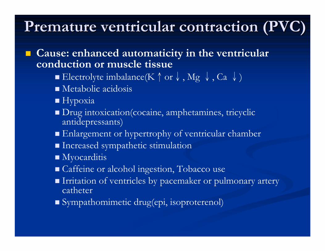
Premature ventricular contraction (PVC)Premature ventricular contraction (PVC)
� Cause: enhanced automaticity in the ventricular conduction or muscle tissue
� Electrolyte imbalance(K↑or↓, Mg ↓, Ca ↓)
� Metabolic acidosis� Hypoxia
� Drug intoxication(cocaine, amphetamines, tricyclic antidepressants)
� Enlargement or hypertrophy of ventricular chamber
� Increased sympathetic stimulation� Myocarditis
� Caffeine or alcohol ingestion, Tobacco use
� Irritation of ventricles by pacemaker or pulmonary artery catheter
� Sympathomimetic drug(epi, isoproterenol)

PVC's are Dangerous When:
• They are frequent (> 30% of complexes) or are increasing in frequency• The come close to or on top of a preceding T-wave (R on T)• Three or more PVC's in a row (run of V-tach)• Any PVC in the setting of an acute MI• PVC's come from different foci ("multifocal" or "multiformed")
These dangerous phenomenon may preclude the occurrence of deadly arrhythmias:
• Ventricular Tachycardia
• Ventricular Fibrillation
sinus beatsUnconverted V-tach r V-fib
V-tach
“R on T phenomenon”
time
The sooner defibrillation takes place,
the increased likelihood of survival

PVC PrognosisPVC Prognosis
LownLown ClassificationClassification
��Class 1: <30PVC/hrClass 1: <30PVC/hr
��Class 2: >30 PVC/hrClass 2: >30 PVC/hr
��Class 3: Multiform Class 3: Multiform
PVCsPVCs
��Class 4a: PVC Class 4a: PVC
coupletscouplets
��Class 4b: PVC Class 4b: PVC
triplets or greatertriplets or greater
��Class 5: R on T Class 5: R on T
�� Post MI Post MI PVCsPVCs and and
LownLown’’ss class 3class 3--5 are 5 are
associated with associated with ↑↑ risk risk
for VT/VF and sudden for VT/VF and sudden
deathdeath

Intervention Intervention
�� Intervention:Intervention:
�� A cardiac originA cardiac origin:: drug to suppress ventricular drug to suppress ventricular
irritability( procainamide, irritability( procainamide, amiodaroneamiodarone and and lidocainelidocaine))
�� Recently PVC: underlying heart disease or complex Recently PVC: underlying heart disease or complex
medical conditionmedical condition
�� Chronic PVC: frequent PVC or dangerous patternChronic PVC: frequent PVC or dangerous pattern

IdioventricularIdioventricular (Escape) rhythm(Escape) rhythm
�� Escape rhythm due to failure of Escape rhythm due to failure of
SA/AVN ventricular activation SA/AVN ventricular activation
or complete conduction blockor complete conduction block
�� Inherent 20Inherent 20--40bpm takes over 40bpm takes over
since it is no longer suppressedsince it is no longer suppressed
�� Regular wide QRSRegular wide QRS
�� EtiologiesEtiologies
�� PostPost--MI, CM, MI, CM, digoxindigoxin toxicitytoxicity

Accelerated Accelerated IdioventricularIdioventricular Rhythm (AIVR)Rhythm (AIVR)
�� May result from accelerated ventricular focus that is May result from accelerated ventricular focus that is faster than the prevailing sinus rate and takes over or faster than the prevailing sinus rate and takes over or can occur as escape rhythm (generally with 3can occur as escape rhythm (generally with 3rdrd degree degree AV block)AV block)
�� Usually 60Usually 60--100 100 bpmbpm (differentiates from VT)(differentiates from VT)�� Regular wide QRSRegular wide QRS�� Usually self limited, rarely see progression to VT/VFUsually self limited, rarely see progression to VT/VF

Accelerated Accelerated IdioventricularIdioventricular Rhythm (AIVR)Rhythm (AIVR)
Enhanced automaticity appears to be the likely electrophysiologic mechanism behind the genesis of AIVR. Enhanced automaticity generally is ascribed to phase-4 depolarization of the action potential of the myocardial cell. AIVR can occur in the His-Purkinje fibers or myocardium under certain abnormal metabolic conditions.
AIVR arises from subordinate or second-order pacemakers and manifests itself when the patient's prevailing sinus rate becomes lower than the accelerated rate (AIVR) of the otherwise suppressed focus. Sinus bradycardiacombined with enhanced automaticity of the subordinate site is the common pathophysiology.
Several conditions, including myocardial ischemia (especially inferior wall ischemia or infarction), digoxin toxicity, electrolyte imbalance (eg, hypokalemia), and hypoxemia may accentuate the phase-4 depolarization in the subordinate pacemaker tissues of the atrioventricular (AV) junction or His-Purkinje system, thus increasing the rate of impulse generation. Frequently, when inferior wall ischemia is present, the subordinate pacemaker acceleration coexists with sinus node depression. The latter permits escape and domination of the pacemaker function, which may occur with AV junctional or ventricular rates of only 60-70 bpm. The ectopic mechanism also can begin after a premature ventricular complex or, as described above, when the ectopic ventricular focus simply can accelerate sufficiently enough to overtake the intrinsic rhythm.
The onset of AIVR is gradual (nonparoxysmal). The ventricular rhythm can be regular or irregular and, occasionally, can show sudden doubling, suggesting the presence of exit block. The ventricular rate, commonly 60-110 bpm, usually stays within 10-15 beats of the sinus rate; therefore, the control of the cardiac rhythm occasionally passes back and forth between these 2 competing pacemaker sites.
Fusion beats often develop at the onset and termination of arrhythmia, which occurs when the pacemakers are competing for control of ventricular depolarization. Because of the slow rate, capture beats also are common. Due to the slow rate and nonparoxysmal onset, precipitation of more rapid ventricular arrhythmias rarely is observed. Rhythm termination generally occurs gradually, while the underlying sinus rhythm accelerates or the AIVR slows down.

Cause and significanceCause and significance�� When all of the heartWhen all of the heart’’s higher pacemakers fail to function s higher pacemakers fail to function
or supraventricular impulse was blockedor supraventricular impulse was blocked
�� Idioventricular rhythm may accompany 3rdIdioventricular rhythm may accompany 3rd--degree heart degree heart
block block
�� Cause: Cause:
�� Myocardial ischemiaMyocardial ischemia
�� Myocardial infarction(MI)Myocardial infarction(MI)
�� Digoxin toxicity, betaDigoxin toxicity, beta--adrenergic blockers, calcium antagonist, tricyclic adrenergic blockers, calcium antagonist, tricyclic
antidepressantantidepressant
�� Pacemaker failurePacemaker failure
�� Metabolic imbalancesMetabolic imbalances
�� Continuous idioventricular rhythm: serious situationContinuous idioventricular rhythm: serious situation
�� Slow rate and loss of atrial kickSlow rate and loss of atrial kick�������� ↓↓↓↓↓↓↓↓cardiac outputcardiac output�������� deathdeath

S/S and interventionS/S and intervention�� Continuous idioventricular rhythm: due to Continuous idioventricular rhythm: due to ↓↓cardiac cardiac outputoutput�� dizziness, lightdizziness, light--headedness, syncope or loss of headedness, syncope or loss of consiousnessconsiousness
�� TxTx: increase HR, improve cardiac output and establish : increase HR, improve cardiac output and establish normal rhythmnormal rhythm�� Atropine: increase HRAtropine: increase HR
�� If hypotension or clinical instabilityIf hypotension or clinical instability�� pacemakerpacemaker
�� TranscutaneousTranscutaneous pacemaker in an emergencypacemaker in an emergency
�� Not to suppress the idioventricular rhythmNot to suppress the idioventricular rhythm�������� never use never use lidocainelidocaine or other or other antiarrhythmicantiarrhythmic to suppress the escape to suppress the escape beatbeat
�� ECG monitor until restore ECG monitor until restore hemodymamichemodymamic stabilitystability
�� Bed restBed rest
�� Education Education

100 > 100 > AIVR AIVR > 30> 30--40 beat/min40 beat/min
Idioventricular rhythm: 30Idioventricular rhythm: 30--40 beat/min40 beat/min

Ventricular tachycardiaVentricular tachycardia�� Usually precedes ventricular fibrillation and sudden cardiac deaUsually precedes ventricular fibrillation and sudden cardiac death.th.
�� <30 sec<30 sec�� few or no symptomsfew or no symptoms
�� SustainedSustained�� immediate immediate TxTx to maintain cardiac outputto maintain cardiac output
�� Cause:Cause:
�� Myocardial ischemiaMyocardial ischemia
�� MIMI
�� Coronary artery diseaseCoronary artery disease
�� ValvularValvular heart diseaseheart disease
�� Heart failureHeart failure
�� CardiomyopathyCardiomyopathy
�� Electrolyte imbalance(Electrolyte imbalance(↓↓K)K)
�� Drug intoxication: procainamide, quinidine or cocaineDrug intoxication: procainamide, quinidine or cocaine
�� ↓↓ventricular refilling time and drop of cardiac outputventricular refilling time and drop of cardiac output��
cardiovascular collapsecardiovascular collapse

Notes on V-tach:•Typical V-tach patient
• MI with complications & extensive necrosis, EF<40%, d wall
motion, v-aneurysm)
•V-tach complexes are likely to be similar and the rhythm regular
• Irregular V-Tach rhythms may be due to to:
• breakthrough of atrial conduction
• atria may “capture” the entire beat beat
• an atrial beat may “merge” with an ectopic ventricular beat
(fusion beat)
Fusion beat - note p-wave in front of PVC and the PVC is narrower than the other PVC’s – this indicates the beat is a product of both the sinus node and an ectopicventricular focus
Capture beat - note that the complex is narrow enough to suggest normal ventricular conduction. This indicates that an atrial impulse has made it through and conduction through the ventricles is relatively normal.

Ventricular Tachycardia (VT)
Rate: 140-220 (200±50); at least 3 ectopic QRS in row
Rhythm: generally regular (may be slightly irregular)
P wave: no related P waves
P-R: none
QRS: normally wide and bizarre; ($ 0.14 sec favors VT)
• Usually associated with MI or other organic HD; unusual in normals
• Often serious requiring quick treatment if sustained
• Mechanism? Reentry or rapid firing ectopic??

ECG diagnosis - clues to rule in VT
• Difficult to rule out SVT with aberrant ventricular conduction
• use more leads whenever possible
• Unrelated P's (independent atrial activity) - rules out atria
• Presence of fusion beats suggests VT as contrasted to SVT
• LVT favored - monophasic pattern in R chest leads (V1 or MCL1) with taller left 'rabbit ear‘• Concordant positivity (all complexes positive) in V leads => favors LV ectopy (rule out WPW)
• concordant negativity = favors RV ectopy (rule out LBBB)• QRS interval > .14 sec (prior tracing available to rule out BBB)
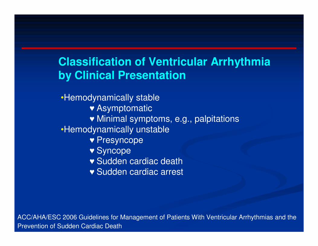
Classification of Ventricular Arrhythmia by Clinical Presentation
•Hemodynamically stable♥ Asymptomatic♥ Minimal symptoms, e.g., palpitations
•Hemodynamically unstable♥ Presyncope♥ Syncope♥ Sudden cardiac death♥ Sudden cardiac arrest
ACC/AHA/ESC 2006 Guidelines for Management of Patients With Ventricular Arrhythmias and the
Prevention of Sudden Cardiac Death

Classification of Ventricular Arrhythmia by Electrocardiography
•Nonsustained ventricular tachycardia (VT)♥ Monomorphic♥ Polymorphic
•Sustained VT♥ Monomorphic♥ Polymorphic
•Bundle-branch re-entrant tachycardia•Bidirectional VT•Torsades de pointes•Ventricular flutter•Ventricular fibrillation
ACC/AHA/ESC 2006 Guidelines for Management of Patients With Ventricular Arrhythmias and the
Prevention of Sudden Cardiac Death

VT VT -- S/S and interventionS/S and intervention�� Weak or absent pulsesWeak or absent pulses
�� Low cardiac outputLow cardiac output�� hypotension, conscious change, angina, hypotension, conscious change, angina, heart failure or organ perfusionheart failure or organ perfusion↓↓
�� Intervention: Intervention:
�� Evaluation of consciousness, respiration and circulationEvaluation of consciousness, respiration and circulation
�� If If pulselesspulseless�� immediate defibrillationimmediate defibrillation
�� Unstable Unstable PP’’tt: ventricular rate > 150 beat/min with S/S: : ventricular rate > 150 beat/min with S/S: hypotension, shortness of breath, chest pain or alternated hypotension, shortness of breath, chest pain or alternated consciousnessconsciousness�� immediate synchronized immediate synchronized cardioversioncardioversion
�� Stable Stable PP’’tt with widewith wide--complex VTcomplex VT and no signs of heart and no signs of heart failurefailure
�� Monomorphic: Monomorphic: IV IV procainimideprocainimide, , amiodaroneamiodarone
�� Polymorphic: Magnesium Polymorphic: Magnesium + ventricular or + ventricular or atrialatrial pacingpacing
�� Chronic, recurrent episodes of VT unresponsive to drug Chronic, recurrent episodes of VT unresponsive to drug therapytherapy�� implantation implantation cardioversioncardioversion--defibrillator (ICD)defibrillator (ICD)
�� Education Education

Torsades De PointesTorsades De Pointes�� Rapid ventricular rate: 250~350 beat/minRapid ventricular rate: 250~350 beat/min
�� Character: QRS complex change back and forth, with amplitude Character: QRS complex change back and forth, with amplitude of each successive complex gradually increasing and decreasingof each successive complex gradually increasing and decreasing
�� DDxDDx: ventricular flutter: rapid, regular, repetitive beating of : ventricular flutter: rapid, regular, repetitive beating of ventricleventricle�� single ventricular focus firing at a rapid rate of single ventricular focus firing at a rapid rate of 250~350 beat/min250~350 beat/min�� smooth and smooth and ““sinesine--wavewave””appearanceappearance

TorsadesTorsades De PointesDe Pointes�� French term meaning "twisting of the pointsFrench term meaning "twisting of the points””
�� torsadetorsade de pointes occurs in the setting of de pointes occurs in the setting of delayed delayed ventricular repolarizationventricular repolarization, evidenced by , evidenced by prolongation of the QT intervalsprolongation of the QT intervals or the or the presence of presence of prominent U waves.prominent U waves.
�� Causes:Causes:
�� Electrolyte imbalances, including hypokalemia, Electrolyte imbalances, including hypokalemia, hypomagnesemia, and less commonly, hypomagnesemia, and less commonly, hypocalcemiahypocalcemia, , which prolong repolarization which prolong repolarization
�� Miscellaneous factors such as severe Miscellaneous factors such as severe bradyarrhythmias, liquid protein diets, and hereditary bradyarrhythmias, liquid protein diets, and hereditary longlong--QT syndromes QT syndromes
From: Goldberger: Clinical Electrocardiography: A Simplified Approach, 6th ed.


TorsadesTorsades De PointesDe Pointes�� TxTx: :
�� Removing or correcting causative factorsRemoving or correcting causative factors such as such as drug toxicity, electrolyte imbalance, or underlying drug toxicity, electrolyte imbalance, or underlying bradycardia. bradycardia.
�� In emergency settings a In emergency settings a temporary pacemakertemporary pacemaker may may be inserted to accomplish "overdrive" suppression be inserted to accomplish "overdrive" suppression of the arrhythmia by increasing the underlying heart of the arrhythmia by increasing the underlying heart rate and thereby decreasing ventricular repolarization rate and thereby decreasing ventricular repolarization time. time.
�� Intravenous magnesium sulfateIntravenous magnesium sulfate has proved highly has proved highly useful for suppressing this arrhythmia. useful for suppressing this arrhythmia.
�� Drug therapyDrug therapy with with isoproterenolisoproterenol or or bretyliumbretylium has has been used in selected cases. been used in selected cases.
�� Sustained episodes of Sustained episodes of torsadetorsade de pointesde pointes��attempted attempted cardioversioncardioversion

Ventricular fibrillationVentricular fibrillation�� VFVF��chaotic, disorganized pattern of electrical activitychaotic, disorganized pattern of electrical activity��
multiple ectopic pacemakermultiple ectopic pacemaker�� no cardiac outputno cardiac output�� sudden sudden cardiac deathcardiac death
�� Cause: Cause: �� CADCAD
�� Myocardial ischemiaMyocardial ischemia
�� MIMI
�� Untreated VTUntreated VT
�� Underlying heart diseaseUnderlying heart disease
�� AcidAcid--base imbalancebase imbalance
�� Electric shockElectric shock
�� Severe hypothermiaSevere hypothermia
�� Drug toxicity (Drug toxicity (digoxindigoxin, quinidine and procainamide), quinidine and procainamide)
�� Electrolyte imbalance (Electrolyte imbalance (↑↓↑↓K, K, ↑↑Ca)Ca)
�� Completely ineffective contraction, cardiac output=0 Completely ineffective contraction, cardiac output=0 ��ventricular standstill and deathventricular standstill and death

ECGECG�� Ventricular rhythm: no Ventricular rhythm: no
pattern or regularitypattern or regularity
�� P wave, QRS complex, PR P wave, QRS complex, PR interval, T wave : caninterval, T wave : can’’t be t be determineddetermined
�� Coarse Coarse fibrillatoryfibrillatory wave: wave: greater chance of successful greater chance of successful electrical electrical cardioversioncardioversion than than small amplitudesmall amplitude

S/S and interventionS/S and intervention
�� Full cardiac arrest, unresponsive, undetectable Full cardiac arrest, unresponsive, undetectable
BPBP
�� Intervention:Intervention:
�� Access VF, confirmAccess VF, confirm
�� Immediate defibrillation is the most effective Immediate defibrillation is the most effective TxTx
�� Unsynchronized electrical shock at 200 JUnsynchronized electrical shock at 200 J
�� Unsynchronized electrical shock at 200 to 300 JUnsynchronized electrical shock at 200 to 300 J
�� Unsynchronized electrical shock at 360 JUnsynchronized electrical shock at 360 J
�� CPR until defibrillator arrives CPR until defibrillator arrives

Intervention Intervention
�� Intervention:Intervention:
�� Drug: Drug: epiepi or vasopressin (for persistent VF)or vasopressin (for persistent VF)
�� AntiarrhythmiaAntiarrhythmia agent: agent: amiodaroneamiodarone,,lidocainelidocaine and Mgand Mg
�� AEDAED-- automated external defibrillatorautomated external defibrillator�� outout--ofof--
hospital settinghospital setting
�� Rapid recognition of the problem and defibrillationRapid recognition of the problem and defibrillation
�� Education, CPREducation, CPR

Wide complex Wide complex tachyarrhythmiastachyarrhythmias
�� QRS greater or equal to 0.12 sec and rate >100 QRS greater or equal to 0.12 sec and rate >100
bpmbpm
�� Not all are of ventricular originNot all are of ventricular origin
�� DifferentialDifferential
�� Ventricular Ventricular tachycardiatachycardia
�� Supraventricular tachycardiaSupraventricular tachycardia with aberrancy with aberrancy
(conduction block) or presence of an accessory (conduction block) or presence of an accessory
pathway with pathway with antegradeantegrade conduction (WPW conduction (WPW
syndrome)syndrome)
�� ArtifactArtifact

VT VT vsvs SVT with AberrancySVT with Aberrancy
�� Both manifest as wide complex Both manifest as wide complex tachycardiastachycardias on ECGon ECG
�� Distinguishing ECG findings: Distinguishing ECG findings:
�� SVT with aberrant conductionSVT with aberrant conduction�� QRS > 0.14 QRS > 0.14
�� Rhythm onset with premature P waveRhythm onset with premature P wave
�� PR interval <100msecPR interval <100msec
�� P wave and QRS are linkedP wave and QRS are linked
�� VagalVagal maneuver slows/terminates rhythmmaneuver slows/terminates rhythm
�� MonomorphicMonomorphic VTVT�� QRS >0.14 QRS >0.14 msecmsec
�� AV dissociation with fusion or capture beatsAV dissociation with fusion or capture beats
�� Absence of RS complex in Absence of RS complex in precordialprecordial leadsleads
�� Extreme axis deviationExtreme axis deviation
�� If above findings fail to be detected, morphologic criteria usedIf above findings fail to be detected, morphologic criteria used: if : if QRS in V1 does NOT look like typical R or L conduction blockQRS in V1 does NOT look like typical R or L conduction block


Physical findings highly suggestive Physical findings highly suggestive
of VT:of VT:
�� Signs of AV dissociation, including:Signs of AV dissociation, including:
�� Canon A waves in the jugular venous pulsationsCanon A waves in the jugular venous pulsations
�� Varying BP measurement from beat to beatVarying BP measurement from beat to beat
�� Varying intensity of SVarying intensity of S11

WQRS WQRS tachycardiatachycardia
algorithmalgorithm(1) presence of AV dissociation;
(2) presence of an initial R-wave
in lead aVR;
(3) did the morphology of the
WCT correspond to bundle
branch (BBB) or fascicular
block (FB)?
(4) estimation of initial (Vi) and
terminal (Vt) ventricular
activation velocity ratio (Vi/Vt)
by measuring the voltage change
on the ECG tracing during the
initial 40 ms (Vi) and the
terminal 40 ms (Vt) of the same
bi- or multiphasic QRS
complex.


Clinical importanceClinical importance
�� Misdiagnosing VT as SVT can lead to fatal errorMisdiagnosing VT as SVT can lead to fatal error
�� Treating VT as SVT with Treating VT as SVT with verapamilverapamil, , diltiazemdiltiazem, ,
and adenosine can precipitate ventricular and adenosine can precipitate ventricular
fibrillation, even if initially stable.fibrillation, even if initially stable.
�� All wide complex All wide complex tachyarrhythmiatachyarrhythmia should be should be
considered VT until proven otherwiseconsidered VT until proven otherwise


General Evaluation for Documented or Suspected VA
Resting Electrocardiogram
Resting 12-lead ECG is indicated in all patients who are evaluated for ventriculararrhythmias.
III IIaIIaIIa IIbIIbIIb IIIIIIIIIIII IIaIIaIIa IIbIIbIIb IIIIIIIIIIII IIaIIaIIa IIbIIbIIb IIIIIIIIIIIaIIaIIa IIbIIbIIb IIIIIIIII
ACC/AHA/ESC 2006 Guidelines for Management of Patients With Ventricular Arrhythmias and the
Prevention of Sudden Cardiac Death
General Evaluation for Documented or Suspected VA
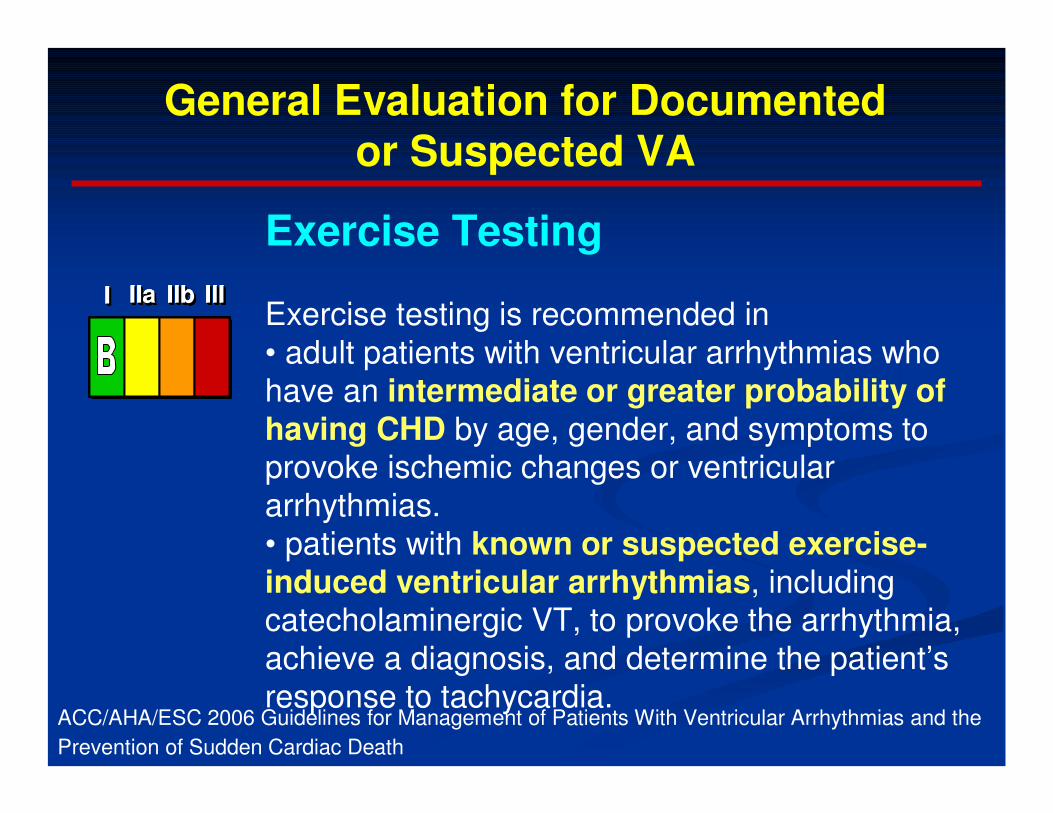
Exercise Testing
Exercise testing is recommended in • adult patients with ventricular arrhythmias who have an intermediate or greater probability of
having CHD by age, gender, and symptoms to provoke ischemic changes or ventricular arrhythmias. • patients with known or suspected exercise-induced ventricular arrhythmias, including catecholaminergic VT, to provoke the arrhythmia, achieve a diagnosis, and determine the patient’s response to tachycardia.
III IIaIIaIIa IIbIIbIIb IIIIIIIIIIII IIaIIaIIa IIbIIbIIb IIIIIIIIIIII IIaIIaIIa IIbIIbIIb IIIIIIIIIIIaIIa IIbIIa IIb IIIIIb IIIIII
ACC/AHA/ESC 2006 Guidelines for Management of Patients With Ventricular Arrhythmias and the
Prevention of Sudden Cardiac Death
General Evaluation for Documented or Suspected VA
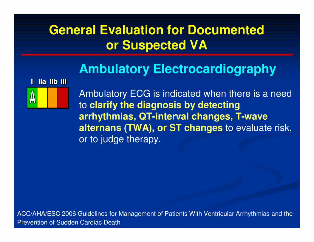
Ambulatory Electrocardiography
Ambulatory ECG is indicated when there is a need to clarify the diagnosis by detecting
arrhythmias, QT-interval changes, T-wave
alternans (TWA), or ST changes to evaluate risk, or to judge therapy.
III IIaIIaIIa IIbIIbIIb IIIIIIIIIIII IIaIIaIIa IIbIIbIIb IIIIIIIIIIII IIaIIaIIa IIbIIbIIb IIIIIIIIIIIaIIaIIa IIbIIbIIb IIIIIIIII
ACC/AHA/ESC 2006 Guidelines for Management of Patients With Ventricular Arrhythmias and the
Prevention of Sudden Cardiac Death

General Evaluation for Documented or Suspected VA
Left Ventricular Function and Imaging
Echocardiography is recommended in • patients with ventricular arrhythmias who are suspected of having structural heart disease. • for the subset of patients at high risk for the
development of serious ventricular arrhythmias
or SCD, such as those with dilated, hypertrophic, or RV cardiomyopathies, AMI survivors, or relatives of patients with inherited disorders associated with SCD.
III IIaIIaIIa IIbIIbIIb IIIIIIIIIIII IIaIIaIIa IIbIIbIIb IIIIIIIIIIII IIaIIaIIa IIbIIbIIb IIIIIIIIIIIaIIa IIbIIa IIb IIIIIb IIIIII
ACC/AHA/ESC 2006 Guidelines for Management of Patients With Ventricular Arrhythmias and the
Prevention of Sudden Cardiac Death

General Evaluation for Documented or Suspected VA
Left Ventricular Function and Imaging
Exercise testing with an imaging modality ― (echocardiography or nuclear perfusion [single-photon emission computed tomography (SPECT)])―is recommended to detect silent ischemia in patients with ventricular
arrhythmias who have an intermediate probability of having CHD by age, symptoms, and gender and in whom ECG assessment is less reliable because of digoxin use, LVH, greater than 1-mm ST-segment depression at rest, Wolf-Parkinson-White (WPW) syndrome, or Left Bundle Branch Block (LBBB).
III IIaIIaIIa IIbIIbIIb IIIIIIIIIIII IIaIIaIIa IIbIIbIIb IIIIIIIIIIII IIaIIaIIa IIbIIbIIb IIIIIIIIIIIaIIa IIbIIa IIb IIIIIb IIIIII
ACC/AHA/ESC 2006 Guidelines for Management of Patients With Ventricular Arrhythmias and the Prevention of Sudden Cardiac Death

Cardiac Cardiac ElectrophysiologyElectrophysiology StudyStudy
�� InducibilityInducibility of VTof VT
�� Reentrant (Reentrant (ischemicischemic VT)VT)
�� Triggered (idiopathic VT)Triggered (idiopathic VT)
�� Assessment of Assessment of antiarrhythmicantiarrhythmic therapy via therapy via
serial drug testingserial drug testing
�� May lead to therapy with May lead to therapy with radiofrequencyradiofrequency
catheter ablationcatheter ablation

AsystoleAsystole�� AKA: ventricular standstillAKA: ventricular standstill
�� Result from a prolonged period of cardiac arrest without Result from a prolonged period of cardiac arrest without effective resuscitationeffective resuscitation
�� DDxDDx with VFwith VF
�� Cause: Cause: �� HypovolemiaHypovolemia
�� MI(coronary thrombosis)MI(coronary thrombosis)
�� Severe electrolyte imbalance(Severe electrolyte imbalance(↑↓↑↓K)K)
�� Massive pulmonary embolismMassive pulmonary embolism
�� HypoxiaHypoxia
�� Drug overdoseDrug overdose
�� HypothermiaHypothermia
�� Cardiac Cardiac tamponadetamponade
�� Tension Tension pneumothoraxpneumothorax
�� No electrical activity, no contractionNo electrical activity, no contraction�� cardiac output=0cardiac output=0�� no no perfusion for vital organperfusion for vital organ

ECGECG
�� No electrical activityNo electrical activity

Intervention Intervention
�� Immediate Immediate TxTx: CPR, oxygen and advanced : CPR, oxygen and advanced
airway control with airway control with intubationintubation
�� Check more than one ECG lead to confirm Check more than one ECG lead to confirm
asystoleasystole
�� IV IV EpiEpi and atropine,and atropine,vasopressinvasopressin
�� Consider terminating resuscitation if persist Consider terminating resuscitation if persist
asystole.asystole.

AntiarrhythmicAntiarrhythmic TherapyTherapy

VaughnVaughn--Williams ClassificationWilliams Classification
�� Based on cellular properties of normal Based on cellular properties of normal
HisHis--Purkinje cellsPurkinje cells
�� Classified on drugClassified on drug’’s ability to block s ability to block
specific ionic currents (i.e. Naspecific ionic currents (i.e. Na++, K, K++, Ca, Ca++++) )
and betaand beta--adrenergicadrenergic receptorsreceptors
�� Advantages:Advantages:
�� Physiologically basedPhysiologically based
�� Highlights beneficial/deleterious effects Highlights beneficial/deleterious effects
of specific drugsof specific drugs

AntiarrhythmicAntiarrhythmic AgentsAgentsVaughnVaughn--Williams ClassificationWilliams Classification
�� Class I Class I -- NaNa+ + -- channel blockers channel blockers (direct membrane action)(direct membrane action)
�� Class II Class II -- SympatholyticSympatholytic agentsagents
�� Class III Class III -- Prolong Prolong repolarizationrepolarization
�� Class IVClass IV-- CaCa++ ++ -- channel blockerschannel blockers
�� PurinergicPurinergic agonistsagonists
�� Digitalis glycosidesDigitalis glycosides

Class I Class I Na+ Channel BlockersNa+ Channel Blockers
�� IA IA -- QuinidineQuinidine//ProcainamideProcainamide//DisopyramideDisopyramide
�� IB IB -- LidocaineLidocaine//MexiletineMexiletine//PhenytoinPhenytoin
�� IC IC -- FlecainideFlecainide//PropafenonePropafenone//EthmozineEthmozine
1
0
2
3
4
ERP RRP
Affects
Phase 0 of
depolarization-the rapid
inflow of sodium through
the sodium channels.

Class IA Class IA -- Na+ Channel BlockersNa+ Channel Blockers
ProcainamideProcainamide//QuinidineQuinidine//DisopyramideDisopyramide
�� ECG changesECG changes
�� Increase PR, QRS (Increase PR, QRS (DisoDiso: PR : PR �������� > QRS > QRS �������� ))
�� Toxicity: Toxicity: QTcQTc increases by 30% or QT > increases by 30% or QT >
0.5 sec0.5 sec
�� CaCa++++ channel blockade / potent channel blockade / potent
anticholinergicanticholinergic ((DisoDiso))

ArrhythmiaArrhythmia--focused Therapyfocused Therapy
•• Class IB Class IB antiarrhythmicsantiarrhythmics are very are very
effective and very safe.effective and very safe.
•• Little or no effect on Little or no effect on ““normalnormal”” tissuestissues
•• First line for First line for ischemicischemic, automatic , automatic
arrhythmia's (arrhythmia's (Ventricular Ventricular tachycardiatachycardia))
•• Not a lot of effect on normal conduction Not a lot of effect on normal conduction
tissue tissue –– not a good medicine for not a good medicine for
reentry and reentry and atrial tachycardiasatrial tachycardias..

Class ICClass ICFlecainideFlecainide//PropafenonePropafenone//EthmozineEthmozine
�� ECG changesECG changes
�� �������� PR, QRSPR, QRS
�� �������� QTcQTc ((PropafenonePropafenone))

Arrhythmia Arrhythmia ––focused Therapyfocused Therapy
�� ICIC’’s have a lot of side effects s have a lot of side effects
that make them appropriate for that make them appropriate for
use only by experienced use only by experienced
providers.providers.

Class II AgentsClass II AgentsBetaBeta--blockersblockers
�� PropranololPropranolol
�� AtenololAtenolol
�� MetoprololMetoprolol
�� NadololNadolol
�� EsmololEsmolol
�� d,ld,l--SotalolSotalol

ArrhythmiaArrhythmia--focused Therapyfocused Therapy
�� BetaBeta--blockers are good for reblockers are good for re--
entry circuits and automatic entry circuits and automatic
dysrhythmiasdysrhythmias. .
�� Their effect of decreasing Their effect of decreasing
contractility may be limiting.contractility may be limiting.

Class III Class III KK++ -- channel blockerschannel blockers
�� PropertiesProperties
�� Prolong Prolong repolarizationrepolarization
�� Prolong action potential durationProlong action potential duration
�� Contractility is unchanged or increasedContractility is unchanged or increased
�� AgentsAgents
�� AmiodaroneAmiodarone
�� SotalolSotalol
�� BretyliumBretylium

ArrhythmiaArrhythmia--focused Therapyfocused Therapy
�� Can be very powerful Can be very powerful antiarrhythmicsantiarrhythmics
but limited indications for firstbut limited indications for first--line line
use use –– beyond the spectrum of primary beyond the spectrum of primary
care providerscare providers
�� AmiodaroneAmiodarone: become a first: become a first--line line
medicine for a broad spectrum of medicine for a broad spectrum of
arrhythmias, currently still higharrhythmias, currently still high--riskrisk


PacemakersPacemakers�� Surgical implantation of electrical leads attached to a Surgical implantation of electrical leads attached to a
pulse generatorpulse generator
�� Over 175,000 implanted per yearOver 175,000 implanted per year

The Pacemaker SystemThe Pacemaker System
�� PatientPatient
Lead
Pacemaker
• Programmer
�� LeadLead
�� PacemakerPacemaker

PacemakersPacemakers
1)1) Leads are inserted via Leads are inserted via subclaviclesubclavicle vein and vein and advanced to the chambers on the vena cava advanced to the chambers on the vena cava (right) side of the heart(right) side of the heart
2)2) Two leads used, one for right atrium, other for Two leads used, one for right atrium, other for right ventricleright ventricle
3)3) Pulse generator containing Pulse generator containing microcircuitrymicrocircuitry and and battery are attached to leads and placed into a battery are attached to leads and placed into a ““pocketpocket”” under the skin near the clavicleunder the skin near the clavicle
4)4) Pulse generator sends signal down leads in Pulse generator sends signal down leads in programmed sequence to contract atria, then programmed sequence to contract atria, then ventriclesventricles

Castle LW, Cook S: Pacemaker radiography. In Castle LW, Cook S: Pacemaker radiography. In EllenbogenEllenbogen KA, Kay GN, KA, Kay GN, WilkoffWilkoff BL [BL [edseds]: Clinical Cardiac Pacing. Philadelphia, WB ]: Clinical Cardiac Pacing. Philadelphia, WB
Saunders, 1995, p 538.Saunders, 1995, p 538.
PacemakersPacemakers

PacemakersPacemakers�� Pulse generator can sense electrical activity generated Pulse generator can sense electrical activity generated
by the heart and only deliver electrical impulses when by the heart and only deliver electrical impulses when needed. needed.
�� Pacemakers can only speed up a heart experiencing Pacemakers can only speed up a heart experiencing bradycardiabradycardia, they cannot alter a condition of , they cannot alter a condition of tachycardiatachycardia

Internal Cardioverter Defibrillator (ICD)
The first full human implantation of an ICDoccurred in February 1980. Since then, ICD technology has become extremely
complex.Current ICDs not only have the ability todefibrillate, but also to pace, terminate ventriculartachycardia (VT) and provide back-up pacing forbradycardia.

Dual chamber pacing with rate response is also now available.
The ICD is extremely expensive.
The total cost with implanting may exceed $40,000.00.
Internal Cardioverter Defibrillator (ICD)(continued)

The Class I indications for ICD implantation inwhich a broad consensus exists include:
• Cardiac arrest caused by VT or ventricular fibrillation (VF) not due to a transient or reversible cause
• Spontaneous sustained VT• Syncope of undetermined origin with clinically
relevant electrophysiologically inducible sustained VT or VF when drug therapy is ineffective or not tolerated or not preferred
Indications for ICD Implantation

• Non-sustained VT in patients with CAD, prior MI, LV dysfunction and electrophysiologicallyinducible VT or VF not suppressed by Class I anti-arrhythmic drugs.
These indications are frequently updated as theresults of outcome trials become available.
Indications for ICD Implantation (continued)

ACC/AHA/ESC 2006 Guidelines for Management of Patients With Ventricular Arrhythmias and the
Prevention of Sudden Cardiac Death

AntiarrhythmicAntiarrhythmic DrugsDrugs
♥♥ Beta Blockers:Beta Blockers: Effectively suppress ventricular Effectively suppress ventricular ectopicectopic beats & beats & arrhythmias; reduce incidence of SCDarrhythmias; reduce incidence of SCD
♥♥ AmiodaroneAmiodarone:: No definite survival benefit; some studies have No definite survival benefit; some studies have shown reduction in SCD in patients with LV dysfunction shown reduction in SCD in patients with LV dysfunction especially when given in conjunction with BB. Has complex drug especially when given in conjunction with BB. Has complex drug interactions and many adverse side effects (pulmonary, hepatic, interactions and many adverse side effects (pulmonary, hepatic, thyroid, thyroid, cutaneouscutaneous))
♥♥ SotalolSotalol:: Suppresses ventricular arrhythmias; is more proSuppresses ventricular arrhythmias; is more pro--arrhythmic than arrhythmic than amiodaroneamiodarone, no survival benefit clearly shown, no survival benefit clearly shown
♥♥ Conclusions:Conclusions: AntiarrhythmicAntiarrhythmic drugs (except for BB) should not be drugs (except for BB) should not be used as used as primaryprimary therapy of VA and the prevention of SCDtherapy of VA and the prevention of SCD
Therapies for VA
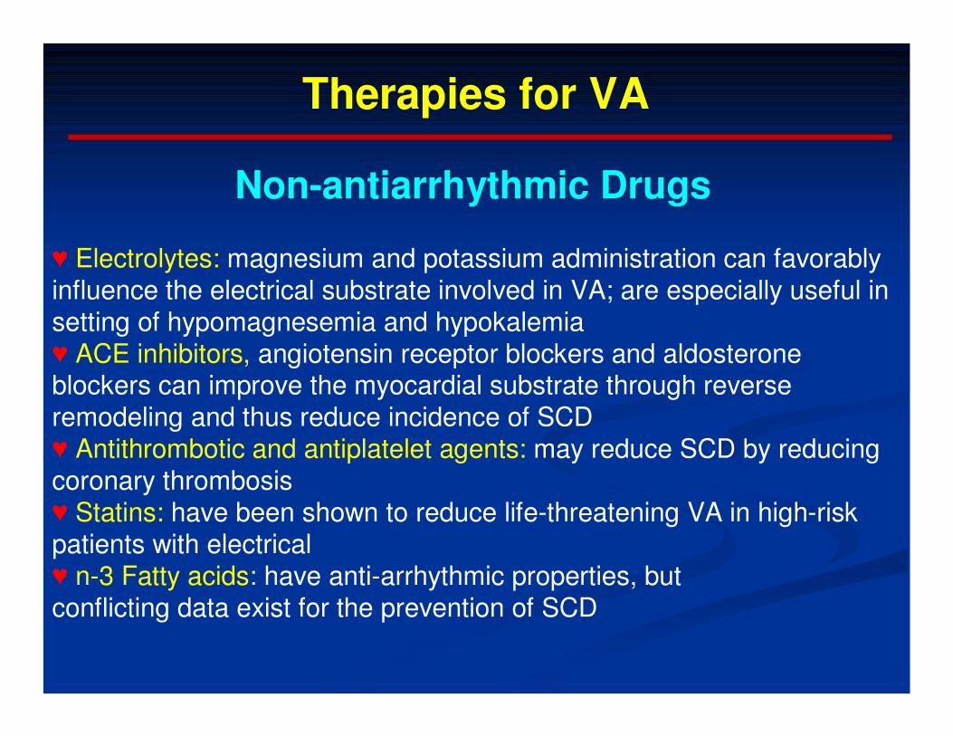
Non-antiarrhythmic Drugs
♥ Electrolytes: magnesium and potassium administration can favorably influence the electrical substrate involved in VA; are especially useful in setting of hypomagnesemia and hypokalemia♥ ACE inhibitors, angiotensin receptor blockers and aldosteroneblockers can improve the myocardial substrate through reverse remodeling and thus reduce incidence of SCD♥ Antithrombotic and antiplatelet agents: may reduce SCD by reducing coronary thrombosis♥ Statins: have been shown to reduce life-threatening VA in high-risk patients with electrical instability♥ n-3 Fatty acids: have anti-arrhythmic properties, but conflicting data exist for the prevention of SCD
Therapies for VA
Therapies for VA
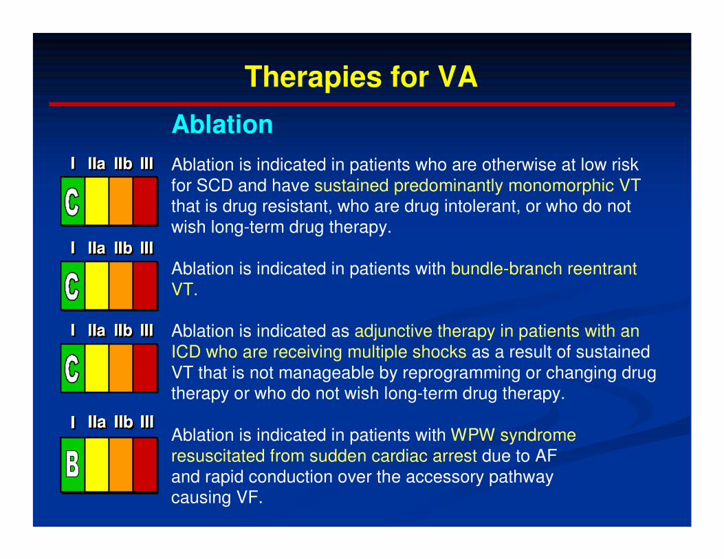
Ablation
Ablation is indicated in patients who are otherwise at low riskfor SCD and have sustained predominantly monomorphic VTthat is drug resistant, who are drug intolerant, or who do notwish long-term drug therapy.
Ablation is indicated in patients with bundle-branch reentrant VT.
Ablation is indicated as adjunctive therapy in patients with an ICD who are receiving multiple shocks as a result of sustainedVT that is not manageable by reprogramming or changing drug therapy or who do not wish long-term drug therapy.
Ablation is indicated in patients with WPW syndrome resuscitated from sudden cardiac arrest due to AF and rapid conduction over the accessory pathway causing VF.
III IIaIIaIIa IIbIIbIIb IIIIIIIIIIII IIaIIaIIa IIbIIbIIb IIIIIIIIIIII IIaIIaIIa IIbIIbIIb IIIIIIIIIIIaIIa IIbIIa IIb IIIIIb IIIIII
III IIaIIaIIa IIbIIbIIb IIIIIIIIIIII IIaIIaIIa IIbIIbIIb IIIIIIIIIIII IIaIIaIIa IIbIIbIIb IIIIIIIIIIIaIIaIIa IIbIIbIIb IIIIIIIII
III IIaIIaIIa IIbIIbIIb IIIIIIIIIIII IIaIIaIIa IIbIIbIIb IIIIIIIIIIII IIaIIaIIa IIbIIbIIb IIIIIIIIIIIaIIaIIa IIbIIbIIb IIIIIIIII
III IIaIIaIIa IIbIIbIIb IIIIIIIIIIII IIaIIaIIa IIbIIbIIb IIIIIIIIIIII IIaIIaIIa IIbIIbIIb IIIIIIIIIIIaIIaIIa IIbIIbIIb IIIIIIIII

Sustained Monomorphic VT
Wide-QRS tachycardia should be presumed to be VTif the diagnosis is unclear.
Direct current cardioversion with appropriate sedationis recommended at any point in the treatmentcascade in patients with suspected sustained monomorphic VT with hemodynamic compromise.
III IIaIIaIIa IIbIIbIIb IIIIIIIIIIII IIaIIaIIa IIbIIbIIb IIIIIIIIIIII IIaIIaIIa IIbIIbIIb IIIIIIIIIIIaIIaIIa IIbIIbIIb IIIIIIIII
III IIaIIaIIa IIbIIbIIb IIIIIIIIIIII IIaIIaIIa IIbIIbIIb IIIIIIIIIIII IIaIIaIIa IIbIIbIIb IIIIIIIIIIIaIIaIIa IIbIIbIIb IIIIIIIII
Acute Management of Specific Arrhythmias

Polymorphic VT
Direct-current cardioversion with appropriate sedation asnecessary is recommended for patients with sustained polymorphic VT with hemodynamic compromise and is reasonable at any point in the treatment cascade.
Intravenous beta blockers are useful for patients with recurrent polymorphic VT, especially if ischemia is suspected or cannot be excluded.
Intravenous loading with amiodarone is useful for patients with recurrent polymorphic VT in the absence of abnormal repolarization related to congenital or acquired LQTS.
III IIaIIaIIa IIbIIbIIb IIIIIIIIIIII IIaIIaIIa IIbIIbIIb IIIIIIIIIIII IIaIIaIIa IIbIIbIIb IIIIIIIIIIIaIIaIIa IIbIIbIIb IIIIIIIII
III IIaIIaIIa IIbIIbIIb IIIIIIIIIIII IIaIIaIIa IIbIIbIIb IIIIIIIIIIII IIaIIaIIa IIbIIbIIb IIIIIIIIIIIaIIa IIbIIa IIb IIIIIb IIIIII
III IIaIIaIIa IIbIIbIIb IIIIIIIIIIII IIaIIaIIa IIbIIbIIb IIIIIIIIIIII IIaIIaIIa IIbIIbIIb IIIIIIIIIIIaIIa IIbIIa IIb IIIIIb IIIIII
Acute Management of Specific Arrhythmias

Polymorphic VT
Urgent angiography with a view to revascularizationshould be considered for patients with polymorphic VT when myocardial ischemia cannot be excluded.
Intravenous lidocaine may be reasonable for treatment of polymorphic VT specifically associated with acute myocardial ischemia or infarction.
III IIaIIaIIa IIbIIbIIb IIIIIIIIIIII IIaIIaIIa IIbIIbIIb IIIIIIIIIIII IIaIIaIIa IIbIIbIIb IIIIIIIIIIIaIIaIIa IIbIIbIIb IIIIIIIII
III IIaIIaIIa IIbIIbIIb IIIIIIIIIIII IIaIIaIIa IIbIIbIIb IIIIIIIIIIII IIaIIaIIa IIbIIbIIb IIIIIIIIIIIaIIaIIa IIbIIbIIb IIIIIIIII
Acute Management of Specific Arrhythmias

Torsades de Pointes
Withdrawal of any offending drugs and correction of electrolyte abnormalities are recommended in patients presenting with torsades de pointes.
Acute and long-term pacing is recommended for patients presenting with torsades de pointes due to heart block andsymptomatic bradycardia.
III IIaIIaIIa IIbIIbIIb IIIIIIIIIIII IIaIIaIIa IIbIIbIIb IIIIIIIIIIII IIaIIaIIa IIbIIbIIb IIIIIIIIIIIaIIaIIa IIbIIbIIb IIIIIIIII
III IIaIIaIIa IIbIIbIIb IIIIIIIIIIII IIaIIaIIa IIbIIbIIb IIIIIIIIIIII IIaIIaIIa IIbIIbIIb IIIIIIIIIIIaIIaIIa IIbIIbIIb IIIIIIIII
Acute Management of Specific Arrhythmias

Torsades de Pointes
Management with intravenous magnesium sulfate is reasonable for patients who present with LQTS and few episodes of torsades de pointes. Magnesium is not likely to be effective in patients with a normal QT interval.
Acute and long-term pacing is reasonable for patients who present with recurrent pause-dependent torsades de pointes.
Beta blockade combined with pacing is reasonable acute therapy for patients who present with torsades de pointes and sinus bradycardia.
Isoproterenol is reasonable as temporary treatmentin acute patients who present with recurrent pause-dependent torsades de pointes who do not have congenital LQTS.
III IIaIIaIIa IIbIIbIIb IIIIIIIIIIII IIaIIaIIa IIbIIbIIb IIIIIIIIIIII IIaIIaIIa IIbIIbIIb IIIIIIIIIIIaIIaIIa IIbIIbIIb IIIIIIIII
III IIaIIaIIa IIbIIbIIb IIIIIIIIIIII IIaIIaIIa IIbIIbIIb IIIIIIIIIIII IIaIIaIIa IIbIIbIIb IIIIIIIIIIIaIIaIIa IIbIIbIIb IIIIIIIII
III IIaIIaIIa IIbIIbIIb IIIIIIIIIIII IIaIIaIIa IIbIIbIIb IIIIIIIIIIII IIaIIaIIa IIbIIbIIb IIIIIIIIIIIaIIaIIa IIbIIbIIb IIIIIIIII
III IIaIIaIIa IIbIIbIIb IIIIIIIIIIII IIaIIaIIa IIbIIbIIb IIIIIIIIIIII IIaIIaIIa IIbIIbIIb IIIIIIIIIIIaIIaIIa IIbIIbIIb IIIIIIIII
Acute Management of Specific Arrhythmias