Ionic Mechanism of Ouabain-Induced Concurrent Apoptosis ...€¦ · Ionic Mechanism of...
13
Ionic Mechanism of Ouabain-Induced Concurrent Apoptosis and Necrosis in Individual Cultured Cortical Neurons Ai Ying Xiao, Ling Wei, Shuli Xia, Steven Rothman, and Shan Ping Yu Department of Neurology and Center for the Study of Nervous System Injury, Washington University School of Medicine, St. Louis, Missouri 63110 Energy deficiency and dysfunction of the Na ,K -ATPase are common consequences of many pathological insults. The na- ture and mechanism of cell injury induced by impaired Na , K -ATPase, however, are not well defined. We used cultured cortical neurons to examine the hypothesis that blocking the Na ,K -ATPase induces apoptosis by depleting cellular K and, concurrently, induces necrotic injury in the same cells by increasing intracellular Ca 2 and Na . The Na ,K -ATPase inhibitor ouabain induced concentration- dependent neuronal death. Ouabain triggered transient neuronal cell swelling followed by cell shrinkage, accompanied by intra- cellular Ca 2 and Na increase, K decrease, cytochrome c release, caspase-3 activation, and DNA laddering. Electron microscopy revealed the coexistence of ultrastructural features of both apoptosis and necrosis in individual cells. The caspase inhibitor Z-Val-Ala-Asp(OMe)-fluoromethyl ketone (Z-VAD- FMK) blocked 50% of ouabain-induced neuronal death. Po- tassium channel blockers or high K medium, but not Ca 2 channel blockade, prevented cytochrome c release, caspase activation, and DNA damage. Blocking of K , Ca 2 , or Na channels or high K medium each attenuated the ouabain- induced cell death; combined inhibition of K channels and Ca 2 or Na channels resulted in additional protection. More- over, coapplication of Z-VAD-FMK and nifedipine produced virtually complete neuroprotection. These results suggest that the neuronal death associated with Na ,K -pump failure consists of concurrent apoptotic and necrotic components, mediated by intracellular depletion of K and accumulation of Ca 2 and Na , respectively. The ouabain-induced hybrid death may represent a distinct form of cell death related to the brain injury of inadequate energy supply and disrupted ion homeostasis. Key words: Na ,K -ATPase; apoptosis; necrosis; hybrid death; potassium channel; calcium; caspase; cytochrome c; DNA fragmentation; ouabain; strophanthidin Apoptosis may play important roles in various disease states (Raff et al., 1993; Ameisen, 1994; Thompson, 1995; Reed, 1999). Neu- ronal apoptosis occurs after an ischemic insult in the brain (Schumer et al., 1992; Chopp and Li, 1996; Du et al., 1996) and after spinal cord injury (Liu et al., 1997). Apoptosis is controlled by an internally encoded suicide program executed by activation of endogenous proteases (caspases) and endonucleases (Vaux et al., 1994; Kroemer et al., 1995; Miura and Yuan, 1996). Although multiple stimuli and signal pathways may contribute to apoptosis in a wide range of cell types, apoptotic cells share similar char- acteristic morphologies such as cell shrinkage, nuclear/chromatin condensation, internucleosomal cleavage of DNA, membrane blebbing, and formation of apoptotic bodies (Kerr et al., 1972; Wyllie et al., 1980; Mills et al., 1999). In contrast, necrosis is distinct from apoptosis in both morphological and biochemical characteristics; it begins with the swelling of cell body and mito- chondrial contents, followed by vacuolization of cytoplasm, irreg- ular breakdown of nuclear DNA, rupture of the cell membrane, and cell lysis (Majno and Joris, 1995). The striking differences in cell volume changes imply that necrosis and apoptosis possess distinguishable ionic mechanisms. Excessive Ca 2 and Na influx and their accumulation in the intracellular space are most likely responsible for cell swelling and necrotic death (Choi, 1988). On the other hand, excessive K efflux and intracellular K depletion may play key roles in cell shrinkage, caspase/endonuclease activation, and apoptotic death (Beauvais et al., 1995; Bortner et al., 1997; Yu et al., 1997, 1998; Dallaporta et al., 1998). Under the “apoptosis versus necrosis” classification, cell death is generally divided into these two categories; however, it is sometimes difficult to exclusively place a cell injury into either group. For example, the exact type of cell death after brain ischemia has been under debate (Deshpande et al., 1992; van Lookeren Campagne and Gill, 1996; Colbourne et al., 1999; Nicotera and Lipton, 1999). Alternatively, it was suggested that these two processes can occur simultaneously in tissues or cell cultures that have been exposed to a toxic stimulus (Ankarcrona et al., 1995; Leist et al., 1996; Shimizu et al., 1996). These discussions dictate reassessment of “mixed cell death” as a het- erogeneous entity combining both active and passive cell death (Hirsch et al., 1997; Kim et al., 1999; Yu et al., 1999a). Consis- tently, recent evidence showed an in vivo “apoptosis–necrosis continuum” in excitotoxically lesioned rat brain (Portera-C ailliau et al., 1997). The present study extends this concept even further, showing, for the first time, the simultaneous appearance of apoptotic and necrotic features in individual cells destined to die after exposure to a Na ,K -ATPase inhibitor. The Na ,K -ATPase, or Na , Received April 6, 2001; revised Nov. 13, 2001; accepted Nov. 27, 2001. This work was supported by grants from the National Science Foundation (9950207N to S.P.Y.), the American Heart Association (I BN-9817151 and 0170064N to S.P.Y.), and National Institutes of Health (NS37337 to L.W. and NS37773 to S.R.). Correspondence should be addressed to Shan Ping Yu, Department of Neurology, Box 8111, 660 South Euclid Avenue, Washington University School of Medicine, St. Louis, MO 63110. E-mail: [email protected]. Copyright © 2002 Society for Neuroscience 0270-6474/02/221350-13$15.00/0 The Journal of Neuroscience, February 15, 2002, 22(4):1350–1362
Transcript of Ionic Mechanism of Ouabain-Induced Concurrent Apoptosis ...€¦ · Ionic Mechanism of...

Ionic Mechanism of Ouabain-Induced Concurrent Apoptosis andNecrosis in Individual Cultured Cortical Neurons
Ai Ying Xiao, Ling Wei, Shuli Xia, Steven Rothman, and Shan Ping Yu
Department of Neurology and Center for the Study of Nervous System Injury, Washington University School of Medicine,St. Louis, Missouri 63110
Energy deficiency and dysfunction of the Na�, K�-ATPase arecommon consequences of many pathological insults. The na-ture and mechanism of cell injury induced by impaired Na�,K�-ATPase, however, are not well defined. We used culturedcortical neurons to examine the hypothesis that blocking theNa�, K�-ATPase induces apoptosis by depleting cellular K�
and, concurrently, induces necrotic injury in the same cells byincreasing intracellular Ca2� and Na�.
The Na�, K�-ATPase inhibitor ouabain induced concentration-dependent neuronal death. Ouabain triggered transient neuronalcell swelling followed by cell shrinkage, accompanied by intra-cellular Ca2� and Na� increase, K� decrease, cytochrome crelease, caspase-3 activation, and DNA laddering. Electronmicroscopy revealed the coexistence of ultrastructural featuresof both apoptosis and necrosis in individual cells. The caspaseinhibitor Z-Val-Ala-Asp(OMe)-fluoromethyl ketone (Z-VAD-FMK) blocked �50% of ouabain-induced neuronal death. Po-tassium channel blockers or high K� medium, but not Ca2�
channel blockade, prevented cytochrome c release, caspaseactivation, and DNA damage. Blocking of K�, Ca2�, or Na�
channels or high K� medium each attenuated the ouabain-induced cell death; combined inhibition of K� channels andCa2� or Na� channels resulted in additional protection. More-over, coapplication of Z-VAD-FMK and nifedipine producedvirtually complete neuroprotection.
These results suggest that the neuronal death associatedwith Na�, K�-pump failure consists of concurrent apoptoticand necrotic components, mediated by intracellular depletionof K� and accumulation of Ca2� and Na�, respectively. Theouabain-induced hybrid death may represent a distinct form ofcell death related to the brain injury of inadequate energysupply and disrupted ion homeostasis.
Key words: Na�, K�-ATPase; apoptosis; necrosis; hybriddeath; potassium channel; calcium; caspase; cytochrome c;DNA fragmentation; ouabain; strophanthidin
Apoptosis may play important roles in various disease states (Raffet al., 1993; Ameisen, 1994; Thompson, 1995; Reed, 1999). Neu-ronal apoptosis occurs after an ischemic insult in the brain(Schumer et al., 1992; Chopp and Li, 1996; Du et al., 1996) andafter spinal cord injury (Liu et al., 1997). Apoptosis is controlledby an internally encoded suicide program executed by activationof endogenous proteases (caspases) and endonucleases (Vaux etal., 1994; Kroemer et al., 1995; Miura and Yuan, 1996). Althoughmultiple stimuli and signal pathways may contribute to apoptosisin a wide range of cell types, apoptotic cells share similar char-acteristic morphologies such as cell shrinkage, nuclear/chromatincondensation, internucleosomal cleavage of DNA, membraneblebbing, and formation of apoptotic bodies (Kerr et al., 1972;Wyllie et al., 1980; Mills et al., 1999). In contrast, necrosis isdistinct from apoptosis in both morphological and biochemicalcharacteristics; it begins with the swelling of cell body and mito-chondrial contents, followed by vacuolization of cytoplasm, irreg-ular breakdown of nuclear DNA, rupture of the cell membrane,and cell lysis (Majno and Joris, 1995).
The striking differences in cell volume changes imply that
necrosis and apoptosis possess distinguishable ionic mechanisms.Excessive Ca2� and Na� influx and their accumulation in theintracellular space are most likely responsible for cell swellingand necrotic death (Choi, 1988). On the other hand, excessive K�
efflux and intracellular K� depletion may play key roles in cellshrinkage, caspase/endonuclease activation, and apoptotic death(Beauvais et al., 1995; Bortner et al., 1997; Yu et al., 1997, 1998;Dallaporta et al., 1998).
Under the “apoptosis versus necrosis” classification, cell deathis generally divided into these two categories; however, it issometimes difficult to exclusively place a cell injury into eithergroup. For example, the exact type of cell death after brainischemia has been under debate (Deshpande et al., 1992; vanLookeren Campagne and Gill, 1996; Colbourne et al., 1999;Nicotera and Lipton, 1999). Alternatively, it was suggested thatthese two processes can occur simultaneously in tissues or cellcultures that have been exposed to a toxic stimulus (Ankarcronaet al., 1995; Leist et al., 1996; Shimizu et al., 1996). Thesediscussions dictate reassessment of “mixed cell death” as a het-erogeneous entity combining both active and passive cell death(Hirsch et al., 1997; Kim et al., 1999; Yu et al., 1999a). Consis-tently, recent evidence showed an in vivo “apoptosis–necrosiscontinuum” in excitotoxically lesioned rat brain (Portera-Cailliauet al., 1997).
The present study extends this concept even further, showing,for the first time, the simultaneous appearance of apoptotic andnecrotic features in individual cells destined to die after exposureto a Na�, K�-ATPase inhibitor. The Na�, K�-ATPase, or Na�,
Received April 6, 2001; revised Nov. 13, 2001; accepted Nov. 27, 2001.This work was supported by grants from the National Science Foundation
(9950207N to S.P.Y.), the American Heart Association (IBN-9817151 and 0170064Nto S.P.Y.), and National Institutes of Health (NS37337 to L.W. and NS37773 toS.R.).
Correspondence should be addressed to Shan Ping Yu, Department of Neurology,Box 8111, 660 South Euclid Avenue, Washington University School of Medicine, St.Louis, MO 63110. E-mail: [email protected] © 2002 Society for Neuroscience 0270-6474/02/221350-13$15.00/0
The Journal of Neuroscience, February 15, 2002, 22(4):1350–1362
K�-pump, is a critical player in maintaining ionic homeostasis;blocking the Na�, K�-pump concomitantly reduces intracellularK� and increases Ca2� and Na� (Budzikowski et al., 1998;Balzan et al. 2000; Ferrandi and Manunta, 2000). We demon-strate that loss of intracellular K� and gain of Ca2� and Na� areresponsible for apoptotic and necrotic injuries in the same cells,respectively. The study of the ionic mechanisms of hybrid celldeath further verified a key role for K� in cytochrome c release,caspase activation, and DNA damage.
This work has been published previously in abstract form(Xiao and Yu, 2000).
MATERIALS AND METHODSNeocortical cultures. Near pure-neuronal cultures and mixed corticalcultures (containing neurons and a confluent glia bed) were prepared asdescribed previously (Rose et al., 1993). Briefly, neocortices were ob-tained at 15–17 d gestation from fetal mice. They were dissociated andplated onto a poly-D-lysine- and laminin-coated base (near-pure neuronalculture) or a previously established glial monolayer (mixed culture), at adensity of 0.35–0.40 hemispheres/ml in 24- or 96-well plates or 35 mmdishes (Falcon, Primaria) depending on experimental requests. Cultureswere maintained in Eagle’s minimal essential medium (MEM; Earle’ssalts) supplemented with 20 mM glucose, 5% fetal bovine serum (FBS),and 5% horse serum (HS). For the pure neuron cultures, cytosinearabinoside (final concentration, 10 �M) was added 3 d later to inhibitglial cell growth and cell division, and no medium change was performeduntil experiments on 11–12 d in vitro (DIV) or at a specified DIV. For themixed cultures, medium was changed after 1 week to MEM containing 20mM glucose and 10% HS, as well as cytosine arabinoside (10 �M) toinhibit cell division. Glial cultures used for glia toxicity and for mixedcultures were prepared from dissociated neocortices of postnatal day 1–3mice. Cells were plated at a density of 0.06 hemispheres/ml in Eagle’sMEM containing 20 mM glucose, 10% FBS, 10% HS, and 10 ng/mlepidermal growth factor (EGF); a confluent glial bed was formed in 1–2weeks. Neuronal identity was confirmed previously by Nissl staining andelectrophysiological characteristics; the glial bed was identified by immu-noreactivity for glial fibrillary acidic protein (Rose et al., 1993).
Assessment of cell death. Neuronal cell death was assessed in 24-wellplates by measuring lactate dehydrogenase (LDH) released into thebathing medium (MEM � 20 mM glucose and 30 mM NaHCO3), using amultiple plate reader (Molecular Devices, Sunnyvale, CA), and con-firmed by staining DNA with propidium iodide (PI) followed by quanti-fication using a fluorometric plate reader (PerSeptive Biosystems, Fram-ingham, MA). Validation of apoptotic or necrotic neuronal death usingLDH release and PI staining has been performed previously (Gottron etal., 1997). Neuronal loss is expressed as either a percentage of LDHreleased or fluorescence measured in each experimental condition nor-malized to the negative (sham wash) and positive controls (completeneuronal death induced by 24 hr exposure to 300 �M NMDA or celldeath induced by ouabain alone). There was no significant glial deathdetected by trypan blue exclusion in injury paradigms except with highconcentrations of ouabain (see Fig. 1C).
Cell volume assay. Cell volume was determined from the maximumcross-sectional area of a cell, assuming that the cell soma swells andshrinks in a spherical manner. This assumption has been validated inneocortical cultures, where cell volume changes measured directly, usingoptical sectioning techniques, were no difference from those calculatedfrom the cross-sectional area (Churchwell et al., 1996). Measurement ofcross-sectional areas was performed using the MetaMorph Imaging Sys-tem (Universal Imaging Corporation, West Chester, PA). Area valueswere normalized to sham controls, expressed as relative cell volumechanges.
Caspase activity assay. Caspase activity was measured as describedpreviously by Armstrong et al. (1997). Briefly, cultures were washed threetimes with PBS and lysed in 80 �l of buffer A (10 mM HEPES, 42 mMKCl, 5 mM MgCl2, 1 mM DTT, 1% Triton X-100, 1 mM PMSF, 1 �g/mlleupeptin, pH 7.4). Lysate (10 �l) was combined in a 96-well plate with90 �l of buffer B (10 mM HEPES, 42 mM KCl, 5 mM MgCl2, 1 mM DTT,1% Triton X-100, 10% sucrose, pH 7.4) containing fluorometric substrate(30 �M) and incubated for 45 min at room temperature in the dark.Formation of fluorogenic product was determined in a cytofluor fluoro-metric plate reader by measuring emission at 460 nm with 360 nmexcitation. Caspase-3-like activity was correlated with cleavage of
N-acetyl-Asp-Glu-Val-Asp-7-amino-4-methylcoumarin (DEVD-AMC)(Thornberry et al., 1997).
Cytochrome c release. Cytochrome c release from mitochondria wasdetermined by Western blot. Cells were harvested by centrifugation at200 � g for 10 min at 4°C. The cell pellets were then resuspended in 50�l of extraction buffer (220 mM mannitol, 68 mM sucrose, 50 mM PIPES-KOH, 50 mM KCl, 5 mM EGTA, 2 mM MgCl2, 1 mM EDTA, 1 mM DTT,10 �g/ml leupeptin, 10 �g/ml aprotinin, pH 7.4). After chilling on ice for30 min, cells were homogenized by the Bio-Vortexer Mixer (No. 1083-MC, Research Products International, Mt. Prospect, IL). The homoge-nate was centrifuged at 750 � g at 4°C and then at 8000 � g for 20 minat 4°C. The 8000 � g pellets were used to obtain the mitochondrialfraction. The supernatant was further centrifuged at 13,000 � g for 60min at 4°C. Protein concentrations were determined by the BCA proteinassay kit (Pierce Inc., Rockford, IL). Approximately 15–35 �g of proteinextracts from cytosol or mitochondria were boiled for 5 min and analyzedon a 14% SDS-polyacrylamide electrophoresis gel and resolved underreducing condition for 90 min at 120 V. Separated proteins were thenelectroblotted onto polyvinylidene difluoride membranes at 130 mA for60 min. Cytochrome c was detected using a monoclonal antibody tocytochrome c (PharMingen, San Diego, CA) at a dilution of 1:500.Cytochrome oxidase (COX) was detected using 1 �g/ml 20E8C12 COXsubunit IV monoclonal (Molecular Probes, Eugene, OR). Blots weredeveloped using an alkaline phosphatase-conjugated secondary antibody(1:1000) and visualized using chromogenic substrates (ProtoBlot West-ern Blot AP System Kit, Promega, Madison, WI). Western blot analysisof �-actin was performed with horseradish peroxidase-conjugated anti-mouse IgG reagents (Sigma Aldrich, St. Louis, MO).
Determination of DNA fragmentation. Cells were washed in PBS,resuspended in lysis buffer (10 mM Tris-HCl, 100 mM EDTA, 0.5% SDS,pH 8.0) for 5 min at room temperature, and then treated with ProteinaseK (300 �g/ml) for 2 hr at 50°C. DNA was precipitated overnight at 4°Cby adding NaCl to a final concentration of 1 M. The lysate was centri-fuged at 13,000 rpm for 1 hr at 4°C followed by extraction of DNA withphenol /chloroform/isoamyl alcohol (25:24:1). The total DNA containedin the aqueous phase was precipitated with isopropanol. The DNA pelletwas washed twice with 70% ethanol and resuspended in TE buffer (10mM Tris-Cl, 1 mM EDTA, pH 7.4) containing RNase at 0.3 mg/ml.Aliquots (10–15 �g of DNA) were analyzed on a 1.5% Agarose gel thatwas run at 75 V for 3 hr. After electrophoresis and staining with ethidiumbromide, the gel was visualized under ultraviolet light and photographed.
Cellular ion measurements. Intracellular K � content was measuredusing a K �-sensitive electrode and inductively coupled plasma massspectrometry (ICP-MS). Intracellular Ca 2� and Na � contents weremeasured by ICP-MS. The ICP-MS technique has been used for deter-mination of trace elements in various materials, including biologicalsamples (Ejima et al., 1999).
The mixed and pure neuronal cortical cultures were washed threetimes at the indicated times with a K �-free, Na �-free, or Ca 2�-freesolution containing 120 mM N-methyl-D-glucamine (NMDG), 2 mMMgCl2, 10 mM glucose, and 10 mM HEPES, pH 7.3. Immediately afterremoval of the wash solution, 0.1% Triton X-100 (25–50 �l) was added toeach well, and solutions from four wells were combined for measurementin triplicate. Comparable cell density in wells was confirmed by proteincontent measured by the BCA protein assay kit (Pierce), and the ionmeasurements were normalized to the protein content.
For ICP-MS assay, 1% nitric acid was added to a final volume of 1 ml,and the sample was digested with a CEM 950 W model 2100 Microwave(CEM Corporation, Matthews, NC). The analyses were performed witha Finnigan Element HR-ICP-Mass spectrometer (Bremen, Germany).Indium was used as an internal standard to compensate for changes inanalytical signals during the operation. Analytical conditions and per-formance of the instrument specific to Na �, Ca 2�, and K � are summa-rized in Table 1. Standards of different concentrations were used forconstruction of the calibration curves for Na �, Ca 2�, and K � assays.Data were corrected for the microwave blank, dilution, and volume oforiginal sample.
Calcium imaging. Intracellular free Ca 2� ([Ca 2�]i) in neuronal cellbodies was measured using ratiometric fluorescence imaging with Fura-2AM (Teflabs, Houston, TX). Fura-2 AM (5 �M) was bath loaded intoneurons at 37°C for 1 hr followed by another hour of incubation at roomtemperature. Fluorescent cells were imaged on an inverted microscope(Nikon Diaphot, Nikon, Melville, NY) using a 40�, 1.3 numericalaperture (NA) fluorite oil immersion objective (Nikon) and a cooledcharge-coupled device camera (Sensys, Photometrics, Tucson, AZ). A 75
Xiao et al. • Concurrent Apoptosis and Necrosis in Individual Neurons J. Neurosci., February 15, 2002, 22(4):1350–1362 1351

W xenon arc lamp was used to provide fluorescence excitation. Ratioimages were obtained by acquiring pairs of images at alternate excitationwavelengths (340/380 nm) and filtering the emission at 510 nm. Imageacquisition and processing were controlled by a computer connected tothe camera and filter wheel, using the commercial software Metafluor(Universal Imaging Corporation). A background image for each wave-length was acquired from a field lacking fluorescent neurons and sub-tracted from each pair of fluorescent images. The actual Ca 2� in theregion of interest was calculated from the formula: [Ca 2�]i � KdB(R �Rmin)/(Rmax � R), where Kd is the Fura-2 dissociation constant for Ca 2�
(224 nM); R is the average ratio of fluorescence intensity at 340 and 380nm wavelength in the region of interest; Rmax and Rmin are the ratios atsaturating Ca 2� and zero Ca 2�, respectively; B is the ratio of thefluorescence intensity of the 380 nm wavelength at zero and saturatingCa 2� (Grynkiewicz et al., 1985). Rmin, Rmax, and B for Fura-2 on ourmicroscope were determined by imaging a droplet (20 �l) that evenlyfilled the microscopic field and contained 0 or 2 mM Ca 2�, 25 �MFura-2/K �, and an artificial intracellular solution. The concentration ofFura-2 in the calibration solution was selected to provide fluorescenceintensity similar to that of dye-loaded neurons.
Electron microscopy. Cultures in 35 mm dishes were fixed in glutaral-dehyde (1% glutaraldehyde, 0.1 M sodium cacodylate buffer, pH 7.4) for30 min at 4°C, washed with 0.1 M sodium cacodylate buffer, and post-fixedin 1.25% osmium tetroxide for 30 min. Cells were then stained en bloc in4% aqueous uranyl acetate for 1 hr, dehydrated through a graded ethanolseries, embedded in Poly/Bed 812 resin (Polysciences Inc., Warrington,PA), and polymerized in a 60°C oven overnight. Thin sections (62 nm)were cut on a Reichert Ultracut Ultramicrotome (Mager Scientific,Dexter, MI), mounted on 150-mesh copper grids, and post-stained inuranyl acetate and Reynold’s lead citrate. Sections were photographedusing a transmission electronic microscope (Zeiss 902, LEO Electronic).
Chemicals. The caspase inhibitor Z-VAD-FMK and an inactive analogN-benzyloxycarbonyl Phe-Ala fluoromethylketone (ZFA) were obtainedfrom Enzyme Systems Products (Dublin, CA); the colorimetric substrateAc-DEVD-AMC and the caspase-1 inhibitor Boc-Asp(OBzl)-CMKwere purchased from Calbiochem (San Diego, CA); MK-801 and nifed-ipine were from RBI (Natick, MA). All other chemicals were purchasedfrom Sigma Aldrich.
Statistics. We used Student’s two-tailed t test for comparison of twoexperimental groups; multiple comparisons were done using one-wayANOVA followed by Dunnett’s test for comparison with a single controlgroup, or by the Tukey or Student–Newman–Keuls test for multiplepairwise comparisons. We report mean values � SEM; changes wereidentified as significant if the p value was �0.05.
RESULTSEffect of ouabain on pure-neuronal andpure-glial culturesOuabain toxicity was first examined in the pure-neuronal cultures.Because ouabain induces membrane depolarization and may in-directly cause excitotoxicity attributable to an enhanced gluta-mate release, the NMDA receptor antagonist MK-801 (1 �M) wascoapplied with ouabain. In the presence of MK-801 alone, LDHrelease was within the normal range of �50 U/ml (34 � 9 U/ml insham controls and 61 � 7 U/ml after 24 hr in MK-801; 453 � 21U/ml LDH was released by the full-kill insult of 300 �M NMDAin sister cultures; n � 8 cultures for each group). After 10–15 hr
incubation with ouabain (80 �M) and MK-801 (1 �M), no celldeath was detected. However, there was a decrease in cell volume(the maximum cross-sectional area was decreased by 11 � 1%from 231.5 � 4.3 to 205.6 � 5.0 �m2; n � 50 cells; p � 0.05) anda marked K� depletion in the cytosolic compartment (72 � 10%loss; n � 3 measurements; p � 0.05), which was attenuated by theK� channel blocker tetraethylammonium (TEA) (5 mM; K� losswas reduced to 42 � 4%; n � 3; p � 0.05). By 20 hr with ouabain,neurons shrank by 18 � 1% (the cross-sectional area � 189.8 �4.7 �m2; n � 50; p � 0.05) (Fig. 1A). A 24 hr exposure to 80 �M
ouabain and 1 �M MK-801 induced 30 � 5% cell death (n � 8cultures). Twenty-four hours after the exposure and after threewashes, the protein content in culture wells treated with ouabainwas similar to sham controls (1.7 � 0.2 and 1.3 � 0.2 mg/ml forouabain and control groups; n � 3; p � 0.05), confirming thatthere was no cell detachment induced by the ouabain treatment asreported in certain epithelial cells (Contreras et al., 1999).
The broad-spectrum caspase inhibitor Z-VAD-FMK (100 �M),which completely blocked caspase-3 cleavage (Polverino andPatterson, 1997) (also see Fig. 5), attenuated 62 � 7% of ouabain-induced neuronal death (Fig. 1B). On the contrary, ZFA (100�M), an inactive Z-VAD-FMK analog, showed no significantprotection against ouabain-induced cell death (data not shown).The large effect of Z-VAD-FMK suggested that there was asignificant apoptotic component in ouabain toxicity, but alsoindicated a component insensitive to caspase blockade. Consis-tent with a major role of K� loss in ouabain toxicity, TEA (5 mM)and elevated extracellular K� concentration (from 5 to 25 mM)attenuated the neuronal death (Fig. 1B). The L-type Ca2� chan-nel antagonist nifedipine (1 �M) also showed marked neuropro-tection against ouabain toxicity, suggesting a Ca2� influx-mediated injury component (Fig. 1B).
In contrast to neurons, glial cells were less sensitive to ouabain.As assessed by LDH release or PI staining, ouabain exposure for48 hr at concentrations up to 200 �M showed no toxic effects onpure glial cultures (Fig. 1C). This observation is consistent withreports that the �3 isoform of Na�, K�-ATPase, which exhibitshigh affinity for ouabain, is expressed in neurons but not in glialcells (McGrail et al., 1991; Watts et al., 1991). The selectiveneuronal injury by ouabain at low concentrations allowed us nextto examine ouabain-induced neuronal death in cortical neuron–glia cultures, a condition more closely mimicking the in vivoenvironment.
Ouabain induced cell volume changes and neuronaldeath in neuron–glia culturesOuabain reduced neuronal viability in cortical neuron–glia cul-tures in a concentration-dependent manner (Fig. 2A). MK-801 (1�M) was coapplied to prevent glutamate-induced excitotoxicity.
Table 1. Experimental conditions of ICP-MS analysis for sodium, calcium, and potassium
Item Sodium Calcium Potassium
Selected isotope 23Na 44Ca 39KChamber gas (sample gas; l /min) 0.98–1.00 0.98–1.00 1.01–1.05Internal standard (Indium; �g/ l) 10 10 10Calibration linear fit R � 0.99991–0.99999 R � 0.99996–1.0000 R � 0.99995–1.0000Lower and upper limits of calibration (�g/ l) 22.07, 2207 31.04, 3104 2.356, 984.06Accuracy (SRM 1643d)
Certified value (mg/l) 22.07 � 0.64 31.0 � 0.5 2.356 � 0.35Measured value (mg/l) 24 � 1 34.0 � 1 2.2 � 0.1 or 2.32 � 0.3
1352 J. Neurosci., February 15, 2002, 22(4):1350–1362 Xiao et al. • Concurrent Apoptosis and Necrosis in Individual Neurons
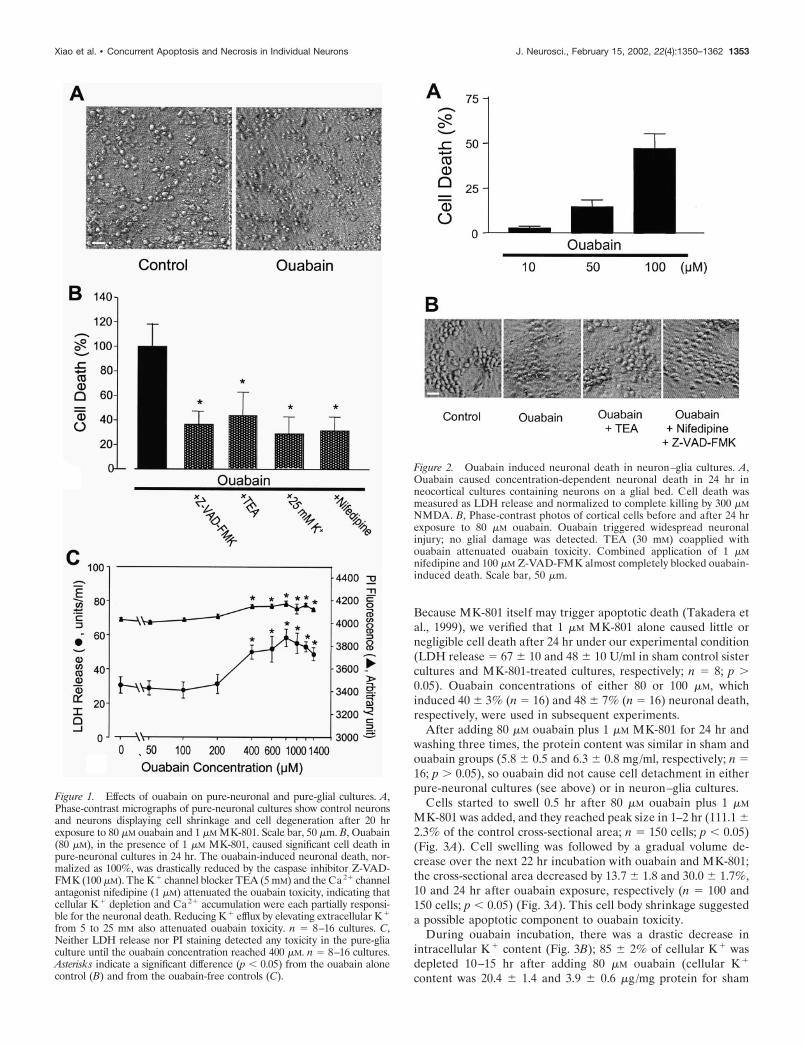
Because MK-801 itself may trigger apoptotic death (Takadera etal., 1999), we verified that 1 �M MK-801 alone caused little ornegligible cell death after 24 hr under our experimental condition(LDH release � 67 � 10 and 48 � 10 U/ml in sham control sistercultures and MK-801-treated cultures, respectively; n � 8; p �0.05). Ouabain concentrations of either 80 or 100 �M, whichinduced 40 � 3% (n � 16) and 48 � 7% (n � 16) neuronal death,respectively, were used in subsequent experiments.
After adding 80 �M ouabain plus 1 �M MK-801 for 24 hr andwashing three times, the protein content was similar in sham andouabain groups (5.8 � 0.5 and 6.3 � 0.8 mg/ml, respectively; n �16; p � 0.05), so ouabain did not cause cell detachment in eitherpure-neuronal cultures (see above) or in neuron–glia cultures.
Cells started to swell 0.5 hr after 80 �M ouabain plus 1 �M
MK-801 was added, and they reached peak size in 1–2 hr (111.1 �2.3% of the control cross-sectional area; n � 150 cells; p � 0.05)(Fig. 3A). Cell swelling was followed by a gradual volume de-crease over the next 22 hr incubation with ouabain and MK-801;the cross-sectional area decreased by 13.7 � 1.8 and 30.0 � 1.7%,10 and 24 hr after ouabain exposure, respectively (n � 100 and150 cells; p � 0.05) (Fig. 3A). This cell body shrinkage suggesteda possible apoptotic component to ouabain toxicity.
During ouabain incubation, there was a drastic decrease inintracellular K� content (Fig. 3B); 85 � 2% of cellular K� wasdepleted 10–15 hr after adding 80 �M ouabain (cellular K�
content was 20.4 � 1.4 and 3.9 � 0.6 �g/mg protein for sham
Figure 1. Effects of ouabain on pure-neuronal and pure-glial cultures. A,Phase-contrast micrographs of pure-neuronal cultures show control neuronsand neurons displaying cell shrinkage and cell degeneration after 20 hrexposure to 80 �M ouabain and 1 �M MK-801. Scale bar, 50 �m. B, Ouabain(80 �M), in the presence of 1 �M MK-801, caused significant cell death inpure-neuronal cultures in 24 hr. The ouabain-induced neuronal death, nor-malized as 100%, was drastically reduced by the caspase inhibitor Z-VAD-FMK (100 �M). The K� channel blocker TEA (5 mM) and the Ca2� channelantagonist nifedipine (1 �M) attenuated the ouabain toxicity, indicating thatcellular K� depletion and Ca2� accumulation were each partially responsi-ble for the neuronal death. Reducing K� efflux by elevating extracellular K�
from 5 to 25 mM also attenuated ouabain toxicity. n � 8–16 cultures. C,Neither LDH release nor PI staining detected any toxicity in the pure-gliaculture until the ouabain concentration reached 400 �M. n � 8–16 cultures.Asterisks indicate a significant difference (p � 0.05) from the ouabain alonecontrol (B) and from the ouabain-free controls (C).
Figure 2. Ouabain induced neuronal death in neuron–glia cultures. A,Ouabain caused concentration-dependent neuronal death in 24 hr inneocortical cultures containing neurons on a glial bed. Cell death wasmeasured as LDH release and normalized to complete killing by 300 �MNMDA. B, Phase-contrast photos of cortical cells before and after 24 hrexposure to 80 �M ouabain. Ouabain triggered widespread neuronalinjury; no glial damage was detected. TEA (30 mM) coapplied withouabain attenuated ouabain toxicity. Combined application of 1 �Mnifedipine and 100 �M Z-VAD-FMK almost completely blocked ouabain-induced death. Scale bar, 50 �m.
Xiao et al. • Concurrent Apoptosis and Necrosis in Individual Neurons J. Neurosci., February 15, 2002, 22(4):1350–1362 1353

control and ouabain-treated cells, respectively; n � 3 and 4; p �0.05). The K� channel blocker TEA (30 mM) antagonized theouabain-induced cell volume decrease and cellular K� depletion(Figs. 2B, 3A,B). The cell shrinkage was also blocked by Z-VAD-FMK (Fig. 3A) and the caspase-1 inhibitor Boc-Asp(Obzl)-CMK(BACMK; 100 �M) (surface area was 97.8 � 1.2% of controlsafter 10 hr in ouabain plus BACMK; p � 005 compared with thecontrol volume). BACMK, however, did not prevent the ouabain-induced neuronal death after 24 hr incubation (data not shown).
Ouabain simultaneously increased intracellular Ca2� contentby 39 � 16% (Ca2� � 2.7 � 1.7 and 3.8 � 0.4 �g/mg protein incontrol and ouabain-treated cells, respectively; n � 6; p � 0.05)measured by the ICP-MS method 15 hr after adding ouabain.Examined by Fura-2 fluorescence videomicroscopy, ouabain in-duced a time-dependent increase in [Ca2�]i. Starting at �30 minafter exposure, the [Ca2�]i level climbed continuously until itreached a plateau level at �90 min ([Ca2�]i � 70 � 4 and 157 �6 nM in sham control and ouabain-treated cells, respectively) (Fig.3C). The ouabain-induced [Ca2�]i increase was largely blockedby 1 �M nifedipine (Fig. 3C), suggesting that the voltage-gatedL-type Ca2� channel was the major route for ouabain-inducedCa 2� influx and [Ca2�]i increase. The residual [Ca2�]i increasenot blocked by nifedipine could be mediated by other pathwayssuch as Na�–Ca 2� exchange or release from intracellular stores.As expected, ouabain incubation (10–15 hr) also increased intra-cellular Na� content by 58 � 13% (Na� � 12.8 � 20.2 and20.2 � 1.5 �g/mg protein in control and ouabain-treated cells;n � 5; p � 0.05; ICP-MS method). Qualitatively and quantita-tively, these ouabain-induced alterations in ionic homeostasis areconsistent with previous reports (Archibald and White, 1974;Lijnen et al., 1986; Ahlemeyer et al., 1992).
Ouabain-induced cytochrome c release, caspaseactivation, and ultrastructural changesCytochrome c release from mitochondria is a critical apoptoticevent; this apoptotic process was triggered by ouabain. Theouabain-elicited cytochrome c release was markedly attenuatedby TEA (30 mM) or 25 mM K� medium but was not reduced bythe Ca2� channel antagonist nifedipine (1 �M) (Fig. 4). Consis-tent with cytochrome c release, ouabain treatment induced acti-vation of caspase-3-like proteases. The caspase activity startedrising after 15 hr in 80 �M ouabain and peaked after 24 hrincubation (Fig. 5). Caspase-3 activation was eliminated by addi-tion of the caspase inhibitor Z-VAD-FMK (100 �M) (Fig. 5); itwas also attenuated by the K� channel blocker TEA, but not bynifedipine (Fig. 5). In fact, addition of nifedipine accelerated the
4
loss was attenuated by 30 mM TEA (Similar results were obtained by theK �-selective electrode and ICP-MS method. Shown in the figure are theresults from the K �-selective electrode assay.) Ouabain also causedincreases in intracellular Na � (see Results). Ouabain induced similar K �
depletion in pure-neuronal cultures (data not shown). n � 3 measure-ments for time-matched sham control and TEA group; n � 6 for ouabain-treated group. The single asterisks in B show p � 0.05 compared with thesham control. The double asterisks in B show a significant difference (p �0.05) from ouabain alone. C, Ouabain-induced [Ca 2�]i increase in corti-cal neurons. Intracellular free Ca 2� concentration was measured byfluorescence imaging with Fura-2 AM. Compared with sham control cells(n � 13), application of 100 �M ouabain gradually increased [Ca 2�]istarting at �30 min after ouabain was added; [Ca 2�]i reached a plateaulevel in 80–90 min (n � 23). The ouabain-induced [Ca 2�]i increase waslargely blocked by coapplied 1 �M nifedipine (n � 28). MK-801 (1 �M)was added in experiments. *p � 0.05 compared with controls; #p � 0.05compared with ouabain alone at the same time points.
Figure 3. Ouabain-induced disruptions of ion homeostasis and cell vol-ume changes. A, Ouabain treatment initiated an acute phase of cell bodyswelling that peaked at 1–2 hr. Approximately 5 hr after ouabain wasadded, cells started to undergo a progressive volume decrease. The cellbody shrinkage was largely prevented by 30 mM TEA; the initial cellswelling was not affected by TEA. The ouabain-induced cell volumedecrease was also prevented by the caspase inhibitor Z-VAD-FMK (100�M). n � 100–150 cells for each time point (n � 150 for Z-VAD-FMKexperiment). The single asterisks in A show p � 0.05 compared with time0 controls. The double asterisks in A show a significant difference (p �0.05) from the ouabain group at the same time points. B, Ouabain (80 �M,10–15 hr exposure) induced a massive depletion of cellular K �. The K �
1354 J. Neurosci., February 15, 2002, 22(4):1350–1362 Xiao et al. • Concurrent Apoptosis and Necrosis in Individual Neurons

process of caspase-3 activation by several hours, so that it peakedby 20 hr (Fig. 5). This phenomenon and an increased cytochromec release observed when nifedipine was added together with TEA(Fig. 4) are consistent with the hypothesis that low [Ca2�]i mayendorse apoptosis (Yu et al., 2001). Further support for anapoptotic contribution to ouabain-induced death is the appear-ance of the characteristic DNA fragmentation (DNA laddering)20–24 hr after the onset of ouabain treatment (Fig. 6). Consis-
tently, DNA laddering was prevented by coapplied TEA orZ-VAD-FMK, but not by nifedipine (Fig. 6).
Although all of these morphological and biochemical featuresare consistent with apoptosis, the caspase inhibitor Z-VAD-FMK, at a concentration (100 �M) that completely and persis-tently prevented caspase-3 activation (Polverino and Patterson,1997) (Fig. 5), blocked only 61 � 7% (n � 16) and 65 � 4% (n �44) of ouabain-induced cell death in pure-neuronal and neuron–glia cultures, respectively (Figs. 1B, 7A). The incomplete block ofcell death implied that a caspase-independent component, likelynecrosis, additionally contributed to ouabain toxicity.
To better characterize ouabain-induced neuronal death, elec-tron microscopy (EM) was used to examine ultrastructural alter-ations. To follow the time course of morphological alterations, weexamined neurons subjected to 2, 5, and 10 hr incubation withouabain (100 �M) and MK-801 (1 �M). Apoptotic changes such asnuclear condensation appeared early; meanwhile, necrotic alter-ations such as swelling of organelles and cytoplasm, formation ofvacuoles, and disruption of membranes were also developed atearly hours, suggesting that the two injurious pathways developedin parallel in ouabain toxicity (Fig. 8). After 15–20 hr exposure toouabain, apoptotic features such as highly condensed pyknoticnuclei and dense chromatin masses were evident. Prominentnecrotic features, including numerous lucent cytoplasmic vacu-oles of different sizes, disruption of cellular organelles, and loss ofplasma membrane integrity were also present in the same cells(Fig. 9). These mixed features of apoptosis and necrosis, referredto as hybrid death, were found in most injured cells, althoughthere were variations in the extent of a particular change.
Consistent with the hybrid cell death mediated by separateionic mechanisms, damaged neurons showed dominant apoptoticmorphology when the Ca2� channel antagonist nifedipine wascoapplied with ouabain. On the other hand, when K� efflux wasattenuated by 25 mM K� medium during ouabain application, EM
Figure 4. Effects of nifedipine, TEA, and potassium on ouabain-inducedcytochrome c release. Cytochrome c release was detected by Western blotin the cytosolic fraction 20 hr after incubation with 80 �M ouabain (toppanel ), with corresponding reduction of mitochondrial cytochrome c(bottom panel ). Cytochrome c release was drastically attenuated by TEA(30 mM) or elevated extracellular K � (25 mM K �); on the other hand, itwas not affected by nifedipine (1 �M). COX in mitochondrial fraction andits absence in cytosolic fraction demonstrated that the intact mitochondriaseparated from cytosol in our analysis. The �-actin analysis was per-formed as an internal control. The results shown are representative ofthree independent experiments. When nifedipine was combined withTEA, there appeared to be more cytochrome c release into the cytosolcompared with the release with TEA alone, suggesting that the membranedepolarization induced by TEA might facilitate the voltage-dependentblock of Ca 2� channels by dihydropyridine derivatives such as nifedipine(Sanguinetti and Kass, 1984) and thus might be favorable for a low Ca 2�
stimulated apoptotic process (Yu et al., 2001).
Figure 5. Effects of TEA and nifedipine on ouabain-induced caspase-3activation. Caspase-3 activity was correlated with the cleavage of thespecific substrate DEVD-AMC. In sham control experiments, caspase-3activity was stable at a low level for 25 hr (f). Incubation with 80 �Mouabain increased the caspase activity in a time-dependent manner (F);the increase was blocked by Z-VAD-FMK (100 �M) (�) and TEA (30mM) (�) but not by nifedipine (1 �M) (Œ). Nifedipine even appeared toaccelerate the process of caspase activation. n � 3–5 independent mea-surements for each time point. *p � 0.05 compared with sham controls atthe same time points.
Xiao et al. • Concurrent Apoptosis and Necrosis in Individual Neurons J. Neurosci., February 15, 2002, 22(4):1350–1362 1355

examination revealed typical necrotic alterations in most cells(Fig. 9).
Ionic mechanisms underlying ouabain-induced hybridcell deathCellular K� homeostasis is maintained by K� efflux and K�
uptake mechanisms. In the presence of MK-801, the major path-way for K� efflux from neurons is the family of the TEA-sensitive,noninactivating delayed rectifier IK channels, whereas the Na�,K�-ATPase is responsible for moving K� back into the cell fromthe extracellular space. We reasoned and demonstrated abovethat as long as K� efflux was prevented throughout the ouabaintreatment, there would be no marked cellular K� loss even if theNa�, K�-pump were blocked. Therefore, should K� efflux andcellular K� depletion be key steps in apoptosis, blocking K�
channels would be able to attenuate ouabain-induced cell death.As expected, the K� channel blocker TEA (30 mM) or tetrapen-tylammonium (TPeA; 10 �M) significantly reduced ouabain-induced cell death (28.9 � 4.3 and 65.4 � 6.5% reduction for theTEA and TPeA groups, respectively) (Fig. 7B). Consistent withthis finding, ouabain induced much less cell death (43% reduc-tion; n � 28) in 25 mM K� medium than in the control mediumof 5 mM K� (Fig. 7C).
To verify the involvement of Na�, K�-ATPase in neurotoxic-ity, we tested another selective Na� pump inhibitor, strophanthi-din (Balzan et al., 2000). Strophanthidin (800 �M) induced �40%neuronal death in 24 hr; the cell injury measured by LDH releasewas reduced from 273 U/ml to 160 U/ml (41 � 1% reduction; n �8; p � 0.05) by 100 �M Z-VAD-FMK, and to 217 U/ml by 25 mM
extracellular K� (21 � 1% reduction; n � 8; p � 0.05), respec-tively. These results confirmed that Na� pump failure caused aK� efflux-related and caspase-dependent apoptotic injury.
Because inhibition of Na�, K�-ATPase increased intracellularCa 2� and Na�, and EM assay revealed a necrotic component inouabain-induced cell death, we tested the idea that Ca2� or Na�
channel blockers might selectively attenuate the necrotic injury ofouabain toxicity. A combination of 80 �M ouabain and the Ca2�
channel antagonist nifedipine (1 �M; n � 23) or Na� channelblocker tetradotoxin (TTX) (1 �M; n � 8) reduced ouabain-induced cell death by 43 � 8 and 32 � 5%, respectively (Fig. 7D).We then compared the protective effects of these channel block-ers alone and in combination with Z-VAD-FMK. Virtually com-plete protection was achieved when nifedipine was coapplied withZ-VAD-FMK (Figs. 2B, 7D). Combined application of TTX andZ-VAD-FMK also brought out additional neuroprotection (Fig.7D), suggesting a role for Na� influx, although less imperativethan Ca2� influx, in necrotic death. Combination of TEA or 25mM K� with nifedipine did not produce full protection, in linewith the incomplete block of K� depletion and some residualcaspase-3 activity in the presence of TEA (Figs. 3B, 5). In agree-ment with this, Z-VAD-FMK enhanced the protective effect of 30mM TEA (Fig. 7B). Higher concentrations of TEA were toxic andnot tested further.
Young cells are more vulnerable to apoptosis. For example,staurosporine induced no appreciable apoptosis in cultured cor-tical neurons older than 16–17 DIV (Koh et al., 1995), which isconsistent with the lack of upmodulation of IK current in thesecells (Yu et al., 1997). However, older cortical neurons (16 DIV)exhibited even higher vulnerability to ouabain toxicity; 80 �M
ouabain, in the presence of 1 �M MK-801, triggered 75% neuro-nal death in these cells compared with �40% death in 11–12 DIVneurons. This enhanced toxicity was unlikely caused by MK-801;the putative pro-apoptotic effect of MK-801 diminishes in corticalneurons older than 12 DIV (Kim-Han et al., 1999). The death in16 DIV cultures was reduced by approximately one-half byZ-VAD-FMF (100 �M), TEA (30 mM), elevated extracellular K�
(25 mM K�), or nifedipine (1 �M) (n � 12 for each treatment; p �0.05 compared with sham controls). Therefore, ouabain triggeredionic disruption and accordant hybrid death in young and oldneurons.
Ouabain-induced death in low Ca2�, lowNa� conditionsIn the ischemic brain, extracellular Ca2� and Na� concentra-tions decline to levels as low as 0.1 and 30–50 mM, respectively(Siesjo, 1992; Xie et al., 1994; Kristian and Siesjo, 1996). Wesuspected that under such conditions, in conjunction with insuf-ficient energy supply, apoptosis might become the dominant formof neuronal death. To model this pathological condition, wetested the effect of ouabain in a low Ca 2� (0.1 vs 1.5 mM CaCl2)or low Na� (60 vs 120 mM NaCl) medium. Osmolarity wasadjusted by adding N-methyl-D-glucamine and HCl to the me-dium, pH 7.4. Incubation for 3–5 hr with this medium alone didnot reduce cell viability 24 hr after the onset of incubation.Adding 80 �M ouabain during the few hours of incubation,however, caused significant (�50%) neuronal death in 24 hr.Most of the cell death was blocked by Z-VAD-FMK, suggestingapoptosis-dominated death under these conditions (Fig. 10). Con-sistent with this, reducing K� efflux by raising the extracellularK� concentration blocked �80% of the cell death in eithermedium (Fig. 10). Nifedipine, added to the low Ca2�/high K�
Figure 6. Ouabain-induced DNA fragmentation. Ouabain (80 �M) ex-posure of 20 hr induced DNA fragmentation (laddering), revealed byagarose gel electrophoresis. The pattern of DNA damage was similar tothat induced by the typical apoptosis inducer staurosporine (0.2 �M). NoDNA fragmentation occurred in control cells. Ouabain-induced DNAladdering was prevented by coapplied TEA (30 mM) or Z-VAD-FMK(100 �M), but not by nifedipine (1 �M). Similar results were obtained fromthree independent experiments. Data shown in the figure were from oneexperiment; the position of columns was rearranged for purpose of clarity.
1356 J. Neurosci., February 15, 2002, 22(4):1350–1362 Xiao et al. • Concurrent Apoptosis and Necrosis in Individual Neurons

medium, provided no additional protection, consistent with thealready reduced Ca2� influx. Under these conditions, TTX fur-ther promoted cell survival by blocking Na� influx (Fig. 10).Nifedipine provided extra protection in the low Na�/normal Ca2�
medium, where Ca2� influx might still normally occur (Fig. 10).
DISCUSSIONCollective evidence agrees that blocking Na�, K�-ATPase in-duces a mixed neuronal death with features of both apoptosis andnecrosis. The caspase-mediated apoptotic component is associ-ated with K� channel activation, K� efflux, and cellular K� loss,whereas the nifedipine-blocked Ca2�-associated cell injury iscaspase independent. In this context, the protective effect ofblocking Na� channels may be mediated indirectly by reducingthe reversed Na�–Ca 2� exchange activity, thereby preventing asecondary [Ca2�]i increase. Although ouabain-induced apoptosishas been reported in a few previous studies (Olej et al., 1998;Verheye-Dua and Bohm, 2000), this investigation provides thefirst evidence of a mixed death in ouabain toxicity. Despite theemerging idea of an overlap of necrosis and apoptosis in tissuesand cell cultures (Toescu 1998), current popular opinion associ-ates these different processes with separate subgroups of cells orconsecutive events (e.g., necrosis followed by apoptosis) (Liptonand Nicotera 1998) (Fig. 11). The present study establishes theconcept and a model of hybrid death as concurrent necrosis andapoptosis in single cells throughout the death process (Fig. 11).
The Na�, K�-ATPase is present in all mammalian cells. Theactivity of Na�, K�-ATPase in brain cortical glial cells shouldhave a significant impact on the microenvironment surroundingneurons and their ionic homeostasis. Glial cells express �1 and �2isoforms of Na�, K�-ATPase; the lack of the �3 isoform of high
ouabain affinity explains the low ouabain toxicity in glial cultures(McGrail et al., 1991; Watts et al., 1991). Although our experi-ments using mixed cultures do not completely exclude interfer-ence from glial cells, it is unlikely that glia have much effect on thenature of hybrid injuries.
The digitalis glycoside, ouabain, has endogenous analogs withintrinsic regulatory properties in vertebrate physiology (Budzi-kowski et al., 1998; Ferrandi and Manunta, 2000). In rats andhumans, “endogenous ouabain” has been detected in all tissuestested (Hamlyn et al., 1996). The level of endogenous ouabain incirculation increases on exposure to stress signals such as hyper-tension and hypoxia/ischemia (Bagrov et al., 1994; De Angelisand Haupert, 1998; Ferrandi and Manunta, 2000). Accordingly,the Na�, K�-ATPase activity in the ischemic heart, brain, andother organs decreases (Lees, 1991; Bundgaard et al., 1997).Ouabain sensitized human and rodent tumor cells to tumornecrosis factor (TNF)-induced apoptosis (Penning et al., 2000),enhanced irradiation-induced apoptosis in human cell lines ofdefined tumor protein p53 status (Verheye-Dua and Bohm,2000), and potentiated anti-Fas-induced apoptosis (Bortner et al.,2001). Thus Na�, K�-ATPase plays an imperative role in apo-ptosis induced by different insults in different cells.
We and others have shown that excessive K� efflux mediatedby K� channels or NMDA receptor channels is a key event in theapoptotic cascade (Yu et al., 1997, 1999a; Colom et al., 1998;Wang et al., 1999; Krick et al. 2001). Cellular K� depletion islikely a prerequisite for activation of two apoptotic mediators:caspases and endonucleases (Dallaporta et al., 1999; Hughes andCidlowski, 1999; Yu et al., 1999b; Wang et al., 2000). In theexperiment with pure-neuronal cultures, 10–15 hr ouabain incu-
Figure 7. Block of ouabain-induced cell death incortical neuron–glia cultures. Ouabain-induced neu-ronal death in cortical cultures containing neuronsand a glial bed was measured by LDH release after 24hr exposure and normalized to the cell death inducedby 80 �M ouabain. A, The broad-spectrum caspaseinhibitor Z-VAD-FMK (100 �M) blocked 65 � 4% ofcell death, whereas its negative control ZFA (100 �M)showed no significant protection ( p � 0.16). B, Potas-sium channel blocker TEA (30 mM) or TPeA (10 �M)partly reduced the ouabain-induced neuronal death;coapplied 1 �M nifedipine or 100 �M Z-VAD-FMKprovided extra protection. TPeA showed substantialprotection, presumably because of its additional non-specific block on Ca 2� channels (Wang et al., 2000).C, Elevated extracellular K � (25 mM KCl) attenuatedouabain-induced death; additional protection was ob-tained with coapplied Ca 2� channel antagonist 2 �Mgadolinium (Gd 3�) or 1 �M nifedipine. D, Nifedipine(1 �M) or the Na � channel blocker TTX (1 �M) alsopartially prevented the ouabain toxicity. Maximalneuroprotection was achieved by combining nifedi-pine with Z-VAD-FMK. n � 12 for each columnexcept for ZFA and TTX (n � 8). *p � 0.05 comparedwith ouabain alone; **p � 0.05 compared withouabain plus one treatment.
Xiao et al. • Concurrent Apoptosis and Necrosis in Individual Neurons J. Neurosci., February 15, 2002, 22(4):1350–1362 1357

bation induced an 11% volume decrease, whereas cells lost 72%of their K�, implying that intracellular K� concentration waslikely decreased by �61%. Presuming that resting intracellularK� concentration is 140 mM and acts as the predominant elementfor cell volume regulation and that water loss is proportional tothe volume loss, the K� concentration would be reduced to �55mM by the ouabain treatment, consistent with the values (50–56mM) reported by others in cells undergoing apoptosis (Barbiero etal., 1995; Hughes et al., 1997).
Blocking K� efflux prevented cytochrome c release, caspase-3activation, and DNA laddering, placing cellular K� loss beforethese apoptotic steps. It reinforces the notion that K� acts as anendogenous modulator of several checkpoints (e.g., cytochrome crelease, caspase cleavage, and endonuclease activation) in theapoptotic cascade. Recent progress suggests that programmedcell death such as that induced by apoptosis-inducing factor(AIF) may be independent of Apaf-1, cytochrome c, and caspases(Joza et al., 2001). Interestingly, the endonuclease activation andDNA damage in AIF-induced programmed death are still K�
dependent (Dallaporta et al., 1998), suggesting that the K� mech-anism may control different forms of programmed death thatcontribute to the hybrid cell death. A nonapoptotic programmedcell death induced by expression of insulin-like growth factor Ireceptor was reported recently (Sperandio et al., 2000). This typeof cell death, although related to caspase-9 activation and proteinsynthesis, lacks almost all morphological features of apoptosis,suggesting that it is not linked to cellular K� depletion and maybe a distinct form of cell death different from the hybrid deathobserved in this study.
The major anti-apoptotic members of the Bcl-2 family, Bcl-2 orBcl-x1, show protective effects against apoptosis induced by block-ing the Na�, K�-pump (Gilbert and Knox, 1997; Kawazoe et al.,1999), presumably because of an enhanced pump activity andmaintaining sufficient mitochondrial ATP/ADP exchange to sus-tain coupled respiration (Gilbert and Knox, 1997; Vander Heidenet al., 1999). Thus, the Bcl-2 family may have a significant influ-ence on apoptosis as well as the mixed form of cell death. Becausethe K� mechanism has been demonstrated in apoptosis inducedby receptor and nonreceptor associated insults (Hughes and Ci-dlowski, 1999; Penning et al., 2000; Bortner et al., 2001), it isconceivable that the apoptotic components associated with eithercytochrome c/caspase-3 cascade or “death receptors,” such as theTNF-� pathway, may both be able to intervene in hybrid celldeath.
The broad-spectrum caspase-inhibitor Z-VAD-FMK pre-vented the ouabain-induced cell volume decrease, in agreementwith observations of some groups (Choi et al. 2000; Lang et al.,2000; Nobel et al., 2000) but in contrast to results from others(Maeno et al., 2000; Yu and Choi, 2000). This discrepancy mayimply a role for specific caspases, but not caspase-3 (see below), incell volume regulation. For example, the apoptotic cell shrinkageinduced by etoposide or methylprednisolone is blocked bycaspase-1 inhibitors in thymocytes (Zhivotovsky et al., 1995).Because casapase-1 activity is relatively uninfluenced by K�
(Hughes et al., 1997; Yu et al., 1999b), its activation may occur inthe absence of excessive K� efflux and cell shrinkage. Our datawith the caspase-1 inhibitor BACMK suggest that this particularcaspase may be activated early and plays an important role inneuronal apoptotic shrinkage. On the other hand, caspase-3 ac-tivation is a relatively delayed event (15 hr later), after cellularK� depletion and cell shrinkage but still before the cell deathmeasured by LDH release. Surprisingly, BACMK did not show
Figure 8. Morphological changes of hybrid cell death at early time pointsof ouabain exposure. EM images reveal ouabain-induced ultrastructuralalterations in cortical neurons; morphology of a normal neuron can beseen in Figure 9. A, Two hours after adding 100 �M ouabain plus 1 �MMK-801, some cells started to show signs of nuclear changes; the electronmicrograph shows an irregular shape of the nucleus, implying a volumedecrease. Meanwhile, swelling mitochondria were observed in many cells.B, Apoptotic features such as nuclear shrinkage and condensation of thenuclear chromatin were advanced after 5 hr in ouabain. Necrotic changessuch as cytoplasm swelling, formation of vacuoles, and disruptions ofcellular organelles and the plasma membrane also appeared at earlierhours. The two cells shown in this micrograph represent different stagesof morphological changes observed at this time. C, Ten hours after onsetof ouabain exposure, injured cells with highly condensed nuclei, chaoticcytoplasm, and disrupted plasma membrane were easily detected. Scalebar, 3.0 �m. N, Nucleus; C, cytoplasm; M, mitochondria; V, vacuole.
1358 J. Neurosci., February 15, 2002, 22(4):1350–1362 Xiao et al. • Concurrent Apoptosis and Necrosis in Individual Neurons
any neuroprotective effect against ouabain toxicity. The explana-tion for the dissociation of BACMK action on cell volume andcell death is obscure and deserves future investigation.
Increases in [Ca2�]i may trigger apoptosis (Lipton and Nico-tera 1998; Toescu 1998). In the present study, blocking of Ca2�
influx and [Ca2�]i increase did not inhibit cytochrome c releaseor caspase-3 activation, suggesting that the ouabain-induced[Ca2�]i increase did not play a primary role in induction ofapoptosis. On the other hand, an increase in [Ca2�]i may explainprotection against apoptosis in sympathetic ganglia and cerebel-lar granule neurons (Johnson et al., 1992). Blocking Ca2� entryand [Ca2�]i increase, however, did not eliminate the anti-apoptotic effect of elevated extracellular K� or K� channelblockers in cortical neurons (Yu et al., 1997). The discrepancymay be attributable to the fact that apoptosis can be mediated bymultiple pathways and that apoptotic mechanisms differ by celltypes. For example, in M1 myeloid leukemia cells, Ca2�-mobilizing compounds like the Ca2� ionophore A23187 and theendoplasmic reticulum Ca2�-ATPase inhibitor thapsigargin caneither suppress or induce apoptosis, depending on activation ofdifferent signal transduction pathways (Lotem et al., 1999). Incerebellar granule cells and vascular smooth muscle, the Na�/K�
ratio, rather than K� concentration or ionic strength, was pro-
posed to determine the outcome of an apoptotic insult (Isaev etal., 2000; Orlov et al., 2000).
Although apoptosis and necrosis are two separate fundamentalaspects of cell death, the most recent findings suggest that celldeath often falls somewhere between the two extremes in thespectrum. Cell death bearing both apoptotic and necrotic featurescan be induced by glutamate, zinc, or oxygen–glucose deprivationin mouse cortical neurons (Gwag et al., 1995; Cheung et al., 1998;Sohn et al., 1998; Kim et al., 1999) and by other insults in variouscells (Papadimitriou et al., 1994; Tsujimoto et al., 1997; Villalba etal., 1997; Okuno et al., 1998; Miller et al., 2000; Park et al., 2000).Features of mixed death may also be found in a number of otherstudies (Molthagen et al., 1996; Warny and Kelly, 1999), includingthose of myocardial cells after coronary artery occlusion andreperfusion in vivo (Takashi and Ashraf, 2000) and in the adult ornewborn rat brain (Portera-Cailliau et al., 1997). After hypoxicischemia in the newborn rat, “hybrid” neuronal cells with inter-mediate ultrastructural characteristics similar to the mixed deathshown in this study were observed (Nakajima et al., 2000). Accu-mulating evidence, therefore, demonstrates that mixed or hybridcell death is common either in vitro or in vivo under differentpathological conditions.
Rather than debating whether atypical cell death with mixed
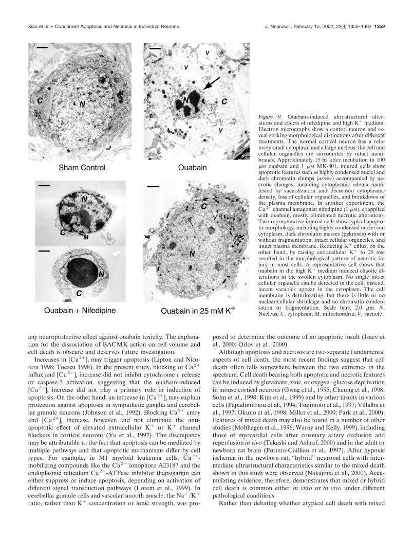
Figure 9. Ouabain-induced ultrastructural alter-ations and effects of nifedipine and high K � medium.Electron micrographs show a control neuron and re-veal striking morphological distinctions after differenttreatments. The normal cortical neuron has a rela-tively small cytoplasm and a large nucleus; the cell andcellular organelles are surrounded by intact mem-branes. Approximately 15 hr after incubation in 100�M ouabain and 1 �M MK-801, injured cells showapoptotic features such as highly condensed nuclei anddark chromatin clumps (arrow) accompanied by ne-crotic changes, including cytoplasmic edema mani-fested by vacuolization and decreased cytoplasmicdensity, loss of cellular organelles, and breakdown ofthe plasma membrane. In another experiment, theCa 2� channel antagonist nifedipine (1 �M), coappliedwith ouabain, mostly eliminated necrotic alterations.Two representative injured cells show typical apopto-tic morphology, including highly condensed nuclei andcytoplasm, dark chromatin masses (pyknosis) with orwithout fragmentation, intact cellular organelles, andintact plasma membrane. Reducing K � efflux, on theother hand, by raising extracellular K � to 25 mMresulted in the morphological pattern of necrotic in-jury in most cells. A representative cell shows thatouabain in the high K � medium induced chaotic al-terations in the swollen cytoplasm. No single intactcellular organelle can be detected in the cell; instead,lucent vacuoles appear in the cytoplasm. The cellmembrane is deteriorating, but there is little or nonuclear/cellular shrinkage and no chromatin conden-sation or fragmentation. Scale bars, 2.0 �m. N,Nucleus; C, cytoplasm; M, mitochondria; V, vacuole.
Xiao et al. • Concurrent Apoptosis and Necrosis in Individual Neurons J. Neurosci., February 15, 2002, 22(4):1350–1362 1359

pathological features fulfills criteria for apoptosis or necrosis, wepropose that this hybrid injury be recognized as a distinct form ofcell death. We believe that this approach is necessary and ofpractical use for classifying widely observed but conceptuallyconfusing lethal cellular events. The identification of hybrid deathis particularly relevant to situations in which cells face multipleinsults accompanied by impaired energy metabolism and elevatedlevels of endogenous ouabain. Study of the hybrid cell injury mayfacilitate the development of better therapies for this broad cat-egory of pathological conditions.
REFERENCESAhlemeyer B, Weintraut H, Schoner W (1992) Cultured chick-embryo
heart cells respond differently to ouabain as measured by the increasein their intracellular Na � concentration. Biochem Biophys Acta1137:135–142.
Ameisen JC (1994) Programmed cell death (apoptosis) and cell survivalregulation: relevance to AIDS and cancer. AIDS 8:1197–1213.
Ankarcrona M, Dypbukt JM, Bonfoco E, Zhivotovsky B, Orrenius S,Lipton SA, Nicotera P (1995) Glutamate-induced neuronal death: asuccession of necrosis or apoptosis depending on mitochondrial func-tion. Neuron 15:961–973.
Archibald JT, White TD (1974) Rapid reversal of internal Na � and K �
contents of synaptosomes by ouabain. Nature 252:595–597.Armstrong RC, Aja TJ, Hoang KD, Gaur S, Bai X, Alnemri ES, Litwack
G, Karanewsky DS, Fritz LC, Tomaselli KJ (1997) Activation of theCED3/ICE-related protease CPP32 in cerebellar granule neurons un-dergoing apoptosis but not necrosis. J Neurosci 17:553–562.
Bagrov AY, Kuznetsova EA, Fedorova OV (1994) Endogenous digoxin-like factor in acute myocardial infarction. J Intern Med 235:63–67.
Balzan S, D’Urso G, Ghione S, Martinelli A, Montali U (2000) Selectiveinhibition of human erythrocyte Na �/K � ATPase by cardiac glycosidesand by a mammalian digitalis like factor. Life Sci 67:1921–1928.
Barbiero G, Duranti F, Bonelli G, Amenta JS, Baccino FM (1995)Intracellular ionic variations in the apoptotic death of L cells by inhib-itors of cell cycle progression. Exp Cell Res 217:410–418.
Beauvais F, Michel L, Dubertret L (1995) Human eosinophils in cultureundergo a striking and rapid shrinkage during apoptosis. Role of K �
channels. J Leukoc Biol 57:851–855.Bortner CD, Hughes Jr FM, Cidlowski JA (1997) A primary role for K �
and Na � efflux in the activation of apoptosis. J Biol Chem272:32436–32442.
Bortner CD, Gomez-Angelats M, Cidlowski JA (2001) Plasma mem-brane depolarization without repolarization is an early molecular eventin anti-Fas induced apoptosis. J Biol Chem 276:4304–4314.
Budzikowski AS, Huang BS, Leenen FH (1998) Brain “ouabain,” aneurosteroid, mediates sympathetic hyperactivity in salt-sensitive hy-pertension. Clin Exp Hypertens 20:119–140.
Bundgaard H, Schmidt TA, Larsen JS, Kjeldsen K (1997) K � supple-mentation increases muscle. J Appl Physiol 82:1136–1144.
Cheung NS, Pascoe CJ, Giardina SF, John CA, Beart PM (1998) Micro-molar L-glutamate induces extensive apoptosis in an apoptotic-necroticcontinuum of insult-dependent, excitotoxic injury in cultured corticalneurones. Neuropharmacology 37:1419–1429.
Choi DW (1988) Calcium-mediated neurotoxicity: relationship to spe-cific channel types and role in ischemic damage. Trends Neurosci11:465–469.
Choi KH, Hama-Inaba H, Wang B, Haginoya K, Odaka T, Yamada T,Hayata I, Ohyama H (2000) UVC-induced apoptosis in human epi-thelial tumor A431 cells: sequence of apoptotic changes, involvement ofcaspase (-8, -3) cascade. J Radiat Res (Tokyo) 41:243–258.
Chopp M, Li Y (1996) Apoptosis in focal cerebral ischemia. Acta Neu-rochir Suppl (Wien) 66:21–26.
Churchwell KB, Wright SH, Emma F, Rosenberg PA, Strange K (1996)NMDA receptor activation inhibits neuronal volume regulation afterswelling induced by veratridine-stimulated Na � influx in rat corticalcultures. J Neurosci 16:7447–7457.
Colbourne F, Sutherland GR, Auer RN (1999) Electron microscopic
Figure 10. Ouabain-induced K � efflux-sensitiveand caspase-dependent neuronal death in lowCa 2� or low Na � conditions. A 3 hr exposure to 80�M ouabain plus 1 �M MK-801 in a low Ca 2� (0.1mM CaCl2 ) or a low Na � (60 mM NaCl) mediuminduced a dominant neuronal death that was highlysensitive to block by 25 mM K � or Z-VAD-FMK(100 �M). Without ouabain, the low Ca 2� or lowNa � medium was not toxic (3 hr exposure; data notshown). In the low Ca 2� medium containing anormal concentration of Na �, the Ca 2� channelantagonist nifedipine (1 �M) did not show anyeffect on the neuroprotection produced by elevatedK �, whereas combination of high K � and the Na �
channel blocker TTX (1 �M) completely preventedcell death. In the low-Na � medium containing nor-mal Ca 2�, an additional protective effect was ob-tained by combining high K � and nifedipine (TTXwas not tested in this paradigm). Cell death isnormalized to the injury induced by 80 �M ouabainin medium containing normal concentrations ofCaCl2 (1.5 mM) and NaCl (120 mM) (MEM sup-plemented with glucose, FBS, HS, and EGF; seeMaterials and Methods). This medium was used towash out ouabain after the 3 hr incubation. Celldeath was measured by LDH release 24 hr after theonset of exposure. Osmolarity was maintained by adding appropriate amounts of NMDG and HCl; pH was 7.4. n � 8 –32. *p � 0.05 comparedwith ouabain alone.
Figure 11. Cell death models for necrosis, apoptosis, and hybrid death.A, The conventional cell death model predicts that necrosis and apoptosisare triggered by separate insults and exhibit typical distinctive morpho-logical changes in injured cells. B, Emerging opinion suggests that thesame insult may induce either necrosis or apoptosis in different cells;alternatively, a necrotic injury may convert to apoptotic injury or viceversa. C, Recent observations and the present study support the thirdpossibility that a single or multiple insult(s) may trigger parallel pathwaysleading to necrotic and apoptotic damages in the same cells, identified ashybrid cell death.
1360 J. Neurosci., February 15, 2002, 22(4):1350–1362 Xiao et al. • Concurrent Apoptosis and Necrosis in Individual Neurons

evidence against apoptosis as the mechanism of neuronal death inglobal ischemia. J Neurosci 19:4200–4210.
Colom LV, Diaz ME, Beers DR, Neely A, Xie WJ, Appel SH (1998)Role of potassium channels in amyloid-induced cell death. J Neuro-chem 70:1925–1934.
Contreras RG, Shoshani L, Flores-Maldonado C, Lazaro A, Cereijido M(1999) Relationship between Na �,K �-ATPase and cell attachment.J Cell Sci 112:4223–4232.
Dallaporta B, Hirsch T, Susin SA, Zamzami N, Larochette N, Brenner C,Marzo I, Kroemer G (1998) Potassium leakage during the apoptoticdegradation phase. J Immunol 160:5605–5615.
Dallaporta B, Marchetti P, de Pablo MA, Maisse C, Duc HT, Metivier D,Zamzami N, Geuskens M, Kroemer G (1999) Plasma membrane po-tential in thymocyte apoptosis. J Immunol 162:6534–6542.
De Angelis C, Haupert Jr GT (1998) Hypoxia triggers release of anendogenous inhibitor of Na �, K �-ATPase from midbrain and adrenal.Am J Physiol 274:F182–F188.
Deshpande J, Bergstedt K, Linden T, Kalimo H, Wieloch T (1992)Ultrastructural changes in the hippocampal CA1 region following tran-sient cerebral ischemia: evidence against programmed cell death. ExpBrain Res 88:91–105.
Du C, Hu R, Csernansky CA, Hsu CY, Choi DW (1996) Very delayedinfarction after mild focal cerebral ischemia: a role for apoptosis?J Cereb Blood Flow Metab 16:195–201.
Ejima A, Watanabe C, Koyama H, Satoh H (1999) Matrix interferencesin the analysis of digested biological tissues with inductively coupledplasma-mass spectrometry. Biol Trace Elem Res 69:99–109.
Ferrandi M, Manunta P (2000) Ouabain-like factor: is this the natri-uretic hormone? Curr Opin Nephrol Hypertens 9:165–171.
Gilbert M, Knox S (1997) Influence of Bcl-2 overexpression on Na �/K �-ATPase pump activity: correlation with radiation-induced pro-grammed cell death. J Cell Physiol 171:299–304.
Gottron FJ, Ying HS, Choi DW (1997) Caspase inhibition selectivelyreduces the apoptotic component of oxygen-glucose deprivation-induced cortical neuronal cell death. Mol Cell Neurosci 9:159–169.
Grynkiewicz G, Poenie M, Tsien RJ (1985) A new generation of Ca 2�
indicators with greatly improved fluorescence properties. J Biol Chem260:3440–3450.
Gwag BJ, Lobner D, Koh JY, Wie MB, Choi DW (1995) Blockade ofglutamate receptors unmasks neuronal apoptosis after oxygen-glucosedeprivation in vitro. Neuroscience 68:615–619.
Hamlyn JM, Hamilton BP, Manunta P (1996) Endogenous ouabain,sodium balance and blood pressure: a review and a hypothesis. J Hy-pertens 14:151–167.
Hirsch T, Marchetti P, Susin SA, Dallaporta B, Zamzami N, Marzo I,Geuskens M, Kroemer G (1997) The apoptosis-necrosis paradox. Ap-optogenic proteases activated after mitochondrial permeability transi-tion determine the mode of cell death. Oncogene 15:1573–1581.
Hughes Jr FM, Cidlowski JA (1999) Potassium is a critical regulator ofapoptotic enzymes in vitro and in vivo. Adv Enzyme Regul 39:157–171.
Hughes Jr FM, Bortner CD, Purdy GD, Cidlowski JA (1997) Intracel-lular K � suppresses the activation of apoptosis in lymphocytes. J BiolChem 272:30567–30576.
Isaev NK, Stelmashook EV, Halle A, Harms C, Lautenschlager M, WeihM, Dirnagl U, Victorov IV, Zorov DB (2000) Inhibition of Na �,K �-ATPase activity in cultured rat cerebellar granule cells prevents theonset of apoptosis induced by low potassium. Neurosci Lett 283:41–44.
Johnson Jr EM, Koike T, Franklin J (1992) A “calcium set-point hypoth-esis” of neuronal dependence on neurotrophic factor. Exp Neurol115:163–166.
Joza N, Susin SA, Daugas E, Stanford WL, Cho SK, Li CY, Sasaki T, EliaAJ, Cheng HY, Ravagnan L, Ferri KF, Zamzami N, Wakeham A,Hakem R, Yoshida H, Kong YY, Mak TW, Zuniga-Pflucker JC,Kroemer G, Penninger JM (2001) Essential role of the mitochondrialapoptosis-inducing factor in programmed cell death. Nature410:549–554.
Kawazoe N, Aiuchi T, Masuda Y, Nakajo S, Nakaya K (1999) Inductionof apoptosis by bufalin in human tumor cells is associated with a changeof intracellular concentration of Na� ions. J Biochem (Tokyo)126:278–286.
Kerr JF, Wyllie AH, Currie AR (1972) Apoptosis: a basic biologicalphenomenon with wide-ranging implications in tissue kinetics. Br JCancer 26:239–257.
Kim YH, Kim EY, Gwag BJ, Sohn S, Koh JY (1999) Zinc-inducedcortical neuronal death with features of apoptosis and necrosis: medi-ation by free radicals. Neuroscience 89:175–182.
Kim-Han JS, Lovett E, Rothman SM, Dugan LL (1999) NMDA recep-tor inhibition induces free radical generation and cell death of corticalneurons. Soc Neurosci Abstr 25:1777.
Koh JY, Wie MB, Gwag BJ, Sensi SL, Canzoniero LM, Demaro J,Csernansky C, Choi DW (1995) Staurosporine-induced neuronal ap-optosis. Exp Neurol 135:153–159.
Krick S, Platoshyn O, Sweeney M, Kim H, Yuan JX (2001) Activation ofK � channels induces apoptosis in vascular smooth muscle cells. Am JPhysiol Cell Physiol 280:C970–C979.
Kristian T, Siesjo BK (1996) Calcium-related damage in ischemia. LifeSci 59:357–367.
Kroemer G, Petit P, Zamzami N, Vayssiere JL, Mignotte B (1995) Thebiochemistry of programmed cell death. FASEB J 9:1277–1287.
Lang F, Madlung J, Siemen D, Ellory C, Lepple-Wienhues A, Gulbins E(2000) The involvement of caspases in the CD95(Fas/Apo-1) but notswelling-induced cellular taurine release from Jurkat T-lymphocytes.Pflugers Arch 440:93–99.
Lees GJ (1991) Inhibition of sodium-potassium-ATPase: a potentiallyubiquitous mechanism contributing to central nervous system neuropa-thology. Brain Res Brain Res Rev 16:283–300.
Leist M, Gantner F, Kunstle G, Bohlinger I, Tiegs G, Bluethmann H,Wendel A (1996) The 55-kD tumor necrosis factor receptor and CD95independently signal murine hepatocyte apoptosis and subsequent liverfailure. Mol Med 2:109–124.
Lijnen P, Hespel P, Lommelen G, Laermans M, M’Buyamba-KabanguJR, Amery A (1986) Intracellular sodium, potassium and magnesiumconcentration, ouabain-sensitive 86rubidium-uptake and sodium-effluxand Na �, K �-cotransport activity in erythrocytes of normal malesubjects studied on two occasions. Methods Find Exp Clin Pharmacol8:525–533.
Lipton SA, Nicotera P (1998) Calcium, free radicals and excitotoxins inneuronal apoptosis. Cell Calcium 23:165–171.
Liu XZ, Xu XM, Hu R, Du C, Zhang SX, McDonald JW, Dong HX, WuYJ, Fan GS, Jacquin MF, Hsu CY, Choi DW (1997) Neuronal andglial apoptosis after traumatic spinal cord injury. J Neurosci17:5395–5406.
Lotem J, Kama R, Sachs L (1999) Suppression or induction of apoptosisby opposing pathways downstream from calcium-activated calcineurin.Proc Natl Acad Sci USA 96:12016–12020.
Maeno E, Ishizaki Y, Kanaseki T, Hazama A, Okada Y (2000) Normo-tonic cell shrinkage because of disordered volume regulation is an earlyprerequisite to apoptosis. Proc Natl Acad Sci USA 97:9487–9492.
Majno G, Joris I (1995) Apoptosis, oncosis, and necrosis. An overviewof cell death. Am J Pathol 146:3–15.
McGrail KM, Phillips JM, Sweadner KJ (1991) Immunofluorescent lo-calization of three Na,K-ATPase isozymes in the rat central nervoussystem: both neurons and glia can express more than one Na,K-ATPase. J Neurosci 11:381–391.
Miller C, Kennington L, Cooney R, Kohjimoto Y, Cao LC, Honeyman T,Pullman J, Jonassen J, Scheid C (2000) Oxalate toxicity in renal epi-thelial cells: characteristics of apoptosis and necrosis. Toxicol ApplPharmacol 162:132–141.
Mills JC, Stone NL, Pittman RN (1999) Extranuclear apoptosis. Therole of the cytoplasm in the execution phase. J Cell Biol 146:703–708.
Miura M, Yuan J (1996) Regulation of programmed cell death byinterleukin-1 beta-converting enzyme family of proteases. Adv ExpMed Biol 389:165–172.
Molthagen M, Schachner M, Bartsch U (1996) Apoptotic cell death ofphotoreceptor cells in mice deficient for the adhesion molecule on glia(AMOG, the beta 2- subunit of the Na, K-ATPase). J Neurocytol25:243–255.
Nakajima W, Ishida A, Lange MS, Gabrielson KL, Wilson MA, MartinLJ, Blue ME, Johnston MV (2000) Apoptosis has a prolonged role inthe neurodegeneration after hypoxic ischemia in the newborn rat.J Neurosci 20:7994–8004.
Nicotera P, Lipton SA (1999) Excitotoxins in neuronal apoptosis andnecrosis. J Cereb Blood Flow Metab 19:583–591.
Nobel CS, Aronson JK, van den Dobbelsteen DJ, Slater AF (2000)Inhibition of Na �/K �-ATPase may be one mechanism contributing topotassium efflux and cell shrinkage in CD95-induced apoptosis. Apo-ptosis 5:153–163.
Okuno S, Shimizu S, Ito T, Nomura M, Hamada E, Tsujimoto Y, MatsudaH (1998) Bcl-2 prevents caspase-independent cell death. J Biol Chem273:34272–34277.
Olej B, dos Santos NF, Leal L, Rumjanek VM (1998) Ouabain inducesapoptosis on PHA-activated lymphocytes. Biosci Rep 18:1–7.
Orlov SN, Taurin S, Thorin-Trescases N, Dulin NO, Tremblay J, HametP (2000) Inversion of the intracellular Na �/K � ratio blocks apoptosisin vascular smooth muscle cells by induction of RNA synthesis. Hyper-tension 35:1062–1068.
Papadimitriou JC, Drachenberg CB, Shin ML, Trump BF (1994) Ultra-structural studies of complement mediated cell death: a biologicalreaction model to plasma membrane injury. Virchows Arch424:677–685.
Park IC, Park MJ, Choe TB, Jang JJ, Hong SI, Lee SH (2000) TNF-�induces apoptosis mediated by AEBSF-sensitive serine protease(s) thatmay involve upstream caspase-3/CPP32 protease activation in a humangastric cancer cell line. Int J Oncol 16:1243–1248.
Penning LC, Denecker G, Vercammen D, Declercq W, Schipper RG,Vandenabeele P (2000) A role for potassium in TNF-induced apopto-sis and gene-induction in human and rodent tumour cell lines. Cyto-kine 12:747–750.
Polverino AJ, Patterson SD (1997) Selective activation of caspases dur-
Xiao et al. • Concurrent Apoptosis and Necrosis in Individual Neurons J. Neurosci., February 15, 2002, 22(4):1350–1362 1361

ing apoptotic induction in HL-60 cells. Effects of a tetrapeptide inhib-itor. J Biol Chem 272:7013–7021.
Portera-Cailliau C, Price DL, Martin LJ (1997) Excitotoxic neuronaldeath in the immature brain is an apoptosis-necrosis morphologicalcontinuum. J Comp Neurol 378:70–87.
Raff MC, Barres BA, Burne JF, Coles HS, Ishizaki Y, Jacobson MD(1993) Programmed cell death and the control of cell survival: lessonsfrom the nervous system. Science 262:695–700.
Reed JC (1999) Dysregulation of apoptosis in cancer. J Clin Oncol17:2941–2953.
Rose K, Goldberg MP, Choi DW (1993) Cytotoxicity in murine corticalcell culture. In: Methods in toxicology, Vol 1, Part A, In vitro biologicalmethods (Tyson CA, Frazier JM, eds), pp 46–60. San Diego:Academic.
Sanguinetti MC, Kass RS (1984) Voltage-dependent block of calciumchannel current in the calf cardiac Purkinje fiber by dihydropyridinecalcium channel antagonists. Circ Res 55:336–348.
Schumer M, Colombel MC, Sawczuk IS, Gobe G, Connor J, O’Toole KM,Olsson CA, Wise GJ, Buttyan R (1992) Morphologic, biochemical,and molecular evidence of apoptosis during the reperfusion phase afterbrief periods of renal ischemia. Am J Pathol 140:831–838.
Shimizu S, Eguchi Y, Kamiike W, Itoh Y, Hasegawa J, Yamabe K, OtsukiY, Matsuda H, Tsujimoto Y (1996) Induction of apoptosis as well asnecrosis by hypoxia and predominant prevention of apoptosis by Bcl-2and Bcl-XL. Cancer Res 56:2161–2166.
Siesjo BK (1992) Pathophysiology and treatment of focal cerebral isch-emia. Part I: Pathophysiology. J Neurosurg 77:169–184.
Sohn S, Kim EY, Gwag BJ (1998) Glutamate neurotoxicity in mousecortical neurons: atypical necrosis with DNA ladders and chromatincondensation. Neurosci Lett 240:147–150.
Sperandio S, de Belle I, Bredesen DE (2000) An alternative, nonapop-totic form of programmed cell death. Proc Natl Acad Sci USA97:14376–14381.
Takadera T, Matsuda I, Ohyashiki T (1999) Apoptotic cell death andcaspase-3 activation induced by N-methyl-D-aspartate receptor antago-nists and their prevention by insulin-like growth factor I. J Neurochem73:548–556.
Takashi E, Ashraf M (2000) Pathologic assessment of myocardial cellnecrosis and apoptosis after ischemia and reperfusion with molecularand morphological markers. J Mol Cell Cardiol 32:209–224.
Thompson CB (1995) Apoptosis in the pathogenesis and treatment ofdisease. Science 267:1456–1462.
Thornberry NA, Rano TA, Peterson EP, Rasper DM, Timkey T, Garcia-Calvo M, Houtzager VM, Nordstrom PA, Roy S, Vaillancourt JP,Chapman KT, Nicholson DW (1997) A combinatorial approach de-fines specificities of members of the caspase family and granzyme B.Functional relationships established for key mediators of apoptosis.J Biol Chem 272:17907–17911.
Toescu EC (1998) Apoptosis and cell death in neuronal cells: where doesCa 2� fit in? Cell Calcium 24:387–403.
Tsujimoto Y, Shimizu S, Eguchi Y, Kamiike W, Matsuda H (1997) Bcl-2and Bcl-xL block apoptosis as well as necrosis: possible involvement ofcommon mediators in apoptotic and necrotic signal transduction path-ways. Leukemia 11[Suppl 3]:380–382.
van Lookeren Campagne M, Gill R (1996) Ultrastructural morphologi-
cal changes are not characteristic of apoptotic cell death following focalcerebral ischaemia in the rat. Neurosci Lett 213:111–114.
Vander Heiden MG, Chandel NS, Schumacker PT, Thompson CB(1999) Bcl-xL prevents cell death following growth factor withdrawalby facilitating mitochondrial ATP/ADP exchange. Mol Cell 3:159–167.
Vaux DL, Haecker G, Strasser A (1994) An evolutionary perspective onapoptosis. Cell 76:777–779.
Verheye-Dua FA, Bohm L (2000) Influence of apoptosis on the en-hancement of radiotoxicity by ouabain. Strahlenther Onkol176:186–191.
Villalba M, Bockaert J, Journot L (1997) Concomitant induction ofapoptosis and necrosis in cerebellar granule cells following serum andpotassium withdrawal. NeuroReport 8:981–985.
Wang L, Xu D, Dai W, Lu L (1999) An ultraviolet-activated K � channelmediates apoptosis of myeloblastic leukemia cells. J Biol Chem274:3678–3685.
Wang X, Xiao AY, Ichinose T, Yu SP (2000) Effects of tetraethylammo-nium analogues on apoptosis and membrane currents in cultured cor-tical neurons. J Pharmacol Exp Ther 295:524–530.
Warny M, Kelly CP (1999) Monocytic cell necrosis is mediated by po-tassium depletion and caspase- like proteases. Am J Physiol276:C717–C724.
Watts AG, Sanchez-Watts G, Emanuel JR, Levenson R (1991) Cell-specific expression of mRNAs encoding Na �,K �-ATPase alpha- andbeta-subunit isoforms within the rat central nervous system. Proc NatlAcad Sci USA 88:7425–7429.
Wyllie AH, Kerr JF, Currie AR (1980) Cell death: the significance ofapoptosis. Int Rev Cytol 68:251–306.
Xiao AY, Yu SP (2000) Ionic mechanism of ouabain-induced concur-rent apoptosis and necrosis in cultured cortical neurons. Soc NeurosciAbstr 26:606.
Xie Y, Dengler K, Zacharias E, Wilffert B, Tegtmeier F (1994) Effectsof the sodium channel blocker tetrodotoxin (TTX) on cellular ionhomeostasis in rat brain subjected to complete ischemia. Brain Res652:216–224.
Yu SP, Choi DW (2000) Ions, cell volume, apoptosis. Proc Natl Acad SciUSA 97:9360–9362.
Yu SP, Yeh CH, Sensi SL, Gwag BJ, Canzoniero LM, Farhangrazi ZS,Ying HS, Tian M, Dugan LL, Choi DW (1997) Mediation of neuronalapoptosis by enhancement of outward potassium current. Science278:114–117.
Yu SP, Farhangrazi ZS, Ying HS, Yeh CH, Choi DW (1998) Enhance-ment of outward potassium current may participate in beta-amyloidpeptide-induced cortical neuronal death. Neurobiol Dis 5:81–88.
Yu SP, Yeh C, Strasser U, Tian M, Choi DW (1999a) NMDA receptor-mediated K � efflux and neuronal apoptosis. Science 284:336–339.
Yu SP, Yeh CH, Gottron F, Wang X, Grabb MC, Choi DW (1999b)Role of the outward delayed rectifier K � current in ceramide-inducedcaspase activation and apoptosis in cultured cortical neurons. J Neuro-chem 73:933–941.
Yu SP, Canzoniero LMT, Choi DW (2001) Ion homeostasis and apo-ptosis. Curr Opin Cell Biol 13:405–411.
Zhivotovsky B, Gahm A, Ankarcrona M, Nicotera P, Orrenius S (1995)Multiple proteases are involved in thymocyte apoptosis. Exp Cell Res221:404–412.
1362 J. Neurosci., February 15, 2002, 22(4):1350–1362 Xiao et al. • Concurrent Apoptosis and Necrosis in Individual Neurons