Expressional profile of apoptosis and anti-apoptosis ......Expressional profile of apoptosis and...
6
Expressional profile of apoptosis and anti-apoptosis proteins in polycystic ovary syndrome patients and related mechanisms. Hua Wei 1* , Xiaowen Wang 2 , Keming Chen 2 , Shengrong Ling 2 , Cunjian Yi 2 1 Department of Obstetrics and Gynecology, the First Clinical Medicine College, Yangtze University, Jingzhou, Hubei, PR China 2 Department of Obstetrics and Gynecology, the First Affiliated Hospital, Yangtze University, Jingzhou, Hubei, PR China Abstract Polycystic Ovary Syndrome (PCOS) is a common endocrine-reproductive disease in females. PCOS patients show decreased ovary granular cells, which is possibly related with cell apoptosis. This study thus aimed to investigate the apoptotic mechanism in PCOS via measuring the protein expressions of apoptosis and anti-apoptosis related molecules in PCOS patients. A total of 30 PCOS patients were recruited in the disease group, in parallel with uterine fibroids patients as control group. TUNEL assay was used to determine the apoptotic index of ovary follicular granule cells. Immunohistochemistry (IHC) staining was performed to test the expressions of Bcl-2, Bax, Fas and Fas-L in various grades of follicular granules. PCOS group had higher apoptosis index of antral follicles compare to those in pre- antral follicles or primordial follicle (P<0.05). With time elapse, apoptosis index was gradually increased. PCOS group had higher apoptosis index in all grades of follicles compared with control group at the same time (P<0.05). Antral follicle had depressed Bcl-2 expression, and elevated Bax, Fas and Fas- L expressions compared with pre-antral, primordial follicles (P<0.05), or even control group (P<0.05). With time elapse, Bcl-2 expression was further decreased with elevated Bax, Fas and Fas-L expression in PCOS group (P<0.05). PCOS patients had elevated apoptosis of follicular granular cells, possibly resulting from down-regulation of Bcl-2 and up-regulation of Bax, Fas and Fas-L. Keywords: Polycystic ovary syndrome (PCOS), Cell apoptosis, Bcl-2, Bax, Fas, Fas-L. Accepted on August 23, 2017 Introduction Polycystic Ovary Syndrome (PCOS) is a disease caused by endocrine disruption and metabolic disorder. Featured as ovulation dysfunction, PCOS is a major disease causing female infertility and menstrual dysregulation in clinic [1]. In a hormonal perspective, PCOS patients may develop into hyperandrogenism, high luteinizing/follicle stimulating hormone levels, or hyperinsulinemias [2]. Large amounts of small follicles exist in the ovary of PCOS patients, but lacking matured follicles, due to dysfunction of hypothalamus- pituitary-ovary axis and possible dysregulation of ovary functions including follicle selection or follicular atresia [3]. Cell apoptosis plays an important role in follicular development, mainly in egg production, oocyte degeneration, follicle selection or even follicle atresia. In reproductive-age females, apoptosis mainly occurs in follicular granular cells, indicating possible dysregulation of cell apoptosis in PCOS individuals [4,5]. An active process exists during apoptosis of follicular granules, which is correlated with caspase family and Bcl-2 family [6]. Cell apoptosis can maintain the homeostasis of internal environment and is an important physiological process of cells. The apoptosis of granule cells is modulated by apoptotic receptors including Fas/Fas-L, in addition to Bcl-2 family [7]. Current studies have revealed dual pathways for cell apoptosis: endogenous pathway, or mitochondrial pathway, which is mainly regulated by Bcl-2 family and exogenous, or death receptor pathway, which is mainly initiated by Fas/Fas-L and induced by cAMP [8,9]. Previous study has been performed by testing the expression levels of apoptosis related proteins Fas/FasL, Bcl-2 and Bax in ovary tissues from PCOS patients, but did not mention the expressional profiles of apoptosis related proteins in ovary follicular cells [10]. This study thus investigated the apoptosis index of ovary granular cells from PCOS patients after surgery and monitored the expression levels of apoptosis/anti-apoptosis proteins Bcl-2, Bax, Fas and Fas-L in an attempt to illustrate the apoptotic mechanism in PCOS patients, as well as its relationship with disease occurrence, progression and treatment efficacy. Materials and Methods General information A total of 30 PCOS women who received the laparoscpopic surgery in the First Clinical Medicine College of Yangtze ISSN 0970-938X www.biomedres.info 7930 Biomed Res 2017 Volume 28 Issue 18 Biomedical Research 2017; 28 (18): 7930-7935
Transcript of Expressional profile of apoptosis and anti-apoptosis ......Expressional profile of apoptosis and...
Expressional profile of apoptosis and anti-apoptosis proteins in polycysticovary syndrome patients and related mechanisms.
Hua Wei1*, Xiaowen Wang2, Keming Chen2, Shengrong Ling2, Cunjian Yi2
1Department of Obstetrics and Gynecology, the First Clinical Medicine College, Yangtze University, Jingzhou, Hubei,PR China2Department of Obstetrics and Gynecology, the First Affiliated Hospital, Yangtze University, Jingzhou, Hubei, PR China
Abstract
Polycystic Ovary Syndrome (PCOS) is a common endocrine-reproductive disease in females. PCOSpatients show decreased ovary granular cells, which is possibly related with cell apoptosis. This studythus aimed to investigate the apoptotic mechanism in PCOS via measuring the protein expressions ofapoptosis and anti-apoptosis related molecules in PCOS patients. A total of 30 PCOS patients wererecruited in the disease group, in parallel with uterine fibroids patients as control group. TUNEL assaywas used to determine the apoptotic index of ovary follicular granule cells. Immunohistochemistry(IHC) staining was performed to test the expressions of Bcl-2, Bax, Fas and Fas-L in various grades offollicular granules. PCOS group had higher apoptosis index of antral follicles compare to those in pre-antral follicles or primordial follicle (P<0.05). With time elapse, apoptosis index was graduallyincreased. PCOS group had higher apoptosis index in all grades of follicles compared with control groupat the same time (P<0.05). Antral follicle had depressed Bcl-2 expression, and elevated Bax, Fas and Fas-L expressions compared with pre-antral, primordial follicles (P<0.05), or even control group (P<0.05).With time elapse, Bcl-2 expression was further decreased with elevated Bax, Fas and Fas-L expression inPCOS group (P<0.05). PCOS patients had elevated apoptosis of follicular granular cells, possiblyresulting from down-regulation of Bcl-2 and up-regulation of Bax, Fas and Fas-L.
Keywords: Polycystic ovary syndrome (PCOS), Cell apoptosis, Bcl-2, Bax, Fas, Fas-L.Accepted on August 23, 2017
IntroductionPolycystic Ovary Syndrome (PCOS) is a disease caused byendocrine disruption and metabolic disorder. Featured asovulation dysfunction, PCOS is a major disease causing femaleinfertility and menstrual dysregulation in clinic [1]. In ahormonal perspective, PCOS patients may develop intohyperandrogenism, high luteinizing/follicle stimulatinghormone levels, or hyperinsulinemias [2]. Large amounts ofsmall follicles exist in the ovary of PCOS patients, but lackingmatured follicles, due to dysfunction of hypothalamus-pituitary-ovary axis and possible dysregulation of ovaryfunctions including follicle selection or follicular atresia [3].Cell apoptosis plays an important role in folliculardevelopment, mainly in egg production, oocyte degeneration,follicle selection or even follicle atresia. In reproductive-agefemales, apoptosis mainly occurs in follicular granular cells,indicating possible dysregulation of cell apoptosis in PCOSindividuals [4,5]. An active process exists during apoptosis offollicular granules, which is correlated with caspase family andBcl-2 family [6]. Cell apoptosis can maintain the homeostasisof internal environment and is an important physiologicalprocess of cells. The apoptosis of granule cells is modulated by
apoptotic receptors including Fas/Fas-L, in addition to Bcl-2family [7]. Current studies have revealed dual pathways forcell apoptosis: endogenous pathway, or mitochondrial pathway,which is mainly regulated by Bcl-2 family and exogenous, ordeath receptor pathway, which is mainly initiated by Fas/Fas-Land induced by cAMP [8,9]. Previous study has beenperformed by testing the expression levels of apoptosis relatedproteins Fas/FasL, Bcl-2 and Bax in ovary tissues from PCOSpatients, but did not mention the expressional profiles ofapoptosis related proteins in ovary follicular cells [10]. Thisstudy thus investigated the apoptosis index of ovary granularcells from PCOS patients after surgery and monitored theexpression levels of apoptosis/anti-apoptosis proteins Bcl-2,Bax, Fas and Fas-L in an attempt to illustrate the apoptoticmechanism in PCOS patients, as well as its relationship withdisease occurrence, progression and treatment efficacy.
Materials and Methods
General informationA total of 30 PCOS women who received the laparoscpopicsurgery in the First Clinical Medicine College of Yangtze
ISSN 0970-938Xwww.biomedres.info
7930Biomed Res 2017 Volume 28 Issue 18
Biomedical Research 2017; 28 (18): 7930-7935
University from January 2015 to January 2016 were recruitedin this study. The age ranged from 25 to 40 y (averageage=29.2 ± 3.5 y). Diagnosis criteria were as follows [11]: (1)Any three of symptoms including oligomenorrhea, hirsutism,infertility and obesity; (2) Serum LH/FSH higher than 3.0,and/or abnormally high testosterone level; (3) More than 10small follicles (less than 10 mm diameter) in unilateral ovary,accompanied with enlarged bilateral ovaries. Another cohort of30 women with uterine fibroids (aged between 25 and 35 y,average age=28.5 ± 3.1) admitted in our hospital wererecruited as a control group. No significant differences existedregarding age or body weight between these two groups, whichwere comparable (P>0.05). All participants signed writteninformed consents. This study has been approved by the ethicalcommittee of the First Clinical Medicine College of YangtzeUniversity.
Inclusive criteria(1) Not using hormone drugs within 3 months; (2) Nomalignant lesion in ovary or uterus; (3) No severe liver/kidneydisorder, autoimmune diseases.
Exclusive criteria(1) Pregnant or lactation women; (2) Endometrial hypertrophy,cervicitis, or uterus inflammation; (3) Long-term user ofhormone replacement treatment; (4) Other malignant disease ofuterus, ovary and ovarian tubes.
Reagents and equipmentDMEM culture medium, streptomycin/penicillin, FoetalBovine Serum (FBS) were purchased from Gibco (USA).Blocking buffer and primary antibody for Bcl-2, Bax, Fas andFasL, plus rabbit anti-mouse secondary antibody kit werepurchased from Shanfeng Chem (China). Ultrapureworkstation (Formal, USA); Inverted microscope (Olympus,Japan); Incubation chamber (Thermo, USA); CO2 incubator(SANYO, Japan); Cold centrifuge (Beckman, UK).
Separation and culture of granular cellsAfter applying standard long/short protocol for ovulation,follicular fluid was collected via vaginal puncture. Follicularfluid was observed to separate Oocyte Coronary ColliculusComplex (OCCC) for in vitro fertilization and embryonicculture. Follicular fluids were centrifuged at 1000 r/min for 10min to discard the supernatant in triplicates. 2 ml PBS+Percollmixture was added and centrifuged again. Granular cells werethen removed from the upper phase and mixed with 30 ml 8.4g/l NH4Cl followed by 37°C vortex and centrifugation. PBSwas used to re-suspend cells and filtered to obtain single cellsuspension. Concentration was adjusted to 1 × 10⁶ per ml. 1 mlcell suspension was then centrifuged, and was re-suspended inDMEM containing 10% FBS and penicillin-streptomycin.After 48 h incubation in culture dish, cells were fixed inparaformaldehyde and were kept at -80°C.
TUNEL assay analysis of apoptosis of folliculargranular cellsTUNEL assay was performed following the manualinstructions. Tissues were prepared into paraffin-based sections(4 μm thickness). After de-waxing, tissue sections were rinsedin PBS, and incubated with 0.2 M HCl at room temperature for10 min. Proteinase K was used to process tissues, followed byaddition of TUNEL substrate. After 1 h incubation at roomtemperature, Conventer-AP was added for 30 min incubation atroom temperature, followed by 20 min dark incubation. Thereaction was stopped, followed by dehydration and mounting.
Apoptotic nucleus showed dark blue staining, 10 fields wererandomly selected for observation under high magnification.The percentage of apoptotic granular cells in each phase offollicles was identified as apoptotic index.
Immunohistochemistry (IHC) staining analysis ofBcl-2, Bax, Fas and Fas-L expressionsGranular cells were mounted onto glass slides for 30 min atroom temperature in 1% Triton-100, followed by 3% H2O2incubation for 30 min at room temperature. Blocking reagentwas added, for 20 min room temperature incubation, followedby addition of primary antibody (1:100 dilution) andsubsequent incubation at 4°C overnight. On the next day,secondary antibody was added for 30 min room temperatureincubation, with the addition of SABC20. DAB substrate wasadded for developing. Hematoxylin counter-staining was thenperformed, followed by 1% HCl-ethanol differentiation,dehydration and mounting.
5 fields were randomly selected to count 100 cells. Negativestaining (-) was identified as no brown granules insidecytoplasm. Weak positive (+) was identified as light browngranules inside cytoplasm. Positive (++) was identified as clearbrown granules inside the cytoplasm. Strong positive (+++)was identified as strong brown granules inside the cytoplasm.
Follicle gradingPrimordial follicle: Consisted of primary oocyte andperipheral single layered granular cells, with diameter0.03~0.06 mm.
Pre-antral follicle: Enlarged primary oocyte and cubicgranular cells in peripheral region, with diameter 0.06~0.20mm.
Antral follicle: Fusion of follicular fluid between granularcells to form follicular cavity, which consists of egg colliculusgranular cells and basal membrane granular cells, withdiameter as large as 16 mm.
Statistical analysisSPSS 17.0 software was used for data analysis. Measurementdata were presented as mean ± Standard Deviation (SD) andwere compared by analysis of variance, while enumeration
Wei/Wang/Chen/Ling/Yi
7931 Biomed Res 2017 Volume 28 Issue 18
data were assessed by chi-square test [12]. A statisticalsignificance was defined when P<0.05.
Results
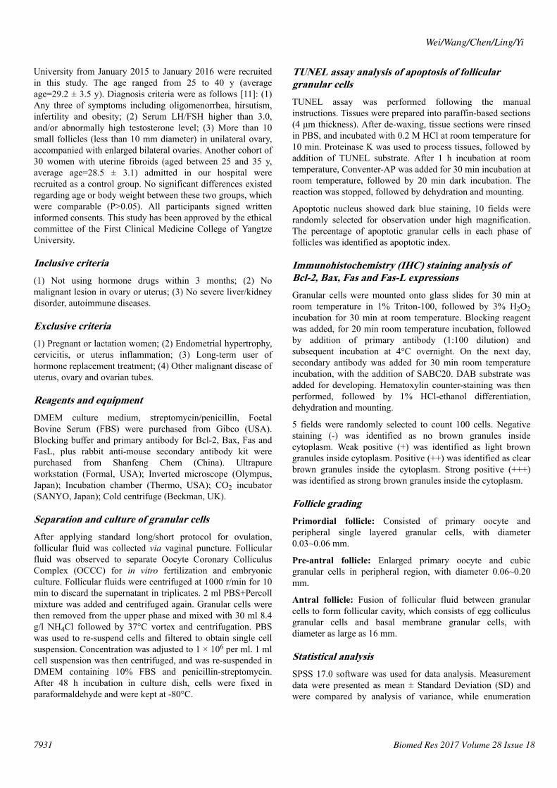
TUNEL assay for analyzing follicular granular cellsapoptosisTUNEL assay was used to measure the apoptotic index offollicular granular cells at all phases [13]. Results showedhigher apoptosis index of antral follicles in PCOS groupcompared with that of pre-antral or primordial follicles. Pre-antral follicles had higher apoptosis index than primordialfollicles (P<0.05). At 12 h, 24 h and 48 h, apoptosis indexes ofall phases of follicles were significantly higher in PCOS groupwith extended time (P<0.05). At the same time point, PCOSgroup had higher apoptosis index at all phases of folliclescompared with control group (P<0.05, Figure 1).
Figure 1. Apoptosis of follicular granular cells by TUNEL assay.aP<0.05 compared to PCOS primordial follicle; bP<0.05 comparedto PCOS pre-antral follicle; cP<0.05 compared to controlledprimordial follicle; dP<0.05 compared to controlled pre-antralfollicle; eP<0.05 compared to controlled antral follicle; fP<0.05compared to 12 h group; gP<0.05 compared to 24 h group.
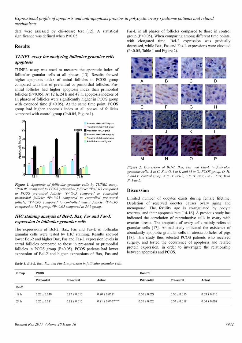
IHC staining analysis of Bcl-2, Bax, Fas and Fas-Lexpression in follicular granular cellsThe expressions of Bcl-2, Bax, Fas and Fas-L in folliculargranular cells were tested by IHC staining. Results showedlower Bcl-2 and higher Bax, Fas and Fas-L expression levels inantral follicles compared to those in pre-antral or primordialfollicles in PCOS group (P<0.05). PCOS patients had lowerexpression of Bcl-2 and higher expressions of Bax, Fas and
Fas-L in all phases of follicles compared to those in controlgroup (P<0.05). When comparing among different time points,with elongated time, Bcl-2 expression was graduallydecreased, while Bax, Fas and Fas-L expressions were elevated(P<0.05, Table 1 and Figure 2).
Figure 2. Expression of Bcl-2, Bax, Fas and Fas-L in folliculargranular cells. A to C, E to G, I to K and M to O: PCOS group. D, H,L and P: control group. A to D: Bcl-2; E to H: Bax; I to L: Fas; M toP: Fas-L.
DiscussionLimited number of oocytes exists during female lifetime.Depletion of reserved oocytes causes ovary aging andmenopause. The fertility age is co-regulated by oocytereserves, and their apoptosis rate [14-16]. A previous study hasindicated the correlation of reproductive cells in ovary withovarian atresia. The apoptosis of ovary cells mainly refers togranular cells [17]. Animal study indicated the existence ofabundantly apoptotic granular cells in atresia follicles of pigs[18]. This study thus selected PCOS patients who receivedsurgery, and tested the occurrence of apoptosis and relatedprotein expression, in order to investigate the relationshipbetween apoptosis and PCOS.
Table 1. Bcl-2, Bax, Fas and Fas-L expression in follicular granular cells.
Group PCOS Control
Primordial Pre-antral Antral Primordial Pre-antral Antral
Bcl-2
12 h 0.28 ± 0.010 0.27 ± 0.013 0.26 ± 0.012d 0.36 ± 0.027 0.35 ± 0.015 0.33 ± 0.016
24 h 0.25 ± 0.021 0.22 ± 0.015 0.21 ± 0.010abcdef 0.35 ± 0.028 0.34 ± 0.017 0.34 ± 0.009
Expressional profile of apoptosis and anti-apoptosis proteins in polycystic ovary syndrome patients and relatedmechanisms
7932Biomed Res 2017 Volume 28 Issue 18
48 h 0.28 ± 0.027 0.20 ± 0.021 0.15 ± 0.097abcdefg 0.36 ± 0.031 0.35 ± 0.009 0.33 ± 0.021
Bax
12 h 0.26 ± 0.017 0.29 ± 0.026 0.31 ± 0.030 d 0.17 ± 0.012 0.18 ± 0.013 0.19 ± 0.013
24 h 0.29 ± 0.018 0.35 ± 0.031 0.46 ± 0.035abcdef 0.18 ± 0.005 0.18 ± 0.021 0.19 ± 0.021
48 h 0.34 ± 0.023 0.39 ± 0.046 0.53 ± 0.044abcdefg 0.17 ± 0.018 0.17 ± 0.028 0.18 ± 0.042
Fas
12 h 0.32 ± 0.012 0.38 ± 0.016 0.41 ± 0.018d 0.14 ± 0.008 0.16 ± 0.015 0.17 ± 0.021
24 h 0.42 ± 0.015 0.51 ± 0.018 0.57 ± 0.022abcdef 0.14 ± 0.010 0.16 ± 0.018 0.18 ± 0.003
48 h 0.56 ± 0.031 0.63 ± 0.025 0.71 ± 0.027abcdefg 0.15 ± 0.004 0.15 ± 0.021 0.17 ± 0.034
Fas-L
12 h 0.26 ± 0.011 0.30 ± 0.013 0.39 ± 0.031d 0.13 ± 0.007 0.14 ± 0.019 0.16 ± 0.024
24 h 0.31 ± 0.013 0.41 ± 0.015 0.48 ± 0.022abcdef 0.14 ± 0.002 0.13 ± 0.027 0.17 ± 0.008
48 h 0.42 ± 0.017 0.47 ± 0.019 0.58 ± 0.032abcdefg 0.13 ± 0.015 0.14 ± 0.011 0.16 ± 0.033
Note: aP<0.05 compared to PCOS primordial follicle; bP<0.05 compared to PCOS pre-antral follicle; cP<0.05 compared to controlled primordial follicle; dP<0.05 comparedto controlled pre-antral follicle; eP<0.05 compared to controlled antral follicle; fP<0.05 compared to 12 h group; gP<0.05 compared to 24 h group.
After artificial ovulation, granular cells were separated forroutine culturing. TUNEL assay was used to test the apoptosisof all follicular granular cells. Antral follicles in PCOS grouphad higher apoptosis index compared with pre-antral orprimordial follicles. With elongated time (12 h, 24 h and 48 h),apoptosis indexes at all phases of follicles in PCOS group weregradually elevated. Moreover, PCOS group had higherapoptosis index at all phase of follicles compared with controlgroup. These results suggested significantly elevated apoptosisof antral follicles in PCOS patients, which is consistent with aprevious study showing the correlation between PCOSoccurrence and apoptosis of ovary follicular granular cells[19]. Follicular granular cells of PCOS patients showedabnormal differentiation, plus the enlargement of lateralgranular cells at the side of oocytes, with filling ofendoplasmic reticulum in cytoplasm, suggesting the potencyfor hormone synthesis of such granular cells. PCOS patientshad undifferentiated pre-antral follicles, suggesting apoptosisof granular cells in those follicles [20]. In addition, thesepatients also had dysregulation of FSH-granular cell axis,which causes apoptosis of follicular granular cells, andinability of androgen to form estradiol, leading to elevation ofbody androgen level, impairment of follicle selectiveadvantage, as well as aggregation of small follicles to cysticformation inside ovary [21]. In PCOS rat model, significantlypotentiated apoptosis of ovary follicular granular cells was alsoobserved, which is consistent with the present study [22].
To further elaborate the changes of apoptosis related proteinsin PCOS patients, this study revealed lower Bcl-2 expression,and higher Bax, Fas and Fas-L expressions in antral follicles inPCOS group compared to those in pre-antral or primordialfollicles. Meanwhile, PCOS group had lower Bcl-2 and higherBax, Fas and Fas-L in all phases of follicles compared withcontrol group (P<0.05). With elongated time (12 h, 24 h and 48
h), Bcl-2 expression was gradually decreased, while Bax, Fasand Fas-L expressions were elevated, indicating theinvolvement of all these proteins in the apoptosis of ovaryfollicular granular cells. PCOS patients had elevated apoptosisof follicular granular cells, especially in antral follicles.Previous studies showed increased apoptosis of granular cellsespecially in antral phase by androgen intervention for humanovary follicular cells, accompanied with lower serum Bcl-2levels in PCOS patients [23,24]. Under the stimulus of highlevel androgen, ovary follicles arrest the development andelimination of atresia, thus entering recruitment of antralfollicles [25]. Literatures have reported that elevatedexpression of Fas and Fas-L in follicular granular cellsindicated the activation of cells, and gradual apoptosis. A basicstudy using human granular cells and luteinizing cells foundFas-mediated facilitation of apoptosis of those cells. Mousestudy showed high expression of Fas in atresia follicles aftertreatment of chorionic gonadotropin for 72 h [26]. This studyindicated down-regulation of Bcl-2 and up-regulation of Bax,Fas and Fas-L in PCOS patients, leading to facilitation of theapoptosis of ovary follicular granular cells especially antralfollicles. However, the major limitation of the present studywas the relatively small sample size. In the future, large cohortclinical study is required to confirm these findings.
In conclusion, elevated apoptosis is observed in ovaryfollicular granular cells of PCOS patients, which isdemonstrated by reduced Bcl-2 expression and elevatedexpressions of Bax, Fas and Fas-L, indicating the participationof these molecules in the apoptosis of ovary granular cells inPCOS patients. These data suggest that therapeuticallytargeting apoptosis might be a novel approach for treatingpatients with PCOS.
Wei/Wang/Chen/Ling/Yi
7933 Biomed Res 2017 Volume 28 Issue 18
References1. Glintborg D, Andersen M. An update on the pathogenesis,
inflammation, and metabolism in hirsutism and polycysticovary syndrome. Gynecol Endocrinol 2010; 26: 281-296.
2. Glintborg D. Endocrine and metabolic characteristics inpolycystic ovary syndrome. Dan Med J 2016; 63.
3. Yin J, Gao Z, He Q, Zhou D, Guo Z, Ye J. Role of hypoxiain obesity-induced disorders of glucose and lipidmetabolism in adipose tissue. Am J Physiol EndocrinolMetab 2009; 296: 333-342.
4. Maciel GA, Baracat EC, Benda JA, Markham SM,Hensinger K, Chang RJ, Erickson GF. Stockpiling oftransitional and classic primary follicles in ovaries ofwomen with polycystic ovary syndrome. J Clin EndocrinolMetab 2004; 89: 5321-5327.
5. Bas D, Abramovich D, Hernandez F, and Tesone M.Altered expression of Bcl-2 and Bax in follicles withindehydroepiandrosterone-induced polycystic ovaries in rats.Cell Biol Int 2011; 35: 423-429.
6. Das M, Djahanbakhch O, Hacihanefioglu B, Saridogan E,Ikram M, Ghali L, Raveendran M, Storey A. Granulosa cellsurvival and proliferation are altered in polycystic ovarysyndrome. J Clin Endocrinol Metab 2008; 93: 881-887.
7. Azziz R, Carmina E, Dewailly D, Diamanti-Kandarakis E,Escobar-Morreale HF, Futterweit W, Janssen OE, LegroRS, Norman RJ, Taylor AE, Witchel SF. Positionsstatement: criteria for defining polycystic ovary syndromeas a predominantly hyperandrogenic syndrome: anAndrogen Excess Society guideline. J Clin EndocrinolMetab 2006; 91: 4237-4245.
8. Cataldo NA, Dumesic DA, Goldsmith PC, Jaffe RB.Immunolocalization of Fas and Fas ligand in the ovaries ofwomen with polycystic ovary syndrome: relationship toapoptosis. Hum Reprod 2000; 15: 1889-1897.
9. Onalan G, Selam B, Baran Y, Cincik M, Onalan R, GunduzU, Ural AU, Pabuccu R. Serum and follicular fluid levels ofsoluble Fas, soluble Fas ligand and apoptosis of luteinizedgranulosa cells in PCOS patients undergoing IVF. HumReprod 2005; 20: 2391-2395.
10. Cui W, Ma J, Wang X, Yang W, Zhang J, Ji Q. Free fattyacid induces endoplasmic reticulum stress and apoptosis ofbeta-cells by Ca2+/calpain-2 pathways. PLoS One 2013; 8:59921.
11. Sheehan MT. Polycystic ovarian syndrome: diagnosis andmanagement. Clin Med Res 2004; 2: 13-27.
12. Tomlinson G, Chimalapati S, Pollard T, Lapp T, Cohen J,Camberlein E, Stafford S, Periselneris J, Aldridge C,Vollmer W, Picard C, Casanova JL, Noursadeghi M, BrownJ. TLR-mediated inflammatory responses to Streptococcuspneumoniae are highly dependent on surface expression ofbacterial lipoproteins. J Immunol 2014; 193: 3736-3745.
13. Yu YS, Sui HS, Han ZB, Li W, Luo MJ, Tan JH. Apoptosisin granulosa cells during follicular atresia: relationship withsteroids and insulin-like growth factors. Cell Res 2004; 14:341-346.
14. Teede H, Deeks A, Moran L. Polycystic ovary syndrome: acomplex condition with psychological, reproductive andmetabolic manifestations that impacts on health across thelifespan. BMC Med 2010; 8: 41.
15. Kose SA, Naziroglu M. N-acetyl cysteine reduces oxidativetoxicity, apoptosis, and calcium entry through TRPV1channels in the neutrophils of patients with polycysticovary syndrome. Free Radic Res 2015; 49: 338-346.
16. Cai L, Ma X, Liu S, Liu J, Wang W, Cui Y, Ding W, Mao Y,Chen H, Huang J, Zhou Z. Effects of upregulation of Hsp27expression on oocyte development and maturation derivedfrom polycystic ovary syndrome. PLoS One 2013; 8:83402.
17. Rzepczynska IJ, Foyouzi N, Piotrowski PC, Celik-OzenciC, Cress A, Duleba AJ. Antioxidants induce apoptosis ofrat ovarian theca-interstitial cells. Biol Reprod 2011; 84:162-166.
18. Honnma H, Endo T, Henmi H, Nagasawa K, Baba T,Yamazaki K, Kitajima Y, Hayashi T, Manase K, Saito T.Altered expression of Fas/Fas ligand/caspase 8 andmembrane type 1-matrix metalloproteinase in atreticfollicles within dehydroepiandrosterone-induced polycysticovaries in rats. Apoptosis 2006; 11: 1525-1533.
19. Yan L, Wang A, Chen L, Shang W, Li M, Zhao Y.Expression of apoptosis-related genes in the endometriumof polycystic ovary syndrome patients during the windowof implantation. Gene 2012; 506: 350-354.
20. Skalba P. Progress in gynecological endocrinology. GinekolPol 2008; 79: 877-881.
21. Jonard S, Dewailly D. The follicular excess in polycysticovaries, due to intra-ovarian hyperandrogenism, may be themain culprit for the follicular arrest. Hum Reprod Update2004; 10: 107-117.
22. Figueroa F, Motta A, Acosta M, Mohamed F, Oliveros L,Forneris M. Role of macrophage secretions on ratpolycystic ovary: its effect on apoptosis. Reproduction2015; 150: 437-448.
23. Garcia-Velasco JA, Arici A. Apoptosis and thepathogenesis of endometriosis. Semin Reprod Med 2003;21: 165-172.
24. Filali M, Frydman N, Belot MP, Hesters L, Gaudin F,Tachdjian G, Emilie D, Frydman R, Machelon V. Oocytein-vitro maturation: BCL2 mRNA content in cumulus cellsreflects oocyte competency. Reprod Biomed Online 2009;4: 4309.
25. Kalaiselvi VS, Krishna GP. The anti mullerian hormone- anovel marker for assessing the ovarian reserve in womenwith regular menstrual cycles. J Clin Diagn Res 2012; 6:1636-1639.
26. Dharma SJ, Kelkar RL, Nandedkar TD. Fas and Fas ligandprotein and mRNA in normal and atretic mouse ovarianfollicles. Reproduction 2003; 126: 783-789.
Expressional profile of apoptosis and anti-apoptosis proteins in polycystic ovary syndrome patients and relatedmechanisms
7934Biomed Res 2017 Volume 28 Issue 18

Department of Obstetrics and Gynecology
The First Clinical Medicine College,
Yangtze University
Jingzhou
Hubei
PR China
Wei/Wang/Chen/Ling/Yi
7935 Biomed Res 2017 Volume 28 Issue 18
*Correspondence toHua Wei