Epidemiology of Streptococcus agalactiae and ... · Epidemiology of Streptococcus agalactiae and...
18
Epidemiology of Streptococcus agalactiae and Streptococcosis in Tilapia Fish Carlos Iregui, Paola Barato, Alba Rey, Gersson Vasquez Veterinary Pathobiology Group, Faculty of Veterinary Medicine Universidad Nacional de Colombia Noel Verjan Veterinary Pathobiology Group, Faculty of Veterinary Medicine Universidad Nacional de Colombia Research Group in Immunobiology and Pathogenesis Universidad del Tolima, Colombia
Transcript of Epidemiology of Streptococcus agalactiae and ... · Epidemiology of Streptococcus agalactiae and...
Epidemiology of Streptococcus agalactiae and Streptococcosis in Tilapia Fish
Carlos Iregui, Paola Barato, Alba Rey, Gersson Vasquez Veterinary Pathobiology Group, Faculty of Veterinary Medicine
Universidad Nacional de Colombia
Noel Verjan
Veterinary Pathobiology Group, Faculty of Veterinary Medicine Universidad Nacional de Colombia
Research Group in Immunobiology and Pathogenesis Universidad del Tolima, Colombia
1 Introduction
Tilapia (Oreochromis sp.) are freshwater fish species belonging to the Cichlidae family, native to Africa and Middle East regions (Trewaves, 1983), and the third largest family of bony fish. Illustrations from Egyptian tombs suggest that Nile tilapia (Oreochromis niloticus) have been cultivated for over 4,000 years (Balarin & Hatton, 1979). However, it was not until the second half of the twentieth century that tilapia were introduced into many tropical, sub-tropical and temperate regions of the world (Pillay, 1990), and it is currently one of the most cultured fish species contributing to the food fish industry worldwide (FAO, 2004).
The estimated world production of tilapia by the year 2012 was 2.5 million metric tons (MT), that when compared to 28,260 MT in 1970 indicates a significant and steadily increased growth rate. Similar-ly, the market value of farmed tilapia increased from about 154 million USD in 1984 to 4,000 million USD in 2010 (FAO, 2012). Among the largest tilapia producers are China, with 706,585 MT and ac-counting for almost 50% of the total global production, followed by Egypt with 167,735 MT, and Philip-pines with 122,390 MT, whereas Colombia was located in the ninth place with 24,000 MT (FAO, 2004; El-Sayed, 2006), although latest report indicates the tilapia production in Colombia has almost doubled to 44,000 MT by 2012 (CCI - MADR, 2012).
Some of the features that have made tilapia an advantageous fish species for culture are: rapid growth rate, firm and white muscle, the ability to survive in poor water conditions, and high reproductive success with limited requirements during incubation (Nandlal & Pickering, 2004). Last but not least, ti-lapia belongs to the base of the food chain, which makes them suitable to consume low cost and wide range of food sources with reduced ecological impact (Beveridge & Baird, 1998).
Initially it was thought that tilapia were more resistant to bacterial, parasitic, fungal and viral dis-eases compared with other farmed fish species (Popma et al., 1999; Klessius et al., 2008; Amal et al., 2011). More recently, it has been shown that tilapia is also susceptible to many bacterial, parasitic, nutri-tional and fungal diseases. The most common bacterial pathogens of tilapia include Streptococcus sp., Flavobacterium columnare, Aeromonas hydrophila and Edwardsiella tarda; and within parasitic infec-tions are found Ichthyophthirius multifiliis and Trichodina sp., (Iregui et al., 2004; Klessius et al., 2008). Of note, infections by the genus Streptococcus sp., are perhaps the most important health problem in farmed tilapia, with serious economic losses that were estimated at 250 million USD in 2008 globally (Klesius et al., 2008). The most significant Streptococcus species that cause disease in the tilapia industry worldwide are S. iniae, S. agalactiae, S. dysgalactiae and Lactococcus garviae (Evans et al., 2006; Netto et al., 2011), while in Colombia Streptococcus agalactiae is the most important bacterial pathogen of tilapia (Iregui et al., 2004; Jimenez et al., 2007), up to date we do not have any evidence for other Gram-positive cocci causing disease in this fish species. Thus, this chapter only deals with the infection and disease caused by Streptococcus agalactiae.
2 Streptococcus
The Genus Streptococcus contains Gram-positive spherical bacteria less than 2 microns diameters that typically grow in pairs and form chains when grown in liquid media. Most members are facultative an-aerobes, meaning that they can grow in absent or limited oxygen conditions; they are catalase negative with varying nutritional requirements, which reflects adaptation as commensals or parasites. Commonly,
Streptococcus is grown in culture media supplemented with red blood cells that may allow a preliminary classification based on the production of hemolysis, which can be classified as β-hemolytic, α-hemolytic (approx. 1% of cases) or non-hemolytic strains (Edwards & Nizet, 2011). In Colombia, the entire Strep-tococcus agalactiae strains isolated from tilapia by the Veterinary Pathobiology Group at the Universidad Nacional de Colombia (VPG, UNC) have been found to be non-hemolytic and belonging to the serotype Ib. The identification scheme followed in our laboratory is based on colony characteristics, hemolytic properties, carbohydrate and protein antigen composition, sugar fermentation and other biochemical reac-tions. More recently, DNA sequencing of 16 and 23S rRNA genes, in addition to molecular typing at the species level has been carried out using multilocus enzyme electropherotype and multilocus sequence typing (MLST) (Unpublished data).
The majority of pathogenic streptococci have a serologically reactive carbohydrate sheath that is antigenically different from one species or group of species to another. These cell wall associated anti-gens are designated A-H and K-V, and are the basis of the Lancefield groups.
2.1 Streptococcus agalactiae and Streptococcosis
Before going further, it is important to clarify the difference between the terms infection and infectious disease. Infection refers to the presence of a microorganism in a host without disease manifestations, whereas infectious disease refers to the expression of clinical signs by the host as a result of the presence and growth of that microorganism within its tissues. Although this explanation seems very simple, it seems to be the cause of most serious mistakes in the livestock practices, daily medical care and research. In this text, Streptococcosis is a word that implies a disease caused by S. agalactiae infection.
2.1.1 Disease (Streptococcosis) by S. agalactiae in Humans, Bovines and Fish
S. agalactiae is the lone member of the Lancefield group B (GBS) and a pathogen that frequently affects three animal species: humans, cattle and fish, in which it is responsible for several pathologies such as neonatal meningitis and sepsis (early-onset and late-onset disease) that is is the primary outcome in hu-man being, mastitis in cattle and meningoencephalitis, epicarditis and choroiditis in fish (Evans et al., 2002; Mitchell, 2003; Oliveira et al., 2006; Mian et al., 2009). Sporadically, S. agalactiae is responsible for disease in many other hosts such as chickens, camels, dogs, horses, cats, bottlenose dolphins, frogs, hamsters, mice, monkeys and otters (Elliot et al., 1990; Yildirim et al., 2002a, b; Hetzel et al., 2003; Johri et al., 2006). S. agalactiae is also considered an obligate parasite of the ruminant mammary gland epithe-lium and tissues; in human S. agalactiae is primarily a commensal bacterium of the gastrointestinal and genitourinary tracts (Timoney, 2010), while the status of S. agalactiae in fish has not yet been elucidated.
There are substantial differences in biochemical, serological and molecular features among S. aga-lactiae strains isolated from human and bovine species, in which this pathogen has been most studied (Pattison et al., 1955; Finch & Martin, 1984). For many years the relationships between GBS strains of human and bovine have been investigated, and so far there is no solid evidence to prove that cattle serve as a reservoir for GBS strains that cause human disease, which is corroborated by the low transmission of GBS from cattle to humans (Finch & Martin, 1984; Sukhanand et al., 2005). Controversial results about if GBS belonging to the hypervirulent clonal complex 17 isolated from humans, are derived from a com-mon bovine GBS ancestor, have been described based on molecular characterization with 15 housekeep-ing genes. Bovine CC67 and human CC17 differed in alleles of seven out of eight additional MLST housekeeping genes examined and they are more distantly related than predicted by analysis based on the
seven MLST gene loci and do not support the conclusion that the highly virulent human CC17 emerged recently form bovine CC67 (Sorensen et al., 2010; Timoney, 2010).
The initial report of Streptococcus sp., in fish was made in rainbow trout (Onchorynchus mykiss) cultured in Japan in 1957 (Hoshina et al., 1958). Subsequently, two outbreaks of disease in golden shiner (Notemigonus crysoleucas) were documented in USA (Robinson & Meyer, 1966). Plumb et al. (1974) isolated Streptococcus sp. in more than 50% of eight different fish species dying during an epidemic out-break in estuarine beaches of Florida, Alabama and the Gulf of Mexico in the U.S. in 1972. In tilapia, the first report of streptococcosis was made by Al-Harbi (1994).
The group B streptococci and particularly S. agalactiae cause significant mortality and morbidity in a wide variety of freshwater and saltwater fish species worldwide (Robinson & Meyer, 1966; Plumb et al., 1974; Evans et al., 2002). There are reports of this pathogen in the U.S, Israel, Japan, Kuwait, Thai-land, Honduras, Costa Rica, Brazil and Colombia. In addition, this microorganism has been isolated from 17 species of fish including rainbow trout, seabream, tilapia, yellowtail, catfish, croaker, killifish, menha-den, mullet, and silver pomfret (Wilkinson et al. 1973; Plumb et al. 1974; Rasheed & Plumb, 1984; Berry et al., 1989; Elliot et al., 1990; Eldar et al., 1995; Vandamme et al., 1997; Evans et al., 2002; Duremdez et al., 2004; Suanyuk et al., 2005; Salvador et al., 2005; Evans et al., 2006; Kim et al., 2007; Garcia et al., 2008). S. agalactiae is so far the only species of Streptococcus isolated in tilapia farms with strepto-coccosis in Colombia (Jimenez et al., 2007).
Conventional serotyping and molecular typing, including multiplex PCR and multilocus sequence typing (MLST), of various capsular strains isolated from fish, dolphins, human and bovine showed that S. agalactiae strains isolated from dolphins and fish in Kuwait, share the same sequence type ST-7 and se-rological type described for human strains that caused neonatal infection in Japan (Evans et al., 2008; Pereira et al., 2010). Although the ability of this pathogen to cross the species barrier, meaning that S. agalactiae of human origin could infect animals, or vice versa, is currently unknown, Pereira et al., (2010) demonstrated that S. agalactiae of human and bovine origin can infect fish and that genetic simi-larity between strains (i.e. a clonal relationship) is not a prerequisite for this phenomenon to occur.
Multilocus sequence typing has allowed the identification of at least four subpopulations of S. aga-lactiae to be present among aquatic isolates; they are the sequence types (ST) ST260, ST261, ST283 and ST491. Both the sequence type ST283, serotype III-4 and its novel single locus variant ST491, were de-tected in fish from Southeast Asia and shared a 3-set identical genotype to the emerging ST283 clone as-sociated with invasive disease of adult humans in Asia. The ST283 serotype Ia, a subpopulation that is normally associated with human carriage, was found in gray seals, suggesting that human effluents may contribute to microbial pollution of surface waters and exposure of sea mammals to human pathogens. The subpopulations ST260 and ST261 serotype Ib consist of non-hemolytic isolates and belong to fish-associated clonal complexes that have never been reported in humans (Delannoy et al., 2013). Finally, the human pathogen S. agalactiae strain ST7, serotype Ia was also detected in fish from Asia (Evans et al., 2008). These findings indicate that some S. agalactiae strains may be an anthropozoonotic hazard.
A number of S. agalactiae isolates from tilapia farmed in Colombia were subjected to routine mi-crobiological tests, molecular (16S RNA gene sequencing, amplification of surface protein-coding genes and mobile genetic elements) and phylogenetic (PFGE and MLST) analyses. From 25 evaluated isolates (period from 2003 to 2011), it was found that all strains corresponded to non-hemolytic mucoid colonies of S. agalactiae serotype Ib, which did not amplify for surface protein-coding genes (bca, rib, alp1 epsi-lon, alp2/3, alp4, and bac) nor to mobile genetic elements for 3 isolates (IS1381, IS861, IS1548, GBSi1, ISSa4, ISSag2, ISSag1). In addition, all 25 strains were ST260 and formed two pulsotypes by PFGE, one
of them grouped 24 isolates and the other one slightly differed but has 95% similarity (Barato et al., 2014, submitted). These results indicate that Colombian´s S. agalactiae isolates have high similarity to strains that affect only fish and had been described in America, Europe and Australia (Delannoy et al., 2013).
3 Pathology and Pathogenesis
The main clinical signs observed in tilapia with streptococcosis by S. agalactiae are loss of appetite, uni-lateral or bilateral exophthalmos, eye hemorrhages, corneal opacity, distended abdomen, curvature of the spinal cord, stiffness, erratic swimming, and bleeding at the base of the fins; some animals may have dif-ficulty to breath, however, other may not show clinical signs before death (Yanong & Francis-Floyd, 2002; Pulido et al., 2004).
Gross findings in tilapia with streptococcosis are the accumulation of bloody fluid in the ab-dominal cavity (hemorrhagic ascites), mucous content with reddish-brown color in the intestine, pale and sometimes enlarged liver, splenomegaly, deposition of a fibrinoid material on the epicardium, and a hem-orrhagic brownish appearance of the retro-bulbar tissue and meninges (Pulido et al., 2004; Zamri-Saad et al., 2010).
Microscopically, most tilapias develop a primary inflammatory response of mononuclear cells with the subsequent formation of granulomatous nodules. Lesions include severe meningoencephalitis that can be hemorrhagic and/or granulomatous in nature with large areas of encephalomalacia (softening of the brain tissue); similar tissue lesions are found in the choroid, sclera and the eyeball. Inflammation may be present at different degrees on the epicardium, whereas other organs are less frequently involved (Pulido et al., 2004).
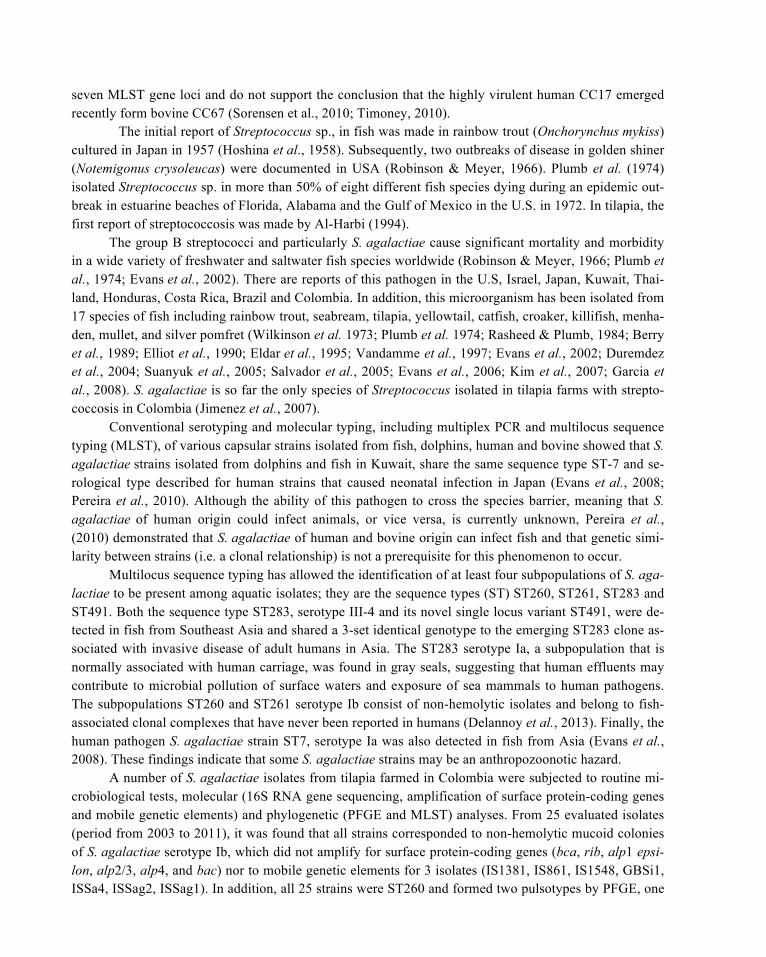
Recognizing the entry route of a systemic pathogen such as S. agalactiae in tilapia, may provide a better understanding of the pathogenesis. In addition, this information is essential to develop novel pre-vention and control measures against the microorganism. To this end, we setup an experiment where ju-venile tilapia (10g-body-weight) were infected with S. agalactiae by two different routes: first by dipping the fish in water with a high concentration of the microorganism (8.5 x 10⁷ CFU/0.1ml), the fish in this group underwent mucus removal and skin (epidermis) scraping before infection. The second group of fish was subjected to an intragastric inoculation with bacteria (7 x 10⁷ CFU/0.1ml) by gavages. The animals in both groups were euthanized periodically beginning at 30 min up to 96 hours post-infection (hpi) and the tissues were collected and processed for indirect immunoperoxidase technique (IIP) to monitor the patho-gen under the microscope.
The result of those experiments showed that the main route of entry of S. agalactiae in tilapia was the gastrointestinal tract regardless of the route of exposure (oral vs. immersion). Of note, S. agalactiae was not consistently found on the skin of fish infected by the immersion route, and after 48hpi it was no more possible to detect S. agalactiae in this organ despite the fact that the fish developed systemic infec-tion. Based on these findings, it was concluded that invasion of S. agalactiae in tilapia through skin or gills is of low frequency, in addition, in our experience with the natural disease by this microorganism, the skin, subcutaneous tissue and muscles are little affected. Interestingly, infection by both oral and im-mersion routes caused severe mucus secretion in the gastric and intestinal lumen of tilapia exposed to S. agalactiae when compared to non-infected control fish; the mucus secretion indicates that an immediate host defense response against the pathogen was stimulated, wherein the mucus substance forms a biologi-
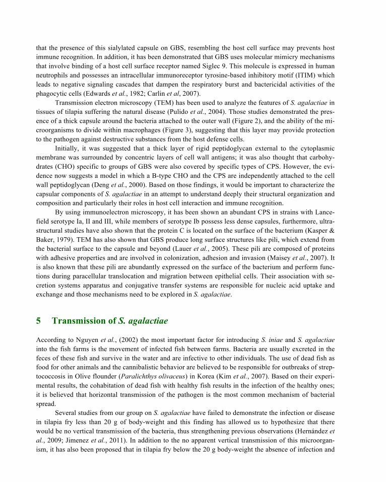
cal mesh which may trap large amounts of bacteria (Figure 1). Under the above experimental conditions some bacteria in the oral (0.5h) or immersion (1h) treated fish had reached the gastric and intestinal epi-thelium despite the abundant mucus secretion. This would indicate that although S. agalactiae can be found in large quantities in the water with easy access to the skin, the main route of entry is the gastroin-testinal tissue of tilapia. Our findings also suggest that S. agalactiae is able to replicate in the lumen of the gastrointestinal tract and perhaps this replication process is a major pathogenic mechanism that may allow permanent colonization of the mucosal epithelium (Comas, 2005; Iregui et al., 2014, Submitted).
Figure 1: Immunohistochemical detection of S. agalactiae in the small intestine of tilapia hybrid experimentally infected by immersion route. Abundant mucus (M) and cell debris are present in the intestinal lumen where the mucus substance forms a biological mesh which appears to trap large amounts of bacteria (arrows).
4 Interaction of S. agalactiae with Host Tissues and Some Virulence Factors
In human infections by S. agalactiae, the amount of group B streptococci transmitted from mother to newborn is considered an important risk factor to infection and disease progression. Infants born from highly infected mothers are more likely to acquire the bacteria on their mucosal surfaces than newborn
from mothers with low colony counts at the time of delivery (Ancona et al., 1980; Jones et al., 1984). Subsequently, the first step that any pathogen should meet during interaction with the host is the adhesion to the host cell surface where the entry process takes place, usually at a mucosal surface. In infections by GBS it is suggested that a microbial alpha C protein mediates the adhesion of the microorganism to epi-thelial cells of the cervix of the woman. In addition, it has been proposed that the bacterial cell wall-associated lipoteichoic acid, given their amphiphilic properties, could mediate low affinity interactions of GBS with host epithelial cells, whereas higher affinity interactions are mediated by hydrophobic surface proteins. Once the pathogen has invaded and traversed the epithelial cell layer, high affinity adhesions are largely mediated by extracellular matrix components such as fibronectin, fibrinogen and laminin; degra-dation of glycosaminoglycans like sulfated hyaluronan and chrondroitin sulfate by secreted hydrolytic enzymes from the pathogen may favor bacterial replication. The capsular polysaccharide has essential role in avoiding the immune system during invasion to different target tissues. It provides antiphagocytic protection by impairing surface deposition of opsonically active complement C3 on the bacterial surface (Maisey et al. 2008). Also, capsular sialic acid plays a role in molecular mimicry of GBS immune avoid-ance (Carlin et al, 2007). Finally, capsule permits resistance to lysis and the beginning of an intracellular stage that facilitates the progression from local to a systemic infection (Baron et al., 2004; Baron et al., 2007).
Growth of GBS in human tissues is attributed to multiple bacterial products that contribute to its virulence, perhaps among the most important are the β-hemolysins that produce lysis of epithelial cells and erythrocytes from different animal species (Weiser & Rubens, 1987). GBS also produces peptidases, enzymes that inactivate the complement component C5a by cleavage of a peptide at the carboxyl termi-nus (Bohnsack et al., 1991). The hyaluronate lyase which cleaves sulfated hyaluronan and chondroitin sulfate is suggested to promote bacterial spread through host tissues, whereas the CAMP factor lyses host cells (co-hemolysin) and causes direct tissue injury. In bovine GBS isolates, a diverse and large number of proteins that are often encoded on pathogenicity islands, are coordinately expressed with capsule com-ponents on the surface of S. agalactiae and seem to have important roles in adhesion, invasion, iron bind-ing, metabolism, transport, and inhibition of phagocytosis (Lindahl et al., 2005).
The capsular polysaccharide (CPS) of many GBS is a virulence factor that seems to play a signifi-cant role after S. agalactiae has traversed the epithelial surfaces and begins its systemic dissemination into the host. Ten serotypes of GBS have been identified based on the CPS (serotypes Ia, Ib, and II-IX), which vary depending on the arrangement of four sugars into a unique repeated unit and are thought to play a key role in virulence (Cieslewicz et al., 2005; Johri et al., 2006). Horizontal transfer of genes cod-ing for capsule biosynthesis accounts for diversity in capsule serotype (Martins et al., 2010; Timoney, 2010), whereas conjugative transfer of large segments of chromosomal DNA among strains of S. agalac-tiae (Brochet et al., 2008) accounts for both strain diversity and the emergence of clonal complexes with combinations of virulence factors and capsular types that are suited to a particular host niche (Timoney, 2010).
The properties of the GBS capsule have been investigated most intensively in members of the sero-type III. A critical element in the epitope of this capsular type is the sialic acid, which appears to confer protection to the microorganism against host immune mechanisms. After treatment with sialidase, the CPS is unable to induce protective antibodies against GBS, however, serotype III GBS treated with sial-idase are apparently more effectively opsonized by complement components (alternative pathway) and are more rapidly engulfed by human PMN in vitro (Edwards et al., 1982; Shigeoka et al., 1983). GBS possess a highly conserved α2 → 3 sialic acid terminal on the capsule that is identical to a sugar epitope widely distributed over the surface of all mammalian cells (Angata & Varki, 2002) and it is suggested
that the presence of this sialylated capsule on GBS, resembling the host cell surface may prevents host immune recognition. In addition, it has been demonstrated that GBS uses molecular mimicry mechanisms that involve binding of a host cell surface receptor named Siglec 9. This molecule is expressed in human neutrophils and possesses an intracellular immunoreceptor tyrosine-based inhibitory motif (ITIM) which leads to negative signaling cascades that dampen the respiratory burst and bactericidal activities of the phagocytic cells (Edwards et al., 1982; Carlin et al, 2007).
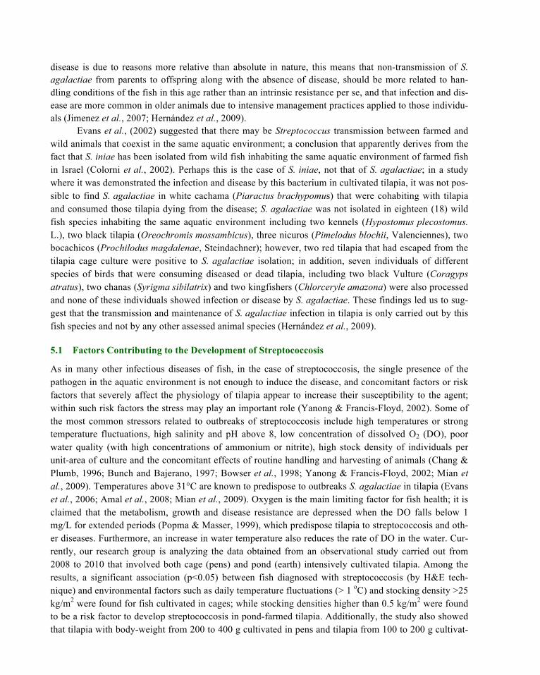
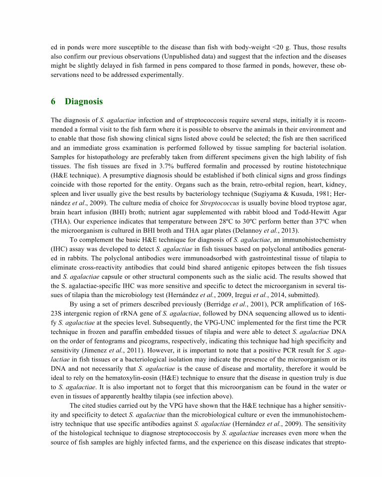
Transmission electron microscopy (TEM) has been used to analyze the features of S. agalactiae in tissues of tilapia suffering the natural disease (Pulido et al., 2004). Those studies demonstrated the pres-ence of a thick capsule around the bacteria attached to the outer wall (Figure 2), and the ability of the mi-croorganisms to divide within macrophages (Figure 3), suggesting that this layer may provide protection to the pathogen against destructive substances from the host defense cells.
Initially, it was suggested that a thick layer of rigid peptidoglycan external to the cytoplasmic membrane was surrounded by concentric layers of cell wall antigens; it was also thought that carbohy-drates (CHO) specific to groups of GBS were also covered by specific types of CPS. However, the evi-dence now suggests a model in which a B-type CHO and the CPS are independently attached to the cell wall peptidoglycan (Deng et al., 2000). Based on those findings, it would be important to characterize the capsular components of S. agalactiae in an attempt to understand deeply their structural organization and composition and particularly their roles in host cell interaction and immune recognition.
By using immunoelectron microscopy, it has been shown an abundant CPS in strains with Lance-field serotype Ia, II and III, while members of serotype Ib possess less dense capsules, furthermore, ultra-structural studies have also shown that the protein C is located on the surface of the bacterium (Kasper & Baker, 1979). TEM has also shown that GBS produce long surface structures like pili, which extend from the bacterial surface to the capsule and beyond (Lauer et al., 2005). These pili are composed of proteins with adhesive properties and are involved in colonization, adhesion and invasion (Maisey et al., 2007). It is also known that these pili are abundantly expressed on the surface of the bacterium and perform func-tions during paracellular translocation and migration between epithelial cells. Their association with se-cretion systems apparatus and conjugative transfer systems are responsible for nucleic acid uptake and exchange and those mechanisms need to be explored in S. agalactiae.
5 Transmission of S. agalactiae
According to Nguyen et al., (2002) the most important factor for introducing S. iniae and S. agalactiae into the fish farms is the movement of infected fish between farms. Bacteria are usually excreted in the feces of these fish and survive in the water and are infective to other individuals. The use of dead fish as food for other animals and the cannibalistic behavior are believed to be responsible for outbreaks of strep-tococcosis in Olive flounder (Paralichthys olivaceus) in Korea (Kim et al., 2007). Based on their experi-mental results, the cohabitation of dead fish with healthy fish results in the infection of the healthy ones; it is believed that horizontal transmission of the pathogen is the most common mechanism of bacterial spread.
Several studies from our group on S. agalactiae have failed to demonstrate the infection or disease in tilapia fry less than 20 g of body-weight and this finding has allowed us to hypothesize that there would be no vertical transmission of the bacteria, thus strengthening previous observations (Hernández et al., 2009; Jimenez et al., 2011). In addition to the no apparent vertical transmission of this microorgan-ism, it has also been proposed that in tilapia fry below the 20 g body-weight the absence of infection and

Figure 2: S agalactiae-containing phagosome from tilapia infected macrophages. Note the prominent filamentous capsule of a viable bacterium (→). There are also lysosomes close to the phagosome (►). (Original magnification × 52000) (Used with permission of Revista AquaTic, Pulido et al., 2004).
Figure 3: Close view of the phagosome where features of viable S. agalactaie are revealed, some of them in the process of cell division (→) (Original magnification × 28500) (Used with permission of Revista AquaTic, Pulido et al., 2004).
disease is due to reasons more relative than absolute in nature, this means that non-transmission of S. agalactiae from parents to offspring along with the absence of disease, should be more related to han-dling conditions of the fish in this age rather than an intrinsic resistance per se, and that infection and dis-ease are more common in older animals due to intensive management practices applied to those individu-als (Jimenez et al., 2007; Hernández et al., 2009).
Evans et al., (2002) suggested that there may be Streptococcus transmission between farmed and wild animals that coexist in the same aquatic environment; a conclusion that apparently derives from the fact that S. iniae has been isolated from wild fish inhabiting the same aquatic environment of farmed fish in Israel (Colorni et al., 2002). Perhaps this is the case of S. iniae, not that of S. agalactiae; in a study where it was demonstrated the infection and disease by this bacterium in cultivated tilapia, it was not pos-sible to find S. agalactiae in white cachama (Piaractus brachypomus) that were cohabiting with tilapia and consumed those tilapia dying from the disease; S. agalactiae was not isolated in eighteen (18) wild fish species inhabiting the same aquatic environment including two kennels (Hypostomus plecostomus. L.), two black tilapia (Oreochromis mossambicus), three nicuros (Pimelodus blochii, Valenciennes), two bocachicos (Prochilodus magdalenae, Steindachner); however, two red tilapia that had escaped from the tilapia cage culture were positive to S. agalactiae isolation; in addition, seven individuals of different species of birds that were consuming diseased or dead tilapia, including two black Vulture (Coragyps atratus), two chanas (Syrigma sibilatrix) and two kingfishers (Chlorceryle amazona) were also processed and none of these individuals showed infection or disease by S. agalactiae. These findings led us to sug-gest that the transmission and maintenance of S. agalactiae infection in tilapia is only carried out by this fish species and not by any other assessed animal species (Hernández et al., 2009).
5.1 Factors Contributing to the Development of Streptococcosis
As in many other infectious diseases of fish, in the case of streptococcosis, the single presence of the pathogen in the aquatic environment is not enough to induce the disease, and concomitant factors or risk factors that severely affect the physiology of tilapia appear to increase their susceptibility to the agent; within such risk factors the stress may play an important role (Yanong & Francis-Floyd, 2002). Some of the most common stressors related to outbreaks of streptococcosis include high temperatures or strong temperature fluctuations, high salinity and pH above 8, low concentration of dissolved O2 (DO), poor water quality (with high concentrations of ammonium or nitrite), high stock density of individuals per unit-area of culture and the concomitant effects of routine handling and harvesting of animals (Chang & Plumb, 1996; Bunch and Bajerano, 1997; Bowser et al., 1998; Yanong & Francis-Floyd, 2002; Mian et al., 2009). Temperatures above 31°C are known to predispose to outbreaks S. agalactiae in tilapia (Evans et al., 2006; Amal et al., 2008; Mian et al., 2009). Oxygen is the main limiting factor for fish health; it is claimed that the metabolism, growth and disease resistance are depressed when the DO falls below 1 mg/L for extended periods (Popma & Masser, 1999), which predispose tilapia to streptococcosis and oth-er diseases. Furthermore, an increase in water temperature also reduces the rate of DO in the water. Cur-rently, our research group is analyzing the data obtained from an observational study carried out from 2008 to 2010 that involved both cage (pens) and pond (earth) intensively cultivated tilapia. Among the results, a significant association (p<0.05) between fish diagnosed with streptococcosis (by H&E tech-nique) and environmental factors such as daily temperature fluctuations (> 1 oC) and stocking density >25 kg/m2 were found for fish cultivated in cages; while stocking densities higher than 0.5 kg/m2 were found to be a risk factor to develop streptococcosis in pond-farmed tilapia. Additionally, the study also showed that tilapia with body-weight from 200 to 400 g cultivated in pens and tilapia from 100 to 200 g cultivat-
ed in ponds were more susceptible to the disease than fish with body-weight <20 g. Thus, those results also confirm our previous observations (Unpublished data) and suggest that the infection and the diseases might be slightly delayed in fish farmed in pens compared to those farmed in ponds, however, these ob-servations need to be addressed experimentally.
6 Diagnosis
The diagnosis of S. agalactiae infection and of streptococcosis require several steps, initially it is recom-mended a formal visit to the fish farm where it is possible to observe the animals in their environment and to enable that those fish showing clinical signs listed above could be selected; the fish are then sacrificed and an immediate gross examination is performed followed by tissue sampling for bacterial isolation. Samples for histopathology are preferably taken from different specimens given the high lability of fish tissues. The fish tissues are fixed in 3.7% buffered formalin and processed by routine histotechnique (H&E technique). A presumptive diagnosis should be established if both clinical signs and gross findings coincide with those reported for the entity. Organs such as the brain, retro-orbital region, heart, kidney, spleen and liver usually give the best results by bacteriology technique (Sugiyama & Kusuda, 1981; Her-nández et al., 2009). The culture media of choice for Streptococcus is usually bovine blood tryptose agar, brain heart infusion (BHI) broth; nutrient agar supplemented with rabbit blood and Todd-Hewitt Agar (THA). Our experience indicates that temperature between 28ºC to 30ºC perform better than 37ºC when the microorganism is cultured in BHI broth and THA agar plates (Delannoy et al., 2013).
To complement the basic H&E technique for diagnosis of S. agalactiae, an immunohistochemistry (IHC) assay was developed to detect S. agalactiae in fish tissues based on polyclonal antibodies generat-ed in rabbits. The polyclonal antibodies were immunoadsorbed with gastrointestinal tissue of tilapia to eliminate cross-reactivity antibodies that could bind shared antigenic epitopes between the fish tissues and S. agalactiae capsule or other structural components such as the sialic acid. The results showed that the S. agalactiae-specific IHC was more sensitive and specific to detect the microorganism in several tis-sues of tilapia than the microbiology test (Hernández et al., 2009, Iregui et al., 2014, submitted).
By using a set of primers described previously (Berridge et al., 2001), PCR amplification of 16S-23S intergenic region of rRNA gene of S. agalactiae, followed by DNA sequencing allowed us to identi-fy S. agalactiae at the species level. Subsequently, the VPG-UNC implemented for the first time the PCR technique in frozen and paraffin embedded tissues of tilapia and were able to detect S. agalactiae DNA on the order of fentograms and picograms, respectively, indicating this technique had high specificity and sensitivity (Jimenez et al., 2011). However, it is important to note that a positive PCR result for S. aga-lactiae in fish tissues or a bacteriological isolation may indicate the presence of the microorganism or its DNA and not necessarily that S. agalactiae is the cause of disease and mortality, therefore it would be ideal to rely on the hematoxylin-eosin (H&E) technique to ensure that the disease in question truly is due to S. agalactiae. It is also important not to forget that this microorganism can be found in the water or even in tissues of apparently healthy tilapia (see infection above).
The cited studies carried out by the VPG have shown that the H&E technique has a higher sensitiv-ity and specificity to detect S. agalactiae than the microbiological culture or even the immunohistochem-istry technique that use specific antibodies against S. agalactiae (Hernández et al., 2009). The sensitivity of the histological technique to diagnose streptococcosis by S. agalactiae increases even more when the source of fish samples are highly infected farms, and the experience on this disease indicates that strepto-
coccosis is the dominant bacterial pathology if not the exclusive one in some tilapia farms in Colombia. Bacteriological isolation provides high specificity, however, isolation is usually difficult and the reasons of why this is so are unknown. On the other hand, histological lesions such as epicarditis, meningoen-cephalitis and choroiditis, alone or in combination, when the agent is S. agalactiae, have high probability (90%) to be present, whereas the involvements of other tissues and organs have low frequency.
In conclusion, visit to the fish farm, histological evaluation of sick fish, and isolation of bacteria from tissues, immunohistochemistry and molecular identification from culture and tissues are the pre-ferred tools to diagnose streptococcosis in farmed tilapia.
7 Prevention and Control
The aquatic environment where fish are grown is considered an important reservoir of microorganisms, however it is currently unclear to what extent S. agalactiae could be found in both the water and in the mud of the ponds of tilapia farms. In the case of tilapia farmed in Colombia, it has not been possible to reliably confirm or refute this hypothesis, despite of what is known from other members of the Strepto-coccus genus affecting fish, which are present in the mud and water of fish ponds (Al-Harbi & Uddin, 2005). Thus, preventing infection by those microorganisms is not an easy task and consequently it is al-ways better to purchase specific pathogen free fish and quarantine new individuals entering the farm, in addition to maintain good routine production practices.
Based on our findings and those from other research groups, it is believed that a very important preventive measure is to reduce the stocking density of fish per-unit-area of culture, and although this management practice alone would not be the most effective to eliminate the infection or disease on a farm, it may significantly reduce morbidity and mortality. In addition, avoiding overfeeding, keeping separated the waters to use in various activities of the fish culture, reducing handling and transport of an-imals to a minimum, frequently removing sick and dead fish, feeding pathogen free rations and imple-menting excellent sanitary measures, are among the most important strategies to reduce pathogens spread within and between fish farms (Inglis et al., 1993; Klesius et al., 2008). Finally, cleaning and disinfecting the equipment, instruments and all facilities, as well as maintaining good water quality are common sense recommendations. Similar to biosecurity practices commonly used in other animal production systems, efforts must be directed to test the efficacy of ¨breaking the cycle¨ as a measure to reduce the spread of pathogens to susceptible fish (Amal et al., 2008). This measure is directed to harvest adult fish of more than 200g before the coming critical period (with high water temperature) and to ensure that only fish of less than 100g are available in cages at critical months. Farmers who still keep fish of 150-300g during critical period are advised to reduce overcrowding by redistributing the fish in cages (Amal & Zamri-Saad, 2011).
To control streptococcosis some antibiotics have been used such as the oxytetracycline, which has been effective in controlling S. iniae in blue tilapia (Darwish & Griffin, 2002). Other reports have docu-mented the effectiveness of antibiotics such as the erythromycin, doxycycline, kitasamycin and lincomy-cin. It should be noted that even in those studies where fish with Streptococcus infections appear to re-cover by antibiotic therapy, the disease cannot be controlled all the way to the slaughter of animals, since the time of antibiotic retirement is higher compared to the time taken by the infection to return. Further-more, it is a matter of time for Streptococcus to develop antibiotic resistance; in fact, some Streptococcus strains are known to have developed resistance to some of these substances (Darwish & Hobbs, 2005).
Vaccination is another practice used to prevent streptococcosis and some reports have documented positive results using vaccines especially against S. iniae in rainbow trout (Eldar et al., 1997). In that study, a formalin-killed S. iniae was used to immunize intraperitoneally rainbow trout and the recorded mortality was only 5% compared to 50% mortality in non-immunized fish in field trials. In experiments with S. agalactiae, Evans et al. (2004) showed relative percentage survival rates from 25 to 80 % in tilap-ias of 5-30g that were vaccinated via intraperitoneally (IP) and by immersion routes and subsequently challenged via IP. They suggested that the vaccine could be effective against S. agalactiae infection by a single IP injection in 30g tilapia. However, those vaccines although are widely used in the US, are not approved in Colombia. Nevertheless, the problem does not seems to lie in the vaccine but in the vaccina-tion procedure that should be applied to immunize hundreds of thousands of small fish such as tilapia at the age they need to be vaccinated, sometimes with a booster dose. It is not an easy task when fish should be inoculated fish by fish, given that the oral route of immunization has not provided good results. Under these circumstances it seems to be appropriate to develop alternative strategies and tools in an attempt to address the problem of streptococcosis by S. agalactiae in tilapia.
8 Future Prospects
Although important developments have been made on fish vaccinology, the development of effective vaccines for fish pathogens is still in its infancy, particularly to control Gram-positive bacteria. This drawback, together with the steady increase in antibiotic resistance, precludes the control of S. agalactiae based on those strategies. In summary, we believe that our findings may contribute to the development of more ecologically feasible prevention and control measures to fish streptococcosis. Importantly, the entry route used by S. agalactiae into the fish appear to be dominated by the gastrointestinal tissue, thus efforts should be focused to stop the entry of the microorganism at this site before it gets distributed systemical-ly. Further, it seems to be true that pathogens are more vulnerable as they are free and have not made con-tact with its host than after they have adhered to the host surfaces. Therefore, preventing pathogen adhe-sion without destroying them seems evolutionarily more strategic than applying biochemical pressure, which only appears to ensure their survival by developing new forms of virulence, as revealed by numer-ous failures on their control.
Within the mechanisms developed by nature to face pathogen encounter there is the production of abundant sticky substances such as sugars, polysaccharides and multiple forms of glycosidic conjugates that based on their physicochemical properties are able to trap and coat foreign material at any mucosal surface. These glycol-conjugates seem to be useful mechanisms to remove microorganisms, and poten-tially harmful substances such as microbial toxins. Consistent with this principle, the VPG-UNC is cur-rently investigating the feasibility to prevent and control infection and disease by S. agalactiae, through the use of sugars that may inhibit specifically its adhesion to the gastrointestinal epithelium of tilapia.
References
Al-Harbi, A.H. (1994). First isolation of Streptococcus sp. from hybrid tilapia (Oreochromis niloticus x O. aureus) in Sau-di Arabia. Aquaculture, 128, 195-201.
Al-Harbi, A.H. & Uddin, N. (2005). Bacterial diversity of tilapia (Oreochromis niloticus) cultured in brackish water in Saudi Arabia. Aquaculture, 250, 566–572.
Amal, A.M.N., Siti-Zahrah, A., Zulkafli, R., Misri, S., Ramley, A. & Zamri-Saad, M. (2008). The effect of water temperature on the incidence of Streptococcus agalactiae infection in cage-cultured tilapia. International Seminar on Manage-ment Strategies on Animal Health and Production Control in Anticipation of Global Warming (pp. 48-51).
Amal, M.N.A. & Zamri-Saad, M. (2011). Streptococcosis in Tilapia (Oreochromis niloticus): A Review. Pertanika Journal of Tropical Agricultural Sciences, 34 (2), 195 – 206.
Ancona, R.J., Ferrieri, P., & Williams, P.P. (1980). Maternal factors that enhance the acquisition of Group B streptococci by newborn infants, Journal of Medical Microbiology, 13, 273–280.
Angata, T. & Varki, A. (2002). Chemical diversity in the sialic acids and related alphaketo acids: an evolutionary perspec-tive, Chemical Reviews, 102, 439–469.
Balarin, J.D. & Hatton, J.P. (1979). Tilapia: A guide to their biology and culture in Africa. Stirling, UK.: University of Stirling.
Barato P., Martins E.R., Melo-Cristino J., Iregui C., Ramirez M. (2014) Persistence of a single clone of Streptococcous agalactiae causing disease in tilapia (Oreochromis sp) cultured in Colombia over 8 years. Journal of Fish Diseases. Submitted
Baron, M., Bolduc, G., Goldberg, M., Auperin, T., & Madoff, L. (2004). Alpha C protein of group B Streptococcus binds host cell surface glycosaminoglycan and enters cells by an actin-dependent mechanism. Journal of biological chem-istry, 279, 24714-23.
Baron, M., Filman, D., Prophete, G., Hogle, J., & Madoff, L. (2007). Identification of a glycosaminoglycan binding region of the alpha C protein that mediates entry of group B Streptococci into host cells. Journal of biological chemistry, 282, 10526-36.
Berridge, B.R., Fuller, J.D., de Azavedo, J., Low, D.E., Bercovier, H., & Frelier, F. (2001). Development of a specific nest-ed oligonucleotide PCR primer for S. iniae 16s-23s ribosomal DNA intergenic spacer. Journal of Clinical Microbi-ology, 36, 2778-2781.
Berry, A.M., Yother, J., Briles, D.E., Hansman, D. & Paton, J.C. (1989) Reduced virulence of a defined pneumolysin-negative mutant of Streptococcus pneumoniae. Infection and Immunity, 57, 2037-2042.
Beveridge, M.C.M., & Baird, D.J. (1998). Feeding mechanism and feeding ecology. In Beverigde M.C.M. & McAndrew, B.J. (Eds.), Tilapias: Their biology and exploitation. London: Chapman and Hall.
Bohnsack, J.F., Mollison, K.W., Buko, A.M., Ashworth, J.C., & Hill, H.R. (1991). Group B streptococci complement com-ponent C5a by enzymic cleavage at the C – terminus, Biochemical Journal, 273, 635-640.
Bowser, P.R., Wooster, G.A., Getchell, R.G., & Timmons, M.B. (1998). S. iniae infection of tilapia O. niloticus in a recircu-lation production facility. Journal of World Aquaculture Society, 29, 335-339.
Brochet, M., Couvé, E., Glaser, P., Guédon, G., & Payot, S. (2008). Integrative conjugative elements and related elements are major contributors to the genome diversity of Streptococcus agalactiae. Journal of Bacteriology, 190 (20), 6913.
Bunch, E.C. & Bajerano, Y. (1997). The effect of environmental factors on the susceptibility of hybrid tilapia Oreochromis niloticus x Oreochromis aureus to streptococcosis. Israeli Journal of Aquaculture, 49, 56-61.
Carlin A.F., Lewis A.L., Varki A & Nizet V (2007) Group B Streptococcal Capsular Sialic Acids Interacts with Siglecs (Immunoglobulin-Like Lectins) on Human Leukocytes. Journal of Bacteriology, 189, 1231-1237.
Chang, P.H. & Plumb, J.A. (1996). Histopathology of experimental Streptococcus sp. infection in tilapia, O. niloticus and channel catfish, Ictalarus punctatus. Journal of Fish Diseases, 19, 235-241.
Cieslewicz, M.J., Chaffin, D., Glusman, G., Kasper, D., Madan, A., Rodrigues, S., Fahey, J., Wessels, J.R., & Rubens, C.E. (2005). Structural and Genetic Diversity of Group B Streptococcus Capsular Polysaccharides. Infection and Immuni-ty, 73(5), 3096–3103.
Colorni, A., Diamant, A., Eldar, A., Kvitt, H., & Zlotkin, A. (2002). S. iniae infections in Red Sea cage cultured and wild fish. Diseases of Aquatic Organisms, 49, 165-170.
Comas, J. (2005). Replicación de la Estreptococosis en Tilapia roja e implementación de la inmunoperoxidasa como téc-nica diagnóstica de la enfermedad en Colombia, Tesis de pregrado, Facultad de Medicina Veterinaria, Universidad Nacional de Colombia.
CCI-MADR. Encuesta nacional piscícola 2012. Sistema de información de la oferta agropecuaria, Colombia.
Darwish, A.M. & Griffin, B.R. (2002). Study shows oxytetracycline controls Streptococcus in tilapia. Global Aquaculture Advocate, 5, 34-35.
Darwish, A.M. & Hobbs, M.S. (2005). Laboratory efficacy of amoxicillin for the control of S. iniae infection in blue tilapia. Journal of Aquatic Animal Health, 17, 197-202.
Delannoy, C.M.J., Crumlish, M. Fontaine, M.C., Pollock, J., Foster, G., Dagleish, M.P., Turnbull, J.F., & Zadoks, R.N. (2013). Human Streptococcus agalactiae strains in aquatic mammals and fish. BMC Microbiology, 13, 41.
Deng, L., Kasper, D.L., Krick, T.P., & Wessels, M.R. (2000). Characterization of the linkage between the type III capsular polysaccharide and the bacterial cell wall of group B Streptococcus, Journal of Biological Chemistry 275, 7497–7504.
Duremdez, R., Al-Marzouk, A., Qasem, J.A., Al-Harbi, A., & Gharaball. H. (2004). Isolation of S. agalactiae from cultured silver pomfret, Pampus argenteus, in Kuwait. Journal of Fish Diseases, 27, 307-310.
Edwards, M.S., Kasper, D.L., Jennings, H.J., Baker, C.J., & Nicholson-Weller, A. (1982). Capsular sialic acid prevents activation of the alternative complement pathway by type III, group B streptococci, Journal of Immunology, 128, 1278–1283.
Edwards, M. & Nizet, V. (2011). Group B Streptococcal Infections, chapter 12. In: Infectious Diseases of the Fetus and Newborn. Jack S. Remington et al., editors. 7th ed. Elsevier Inc (pp. 419-46).
Eldar, A., Bejerano, Y., Livoff, A., Horovitz, A., & Bercovier, H. (1995). Experimental streptococcal meningo-encephalitis in cultured fish. Veterinary Microbiology, 43, 33-40.
Eldar, A., Horovitz, A., & Bercovier, H. (1997). Development of a vaccine against S. iniae infection in farmed rainbow trout. Veterinary Immunology and Immunopathology, 56, 175-183.
El-Sayed, A.F.M. (2006). Tilapia culture. Oceanography Department, Faculty of Science, Alexandria University, Egypt. CABI Publishing.
Elliot, J.A., Facklam, R.R., & Ritchter, C.B. (1990). Whole-cell protein patterns of nonhemolytic group B, types 1b, strep-tococci isolated from humans, mice, cattle, frogs, and fish. Journal of Clinical Microbiology. 28, 628–630.
Evans, J.J., Klesius, P.H., Gilbert, P.M., Shoemaker, C.A., Al-Sarawi, M.A., Landsberg J., Durendez, R., Al-Marzouk, A., & Al-Zenki, S. (2002). Characterization of β-hemolytic group B Streptococcus agalactiae in cultured seabream, Spa-rus auratus and mullet, Liza klunzingeri, in Kuwait. Journal of Fish Diseases, 25, 505-513.
Evans, J.J., Klesius, P.H., & Shoemaker, C.A. (2004). Efficacy of Streptococcus agalactiae (group B) vaccine in tilapia (Oreochromis niloticus) by intraperitoneal and bath immersion administration. Vaccine, 22, 3769–3773.
Evans, J.J., Pasnik, D.J., Klesius, P.H., & Shoemaker, C.A. (2006). Identification and epidemiology of Streptococcus iniae and Streptococcus agalactiae in tilapia, Oreochromis spp. International Symposium on Tilapia in Aquaculture 7. Charles Town, WV, USA, American Tilapia Association (pp. 25-42).
Evans, J., Bohnsack, J.F., Klesius, P.H., Whiting, A.A., Garcia, J.C., Shoemaker, C.A., & Takahashi, Shinji. (2008). Phylo-genetic relationships among Streptococcus agalactiae isolated from piscine, dolphin, bovine and human sources: a dolphin and piscine lineage associated with a fish epidemic in Kuwait is also associated with human neonatal infec-tions in Japan. Journal of Medical Microbiology, 57, 1369 -1376.
FAO (Food and Agriculture Organization of the United Nations). (2004). Fishstat plus. FAO. Rome.
FAO (Food and Agriculture Organization of the United Nations). (2012). Yearbook 2010, Fishery and Aquaculture Statis-tics. Rome.
Finch, L.A. & Martin, D.R. (1984). Human and bovine group B streptococci: two distinct populations, Journal of Applied Bacteriology, 57, 273–278.
Garcia, J.C., Klesius, P.H., Evans, J.J., & Shoemaker, C.A. (2008). Non infectivity of cattle S. agalactiae in Nile tilapia (O. niloticus) and channel catfish (Ictalurus ounctatus). Aquaculture, 281, 151-154.
Hernández, E., Figueroa, J., & Iregui, C.A. (2009). Streptococcosis on a red tilapia, Oreochromis sp., farm: a case study Journal of Fish Diseases, 32, 247–252.
Hetzel, U., Ko¨nig, A., Yildirim, A.O¨., La¨mmler, C., & Kipar, A. (2003). Septicaemia in emerald monitors (Varanus pras-inus Schlegel 1839) caused by Streptococcus agalactiae acquired from mice. Veterinary Microbiology 95, 283–293.
Hoshina, T., Sano, T., & Marimoto, Y. (1958). A Streptococcus pathogenic to fish. Journal of Tokyo University Fisheries, 44, 57-58.
Inglis, V., Robert, R.J., & Bromage, N.R. (1993). Bacterial disease of fish (pp. 196-207).
Iregui C.A. Comas J., Vasquez G., Verjan N. (2014) Experimental early pathogenesis of Streptococcus agalactiae infection in red tilapia (Oreochromis spp.). Journal of Fish Diseases, submitted.
Iregui, C.A., Hernández, E., Jiménez, A.P., Pulido, E.A., Rey, A.L., Comas, J., Peña, L.C., & Rodríguez, M. (2004). Primer Mapa Epidemiológico de las lesiones y enfermedades de los peces en Colombia [First epidemiological map of le-sions and diseases of fish in Colombia]. Universidad Nacional de Colombia, MADR, Bogotá DC, Colombia.
Jimenez, A.P., Rey A.L., Penagos, L.G., Ariza, M.F., Figueroa, J., & Iregui, C.A. (2007). Streptococcus agalactiae: Hasta ahora el único Streptococcus patógeno de tilapias cultivadas en Colombia. Revista de Medicina Veterinaria y de Zootecnia, 54, 285-294.
Jimenez, A., Tibatá, V.M., Junca, H., Ariza, F., Verjan, N., & Iregui, C.A. (2011). Evaluating a nested-PCR assay for de-tecting Streptococcus agalactiae in red tilapia (Oreochromis sp.) tissue. Aquaculture, 321, 203–206.
Johri, A.K., Paoletti, L.C., Glaser, P., Dua, M., Sharma, P.K., Grandi, G., & Rappuolli, R. (2006). Group B Streptococcus: global incidence and vaccine development. Nature Reviews of Microbiology, 4, 932–942.
Jones, D., Kanarek, K., & Lim, D. (1984). Group B Streptococcal colonization patterns in mothers and their infants. Jour-nal of clinical microbiology, 20, 438-440.
Kasper, D.L. & Baker, C.J. (1979). Electron microscopic definition of surface antigens of group B Streptococcus. Journal of Infection Diseases, 139, 147–151.
Kim, J.H., Gomez, D.K., Choresca, C.H., & Park, S.C. (2007). Detection of major bacterial and viral pathogens in trash fish used to feed cultured flounder in Korea. Aquaculture, 272, 105-110.
Klesius, P.H., Shoemaker, C.A., & Evans, J.J. (2008). Streptococcus: A worldwide fish health problem. 8th International Symposium on Tilapia in Aquaculture. Cairo, 83-107.
Lindahl, G., Stålhammar-Carlemalm, M., & Areschoug, T. (2005). Surface proteins of Streptococcus agalactiae and relat-ed proteins in other bacterial pathogens. Clinical Microbiology Rev., 18, 102-127.
Lauer, P., Rinaudo, C.D., Soriani, M., Margarit, I., Maione, D., Rosini, R., Taddei, A.R., Mora, M., Rappuoli, R., Grandi, G., & Telford, J.L. (2005). Genome analysis reveals pili in group B Streptococcus. Science, 309, 105.
Maisey, H.C., Hensler, M., Nizet, V., & Doran, K.S. (2007). Group B streptococcal pilus proteins contribute to adherence to and invasion of brain microvascular endothelial cells. Journal of Bacteriology, 189, 1464–1467.
Maisey H.C., Doran K.S. & Nizet V. (2008) Recent advances in understanding the molecular basis of group B Streptococ-cus virulence. Expert Reviews in Molecular Medicine, 10, e27.
Martins, E.R., Melo-Cristino, J., & Ramirez, M. (2010). Evidence for Rare Capsular Switching in Streptococcus agalac-tiae. Journal of Bacteriology, 192(5), 1361–1369.
Mian, G.F., Godoy, D.T., Leal, C.A.G., Yuhara, T.Y., Costa, G.M., & Figueiredo, H.C.P. (2009). Aspects of the natural history and virulence of S. agalactiae infection in Nile tilapia. Veterinary Microbiology, 136, 180–183.
Mitchell, T.J. (2003). The pathogenesis of streptococcal infections: from tooth decay to meningitis. Nature Reviews of Mi-crobiology, 1, 219–230.
Nandlal, S. & Pickering, T. (2004). Tilapia fish farming in Pacific Island countries. Vol 1. Tilapia Hatchery Operation. Noumea, New Caledonia: Secretariat of the Pacific Community.
Netto, L.N., Leal, C.A., & Figueiredo, H.C. (2011). Streptococcus dysgalactiae as an agent of septicaemia in Nile tilapia, Oreochromis niloticus (L.), Journal of Fish Diseases, 34(3), 251-254.
Nguyen, H.T., Kanai, K., & Yoshikoshi, K. (2002). Ecological investigation of S. iniae isolated in cultured Japanese Floud-er, Paralicthys olivaceus using selective isolation procedure. Aquaculture, 205, 7-17.
Oliveira, I.C.M., Mattos, M.C. de, Areal, M.F.T., Ferreira-Carvalho, B.T., Figueiredo, A.M.S., & Benchetrit, L.C. (2005). Pulsed-field gel electrophoresis of human group B streptococci isolated in Brazil. Journal Chemotherapy, 17, 258–263.
Pattison, I.H., Matthews, P.R.J., & Maxted, W.R. (1955). Type classification by Lancefields precipitin method of human and bovine group B streptococci isolated in Britain. Journal of Pathology and Bacteriology, 69, 43-50.
Pereira, U.P., Mian, G.F., Oliveira, I.C.M., Benchetrit, L.C., Costa, G.M., & Figueiredo H.C.P. (2010). Genotyping of Streptococcus agalactiae strains isolated from fish, human and cattle and their virulence potential in Nile tilapia. Veterinary Microbiology, 140, 186-192.
Pillay, T.V.R. (1990). Aquaculture principles and practices. Fishing News Books, Blackwell Science, Oxford, UK.
Plumb, J.A., Schachte, J.H., Gaines, J.L., Peltier, W., & Carrol, B. (1974). Streptococcus sp. from marine fishes along the Alabama and northwest Florida coast of the Gulf of Mexico. Transactions of the American Fisheries Society, 103, 358-361.
Popma, T. & Masser, M. (1999). Tilapia life story and biology. Southern Regional Aquaculture Center Publication No. 283.
Pulido, E.A., Iregui, C.A., Figueroa, J., & Klesius, P. (2004). Estreptococosis en tilapias (Oreochromis spp.) cultivadas en Colombia. Revista AquaTic, 20, 97-106.
Rasheed, V. & Plumb, J.A. (1984). Pathogenicity of non-hemolytic group B Streptococcus sp. in gulf killfish, Fundulus grandis. Aquaculture, 37, 97-105.
Robinson, J.A. & Meyer, F.P. (1966). Streptococcal fish pathogen. Journal of Bacteriology, 92, 512.
Salvador, R., Muller, E.E., Freitas, J.C., Leonhadt, J.H., Giordano, L.G.P., & Dias, J.A. (2005). Isolation and characteri-zation of Streptococcus spp. group B in Nile Tilapia (Oreochromis niloticus) reared in hapas nets and earth nurse-ries in the northern region of Parana State, Brazil. Ciencia Rural. Santa Maria, 35, 1374-1378.
Sharon, N. (2006). Carbohydrates as future anti-adhesion drugs for infectious diseases. Biochimie, 1760(4), 527-37.
Shigeoka, A.O., Rote, N.S., Santos, J.I., & Hill, H.R. (1983). Assessment of the virulence factors of group B streptococci: correlation with sialic acid content, Journal of Infection Diseases, 147, 857–863.
Sorensen U.B, Poulsen K, Ghezoo C., Margarit I & Kilian M. (2010) Emergence and global dissemination of host-specific Streptococcus agalactiae clones. MBio, 1, e00178-10.
Suanyuk, N., Kanghear, H., Khongpradit, R., & Supamattaya, K. (2005). S. agalactiae infection in tilapia (O. niloticus) Songklanakarin. Journal of Science and Technology in Aquaculture Science, 27, 307-319.
Sugiyama, A. & Kusuda, R. (1981). Studies on the characters of Staphylococcus epidermidis isolated from diseased fishes. Fish Pathology, 16, 35-41.
Sukhanand, S., Dogam, B., Ayodele, M.O., Zadoks, R.N., Graver, M.P.J., Dumas, N.B., Schukken, Y.H., Boor, K.J., & Wiedmann, M. (2005). Molecular subtyping and characterization of bovine and human Streptococcus agalactiae iso-lates. Journal of Clinical Microbiology, 43, 1177–1186.
Timoney, J.F. (2010). Streptococcus. In: Gyles, C.L., Prescott, J.F., Songer, J.G., Thoen, C.O. Pathogenesis of Bacterial Infections in Animals, 4th Edition. Blackwell Publishing. (pp. 51-74)
Trewaves, E. (1983). Tilapia fishes of the Genera Sarotherodon, Oreochromis. Danakilia. British Museum (Natural Histo-ry).
Vandamme, L., Devriese, L.A., Kersters, P.K., & Melin, P. (1997). S. difficile is a non-hemolytic group B, type Ib Strepto-coccus. International Journal of Systemic Bacteriology, 47, 81-85.
Weiser, J.N., & Rubens, C.E. (1987). Transposon mutagenesis of group B streptococcus beta-hemolysin biosynthesis. In-fection and Immunity, 55, 2314-2316.
Wilkinson, H.W., Thacker, L.G., & Facklam, R.R. (1973). Nonhemolytic group B streptococci of human, bovine, and ich-thyic origin. Infection and Immunity, 7, 496-498.
Yanong, R.P.E. & Francis-Floyd, R. (2002). Streptococcal infections of fish. Report from University of Florida. Series from the Department of Fisheries and Aquatic Sciences, Florida Cooperative Extension Service, Institute of Food and Agricultural Sciences, University of Florida.
Yildirim, A.O, La¨mmler, C., & Weiß, R. (2002a). Identification and characterization of Streptococcus agalactiae isolated from horses. Veterinary Microbiology, 85, 31–35.
Yildirim, A.O, La¨mmler, C., Weiß, R., & Kopp, P. (2002b). Pheno and genotypic properties of streptococci of serological group B of canine and feline origin. FEMS Microbiology, 212, 187–192.
Zamri-Saad. M., Amal, M.N., & Siti-Zahrah, A. (2010). Pathological changes in red tilapias (Oreochromis spp.) naturally infected by Streptococcus agalactiae. Journal of Comparative Pathology, 143(2-3), 227-229.