Chapter 2 definitions final - UNM Digital Repository
37
Chapter 2 TERMINOLOGY, STANDARDS AND MASS SPECTROMETRY Contents 2.1 Overview ....................................................................................................................... 1 2.2 Isotopes, Isotopologues, Isotopomers, and Mass Isotopomers ..................................... 1 2.2.1 ‘Isotope’ vs. ‘Isotopic’ ........................................................................................... 2 2.3 The Delta Value ............................................................................................................ 2 2.4 The Fractionation Factor ........................................................................................... 8 2.5 1000ln, , and the Value ....................................................................................... 10 2.6 Reference Standards.................................................................................................... 12 2.6.1 Hydrogen.............................................................................................................. 13 2.6.2 Carbon .................................................................................................................. 17 2.6.3 Nitrogen ............................................................................................................... 17 2.6.4 Oxygen ................................................................................................................. 18 2.6.5 Sulfur.................................................................................................................... 19 2.6.6. Silicon ................................................................................................................. 19 2.6.7. Chlorine............................................................................................................... 20 2.7 Isotope Ratio Mass Spectrometry ............................................................................... 20 2.7.1 Components of a mass spectrometer.................................................................... 21 2.7.1.1 The ion source ............................................................................................... 22 2.7.1.2. The analyzer ................................................................................................. 23 2.7.1.3. Collector assembly ....................................................................................... 25 2.7.1.4 Dual inlet mass spectrometer inlet system .................................................... 26 2.7.2 Gas Chromatograph Isotope Ratio Mass Spectrometry (GC-IRMS) .................. 27 2.7.3 Gases measured in isotope ratio mass spectrometry ............................................ 28 2.7.4 Relations between measured and desired isotopic ratios ..................................... 29 2.8 Laser absorption mass spectroscopy ........................................................................... 30 REFERENCES ................................................................................................................. 33
Transcript of Chapter 2 definitions final - UNM Digital Repository

Chapter 2 TERMINOLOGY, STANDARDS AND MASS SPECTROMETRY
Contents 2.1 Overview ....................................................................................................................... 1 2.2 Isotopes, Isotopologues, Isotopomers, and Mass Isotopomers ..................................... 1
2.2.1 ‘Isotope’ vs. ‘Isotopic’ ........................................................................................... 2 2.3 The Delta Value ............................................................................................................ 2 2.4 The Fractionation Factor ........................................................................................... 8 2.5 1000ln, , and the Value ....................................................................................... 10 2.6 Reference Standards.................................................................................................... 12
2.6.1 Hydrogen.............................................................................................................. 13 2.6.2 Carbon .................................................................................................................. 17 2.6.3 Nitrogen ............................................................................................................... 17 2.6.4 Oxygen ................................................................................................................. 18 2.6.5 Sulfur.................................................................................................................... 19 2.6.6. Silicon ................................................................................................................. 19 2.6.7. Chlorine............................................................................................................... 20
2.7 Isotope Ratio Mass Spectrometry ............................................................................... 20 2.7.1 Components of a mass spectrometer.................................................................... 21
2.7.1.1 The ion source ............................................................................................... 22 2.7.1.2. The analyzer ................................................................................................. 23 2.7.1.3. Collector assembly ....................................................................................... 25 2.7.1.4 Dual inlet mass spectrometer inlet system .................................................... 26
2.7.2 Gas Chromatograph Isotope Ratio Mass Spectrometry (GC-IRMS) .................. 27 2.7.3 Gases measured in isotope ratio mass spectrometry ............................................ 28 2.7.4 Relations between measured and desired isotopic ratios ..................................... 29
2.8 Laser absorption mass spectroscopy ........................................................................... 30 REFERENCES ................................................................................................................. 33
Sharp, Z.D. Principles of Stable Isotope Geochemistry
2-1
Chapter 2 TERMINOLOGY, STANDARDS AND MASS SPECTROMETRY 2.1 Overview
Most of the accepted terms and symbols used in stable isotope geochemistry are precise and were developed by the earliest workers who gave the matter considerable thought. Arguably, some of the terms could be improved, and some recent workers have unilaterally coined new symbols and expressions for reasons known only to themselves. Unfortunately this practice has caused considerable confusion among new workers and more and more improper usage is finding its way into the literature and into oral presentations. In this text the terms established by the founders of our discipline will be used, both in homage to them and because these terms are, for the most part, logical and grammatically correct. In Table 2.1 a number of terms and phrases are presented that are considered to be mistakes, with the reasons why they are unacceptable, and recommended alternatives. All examples were culled from the literature. Although some of these common mistakes can be seductive in their simplicity, they should be avoided, in part to preserve the historical purity of the discipline but, most importantly, because they are indeed mistakes and not simply a matter of style.
In this chapter, the nomenclature commonly used in stable isotope geochemistry is developed, the mysterious and arcane standardization protocols and reference standards are explained and the principles of the mass spectrometer are presented. 2.2 Isotopes, Isotopologues, Isotopomers, and Mass Isotopomers Most definitions of the word isotope include something to the extent 'one of two or more forms of an element' due to differing numbers of neutrons. Or 'one of two or more atoms having the same atomic number but different mass numbers'. However these definitions fail to describe monoisotopic elements such as fluorine. A better definition might be 'a particular form of an element defined by a specific number of neutrons' or 'a variety an element with a fixed number of neutrons". In applications to natural processes, we are obviously not concerned with monoisotopic elements, so perhaps both definitions are equally valid. All stable isotope studies report the stable isotope composition of a particular element in a molecule or compound. For example, we measure the carbon isotope composition of CO2 or calcite, and the hydrogen isotope composition of water. According to recommendations made in 1994 by the International Union of Pure and Applied Chemistry (IUPAC), isotopologues are molecules that differ from one another only in isotopic composition. It is therefore appropriate to talk about the different 'isotopologues' of water, but not the different 'isotopes' of water because water doesn't have isotopes – its constituent elements H and O do. Writing about 'water isotopes' may sound short and concise, but it is wrong. Just as petrologists don’t talk about ‘rock isotopes’, so hydrologists should avoid talking about ‘water isotopes’.
Isotopologues can have the same or different masses. For example, 12C17O has the same mass as 13C16O. Although they have the same mass, they are distinctly different isotopologues of carbon monoxide. The word isotopologue, when used with care, is the

Chapter 2. Terminology, Standards and Mass Spectrometry
2-2
appropriate term to describe molecules that are encountered in stable isotope geochemistry and will be employed frequently in this text.
Isotopomers (contraction of isotopic isomers) are isotopologues that differ from one another only in the positions or locations of the isotopic elements. Studies of isotopomers is common in pharmaceutical and biochemical research, where the position of atoms provides important information about metabolic processes. Isotopomers always comprise the same number of each isotope and thus always have the same mass. They differ from one another in the positions or locations of the isotopic elements and thus the connection to isomerism. Two different isotopic forms of acetaldehyde provide an example of isotopomers: CH2DCH=O and CH3CD=O. They have the same isotopic composition, but the D atom is bonded to the methyl group carbon in the first case and to the carboxyl carbon in the second. Isotopic forms of nitrous oxide (15N14NO and 14N15NO), and ozone (16O18O16O and 18O16O16O) are among the few isotopomers studied by stable isotope geochemists (e.g., Michalski and Bhattacharya, 2009). In mass spectrometry, the expression mass isotopomer, normally an organic compound, is used to describe a family of isotopologues that have the same mass. Because mass isotopomers are collected simultaneously on the same collectors of a mass spectrometer, they pose a problem in isotopic analysis. The molecules 13C16O and 12C17O are mass isotopomers that each have mass 29.
2.2.1 ‘Isotope’ vs. ‘Isotopic’ These two words appear to be used randomly and interchangeably because the proper use of one or the other is not immediately clear. My mentor, Jim O’Neil, was confronted with this dilemma as a U.S.G.S. employee. He consulted the Technical Reports Unit at the USGS for guidance. After some research, it was decided that ‘isotope’ is used when modified and ‘isotopic’ is used as a stand-alone adjective. One therefore should write “The oxygen isotope composition of . . .” and “The isotopic composition of . . .”. In the first case, ‘oxygen’ modifies ‘isotope’ and in the second ‘isotopic’ stands alone. 2.3 The Delta Value Relative differences in isotopic ratios can be determined far more precisely than absolute isotopic ratios. McKinney et al. (1950) introduced the delta () notation to report stable isotope data for all materials except certain extraterrestrial materials whose isotopic ratios and variations are occasionally so large that absolute ratios are used in publications (see Chapter 13). The delta value is given by1
1000
std
stdx
R
RR 2.1a
1 Some people have suggested that the ‘× 1000’ part of the definition of delta should be left out. The delta numbers would then be very small, and they would be reported in per mil (‰), percent (%) or per meg (ppm), with the actual value being multiplied by 1,000, 100 or 106, respectively. There is some sense to this argument, but the 50+ years of defining the delta with the ×1000 part has worked wonderfully, and as the saying goes ‘if it ain’t broke, don’t fix it’.

Sharp, Z.D. Principles of Stable Isotope Geochemistry
2-3
or equivalently
10001
std
x
R
R 2.1b
where R is the ratio of the abundance of the heavy to light isotope, x denotes the sample, and std is an abbreviation for standard. For the elements, hydrogen, carbon, nitrogen, oxygen, sulfur,, silicon and chlorine, R is given by 2H/1H (or D/H), 13C/12C, 15N/14N, 18O/16O (and 17O/16O), 34S/32S (and 33S/32S, 36S/32S), 30Si/28Si (and 29Si/28Si), and 37Cl/35Cl, respectively. The notation 2H/1H is strictly correct for hydrogen isotope ratios (Coplen, 1994) and is used almost exclusively in the hydrological literature, but, for historical reasons, D/H is routinely used by many workers in geological studies. Delta values are reported in per mil, or parts per thousand, and the symbol for per mil is ‰. A positive value means that the ratio of heavy to light isotope is higher in the sample than it is in the standard, and a negative value has the opposite meaning. A sample with a 18O value of +19.7‰ has an 18O/16O ratio that is 19.7 per mil, or 1.97 per cent, higher than that of the standard. Similarly, a negative D value of –65.2‰ means that the D/H ratio of the sample is 65.2 per mil or 6.52 per cent lower than that of the standard. The value is computed from the intensities of ion signals measured in the Table 2.1. Common Mistakes in Terminology and Phraseology
Mistake Recommended Expressions Explanation referring to the symbol as ‘del’ Since the time of the early Greeks, the
name of this symbol has been and remains delta.
The word del describes either of two things in mathematics and science: an operator () or the partial derivative ()
13C composition 13C value; carbon isotope composition 13C values are numbers and a composition of numbers has no meaning.
Isotopically depleted water 18O (or D) depleted water A given sample of water is neither depleted nor enriched in isotopes.
Stable water isotopes have been widely used as tracers. . .
Stable O and H isotope ratios in water have been used. . .
Only elements have isotopes. It is the H
and O that has the isotopes, not H2O.
heavy (light) 18O values high (low) 18O values As numbers, -values can be high or low, positive or negative, but not heavy or light.
Isotopically negative relatively low values Isotopic ratios are not negative or positive; they are lower or higher than those of the standards.
depleted 13C value enriched (depleted) carbonates. enriched (depleted) compositions depleted carbon reservoir
low 13C value (relative to another) isotopically heavy (light) carbonates
(relatively) 18O-rich or 13C-poor carbonates reservoir of (isotopically) light carbon
13C values are numbers and, as such, they cannot be depleted or enriched. The words enrich and deplete are overused and much abused. These words should be reserved for describing a process that changes the content of the heavy isotope of the element in some substance.
oxygen isotopes in chert; inferred from carbon isotopes; isotopes of soil water
oxygen isotope ratio (composition) of chert; inferred from carbon isotope measurements; isotopic composition of soil water
Such written mistakes are a carryover from loose oral communication.

Chapter 2. Terminology, Standards and Mass Spectrometry
2-4
The isotopic composition of the
water was 18O = 4.3‰. The 18O value of the water was 4.3‰.
A matter of redundancy.
The isotopic value changed. The isotopic composition changed. The
18O value changed.
The phrase isotopic value is ambiguous. R?, ? Which element?
The isotopic signature of the rock
was 18O = 5.7‰. The 18O value of the rock was 5.7‰. Thus this rock has the oxygen isotope signature of the mantle.
The word signature should be used to describe the isotopic composition of a significant reservoir like the mantle, the ocean, or a major part of the system being studied, not to the isotopic composition of ordinary samples.
15, 18, 13, etc.; 151813 etc.; 151813, etc.
15N, 18O, 13C, etc. Introduction of new symbols that save one character of space is unnecessary at best and confusing at worst.
Sulfur was measured the sulfur isotope composition was measured
Misleading because the sulfur content of a rock or mineral may be understood.
The 13C content of . . The 13C/12C ratio of .. 13C content refers to how much 13C there is in a rock. A sample of coal has a lot of 13C (high 13C content) but a low 13C/12C ratio compared to most materials.
In general the water isotopes are valuable proxies of temperature variations in high latitudes
In general the isotopologues of water are valuable proxies of temperature variations in high latitudes
Water does not have isotopes. It is composed of H and O that have multiple isotopes.
. . . using data assimilation of water-isotope ratios . .
Same as above Just as it would be wrong to say “using data of fish-isotope ratios” so it is wrong to say ‘water-isotope ratios’.
mass spectrometer (see section 2.8). Mass spectrometric analyses of pure gases other than H2 are reproducible to 0.01‰ or better. Excellent reproducibility like this does not necessarily represent the precision of an actual sample because errors can be introduced from the collection and chemical preparation procedures employed. To get a true precision of ± 0.01‰ requires extreme care. Reporting a precision for an analysis based on the precision obtained on the measurement of the gas in a mass spectrometer will always result in a precision that is higher than the actual precision of the overall analysis and should be avoided. Oxygen isotope compositions are reported using the symbol 18O, those of carbon by 13C, and so forth. The symbol is the lower case Greek letter delta and is commonly used in many disciplines to express a difference. The unrelated symbol (del) crept into the parlance of stable isotope geochemistry about 20-30 years ago, presumably as some kind of abbreviation for the correct word delta2. This incorrect usage should be abandoned because the word del has been used for centuries to denote the symbol , the partial derivative sign and is not the greek letter 'delta'.
The delta notation provides a very convenient means to express the small relative differences in isotopic ratios between samples and standards that are measured by isotope ratio mass spectrometry. The effective precision of a stable isotope measurement is much higher than is immediately apparent from the stated precision of a value, which in the best case is 0.01‰. For example, the absolute 18O/16O ratio of one of the international reference standards SMOW is (2005.20±0.45)10-6. This ratio was
2 I first encountered students using in place of because the symbol took only one stroke of a keyboard (on a Mac) instead of two for the . Needless to say, this was not a convincing reason to abandon the correct notation.

Sharp, Z.D. Principles of Stable Isotope Geochemistry
2-5
determined by comparing the isotopic value of a sample of SMOW to synthetic mixtures of pure D2
18O and H216O (Baertschi, 1976b), and is known to five significant figures, or
to four parts in 105. The 18O/16O ratio of a gas whose 18O value is +2.06‰ relative to SMOW (18O of SMOW = 0.00‰ by definition) is 0.00200933, the value obtained by substituting 2.06 in equation 2.1
10001020.2005
1020.2005O/O06.2
6
6sample
1618
, 2.2
A 18O value of +2.05‰ corresponds to an absolute ratio of 0.002009311, different from a 18O value of +2.06‰ by ±0.00000002. Obviously, given the uncertainties in the absolute value of our reference, the absolute ratio is not known anywhere near the calculated precision. Nevertheless, by accepting a value for the absolute 18O/16O ratio of a reference standard (SMOW in this case), we can determine relative differences in the 18O/16O ratios of two substances at the remarkable level of two parts in the eighth decimal place! The ability to determine relative differences in small isotopic ratios at this level of precision makes stable isotope measurements among the most precise attainable in all of geochemistry. The delta scale leads to some interesting peculiarities which fortunately, for the most part, can be ignored. Consider the oxygen isotope composition of a gas, given by
18
16sample18
18
16standard
OO
δ O 1 1000O
O
2.3.
If we have a sample with no 18O, then the 18O/16O ratio of the sample is 0, and the D18O value is -1000‰. The isotope scale 'bottoms out' at -1000‰. At the other extreme, a sample containing only 18O has a 18O/16O ratio of ∞, and the 18O value is ∞‰. The value of a mixture of two materials can be approximated by taking the average of the two delta values of the unmixed materials in their relative proportions, and in practice, this works well, although mathematically it is not correct. Just consider mixing equal proportions of pure H2
18O and pure H216O. The mixture has a composition
of H16O18O, with a 18O value of 497753‰, but if we were just to take the average of the two endmember delta values (-1000 and ∞‰), the answer would not be correct. Fortunately, the rare isotope is generally in low concentrations, so that for most geological applications we can use delta values additively because the denominator (the common isotope) has a fraction close to 1.
Isotopic compositions of samples are measured relative to the isotopic composition of a reference gas, the working standard, in a mass spectrometer. To convert the value of sample X from one scale (reference standard A – the working gas value) to another scale (reference standard B – the international standard value), the following equation is used:

Chapter 2. Terminology, Standards and Mass Spectrometry
2-6
B-AA-XBAA-XBX 0.001 + + = 2.4. This simple calculation, analogous to converting temperatures on the Celsius scale to temperatures on the Fahrenheit scale, is made in every stable isotope laboratory in the world3. Laboratory working standards are calibrated relative to international reference standards, precious materials which are distributed to qualified workers by the International Atomic Energy Agency (IAEA) or the National Institute of Standards and Technology (NIST). In most stable isotope laboratories there are supplies of gases like CO2, N2, H2, etc. contained in metal or glass tubes and tanks that are fitted with appropriate valves to allow aliquots of the gases to be taken for use as working standards or for calibration purposes. The values of the gases are well known from repeated measurements relative to the values of primary or secondary reference standards, which are analyzed sparingly. Suppose that the CO2 working standard (WS) used in a given mass spectrometer has a 13C value of +4.75‰ relative to the international standard PDB (WS-PDB). When an unknown sample is analyzed in the mass spectrometer, the difference in the isotopic composition of the sample and the working standard is measured. If sample X has a 13C value of 22.32‰ relative to WS (X-WS), the 13C value of X on the PDB scale is 4.75 22.32 + 0.001(4.75)(22.32) = 17.68‰. 2.5 That is, values are converted from the working standard of the mass spectrometer to PDB, or to any international reference standard, by simply adding a scaling term 10-3(X-WS)(WS-PDB) to the sum of the two delta values X-WS and WS-PDB. Another equation (essentially the same equation rearranged in a slightly different format) frequently used to calculate a scale change like this has, for the case above, the form 1.00475(22.32) + 4.75 = 17.68‰ 2.6 The difference of 4.75‰ between the isotopic composition of the working standard and PDB must be added to the measured raw value (-22.32), but only after the raw value has been corrected for scale expansion, equivalent in this case to a multiplicative factor of 1.00475 (equal to 1 + 0.001WS-PDB). Note that there is a contraction or expansion of scales involved in these calculations and this term is directly related to the magnitude of the difference in values between the two standards. The size of values changes from one scale to another. When converting between scales, one must apply both an additive and multiplicative factor to the raw data. If the value of a working standard is 12.34‰, the multiplicative factor is 1.01234 (i.e., 1 + 0.001) and the additive factor is 12.34. If the value of another working standard is –6.78‰, the multiplicative factor is 0.99322 (1 + 0.001 -6.78), which in this case contracts the scale, and the additive factor is –6.78. The application of this equation is familiar to many geochemists analyzing carbonates. There are two references scales for oxygen, the SMOW and PDB scale. The value of
3 Certainly, many workers are not aware that such calculations are being made, as the conversion equations are hidden in the software packages provided with their mass spectrometers.

Sharp, Z.D. Principles of Stable Isotope Geochemistry
2-7
PDB on the SMOW scale (or VPDB on the VSMOW scale) is 30.91‰. In order to convert a 18O value from the SMOW to PDB scale the equation 18OSMOW = 1.03091×18OPDB + 30.91 2.7 is used. In order to reduce the size of the multiplicative factor, international reference standards are prepared with isotopic compositions that are as close as possible to the range of isotopic compositions of natural materials expected to be analyzed by most workers. In the same vein, a researcher can choose working standards whose isotopic compositions are close to those of the materials most commonly met in the research at hand. If given the choice, it would be better to have a working gas with a higher delta value (higher proportion of rare isotope) than the sample gas to minimize extrapolation. The practice of having similar reference and sample gas compositions results in only small improvements in precision, but is a worthwhile practice in any case. Over the years, stable isotope geochemists have tacitly developed a certain uniformity in the presentation of their data. Some notations used in the past have all but disappeared in the modern literature, but are noted for the sake of completeness. In the early literature you will see the expressions (O18/O16), O18 (D/H), and so forth, but these strictly more correct notations soon gave way to the simpler expressions 18O and D. Prior to the mid-1970s the mass number was always written as a right superscript of the symbol of the element as in O18, C14, U235, etc. It is for this reason that one usually hears the element name (or symbol) and number spoken in that order, as in “C-14 dating”, “Sr-90 contamination”, or “delta O-18 values”. Subsequently, IUPAC officially changed the order in which mass number and symbol are written to allow oxidation states and other identifying marks to be written to the right of the element symbol. In the early literature, D and 13C values were often given in per cent rather than in per mil.
Box 2.1 Why 12C is the official reference mass for atomic mass units? Prior to the 1970s, two conventions were used for determining relative atomic masses. Physicists related their mass spectrometric determinations to the mass of 16O, the most abundant isotope of oxygen, and chemists used the weighted mass of all three isotopes of oxygen 16O, 17O, and 18O. At an international congress devoted to standardization of scientific weights and measures, the redoubtable A.O. Nier proposed a solution to these disparate conventions whose negative consequences were becoming serious. He suggested that the carbon-12 isotope (12C) be the reference for the atomic mass unit (amu). By definition, its mass would be exactly 12 amu, a convention that would be acceptable to the physicists. By adopting this convention, the average mass for oxygen (the weighted sum of the three naturally occurring isotopes) becomes 15.9994 amu, a number close enough to 16 to satisfy the chemists.

Chapter 2. Terminology, Standards and Mass Spectrometry
2-8
2.4 The Fractionation Factor The isotopic fractionation factor between two substances A and B is defined as
A BA
B
R
R = 2.8.
The value is the ratio of the ratios for the rare to heavy isotope in any two substances A and B. In terms of values this expression becomes
δ1 + 1000 + δ 1000α = =
δ 1000 + δ1 + 1000
A
AA B
B B 2.9.
The value is a measure of the isotope fractionation between any two phases and is extremely important in terms of understanding equilibrium isotope exchange. This concept is the foundation of our field, where the early practitioners realized that the enrichment of the heavy isotope in one phase relative to coexisting phase in isotopic equilibrium is a function almost exclusively of temperature. Isotope exchange reactions are considered in terms of equilibrium thermodynamics in which isotopes of a single element are exchanged between two substances until equilibrium is reached. (Kinetic reactions that do not reach equilibrium are important for understanding the mechanisms of chemical reactions or mineral formation, but should not be considered in terms of the equilibrium fractionation factor ). The chemical makeup of reactants and products in an isotope exchange reaction are identical. For the general case of an isotope exchange reaction between substances A and B, where the subscripts 1 and 2 refer to molecules totally substituted by the light and heavy isotope, respectively and a and b refer to the coefficients necessary to balance the reaction, we have aA1 + bB2 = aA2 + bB1 2.10a. An example of such a reaction is given by 12CH4 + 13CO2 = 13CH4 + 12CO2 2.10b. In a real system, there would be only one methane phase and one CO2 phase, nevertheless it is thermodynamically valid to consider the components 12CH4 and 13CH4 as making up the CH4 phase. The equilibrium constant for equation 2.10a the above reaction is written in the usual way
b
a
ba
ba
BB
AA
BA
BAK
12
12
21
12 = 2.11.
The terms in parentheses are activities but, in practice, ratios are normally used. The difference between concentrations and ratios of isotopologues is normally negligible (i.e.

Sharp, Z.D. Principles of Stable Isotope Geochemistry
2-9
the activity coefficient 1), so that substituting ratio for activities is valid. For the reaction given by equation 2.10b, the equilibrium constant is
13 12 13 124 2 4 4
12 13 13 124 2 2 2
= CH CO CH CH
KCH CO CO CO
2.12.
Finally, because the reaction is written with the hydrogen (in methane) and oxygen (in CO2) as having the same value in the numerators and denominators (we are considering their isotopic composition fixed), equation 2.12 becomes simply
13 12
13 12
2
= methane
CO
C CK
C C 2.13,
which is identical to the value If the isotopes are randomly distributed over all possible sites or positions in substances A and B, the fractionation factor () is related to the equilibrium constant (K) for isotope exchange reactions in the following way:
= K 1/n 2.14 where n is the number of atoms exchanged, normally 1 as in the example above. For the isotope exchange reaction between CO2 and SiO2, we have
2
2
18 16
CO
18 16
SiO
O O = α =
O OK 2.15.
Values of are normally very close to unity, typically 1.00X, and the fractionation is often considered informally to be equal to X. As a true thermodynamic equilibrium constant, is a function of temperature4, so values of are meaningful only when temperature is specified. For example, the sulfur isotope fractionation between sphalerite (ZnS) and galena (PbS) is 1.00360 at 200°C. It is accepted parlance to state that, at 200°C, (1) the sphalerite-galena fractionation is 3.60 per mil, or (2) sphalerite concentrates 34S by 3.60 per mil relative to galena5. It is common to report the difference in the isotopic composition as
4 Chemical thermodynamic reactions are a function of both temperature and pressure. Because the volume change for the isotope exchange reactions 2.10a is extremely small, pressure can be ignored except in extreme cases. See Horita et al.(2002).
5 Note that the difference of 3.60‰ is only correct if the 34S value of the galena is 0.

Chapter 2. Terminology, Standards and Mass Spectrometry
2-10
- = BABA 2.16. is the upper case symbol for the Greek letter delta and is frequently expressed orally as “big delta” or "cap delta". 2.5 1000ln, , and the Value For the 34S/32S example mentioned above, if the 34S values of sphalerite and galena are 3.6‰ and 0.0‰, respectively, then = 1.0036 and the difference between the 34S values of sphalerite and galena is 3.6‰. It is a useful mathematical fact that 1000ln(1.00X) is approximately equal to X. For our sphalerite-galena example, 1000ln = 3.594. That is, 1000ln is the fractionation between sphalerite and galena. It is close the difference in the isotopic composition of the two phases, and is independent on their actual isotopic composition. It is sometimes called the per mil fractionation, but this terminology is strictly not correct because is unitless. This logarithmic function has added theoretical significance. For perfect gases, ln varies as 1/T2 and 1/T in the high and low temperature regions, respectively. As in any expressions or calculations in thermodynamics, T is absolute temperature in kelvins. The fractionation expressed as 1000ln is of prime importance in stable isotope geochemistry. This quantity is very well approximated by the value, but it is important to realize that the two are not exactly the same: BABABA ln -1000 - = 2.17. That is, merely subtracting values is a good approximation to the per mil fractionation given by 1000ln and identical to it within the limits of analytical error when the individual values of A
and B as well as A-B are less than about 10‰. As the numbers in Table 2.2 indicate however, the differences between A-B and 1000ln become significant when the fractionations or the values are greater than 10. For all fractionations that are assumed to be at thermodynamic equilibrium, 1000ln should be reported. Reporting the differences in delta values is valid when non-equilibrium fractionations are being studied, such as in biological processes. A few authors have used the symbol to designate an isotopic fractionation and define it as = ( 1)1000. Again, for small values of , this function is almost identical to 1000ln (Table 2.2). It is recommended that not be used for equilibrium reactions. Instead is more commonly used in kinetic, non-equilibrium processes, where the isotopic composition of two phases can be measured, but because they are out of chemical equilibrium, do not follow the rules of classical thermodynamics.
Table 2.2. Comparison between values obtained using different expressions for isotopic fractionations.
A B A-B A-B 103ln A-B 1.0 0.0 1.00100 1.00 1.00 1.000 5.0 0.0 1.00500 5.00 5.00 4.988 10.0 5.0 1.00498 5.00 4.98 4.963

Sharp, Z.D. Principles of Stable Isotope Geochemistry
2-11
10.0 0.0 1.01000 10.00 10.00 9.950 12.0 0.0 1.01200 12.00 12.00 11.929 15.0 0.0 1.01500 15.00 15.00 14.889 20.0 0.0 1.02000 20.00 20.00 19.803 20.0 5.0 1.01493 15.00 14.93 14.815 30.0 5.0 1.02488 25.00 24.88 24.571 30.0 20.0 1.00980 10.00 9.80 9.756 30.0 15.0 1.01478 15.00 14.78 14.670 30.0 10.0 1.01980 20.00 19.80 19.608
The function is an integral part of a variety of analytical techniques. For
example, the 18O value of a sample of liquid (l) water is determined by equilibrating the water with a small amount of CO2 gas at a constant temperature and then measuring the oxygen isotope composition of the equilibrated CO2 gas in a mass spectrometer. At 25°C, the fractionation factor between CO2(gas) and H2Ol is 1.04120. This is approximately equivalent to stating that CO2 is 40.37‰ (103ln = 40.37‰) heavier than the water with which it was equilibrated, but the actual 18O value of the water is not equal to the 18O value of CO2 - 40.37‰. The 18O value of CO2 in equilibrium with H2O is determined using equation 2.9.
As another example, the isotopic compositions of carbon and oxygen in carbonates are determined by reacting the carbonates with 100% phosphoric acid and measuring the CO2 that is released during the decarbonation reaction. All of the carbon is released during this procedure and the 13C value of the CO2 gas is identical to the 13C value of the original carbonate. Only 2/3 of the oxygen in the carbonate is transferred to the CO2 gas. There is a temperature dependent isotopic fractionation between the oxygen in the evolved CO2 and the oxygen in the original carbonate. To a first-approximation, as long as the temperature of acid dissolution reaction is held constant, the fractionation between the carbonate and liberated CO2 is constant. This provides us with the so-called acid fractionation factor, for CO2 liberated from a carbonate sample. If we know the value between evolved CO2 gas and carbonate at the reaction temperature, we can calculate the 18O value of the carbonate itself. At 25°C, CO2-calcite) for the phosphoric acid reaction is 1.01025. That is, the liberated CO2 is about 10‰ heavier than the calcite. From equation 2.9, we have
calcite
CO
1000
100001025.1 2 2.18.
If the 18O value of the calcite is -6.78‰, then the liberated CO2 gas will have a 18O value of 3.40‰. Interestingly, this value is not the same as the equilibrium fractionation between CO2 and calcite. At 25°C, the equilibrium CO2-calcite value is 1.01258. The 1.01025 value is an empirical determination of the fractionation between a calcite sample and the CO2 liberated in a non-equilibrium, but repeatable, fractionation that occurs during acid dissolution. It would probably be more correct to use for this equation, because the fractionation is not the same as the equilibrium value for CO2 and calcite.
Chapter 2. Terminology, Standards and Mass Spectrometry
2-12
2.6 Reference Standards Very precise comparisons of isotopic compositions of materials can be determined in a given laboratory, but to allow for accurate intercomparisons of data obtained in different laboratories, an internationally accepted set of reference standards is available to all workers in the field. The measured isotopic composition of any substance should be the same in all laboratories after calibrations have been made with these international reference standards. Beginning in the 1970s, committees of stable isotope geochemists convened periodically in Vienna to select standard materials and to establish protocols for calibrating mass spectrometer analyses and presenting stable isotope data (Coplen and Clayton, 1973; Coplen et al., 1983; Hut, 1987; Coplen, 1996). These reference materials (Appendix 1) are available from the National Institute for Standards and Technology (NIST) in Gaithersburg, Maryland and from the International Atomic Energy Agency (IAEA) in Vienna. International reference standards are in limited supply and are not intended for use as working standards. They are provided in small quantities to allow workers to establish larger supplies of secondary reference materials (solids, liquids and gases) that in turn can be used on a daily basis as working standards, for calibrating extraction techniques, and so on. The history of stable isotope reference materials is long and complex and unfortunately has led to considerable confusion. The early Chicago group reported 13C and 18O values of carbonates relative to the carbon and oxygen isotope compositions of a powdered specimen of Belemnitella americana from the Upper Cretaceous Peedee formation of South Carolina. They called this calcite standard PDB (PeeDee Belemnite). When the original supply of this material became exhausted, another sample was prepared and named PDB II, a standard that was later replaced by PDB III. In each case the new standard was carefully calibrated against the isotopic composition of the original sample of PDB. Despite the fact that the original supply of PDB is exhausted, PDB remains the standard used in reporting all carbon isotope analyses and most of the oxygen isotope analyses of low-temperature carbonates. Secondary standards have been developed with isotopic compositions that are calibrated to the original PDB.
The Chicago group also created an Mean Ocean Water by taking the average ocean water samples collected at depths ranging from 500 and 2000 meters, with the goal of creating a sample that was representative of the average oxygen isotope composition of the ocean (Epstein and Mayeda, 1953). Each ocean has a slightly different 18O and D value (Table 2.3). In order to standardize the average ocean water value, Harmon Craig compared the 18O and D values of these ocean waters to the National Bureau of Standards Potomac River water (NBS-1). He coined the term Standard Mean Ocean Water, or SMOW as the average of the different ocean waters, with values defined in terms of NBS 1 by the following relationships:
D/H (SMOW) ≡ 1.050 D/H (NBS-1) 18O/16O (SMOW) ≡ 1.008 18O/16O (NBS-1) 2.20.
This allowed workers everywhere to standardize their 'ocean water' values to the widely distributed NBS-1 (Fig. 2.1). Ultimately an actual water standard with D and 18O values equal to the defined SMOW was made by mixing waters with different isotopic

Sharp, Z.D. Principles of Stable Isotope Geochemistry
2-13
compositions. This physical sample is called VSMOW (or V-SMOW), where the V is an abbreviation for 'Vienna', the headquarters for the International Atomic Energy Agency that distributes the standard. Unfortunately the original VSMOW has been used up, and a second standard VSMOW2 was made by the IAEA Isotope Hydrology Laboratory in 2006. It is thought to be essentially identical to the original VSMOW (except perhaps for its 17O value) and is available for distribution through the IAEA. Many other accepted standards are available from the IAEA, so that standardization procedures are now relatively routine and stable isotope analyses made anywhere in the world are, for the most part, easily comparable.
Table 2.3. 18O and D values of the average deep water samples from the different oceans. Compilation from Craig (1961).
Location (sample) D‰ 18O (‰) Atlantic -0.7 +0.14 Pacific +0.9 +0.04 Indian +0.1 -0.07 NBS-1 -47.60 -7.94 A further complication has developed because of the use of non-quantitative
techniques for determining stable isotope ratios. Carbon and nitrogen isotope analyses of organic matter, and to a lesser extent sulfur isotope analyses, are now made almost exclusively using an elemental analyzer. This methodology consists of combusting organic matter (or S-bearing phase) in a helium stream and excess oxygen gas. The C is converted to CO2, N is converted to N2 and S is converted to SO2. The gases are separated in a gas chromatograph using He as a carrier gas and measured in 'continuous flow mode' in the mass spectrometer (see section 2.7.2 for a discussion of continuous flow mass spectrometry). Unfortunately, the measured isotopic composition is often not the same as the actual composition of the sample due to a number of factors, including incomplete reaction, contamination from other C and N sources in the organic matter and fractionation at the open split. Many laboratories and the IAEA have developed standards that can be used to compare the isotopic compositions of specific isotopic compounds, including cellulose, benzoic acid and caffeine. An outline of the reference materials for selected elements is given below. 2.6.1 Hydrogen In much of the early literature on the abundance of deuterium in natural materials, a sample of Lake Michigan water was used as a reference standard. The D value of the Lake Michigan standard is 42.4‰ on the modern VSMOW scale. Today, all hydrogen isotope analyses are reported relative to VSMOW, a logical geochemical reference material because ocean water is by far the largest terrestrial reservoir of water. By definition, the D value of VSMOW is equal to zero. VSMOW has a D/H ratio that is higher than the ratios of most other materials on Earth, an interesting geochemical fact in itself. Thus most D values of natural materials on our planet are negative on this scale in

Chapter 2. Terminology, Standards and Mass Spectrometry
2-14
contrast to D values of extraterrestrial substances which can be extremely positive for reasons explained in Chapter 13.
SMOW was originally defined relative to NBS-1 (Fig. 2.1) by equation 2.19. A physical sample of water with an isotopic composition equal to SMOW was made by Harmon Craig and Ray Weiss, who distilled a large sample of ocean water (Fig. 2.2 and Fig. 2.3) and adjusted its hydrogen and oxygen isotope compositions to match SMOW by carefully adding appropriate amounts of other waters of different isotopic compositions. This was the original VSMOW sample. By definition, it has a D value ≡ 0‰ on the VSMOW scale. Practitioners in the field should realize that data presented in older literature using the SMOW reference are identical to those using the VSMOW or VSMOW2 reference. No additional corrections are needed in order to compare data reported relative to either reference. In other words, the D and 18O values of SMOW and VSMOW are identical6.
All hydrogen isotope ratios are measured using H2 gas in the mass spectrometer and H2O gas with laser spectroscopy7 (Table 2.4). The raw D value of a sample whose D/H ratio is quite different, say 20-30‰ or more from that of the working standard, will generally be very slightly different when measured on different mass spectrometers. The factor most responsible for this effect is the inevitable production of the ion H3
+ (the same mass 3 as DH+) in the source of the mass spectrometer (or non-linearities in the case of laser spectroscopy). In order to resolve this problem, an isotopically light natural water from
Antarctica was selected as an additional reference standard for use in determining the stretching factor for individual mass spectrometers (see Appendix 2 for further discussion). The stretching factor is especially important for hydrogen isotope measurements because the variation in D values of natural materials are about ten times larger than variations in any other element. This standard was given the acronym SLAP (Standard Light Antarctic Precipitation) and has a D value of 428‰ on the basis of a comparison study made in many of the major stable isotope laboratories in the world in the 1970s. (SLAP2 has a D value of -427.5±0.3‰). In order to calibrate a machine for
6 see https://nucleus.iaea.org/rpst/Documents/VSMOW2_SLAP2.pdf for details of VSMOW2 7 See sections 2.7 and 2.8 for methodological details
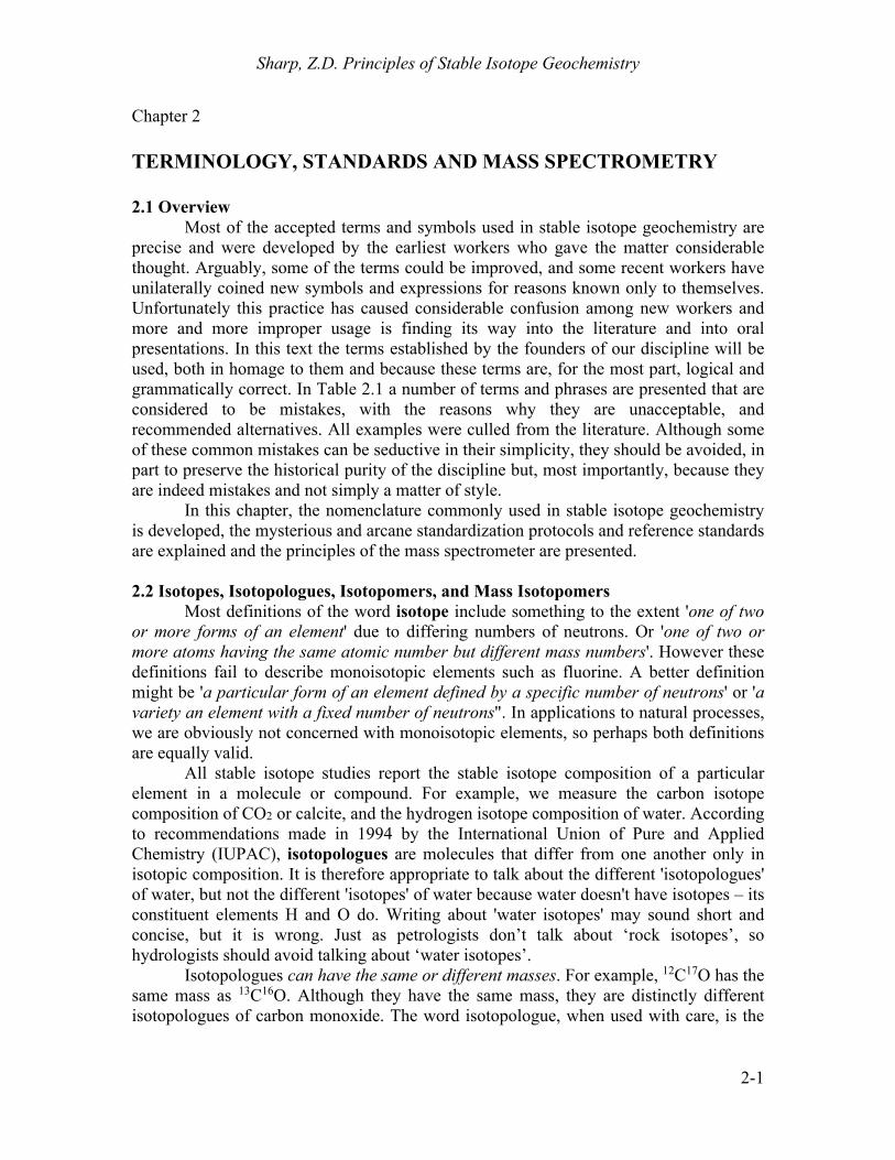
Fig. 2.1. Picture of an ampoule (glass break-seal tube) containing NBS-1 standard, a reference standard that was formerly distributed by the National Bureau of Standards (now NIST). The label reads: Isotope Reference Sample #1. PROTIUM OXIDE (Ordinary Water)

Sharp, Z.D. Principles of Stable Isotope Geochemistry
2-15
Fig. 2.2. The ocean pier at the Scripps Institute in San Diego, where Harmon Craig and Ray Weiss collected water for VSMOW. Photo by author.
Table 2.4. Gases commonly measured in conventional gas source isotope ratio mass spectrometers.
Element Gas Masses of Isotopologues Measured
Hydrogen H2
H2O (g) 2 , 3 (interference from H3
+) (H2O, HDO in laser spectroscopy systems)
Carbon CO2 44, 45 Nitrogen N2 28, 29 (and 30 for artificially enriched
samples) Oxygen CO2
O2 (fluorination)
CO (pyrolysis) H2O (g)
44, 46 32, (33), 34
28, 30
(H216O, H2
18O in laser spectroscopy systems) Sulfur SO2
SF6 64, 66
146, (147), 148, (150) Silicon SiF4 85, 86, 87
Chlorine CH3Cl 50, 52

Chapter 2. Terminology, Standards and Mass Spectrometry
2-16
D determinations, hydrogen isotope analyses of both SMOW and SLAP are analyzed relative to the working standard, and the difference obtained multiplied by a factor so that DSLAP-
SMOW = 428‰ (Coplen, 1988). Absolute ratios of D/H
determined for both SMOW and SLAP and the absolute ratio of 18O/16O and 17O/16O determined for SMOW are given in Table 2.5. The absolute values were determined by mixing waters that were extremely pure samples of 1H2
16O, 1H218O,
D216O and H2
17O. It is very difficult to prepare water that has no deuterium or 18O and 17O in it, but the best job possible was done at the time the determination was made. By careful mixing, the D/H and 18O/16O ratios of these synthetic waters were known, but with significant uncertainty. SMOW and SLAP were
then measured relative to the isotopic compositions of these waters to derive their absolute D/H and 18O/16O ratios. While it is desirable to have reliable determinations of the absolute ratios of these standards, keep in mind that knowledge of the absolute ratios is not necessary to conduct research in stable isotope geochemistry and is not used in determining stable isotope ratios on a mass spectrometer.
Table 2.5. Determinations of the absolute ratios of the isotope ratios of selected elements and internationally accepted standards.
Ratio Standard Value Reference D/H SMOW (155.76±0.05)×10-6 (Hagemann et al., 1970) D/H SMOW (155.75±0.08)×10-6 (de Wit et al., 1980) D/H SMOW (155.60±0.12)×10-6 (Tse et al., 1980)
13C/12C PDB (11179±20)×10-6 (Zhang et al., 1990)
15N/14N AIR (3670±40)×10-6 (Junk and Svec, 1958) 17O/16O SMOW (379.9±0.8)×10-6 (Li et al., 1988) 18O/16O SMOW (2005.20±0.45)×10-6 (Baertschi, 1976a) 33S/32S VCDT (78.77±0.03)×10-4 (Ding et al., 2001) 34S/32S VCDT (441.626±0.039)×10-4 (Ding et al., 2001)
29Si/28Si NBS-28 (508.1±0.1)×10-4 (De Bièvre et al., 1994) 30Si/28Si NBS-28 (335.3±0.7)×10-4 (De Bièvre et al., 1994) 37Cl/35Cl SMOC (ISL 354) 0.31977±0.00009 (Xiao et al., 2002)
Fig. 2.3. The original container of VSMOW. From https://en.wikipedia.org/wiki/Vienna_Standard_Mean_Ocean_Water.
Sharp, Z.D. Principles of Stable Isotope Geochemistry
2-17
2.6.2 Carbon Carbon isotope ratios are reported relative to the PDB standard described above
and, by definition, the 13C value of PDB is zero. The calculated 13C/12C ratio for VPDB is given in Table 2.5. Several secondary carbonate standards (e.g. Carrara marble and Solenhofen limestone) were measured relative to PDB in the early years and these standards are still in use in some older laboratories. The international isotope reference standard NBS-19 (and most recently IAEA-603) is now the accepted means of calibrating to the PDB scale. NBS-19 was originally the TS (Toilet Seat) limestone working standard used in the laboratory of Irving Friedman and colleagues at the U. S. Geological Survey (Friedman et al., 1982). It has a 13C value of +1.95‰ relative to PDB and VPDB. In other words, the 13C value of NBS-19 ≡ +1.95‰ on the VPDB scale. Unfortunately, NBS-19 is now out of stock, and a new calcite standard IAEA-603, a sample of the Carrara marble is available with a 13C value of +2.46‰ ± 0.01 on the VPDB scale8. Other IAEA carbon isotope standards include oil, graphite and caffeine.
As in the case for SMOW, it is recommended to append the letter V to the acronym PDB. Carbon isotope geochemistry has remained a very active discipline since its inception, and many thousands of carbon isotope analyses are reported every year. The carbon analyzed is present in a variety of substances including the various carbonate minerals, organic matter in sediments, organic matter in meteorites, petroleum products, collagen extracted from plant material, graphite, carbonate in the apatite of bones and teeth, carbon present in trace quantities in rocks (e.g., basalts) and minerals (e.g., goethite) and in archaeological and anthropological specimens. There are major differences in the extraction techniques used for these various carbonaceous materials and the errors assigned to an analysis depend on the complexities of the extraction method employed. In almost all cases, however, the carbon is put into the form of CO2 for mass spectrometric analysis (Table 2.4), and therefore can be compared to the CO2 liberated from one of the IAEA standards calibrated to PDB. 2.6.3 Nitrogen
Nitrogen isotope values are measured using N2 gas in the mass spectrometer. The reference standard for nitrogen isotope analyses is atmospheric nitrogen and is called AIR. The 15N value of atmospheric nitrogen is almost constant everywhere on Earth and is 0‰ by definition (Mariotti, 1983). The 15N/14N ratio of air is given in Table 2.5. One N2 gas standard called NSVEC (15N = -2.8‰ vs AIR) is distributed by IAEA. Several other solid standards (nitrates) are also available from IAEA. Any laboratory can produce their own nitrogen reference by purifying air. Oxygen is removed by combusting the air sample with Cu metal, but contaminant Ar in the product can interfere with the nitrogen isotope analyses9.
8 For details, see: https://nucleus.iaea.org/rpst/referenceproducts/referencematerials/Stable_Isotopes/13C18and7Li/IAEA-603/RM603_Reference_Sheet_2016-08-16.pdf
9 Note that V-AIR has not yet been proposed.

Chapter 2. Terminology, Standards and Mass Spectrometry
2-18
2.6.4 Oxygen Two international reference standards are used to report variations in oxygen isotope ratios, VPDB and VSMOW. SMOW was originally defined in terms of NBS-1
18 18
16 16
1
O O1.008
O O
SMOW NBS
2.21.
SMOW has a 18O value of 8.00‰ relative to NBS-1, and NBS-1 has a 18O value of -7.94‰ versus SMOW. As with hydrogen, a stretching factor should be applied to oxygen isotope analyses and this factor is calibrated by analyzing VSMOW whose 18O 0‰ and SLAP (Standard Light Antarctic Precipitation) whose 18O = –55.5‰ (See Appendix 2). By calibrating in this manner, analyses of water reported on the VSMOW scale are reliable. The stretching factor is much smaller for oxygen isotope analyses than for hydrogen isotope analyses and is often ignored.
Use of the PDB standard for reporting oxygen isotope compositions is restricted to analyses of carbonates of low-temperature origin (oceanic, lacustrine, or pedogenic) in studies of paleoclimate, paleoceanography and carbonate diagenesis. As mentioned above, oxygen isotope compositions of carbonates are determined by analyses of CO2 generated from them by reaction with 100% H3PO4 at a fixed temperature. It is emphasized that the PDB standard is the solid carbonate, not the acid-liberated CO2 that is actually introduced to the mass spectrometer. The 18O of PDB is 0‰ on the VPDB scale by definition and analysis of NBS-19 is the accepted means of relating oxygen isotope analyses to VPDB. The 18O value of NBS-19 is 2.20‰ on the VPDB scale. Additional international secondary reference standards for carbonates are available (Appendix 1), and relating analyses to PDB no longer poses any ambiguities. Because the 18O value of VPDB is 30.91‰ higher than VSMOW (on the VSMOW scale), the conversion between VSMOW and VPDB scales is given by equation 2.7. There is a difference of 0.28‰ (Fig. 6.1) between CO2 in equilibrium with VSMOW (CO2-H2O = 1.04120) at 25oC and CO2 liberated from PDB at 25oC (CO2-CaCO3 = 1.01025) and all these values were used in deriving equation 2.7 (See Chapter 6 for further explanation). Marine carbonates have 18O values near zero on the VPDB scale, while ocean waters have 18O values near zero on the VSMOW scale. As a result of this relationship, it is not uncommon to see the two scales mixed in published reports, with data for carbonates reported on the VPDB scale and data for waters reported on the VSMOW scale. If their 18O values were replotted on the same scale, however, they would be approximately 30‰ apart! Care must be taken not to mix scales when presenting oxygen isotope data. Traditionally, the 17O/16O ratios of terrestrial materials are not measured, because they provide no additional information than the 18O/16O ratios alone. The 17O/16O ratio correlates well with the 18O/16O ratio by the equation
λ17 18
16 16O O
O O 2.22,
where the lambda () is an empirical best fit of ~0.527 for solids and 0.528 for waters. Recently, it has been recognized that there are small departures in the 17O values

Sharp, Z.D. Principles of Stable Isotope Geochemistry
2-19
predicted by equation 2.22 (Luz and Barkan, 2010; Pack and Herwartz, 2014), so that high precision 17O studies have relevance and are beginning to be made on a more routine basis. The 17O value of VSMOW is by definition 0‰. Oxygen isotope measurements are made on one of several gases. Generally, samples are converted to or equilibrated with CO2¸which is analyzed in the mass spectrometer. O2 is routinely used for analyses of silicates which are fluorinated to produce O2 gas. Analysis of O2 is required for 17O analyses. Finally, oxygen from organic matter is frequently analyzed as CO by reaction with carbon at very high temperatures. 2.6.5 Sulfur Sulfur has four stable isotopes 32S, 33S, 34S and 36S (Chapter 10). Like the triple oxygen isotope system, to a first approximation the four isotopes of sulfur fractionate in predictable proportions, so that measuring all four isotopes is redundant. (In some cases multiple sulfur isotopes can provide important information about low temperature processes – e.g., Farquhar and Wing, 2003; Ono et al., 2003). In general, only the 34S/32S ratios are measured, as these are the two most abundant isotopes of sulfur. Sulfur isotope ratios are given by 34S notation. 34S values are reported relative to the CDT (Cañon Diablo Troilite) standard, a sample of meteoritic troilite (FeS) from Meteor Crater in Arizona. The 34S/32S value of CDT is given in Table 2.5. As with the other primary standards, 34S(CDT) 0.0‰ by definition. Unfortunately, the CDT standard is not as homogeneous as originally thought, at least on a scale smaller than several milligrams (Beaudoin et al., 1994). The IAEA now distributes two synthetic Ag2S standards with defined 34S values relative to CDT (Appendix 1). Sulfur isotope analyses are calibrated to CDT in each laboratory by analyses of these Ag2S standards whose 34S values differ by 22‰. CDT now joins the ranks of PDB in the sense that they are the accepted international reference standards for reporting all sulfur and carbon isotope analyses, but neither is distributed and both are defined in terms of recommended values of secondary solid standards. Sulfur analyses are made on either SO2 gas or SF6 gas (Table 2.4). The advantage of each is discussed in Chapter 10. The 34S values of SO2 gas are determined by measuring masses 66 (34SO2) and 64 (32SO2). Unfortunately, there is an isobaric interference at mass 66 from 32S16O18O, which must be corrected for. This problem does not exist with SF6 because F is monoisotopic, however SF6 is more difficult to produce in the laboratory and requires a mass spectrometer specially configured for the high masses (Table 2.4). 2.6.6. Silicon Silicon has three stable isotopes, 28Si, 29Si, and 30Si. The abundance of the major isotope 28Si is 92.2%, while 29Si and 30Si have abundances of 4.6 and 3.0%, respectively. The three isotope system follows well-established mass dependent fractionation for terrestrial materials, so that there is no need to measure all three isotopes. In general, the 30Si/28Si ratios are measured, reported in terms of 30Si values. The total range of 30Si values for terrestrial materials range from ~-4 to +4‰. Silicon isotope ratios are reported relative to the NIST standard NBS 28 quartz, with a defined value ≡0‰. Si isotope ratios are measured using either gas source mass spectrometery with SiF4 as the sample gas or

Chapter 2. Terminology, Standards and Mass Spectrometry
2-20
using multicollector ICPMS. Each method has equivalent precision. Samples measured with gas source mass spectrometry measure the ion fragment SiF3
+ at masses 85, 86 and 87. 2.6.7. Chlorine Chlorine has two stable isotopes, 35Cl and 37Cl. Unlike the other isotope systems commonly studied, the abundance the rare isotope is very high, leading to a non-integer mass for the average atomic weight of Cl (35.453 amu). Originally analyzed as HCl (g) (Hoering and Parker, 1961) or CsCl on a thermal ionization mass spectrometer (Magenheim et al., 1994), Cl isotope ratios are now analyzed exclusively using CH3Cl (Long et al., 1993). Seawater, with an extremely long residence time for Cl- is well mixed, and is therefore the accepted standard, defined as Standard Mean Ocean Chloride (SMOC) with a 37Cl value ≡ 0.0‰. Several Cl isotope standards have been made (Appendix 1), although any ocean sample has a Cl isotope composition equal to 0.0‰, unless extreme contamination has occurred. The ocean has an essentially constant Cl isotope composition, although some publications suggest that there are variations, a conclusion that I believe in general is not correct. 2.7 Isotope Ratio Mass Spectrometry The mass spectrometer is the heart of nearly all stable isotope laboratories Some labs are beginning to employ laser spectroscopy systems, but for the last century, most analyses have been made using a mass spectrometer. The foundations of the mass spectrometer can be traced to the Cavendish Laboratory, University of Cambridge, where noted scientists including J.J. Thomson, E. Rutherford and F.W. Aston developed some of the first mass spectrographs10. By 1927, Aston had built a second generation machine that allowed for the discovery of isotopes and accurate determinations of their atomic weights. A description of Aston's early machine is discussed in detail and is worth reading (Aston, 1927). In the early to mid-20th century, most isotope ratios were determined gravimetrically, where the mass of a sample was determined using a precise density balance. The difference in the atomic weight of oxygen in air and water – the basis for the 'Dole Effect' – was determined in this way (Dole, 1936). Major improvements were made in the sensitivity and precision of mass spectrometers in the 1940's so that variations in the isotopic composition of natural materials could be measured with the necessary precision. These machines were operated by talented physicists, and extracting high-quality data required skilled practitioners. Only starting in the 1970s and 80s did the mass spectrometer manufacturers begin to offer mass spectrometers that could be used without a great deal of training and infrastructure. Today, there are a wide range of mass spectrometers available, from simple desktop units for low-precision analyses used for simple tracer experiments, to complex doubly-focusing machines that have high sensitivities and mass resolution for analyses of exotic isotopologues from natural samples. At the same time, there is a growing industry of laser spectroscopy systems in which the isotope ratios are measured based on the absorption of infrared radiation by the different isotopologues of a particular gas, such as CO2, H2O, CH4 and N2O (see section 2.8 for further details).
10 Mass spectrographs used photographic plates to measure the position and intensity of ion beams. Mass spectrometers use electronic collectors which measure the intensity (current) of the ion beam.

Sharp, Z.D. Principles of Stable Isotope Geochemistry
2-21
2.7.1 Components of a mass spectrometer All mass spectrometers are based on the principle of deflecting an energetic, focused ion beam in a magnetic and/or electrostatic field. The degree of deflection is a function of mass and charge. The relative intensities of the ion beams of different masses can then be used to calculate isotopologue masses or isotope ratios. The mass spectrometer consists of three primary components (Fig. 2.4): 1) the source, where a sample is ionized, accelerated to a given energy and collimated into a well-focused beam; 2) the analyzer, which acts to deflect the ion as a function of mass. It is the ‘prism’ of the system (See Criss, 1999 for a nice review); and 3) the collector assembly for measuring the relative intensities of the different ion beams. In addition, the mass spectrometer system also has an inlet system for introducing the gas into the source without
BELLOWS
valve
capillary
MAGNET
COLLECTORASSEMBLY
SOURCE
SWITCHINGBLOCK
H.V.pump
capillary
crimp
FLIGHTTUBE
MICROVOLUME
samplegas inlet
reference side sample side
(not drawn to scale) highvacuum
side
lowvacuumside
Fig. 2.4. Schematic of a typical modern mass spectrometer. The isotopic composition of two gases (reference and sample) are measured relative to one another. The pressures of both gases in the source region are adjusted to the same value by compressing or expanding the bellows. The gas passes through a capillary about 1 meter long with a crimp at the mass spectrometer end to help reduce the flow rate and to assure viscous flow. The capillary/crimp prevents a fractionation of the gas as it enters the high vacuum of the mass spectrometer. The bleed rate of the gas into the mass spectrometer is slow enough so that the reference gas will remain in the bellows system for a full day of measurement (except for hydrogen). The gas enters the source region, where it is ionized, focused into a coherent beam and accelerated down the flight tube. The ion beams are deflected in a magnetic field in relation to the charge/mass ratio of the ion. The lighter ions are deflected to a greater degree than the heavy ones of the same charge. The ions enter the various collectors (Faraday cups) where the current developed is sent through resistors to produce voltages that are amplified and registered on a recording system (not shown). The intensities of these ion beams are proportional to the abundance of the isotopologue collected. The isotopic ratios and delta values are automatically calculated and recorded by the software that operates the machine.

Chapter 2. Terminology, Standards and Mass Spectrometry
2-22
fractionation. (see Sharp, 2014 for a detailed discussion of gas source mass spectrometers). 2.7.1.1 The ion source The ion source both ionizes the sample gas, accelerates it to a near-constant energy and focuses the beam in the direction of the analyzer. Sample gas is admitted into the source at low pressures and a fraction of the gas is ionized (~0.1%) with an unknown but significant isotope fractionation. The ideal ion source has high ionization efficiency, a well collimated ion beam and a near-constant, or at least linear isotope source fractionation over a wide range of source pressures . The gas source mass spectrometer uses electron impact ionization, in which a hot tungsten filament generates electrons in the ionization box. The ionization box has a filament on one side and an anode on the other, so that the electrons are accelerated towards an anode with an energy of ~90eV (Fig. 2.5). This energy is more than enough to cause the sample gas to reach its first ionization potential, which ranges from 12.08eV for O2 to 15.58 for N2. Interestingly, above 100eV, ionization efficiencies begin to decrease as molecules become ‘transparent’ to the electrons (de Hoffmann et al., 1996). A magnetic field is applied to the electron beam with two small permanent magnets on either side of the ionization box which causes the electrons to spiral towards the anode, thereby increasing ion efficiency. The electron impact knocks an electron off a molecule of sample gas causing it to become positively charged. The source is held at a positive electric potential of 2 and 10 kV relative to ground. The positively ionized gas is accelerated out of the ionization box by the repeller plate and towards a stack of
electrostatic lenses which focus the ions into a tight beam. Each lens is at a higher potential relative to the ionization box, so that the ions reach a nearly mono-energetic value equal to the electric potential of the source relative to ground. In addition to imparting energy to the ions, the lens stack acts as a telescope, focusing the beam at the collector assembly. While the ideal electron impact source ionizes the sample gas by removing one electron, ion fragmentation and double ionization (removal to two or more electrons) invariably occur. If the source conditions remain nearly identical for both sample and reference gas, the fragmentation problems can generally be ignored, because it
Fig. 2.5. Schematic of the ion source. Gas is bled into the ionization box and is ionized by a stream of energetic electrons generated at the hot (2000°C) tungsten filament. The electrons strike the gas, knocking off one (or more) electrons, producing positively charged ions. The ions are accelerated out of the ionization box by a combination of repulsion from the repeller plate and acceleration towards the focusing electrostatic lenses. Each lens is at an ever-higher potential relative to the trapping box (closer to ground), thereby further increasing both the energy and the focus of the ion beam.

Sharp, Z.D. Principles of Stable Isotope Geochemistry
2-23
will affect both gases to the same extent. Consider CO2 as an example. CO2 will ionize primarily to CO2
+, but other ion fragments, such as such as CO+, C+, and O+ and CO22+. A
mass spectrum of CO2 gas will show peaks at masses 12, 16, 22 (doubly ionized CO2), 28 and 44. The ionization efficiency of an electron impact source is on the order of 0.1 to 0.15% (for CO2), with a large and generally unknown isotope fractionation. Again, if source conditions (i.e., pressure) are kept the same both for sample and standard, this fractionation will not affect the relative isotopic ratios of reference and sample gas. How the isotope fractionation changes with source pressure is known as linearity. Increasing the draw-out potential from the ionization box increases linearity by minimizing ion-ion interactions, but also reduces the sensitivity of the source. A tradeoff must be made between linearity and sensitivity. If gas quantities are not an issue, then the source should be focused for highly linear conditions. 2.7.1.2. The analyzer The mass analyzer is the portion of the mass spectrometer where the ion beams are separated from each other on the basis of their mass. It is equivalent to a prism, which separates light according to wavelength. Most stable isotope mass spectrometers use a magnetic field to separate ions by mass, but doubly-focusing mass spectrometers also use an electrostatic lens in order to attain very high mass resolution. The effects of electric and magnetic fields on an ion beam is given by Lorentz’s law by BvEqF 2.23, where F is the force acting on the ion, (which determines the degree of deflection in a circular field), q is the ion charge, v is velocity and E and B are the electric and magnetic forces, respectively. The energy imparted by the ion source is given by qV = ½ mv2 2.24, where q is the charge, V is the voltage potential between the source and ground, m is the ion mass and v is the ion velocity. The force imparted by an electrostatic field, composed of two charged plates in a semi-circular arc results in a deflection of the ion beam given by
qE
mvr
2
2.25,
where E is the electric force. All ions of equal charge traveling through a curved electrostatic field with radius r will have a constant kinetic energy (½mv2). Higher or lower energetic ions will have a different radius of curvature and can be physically removed from the beam with proper placement of slit apertures. An electrostatic field is therefore an energy filter, imparting a constant energy to the ion beam.

Chapter 2. Terminology, Standards and Mass Spectrometry
2-24
A magnetic field generates a force equal to qvB which imparts a centripetal force equal to mv2/r. The radius of curvature is given by
qB
mvr 2.26.
Unlike an electrostatic field, where the deflection is related to mv2, a magnetic field deflects ions proportional to mv or momentum. The radius of singly charged particles at constant velocity is therefore related to the mass. For a constant energy ion beam, ions
with a smaller mass will be deflected more than ions with a higher mass. High resolution doubly focusing mass spectrographs have been around for close to a century (e.g., Bainbridge and Jordan, 1936). For most stable isotope studies, sufficient mass separation is achieved with a magnetic field alone. However, for very high mass resolution and when the ions are produced with variable energy, the addition of energy filtering using an electrostatic field is required. Both an ion microprobe and high resolution ICP MS require both electrostatic and magnetic field focusing.
In a magnetic sector mass spectrometer, the magnet separates ions of different masses, but also focuses the ion beam at the point of the collector assembly (Fig. 2.6). Interestingly, it is the radius of the magnetic field, and not the total arc fraction (e.g., 60° vs. 180°) that defines the degree of separation and focus (Nier, 1940)11. An ion beam leaving the source will necessarily have some degree of divergence. As shown in Fig. 2.6a, the beam will enter the magnetic field and be deflected in such a way as to be refocused on a plane perpendicular to the arc of the magnetic field that includes the ion source and the point of refocusing. Beam divergence also occurs perpendicular to the plane defined by the ion path (the z direction). It has been shown that if the beam crosses the fringing field (edge) of a magnetic field non-perpendicular to the edge of the magnet, the beam will be deflected perpendicular to the median plane (in a vertical direction in a flat-lying magnetic arc – 26.5° angle in Fig. 2.6b). By adjusting the angle of the fringing field, the beam can be deflected – and thereby focused – in a vertical plane at the point of
11 Commercially available mass spectrometers use 60° or 90° sector field with comparable results.
Fig. 2.6. Schematic of mass spectrometer focusing. The dispersion of the beam from the source is eliminate by focusing through the analyzer in the x-y plane (2.4a top). The beam will refocus on a line perpendicular to the magnetic arc which includes the source. In the z-plane, focusing is made by ‘fringe focusing’, in which the edge of the magnet is designed to be non-perpendicular to the ion beam (2.4b bottom).

Sharp, Z.D. Principles of Stable Isotope Geochemistry
2-25
the collector assembly. The angle between the beam and magnet edge is a function of the arc and the path lengths on either side of the magnet. If properly designed, the deflection that occurs upon entering the magnet causes the beam to become parallel to the magnetic plates, and during the exit deflection, the beam is deflected to focus at the focal plane of the ion beam (e.g., at the collector). 2.7.1.3. Collector assembly The final component of the gas source mass spectrometer is the collector assembly. The early machines were mass spectrographs, in which the ion beams were focused on photographic plates. The intensity and position of the exposed images gave the position and abundance of the isotopologues. The accuracy attained with photographic film was so high that Francis Aston in 1937 was able to determine the atomic weights of over twenty isotopes to better than 0.008 amu of today’s accepted value (when corrected for different scales) (Aston, 1937). All modern mass spectrometers use electronic detectors to measure the intensity (current) of an ion beam. Positive ions enter a collector which acts as an electron source for charge compensation. Neutralization of the collectors generates an extremely small electric current. The current (total charge per unit time) is related to the intensity of the ion beam. Current is related to voltage by Ohm’s law V = IR 2.27 where V is voltage, I is current and R is resistance. By using a very large resistor (108 to 1012 ), a high voltage can be generated from a very small current. A voltage-to-frequency converter is then employed to give a signal that is related to the beam strength. When multiple beams from different isotopologues are collected simultaneously, each collector is fitted with appropriate resistors so that the output voltage (current) of each ion beam are close to the same. For example, the relative abundance of the isotopologues of CO2 are roughly the following: 12C16O16O – 0.984; 13C16O16O – 0.011; 12C18O16O – 0.004. Voltages for these three beams will be similar if resistors of 1 108, 11010 and 31010 are used for masses 44, 45 and 46, respectively. For most applications in gas source mass spectrometry, Faraday collectors are used to measure beam intensity. Positive ions are neutralized in the collector and draw an electron for charge compensation. Ideally, the collector acts as a perfect black body surface for reception of the ions. Reflection of ions is minimized by coating the inside of the collector with soft carbon. Some sputtering of the ions occurs, generating secondary electrons. The loss of such an electron would negate the charge contribution from a positive
Fig. 2.7. Schematic of a Faraday collector. The ion beam passes through a narrow slit a and is neutralized in the collector, generating a current. A charged electrode b prevents secondary electrons generated in the collector cup from exiting the assembly.

Chapter 2. Terminology, Standards and Mass Spectrometry
2-26
ion. Collectors are therefore manufactured as deep wells with narrow entrance slits to prevent the loss of secondary electrons. The collector consists of a narrow slit to prevent the introduction of stray ions and a charged electrode at the entrance to the collector to help prevent the secondary ions produced in the collector from escaping back through the entrance slit (Fig. 2.7). Multiple collectors are generally used in order to simultaneously measure the intensities of all relevant isotopologues of a given gas. In this way, electronic drift in the system is minimized. Instrumental designs include either multiple narrow detectors positioned for analysis of a single gas type or ‘universal collectors’, in which several wide cups are paired with a narrow cup. The multiple, narrow collector design is unquestionably superior to the universal collector system, but it can be costly, and flexibility is limited because the positioning of the collectors is generally fixed. For users wishing to be able to analyze multiple gases on a single instrument, the number of collectors required can be quite high. The universal collector design is simple and less costly, but inherently runs the risk of having a greater loss of secondary electrons or reflected ions and the possibility of stray ions entering the cup. In practice, by comparing standard and sample, these effects are minimized. 2.7.1.4 Dual inlet mass spectrometer inlet system In gas source mass spectrometry, a dynamic inlet system is used. Gas continuously bleeds into the source, is partially ionized, and the excess non-ionized gas is pumped away. A dynamic inlet system avoids the problems of drift due to preferential ionization of one isotopologue relative to another by continuously streaming fresh sample gas into the source. Source pressures must be kept low to avoid molecular collisions in the flight tube which would otherwise broaden the well-focused beam. Therefore some method of pressure reduction is required between the gas reservoir and the source. Whereas the gas pressure in the sample reservoir is on the order of 102-103 mbar, pressures are reduced to 10-4 bars in the ionization chamber and 10-6 to 10-8 bars in the flight tube. The pressure drop from sample volume to source could be accomplished by having a pinhole orifice between the gas volume and the source, but such a geometry causes an isotopic fractionation, with the lighter isotopologues preferentially passing through the orifice due to their higher translational velocities (Graham's Law). To eliminate this fractionation, gas source mass spectrometers are equipped with a capillary inlet system. A long (~1 m) 0.1mm I.D. capillary is connected to the gas volume adjusted to between 10-100 mbar pressure. At the other end – the interface with the high vacuum of the mass spectrometer – the capillary is crimped, so that the pressure drops to 10-4 bar in the source. The light molecules will preferentially pass through the crimp, but because the mean free path of individual molecules in the capillaries is a fraction of the capillaries’ internal diameter (~10-7 m), the gas migrates along the capillary into the mass spectrometer under viscous flow conditions. Like leaves floating on a swift moving stream, the random motion of individual molecules is overwhelmed by the overall flow direction of the gas through the capillary into the mass spectrometer. Using this type of geometry, there is no fractionation in the capillary inlet system as long as the pressure in the capillary is kept in the 10-100 mbar range.

Sharp, Z.D. Principles of Stable Isotope Geochemistry
2-27
High precision is achieved by rapidly switching between sample and reference gas using a system of changeover valves ('switching block' in Fig. 2.4). First described by Murphey (1947) and then McKinney et al. (1950), two identical capillary systems feed through a system of four change-over valves so that either the reference gas or the sample gas enters the mass spectrometer. Gas always flows through the two capillaries to achieve steady state flow. The switching valves change the flow path between the source and a waste pump. Switching between reference and sample gas can be accomplished rapidly, so that the intensities of all ion beams for the sample and reference gas can be compared almost simultaneously. This rapid switching minimizes any errors associated with drifting electronics. In order to keep conditions in the source as similar as possible, both reference and sample gas are adjusted to be nearly the same pressure. This was originally accomplished using mercury pistons, but is now done using flexible steel bellows controlled by stepping motors ('bellows' in Fig. 2.4). 2.7.2 Gas Chromatograph Isotope Ratio Mass Spectrometry (GC-IRMS)
In recent years, the continuous flow method of gas introduction has revolutionized isotope ratio mass spectrometry and opened up a panoply of research avenues (Matthews and Hayes, 1978). In place of analyzing gas using the dual inlet method of measurement, samples of gas are entrained in a He stream, purified in a gas
Fig. 2.8. Schematic of a GC-IRMS system. Samples are introduced into a He stream and the different gases are separated on a gas chromatographic column. They pass through an open split into the mass spectrometer. The open split allows for a constant pressure to be maintained in the source. A pressure drop from 1 bar at the open split to 10-5 - 10-6 bars occurs along the length of the capillary. Multiple turbomolecular pumps separated by a diaphragm slit keeps the pressure in the flight tube low. Signals for the different isotopolouges of a given gas are measured simultaneously as Gaussian-shaped peaks. The integrated area of each peak relative to those of a reference gas or standard sample can be used to determine the isotope ratio in conventional delta format.

Chapter 2. Terminology, Standards and Mass Spectrometry
2-28
chromatograph and (with or without first oxidizing or reducing the gas by passage through an oven) introduced directly into the source of the mass spectrometer. Initial applications of the GC were mainly to purify the gas. Early online applications included the now routine high temperature combustion of very small C and/or N-bearing samples in an elemental analyzer or equivalent oxidation furnace (Fig. 2.8). Another important application is compound specific analysis of organic molecules (Hayes et al., 1989). Organic species are separated on a GC column and oxidized (or reduced) sequentially before being analyzed in the mass spectrometer. A simple example would be the analysis of a natural gas sample. The gas consists of methane (CH4), ethane (C2H6), propane (C3H8), etc. The lighter alkanes pass through the GC column faster than heavier ones, and with a judicious choice of GC columns, flow rates, temperatures, etc, it is possible to get a quantitative separation of the different alkanes. The methane exits the GC column first and is carried by the He stream through a copper oxide furnace where it is oxidized to CO2 and introduced into the mass spectrometer. The 13C vale of the methane-derived CO2 is determined as it flows into the source. Ethane and higher alkanes follow sequentially, allowing for the 13C values of each species to be determined. The range of isotopic variations within a single sedimentary organic sample can often exceed the total variation of different bulk samples (Freeman et al., 1990). This information has been used extensively for paleoclimate reconstruction, ecology and metabolic studies.
GC-IRMS (Gas Chromatography- Isotope Ratio Mass Spectrometry) technology has been modified so that it can be used in place of conventional extraction techniques for almost every material that has been analyzed conventionally. The advantages of GC-IRMS is that gases can be purified easily and efficiently, analysis is extremely rapid, and sample sizes are reduced by several orders of magnitude. Systems that combine gas chromatography and continuous flow technology continue to find important applications in the earth and biological sciences. Recent improvements in analyses of water and carbonates effectively replace analytical methods that have been in use with only minor modification for over half a century. There is a slight loss of precision relative to the traditional dual inlet system, although the gap is reduced every year.
2.7.3 Gases measured in isotope ratio mass spectrometry
Gases that are commonly introduced into isotope ratios mass spectrometers and masses of the isotopologues measured are given in Table 2.4. Hydrogen isotope ratios are almost always determined from the 3/2 ratios of H2. Carbon isotope ratios are almost always determined from 45/44 ratios of CO2 gas. Nitrogen isotope ratios are determined from 29/28 and 30/28 ratios of N2. Because the concentration of 15N15N is negligible in nature, measurements are made of mass 30 only when materials artificially enriched in 15N are being studied. Oxygen isotope ratios are most commonly determined from measurements of 46/44 ratios of CO2 but, more and more frequently, the gases CO and O2 are being used. 18O/16O ratios are determined from the 30/28 mass ratio of CO and the 34/32 mass ratio of O2 . 17O/16O ratios are determined from 33/32 ratios of O2. CO is the gas of choice for emerging pyrolysis applications.
As discussed in section 2.6.5., sulfur isotope ratios can be measured on either SO2 or SF6. SO2 is easily produced from most samples and can be analyzed in most mass spectrometers, but has a number of disadvantages, most notably, it quickly dirties the source of the mass spectrometer (see section 10.2). This problem is reduced significantly

Sharp, Z.D. Principles of Stable Isotope Geochemistry
2-29
in continuous flow systems where only minor amounts of SO2 are introduced into the mass spectrometer. SF6 is cleaner and allows for the rare isotope ratios to be measured, but it is difficult to produce and requires a large-radius mass spectrometer especially configured for SF6.
Silicon isotope ratios are measured on SiF4 gas. The fragment SiF3+ is analyzed
because it is the major isobar produced in the source. Masses 85 (28SiF3+), 86 (29SiF3
+) and 87 (30SiF3
+) are analyzed. Chlorine isotope ratios are made using CH3Cl. Masses 50 (CH3
35Cl) and 52 (CH337Cl) are analyzed. Many normal triple collector assemblies are
not quite configured correctly for simultaneous analysis of masses 50 and 52, so that mass spectrometers must be fitted with special collectors for CH3Cl analysis. 2.7.4 Relations between measured and desired isotopic ratios The relationship between intensities of measured gasses in a mass spectrometer and the actual isotopic ratio of a given element is different for each gas. A few of the salient points pertinent to the analyses of three gases (H2, N2, and CO2) will be made here to gain an appreciation of this important aspect of isotope ratio mass spectrometry.
After experimenting with various hydrogen containing gases, Irving Friedman (1953) concluded that diatomic hydrogen was the most suitable for determination of hydrogen isotope ratios and H2 remains the gas of choice to this day. Isotopic analysis of hydrogen poses a unique analytical problem. One of the ionic fragments produced in the source of the mass spectrometer is H3
+ which has the same mass 3 as HD+. Collection of this ion results in an apparent 3/2 ratio that is higher than the true HD/HH ratio. The production of H3
+ is proportional to the pressure of hydrogen gas in the source because this ion is produced by the reaction H2 + H2
+ H3+ + H. The H3
+ contribution to mass 3 can be minimized, but not eliminated, by using a high accelerating voltage and a low gas pressure. The contribution of H3
+ is evaluated by measuring the 3/2 ratio as a function of source pressure, a procedure that is done routinely on modern mass spectrometers. The correlation is linear on most modern machines (at low pressures, less H3
+ is produced). Once the relationship between pressure and contribution of H3
+ is determined, a correction algorithm can be applied that removes the mass 3 contribution from the H3
+ ion (i.e., extrapolate back to zero pressure).
Nitrogen isotope analysis is relatively straightforward. Measurements are made of two isotopologues of molecular nitrogen whose masses are 28 and 29. The isotopologue with mass 28 consists solely of 14N14N while that of mass 29 is a sum of the two possible orientations of 15N and 14N atoms in the molecule, 15N14N and 14N15N. The desired mass ratio is then obtained from the relatively simple relation12:
15
14
292
28
N
N 2.28
12 The actual relationship is 29 152
29 28 14
or 15229 141528 1 14
. If the 15/14 ratio is 3670×10-6, the
actual stochastic 29/28 ratio is 7367×10-6, whereas equation 2.28 gives a 29/28 ratio of 7340×10-6.

Chapter 2. Terminology, Standards and Mass Spectrometry
2-30
The molecular mass ratio 29/28 is twice the atomic mass ratio of 15/14 and is the one used to determine nitrogen isotope ratios. This same mathematical correction is used for CO2 as well.
The nine isotopologues of CO2 that are measured on a mass spectrometer are given in Table 2.7. An additional 5 are included when mass 47 is measured in 'clumped isotope' studies (see section 6.7). Deconvolution of the three masses 44, 45 and 46 to 13C/12C and 18O/16O ratios can be made assuming that the relationship between 17O/16O and 18O/16O ratios (equation 2.2) is valid. Given that (17O/16O) (18O/16O)0.5x, the three masses 44, 45 and 46 of CO2 can be used to determine both 13C/12C and 18O/16O ratios (Craig, 1957; Santrock et al., 1985). The 46/44 ratio measured in the mass spectrometer is close to the 18O/16O ratio, because 12C16O18O makes up >99.7% of the total isotopologues at mass 46. On the other hand, the 13C/12C ratio of CO2 gas is not the same as the 45/44 ratio because the 12C17O16O isotopologue is on the order of 7% of the total mass 45 isobar. For 13C analyses, the common '17O correction' must be made. The 17O value of the CO2 sample is estimated from the measured 18O value and the relationship between 17O/16O and 18O/16O (equation 2.2). The 12C17O16O contribution to mass 45 is then subtracted from the total 45 isobar intensity in order to obtain the contribution of just 13C16O2.
Table 2.6 Masses and possible configurations of CO2 isotopologues. Mass Isotopologue
44 16O12C16O 45 16O13C16O, 17O12C16O, 16O12C17O 46 17O13C16O, 16O13C17O, 17O12C17O, 18O12C16O, 16O12C18O
47 16O13C18O, 18O13C16O, 17O13C17O, 17O12C18O, 18O12C17O
48 18O12C18O, 18O13C17O, 17O13C18O
49 18O13C18O,
2.8 Laser absorption mass spectroscopy In recent years, laser absorption spectroscopy has been modified for use in stable isotope analysis. This method involves measuring the absorption of electromagnetic radiation (generally infrared) generated by a laser at a frequency specific to the absorption of a single isotopologue of a gas molecule (Fig. 2.9). There are a large number of absorption bands for any molecule that has a dipole13, and bands are chosen in the frequency range of the laser which are close to one another without any interferences from other absorption lines. Absorption is directly related to concentration following Beer-Lambert law, given by I = Ioexp(-Cl) 2.29,
where Io and I are the intensity of the electromagnetic radiation entering and leaving the chamber containing the gas, C is the concentration of the absorber, l is the sample path length and is the absorption cross section at the appropriate wavelength. The laser light
13 A dipole has a permanent bond polarization – two atoms with different electronegativity. O2 and N2 consist of two atoms with the same electronegativity and have no dipole.

Sharp, Z.D. Principles of Stable Isotope Geochemistry
2-31
is directed through the input mirror (with most being reflected back to the laser) and reflects back and forth between two parallel mirrors. The intensity of the reflecting light is measured by a photo-sensitive detector behind the output mirror. The concentration of a molecular species is determined from the I/Io value.
Several systems are now commercially available that take advantage of low-cost tunable diode lasers. Two distinct methods are employed. The first, called off axis cavity output spectroscopy, measures changes in intensity of the laser beam as it scans across the wavelength of interest (Baer et al., 2002). By having the beam reflect multiple times between the two mirrors in the sample cavity, it is possible to achieve path lengths of several km, thereby drastically increasing the degree of absorption (Fig. 2.10). The second method, called cavity ring-down laser absorption spectroscopy, measures changes in the intensity of
the reflecting beam as a function of time. A pulse of laser light charges the cavity and the laser is quickly shut off. The rate of decay (the ‘ring-down’ time) of the laser beam (on the order of several microseconds) is a function of the concentration of the absorbing species. The higher the concentration, the more rapid the ring-down time. Each method has certain advantages and disadvantages, and at present, it appears that the precision of the two systems are more-or-less equivalent (Johnson et al., 2011). The tunable diode laser systems are used primarily for H and O isotope ratios of water and C and O isotope ratios of CO2, carbon from methane, and N and O in N2O (Baer et al., 2002; Crosson et al., 2002; Lis et al., 2008; Brand et al., 2009). Analyses can only be made on gases which are transferred into the laser cavity in a carrier gas, either N2 or dry air. Precision is only slightly lower than that attainable from conventional mass spectrometry. At low concentrations of water vapor, serious analytical bias is observed, however. Care must be taken to avoid interferences from contaminant gases (Brand et al., 2009). For all of the commercially available laser systems, the sample flows through the chamber continuously throughout the measurement, although efforts to introduce a small
Fig. 2.9. Illustration of several absorption bands for CO2 gas for different isotopologues. There are a large number of bands to chose from. Ideally, absorption bands will be chosen for the isotopologues of interest that are close to each other and do not have any other interferences. The 13C16O16O and 12C16O16O in bold are ideal pairs to analyze.

Chapter 2. Terminology, Standards and Mass Spectrometry
2-32
amount of gas into the cavity in a static mode has met with success (Berryman et al., 2011). The future outlook for laser spectroscopy systems is quite favorable. Simultaneous analyses of both D/H and 18O/16O ratios are possible because the absorption bands of 1H2
16O, 1H2H16O, and 1H218O are all distinct and large enough for accurate
analysis, and recently manufactures have added 17O to the total analysis (although non-linearity problems persist). Similarly, analyses of N2O and CO2 may allow for isotopologues of the rare isotopes to be measured with far greater ease than by traditional mass spectrometry methods. Ono et al. (2014) have successfully developed a laser absorption spectroscopy system that is able to measure the isotopic composition of the
rare methane isotopologue 13CH3D. Laser spectroscopy systems have also been used for measuring the rare ‘clumped’ isotope 13C16O18O (mass 47) and 17O/16O ratios in CO2 gas (Sakai et al., 2017) The ability to measure rare isotopologues and separate the contributions of different same-mass isotopologues (e.g., 13CH4 and 12CH3D) suggests exciting applications yet to come. It is doubtful that the laser systems will supplant the gas source methods entirely, although for routine analyses, the cost of the instruments are far lower than for gas source mass spectrometers and the off-line preparation of samples such as water is virtually eliminated. The laser systems are compact and easily deployable to the field. It is likely that continued advancements to the laser systems will be made, resulting in lowered price and greater versatility.
Fig. 2.10. Different configurations for laser absorption spectroscopy. Off axis cavity spectroscopy (top) causes the laser beam to reflect between two parallel mirrors multiple times before being analyzed at the detector, thereby increasing the effective cavity length (Baer et al., 2002). The second method (bottom) is the cavity ringdown laser, in which the cavity is charged with 'light' which reflects between several mirrors. The light source is switched off and the rate at which the light intensity decays (rings down) is measured by a detector, which allows a fraction of the radiation to pass through the mirror to the detector. The rate of decay (ringdown rate) is a function of the concentration of the absorbing gas in the cavity (Crosson et al., 2002).

Sharp, Z.D. Principles of Stable Isotope Geochemistry
2-33
REFERENCES Aston, F.W. (1927) Bakerian Lecture. A new mass-spectrograph and the whole number
rule. Proceedings of the Royal Society of London, Series A 115, 487-514. Aston, F.W. (1937) A Second-Order Focusing Mass Spectrograph and Isotopic Weights
by the Doublet Method. Proceedings of the Royal Society of London. Series A 163, 391-404.
Baer, D.S., Paul, J.B., Gupta, M. and O'Keefe, A. (2002) Sensitive absorption measurements in the near-infrared region using off-axis integrated-cavity-output spectroscopy. Applied Physics B DOI: 10.1007/s00340-002-0971-z.
Baertschi, P. (1976a) Absolute 18-O content of standard mean ocean water. Earth and Planetary Science Letters 31, 341-344.
Baertschi, P. (1976b) Absolute18O content of standard mean ocean water. Earth and Planetary Science Letters 31, 341-344.
Bainbridge, K.T. and Jordan, E.B. (1936) Mass spectrum analysis 1. The mass spectrograph. 2. The existence of isobars of adjacent elements. Physical Review 50, 282-296.
Beaudoin, G., Taylor, B.E., Rumble, D.I. and Thiemens, M. (1994) Variations in the sulfur isotope composition of troilite from the Cañon Diablo iron meteorite. Geochimica et Cosmochimica Acta 58, 4253-4255.
Berryman, E.M., Marshall, J.D., Rahn, T., Cook, S.P. and Litvak, M. (2011) Adaptation of continuous-flow cavity ring-down spectroscopy for batch analysis of 13C of CO2 and comparison with isotope ratio mass spectrometry. Rapid Communications in Mass Spectrometry 25, 2355-2360.
Brand, W.A., Geilmann, H., Crosson, E.R. and Rella, C.W. (2009) Cavity ring-down spectroscopy versus high-temperature conversion isotope ratio mass spectrometry; a case study on 2H and 18O of pure water samples and alcohol/water mixtures. Rapid Communications in Mass Spectrometry 23, 1879-1884.
Coplen, T.B. and Clayton, R.N. (1973) Hydrogen isotopic composition of NBS and IAEA stable isotope water reference samples. Geochimica et Cosmochimica Acta 37, 2347-2349.
Coplen, T.B., Kendall, C. and Hopple, J. (1983) Comparison of stable isotope reference samples. Nature 302, 236-238.
Coplen, T.B. (1988) Normalization of oxygen and hydrogen isotope data. Chemical Geology 72, 293-297.
Coplen, T.B. (1994) Reporting of stable hydrogen, carbon, and oxygen isotopic abundances (Technical Report). Pure and Applied Chemistry 66, 273-276.
Coplen, T.B. (1996) New guidelines for reporting stable hydrogen, carbon, and oxygen isotope-ratio data. Geochimica et Cosmochimica Acta 60, 3359-3360.
Craig, H. (1957) Isotopic standards for carbon and oxygen and correction factors for mass-spectrometric analysis of carbon dioxide. Geochimica et Cosmochimica Acta 12, 133-149.
Craig, H. (1961) Standard for reporting concentrations of deuterium and oxygen-18 in natural waters. Science 133, 1833-1834.
Criss, R.E. (1999) Prinicples of stable isotope distribution. Oxford University Press, New York.

Chapter 2. Terminology, Standards and Mass Spectrometry
2-34
Crosson, E.R., Ricci, K.N., Richman, B.A., Chilese, F.C., Owano, T.G., Provencal, R.A., Todd, M.W., Glasser, J., Kachanov, A.A. and Paldus, B.A. (2002) Stable Isotope Ratios Using Cavity Ring-Down Spectroscopy: Determination of 13C/12C for Carbon Dioxide in Human Breath. Analytical Chemistry 74, 2003–2007.
De Bièvre, P., Valkiers, S. and Peiser, H.S. (1994) New values for silicon reference materials, certified for isotope abundance ratios. Journal of research of the National Institute of Standards and Technology 99, 201-202.
de Hoffmann, E., Charette, J. and Stroobant, V. (1996) Mass Spectrometry. Principles and Applications. John Wiley & Sons, Chichester.
de Wit, J.C., van der Straaten, C.M. and Mook, W.G. (1980) Determination of the absolute hydrogen isotopic ratio of V-SMOW and SLAP. Geostandards Newsletter 4, 33-36.
Ding, T., Valkiers, S., Kipphardt, H., De Bièvre, P., Taylor, P.D.P., Gonfiantini, R. and Krouse, R. (2001) Calibrated sulfur isotope abundance ratios of three IAEA sulfur isotope reference materials and V-CDT with a reassessment of the atomic weight of sulfur. Geochimica et Cosmochimica Acta 65, 2433-2437.
Dole, M. (1936) The relative atomic weight of oxygen in water and in air. A discussion of the atmospheric distribution of the oxygen isotopes and the chemical standard of atomic weights. The Journal of Chemical Physics 4, 268-275.
Epstein, S. and Mayeda, T.K. (1953) Variation of 18O content of waters from natural sources. Geochimica et Cosmochimica Acta 4, 213-224.
Farquhar, J. and Wing, B.A. (2003) Multiple sulfur isotopes and the evolution of the atmosphere. Earth and Planetary Science Letters 213, 1-13.
Freeman, K.H., Hayes, J.M., Trendel, J.-M. and Albrecht, P. (1990) Evidence from carbon isotope measurements for diverse origins of sedimentary hydrocarbons. Nature 343, 254-256.
Friedman, I. (1953) Deuterium content of natural water and other substances. Geochimica et Cosmochimica Acta 4, 89-103.
Friedman, I., O'Neil, J.R. and Cebula, G. (1982) Two new carbonate stable-isotope standards. Geostandards Newsletter 6, 11-12.
Hagemann, R., Nief, G. and Roth, E. (1970) Absolute isotopic scale for deuterium analysis of natural waters - absolute D/H ratio for SMOW. Tellus 22, 712-715.
Hayes, J.M., Freeman, K.H., Hoham, C.H. and Popp, B.N. (1989) Compound-specific isotopic analyses: a novel tool for reconstruction of ancient biogeochemical processes. Organic Geochemistry 16, 1115-1128.
Hoering, T. and Parker, P.L. (1961) The geochemistry of the stable isotopes of chlorine. Geochimica et Cosmochimica Acta 23, 186-199.
Horita, J., Cole, D.R., Polyakov, V.B. and Driesner, T. (2002) Experimental and theoretical study of pressure effects on hydrogen isotope fractionation in the system brucite-water at elevated temperatures. Geochimica et Cosmochimica Acta 66, 3769-3788.
Hut, G. (1987) Consultants' group meeting on stable isotope reference samples for geochemical and hydrological investigations. International Atomic Energy Agency, Vienna.
Johnson, L.R., Sharp, Z.D., Galewsky, J., Strong, M., Van Pelt, A., Dong, F. and Noone, D. (2011) Isotope correction for laser instrument measurement bias at low water

Sharp, Z.D. Principles of Stable Isotope Geochemistry
2-35
vapor concentration using flasks: application to measurements from Mauna Loa Observatory, Hawaii. Rapid Communications in Mass Spectrometry 25, 608-616.
Junk, G. and Svec, H.J. (1958) The absolute abundance of the nitrogen isotopes in the atmosphere and compressed gas from various sources. Geochimica et Cosmochimica Acta 14, 234-243.
Li, W.J., Ni, B.L., Jin, D.Q. and Zhang, Q.G. (1988) Measurement of the absolute abundance of O-17 in V-SMOW. Kexue Tongbao 33, 1610-1613.
Lis, G., Wassenaar, L.I. and Hendry, M.J. (2008) High-precision laser spectroscopy D/H and 18O/16O measurements of microliter natural water samples. Analytical Chemistry 80, 287-293.
Long, A., Eastoe, C.J., Kaufmann, R.S., Martin, J.G., Wirt, L. and Finley, J.B. (1993) High-precision measurement of chlorine stable isotope ratios. Geochimica et Cosmochimica Acta 57, 2907-2912.
Luz, B. and Barkan, E. (2010) Variations of 17O/16O and 18O/16O in meteoric waters. Geochimica et Cosmochimica Acta 74, 6276–6286.
Magenheim, A.J., Spivack, A.J., Volpe, C. and Ransom, B. (1994) Precise determination of stable chlorine isotopic ratios in low-concentration natural samples. Geochimica et Cosmochimica Acta 58, 3117-3121.
Mariotti, A. (1983) Atmospheric nitrogen is a reliable standard for natural 15N abundance measurements. Nature 303, 685-687.
Matthews, D.E. and Hayes, J.M. (1978) Isotope-ratio-monitoring gas chromotagraphy-mass spectrometry. Analytical Chemistry 50, 1465-1473.
McKinney, C.R., McCrea, J.M., Epstein, S., Allen, H.A. and Urey, H.A. (1950) Improvements in mass spectrometers for the measurement of small differences in isotope abundance ratios. Review of Scientific Instruments 21, 724-730.
Michalski, G. and Bhattacharya, S.K. (2009) The role of symmetry in the mass independent isotope effect in ozone. Proceedings of the National Academy of Sciences 106, 5493-5496.
Murphey, B.F. (1947) The Temperature Variation of the Thermal Diffusion Factors for Binary Mixtures of Hydrogen, Deuterium, and Helium. Physical Review 72, 834-837.
Nier, A.O. (1940) A mass spectrometer for routine isotope abundance measurements. Review of Scientific Instruments 11, 212-216.
Ono, S., Eigenbrode, J.L., Pavlov, A.A., Kharecha, P., Rumble, D., Kasting, J.F. and Freeman, K.H. (2003) New insights into Archean sulfur cycle from mass-independent sulfur isotope records from the Hamersley Basin, Australia. Earth and Planetary Science Letters 213, 15-30.
Ono, S., Wang, D.T., Gruen, D.S., Sherwood Lollar, B., Zahniser, M.S., McManus, B.J. and Nelson, D.D. (2014) Measurement of a doubly substituted methane isotopologue, 13CH3D, by tunable infrared laser direct absorption spectroscopy. Analytical Chemistry 86, 6487-6494.
Pack, A. and Herwartz, D. (2014) The triple oxygen isotope composition of the Earth mantle and Δ17O variations in terrestrial rocks. Earth and Planetary Science Letters 390, 138-145.
Sakai, S., Matsuda, S., Hikida, T., Shimono, A., McManus, J.B., Zahniser, M., Nelson,
D.D., Dettman, D.L., Yang, D. and Ohkouchi, N. (2017) High-Precision

Chapter 2. Terminology, Standards and Mass Spectrometry
2-36
Simultaneous 18O/16O, 13C/12C, and 17O/16O Analyses for Microgram Quantities of CaCO3 by Tunable Infrared Laser Absorption Spectroscopy. Analytical Chemistry 89, 11846-11852.
Santrock, J., Studley, S.A. and Hayes, J.M. (1985) Isotopic analyses based on the mass
spectrum of carbon dioxide. Analytical Chemistry 57, 1444-1448. Sharp, Z.D. (2014) Stable Isotope Techniques for Gas Source Mass Spectrometry-15.16,
in: McDonough, W.F. (Ed.), Treatise on Geochemistry. Elsevier, pp. 291-307. Tse, R.S., Wong, S.C. and Yuen, C.P. (1980) Determination of deuterium/hydrogen
ratios in natural waters by Fourier transform nuclear magnetic resonance spectrometry. Analytical Chemistry 52, 2445-2445.
Wang, Z., Nelson, D.D., Dettman, D.L., McManus, J.B., Quade, J., Huntington, K.W., Schauer, A.J., Sakai, S., 2020. Rapid and Precise Analysis of Carbon Dioxide Clumped Isotopic Composition by Tunable Infrared Laser Differential Spectroscopy. Analytical Chemistry 92, 2034-2042.
Xiao, Y.K., Yinming, Z., Qingzhong, W., Haizhen, W., Weiguo, L. and Eastoe, C.J. (2002) A secondary isotopic reference material of chlorine from selected seawater. Chemical Geology 182, 655-661.
Zhang, Q.L., Chang, T.L. and Li, W.J. (1990) A calibrated measurement of the atomic-weight of carbon. Chinese Science Bulletin 35, 290-296.