Cbl popliteal fossa,leg and foot
21
CASE BASED LEARNING (Popliteal fossa, leg and foot) By Dr. Abdul Waheed Ansari Chairperson & Prof. Anatomy, RAKCOMS. RAKMHSU. 1/14/2016 1
-
Upload
abdul-ansari -
Category
Health & Medicine
-
view
844 -
download
0
Transcript of Cbl popliteal fossa,leg and foot

CASE BASED LEARNING
(Popliteal fossa, leg and foot)
By Dr. Abdul Waheed Ansari
Chairperson & Prof. Anatomy, RAKCOMS. RAKMHSU.
1/14/2016 1
A case of peripheral vascular disease and neuropathy
• A 65-year-old woman with a long history of diabetes has been suffering from worsening numbness and pain in the right leg and foot.
• She was admitted to the hospital as a case of peripheral vascular disease with neuropathy.
• The examining physician found that both the dorsalis pedis pulse and the popliteal pulsations were weak.
• Neurological examination revealed an area of skin paresthesia over the lateral aspect of the right leg.
• He recommended doing arteriography to assess the extent of vascular occlusion.1/14/2016 2
The learning outcomes• What are the muscles, vessels, and nerves in the
popliteal fossa?
• What are the structures in the anterior compartment of leg?
• What are the effects of injury to the nerve of the lateral compartment of leg?
• What are the effects of injury to the structures in the posterior compartment of leg?
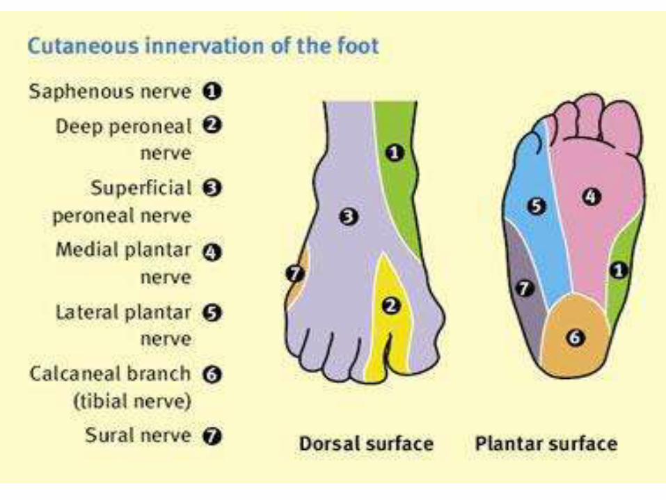
• What are the structures on the dorsum of foot and in the layers of sole?
1/14/2016 3
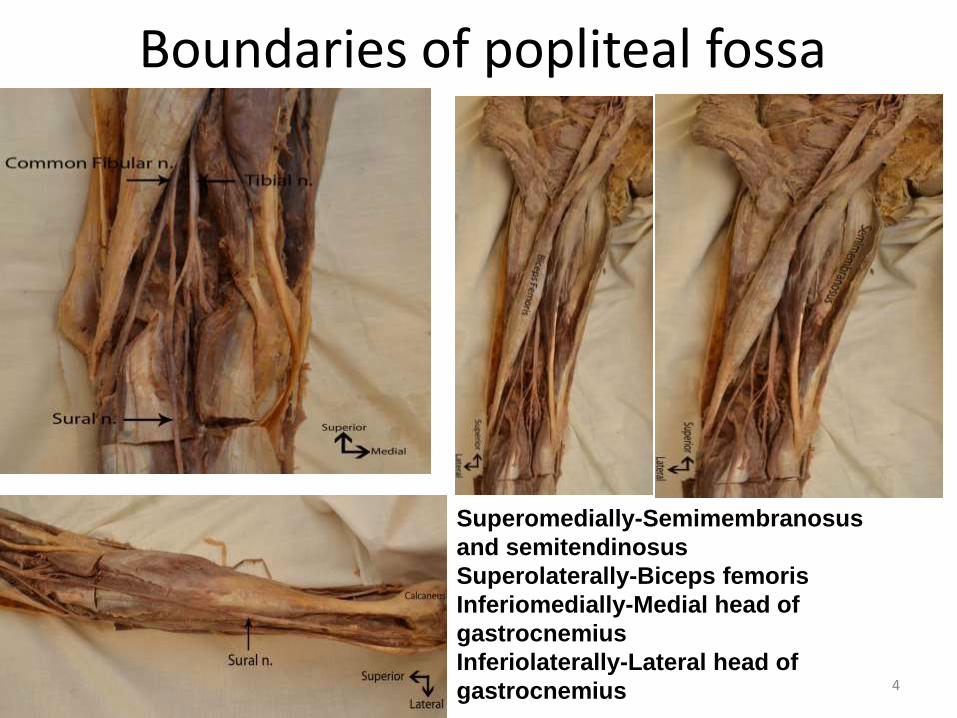
Boundaries of popliteal fossa
1/14/2016 4
Superomedially-Semimembranosus
and semitendinosus
Superolaterally-Biceps femoris
Inferiomedially-Medial head of
gastrocnemius
Inferiolaterally-Lateral head of
gastrocnemius
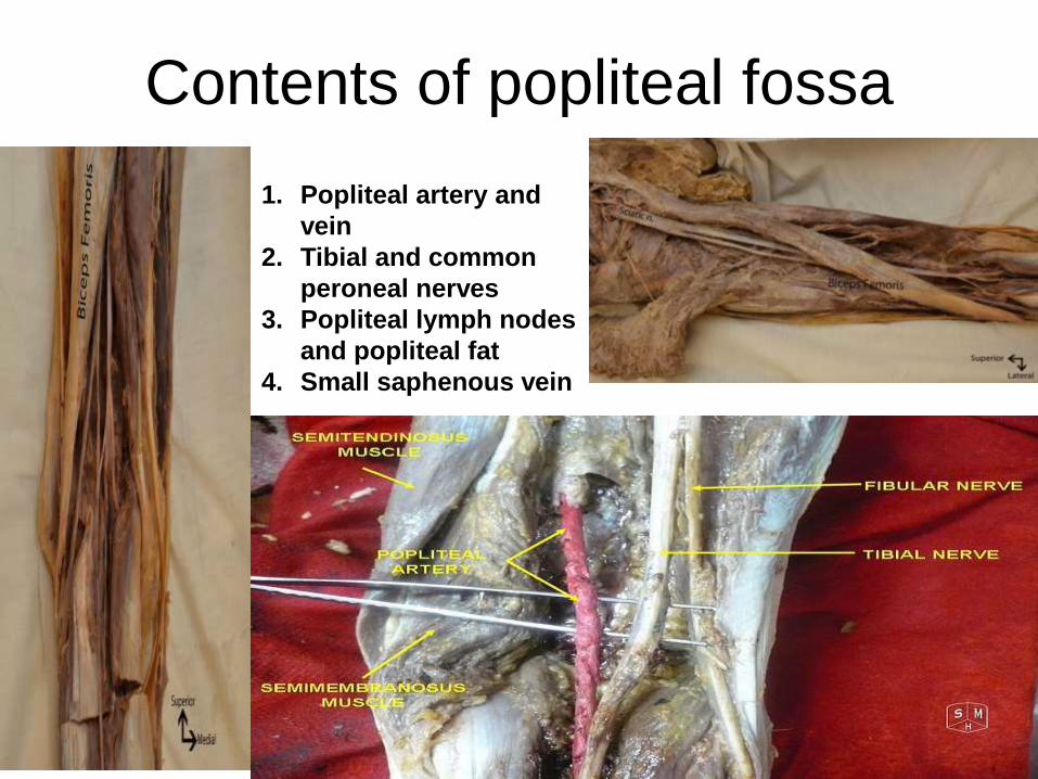
Contents of popliteal fossa
1/14/2016 5
1. Popliteal artery and
vein
2. Tibial and common
peroneal nerves
3. Popliteal lymph nodes
and popliteal fat
4. Small saphenous vein
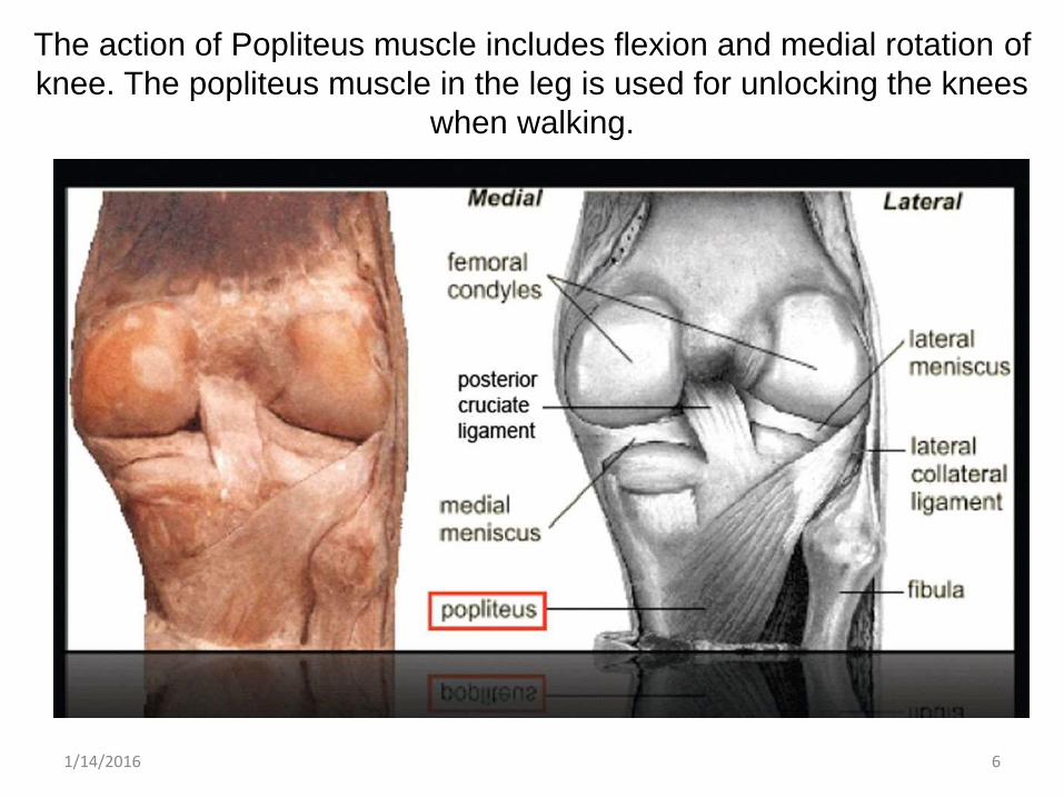
The action of Popliteus muscle includes flexion and medial rotation of
knee. The popliteus muscle in the leg is used for unlocking the knees
when walking.
1/14/2016 6
A clinical case of fracture of neck of
fibula
• A 24-year-old football player was taken to the emergency room after receiving a blow to the left leg that resulted in severe pain and inability to stand up.
• The attending physician was able to locate a very painful area just below the knee and suspected a fracture to the fibula.
• He ordered a plain AP and lateral x-ray of the leg and knee.
• A clear spiral fracture in the left fibular neck and a cracked tibial shaft were shown on the x-ray.
• The patient was given analgesics, and a thorough neurological examination was done. No signs of nerve injury were detected.
• A plaster cast was applied, and the patient was discharged.
1/14/2016 7
Leg compartments
• There are three compartment of leg.
• Anterior compartment
• Lateral compartment
• Posterior compartment
• Anterior compartment muscles are:-
• T.A.+E.H.L.+E.D.L.+P.T.
• Muscles in the lateral compartment are:-
• P.L.+P.B.
• The muscles in the posterior compartment are:-
• Gastrocnemius +Soleus + Plantaris.
• Flexor hallucis longus+Flexordigitorum longus+tibialisposterior.
• The nerve of anterior compartment of leg is deep peroneal nerve.
• The superficial peroneal nerve is for lateral compartment.
• Tibial nerve is for posterior compartment of leg.
1/14/2016 8
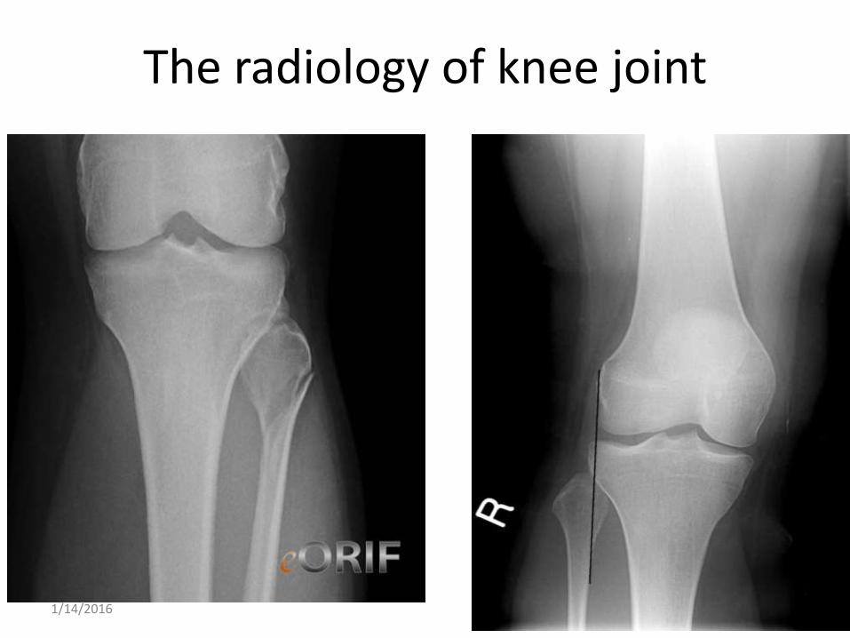
The radiology of knee joint
1/14/2016 9
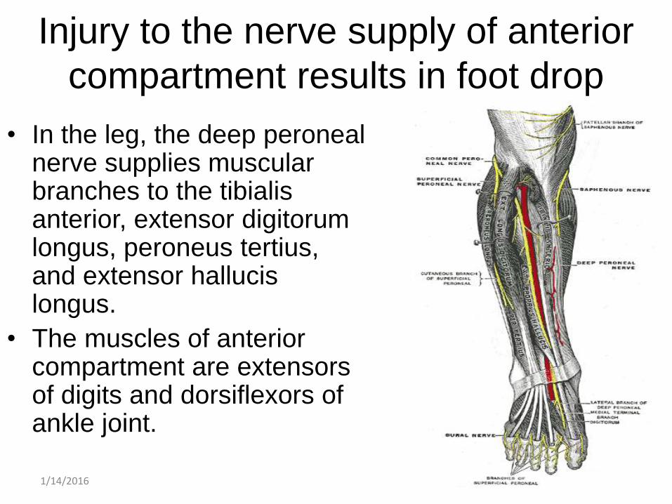
Injury to the nerve supply of anterior
compartment results in foot drop
• In the leg, the deep peroneal nerve supplies muscular branches to the tibialis anterior, extensor digitorumlongus, peroneus tertius, and extensor hallucislongus.
• The muscles of anterior compartment are extensors of digits and dorsiflexors of ankle joint.
1/14/2016 10
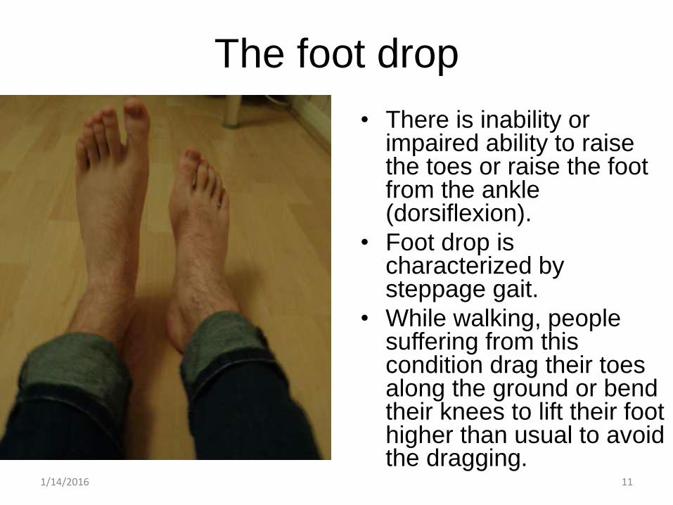
The foot drop
• There is inability or impaired ability to raise the toes or raise the foot from the ankle (dorsiflexion).
• Foot drop is characterized by steppage gait.
• While walking, people suffering from this condition drag their toes along the ground or bend their knees to lift their foot higher than usual to avoid the dragging.
1/14/2016 11
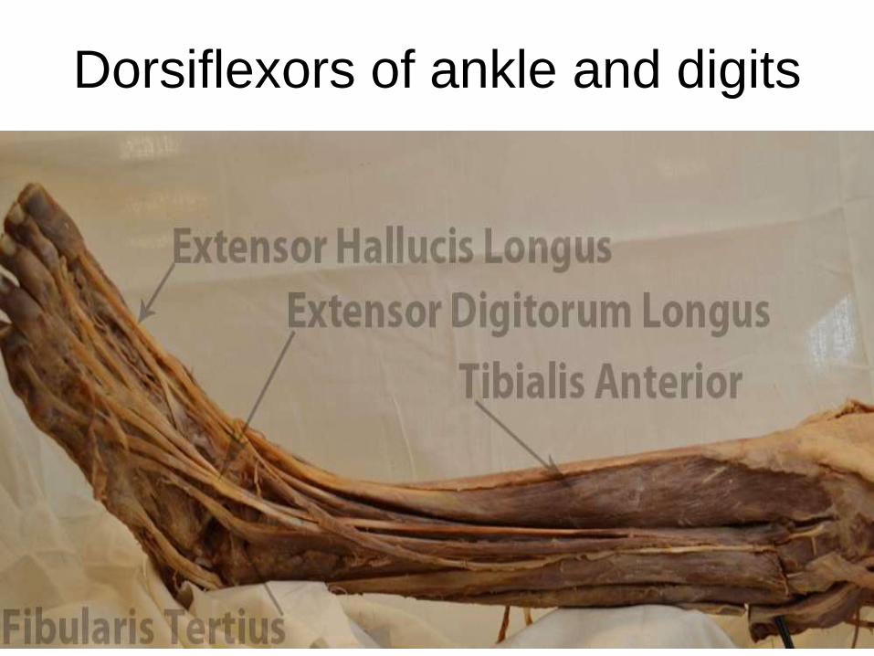
Dorsiflexors of ankle and digits
1/14/2016 12
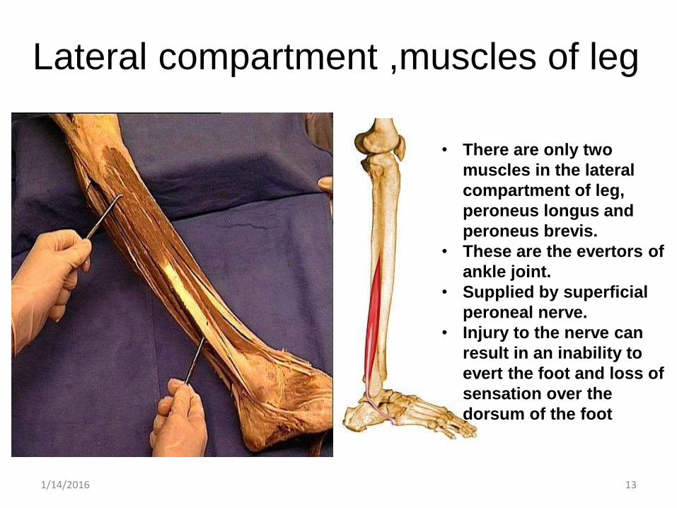
Lateral compartment ,muscles of leg
1/14/2016 13
• There are only two
muscles in the lateral
compartment of leg,
peroneus longus and
peroneus brevis.
• These are the evertors of
ankle joint.
• Supplied by superficial
peroneal nerve.
• Injury to the nerve can
result in an inability to
evert the foot and loss of
sensation over the
dorsum of the foot
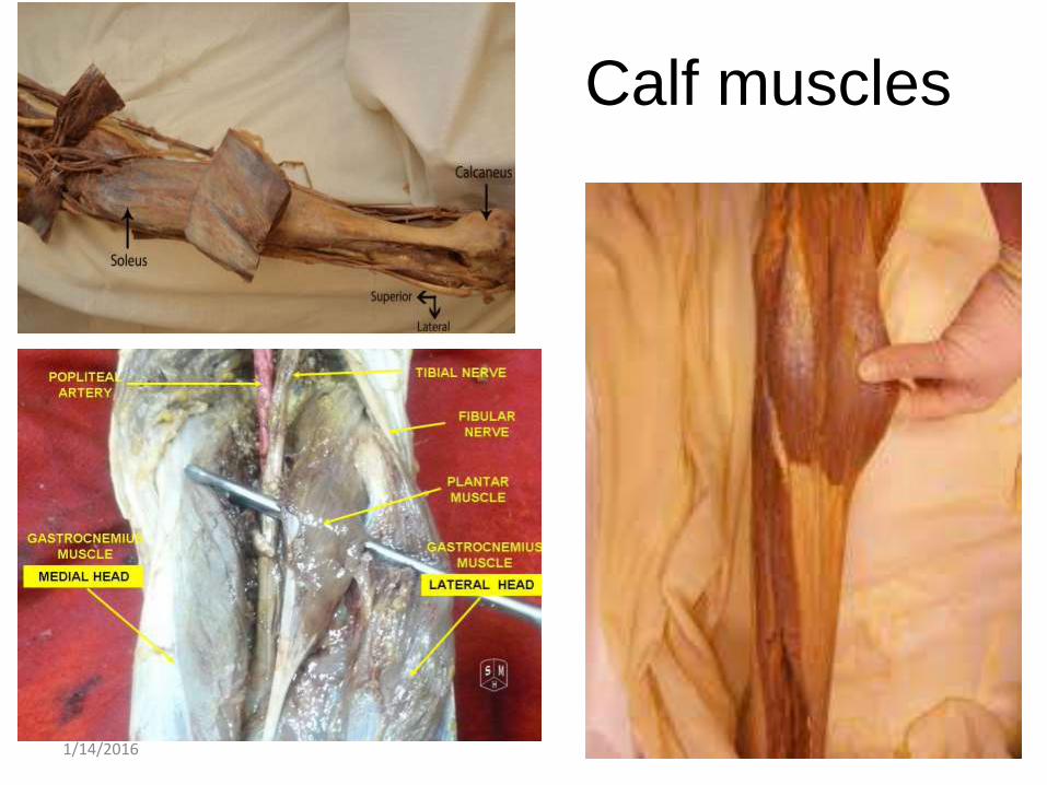
Calf muscles
1/14/2016 14
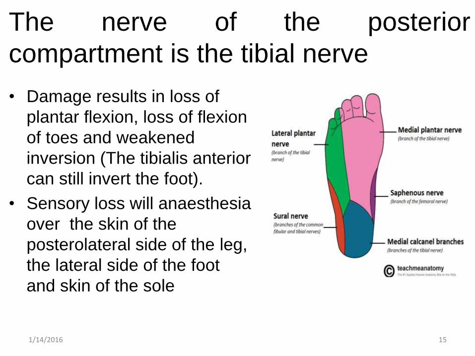
The nerve of the posterior
compartment is the tibial nerve
• Damage results in loss of
plantar flexion, loss of flexion
of toes and weakened
inversion (The tibialis anterior
can still invert the foot).
• Sensory loss will anaesthesia
over the skin of the
posterolateral side of the leg,
the lateral side of the foot
and skin of the sole
1/14/2016 15
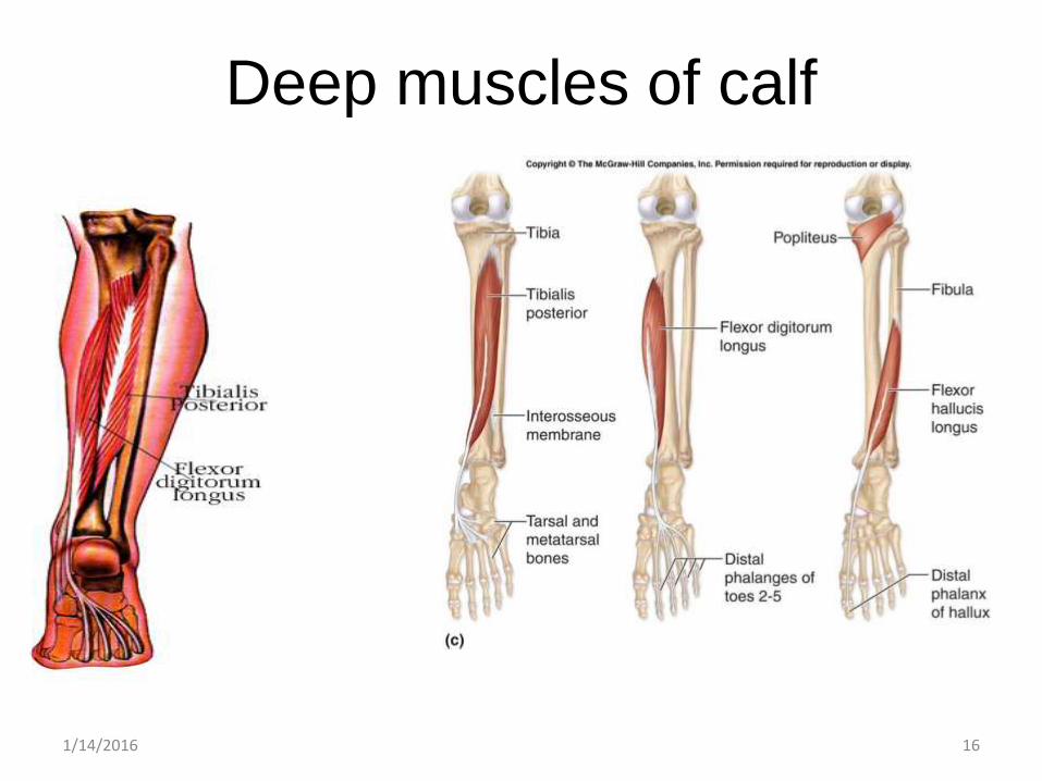
Deep muscles of calf
1/14/2016 16
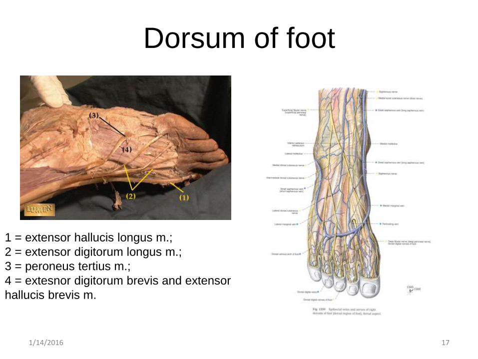
Dorsum of foot
1/14/2016 17
1 = extensor hallucis longus m.;
2 = extensor digitorum longus m.;
3 = peroneus tertius m.;
4 = extesnor digitorum brevis and extensor
hallucis brevis m.
1/14/2016 18
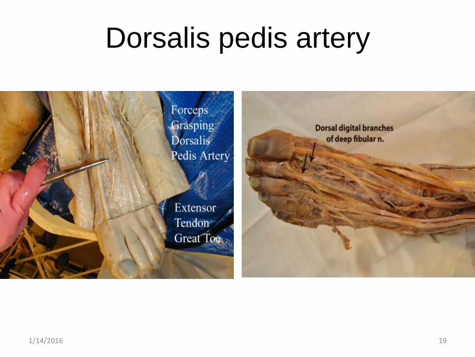
Dorsalis pedis artery
1/14/2016 19
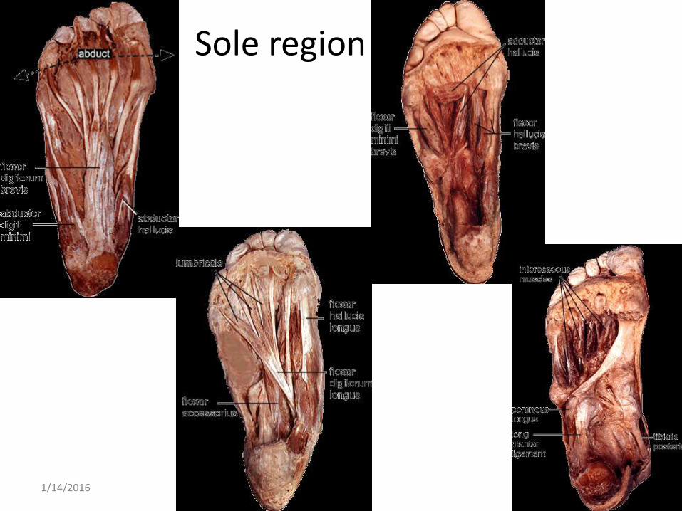
Sole region
1/14/2016 20
References
• http://www.rad.washington.edu/academics/academic-sections/msk/muscle-atlas/lower-body/peroneus-brevis
• http://www.rad.washington.edu/academics/academic-sections/msk/muscle-atlas/lower-body/peroneus-longus
• http://www.gla.ac.uk/t4/~fbls/files/fab/tutorial/anatomy/sole3.html
• https://www.studyblue.com/notes/note/n/lower-limb/deck/1431917
• http://www.slideshare.net/Ramzanken/lower-limb-mcqs
1/14/2016 21


![A Cystic Mass in the Popliteal Fossa and Its Differential ......[2]. Therefore, surgeons may mistake ganglionic cysts in the popliteal fossa for Baker’s cysts or meniscal cysts.](https://static.fdocuments.in/doc/165x107/5f8ba0d5beaa983e540e6dd7/a-cystic-mass-in-the-popliteal-fossa-and-its-differential-2-therefore.jpg)