Popliteal angle: Bilateral
49
28‐05‐14 1 DEFICIENCE MOTRICE CEREBRALE: INTERVENTIONS EN ORTHOPEDIE PEDIATRIQUE CAROLINE FORSYTHE MD, FRCSC RESUME • SURVEILLANCE DES HANCHES • TRAITMENTS NONCHIRURGICALES – BOTOX – PLATRES D’INHIBITION • APPROCHE CHIRURGICALE
Transcript of Popliteal angle: Bilateral

28‐05‐14
1
DEFICIENCE MOTRICE CEREBRALE:
INTERVENTIONS EN ORTHOPEDIE PEDIATRIQUE
CAROLINE FORSYTHE
MD, FRCSC
RESUME
• SURVEILLANCE DES HANCHES
• TRAITMENTS NONCHIRURGICALES
– BOTOX
– PLATRES D’INHIBITION
• APPROCHE CHIRURGICALE

28‐05‐14
2
CEREBRAL PALSY
• NON PROGRESSIVE INJURY TO BRAIN
OCCURING PRIOR TO AGE 2 YEARS
• PROGRESSIVE MUSCULOSKELETAL
PATHOLOGY
• PREVALENCE 2‐3/1000
Gross Motor Function Classification SystemGold Standard
GMFCS Levels
Ambulatory Cerebral Palsy : 65%
• Level I: Walks and runs
• Level II: Walks; no aids
• Level III: Walks with aids
Non-Ambulatory Cerebral Palsy : 35%
• Level IV: Stands; wheelchair
• Level V: Wheelchair; dependant

28‐05‐14
3
GMFCS Level I
GMFCS Level II

28‐05‐14
4
GMFCS Level III
GMFCS IV

28‐05‐14
5
GMFCS Level V
PROGRESSIVE MUSCULOSKELETAL DEFORMITY

28‐05‐14
6
Progressive Musculoskeletal Pathology
Primary AbnormalitiesAbnormal muscle toneMuscle imbalanceWeakness
Dynamic Contracture
Developmental Delay
Static Contracture
Bony Deformity
Joint Instability
•Functional Impairment/ Disability
•Degenerative Arthritis
•Pain
Effect of Bone Growth
Longitudinal Muscle Growth
Static Physical ExaminationHip Flexors (Ilio-Psoas)
• Thomas test: for hip flexion contracture• Flex contralateral hip to neutralize lumbar lordosis

28‐05‐14
7
• Hip Abduction– Hips & knees flexed– Add longus,brevis,magnus
• Hip Abduction:– Hips & knees extended– Tests for Gracilis tightness
Static Physical Examination
Hip Adductors
Static Physical ExaminationKnee: Hamstring Length
• Straight leg raise (hamstring tightness)
• Knee extension: to measure flexioncontracture

28‐05‐14
8
Popliteal angle: Unilateral(Test of Hamstrings)
• Amount of kneeextension with hipflexed at 90º
• “Functional HScontracture”
• Normal Range– 0º to 30º (< 6 yrs)– 15º to 40º (7-12 yrs)– 20º to 50º (13 - 18yrs)
Popliteal angle: Bilateral(Hamstring Shift Test)
• Popliteal angle withcontralateral hip & kneeflexed so that pelvis isneutralized (tippedposteriorly)
• “True hamstringcontracture”
• Difference between uni-and bilateral poplitealangle: Hamstring Shift

28‐05‐14
9
Popliteal angle: Bilateral(Hamstring Shift Test)
• Hamstring shift > 20ºindicates excessiveanterior pelvic tilt– Tight hip flexors
– Weak abdominals
– Weak hip extensors
• In presence of increasedlordosis: Apparent HScontracture with normalHS length
Static Physical ExaminationRectus Femoris
• Duncan Ely test (Grade 0 – 3)– Rectus femoris tightness is assessed prone

28‐05‐14
10
Static Physical ExaminationDorsiflexion of Ankle
Dorsiflexion with knee flexedtests tightness of soleus &
gastroc
Dorsiflexion with knee extended(Silverskjiold test) tests tightness ofgastrocnemius
Foot must be supinated to lock subtalar joint prior todorsiflexion to prevent midfoot break (valgus escape)
Static Physical Examination: Foot & Ankle• Hindfoot varus
– Tibialis posterior tightness– Weak peroneals
• Hindfoot valgus– Tight peroneals– Equinus
• Supination– Tight tibialis anterior
• Tibialis anterior– Strength– Voluntary (selective) control– Confusion test
• Hallux valgus

28‐05‐14
11
Tibial Torsional Profile
Thigh Axis
Foot Axis
Bimalleolar Axis
Static Physical ExaminationTorsional Profile (prone)
• Thigh foot angle– Normal: 10º
– (0º - 20º external)
• Trans (bi)malleolar axis– Measures tibial torsion
– Normal: 20º (0º to 35º external)

28‐05‐14
12
Measuring Femoral Anteversion
Femoral Anteversion+ External Tibial Torsion

28‐05‐14
13
Abnormal Muscle Tone• Spasticity
–––––
Velocity dependent hypertonia: Clasp knifeTardieu: R1 & R2 catchHyperreflexia: increased deep tendon reflexesClonusQuantified by modified Ashworth scale
• Dystonia–––––
Abnormal and distorted postures (trunk)Variable muscle tone induced by movementLow tone in supine positionMay “shake loose”No hyper reflexia
• Choreoathetosis––––
When patient initiates a movementMassive involuntary movementsMotor overflow to other muscle groupsPosturing of fingers or limbs
•
•
Mixed tone
Hypotonia
Muscle Strength & Control• Standard Manual Testing
– Graded 0 – 5
– All relevant muscle groups– Tested if adequate selective control is present
• Selectivity0 = Patterned movement only1 = Partially isolated movements (reactions/substitutions)2 = Completely isolated movement
• Confusion test (mass flexion pattern)– Sitting: antigravity hip flexion associated with ankle
dorsiflexion
• Sitting balance/Trunk balance

28‐05‐14
14
IGA
INSTRUMENTED GAIT ANALYSIS
Spastic diplegia –pre‐op gait lab

28‐05‐14
15
CINEMATIQUE
CINEMATIQUE, MOMENT, PUISSANCE

28‐05‐14
16
EMG
TREATMENT

28‐05‐14
17
TREATMENT PRINCIPLES
• Prevention of contractures, hip subluxation• Strengthening
• Optimizing biomechanics
• Correction of contractures & deformities
SUIVI DES HANCHES

28‐05‐14
18
CP – Hip Surveillance – Australian Consensus
• GMFCS 1 – AP Pelvis at 12 – 24 months age
• Review at 3 yo
• Review at 5 yo
• GMFCS II AP PELVIS AT 12 – 24 MONTHS AGE
• Repeat assessment q 12 months until MP stable
• Review age 4‐5, then age 8‐10
CP – Hip Surveillance – Australian Consensus
• GMFCS III, IV, V– AP PELVIS AT 12‐24 MONTHS
– SURVEILLANCE Q 6 MONTHS
– REVIEW AT 7 YEARS
– IF MP STABLE AND LESS THAN 30%, DISCONTINUE
UNTIL PRE‐PUBERTY. RESUME Q 12MONTH AP PELVIS
AT PRE‐PUBERTY UNTIL SKELETAL MATURITY
28‐05‐14
19
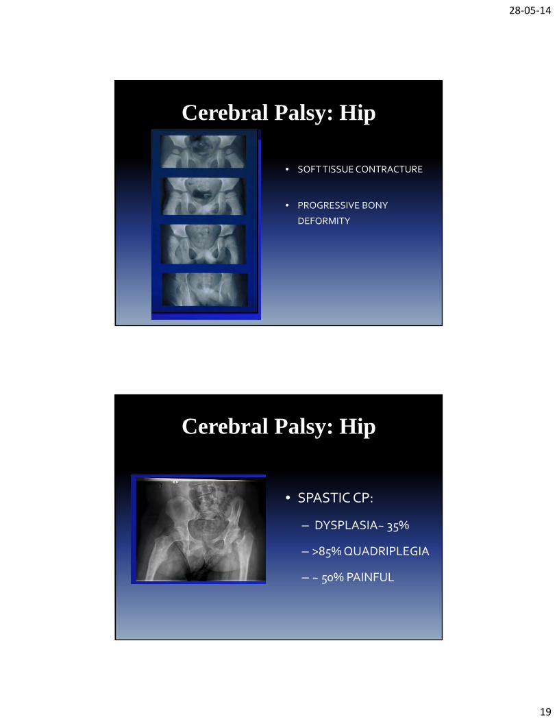
Cerebral Palsy: Hip
• SOFT TISSUE CONTRACTURE
• PROGRESSIVE BONY
DEFORMITY
Cerebral Palsy: Hip
• SPASTIC CP:
– DYSPLASIA~ 35%
– >85% QUADRIPLEGIA
– ~ 50% PAINFUL
28‐05‐14
20
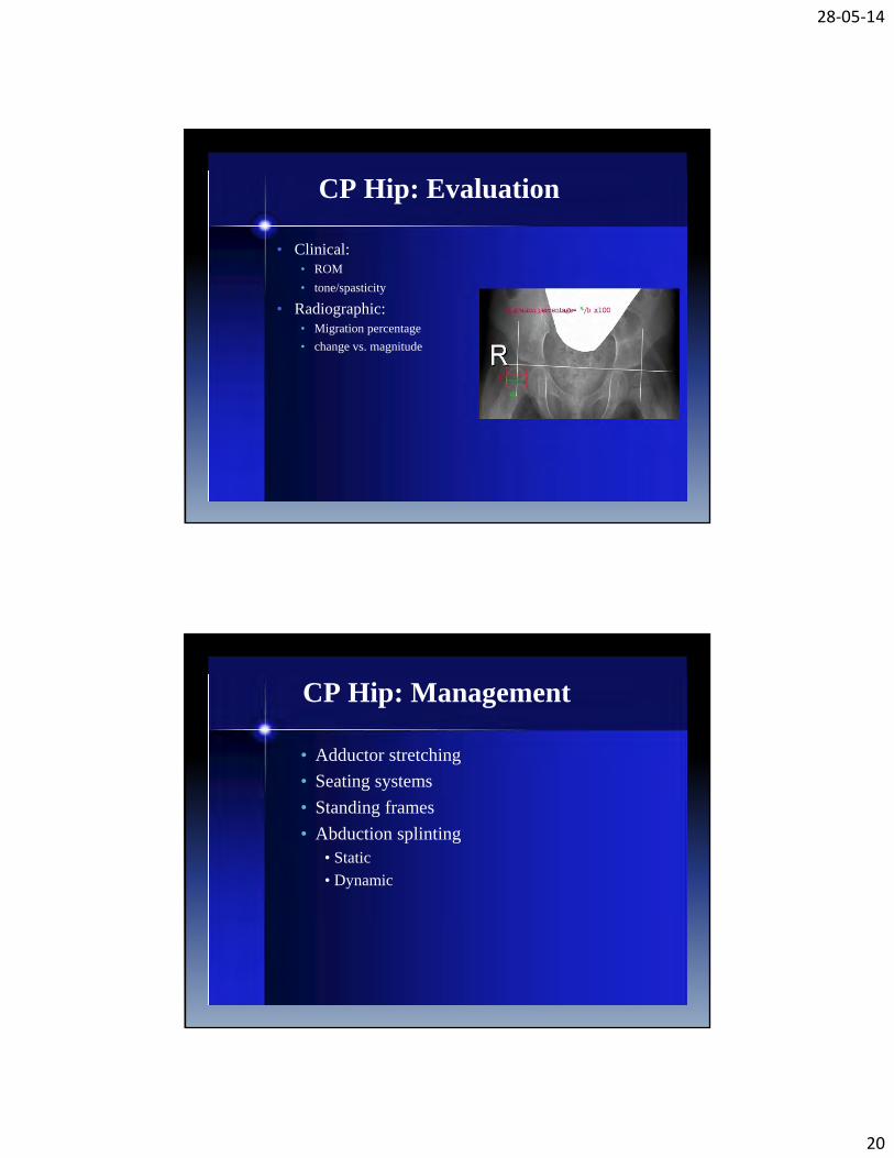
CP Hip: Evaluation
• Clinical:• ROM• tone/spasticity
• Radiographic:• Migration percentage• change vs. magnitude
CP Hip: Management
••••
Adductor stretchingSeating systemsStanding framesAbduction splinting
• Static• Dynamic

28‐05‐14
21
CP Hip: Management
• Medical tone management• Baclofen
• Parenteral therapy• Phenol to obturator nerve• Botox to adductors
• associated stretching/splinting/etc.
CP Hip: Surgery
• Soft tissue releases:• <30% subluxation/<4 y.o.• Adductor longus• Iliopsoas: medial approach
vs.. over-the-brim• Anterior Obturator
neurectomy in non-ambulators
• Early release may preventsubluxation in up to 60%

28‐05‐14
22
CP Hip: Surgery
• Bony: 30 - 50%subluxation• Varus Derotation
Osteotomy• Unilateral vs.. Bilateral• Associated soft-tissue
releases
Management Summary
• <4 yo with abduction < 30 degrees and > 30% Reimer
Migration index: Soft tissue release
• <8 yo with >50% Reimer migration index – VDRO +/‐
DEGA
• > 8 yo with > 40% Reimer migration index – VDRO +/‐
DEGA

28‐05‐14
23
AUSTRALIE
• 8 YO MALE WITH
BILATERAL CP

28‐05‐14
24
CHIRURGIE BILATERALE SANS PLATRE
CANADA
• FILLE 6 ANS
• GMFCS 4
• BILATERAL DYSPLASIE
AVEC DYSPLASIE DU
BASSIN
28‐05‐14
25
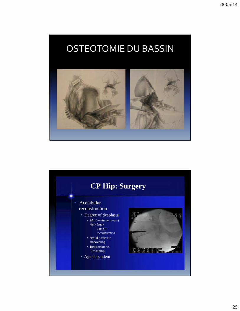
OSTEOTOMIE DU BASSIN
CP Hip: Surgery
• Acetabularreconstruction• Degree of dysplasia
• Must evaluate area ofdeficiency
?3D CTreconstruction
• Avoid posterioruncovering
• Redirection vs.Reshaping
• Age dependent

28‐05‐14
26
HIP SURVEILLANCE
• REGISTRY OF CEREBRAL PALSY
• ORGANIZE MEETING OF STAKEHOLDERS
TO DEVELOP SURVEILLANCE PLAN
BOTOX

28‐05‐14
27
BOTOX
• INHIBITS THE RELEASE OF ACETYLCHOLINE
AT NEUROMUSCULAR JUNCTION
• RESULTS IN TEMPORARY REVERSIBLE
MUSCLE WEAKNESS
BOTOX IN CP
• LOWER EXTREMITIES
– USED FOR MORE THAN 15 YEARS
– EFFECTIVE FOR TREATMENT OF SPASTICITY (LARGE
R1>R2)
– MAX DOSE 12‐16 UNITS/KG, NO MORE THAN 300‐400
UNITS
– Q 3 MONTHS OR MORE
– NO MAXIMAL NUMBER OF INTERVENTIONS

28‐05‐14
28
POST BOTOX SPLINTING
• ADDUCTORS –ABDUCTOR PILLOW AT
NIGHT
• HAMSTRINGS – SPLINTS AT NIGHT
• GASTROC – SERIAL CASTING UNTIL 10
DEGREES DORSIFLEXION THEN FULL TIME
AFOS
EUROPEAN JOURNAL OF NEUROLOGY CONSENSUS STATEMENT
• BOTOX EFFECTIVE IN MANAGEMENT OF
SPASTIC EQUINUS (LEVEL A)
• SIMILAR TO SERIAL CASTING IN
MANAGEMENT OF EQUINUS
• INJECTION IN ADDUCTOR AND HAMSTRINGS
MAY DELAY HIP DISPLACEMENT, BUT DOES
NOT AFFECT LONG TERM OUTCOME

28‐05‐14
29
CONSENSUS STATEMENT – EXPERT OPINION
• SERIAL CASTING SHOULD FOLLOW BOTOX
• AFOS ARE EFFECTIVE ADJUNCT
• PROLONGED STRETCHING IS AN ADJUNCT
INTERVENTION
• STRENGTHENING IS AN ESSENTIAL
ADJUNCT INTERVENTION
DELAYED VERSUS IMMEDIATE SERIAL CASTING AFTER BOTOX
• CAST REPLACED WEEKLY FOR THREE
WEEKS
• BENEFIT TO DELAY CASTS FOR 4 WEEKS
VERSUS PLACING SAME DAY

28‐05‐14
30
SERIAL CASTING
• KAY ET AL, JBJS 2004 BOTOX + SERIAL
CASTING VERSUS SERIAL CASTING ALONE
• CASTS CHANGED Q 2 WEEKS UNTIL >5
DEGREES DORSIFLEXION
• ADDITION OF BOTOX LED TO EARLIER
RECURRENCE SPASTICITY AND EQUINUS
CONTRACTURE
BOTOX UPPER EXTREMITY
• 2U/KG FOREARM
• 4 U/KG UPPER ARM
• UP TO Q 3MONTHS
• SPLINTING/EXERCISE POST BOTOX BASED
ON GOALS

28‐05‐14
31
BOTOX + SWASH FOR HIP DISPLACEMENTJBJS 2008
• BOTOX Q 6 MONTHS FOR 3 YEARS + SWASH
6 HOURS PER DAY
• SMALL BENEFIT IN PREVENTING
PROGRESSION OF CONTRACTURES
• HIP DYSPLASIA CONTINUED TO PROGRESS.
• NO CHANGE IN GMFCS
CONSERVATIVE TREATMENT

28‐05‐14
32
Management: Therapy & Orthotics
Physical Therapy
• Developmental
• Range of motion
• Stretching
• Strengthening
Orthotic Management
• Goals– Prevent deformity
– Protect a part
– Improve function
• Dynamic bracing– AFOs
• Resting splints– Maintain stretch
• Hip abduction
• Knee immobilizers
• Night time use
• Serial casting
Spasticity Management• Multi-disciplinary approach• Pharmacologic agents
– Local (injection)• Botulinum toxin• Phenol
– Systemic (oral)• Baclofen
• Adjuncts– Casting– Orthotics
• Surgical (neurosurgical)– Selective dorsal rhizotomy– Intra-thecal baclofen
• Optimal management of spasticity to avoid ordelay orthopaedic surgery

28‐05‐14
33
ORTHOPAEDIC SURGERY
Orthopaedic ManagementAmbulatory Cerebral Palsy
• Goals: Maintain or improve– Gait efficiency / Gait appearance
– Function: activities & participation
– Quality of life
• Soft tissue Procedures– Muscle/Tendon lengthening
– Tendon transfers
• Bony procedures– Corrective osteotomies
• Joint procedures– Correction of joint deformities

28‐05‐14
34
Mercer Rang’s “Birthday Syndrome”
PROGRESSIVE MS PATHOLOGY CAUSES DECLINE IN GMFCS AND GAIT
28‐05‐14
35
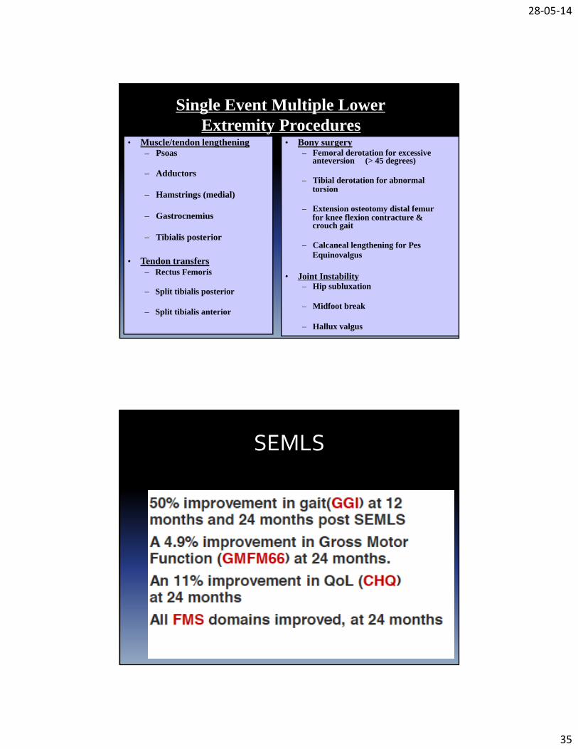
Single Event Multiple LowerExtremity Procedures
• Muscle/tendon lengthening– Psoas
– Adductors
– Hamstrings (medial)
– Gastrocnemius
– Tibialis posterior
• Tendon transfers– Rectus Femoris
– Split tibialis posterior
– Split tibialis anterior
• Bony surgery– Femoral derotation for excessive
anteversion (> 45 degrees)
– Tibial derotation for abnormaltorsion
– Extension osteotomy distal femurfor knee flexion contracture &crouch gait
– Calcaneal lengthening for PesEquinovalgus
• Joint Instability– Hip subluxation
– Midfoot break
– Hallux valgus
SEMLS

28‐05‐14
36
Soft Tissue SurgeryIntra-muscular Lengthening
• Psoas-over- the-brim IM tenotomy
• (Medial) Hamstring lengthening– Semitendinosis (IM tenotomy)– Gracilis (IM tenotomy)– Semimembranosus (aponeurosis)
• Rectus femoris transfer tosemitendinosis
• Gastrocnemius lengthening– Strayer recession– Vulpius– Baker
• Tibialis posterior intramusculartenotomy
Psoas Release at the Pelvic Brim in Ambulatory Patients with Cerebral Palsy:Operative Technique and Functional Outcome
Sutherland, D. H. M.D.*†; Zilberfarb, J. L. M.D.‡; Kaufman, K. R. Ph.D.*†; Wyatt, M. P. P.T., M.A.*; Chambers, H.

28‐05‐14
37
Lengthening and transfer of hamstrings for aflexion deformity of the knee in children with
bilateral cerebral palsyMa et al, JBJS (Br) VOL. 88‐B, No. 2, FEBRUARY 2006
Bony Surgery: Lever Arm Disease
Derotational Osteotomies forexcessive femoral anteversion
Derotational Osteotomies forexcessive tibial torsional deformity

28‐05‐14
38
••••••••••
Ankle equinus: Calf dominance
Knee and hip extended
Knee sometimes recurvatum
Other levels are usually tight
Over active Planter Flexion-KneeExtension (PF-KE) couple
Management
••
••
••
Bilateral CP– Selective gastroc lengthening
Unilateral CP– Tendoachilles lengthening
Hinged or Posterior leaf spring AFO
TYPE I: EQUINUS GAIT – TOE WALKER
• Avoid TAL in bilateral CP to avoid crouch gait
TYPE I: EQUINUS GAIT – TOE WALKER

28‐05‐14
39
––––––
Selective gastroc (soleus)Medial hamstrings lengtheningRectus femoris transfer for stiffknee gait
– IM Psoas lengthening
TYPE II: JUMP GAIT
• Ankle equinus• Knee/hip flexion• Contracture at each level• Strong PF-KE coupleManagement• Balance all levels as needed
• No heel contact: toe-toe or toe-heelgait
• Ankle neutral: No fixed equinus• Hip and knee flexed
– Proximal contractures
• PF-KE couple neutralManagement• Lengthen at knee/hip• Rectus transfer for stiff knee gait• No botox or surgery to calf• Ground Reaction AFO or solid AFO
Type III: APPARENT EQUINUS

28‐05‐14
40
•••
Ankle dorsiflexedKnee/hip flexedPatella alta: inferior pole avulsion fx
Causes•••••
Tight Psoas & HamstringsWeak quadsWeak soleusNatural historyIatrogenic from (over) TAL in bilateral CP
Management
•••
•
•
Lengthen at hip and kneeHS length may be normal with increased ant pelvic tiltExtension osteotomy of distal femur +/- patellartendon advancement or shorteningMay need lateral column lengthening to correctplanovalgus & forefoot abductionGround Reaction AFO
TYPE IV: CROUCH GAIT
FOOT & ANKLE PATHOLOGY IN CP
• Equinus
• Equinovarus
• Equinovalgus
• Hallux Valgus
• Tibial Torsion

28‐05‐14
41
EQUINOVALGUS DEFORMITY IN CP
• Common in spastic diplegia
• Tight gastrocnemius & weak tibialis posterior
• Peroneals may become secondarily short
• Midfoot break to accommodate plantarflexion whilekeeping foot flat on the ground
• Forefoot abducted/externally rotated
EQUINOVALGUS DEFORMITY IN CP
•Incompetent subtalar joint cannot “lock” (mid-foot break)
•Lever arm disease: weak push off
•Ground reaction force passes posterior and lateral to knee
•Genu valgum
•Callosity/pain over prominence of talar head medially
•Shoe fitting and shoe wear problems

28‐05‐14
42
Spastic DiplegiaEquino/Plano Valgus Deformity
• Rigid AFO: if flexible
• Lateral column (calcaneal) lengthening, medial tightening
• Subtalar fusion
EQUINOVARUS IN CP
• Common in hemiplegia
• Imbalance betweeninverters and everters– Overactive tibialis posterior
(varus in stance & swing)– Overactive tibialis anterior
(varus in swing phase)– Weak peroneals
• Intoed gait• Inverted heel (tib post)• Supinated forefoot (tib ant)

28‐05‐14
43
CONSEQUENCES OF EQUINOVARUS• Impaired stability in
stance phase
• Poor foot clearance
• Callosity along lateralborder and pain
• Shoe fitting/ shoe wear
• Lever arm disease
ASSESSMENT OF EQUINOVARUS
• Static range of motion
• Gait assessment– Video of foot & ankle– Kinematics & kinetics– Pedobarograph
• Dynamic EMG– Surface: gastroc, tib ant– Fine wire: tib post
• Radiographs

28‐05‐14
44
Spastic HemiplegiaEquino-varus deformity
• Split tibialis posterior tendon transfer (SPOTT)••••
Younger childFlexible deformityStance phase varusNo significant supination
• Split tibialis anterior tendon transfer (SPLATT)• Swing phase varus,• Dropfoot• Forefoot supination
• Split tibialis anterior tendon transfer +intra-muscular lengthening of tibialis posteriorand gastrocsoleus lengthening (as needed)
• Older child• Stiffer deformity• When both tendons are involved in varus
SPLATTEquinovarus Deformity

28‐05‐14
45
SPOTTEquinovarus Deformity
Hallux Valgus• Common in diplegics with
planovalgus feet
• Painful bunion/ callosity over1st MT head
• Associated foot malalignment– Equinovalgus
– External tibial torsion
• Management– 1st MTP fusion
– Corrected ass. malalignment

28‐05‐14
46
1. What type of cerebral palsy?
2. What surgery for equinus?
3. What is a common complication of surgery for equinusin this type of CP?
1. What type of cerebral palsy? Unilateral or R Hemiplegia
2. What surgery for equinus? R TAL
3. What is a common complication of surgery for equinusin this type of CP? Recurrent equinus

28‐05‐14
47
SpasticDiplegia
Age 5
Age 72 yearspost ?
1.What type of cerebral palsy?
2.What surgery did this child have?
3.What is the outcome?
SpasticDiplegia
Age 5
Age 72 years
postTAL’s
1.What type of cerebral palsy? Bilateral SpasticDiplegia
2.What surgery did this child have? Bil TAL’s
3.What is the outcome? Calcaneus/crouch

28‐05‐14
48
• FEMORAL
SHORTENING +/‐
HAMSTRINGS
LENGTHENING +
PATELLAR TENDON
PLICATION

28‐05‐14
49
PATELLAR TENDON PLICATION