COVER 200 mm PAGE 5 199 mmPAGE 6 199 mm … · nerve block and fascia iliaca block INTERNAL...
8
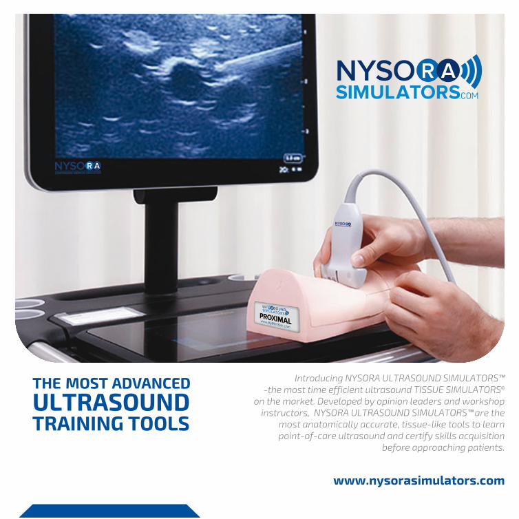
Introducing NYSORA ULTRASOUND SIMULATORS™ -the most time efficient ultrasound TISSUE SIMULATORS © on the market. Developed by opinion leaders and workshop instructors, NYSORA ULTRASOUND SIMULATORS™ are the most anatomically accurate, tissue-like tools to learn point-of-care ultrasound and certify skills acquisition before approaching patients. THE MOST ADVANCED ULTRASOUND TRAINING TOOLS MS2-PVT MS2-TAP www.nysorasimulators.com MS2-VASBCL
Transcript of COVER 200 mm PAGE 5 199 mmPAGE 6 199 mm … · nerve block and fascia iliaca block INTERNAL...
EUROPE:Slachthuislaan 1, Bus 1023000 Leuven, Belgium+32 492 80 76 76
USA:2581 Broadway, Suite 183New York, NY 10025+ 212-726-9900
[email protected] http://medxpress.pro/
Transverse Abdominis Plane SIMULATOR™Epidural SIMULATOR™
Paravertebal (PVT) and Errectores Spinae Blocks SIMULATOR™TAP and Quadratus Lumborum SIMULATOR™
ULTRASOUND SIMULATORS™ are shipped with carry/storage base and case, cover protectors, conven-ient accessory storage tray, work surface tray, convenient laminated poster with instructions for use and
care for each model.
ESSENTIAL TRAINERULTRASOUND SIMULATOR™
Facilitates acquisition of essential skills of using ultrasoundAllows needle-target practice in plane and out of plane.Absolute must for anyone wanting to start utilizing point of care ultrasound
INTERNAL LANDMARKST3-T8; Ribs 3-8; Paravertebral space; Pleura; Spinous processes; Vertebrae T3 to T8
EXTERNAL LANDMARKS
OPTIONAL
Left and right inferior angle of scapula; Vertebra prominens; Spinous processes, Paravertebral muscles
Digital camera connected to a tablet for monitoring of needle and catheter entry into the paravertebral space and to confirm the correct ultrasound-guided placement.
OPTIONALDigital camera connected to a tablet for monitoring of needle and catheter entry into the epidural space and to confirm the tactile or ultrasound information.
Facilitates learning essential anatomy for TAP and QL blocks
Allows training on injection techniques for TAP and QL
INTERNAL LANDMARKSTransverse abdominis muscles; Internal and external oblique muscles; Quadratus lumborum muscles; Latissimus dorsi muscles
EXTERNAL LANDMARKSUmbilicus; Rectus abdominous
Facilitates learning of anatomy, identification of interspaces and placement of the needle in the epidural space with loss of resistance technique or ultrasound.Options include a camera for visual confirmation of the needleplacement
INTERNAL LANDMARKSL1-L5; Sacrum; Epidural space; Spinal processes; Laminae; Articulate joints; Intervertebral space; Dura and Ligamentum Flavum
Full size anatomical structures in easy to carry footprint.
EXTERNAL LANDMARKSIliac crest
Facilitates acquisition of essential skills of using ultrasoundAllows needle-target practice in plane and out of planeAbsolute must for anyone wanting to start utilizing point ofcare ultrasound
INTERNAL LANDMARKSArtery; Vein; Nerve
EXTERNAL LANDMARKSNot applicable
Introducing NYSORA ULTRASOUND SIMULATORS™ -the most time efficient ultrasound TISSUE SIMULATORS©
on the market. Developed by opinion leaders and workshop instructors, NYSORA ULTRASOUND SIMULATORS™ are the
most anatomically accurate, tissue-like tools to learn point-of-care ultrasound and certify skills acquisition
before approaching patients.
THE MOST ADVANCEDULTRASOUNDTRAINING TOOLS
MS2
-QU
AM
S2-E
PI
TORSO (cont.) TORSO
MS2
-TR
N
MS2
-PV
TM
S2-T
AP
www.nysorasimulators.com
COVER 200 mm PAGE 5 199 mmPAGE 6 199 mm BACKCOVER 200 mm
VASCULAR ACCESS
ABOUT USNYSORA US-SIMULATORS™ are manufactured in EU and distributed by MedXpress.Pro.MedXPress.Pro is founded and managed by leaders and innovators in ultrasonography and education. We use our keen insight into real life clinical practice and educational challenges to develop products that improve patient safety. We believe that our products add additional functionality, facilitate ultra-sound skills knowledge acquisition, increase the educational experience, help standardize training and allow introduction of the testing process to document knowledge-acquisition and retention.
DESIGN Each ULTRASOUND SIMULATOR™ utilizes TISSUE SIMULATOR™ Technology TST has been designed to optimize the develop-ment of specific knowledge and skills using landmark and ultrasound guided procedural techniques. All models contain internal and external landmarks necessary to practice ultra-sound visualization and placement of needle and catheter necessary to accomplish peripheral nerve blocks, vascular access, or perform other Point-Of-Care ultrasound interven-tions.
FOOTPRINTThe system footprint is small to facilitate the utilization of mul-tiple units simultaneously, making it ideal for workshops and simulation labs. Small footprint and standardized packing size allows for easy storage and transport.
EXPERT INPUTEach ULTRASOUND SIMULATOR™ has been reviewed by lead-ers in the field of Point-Of-Care ultrasound and approved to ensure that all the relevant external and internal landmarks for taught procedures are included and accurate.
NYSORA ULTRASOUND SIMULATORS™
Subclavian Vein Canulation SIMULATOR™
MS2
-VAS
BCL
Facilitates acquisition of essential skills of using ultrasoundAllows needle-target practice in plane and out of planeAbsolute must for anyone wanting to start utilizing point of care ultrasound
INTERNAL LANDMARKSRectus abdominis; Transverse abdominis; Internal oblique; External oblique; Pectineus; Essential Sheaths
EXTERNAL LANDMARKSUmbilicus; Rectus abdominis; Abdominal wall
Allows learning imaging of the subclavian veinRealistically built to mimic challenging needle insertionTeaches skills in avoiding coracoid process for success-ful needle placement
INTERNAL LANDMARKSCoracoid process, clavicle, subclavian/axillary vein
EXTERNAL LANDMARKSSubclavian space, shoulder girdle, clavicle
ML006 RevD
Mid-Axillary line
Spinous Processes
Umbilicus
Optional Camera to Visually ConfirmCorrect Needle Placement
L4/L5
UltrasoundTissue Simulator(TM)
Ribs
Delto-pectoral grooveCoracoid Process
Actual ULTRASOUND SIMULATORS image Actual ULTRASOUND SIMULATORS image
Actual ULTRASOUND SIMULATORS image
Actual ULTRASOUND SIMULATORS image
Actual ULTRASOUND SIMULATORS image
Actual ULTRASOUND SIMULATORS image Actual ULTRASOUND SIMULATORS image
LOWER EXTREMITY NERVE BLOCK SIMULATORS
Facilitates knowledge and skills acquisition to perform femoral nerve block and fascia iliaca block
INTERNAL LANDMARKSFemoral, obturator, lateral cutaneous nerves; Saphenous nerves; Facial ilica; Facia lata; Iliopsoas muscle; Pectineus muscle; Sartorius muscle; Adductor muscles; Femoral artery; Femoral vein
EXTERNAL LANDMARKSInguinal crease; Anterior superior iliac spine; Proximal thigh; Hips Pubic tubercle; Inguinal Ligament; Femoral Crease
INTERNAL LANDMARKSGreat trochanter; Ischial tuberosity; Sciatic nerve; Gluteus maximus
EXTERNAL LANDMARKSGluteus maximus; Gluteal crease; Femur Trochanter
Facilitates knowledge and skills acquisition to recognize ulnar and radial /rarteries and perform ulnar and median nerve blocks.
INTERNAL LANDMARKSRadius, ulnar carpal bones; Radial and ulnar arteries; Median and ulnar nerves; Palmares longus; Radial Artery; Ulnar artery; Deep and supperficial muscles of the forearm
EXTERNAL LANDMARKSUlnar head; Radius; Ulna; Palmar crease; Tendons of wrist flexors; Thenar ligament; Veins; Hypothenar ligament
Facilitates knowledge and skills acquisition to perform Axillary BlockAllows injection practice
Allows injection practice
INTERNAL LANDMARKSRadial nerve; Ulnar nerve; Median nerve; Musculocutaneous nerves; Axillary artery; Axillary Vein; Deltoid muscle; Biceps muscle; Coracobrachialis muscle
EXTERNAL LANDMARKSAxillary fossa; Pectoralis muscle; Trapezus muscle; latissimus Biceps Brachialis muscle; Deltoid Muscle
dorsi muscle;
Facilitates knowledge and skills acquisition to perform popliteal sciatic and IPAC blocks
INTERNAL LANDMARKSPopliteal artery and vein; Common peroneal nerve; Tibial nerve;Semitendinosus muscle; Semimembranosus muscle; Biceps femoris muscle; Distal part of femur
EXTERNAL LANDMARKSPopliteal fossa; Tendon of Biceps Femoris Muscle; Tendon of Semimembranosus Muscle; Popliteal Fossa; Ppopliteal Crease; Popliteal triangle
INTERNAL LANDMARKSTibial nerve; Peroneal nerves (superficial and deep); Sural nerve;Saphenous nerve; Posterior and anterior tibial artery and vein;Tibia Fibula; Medial malleolus
EXTERNAL LANDMARKSTibia; Lateral malleolus; Medial malleolus; Side of the foot;Dorsus of the foot
Facilitates knowledge and skills acquisition to perform Suprascapular nerve block for analgesia of the shoulder without phrenic nerve paralysis
INTERNAL LANDMARKSClavicle; Scapula; Humerus; Suprascapular nerves; Infraspinatus muscle; Supraspinatus muscle; Suprascapular artery; Trapezius muscle; Circumflex artery
EXTERNAL LANDMARKSScapula; Clavicle
Facilitates knowledge and skills acquisition to perform femoral triangle and adductor canal block for analgesia of the kneeAllows injection practice
INTERNAL LANDMARKSSartorius muscle; Vastus medialis muscle; Adductors femur; Femoral artery; Femoral vein; Saphenous nerve; Relevant fascial sheats
EXTERNAL LANDMARKSMid and distal thigh; Knee; Quadriceps Femoris muscle; Sartorius Muscle
INTERNAL LANDMARKSSternocleidomastoid muscles; Carotid artery; Internal jugular vein;Subclavian artery; Clavicle; First rib; Thiroid Gland; Pleura
EXTERNAL LANDMARKSSternocleidomastoid muscles; Clavicle; Cricoid cartilage; Supraclavicular fossa; Neck; Mandible
Facilitates understanding of the anatomy of the breast and tissue layers of importance in breast surgery and local anesthetic infiltration techniques
INTERNAL LANDMARKSPectoralis major and minor muscles; Serratus muscles; Axillary arteryAxillary vein; Cords of the brachial plexus; Clavicle; Coracoid process;Ribs 1-4
EXTERNAL LANDMARKSBreast anatomy; Sternocleidomastoid muscle
Facilitates skills acquisition to perform Infraclavicular brachial plexus and PEC blocks
INTERNAL LANDMARKSPectoralis major and minor muscles; Serratus muscles; Axillary artery and vein; Chords of the brachial plexus; Clavicle; Coracoid process; Ribs 1-4; Pleura and Chest Cavity
EXTERNAL LANDMARKSClavicle; Deltopectoral groove; Pectoralis muscle; Chest wall;Coracoid process; Shoulder; Deltoid muscle
Combined Infraclavicular/PEC Nerve Block SIMULATOR™ Wrist Block SIMULATOR™ Popliteal and IPAC Nerve Block SIMULATOR™ Ankle Block SIMULATOR™
Open Breast Infraclavicular Visual SIMULATOR™
Suprascapular Nerve Block SIMULATOR™ Adductor/Saphenous Combined Nerve Block SIMULATOR™
Axillary Brachial Plexus SIMULATOR™ Femoral and Fascia Iliaca SIMULATOR™ Sciatic Transgluteal SIMULATOR™
VASCULAR ACCESSVascular Access IJ SIMULATOR™
INTERNAL LANDMARKSAnterior scalene muscle; Middle scalene muscle; Sternocleido-mastoid muscle; Carotid artery; Internal jugular vein; Subclavian artery; Subclavian vein; Clavicle; Stellate ganglion; C2-C7; First rib; Clavicle; Brachial plexus; Superficial cervical plexus; Stellate ganglion thyroid
EXTERNAL LANDMARKSSternocleidomastoid muscle; Clavicle; Lateral neck; Chest wall;Thyroid notch
Interscalene Brachial Plexus Block SIMULATOR™
MS2
-IN
TM
S2-I
NF/
PEC
MS2
-BA
E
MS2
-AB
PM
S2-M
EDM
S2-S
SC
MS2
-FEM
MS2
-PO
PM
S2-A
DD
MS2
-STG
MS2
-AN
KM
S2-V
AIJ
PAGE 3 200 mm PAGE 4 199 mmPAGE 1 199 mm PAGE 2 200 mm
UPPER EXTREMITY NERVE BLOCK SIMULATORS
Infraclavicular fossa
Axillary VesselsPec Minor M
Rib CagePec Major M
Axillary Fossa
Quadriceps M
Pop Fossa Crease
Vastus Lateralis M
Semitendinosus MBiceps Femoris M
Femoral CreaseASIS
Carpal Tunnel
Scapula
Suprascapular M
Chest Wall
Middle andAnteriorSceleneMuscles
Greater Trochanter
Superf Peroneal NLat MalleolusDeep Peron Nerve
Sternocleidomastoid M Clavicle
Gluteus M
Adductor Longus M
Actual ULTRASOUND SIMULATORS image
Actual ULTRASOUND SIMULATORS image
Actual ULTRASOUND SIMULATORS image
Actual ULTRASOUND SIMULATORS image
Actual ULTRASOUND SIMULATORS image
Actual ULTRASOUND SIMULATORS image
Actual ULTRASOUND SIMULATORS image
Actual ULTRASOUND SIMULATORS image
Actual ULTRASOUND SIMULATORS imageActual ULTRASOUND SIMULATORS image
EUROPE:Slachthuislaan 1, Bus 1023000 Leuven, Belgium+32 492 80 76 76
USA:2581 Broadway, Suite 183New York, NY 10025+ 212-726-9900
[email protected] http://medxpress.pro/
Transverse Abdominis Plane SIMULATOR™Epidural SIMULATOR™
Paravertebal (PVT) and Errectores Spinae Blocks SIMULATOR™TAP and Quadratus Lumborum SIMULATOR™
ULTRASOUND SIMULATORS™ are shipped with carry/storage base and case, cover protectors, conven-ient accessory storage tray, work surface tray, convenient laminated poster with instructions for use and
care for each model.
ESSENTIAL TRAINERULTRASOUND SIMULATOR™
Facilitates acquisition of essential skills of using ultrasoundAllows needle-target practice in plane and out of plane.Absolute must for anyone wanting to start utilizing point of care ultrasound
INTERNAL LANDMARKST3-T8; Ribs 3-8; Paravertebral space; Pleura; Spinous processes; Vertebrae T3 to T8
EXTERNAL LANDMARKS
OPTIONAL
Left and right inferior angle of scapula; Vertebra prominens; Spinous processes, Paravertebral muscles
Digital camera connected to a tablet for monitoring of needle and catheter entry into the paravertebral space and to confirm the correct ultrasound-guided placement.
OPTIONALDigital camera connected to a tablet for monitoring of needle and catheter entry into the epidural space and to confirm the tactile or ultrasound information.
Facilitates learning essential anatomy for TAP and QL blocks
Allows training on injection techniques for TAP and QL
INTERNAL LANDMARKSTransverse abdominis muscles; Internal and external oblique muscles; Quadratus lumborum muscles; Latissimus dorsi muscles
EXTERNAL LANDMARKSUmbilicus; Rectus abdominous
Facilitates learning of anatomy, identification of interspaces and placement of the needle in the epidural space with loss of resistance technique or ultrasound.Options include a camera for visual confirmation of the needleplacement
INTERNAL LANDMARKSL1-L5; Sacrum; Epidural space; Spinal processes; Laminae; Articulate joints; Intervertebral space; Dura and Ligamentum Flavum
Full size anatomical structures in easy to carry footprint.
EXTERNAL LANDMARKSIliac crest
Facilitates acquisition of essential skills of using ultrasoundAllows needle-target practice in plane and out of planeAbsolute must for anyone wanting to start utilizing point ofcare ultrasound
INTERNAL LANDMARKSArtery; Vein; Nerve
EXTERNAL LANDMARKSNot applicable
Introducing NYSORA ULTRASOUND SIMULATORS™ -the most time efficient ultrasound TISSUE SIMULATORS©
on the market. Developed by opinion leaders and workshop instructors, NYSORA ULTRASOUND SIMULATORS™ are the
most anatomically accurate, tissue-like tools to learn point-of-care ultrasound and certify skills acquisition
before approaching patients.
THE MOST ADVANCEDULTRASOUNDTRAINING TOOLS
MS2
-QU
AM
S2-E
PI
TORSO (cont.) TORSO
MS2
-TR
N
MS2
-PV
TM
S2-T
AP
www.nysorasimulators.com
COVER 200 mm PAGE 5 199 mmPAGE 6 199 mm BACKCOVER 200 mm
VASCULAR ACCESS
ABOUT USNYSORA US-SIMULATORS™ are manufactured in EU and distributed by MedXpress.Pro.MedXPress.Pro is founded and managed by leaders and innovators in ultrasonography and education. We use our keen insight into real life clinical practice and educational challenges to develop products that improve patient safety. We believe that our products add additional functionality, facilitate ultra-sound skills knowledge acquisition, increase the educational experience, help standardize training and allow introduction of the testing process to document knowledge-acquisition and retention.
DESIGN Each ULTRASOUND SIMULATOR™ utilizes TISSUE SIMULATOR™ Technology TST has been designed to optimize the develop-ment of specific knowledge and skills using landmark and ultrasound guided procedural techniques. All models contain internal and external landmarks necessary to practice ultra-sound visualization and placement of needle and catheter necessary to accomplish peripheral nerve blocks, vascular access, or perform other Point-Of-Care ultrasound interven-tions.
FOOTPRINTThe system footprint is small to facilitate the utilization of mul-tiple units simultaneously, making it ideal for workshops and simulation labs. Small footprint and standardized packing size allows for easy storage and transport.
EXPERT INPUTEach ULTRASOUND SIMULATOR™ has been reviewed by lead-ers in the field of Point-Of-Care ultrasound and approved to ensure that all the relevant external and internal landmarks for taught procedures are included and accurate.
NYSORA ULTRASOUND SIMULATORS™
Subclavian Vein Canulation SIMULATOR™
MS2
-VAS
BCL
Facilitates acquisition of essential skills of using ultrasoundAllows needle-target practice in plane and out of planeAbsolute must for anyone wanting to start utilizing point of care ultrasound
INTERNAL LANDMARKSRectus abdominis; Transverse abdominis; Internal oblique; External oblique; Pectineus; Essential Sheaths
EXTERNAL LANDMARKSUmbilicus; Rectus abdominis; Abdominal wall
Allows learning imaging of the subclavian veinRealistically built to mimic challenging needle insertionTeaches skills in avoiding coracoid process for success-ful needle placement
INTERNAL LANDMARKSCoracoid process, clavicle, subclavian/axillary vein
EXTERNAL LANDMARKSSubclavian space, shoulder girdle, clavicle
ML006 RevD
Mid-Axillary line
Spinous Processes
Umbilicus
Optional Camera to Visually ConfirmCorrect Needle Placement
L4/L5
UltrasoundTissue Simulator(TM)
Ribs
Delto-pectoral grooveCoracoid Process
Actual ULTRASOUND SIMULATORS image Actual ULTRASOUND SIMULATORS image
Actual ULTRASOUND SIMULATORS image
Actual ULTRASOUND SIMULATORS image
Actual ULTRASOUND SIMULATORS image
Actual ULTRASOUND SIMULATORS image Actual ULTRASOUND SIMULATORS image

LOWER EXTREMITY NERVE BLOCK SIMULATORS
Facilitates knowledge and skills acquisition to perform femoral nerve block and fascia iliaca block
INTERNAL LANDMARKSFemoral, obturator, lateral cutaneous nerves; Saphenous nerves; Facial ilica; Facia lata; Iliopsoas muscle; Pectineus muscle; Sartorius muscle; Adductor muscles; Femoral artery; Femoral vein
EXTERNAL LANDMARKSInguinal crease; Anterior superior iliac spine; Proximal thigh; Hips Pubic tubercle; Inguinal Ligament; Femoral Crease
INTERNAL LANDMARKSGreat trochanter; Ischial tuberosity; Sciatic nerve; Gluteus maximus
EXTERNAL LANDMARKSGluteus maximus; Gluteal crease; Femur Trochanter
Facilitates knowledge and skills acquisition to recognize ulnar and radial /rarteries and perform ulnar and median nerve blocks.
INTERNAL LANDMARKSRadius, ulnar carpal bones; Radial and ulnar arteries; Median and ulnar nerves; Palmares longus; Radial Artery; Ulnar artery; Deep and supperficial muscles of the forearm
EXTERNAL LANDMARKSUlnar head; Radius; Ulna; Palmar crease; Tendons of wrist flexors; Thenar ligament; Veins; Hypothenar ligament
Facilitates knowledge and skills acquisition to perform Axillary BlockAllows injection practice
Allows injection practice
INTERNAL LANDMARKSRadial nerve; Ulnar nerve; Median nerve; Musculocutaneous nerves; Axillary artery; Axillary Vein; Deltoid muscle; Biceps muscle; Coracobrachialis muscle
EXTERNAL LANDMARKSAxillary fossa; Pectoralis muscle; Trapezus muscle; latissimus Biceps Brachialis muscle; Deltoid Muscle
dorsi muscle;
Facilitates knowledge and skills acquisition to perform popliteal sciatic and IPAC blocks
INTERNAL LANDMARKSPopliteal artery and vein; Common peroneal nerve; Tibial nerve;Semitendinosus muscle; Semimembranosus muscle; Biceps femoris muscle; Distal part of femur
EXTERNAL LANDMARKSPopliteal fossa; Tendon of Biceps Femoris Muscle; Tendon of Semimembranosus Muscle; Popliteal Fossa; Ppopliteal Crease; Popliteal triangle
INTERNAL LANDMARKSTibial nerve; Peroneal nerves (superficial and deep); Sural nerve;Saphenous nerve; Posterior and anterior tibial artery and vein;Tibia Fibula; Medial malleolus
EXTERNAL LANDMARKSTibia; Lateral malleolus; Medial malleolus; Side of the foot;Dorsus of the foot
Facilitates knowledge and skills acquisition to perform Suprascapular nerve block for analgesia of the shoulder without phrenic nerve paralysis
INTERNAL LANDMARKSClavicle; Scapula; Humerus; Suprascapular nerves; Infraspinatus muscle; Supraspinatus muscle; Suprascapular artery; Trapezius muscle; Circumflex artery
EXTERNAL LANDMARKSScapula; Clavicle
Facilitates knowledge and skills acquisition to perform femoral triangle and adductor canal block for analgesia of the kneeAllows injection practice
INTERNAL LANDMARKSSartorius muscle; Vastus medialis muscle; Adductors femur; Femoral artery; Femoral vein; Saphenous nerve; Relevant fascial sheats
EXTERNAL LANDMARKSMid and distal thigh; Knee; Quadriceps Femoris muscle; Sartorius Muscle
INTERNAL LANDMARKSSternocleidomastoid muscles; Carotid artery; Internal jugular vein;Subclavian artery; Clavicle; First rib; Thiroid Gland; Pleura
EXTERNAL LANDMARKSSternocleidomastoid muscles; Clavicle; Cricoid cartilage; Supraclavicular fossa; Neck; Mandible
Facilitates understanding of the anatomy of the breast and tissue layers of importance in breast surgery and local anesthetic infiltration techniques
INTERNAL LANDMARKSPectoralis major and minor muscles; Serratus muscles; Axillary arteryAxillary vein; Cords of the brachial plexus; Clavicle; Coracoid process;Ribs 1-4
EXTERNAL LANDMARKSBreast anatomy; Sternocleidomastoid muscle
Facilitates skills acquisition to perform Infraclavicular brachial plexus and PEC blocks
INTERNAL LANDMARKSPectoralis major and minor muscles; Serratus muscles; Axillary artery and vein; Chords of the brachial plexus; Clavicle; Coracoid process; Ribs 1-4; Pleura and Chest Cavity
EXTERNAL LANDMARKSClavicle; Deltopectoral groove; Pectoralis muscle; Chest wall;Coracoid process; Shoulder; Deltoid muscle
Combined Infraclavicular/PEC Nerve Block SIMULATOR™ Wrist Block SIMULATOR™ Popliteal and IPAC Nerve Block SIMULATOR™ Ankle Block SIMULATOR™
Open Breast Infraclavicular Visual SIMULATOR™
Suprascapular Nerve Block SIMULATOR™ Adductor/Saphenous Combined Nerve Block SIMULATOR™
Axillary Brachial Plexus SIMULATOR™ Femoral and Fascia Iliaca SIMULATOR™ Sciatic Transgluteal SIMULATOR™
VASCULAR ACCESSVascular Access IJ SIMULATOR™
INTERNAL LANDMARKSAnterior scalene muscle; Middle scalene muscle; Sternocleido-mastoid muscle; Carotid artery; Internal jugular vein; Subclavian artery; Subclavian vein; Clavicle; Stellate ganglion; C2-C7; First rib; Clavicle; Brachial plexus; Superficial cervical plexus; Stellate ganglion thyroid
EXTERNAL LANDMARKSSternocleidomastoid muscle; Clavicle; Lateral neck; Chest wall;Thyroid notch
Interscalene Brachial Plexus Block SIMULATOR™
MS2
-IN
TM
S2-I
NF/
PEC
MS2
-BA
E
MS2
-AB
PM
S2-M
EDM
S2-S
SC
MS2
-FEM
MS2
-PO
PM
S2-A
DD
MS2
-STG
MS2
-AN
KM
S2-V
AIJ
PAGE 3 200 mm PAGE 4 199 mmPAGE 1 199 mm PAGE 2 200 mm
UPPER EXTREMITY NERVE BLOCK SIMULATORS
Infraclavicular fossa
Axillary VesselsPec Minor M
Rib CagePec Major M
Axillary Fossa
Quadriceps M
Pop Fossa Crease
Vastus Lateralis M
Semitendinosus MBiceps Femoris M
Femoral CreaseASIS
Carpal Tunnel
Scapula
Suprascapular M
Chest Wall
Middle andAnteriorSceleneMuscles
Greater Trochanter
Superf Peroneal NLat MalleolusDeep Peron Nerve
Sternocleidomastoid M Clavicle
Gluteus M
Adductor Longus M
Actual ULTRASOUND SIMULATORS image
Actual ULTRASOUND SIMULATORS image
Actual ULTRASOUND SIMULATORS image
Actual ULTRASOUND SIMULATORS image
Actual ULTRASOUND SIMULATORS image
Actual ULTRASOUND SIMULATORS image
Actual ULTRASOUND SIMULATORS image
Actual ULTRASOUND SIMULATORS image
Actual ULTRASOUND SIMULATORS imageActual ULTRASOUND SIMULATORS image
EUROPE:Slachthuislaan 1, Bus 1023000 Leuven, Belgium+32 492 80 76 76
USA:2581 Broadway, Suite 183New York, NY 10025+ 212-726-9900
[email protected] http://medxpress.pro/
Transverse Abdominis Plane SIMULATOR™Epidural SIMULATOR™
Paravertebal (PVT) and Errectores Spinae Blocks SIMULATOR™TAP and Quadratus Lumborum SIMULATOR™
ULTRASOUND SIMULATORS™ are shipped with carry/storage base and case, cover protectors, conven-ient accessory storage tray, work surface tray, convenient laminated poster with instructions for use and
care for each model.
ESSENTIAL TRAINERULTRASOUND SIMULATOR™
Facilitates acquisition of essential skills of using ultrasoundAllows needle-target practice in plane and out of plane.Absolute must for anyone wanting to start utilizing point of care ultrasound
INTERNAL LANDMARKST3-T8; Ribs 3-8; Paravertebral space; Pleura; Spinous processes; Vertebrae T3 to T8
EXTERNAL LANDMARKS
OPTIONAL
Left and right inferior angle of scapula; Vertebra prominens; Spinous processes, Paravertebral muscles
Digital camera connected to a tablet for monitoring of needle and catheter entry into the paravertebral space and to confirm the correct ultrasound-guided placement.
OPTIONALDigital camera connected to a tablet for monitoring of needle and catheter entry into the epidural space and to confirm the tactile or ultrasound information.
Facilitates learning essential anatomy for TAP and QL blocks
Allows training on injection techniques for TAP and QL
INTERNAL LANDMARKSTransverse abdominis muscles; Internal and external oblique muscles; Quadratus lumborum muscles; Latissimus dorsi muscles
EXTERNAL LANDMARKSUmbilicus; Rectus abdominous
Facilitates learning of anatomy, identification of interspaces and placement of the needle in the epidural space with loss of resistance technique or ultrasound.Options include a camera for visual confirmation of the needleplacement
INTERNAL LANDMARKSL1-L5; Sacrum; Epidural space; Spinal processes; Laminae; Articulate joints; Intervertebral space; Dura and Ligamentum Flavum
Full size anatomical structures in easy to carry footprint.
EXTERNAL LANDMARKSIliac crest
Facilitates acquisition of essential skills of using ultrasoundAllows needle-target practice in plane and out of planeAbsolute must for anyone wanting to start utilizing point ofcare ultrasound
INTERNAL LANDMARKSArtery; Vein; Nerve
EXTERNAL LANDMARKSNot applicable
Introducing NYSORA ULTRASOUND SIMULATORS™ -the most time efficient ultrasound TISSUE SIMULATORS©
on the market. Developed by opinion leaders and workshop instructors, NYSORA ULTRASOUND SIMULATORS™ are the
most anatomically accurate, tissue-like tools to learn point-of-care ultrasound and certify skills acquisition
before approaching patients.
THE MOST ADVANCEDULTRASOUNDTRAINING TOOLS
MS2
-QU
AM
S2-E
PI
TORSO (cont.) TORSO
MS2
-TR
N
MS2
-PV
TM
S2-T
AP
www.nysorasimulators.com
COVER 200 mm PAGE 5 199 mmPAGE 6 199 mm BACKCOVER 200 mm
VASCULAR ACCESS
ABOUT USNYSORA US-SIMULATORS™ are manufactured in EU and distributed by MedXpress.Pro.MedXPress.Pro is founded and managed by leaders and innovators in ultrasonography and education. We use our keen insight into real life clinical practice and educational challenges to develop products that improve patient safety. We believe that our products add additional functionality, facilitate ultra-sound skills knowledge acquisition, increase the educational experience, help standardize training and allow introduction of the testing process to document knowledge-acquisition and retention.
DESIGN Each ULTRASOUND SIMULATOR™ utilizes TISSUE SIMULATOR™ Technology TST has been designed to optimize the develop-ment of specific knowledge and skills using landmark and ultrasound guided procedural techniques. All models contain internal and external landmarks necessary to practice ultra-sound visualization and placement of needle and catheter necessary to accomplish peripheral nerve blocks, vascular access, or perform other Point-Of-Care ultrasound interven-tions.
FOOTPRINTThe system footprint is small to facilitate the utilization of mul-tiple units simultaneously, making it ideal for workshops and simulation labs. Small footprint and standardized packing size allows for easy storage and transport.
EXPERT INPUTEach ULTRASOUND SIMULATOR™ has been reviewed by lead-ers in the field of Point-Of-Care ultrasound and approved to ensure that all the relevant external and internal landmarks for taught procedures are included and accurate.
NYSORA ULTRASOUND SIMULATORS™
Subclavian Vein Canulation SIMULATOR™
MS2
-VAS
BCL
Facilitates acquisition of essential skills of using ultrasoundAllows needle-target practice in plane and out of planeAbsolute must for anyone wanting to start utilizing point of care ultrasound
INTERNAL LANDMARKSRectus abdominis; Transverse abdominis; Internal oblique; External oblique; Pectineus; Essential Sheaths
EXTERNAL LANDMARKSUmbilicus; Rectus abdominis; Abdominal wall
Allows learning imaging of the subclavian veinRealistically built to mimic challenging needle insertionTeaches skills in avoiding coracoid process for success-ful needle placement
INTERNAL LANDMARKSCoracoid process, clavicle, subclavian/axillary vein
EXTERNAL LANDMARKSSubclavian space, shoulder girdle, clavicle
ML006 RevD
Mid-Axillary line
Spinous Processes
Umbilicus
Optional Camera to Visually ConfirmCorrect Needle Placement
L4/L5
UltrasoundTissue Simulator(TM)
Ribs
Delto-pectoral grooveCoracoid Process
Actual ULTRASOUND SIMULATORS image Actual ULTRASOUND SIMULATORS image
Actual ULTRASOUND SIMULATORS image
Actual ULTRASOUND SIMULATORS image
Actual ULTRASOUND SIMULATORS image
Actual ULTRASOUND SIMULATORS image Actual ULTRASOUND SIMULATORS image

EUROPE:Slachthuislaan 1, Bus 1023000 Leuven, Belgium+32 492 80 76 76
USA:2581 Broadway, Suite 183New York, NY 10025+ 212-726-9900
[email protected] http://medxpress.pro/
Transverse Abdominis Plane SIMULATOR™Epidural SIMULATOR™
Paravertebal (PVT) and Errectores Spinae Blocks SIMULATOR™TAP and Quadratus Lumborum SIMULATOR™
ULTRASOUND SIMULATORS™ are shipped with carry/storage base and case, cover protectors, conven-ient accessory storage tray, work surface tray, convenient laminated poster with instructions for use and
care for each model.
ESSENTIAL TRAINERULTRASOUND SIMULATOR™
Facilitates acquisition of essential skills of using ultrasoundAllows needle-target practice in plane and out of plane.Absolute must for anyone wanting to start utilizing point of care ultrasound
INTERNAL LANDMARKST3-T8; Ribs 3-8; Paravertebral space; Pleura; Spinous processes; Vertebrae T3 to T8
EXTERNAL LANDMARKS
OPTIONAL
Left and right inferior angle of scapula; Vertebra prominens; Spinous processes, Paravertebral muscles
Digital camera connected to a tablet for monitoring of needle and catheter entry into the paravertebral space and to confirm the correct ultrasound-guided placement.
OPTIONALDigital camera connected to a tablet for monitoring of needle and catheter entry into the epidural space and to confirm the tactile or ultrasound information.
Facilitates learning essential anatomy for TAP and QL blocks
Allows training on injection techniques for TAP and QL
INTERNAL LANDMARKSTransverse abdominis muscles; Internal and external oblique muscles; Quadratus lumborum muscles; Latissimus dorsi muscles
EXTERNAL LANDMARKSUmbilicus; Rectus abdominous
Facilitates learning of anatomy, identification of interspaces and placement of the needle in the epidural space with loss of resistance technique or ultrasound.Options include a camera for visual confirmation of the needleplacement
INTERNAL LANDMARKSL1-L5; Sacrum; Epidural space; Spinal processes; Laminae; Articulate joints; Intervertebral space; Dura and Ligamentum Flavum
Full size anatomical structures in easy to carry footprint.
EXTERNAL LANDMARKSIliac crest
Facilitates acquisition of essential skills of using ultrasoundAllows needle-target practice in plane and out of planeAbsolute must for anyone wanting to start utilizing point ofcare ultrasound
INTERNAL LANDMARKSArtery; Vein; Nerve
EXTERNAL LANDMARKSNot applicable
Introducing NYSORA ULTRASOUND SIMULATORS™ -the most time efficient ultrasound TISSUE SIMULATORS©
on the market. Developed by opinion leaders and workshop instructors, NYSORA ULTRASOUND SIMULATORS™ are the
most anatomically accurate, tissue-like tools to learn point-of-care ultrasound and certify skills acquisition
before approaching patients.
THE MOST ADVANCEDULTRASOUNDTRAINING TOOLS
MS2
-QU
AM
S2-E
PI
TORSO (cont.) TORSOM
S2-T
RN
MS2
-PV
TM
S2-T
AP
www.nysorasimulators.com
COVER 200 mm PAGE 5 199 mmPAGE 6 199 mm BACKCOVER 200 mm
VASCULAR ACCESS
ABOUT USNYSORA US-SIMULATORS™ are manufactured in EU and distributed by MedXpress.Pro.MedXPress.Pro is founded and managed by leaders and innovators in ultrasonography and education. We use our keen insight into real life clinical practice and educational challenges to develop products that improve patient safety. We believe that our products add additional functionality, facilitate ultra-sound skills knowledge acquisition, increase the educational experience, help standardize training and allow introduction of the testing process to document knowledge-acquisition and retention.
DESIGN Each ULTRASOUND SIMULATOR™ utilizes TISSUE SIMULATOR™ Technology TST has been designed to optimize the develop-ment of specific knowledge and skills using landmark and ultrasound guided procedural techniques. All models contain internal and external landmarks necessary to practice ultra-sound visualization and placement of needle and catheter necessary to accomplish peripheral nerve blocks, vascular access, or perform other Point-Of-Care ultrasound interven-tions.
FOOTPRINTThe system footprint is small to facilitate the utilization of mul-tiple units simultaneously, making it ideal for workshops and simulation labs. Small footprint and standardized packing size allows for easy storage and transport.
EXPERT INPUTEach ULTRASOUND SIMULATOR™ has been reviewed by lead-ers in the field of Point-Of-Care ultrasound and approved to ensure that all the relevant external and internal landmarks for taught procedures are included and accurate.
NYSORA ULTRASOUND SIMULATORS™
Subclavian Vein Canulation SIMULATOR™
MS2
-VAS
BCL
Facilitates acquisition of essential skills of using ultrasoundAllows needle-target practice in plane and out of planeAbsolute must for anyone wanting to start utilizing point of care ultrasound
INTERNAL LANDMARKSRectus abdominis; Transverse abdominis; Internal oblique; External oblique; Pectineus; Essential Sheaths
EXTERNAL LANDMARKSUmbilicus; Rectus abdominis; Abdominal wall
Allows learning imaging of the subclavian veinRealistically built to mimic challenging needle insertionTeaches skills in avoiding coracoid process for success-ful needle placement
INTERNAL LANDMARKSCoracoid process, clavicle, subclavian/axillary vein
EXTERNAL LANDMARKSSubclavian space, shoulder girdle, clavicle
ML006 RevD
Mid-Axillary line
Spinous Processes
Umbilicus
Optional Camera to Visually ConfirmCorrect Needle Placement
L4/L5
UltrasoundTissue Simulator(TM)
Ribs
Delto-pectoral grooveCoracoid Process
Actual ULTRASOUND SIMULATORS image Actual ULTRASOUND SIMULATORS image
Actual ULTRASOUND SIMULATORS image
Actual ULTRASOUND SIMULATORS image
Actual ULTRASOUND SIMULATORS image
Actual ULTRASOUND SIMULATORS image Actual ULTRASOUND SIMULATORS image
EUROPE:Slachthuislaan 1, Bus 1023000 Leuven, Belgium+32 492 80 76 76
USA:2581 Broadway, Suite 183New York, NY 10025+ 212-726-9900
[email protected] http://medxpress.pro/
Transverse Abdominis Plane SIMULATOR™Epidural SIMULATOR™
Paravertebal (PVT) and Errectores Spinae Blocks SIMULATOR™TAP and Quadratus Lumborum SIMULATOR™
ULTRASOUND SIMULATORS™ are shipped with carry/storage base and case, cover protectors, conven-ient accessory storage tray, work surface tray, convenient laminated poster with instructions for use and
care for each model.
ESSENTIAL TRAINERULTRASOUND SIMULATOR™
Facilitates acquisition of essential skills of using ultrasoundAllows needle-target practice in plane and out of plane.Absolute must for anyone wanting to start utilizing point of care ultrasound
INTERNAL LANDMARKST3-T8; Ribs 3-8; Paravertebral space; Pleura; Spinous processes; Vertebrae T3 to T8
EXTERNAL LANDMARKS
OPTIONAL
Left and right inferior angle of scapula; Vertebra prominens; Spinous processes, Paravertebral muscles
Digital camera connected to a tablet for monitoring of needle and catheter entry into the paravertebral space and to confirm the correct ultrasound-guided placement.
OPTIONALDigital camera connected to a tablet for monitoring of needle and catheter entry into the epidural space and to confirm the tactile or ultrasound information.
Facilitates learning essential anatomy for TAP and QL blocks
Allows training on injection techniques for TAP and QL
INTERNAL LANDMARKSTransverse abdominis muscles; Internal and external oblique muscles; Quadratus lumborum muscles; Latissimus dorsi muscles
EXTERNAL LANDMARKSUmbilicus; Rectus abdominous
Facilitates learning of anatomy, identification of interspaces and placement of the needle in the epidural space with loss of resistance technique or ultrasound.Options include a camera for visual confirmation of the needleplacement
INTERNAL LANDMARKSL1-L5; Sacrum; Epidural space; Spinal processes; Laminae; Articulate joints; Intervertebral space; Dura and Ligamentum Flavum
Full size anatomical structures in easy to carry footprint.
EXTERNAL LANDMARKSIliac crest
Facilitates acquisition of essential skills of using ultrasoundAllows needle-target practice in plane and out of planeAbsolute must for anyone wanting to start utilizing point ofcare ultrasound
INTERNAL LANDMARKSArtery; Vein; Nerve
EXTERNAL LANDMARKSNot applicable
Introducing NYSORA ULTRASOUND SIMULATORS™ -the most time efficient ultrasound TISSUE SIMULATORS©
on the market. Developed by opinion leaders and workshop instructors, NYSORA ULTRASOUND SIMULATORS™ are the
most anatomically accurate, tissue-like tools to learn point-of-care ultrasound and certify skills acquisition
before approaching patients.
THE MOST ADVANCEDULTRASOUNDTRAINING TOOLS
MS2
-QU
AM
S2-E
PI
TORSO (cont.) TORSO
MS2
-TR
N
MS2
-PV
TM
S2-T
AP
www.nysorasimulators.com
COVER 200 mm PAGE 5 199 mmPAGE 6 199 mm BACKCOVER 200 mm
VASCULAR ACCESS
ABOUT USNYSORA US-SIMULATORS™ are manufactured in EU and distributed by MedXpress.Pro.MedXPress.Pro is founded and managed by leaders and innovators in ultrasonography and education. We use our keen insight into real life clinical practice and educational challenges to develop products that improve patient safety. We believe that our products add additional functionality, facilitate ultra-sound skills knowledge acquisition, increase the educational experience, help standardize training and allow introduction of the testing process to document knowledge-acquisition and retention.
DESIGN Each ULTRASOUND SIMULATOR™ utilizes TISSUE SIMULATOR™ Technology TST has been designed to optimize the develop-ment of specific knowledge and skills using landmark and ultrasound guided procedural techniques. All models contain internal and external landmarks necessary to practice ultra-sound visualization and placement of needle and catheter necessary to accomplish peripheral nerve blocks, vascular access, or perform other Point-Of-Care ultrasound interven-tions.
FOOTPRINTThe system footprint is small to facilitate the utilization of mul-tiple units simultaneously, making it ideal for workshops and simulation labs. Small footprint and standardized packing size allows for easy storage and transport.
EXPERT INPUTEach ULTRASOUND SIMULATOR™ has been reviewed by lead-ers in the field of Point-Of-Care ultrasound and approved to ensure that all the relevant external and internal landmarks for taught procedures are included and accurate.
NYSORA ULTRASOUND SIMULATORS™
Subclavian Vein Canulation SIMULATOR™
MS2
-VAS
BCL
Facilitates acquisition of essential skills of using ultrasoundAllows needle-target practice in plane and out of planeAbsolute must for anyone wanting to start utilizing point of care ultrasound
INTERNAL LANDMARKSRectus abdominis; Transverse abdominis; Internal oblique; External oblique; Pectineus; Essential Sheaths
EXTERNAL LANDMARKSUmbilicus; Rectus abdominis; Abdominal wall
Allows learning imaging of the subclavian veinRealistically built to mimic challenging needle insertionTeaches skills in avoiding coracoid process for success-ful needle placement
INTERNAL LANDMARKSCoracoid process, clavicle, subclavian/axillary vein
EXTERNAL LANDMARKSSubclavian space, shoulder girdle, clavicle
ML006 RevD
Mid-Axillary line
Spinous Processes
Umbilicus
Optional Camera to Visually ConfirmCorrect Needle Placement
L4/L5
UltrasoundTissue Simulator(TM)
Ribs
Delto-pectoral grooveCoracoid Process
Actual ULTRASOUND SIMULATORS image Actual ULTRASOUND SIMULATORS image
Actual ULTRASOUND SIMULATORS image
Actual ULTRASOUND SIMULATORS image
Actual ULTRASOUND SIMULATORS image
Actual ULTRASOUND SIMULATORS image Actual ULTRASOUND SIMULATORS image

EUROPE:Slachthuislaan 1, Bus 1023000 Leuven, Belgium+32 492 80 76 76
USA:2581 Broadway, Suite 183New York, NY 10025+ 212-726-9900
[email protected] http://medxpress.pro/
Transverse Abdominis Plane SIMULATOR™Epidural SIMULATOR™
Paravertebal (PVT) and Errectores Spinae Blocks SIMULATOR™TAP and Quadratus Lumborum SIMULATOR™
ULTRASOUND SIMULATORS™ are shipped with carry/storage base and case, cover protectors, conven-ient accessory storage tray, work surface tray, convenient laminated poster with instructions for use and
care for each model.
ESSENTIAL TRAINERULTRASOUND SIMULATOR™
Facilitates acquisition of essential skills of using ultrasoundAllows needle-target practice in plane and out of plane.Absolute must for anyone wanting to start utilizing point of care ultrasound
INTERNAL LANDMARKST3-T8; Ribs 3-8; Paravertebral space; Pleura; Spinous processes; Vertebrae T3 to T8
EXTERNAL LANDMARKS
OPTIONAL
Left and right inferior angle of scapula; Vertebra prominens; Spinous processes, Paravertebral muscles
Digital camera connected to a tablet for monitoring of needle and catheter entry into the paravertebral space and to confirm the correct ultrasound-guided placement.
OPTIONALDigital camera connected to a tablet for monitoring of needle and catheter entry into the epidural space and to confirm the tactile or ultrasound information.
Facilitates learning essential anatomy for TAP and QL blocks
Allows training on injection techniques for TAP and QL
INTERNAL LANDMARKSTransverse abdominis muscles; Internal and external oblique muscles; Quadratus lumborum muscles; Latissimus dorsi muscles
EXTERNAL LANDMARKSUmbilicus; Rectus abdominous
Facilitates learning of anatomy, identification of interspaces and placement of the needle in the epidural space with loss of resistance technique or ultrasound.Options include a camera for visual confirmation of the needleplacement
INTERNAL LANDMARKSL1-L5; Sacrum; Epidural space; Spinal processes; Laminae; Articulate joints; Intervertebral space; Dura and Ligamentum Flavum
Full size anatomical structures in easy to carry footprint.
EXTERNAL LANDMARKSIliac crest
Facilitates acquisition of essential skills of using ultrasoundAllows needle-target practice in plane and out of planeAbsolute must for anyone wanting to start utilizing point ofcare ultrasound
INTERNAL LANDMARKSArtery; Vein; Nerve
EXTERNAL LANDMARKSNot applicable
Introducing NYSORA ULTRASOUND SIMULATORS™ -the most time efficient ultrasound TISSUE SIMULATORS©
on the market. Developed by opinion leaders and workshop instructors, NYSORA ULTRASOUND SIMULATORS™ are the
most anatomically accurate, tissue-like tools to learn point-of-care ultrasound and certify skills acquisition
before approaching patients.
THE MOST ADVANCEDULTRASOUNDTRAINING TOOLS
MS2
-QU
AM
S2-E
PI
TORSO (cont.) TORSO
MS2
-TR
N
MS2
-PV
TM
S2-T
AP
www.nysorasimulators.com
COVER 200 mm PAGE 5 199 mmPAGE 6 199 mm BACKCOVER 200 mm
VASCULAR ACCESS
ABOUT USNYSORA US-SIMULATORS™ are manufactured in EU and distributed by MedXpress.Pro.MedXPress.Pro is founded and managed by leaders and innovators in ultrasonography and education. We use our keen insight into real life clinical practice and educational challenges to develop products that improve patient safety. We believe that our products add additional functionality, facilitate ultra-sound skills knowledge acquisition, increase the educational experience, help standardize training and allow introduction of the testing process to document knowledge-acquisition and retention.
DESIGN Each ULTRASOUND SIMULATOR™ utilizes TISSUE SIMULATOR™ Technology TST has been designed to optimize the develop-ment of specific knowledge and skills using landmark and ultrasound guided procedural techniques. All models contain internal and external landmarks necessary to practice ultra-sound visualization and placement of needle and catheter necessary to accomplish peripheral nerve blocks, vascular access, or perform other Point-Of-Care ultrasound interven-tions.
FOOTPRINTThe system footprint is small to facilitate the utilization of mul-tiple units simultaneously, making it ideal for workshops and simulation labs. Small footprint and standardized packing size allows for easy storage and transport.
EXPERT INPUTEach ULTRASOUND SIMULATOR™ has been reviewed by lead-ers in the field of Point-Of-Care ultrasound and approved to ensure that all the relevant external and internal landmarks for taught procedures are included and accurate.
NYSORA ULTRASOUND SIMULATORS™
Subclavian Vein Canulation SIMULATOR™
MS2
-VAS
BCL
Facilitates acquisition of essential skills of using ultrasoundAllows needle-target practice in plane and out of planeAbsolute must for anyone wanting to start utilizing point of care ultrasound
INTERNAL LANDMARKSRectus abdominis; Transverse abdominis; Internal oblique; External oblique; Pectineus; Essential Sheaths
EXTERNAL LANDMARKSUmbilicus; Rectus abdominis; Abdominal wall
Allows learning imaging of the subclavian veinRealistically built to mimic challenging needle insertionTeaches skills in avoiding coracoid process for success-ful needle placement
INTERNAL LANDMARKSCoracoid process, clavicle, subclavian/axillary vein
EXTERNAL LANDMARKSSubclavian space, shoulder girdle, clavicle
ML006 RevD
Mid-Axillary line
Spinous Processes
Umbilicus
Optional Camera to Visually ConfirmCorrect Needle Placement
L4/L5
UltrasoundTissue Simulator(TM)
Ribs
Delto-pectoral grooveCoracoid Process
Actual ULTRASOUND SIMULATORS image Actual ULTRASOUND SIMULATORS image
Actual ULTRASOUND SIMULATORS image
Actual ULTRASOUND SIMULATORS image
Actual ULTRASOUND SIMULATORS image
Actual ULTRASOUND SIMULATORS image Actual ULTRASOUND SIMULATORS image


![homelessness nyc - New York – São Paulo Exchange | The ... · 2/6/2010 · homelessness [nyc] homelessness ... ... 199 2 199 3 199 4 199 5 199 6 199 7 199 8 199 9 200 0 200 1 200](https://static.fdocuments.in/doc/165x107/5c622b0c09d3f2223c8b45ae/homelessness-nyc-new-york-sao-paulo-exchange-the-262010-homelessness.jpg)