Lower Extremity Anatomy - mc.vanderbilt.edu · (unless thigh tourniquet) Popliteal Fossa! Both...
38
Lower Extremity Anatomy for Blocks Regional/APS Rotations Slides by Randall J. Malchow, MD
Transcript of Lower Extremity Anatomy - mc.vanderbilt.edu · (unless thigh tourniquet) Popliteal Fossa! Both...
Lower Extremity Anatomy for Blocks
Regional/APS RotationsSlides by Randall J. Malchow, MD

Anatomy for LE Blocks§ Lumbar Plexus Anatomy:
§ Lumbar Plexus Block§ Femoral Nerve Block§ Fascia Iliaca Block§ Lateral Femoral
Cutaneous Block§ Obturator Block§ Saphenous Nerve Block
§ Lumbosacral Plexus Anatomy:§ Parasacral Plexus Block§ Sciatic Block -

Lower ExtremitySensory
Exam
§ Cutaneous only (not deep)
§ Colors:§ LP: multi§ Sciatic:Yellow
Femoral
Obturator
Lat Fem CutPost Cut N Thigh
Saphenous
Sural
Sup Per N

LE Motor ExamSciatic
Lat Fem Cut (sensory only) Femoral
Obturator
(Motor onsetbefore sens)

Lumbar Plexus
-Formed fr. L1-4 ventral rami
Lat Fem Cut NL2,3 post
Fem N, L2,3,4 postObt N
L,2,3,4 ant

Lumbar Plexus Anatomy
3cm
9cm (1-2cm ant to T.P.)Multifidus
Erector Spinae
Quad Lumb
Psoas
Gen Fem N. Sympathetics
FNObt
LFCN
Lumb Plx

Lumbar Plexus
Lat Fem Cut N. Exits m. laterallyFemoral N.Exits m. lat 2/3rd way down psoasObturator:Exits m. medially at S1, Lat bladder wall to obturator foramen
Psoas
Fem N.
Lat Fem Cut
Obturator

Psoas Compartment/ Lumbar Plexus Block
§ N. Stim Pattern:§ Quad’s only = target§ Pelvic tilt = Quad Lumb, too
lateral§ Hamstrings = L4/5 roots, too
medial (risk epidural)§ LFCN 97%; Obt 77%
Winnie

Femoral Nerve Anatomy
§ L2,3,4 nerve roots§ 80% motor§ Exits lat body of psoas, runs in
groove between psoas/iliacus m.s.
§ Exits under ing lig, lat fem.a.§ 2 Fascia: Lata & Iliacus§ Divides 2 Branches:
§ Anterior: § Sensory: ant thigh § Motor: Sartorius, Pectineus
§ Posterior: § Sensory: hip/knee;
saphenous (sens ant/medial leg/foot)
§ Motor: quad’s
Lateral Medial
FN FA FV Pectineus
Iliopsoas
RectFem
Sart

Inguinal crease ideally1 cm lateral to femoral artery22 gu x 2in stimuplex (paresthesias are unlikely)Patellar snap- critical30cc of local anesthesiaUnreliable method for other:
-LFCN: 60% -Obturator: Ant 13-50% Motor-0% Redirection Clues: Sartorius= too medial and/or superficial Adduction= Pectineus: stim ant div or too deep
Femoral Nerve Block

Fascia Iliaca-Alternative to “3 in 1” block-Useful in fracture pt’s and kids-No paresthesia/n.stim 22gu x 1.5in adv at junction of lateral and med two- thirds of inguinal ligament, 1-3 cm below, aim 45 deg rostral-“Pop” thru fascia lata, then fascia iliaca, then adv another 2mm
-Femoral 100%-LFCN 92% -Obturator <50%Iliopsoas

Lateral
Anatomy:sensory onlyatop iliacus m., medial to ASIS, under ing.ligPierces fascia lata below ing ligBranches into ant and post branches Block:1cm med, 1cm inf to ASIS22gu x 1.5 in adv5-10ml vol infiltrateN.stim useful

Obturator Nerve Anatomy: L2,3,4
Obturator N.
Ant Br
AdductorLongus
AdductorMagnus
Pectineus
Post Br’s
Gracilis
Pectineus
AddBrevisAnterior branch:
motor: add long /brev, gracilis sensory: med.thigh, hip jointPosterior branch: motor add.brev & magnus,
obt.ext. sensory: knee joint
Iliacus

Obturator

SaphenousNerve
(could stim v.med)

SaphenousNerve Block
Trans-sartorial§ Supine pos’n; active hip
flexion with knee extended§ 18/20 gu Touhy needle w/
LOR§ Depth 1.5-3.0cm§ 10-20 cc’s of local
anesthesia§ No quadricep weakness

SaphenousNerve
Anatomy
§ Tibial Tuberosity (50% success)
§ Medial Malleolus (high success)
§ Tech:§ SQ§ U/S, tourniquet

History of Sciatic Block
§ George W. Crile:§ 1897, Clev Cl§ 7yo boy cut-down
§ Gaston Labat§ 1923, classic§ paresthesia
§ Alon Winnie’s mod, 1974§ P.P. Raj, 1975§ Popliteal:
§ 1980, Rorie, Post, Mayo
§ 1993, Collum, Lateral
Labat: 1923
Winnie

L5
S1,2,3 (not a branch of sciatic)L4-S32cm width
ComPer N (post div ofventral rami)
Tib N (ant div of ventral rami)
S1

Sciatic Anatomy
B.F.
Glut MaxGlut Med
Piriformis
Sup Glut n. & a.
Inf Glut n.& a.
Post Fem Cut N.
Quad Fem
Glut Min§ 1/3 rule betw GrTr and Isch Tub.
§ Gl.Max-Hip Ext§ Gl.Med/Min- Abd§ Pyriformis, Gem.
Obt Int., Quad Fem= Ext Rotation/Abd
§ N.s. to Hamstrings: med aspect of prox sciatic n.
S.T.
Add. Mag
G.T.

Sciatic Nerve Block - Indications
§ Single Shot:§ Total Knee Arthroplasty +/-§ Knee Ligament (if primary tech)§ Femur ORIF
§ CPNB:§ Tibial Plateau ORIF§ Complex Ankle ORIF§ Calcaneal ORIF§ AKA/ BKA

Parasacral Nerve Block-Mansour

Sciatic
PSIS
Gr Tr
Isch.Tub.
X
10cm

Sims Position21gu x 4in insulated, adv thru gl.max.,+\- piriformis7-10cm depthAnkle twitch (dorsi/plant)20-30ml volume
Sciatic Nerve Block-Mod.Labat(Winnie)

SciaticNerveBlock§ Supine
Position, with hip/knee flexed at 90 deg
§ Midpt between Gr.Tr.-Isch.Tub
§ Adv 21gu x 4in, thru glut.max.
§ Ave depth 2-2.5in
§ Ankle twitch§ 20-30ml
volume
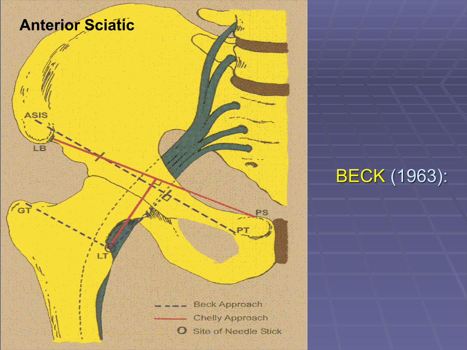
BECK (1963):
Anterior Sciatic

SciaticNerveBlock
Anterior§ Adv 21gu x 6in
insulated§ Obs for fem n. § Adv to lesser
troch, then 4-5cm post/med;
§ 10.5cm ave total depth
§ Hip rotation may help (eg. Int rot above l.t.)
§ Ankle twitch§ 20-30 ml volume
Quad Fem
Gluteus Max
Sartorius
Pectineus
VastMed

Subgluteal Block(Infragluteal)
§ Position: Sims§ Indication: CPNB§ Needle placement:
§ Intersection of gluteal fold and lateral border of the biceps femoris muscle (still thru glut max)
§ Insertion point is 1 cm distal to the intersection
§ Slight cranial angulation of needle
§ Ave 7cm depth§ Accepted stimulation:
Plantar flexion or inversion
§ Will miss Post cut N of

Popliteal Block - Indications§ Single Shot:
§ Ankle arthroscopy§ Metatarsal ORIF§ Excision of mass, feet
§ CPNB:§ Ankle Fractures§ Ankle Reconstruction§ Hallux Valgus Correction
§ Combine with Saphenous Nerve Block (unless thigh tourniquet)
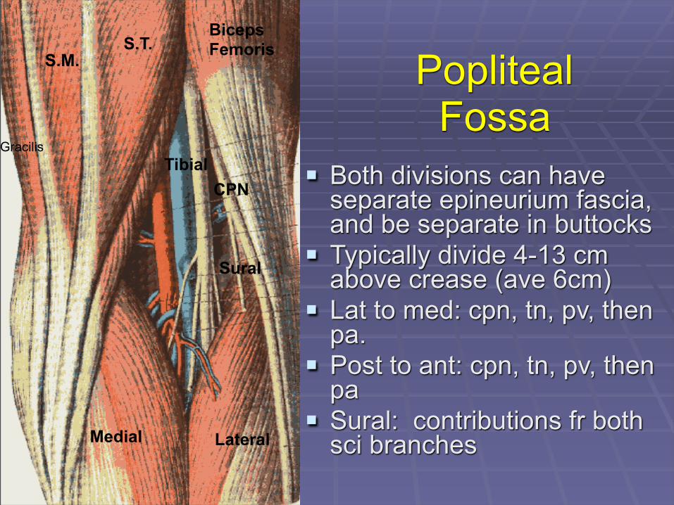
PoplitealFossa
§ Both divisions can have separate epineurium fascia, and be separate in buttocks
§ Typically divide 4-13 cm above crease (ave 6cm)
§ Lat to med: cpn, tn, pv, then pa.
§ Post to ant: cpn, tn, pv, then pa
§ Sural: contributions fr both sci branches
TibialCPN
S.T.BicepsFemorisS.M.
Sural
Gracilis
Medial Lateral

Popliteal§ 22gu x 2in at 7cm
above crease and 1cm lat midline
§ Advanced at 45 deg cephalad
§ Dorsi/plantar flexion of ankle
§ 35-40 ml single injection or
§ 15ml x 2 separate injections
§ >95% w/ high volumes
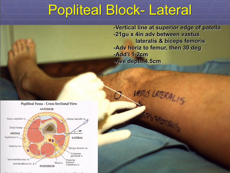
Popliteal Block- Lateral-Vertical line at superior edge of patella-21gu x 4in adv between vastus lateralis & biceps femoris-Adv horiz to femur, then 30 deg-Add’l 1-2cm-Ave depth 4.5cm

CommonPeroneal
NerveTib.Ant.
E.H.L.Pr.Br.
Pr.Lg.
Dp Pr N
Sup Pr N
TA
EHL
EDL

Deep Peroneal Nerve Blocks
§ Adv 22gu-25gu lat to EHL and either side of Dor.Ped.A.
§ Deep to Ext Retic§ To bone, back 2mm§ 5ml volume
Tib Ant
EHL
EDL
Dp Per N
Ext Ret
Dor Ped A
Dp Per N
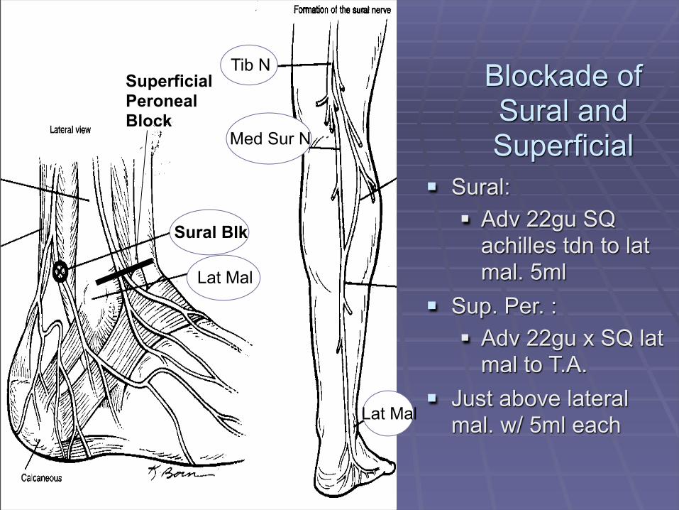
Blockade of Sural and Superficial
§ Sural: § Adv 22gu SQ
achilles tdn to lat mal. 5ml
§ Sup. Per. :§ Adv 22gu x SQ lat
mal to T.A.§ Just above lateral
mal. w/ 5ml each
Superficial PeronealBlock
Sural Blk
Lat Mal
Tib N
Med Sur N
Lat Mal

Medial Plantar
LateralPlantar
Tibial Nerve Anatomy
Soleus
F.D.L. F.H.L.
Tib Post
Calcaneal

PosteriorTibialNerveBlock
§ Midpoint achilles tdn. to med.mall.
§ 22 x 2in§ Just post to
post.tib.art.§ Paresthesia,
stim, U/S§ To bone, back
2mm§ 5ml volume

Questions?


![Fluorophores Structure-Activity Relationship of Nerve-Highlighting · sequence that largely highlights the epineurium with some binding to the endoneurium [13]. The three classes](https://static.fdocuments.in/doc/165x107/5f4b11293c40001f7f62f92e/fluorophores-structure-activity-relationship-of-nerve-highlighting-sequence-that.jpg)