Understanding ultrafine nanodiamond formation using ... fileUnderstanding ultrafine nanodiamond...
6
Understanding ultrafine nanodiamond formation using nanostructured explosives Vincent Pichot 1 , Benedikt Risse 1 , Fabien Schnell 1 , Julien Mory 2 & Denis Spitzer 1 1 NS3E «Nanomate ´riaux pour les Syste `mes Sous Sollicitations Extre ˆmes» UMR ISL-CNRS-UDS 3208, French-German Research Institute of Saint-Louis, 5 rue du Ge ´ne ´ral Cassagnou 68301 Saint-Louis, France, 2 French-German Research Institute of Saint-Louis, 5 rue du Ge ´ne ´ral Cassagnou 68301 Saint-Louis, France. The detonation process is able to build new materials with a bottom-up approach. Diamond, the hardest material on earth, can be synthesized in this way. This unconventional synthesis route is possible due to the presence of carbon inside the high-explosive molecules: firing high-explosive mixtures with a negative oxygen balance in a non-oxidative environment leads to the formation of nanodiamond particles. Trinitrotoluene (TNT) and hexogen (RDX) are the explosives primarily used to synthesize nanodiamonds. Here we show that the use of nanostructured explosive charges leads to the formation of smaller detonation nanodiamonds, and it also provides new understanding of nanodiamond formation-mechanisms. The discontinuity of the explosive at the nanoscale level plays the key role in modifying the diamond particle size, and therefore varying the size with microstructured charges is impossible. S ynthesis of detonation nanodiamonds involves the following four research areas: decrease in particle size, control of particle aggregation during synthesis, optimization of functional surface groups, and controlled doping of such particles 1 . The conditions surrounding the synthesis of nanodiamonds have already been investigated with a view to controlling particle-size, and modifying the reaction environment and the configura- tion of the explosive charges only had a slight influence on the nanodiamond particle-size 2–5 . Research into the synthesis of detonation nanodiamonds began in the USSR in the Sixties and was later developed in other countries 6 . The unique properties of the nanodiamond—hardness, thermal conductivity, biocompatibility, photolumi- nescence, surface chemistry, electrical insulation—make it a very attractive material for various applications 7 . Medicine 7–9 , magnetometry 10–12 and sensors 13 are only some of the many fields with applications where nano- diamonds may lead to groundbreaking innovations. Although the intrinsic properties of diamonds are already of great interest, the main breakthrough should arise from the size of nanodiamond particles 5 . The smallest nano- particles are of great importance for fundamental and applicative research. Their physical and chemical prop- erties can be modified according to their intended applications. For diamonds with a size smaller than 2 nm, theoretical works predict quantum confinement effects due to an increase in the band gap 14 . The diamond nanoparticles may help understanding the appearance and the evolution of these quantum effects when decreas- ing the size of the particles. The ability to use these particles as quantum dots in applications such as quantum computers, cryptography or medical imagery can enable major progress in numerous scientific domains. The detonation of high-explosive mixtures with a negative oxygen balance leads to the formation of nanodia- monds with sizes of between 2 and 20 nanometres. Their size distributions have a lognormal shape centred at 4 to 5 nm and with a long trailing edge. When in suspension, only a very small fraction (0.01 wt%) of the smallest nanodiamonds (i.e. particle sizes of between 2 and 4 nanometres) can be isolated by ultracentrifugation 15 . Despite intensive research focused on the understanding of the diamond-formation process, its fundamental mechanisms are still subject to discussion 2–4,16–20 . Various possible mechanisms based on thermodynamic assumptions, and the evaluation of the final product, were used to explain the nanodiamond formation-mechan- isms during the detonation process 4,17–19 . Remarkable works on the subject of nanodiamond formation accomp- lished by Titov and co-workers based on in-situ measurements using synchrotron radiation 3,16,20 have investigated the detonation process. Here we propose a pioneering approach which uses nanostructured explosive charges for the synthesis of smaller nanodiamonds. Due to the nanostructure of the explosive, the grain boundaries form a discontinuity in OPEN SUBJECT AREAS: NANOPARTICLES SYNTHESIS AND PROCESSING MATERIALS SCIENCE NANOSCALE MATERIALS Received 22 March 2013 Accepted 20 June 2013 Published 8 July 2013 Correspondence and requests for materials should be addressed to D.S. (denis.spitzer@isl. eu) SCIENTIFIC REPORTS | 3 : 2159 | DOI: 10.1038/srep02159 1
Transcript of Understanding ultrafine nanodiamond formation using ... fileUnderstanding ultrafine nanodiamond...
Understanding ultrafine nanodiamondformation using nanostructuredexplosivesVincent Pichot1, Benedikt Risse1, Fabien Schnell1, Julien Mory2 & Denis Spitzer1
1NS3E «Nanomateriaux pour les Systemes Sous Sollicitations Extremes» UMR ISL-CNRS-UDS 3208, French-German ResearchInstitute of Saint-Louis, 5 rue du General Cassagnou 68301 Saint-Louis, France, 2French-German Research Institute of Saint-Louis, 5rue du General Cassagnou 68301 Saint-Louis, France.
The detonation process is able to build new materials with a bottom-up approach. Diamond, the hardestmaterial on earth, can be synthesized in this way. This unconventional synthesis route is possible due to thepresence of carbon inside the high-explosive molecules: firing high-explosive mixtures with a negativeoxygen balance in a non-oxidative environment leads to the formation of nanodiamond particles.Trinitrotoluene (TNT) and hexogen (RDX) are the explosives primarily used to synthesize nanodiamonds.Here we show that the use of nanostructured explosive charges leads to the formation of smaller detonationnanodiamonds, and it also provides new understanding of nanodiamond formation-mechanisms. Thediscontinuity of the explosive at the nanoscale level plays the key role in modifying the diamond particle size,and therefore varying the size with microstructured charges is impossible.
Synthesis of detonation nanodiamonds involves the following four research areas: decrease in particle size,control of particle aggregation during synthesis, optimization of functional surface groups, and controlleddoping of such particles1. The conditions surrounding the synthesis of nanodiamonds have already been
investigated with a view to controlling particle-size, and modifying the reaction environment and the configura-tion of the explosive charges only had a slight influence on the nanodiamond particle-size2–5. Research into thesynthesis of detonation nanodiamonds began in the USSR in the Sixties and was later developed in othercountries6.
The unique properties of the nanodiamond—hardness, thermal conductivity, biocompatibility, photolumi-nescence, surface chemistry, electrical insulation—make it a very attractive material for various applications7.Medicine7–9, magnetometry10–12 and sensors13 are only some of the many fields with applications where nano-diamonds may lead to groundbreaking innovations. Although the intrinsic properties of diamonds are already ofgreat interest, the main breakthrough should arise from the size of nanodiamond particles5. The smallest nano-particles are of great importance for fundamental and applicative research. Their physical and chemical prop-erties can be modified according to their intended applications. For diamonds with a size smaller than 2 nm,theoretical works predict quantum confinement effects due to an increase in the band gap14. The diamondnanoparticles may help understanding the appearance and the evolution of these quantum effects when decreas-ing the size of the particles. The ability to use these particles as quantum dots in applications such as quantumcomputers, cryptography or medical imagery can enable major progress in numerous scientific domains.
The detonation of high-explosive mixtures with a negative oxygen balance leads to the formation of nanodia-monds with sizes of between 2 and 20 nanometres. Their size distributions have a lognormal shape centred at 4 to5 nm and with a long trailing edge. When in suspension, only a very small fraction (0.01 wt%) of the smallestnanodiamonds (i.e. particle sizes of between 2 and 4 nanometres) can be isolated by ultracentrifugation15.
Despite intensive research focused on the understanding of the diamond-formation process, its fundamentalmechanisms are still subject to discussion2–4,16–20. Various possible mechanisms based on thermodynamicassumptions, and the evaluation of the final product, were used to explain the nanodiamond formation-mechan-isms during the detonation process4,17–19. Remarkable works on the subject of nanodiamond formation accomp-lished by Titov and co-workers based on in-situ measurements using synchrotron radiation3,16,20 have investigatedthe detonation process.
Here we propose a pioneering approach which uses nanostructured explosive charges for the synthesis ofsmaller nanodiamonds. Due to the nanostructure of the explosive, the grain boundaries form a discontinuity in
OPEN
SUBJECT AREAS:NANOPARTICLES
SYNTHESIS AND PROCESSING
MATERIALS SCIENCE
NANOSCALE MATERIALS
Received22 March 2013
Accepted20 June 2013
Published8 July 2013
Correspondence andrequests for materials
should be addressed toD.S. (denis.spitzer@isl.
eu)
SCIENTIFIC REPORTS | 3 : 2159 | DOI: 10.1038/srep02159 1
the explosive matrix on a very small scale which strongly influencesthe quality of the synthesized nanodiamonds. These novel experi-ments have provided new insight into the comprehension of nano-diamond formation.
ResultsMicrostructured and nanostructured hexolites composed of 60 wt%of hexogen and 40 wt% of trinitrotoluene were chosen as the explos-ive formulation due to the fact that they provide the highest nano-diamond output. By means of careful preparation, explosive pressedcharges with the same composition (60.2/39.8) and the same density(1.67 g.cm23) were obtained. Scanning Electron Microscopy (SEM)images of the explosive charges shown in Fig. 1 reveal a strong dif-ference between microstructured and nanostructured explosivecharges.
The microstructured charge exhibits large TNT and RDX particleswith sizes between 5 and 100 mm, while in the nanostructured chargethe particle size is between 50 and 200 nm.
Using Raman spectroscopy, RDX and TNT can be differentiatedby means of their spectral signatures. The intensity of the signallocated at 1618 cm21 corresponding to the C5C aromatic stretchingvibration in TNT, and the band at 1272 cm21 corresponding to theN-O stretching in RDX, have been selected to indicate the distri-bution of both explosives in the different samples21,22.
Raman spectroscopy mapping shows a much more homogeneousdistribution of RDX and TNT in the nanostructured charge than inthe microstructured charge (Fig. 1f and 1g). The TNT (green) andRDX (red) signals reveal a uniform distribution of both componentsin the nanostructured charge, whereas the microstructured charge isdominated by micron-sized TNT and RDX regions. Due to the lim-iting resolution of Raman spectroscopy, individual RDX and TNTparticles could not be discriminated by this technique. CertainRaman spectra may result from the superposition of multiple part-icles; however, this technique does reveal the homogeneity degree ofthe explosive charges.
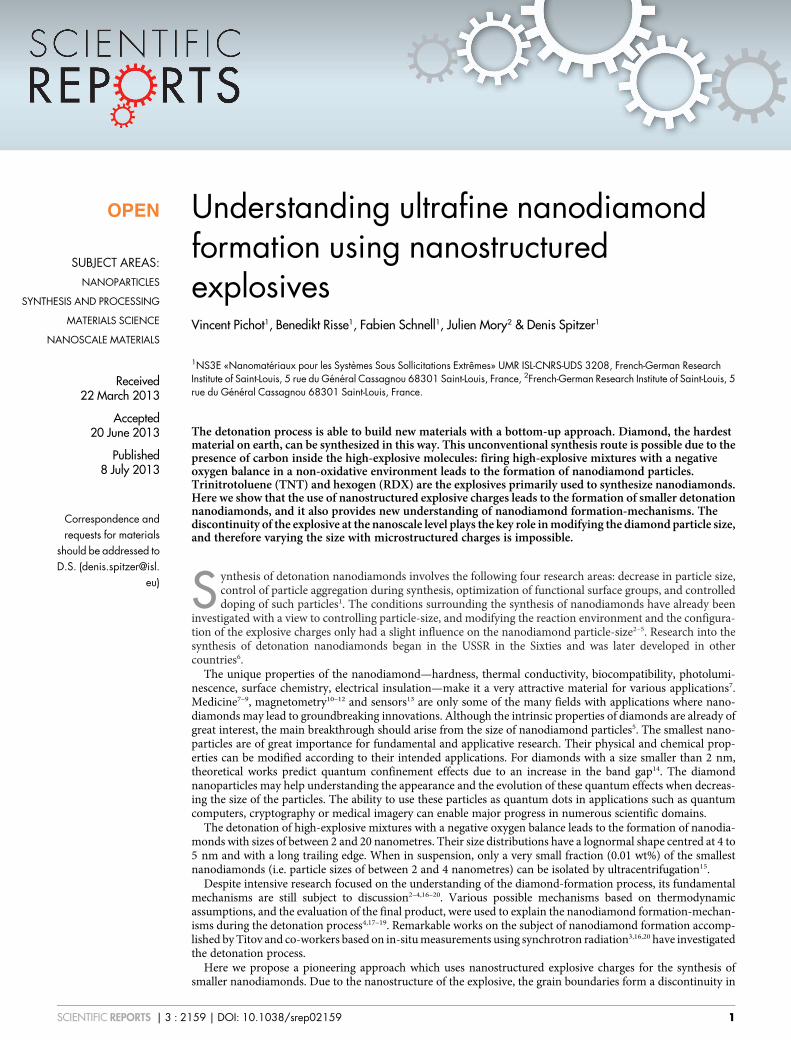
X-Ray Diffraction (XRD) patterns of the microstructured andnanostructured hexolites are quite similar (Fig. 2), indicating thatthe nanohexolite is composed of individual TNT and RDX particlesand neither co-crystallization nor core-shell structures occurred. Itcan be noticed that an enlargement of the diffraction peaks of TNTand RDX is observed, indicating finer elementary crystallites in thecase of nanostructured hexolite.
After firing both charges, detonation soot was recovered and char-acterized. Transmission Electron Microscopy (TEM) images clearlyshow that smaller nanodiamonds were formed by the nanostruc-tured charge (Fig. 3).
Particle-size distribution curves from the collected nanodiamondswere obtained for the nanostructured and the corresponding micro-structured charge by a careful statistical evaluation of the size of morethan 500 individual particles for each sample (the dimensions ofnanodiamonds were manually measured by 4 different persons,ensuring the validity of the results). The nanodiamond particle-sizedistribution obtained from the nanostructured hexolite is narrowerand therefore more homogeneous than in the case of the microstruc-tured charge, in which nanodiamonds with sizes of over 10 nm canbe obtained. Their mean particle diameters are 4.2 and 6.3 nmrespectively. In both cases, lognormal law allows representation ofparticle size distribution.
The cumulative frequencies were calculated from the size distri-bution data of both samples. Two main results can be highlighted:
- For the nanostructured charge, the fraction of nanodiamondshaving a size of below 3 nm is nearly 20%, in contrast to only4% of those formed by the microstructured charge.
Figure 1 | Explosive charge structures. (a), Photograph of the explosive
charge. (b–e), SEM micrographs of the microstructured (b–c) and
nanostructured (d–e) explosive pellets showing enlarged views of the
samples. (f–g), Raman mapping of the microstructured (f) and
nanostructured (g) explosive pellets.Figure 2 | Diffraction patterns of explosive materials. XRD patterns of
nanostructured (red) and microstructured (black) hexolites.
www.nature.com/scientificreports
SCIENTIFIC REPORTS | 3 : 2159 | DOI: 10.1038/srep02159 2
- The largest nanodiamonds formed by the nanostructured chargehave a size of about 8 nm, whereas in the microstructured charge,nanodiamonds with sizes up to 23 nm could be found.
XRD experiments were conducted on the obtained nanodia-monds, the elementary crystallite sizes were measured by using theScherrer equation on the diffraction peak of the (111) planes ofdiamond giving 2.9 nm and 4.7 nm for the nanodiamonds synthe-sized by the nanostructured and the microstructured explosivecharge respectively. By taking into account the fact that there maybe one graphitic layer or fullerene-like structure on the nanodia-mond surface, 0.6 nm in diameter have to be added to the diamondcrystalline particle measured by XRD, leading to particle sizes of 3.5and 5.3 nm respectively which is in good agreement with the meansize of the size distribution.
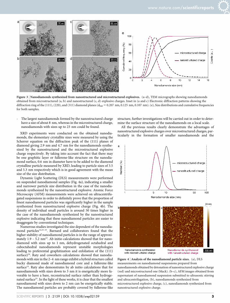
Dynamic Light Scattering (DLS) measurements were performedon suspended nanodiamond samples (Fig. 4a), indicating a smallerand narrower particle size distribution in the case of the nanodia-monds synthesized by the nanostructured explosive. Atomic ForceMicroscopy (AFM) measurements were achieved on ultracentrifu-gated suspensions in order to definitely prove that the proportion offinest nanodiamond particles was significantly higher in the samplesynthesized from nanostructured explosive charge (Fig. 4b). Thedensity of individual small particles is around 50 times higher inthe case of the nanodiamonds synthesized by the nanostructuredexplosive indicating that these nanodiamond particles are easier todeaggregate by conventional techniques.
Numerous studies investigated the size dependent of the nanodia-mond particles14,23–25. Barnard and collaborators found that thehigher stability of nanodiamond particles is in the range of approxi-mately 1.9 – 5.2 nm23. Ab initio calculations showed that for nano-diamond with sizes up to 1 nm, dehydrogenated octahedral andcuboctahedral nanodiamonds represent unstable morphologiesleading to preferential graphitization and exfoliation of the (111)surface)24. Raty and coworkers calculations showed that nanodia-monds with size in the 2–4 nm range exhibit a hybrid structure calledbucky diamond made of nanodiamond core and a fullerene-likesurface14. Raty also demonstrates by ab initio calculations that fornanodiamonds with sizes down to 3 nm it is energetically more fa-vorable to have a bare, reconstructed surface rather than hydroge-nated surface25. In the light of these works, it is clear that the smallestnanodiamond with sizes down to 2 nm can be energetically stable.The nanodiamond particles are probably covered by fullerene-like
structure, further investigations will be carried out in order to deter-mine the surface structure of the nanodiamonds on a local scale.
All the previous results clearly demonstrate the advantages ofnanostructured explosive charges over microstructured charges, par-ticularly in the formation of smaller nanodiamonds and the
Figure 3 | Nanodiamonds synthesized from nanostructured and microstructured explosives. (a–d), TEM micrographs showing nanodiamonds
obtained from microstructured (a, b) and nanostructured (c, d) explosive charges. Inset in (a and c) Electronic diffraction patterns showing the
diffraction ring of the (111), (220), and (311) diamond planes (dhkl 5 0.207 nm, 0.125 nm, 0.107 nm). (e), Size distributions and cumulative frequencies
for both samples.
Figure 4 | Analysis of the nanodiamond particle sizes. (a), DLS
measurements on nanodiamond suspensions prepared from
nanodiamonds obtained by detonation of nanostructured explosive charge
(red) and microstructured one (black). (b–c), AFM images obtained from
supernatant of nanodiamond suspension submitted to ultrasonic stirring
and ultracentrifugation, (b), nanodiamonds synthesized from
microstructured explosive charge, (c), nanodiamonds synthesized from
nanostructured explosive charge.
www.nature.com/scientificreports
SCIENTIFIC REPORTS | 3 : 2159 | DOI: 10.1038/srep02159 3
obtaining of narrower particle-size distribution. This striking discov-ery furthers understanding of this type of synthesis as it opens thesynthesis route for ultra-small detonation nanodiamonds for use invarious target applications such as quantum computers, biology,electronics and optronics which necessitate ultra-small diamonds.
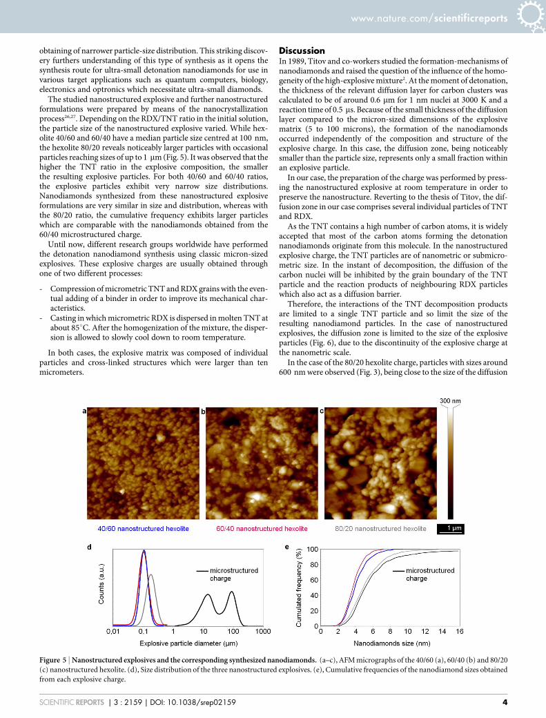
The studied nanostructured explosive and further nanostructuredformulations were prepared by means of the nanocrystallizationprocess26,27. Depending on the RDX/TNT ratio in the initial solution,the particle size of the nanostructured explosive varied. While hex-olite 40/60 and 60/40 have a median particle size centred at 100 nm,the hexolite 80/20 reveals noticeably larger particles with occasionalparticles reaching sizes of up to 1 mm (Fig. 5). It was observed that thehigher the TNT ratio in the explosive composition, the smallerthe resulting explosive particles. For both 40/60 and 60/40 ratios,the explosive particles exhibit very narrow size distributions.Nanodiamonds synthesized from these nanostructured explosiveformulations are very similar in size and distribution, whereas withthe 80/20 ratio, the cumulative frequency exhibits larger particleswhich are comparable with the nanodiamonds obtained from the60/40 microstructured charge.
Until now, different research groups worldwide have performedthe detonation nanodiamond synthesis using classic micron-sizedexplosives. These explosive charges are usually obtained throughone of two different processes:
- Compression of micrometric TNT and RDX grains with the even-tual adding of a binder in order to improve its mechanical char-acteristics.
- Casting in which micrometric RDX is dispersed in molten TNT atabout 85uC. After the homogenization of the mixture, the disper-sion is allowed to slowly cool down to room temperature.
In both cases, the explosive matrix was composed of individualparticles and cross-linked structures which were larger than tenmicrometers.
DiscussionIn 1989, Titov and co-workers studied the formation-mechanisms ofnanodiamonds and raised the question of the influence of the homo-geneity of the high-explosive mixture2. At the moment of detonation,the thickness of the relevant diffusion layer for carbon clusters wascalculated to be of around 0.6 mm for 1 nm nuclei at 3000 K and areaction time of 0.5 ms. Because of the small thickness of the diffusionlayer compared to the micron-sized dimensions of the explosivematrix (5 to 100 microns), the formation of the nanodiamondsoccurred independently of the composition and structure of theexplosive charge. In this case, the diffusion zone, being noticeablysmaller than the particle size, represents only a small fraction withinan explosive particle.
In our case, the preparation of the charge was performed by press-ing the nanostructured explosive at room temperature in order topreserve the nanostructure. Reverting to the thesis of Titov, the dif-fusion zone in our case comprises several individual particles of TNTand RDX.
As the TNT contains a high number of carbon atoms, it is widelyaccepted that most of the carbon atoms forming the detonationnanodiamonds originate from this molecule. In the nanostructuredexplosive charge, the TNT particles are of nanometric or submicro-metric size. In the instant of decomposition, the diffusion of thecarbon nuclei will be inhibited by the grain boundary of the TNTparticle and the reaction products of neighbouring RDX particleswhich also act as a diffusion barrier.
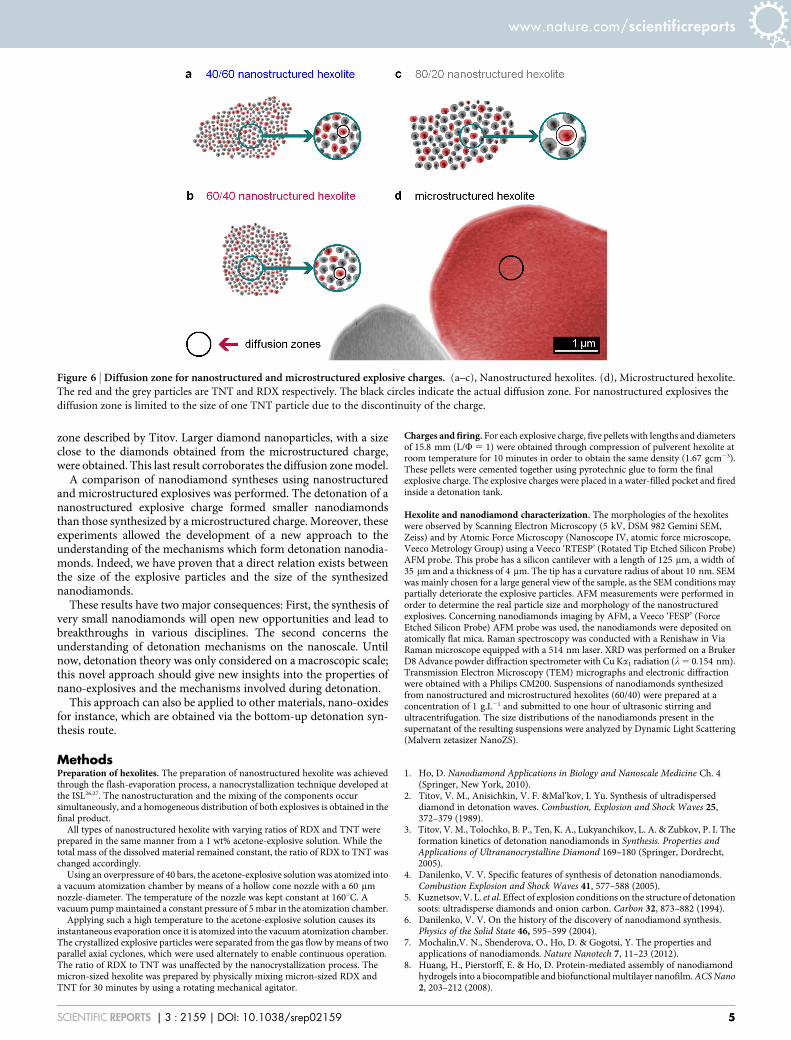
Therefore, the interactions of the TNT decomposition productsare limited to a single TNT particle and so limit the size of theresulting nanodiamond particles. In the case of nanostructuredexplosives, the diffusion zone is limited to the size of the explosiveparticles (Fig. 6), due to the discontinuity of the explosive charge atthe nanometric scale.
In the case of the 80/20 hexolite charge, particles with sizes around600 nm were observed (Fig. 3), being close to the size of the diffusion
Figure 5 | Nanostructured explosives and the corresponding synthesized nanodiamonds. (a–c), AFM micrographs of the 40/60 (a), 60/40 (b) and 80/20
(c) nanostructured hexolite. (d), Size distribution of the three nanostructured explosives. (e), Cumulative frequencies of the nanodiamond sizes obtained
from each explosive charge.
www.nature.com/scientificreports
SCIENTIFIC REPORTS | 3 : 2159 | DOI: 10.1038/srep02159 4
zone described by Titov. Larger diamond nanoparticles, with a sizeclose to the diamonds obtained from the microstructured charge,were obtained. This last result corroborates the diffusion zone model.
A comparison of nanodiamond syntheses using nanostructuredand microstructured explosives was performed. The detonation of ananostructured explosive charge formed smaller nanodiamondsthan those synthesized by a microstructured charge. Moreover, theseexperiments allowed the development of a new approach to theunderstanding of the mechanisms which form detonation nanodia-monds. Indeed, we have proven that a direct relation exists betweenthe size of the explosive particles and the size of the synthesizednanodiamonds.
These results have two major consequences: First, the synthesis ofvery small nanodiamonds will open new opportunities and lead tobreakthroughs in various disciplines. The second concerns theunderstanding of detonation mechanisms on the nanoscale. Untilnow, detonation theory was only considered on a macroscopic scale;this novel approach should give new insights into the properties ofnano-explosives and the mechanisms involved during detonation.
This approach can also be applied to other materials, nano-oxidesfor instance, which are obtained via the bottom-up detonation syn-thesis route.
MethodsPreparation of hexolites. The preparation of nanostructured hexolite was achievedthrough the flash-evaporation process, a nanocrystallization technique developed atthe ISL26,27. The nanostructuration and the mixing of the components occursimultaneously, and a homogeneous distribution of both explosives is obtained in thefinal product.
All types of nanostructured hexolite with varying ratios of RDX and TNT wereprepared in the same manner from a 1 wt% acetone-explosive solution. While thetotal mass of the dissolved material remained constant, the ratio of RDX to TNT waschanged accordingly.
Using an overpressure of 40 bars, the acetone-explosive solution was atomized intoa vacuum atomization chamber by means of a hollow cone nozzle with a 60 mmnozzle-diameter. The temperature of the nozzle was kept constant at 160uC. Avacuum pump maintained a constant pressure of 5 mbar in the atomization chamber.
Applying such a high temperature to the acetone-explosive solution causes itsinstantaneous evaporation once it is atomized into the vacuum atomization chamber.The crystallized explosive particles were separated from the gas flow by means of twoparallel axial cyclones, which were used alternately to enable continuous operation.The ratio of RDX to TNT was unaffected by the nanocrystallization process. Themicron-sized hexolite was prepared by physically mixing micron-sized RDX andTNT for 30 minutes by using a rotating mechanical agitator.
Charges and firing. For each explosive charge, five pellets with lengths and diametersof 15.8 mm (L/W 5 1) were obtained through compression of pulverent hexolite atroom temperature for 10 minutes in order to obtain the same density (1.67 gcm23).These pellets were cemented together using pyrotechnic glue to form the finalexplosive charge. The explosive charges were placed in a water-filled pocket and firedinside a detonation tank.
Hexolite and nanodiamond characterization. The morphologies of the hexoliteswere observed by Scanning Electron Microscopy (5 kV, DSM 982 Gemini SEM,Zeiss) and by Atomic Force Microscopy (Nanoscope IV, atomic force microscope,Veeco Metrology Group) using a Veeco ‘RTESP’ (Rotated Tip Etched Silicon Probe)AFM probe. This probe has a silicon cantilever with a length of 125 mm, a width of35 mm and a thickness of 4 mm. The tip has a curvature radius of about 10 nm. SEMwas mainly chosen for a large general view of the sample, as the SEM conditions maypartially deteriorate the explosive particles. AFM measurements were performed inorder to determine the real particle size and morphology of the nanostructuredexplosives. Concerning nanodiamonds imaging by AFM, a Veeco ‘FESP’ (ForceEtched Silicon Probe) AFM probe was used, the nanodiamonds were deposited onatomically flat mica. Raman spectroscopy was conducted with a Renishaw in ViaRaman microscope equipped with a 514 nm laser. XRD was performed on a BrukerD8 Advance powder diffraction spectrometer with Cu Ka1 radiation (l 5 0.154 nm).Transmission Electron Microscopy (TEM) micrographs and electronic diffractionwere obtained with a Philips CM200. Suspensions of nanodiamonds synthesizedfrom nanostructured and microstructured hexolites (60/40) were prepared at aconcentration of 1 g.L21 and submitted to one hour of ultrasonic stirring andultracentrifugation. The size distributions of the nanodiamonds present in thesupernatant of the resulting suspensions were analyzed by Dynamic Light Scattering(Malvern zetasizer NanoZS).
1. Ho, D. Nanodiamond Applications in Biology and Nanoscale Medicine Ch. 4(Springer, New York, 2010).
2. Titov, V. M., Anisichkin, V. F. &Mal’kov, I. Yu. Synthesis of ultradisperseddiamond in detonation waves. Combustion, Explosion and Shock Waves 25,372–379 (1989).
3. Titov, V. M., Tolochko, B. P., Ten, K. A., Lukyanchikov, L. A. & Zubkov, P. I. Theformation kinetics of detonation nanodiamonds in Synthesis. Properties andApplications of Ultrananocrystalline Diamond 169–180 (Springer, Dordrecht,2005).
4. Danilenko, V. V. Specific features of synthesis of detonation nanodiamonds.Combustion Explosion and Shock Waves 41, 577–588 (2005).
5. Kuznetsov, V. L. et al. Effect of explosion conditions on the structure of detonationsoots: ultradisperse diamonds and onion carbon. Carbon 32, 873–882 (1994).
6. Danilenko, V. V. On the history of the discovery of nanodiamond synthesis.Physics of the Solid State 46, 595–599 (2004).
7. Mochalin,V. N., Shenderova, O., Ho, D. & Gogotsi, Y. The properties andapplications of nanodiamonds. Nature Nanotech 7, 11–23 (2012).
8. Huang, H., Pierstorff, E. & Ho, D. Protein-mediated assembly of nanodiamondhydrogels into a biocompatible and biofunctional multilayer nanofilm. ACS Nano2, 203–212 (2008).
Figure 6 | Diffusion zone for nanostructured and microstructured explosive charges. (a–c), Nanostructured hexolites. (d), Microstructured hexolite.
The red and the grey particles are TNT and RDX respectively. The black circles indicate the actual diffusion zone. For nanostructured explosives the
diffusion zone is limited to the size of one TNT particle due to the discontinuity of the charge.
www.nature.com/scientificreports
SCIENTIFIC REPORTS | 3 : 2159 | DOI: 10.1038/srep02159 5
9. Ho, D. Beyond the sparkle: The impact of nanodiamonds as biolabeling andtherapeutic agents. ACS Nano 3, 3825–3829 (2009).
10. Waldherr, G. et al. High-dynamic-range magnetometry with a single nuclear spinin diamond. Nature Nanotech 7, 105–108 (2012).
11. Nusran, N. M., Momeen, M. U. & Dutt, M. V. G. High-dynamic-rangemagnetometry with a single electronic spin in diamond. Nature Nanotech 7,109–113 (2012).
12. Balasubramanian, G. et al. Nanoscale imaging magnetometry with diamond spinsunder ambient conditions. Nature 455, 648–651 (2008).
13. Maletinsky, P. et al. A robust scanning diamond sensor for nanoscale imagingwith single nitrogen-vacancy centres. Nature Nanotech 7, 320–324 (2012).
14. Raty, J.-Y., Galli, G., Bostedt, C., van Buuren, T. W. & Terminello, L. J. Quantumconfinement and fullerenelikesurface reconstruction in nanodiamonds. Phys. Rev.Lett. 90, 037401 (2003).
15. Schmidlin, L. et al. Two-dimensional nanodiamond monolayers deposited bycombined ultracentrifugation and electrophoresis techniques. App. Phys. Lett.101, 253111 (2012).
16. Titov, V. M., Tolochko, B. P., Ten, K. A., Lukyanchikov, L. A. & Pruuel, E. R.Where and when are nanodiaomnds formed under explosion? Diam. Relat.Mater. 16, 2009–2013 (2007).
17. Anischkin, V. F. Isotope Studies of Detonation Mechanisms of TNT, RDX, andHMX. Combustion, Explosion, and Shock Waves 43, 580–586 (2007).
18. Kozyrev, N. V. & Golubeva, E. S. Investigation of the synthesis of ultradisperseddiamonds in mixtures of TNT with RDX, HMX and PETN. Combustion,Explosion, and Shock Waves 28, 560–564 (1992).
19. Malkov, I. Yu. et al. Formation of diamond from the liquid phase carbon.Combustion, Explosion, and Shock Waves 29, 542–544 (1993).
20. Chernishev, A. P. et al. Physical–chemical model of nanodiamond formation atexplosion. Nuclear Instruments and Methods in Physics Research A 575, 72–74(2004).
21. Wang, X. et al. Detection of TNT in acetone using Raman spectroscopic signature.Proc. Of International Symposium on Photoelectronic Detection and Imaging 2007:Laser, Ultraviolet, and Terahertz Technology, edited by Liwei Zhou, Proc. of SPIEVol. 6622, 662219 (2008) doi: 10.1117/12.790827.
22. Karpowicz, R. J. & Brill, T. B. Comparison of the molecular structure ofhexahydro-1,3,5-trinitro-s-triazine in the vapor, solution and solid phases.J. Phys. Chem. 88, 348–352 (1984).
23. Barnard, A. S., Russo, S. P. & Snook, I. K. Size dependent phase stability of carbonNanoparticles: nanodiamond versus fullerenes, J. Chem. Phys. 118, 5094–5097(2003).
24. Barnard, A. S., Russo, S. P. & Snook, I. K. Structural relaxation and relative stabilityof nanodiamond morphologies, Diam. Relat. Mater. 16, 1867–1872 (2003).
25. Raty, J. Y. & Galli. Ultradispersity of diamond at the nanoscale, Nat. Mater. 2,792–795 (2003).
26. Risse, B. et al. Continuous formation of submicron energetic particles by the flash-evaporation technique. Chem. Eng. J. 203, 158–165 (2012).
27. Risse, B., Spitzer, D. & Hassler, D. Patent, FR 1251143, Preparation denanoparticules d’explosif par evaporation flash.
AcknowledgementsLoıc Vidal at the ‘‘Institut de Science des Materiaux de Mulhouse’’ (IS2M), for the TEMmicrographs.
Author contributionsD.S. conceived the original idea and D.S. and V.P. directed the research. B.R. synthesized thenanostructured explosives and made the Raman spectroscopy experiments. J.M. preparedthe explosive charges. F.S. performed the SEM experiments on the explosive samples. V.P.conducted the AFM experiments on explosive nanoparticles, synthesized the nanodiamondparticles. All authors participated to the analysis of the data and discussed the results. V.P.and D.S. wrote the paper, and all authors provided their feedback.
Additional informationCompeting financial interests: The authors declare no competing financial interests.
How to cite this article: Pichot, V., Risse, B., Schnell, F., Mory, J. & Spitzer, D.Understanding ultrafine nanodiamond formation using nanostructured explosives. Sci.Rep. 3, 2159; DOI:10.1038/srep02159 (2013).
This work is licensed under a Creative Commons Attribution-NonCommercial-NoDerivs 3.0 Unported license. To view a copy of this license,
visit http://creativecommons.org/licenses/by-nc-nd/3.0
www.nature.com/scientificreports
SCIENTIFIC REPORTS | 3 : 2159 | DOI: 10.1038/srep02159 6