Therapeutic Hypothermia, Patient Outcomes and Post...
80
ACTA UNIVERSITATIS UPSALIENSIS UPPSALA 2014 Digital Comprehensive Summaries of Uppsala Dissertations from the Faculty of Medicine 1021 Post-Cardiac Arrest Care Therapeutic Hypothermia, Patient Outcomes and Relatives’ Experiences ING-MARIE LARSSON ISSN 1651-6206 ISBN 978-91-554-9009-6 urn:nbn:se:uu:diva-229758
Transcript of Therapeutic Hypothermia, Patient Outcomes and Post...
ACTAUNIVERSITATIS
UPSALIENSISUPPSALA
2014
Digital Comprehensive Summaries of Uppsala Dissertationsfrom the Faculty of Medicine 1021
Post-Cardiac Arrest Care
Therapeutic Hypothermia, Patient Outcomes andRelatives’ Experiences
ING-MARIE LARSSON
ISSN 1651-6206ISBN 978-91-554-9009-6urn:nbn:se:uu:diva-229758
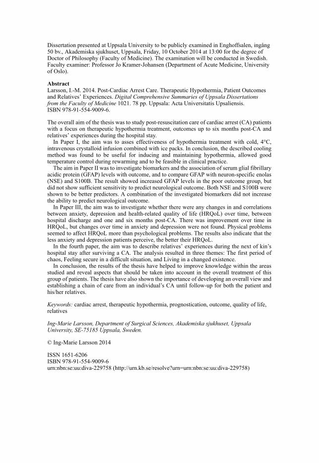
Dissertation presented at Uppsala University to be publicly examined in Enghoffsalen, ingång50 bv., Akademiska sjukhuset, Uppsala, Friday, 10 October 2014 at 13:00 for the degree ofDoctor of Philosophy (Faculty of Medicine). The examination will be conducted in Swedish.Faculty examiner: Professor Jo Kramer-Johansen (Department of Acute Medicine, Universityof Oslo).
AbstractLarsson, I.-M. 2014. Post-Cardiac Arrest Care. Therapeutic Hypothermia, Patient Outcomesand Relatives’ Experiences. Digital Comprehensive Summaries of Uppsala Dissertationsfrom the Faculty of Medicine 1021. 78 pp. Uppsala: Acta Universitatis Upsaliensis.ISBN 978-91-554-9009-6.
The overall aim of the thesis was to study post-resuscitation care of cardiac arrest (CA) patientswith a focus on therapeutic hypothermia treatment, outcomes up to six months post-CA andrelatives’ experiences during the hospital stay.
In Paper I, the aim was to asses effectiveness of hypothermia treatment with cold, 4°C,intravenous crystalloid infusion combined with ice packs. In conclusion, the described coolingmethod was found to be useful for inducing and maintaining hypothermia, allowed goodtemperature control during rewarming and to be feasible in clinical practice.
The aim in Paper II was to investigate biomarkers and the association of serum glial fibrillaryacidic protein (GFAP) levels with outcome, and to compare GFAP with neuron-specific enolas(NSE) and S100B. The result showed increased GFAP levels in the poor outcome group, butdid not show sufficient sensitivity to predict neurological outcome. Both NSE and S100B wereshown to be better predictors. A combination of the investigated biomarkers did not increasethe ability to predict neurological outcome.
In Paper III, the aim was to investigate whether there were any changes in and correlationsbetween anxiety, depression and health-related quality of life (HRQoL) over time, betweenhospital discharge and one and six months post-CA. There was improvement over time inHRQoL, but changes over time in anxiety and depression were not found. Physical problemsseemed to affect HRQoL more than psychological problems. The results also indicate that theless anxiety and depression patients perceive, the better their HRQoL.
In the fourth paper, the aim was to describe relatives’ experiences during the next of kin’shospital stay after surviving a CA. The analysis resulted in three themes: The first period ofchaos, Feeling secure in a difficult situation, and Living in a changed existence.
In conclusion, the results of the thesis have helped to improve knowledge within the areasstudied and reveal aspects that should be taken into account in the overall treatment of thisgroup of patients. The thesis have also shown the importance of developing an overall view andestablishing a chain of care from an individual’s CA until follow-up for both the patient andhis/her relatives.
Keywords: cardiac arrest, therapeutic hypothermia, prognostication, outcome, quality of life,relatives
Ing-Marie Larsson, Department of Surgical Sciences, Akademiska sjukhuset, UppsalaUniversity, SE-75185 Uppsala, Sweden.
© Ing-Marie Larsson 2014
ISSN 1651-6206ISBN 978-91-554-9009-6urn:nbn:se:uu:diva-229758 (http://urn.kb.se/resolve?urn=urn:nbn:se:uu:diva-229758)

To my family

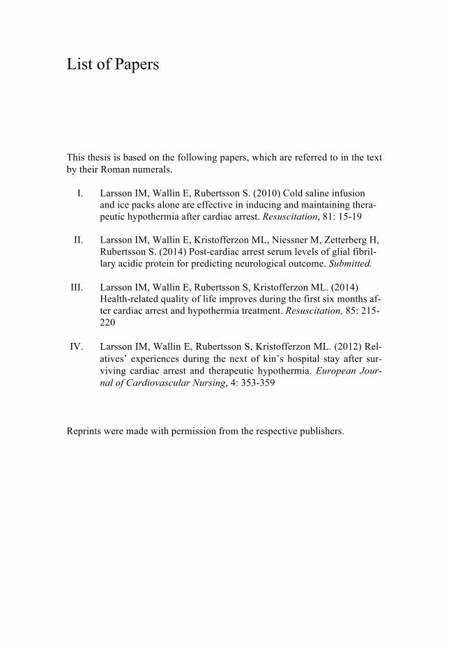
List of Papers
This thesis is based on the following papers, which are referred to in the text by their Roman numerals.
I. Larsson IM, Wallin E, Rubertsson S. (2010) Cold saline infusion
and ice packs alone are effective in inducing and maintaining thera-peutic hypothermia after cardiac arrest. Resuscitation, 81: 15-19
II. Larsson IM, Wallin E, Kristofferzon ML, Niessner M, Zetterberg H, Rubertsson S. (2014) Post-cardiac arrest serum levels of glial fibril-lary acidic protein for predicting neurological outcome. Submitted.
III. Larsson IM, Wallin E, Rubertsson S, Kristofferzon ML. (2014)
Health-related quality of life improves during the first six months af-ter cardiac arrest and hypothermia treatment. Resuscitation, 85: 215-220
IV. Larsson IM, Wallin E, Rubertsson S, Kristofferzon ML. (2012) Rel-atives’ experiences during the next of kin’s hospital stay after sur-viving cardiac arrest and therapeutic hypothermia. European Jour-nal of Cardiovascular Nursing, 4: 353-359
Reprints were made with permission from the respective publishers.

Contents
Introduction ................................................................................................... 11
Background ................................................................................................... 13Surviving cardiac arrest ............................................................................ 13
Post resuscitation care .......................................................................... 13Brain injury after cardiac arrest ........................................................... 14
Therapeutic hypothermia .......................................................................... 15Neuroprotective mechanisms ............................................................... 15Applications of therapeutic hypothermia after cardiac arrest .............. 16
Prognostication ......................................................................................... 18The natural course of recovery after hypoxic brain injury .................. 18Prediction of neurological outcome ..................................................... 19Biomarkers ........................................................................................... 20Withdrawal of care ............................................................................... 21
Outcome after cardiac arrest ..................................................................... 21Neurological outcome .......................................................................... 22Health-related quality of life ................................................................ 22Anxiety and depression ........................................................................ 23
Relatives ................................................................................................... 23Relatives of critically ill persons .......................................................... 23Relatives of cardiac arrest patients ...................................................... 24
Rationale for the thesis ............................................................................. 24
Aims .............................................................................................................. 26
Materials and methods .................................................................................. 27Paper I ....................................................................................................... 28
Settings and participants ...................................................................... 28Cooling method .................................................................................... 29Treatment protocol ............................................................................... 29Data collection and monitoring ............................................................ 29Statistical analysis ................................................................................ 30
Paper II ..................................................................................................... 30Settings and participants ...................................................................... 30Data collection ..................................................................................... 30Analysis of biomarkers ........................................................................ 31Statistical analysis ................................................................................ 31
Paper III .................................................................................................... 32Settings and participants ...................................................................... 32Data collection ..................................................................................... 32Statistical analysis ................................................................................ 34
Paper IV .................................................................................................... 34Setting and participants ........................................................................ 34Data collection ..................................................................................... 35Data analysis ........................................................................................ 35
Ethical considerations ............................................................................... 36
Results ........................................................................................................... 37Paper I ....................................................................................................... 37
Temperature control ............................................................................. 37Outcome ............................................................................................... 38
Paper II ..................................................................................................... 39GFAP ................................................................................................... 40NSE ...................................................................................................... 40S100B ................................................................................................... 40Comparisons of biomarkers ................................................................. 41
Paper III .................................................................................................... 42Outcome ............................................................................................... 42Changes over time in anxiety, depression and HRQoL ....................... 43Correlation between anxiety and depression and HRQoL ................... 44Descriptive distributions of the questionnaires .................................... 46
Paper IV .................................................................................................... 47The first period of chaos ...................................................................... 47Feeling secure in a difficult situation ................................................... 47Living in a changed existence .............................................................. 48
Discussion ..................................................................................................... 49Therapeutic hypothermia .......................................................................... 49Prognostication ......................................................................................... 51Outcome .................................................................................................... 52Relative’s experiences .............................................................................. 55Methodological considerations ................................................................. 56
Conclusions ................................................................................................... 60
Clinical implications and future perspectives ............................................... 61
Svensk sammanfattning (Swedish summary) ............................................... 63
Acknowledgements ....................................................................................... 65
References ..................................................................................................... 67
Abbreviations
AUC CA CBF CPC CPR CT ECG EEG EQ5D EQ-VAS FPR GCS GFAP HADS HRQoL ICU M1 M2 M3 MCS MRI mRS NSE OHCA PCI PCS PEA PTSD QoL RLS ROC ROSC SF12 SSEP TH VF
Area Under the Curve Cardiac Arrest Cerebral Blood Flow Cerebral Performance Categories Cardiopulmonary Resuscitation Computed Tomography Electrocardiogram Electroencephalogram Euroqol Euroqol Visual Analogue Scale False Positive Rate Glasgow Coma Scale Glial Fibrillary Acidic Protein Hospital Anxiety and Depression Scale Health-related Quality of Life Intensive Care Unit Measurement Occasion 1 Measurement Occasion 2 Measurement Occasion 3 Mental Component Score Magnetic Resonance Imaging Modified Rankin Scale Neuron-Specific Enolas Out of Hospital Cardiac Arrest Percutaneous Coronary Intervention Physical Component Score Pulseless Electric Activity Post Traumatic Stress Disorder Quality of Life Reaction Level Scale Receiver Operating Characteristic Return of Spontaneous Circulation Short form 12 Somatosensory Evoked Potentials Therapeutic Hypothermia Ventricular Fibrillation

VT
Ventricular Tachycardia
11
Introduction
Cardiac arrest (CA) is usually an unexpected event and extremely stressful for the people nearby, who are often relatives. It is a life-threatening condi-tion, and the survival rate is low. In Sweden in 2012, the survival rate one month after a CA – which occurred out of hospital and was treated by ambu-lance personnel – was 10.3%.1 In Europe, approximately 275.000 CA cases are treated annually by the emergency medical service, and of those, about 10% are expected to survive until hospital discharge,2 but there is great vari-ation in the reported survival rate.3 Survivors are treated in an intensive care unit (ICU), and the prognosis is uncertain not only due to the heart disease, but mostly with regard to neurological symptoms and the sequelae of the asphyxia during the CA. For the relatives, it is a time characterized by being torn between hope and despair.
In my profession as an intensive care nurse, I meet CA patients and their relatives. In the ICU, we treat the patients with life support to try to ensure survival with the best possible outcome. Due to the serious disease in addi-tion affected by asphyxia, there are questions concerning the patient's prog-nosis and ability to return to a life of dignity. When caring for these patients we are facing relatives who have questions and need support to encounter this situation.
One CA patient is a man, 68 years old. He had his CA at home and his
wife started cardiopulmonary resuscitation (CPR). He arrived at hospital unconscious and therapeutic hypothermia (TH) was initiated at the emer-gency ward. At the ICU, three days after the CA, he had reached normal temperature and started to wake up. He could not talk and was motor agitat-ed. The next day he was moved to the medical ward. He began to talk, but asked the same question over and over again, forgetting the answer immedi-ately. He was still motor agitated and had sleeping problems. Day by day he got better and 12 days after the CA, he was discharged from hospital. He had no memory from the event but thought he felt quite all right and was looking forward to going home. His wife was worried, she did not think he looks the same and she did not know what will happen.
Another CA patient is a 75-years old woman who easy reached the target temperature for hypothermia but took long time to rewarm, sedation had been removed but she was still unconscious. She initiated spontaneous
12
breathing through the ventilator, coughed and had opened her eyes, but has made no contact. Her relatives were worried but hopeful.
This thesis will focus on CA patients who reach the ICU and the chain of
care for patients and their relatives. It covers a method of therapeutic hypo-thermia, prognostication with biomarkers, patients’ outcomes of health-related quality of life (HRQoL), anxiety and depression until six months post-CA as well as relatives’ experiences during the first six weeks after the next of kin’s CA.
13
Background
Surviving cardiac arrest In 1991, the first Utstein guidelines were presented to help medical profes-sionals report CA in a uniform manner.4 The Utstein templates have been revised, and been used in published studies and registries, thus enabling comparisons of findings. This has resulted in improved knowledge about CA, CPR and patient outcomes, knowledge that has come to form the foun-dation of international consensus and resuscitation guidelines.5
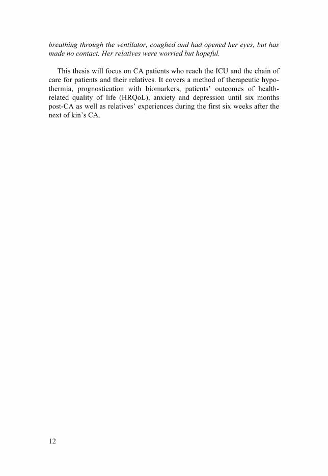
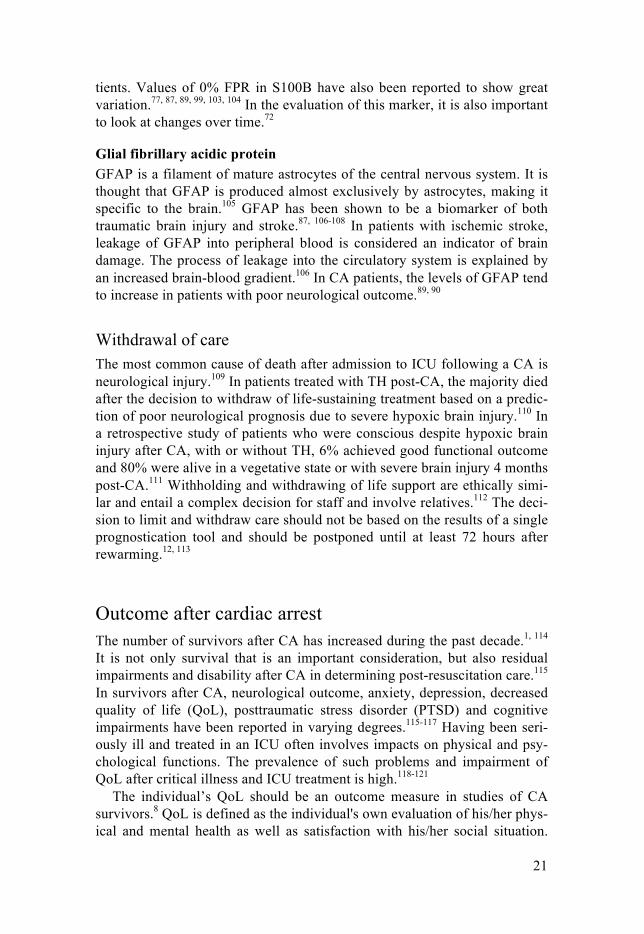
To survive a CA with a good outcome, there are many pieces that must fall into place. The chain of survival (Figure 1) illustrates the interventions that need to work in order to optimize the survival with a good outcome after a CA.6
Figure 1. The European Resuscitation Council’s; Chain of survival.6
Post resuscitation care After resuscitation and return of spontaneous circulation (ROSC), the post-CA care plays an important role in increasing survival and reducing morbidi-ty.7, 8 After ROSC, a phase of pathophysiological processes called post-
14
cardiac arrest syndrome occurs. The syndrome includes brain injury, myo-cardial dysfunction, systemic ischemia/reperfusion response, the process that caused the CA and the patient’s co-morbidities.8 The syndrome is complex and patients have sepsis-like symptoms.9
In the immediate and early phase after ROSC, care of the patient is re-source-intensive and a multidisciplinary team of healthcare providers is needed.8 To enhance the quality of care, treatment protocols have been shown to improve post-CA care and patient outcomes.10, 11 Post-resuscitation care should follow guidelines for severely ill patients. Treatment of circula-tion should include hemodynamic stabilization and optimization. Adequate circulation is necessary for the brain and the myocardium. Hemodynamic instability, hypotension and arrhythmias need to be treated, but the optimal mean arterial pressure is not known. Coronary artery disease is common in CA patients, and early diagnosis and treatment with angiography and percu-taneous coronary intervention (PCI) are recommended. Ventilation and oxy-genation need to be optimized. Controlled mechanical ventilation should be initiated as early as possible to achieve normoventilation and avoid hypoxia.7, 8, 11-13 Hyperoxia and hyperventilation should also be avoided as they may worsen brain injury.14 Metabolic controls are necessary, including blood glucose, electrolytes, lactate and acid-base and normal values are pur-sued. Post-CA care should also include TH for protection of the brain.7, 8, 11-13 Fever post-CA have been shown to be associated with unfavorable outcome and should be avoided, also after completing TH.15-17
Brain injury after cardiac arrest The brain is particularly vulnerable to hypoxic ischemia due to its limited tolerance and its sensitivity to reperfusion.8 In animal models, cerebral is-chemia for more than 5 minutes showed necrotic neurons to such a degree that irreversible damage occurred 18 and after 15 minutes of global ischemia, 95% of the brain tissue was damaged.19 Interruption of circulation leads to biochemical changes. The event includes reduction of ATP production, changes in the intra- and extracellular electrolyte composition, which include disrupted calcium homeostasis, acidosis, releases of glutamate and free radi-cals and increased production of nitric oxide. Also functional changes of the cell with mitochondrial damage are seen. All this leads to cell damage. Dur-ing reperfusion the toxic damage is ongoing, triggered by a second cascade of the events.20 Ischemic cell damage and death is often delayed, and the delay varies between a few hours to several days after ROSC, the process is complex and there is an interaction between different changes.21 The cere-bral perfusion and cerebral blood flow (CBF) are affected after CA and ROSC. During the first minutes after ROSC, the CBF is increased.22, 23 After the hyperaemic period, there are a period followed by hypoperfusion, which lasts for 6-24 hours after the CA and before normal CBF is achieved.23, 24
15
Cerebral autoregulation is affected,25 which may affect the risk of cerebral damage. A secondary, delayed window of cerebral metabolic changes be-gins, lasting for several days.26
Therapeutic hypothermia Hypothermia is a body temperature below normal (35°C) and TH is defined as a controlled lowering of the body temperature.27
TH has been used for patients undergoing intracerebral aneurysm and cardiac surgery since 1950s. During the 1950s and 1960s deep TH (below 30°C) was used in clinical trials and resulted in a large number of side ef-fects.27 Clinical use of TH after CA was first presented in 1958.28 In 1990, it was reported that mild hypothermia (34°C) after CA improved neurological outcomes in dogs.29 In 2002, two randomized and controlled studies revealed improved neurological outcomes30, 31 and survival31 after CA in patients treated with TH 32-34°C. In 2013, a randomized and controlled study, com-pared TH at 33°C and 36°C and found no differences in outcome.16
Neuroprotective mechanisms TH affects the cerebral damaging mechanisms along different pathways at the same time and decreases cell death, apoptosis and brain infarct volume.32
Cerebral metabolism is temperature dependent and decreases with low-ered temperature. Cerebral metabolism is reduced by between 5 and 7% per decreased degree body temperature.27 TH protection of the brain is greater than can be explained by decrease in metabolism, therefore other mecha-nisms also result in neuroprotection.33 In a non-injured brain, CBF decreases with lowered body temperature and there is also a relation between metabo-lism and CBF. However, the data on CBF and hypothermia after hypoxic brain injury have been conflicting.33
Hypothermia’s non-metabolic effects include reduction of glutamate re-lease, intracellular acidosis and free radical production. Hypothermia also includes a reduction of the inflammatory processes with reduction of cyto-kine production and leucocyte infiltration. It also suppresses the mechanisms related to blood-brain barrier degeneration.27, 32-34
Most of these mechanisms are effective during the ischemia and reperfu-sion period, but cooling also has a long-lasting neuroprotective effect.33 Pro-tection has also been observed with delayed cooling.31 Delayed cooling may be effective by attenuating events triggered by a secondary window.33
16
Applications of therapeutic hypothermia after cardiac arrest TH is recommended for all comatose patients with witnessed CA when the first recorded rhythm is ventricular tachycardia (VT) or ventricular fibrilla-tion (VF), but should also be considered for other first recorded electrocardi-ogram (ECG) rhythms if active treatment is decided. Before cooling, all patients need to be intubated, mechanically ventilated and sedated.7, 12, 13
Phases of treatment TH is usually divided in to three phases: induction, maintenance and re-warming. Induction should be carried out as quickly as possible to minimize the risk of side effects. During the maintenance phase, the temperature should be controlled at the target temperature with minor fluctuations.35 Well-controlled hypothermia has been shown to correlate with favourable neurological outcomes.36 Cooling below 32°C should be avoided.37 The re-warming phase should be slow and controlled at no more than 0.2-0.5°C/hour to avoid rapid changes of metabolism and cerebral blood flow.35 However, in a retrospective analysis, no significant difference in poor out-come was found between rewarming at a rate >0.5°C/hour compared to re-warming <0.5°C/hour.38 The same study, found no association between fever after rewarming and poor outcome,38 while other have found fever post-CA associated with unfavourable outcome.15-17
Duration and initiation The optimal duration of TH is unknown,32, 39 but the time used in several studies ranges from 12 to 24 hours.30, 31, 40-43. Recommendations in clinical guidelines are also that TH should be continued for 12-24 hours.7, 12, 13 The optimal time at which to initiate TH is also unknown. Favourable neurologi-cal outcomes have been shown when the target temperature is achieved 8 hours from CA,31 and in a registry-based report, time to initiation and time to target temperature had no significant association with outcome.44 In another study, early achievement of target temperature was an independent factor for good outcome.45 In an animal study, good neurological outcomes were found when TH was initiated within 4 hours from CA, and there was no difference in outcome by duration of TH (24 versus 48 hours), though the surviving neuron count was greater after 48 hours TH than after 24 hours.46
Target temperature The target temperature used in randomized clinical trials has been 33°C30 or 32-34°C.31 The recommendation in the guidelines is a target temperature of 32-34°C.7, 12, 13 The optimal target temperature has not yet been established. A recently published study compared 33°C to 36°C and found no difference in neurological outcome or mortality.16
17
Cooling methods Several methods have been described, but the ideal method for TH is not known. For induction, intravenous infusion of ice-cold fluid (30-40 ml/kg) can easily be used and is rapid and effective.47-50 On the other hand, pre-hospital cooling with cold infusion have shown increased rates if rearrests before admission to hospital,51 and in an animal model of CA, intravenous volume was associated with decreased coronary artery perfusion pressure.52 Cold infusion alone has been shown to be inadequate for maintaining hypo-thermia.47 Different cooling methods and devices for maintaining TH have been described. For surface cooling, different devices can be used, such as ice packs, which are inexpensive, safe and easy to use.30, 40 Another type of surface cooling is with special pads that are described as feasible, fast and safe to use.53 Special suits and blankets with circulating air or water are also available.54, 55 Even caps/helmets for TH are described.56 A combination of different surface cooling devices can be used to reach target temperature as fast as possible.54, 57 Endovascular cooling – cooling with a closed-loop endovascular system and a catheter usually placed in the femoral vein – has been described as feasible, safe and as resulting in a steady temperature.49, 58,
59 Transnasal cooling is another method tested in studies for both induction and maintenance, either with gas or cold water circulating in balloons.60, 61
Among these methods, surface cooling is generally considered the least expensive and is the most widely used. However, there is no research indi-cating which TH methods are superior.13 Two studies that compared surface cooling with endovascular cooling found no difference in cooling rate, sur-vival and neurological outcome.62, 63 All methods have their strengths and weaknesses, therefore it is important for each unit to use a method that suits its logistics and that the personnel are familiar with.64
Physiological side effects During TH, different possible side effects may occur. It is important to be aware of these side effects as they may reduce the positive effects of TH if they are not prevented.
Shivering is common, especially during induction. Shivering increases oxygen consumption and metabolic rate and makes it more difficult to rapid-ly decrease the body temperature to the target temperature. The most im-portant method of avoiding shivering is to use sedation and analgesia in suf-ficient amounts. Another side effect is hypovolemia due to cold diuresis, which can result in hypotension and hemodynamic instability.35 Bradycardia is seen during TH and is a positive effect, thus usually not necessary to ag-gressively treat.65, 66 The risk of severe arrhythmias is low at temperatures >30°C. Cooling can also affect electrolyte levels with loss of potassium, sodium, magnesium, phosphate and calcium. This involves a combination of intracellular shift and tubular dysfunction. Electrolyte levels should be kept
18
in the normal range to avoid complications related to low levels. During rewarming, there is a risk of hyperkalemia due to extracellular shift. TH causes a decrease in insulin sensitivity and secretion. To maintain normogly-cemia, supply of insulin may be required during the induction and mainte-nance phases, but the need for insulin decreases during rewarming. A de-crease in metabolic rate affects drug clearance.35 A decrease in the metabo-lism alters CO2 production, and adjustment of ventilation is needed to avoid hypocapnia.67 TH causes mild metabolic acidosis, which in most patients does not require treatment. Other side effects that may occur are impaired coagulation and increased risk for infection.35
Adverse events that have been reported after TH are hyperglycemia, elec-trolyte disorders, seizures, arrhythmias and pneumonia. Less frequent are bleeding and sepsis.68 Pneumonia is a frequent problem in ventilated CA patients and TH is a risk factor.69 One systematic review found that most adverse events and complications after CA do not differ between hypother-mia and nonhypothermia groups, except for hypokalaemia and arrhythmias.70 Adverse effects of TH can be controlled and treated with ex-tensive and proper intensive care.32
Prognostication The prognosis and the long-term outcome are difficult to predict in the early phase after CA, and prognostication may be performed not earlier than 3 days post-CA.71 Since TH has become a recommended treatment in the post-CA care, prognostication is delayed until 72 hours after normothermia.72 TH and sedation affect awakening, due to the prolonged metabolism of drugs and the effect of hypothermia.73-75
The natural course of recovery after hypoxic brain injury Neurological recovery follows a certain pattern and is dependent on the se-verity of the hypoxic brain injury. Initial recovery comprises a return of brainstem functions, with cranial nerve reflexes and spontaneous breathing. This is followed by return of activity in deeper structures of the brain, with defensive reactivity, and eventually consciousness, with gradual return of eye-orientation, speech, motor functions, orientation and memory function.76 The brainstem is less sensitive to hypoxia, and recovery of brainstem func-tions, like breathing, is common even in patients with severe hypoxic brain injury.73

19
Prediction of neurological outcome Several methods for predicting the long-term neurological outcome after CA have been reported: clinical neurological examination, neurophysiological examination (electroencephalogram (EEG), somatosensory evoked poten-tials (SSEP)), imaging (magnetic resonance imaging (MRI), computed to-mography (CT)) and biomarkers.72, 77-79 For prognostication, a continuous evaluation of the patient’s prognosis is needed and a combination of predic-tors is recommended.77
Clinical neurological examination Clinical neurological examination should include brainstem reflexes, motor response and presence of myoclonus.80 Signs with high specificity for poor outcome when examining of brainstem reflexes are: bilateral absence of pupillary reflex to light, bilateral absence of corneal reflexes and no motor response or an extension pattern to pain stimuli.71, 80, 81 Observed spontane-ous myoclonus is associated with poor outcome,74, 82 but neurological recov-ery cannot be ruled out.83, 84
Neurophysiological examination EEG is the recording of electrical activity generated from voltage changes across neuronal membranes and is a marker of neuronal activity. It is rec-ommended that EEG be performed early in comatose patients either inter-mittently (standard EEG) or continuously (cEEG).12 EEG is used to detect seizures and the EEG pattern has a prognostic value.85, 86
SSEP involves stimulation of a peripheral nerve, usually the median nerve at the wrist, and the response is registered at the plexus brachialis, brainstem and cortex.73 Bilateral absence of cortical response is a predictor of poor outcome.75, 77 The examination should be performed when the patient has reached normothermia and can be used with good reliability even when the patient is sedated.80
Imaging CT-scan is recommended in the early phase after CA to detect differential diagnoses to the CA, such as intracranial haemorrhages or high cervical frac-tures after trauma.72 Brain swelling may be present after hypoxic brain injury and is detectable with CT scan.77 The prognostic value of CT scan is limited and is not supported in the guidelines.12
MRI 3-5 days after CA is a more sensitive tool than CT scan for detecting hypoxic brain injury. Knowledge of the prognostic value of MRI and its independent value as a predictor of outcome is still limited.72, 77 The exami-nation is not recommended as a routine in guidelines.12
20
Biomarkers Biomarkers can be proteins released into the cerebrospinal fluid after brain injury and through the blood-brain-barrier into systemic circulation.87 An ideal biomarker of brain injury should have certain properties for prediction of outcome. It should originate specifically from the damaged neurons in the brain, provide information about the severity of the brain damage, have high specificity and sensitivity, be easy to sample and analyze, be independent from the effect of sedative drugs and be able to predict outcome in an early phase. Regarding the state of knowledge today, there are limitations in the reliability of biomarkers. Different cut-off values for predicting poor out-comes are presented and the influence of hemolysis in the sample can affect the response.72, 77, 87 Another factor is that levels might differ among labora-tories.88
The most commonly studied biomarkers of brain injury after CA are neu-ron-specific enolase (NSE) and S100B.77, 87 Other brain-specific markers that have been investigated in CA patients are Glial fibrillary acidic protein (GFAP),89, 90 Amyloid beta (Aβ),91 Tau,92 brain-derived neurotrophic factor (BDNF),89 neurofilament light (NfL)93 and neurofilament heavy (NfH).94 Given post-CA syndrome and that a sepsis-like syndrome occurs after CA, inflammatory response95 and procalcitonin96, 97 have been investigated as possible part of the prognostication of outcome.72, 87
NSE NSE is a dimeric enzyme that is present in neurons and neuroendocrine cells. NSE is also present in erythrocytes and platelets, which is a source in the clinical setting where hemolysis may affect the analysis. Hypoxic brain inju-ry releases NSE into the circulation, and its half-life is estimated to be 24-30 hours.87 Following CA, the serum concentration usually rises after 24 hours to a high sustained level for 48-72 hours post-CA.98 Elevations of NSE after CA are associated with poor outcome in comatose patients. Cut-off values with 0% false positive rate (FPR) for predicting poor outcome are conflicting and show great variation.75, 77, 87, 98-100 In one study, changes in elevation of NSE between admission and 48 hours post-CA were shown to be associated with poor outcome.101 At least two samples should be analysed to evaluate the trend.72
S100B S100B is an intracellular calcium-binding protein that is expressed particu-larly in astroglial cells in the white matter of the brain. S100B has a short half-life, about 2 hours, which means it has its highest sensitivity during the first 24 hours after hypoxic brain injury.87 Its serum levels may also be influ-enced by release from fat and skeletal tissues.102 Serum levels of S100B have been examined in relation to outcome in several studies involving CA pa-
21
tients. Values of 0% FPR in S100B have also been reported to show great variation.77, 87, 89, 99, 103, 104 In the evaluation of this marker, it is also important to look at changes over time.72
Glial fibrillary acidic protein GFAP is a filament of mature astrocytes of the central nervous system. It is thought that GFAP is produced almost exclusively by astrocytes, making it specific to the brain.105 GFAP has been shown to be a biomarker of both traumatic brain injury and stroke.87, 106-108 In patients with ischemic stroke, leakage of GFAP into peripheral blood is considered an indicator of brain damage. The process of leakage into the circulatory system is explained by an increased brain-blood gradient.106 In CA patients, the levels of GFAP tend to increase in patients with poor neurological outcome.89, 90
Withdrawal of care The most common cause of death after admission to ICU following a CA is neurological injury.109 In patients treated with TH post-CA, the majority died after the decision to withdraw of life-sustaining treatment based on a predic-tion of poor neurological prognosis due to severe hypoxic brain injury.110 In a retrospective study of patients who were conscious despite hypoxic brain injury after CA, with or without TH, 6% achieved good functional outcome and 80% were alive in a vegetative state or with severe brain injury 4 months post-CA.111 Withholding and withdrawing of life support are ethically simi-lar and entail a complex decision for staff and involve relatives.112 The deci-sion to limit and withdraw care should not be based on the results of a single prognostication tool and should be postponed until at least 72 hours after rewarming.12, 113
Outcome after cardiac arrest The number of survivors after CA has increased during the past decade.1, 114 It is not only survival that is an important consideration, but also residual impairments and disability after CA in determining post-resuscitation care.115 In survivors after CA, neurological outcome, anxiety, depression, decreased quality of life (QoL), posttraumatic stress disorder (PTSD) and cognitive impairments have been reported in varying degrees.115-117 Having been seri-ously ill and treated in an ICU often involves impacts on physical and psy-chological functions. The prevalence of such problems and impairment of QoL after critical illness and ICU treatment is high.118-121
The individual’s QoL should be an outcome measure in studies of CA survivors.8 QoL is defined as the individual's own evaluation of his/her phys-ical and mental health as well as satisfaction with his/her social situation.
22
HRQoL measures are used to show how health status, such as illness or dis-ability, affects QoL, and HRQoL refers to how the individual’s wellbeing may be affected over time by a disease or a disorder.122
CA arrest survivors are often seen as “cardiac patients”, but also need to be seen as “neurological patients” due to the potential hypoxic brain injury that may affect the long-term outcome.116
Neurological outcome Reports on neurological outcome after CA, often use the Pittsburgh Cerebral Performance Categories (CPC) scale,123 which is also recommended in guidelines.5 The scale ranges from 1 to 5 and the different levels of function are presented in Table 1. The CPC scale is usually dichotomized into good or poor outcome, where CPC 1-2 corresponds to good outcome and CPC 3-5 to poor outcome. The ability of the CPC scale to assess patients’ outcome regarding both physical and psychological problems is limited.124, 125 How-ever, in 2002, two randomized controlled studies showed improved neuro-logical outcome after CA in patients treated with TH.30, 31 A Cochrane re-view found that TH seem to improve neurological outcome compared to standard post-resuscitation care.126
Table 1. Cerebral Performance Categories (CPC) scale. CPC score Function level CPC 1 Good cerebral performance; conscious, alert, able to work CPC 2 Moderate cerebral performance; conscious, can carry out independent activi-
ties CPC 3 Severe cerebral disability; conscious, dependent on others for daily support CPC 4 Coma or vegetative state CPC 5 Death
Health-related quality of life HRQoL is negatively affected and related to cognitive dysfunctions in CA survivors. In one review, cognitive problems, in particular memory prob-lems, were found in between 6-100% of CA survivors.116 Other factors af-fecting HRQoL are fatigue, emotional problems, PTSD and difficulties in daily activities.127 Results from previous studies on HRQoL after CA are conflicting. Some have reported decreased HRQoL after CA, though not always statistically significant differences from the general population.128-130 In one study, HRQoL after CA was lower than for the general population.131 Others have shown that CA survivors have acceptable or good HRQoL, though not necessarily the same HRQoL as before the CA.115 Studies com-paring CA patients treated with or without TH found no difference in cogni-tive function or HRQoL.128, 132 One study, comparing CA patients with or
23
without hypoxic brain injury, found no difference in HRQoL between the groups.133 In any event, CA survivors stated they were satisfied with life as a whole.134
Anxiety and depression Depressive symptoms are common in ICU survivors in general and may negatively impact HRQoL.120 In CA patients, anxiety and depression have been shown to be strongly related to HRQoL.127 Anxiety and depression are present in CA-survivors,131, 135 but figures of the occurrence of anxiety and depression vary across studies investigating this problem. The proportion of patients affected varies between 13-61% for anxiety and 14-45% for depres-sion.117
Relatives Relatives of critically ill persons The experience of being a relative of a person who has been critically ill and hospitalized at an ICU is described as one of chock, stress and of feeling that time has stood still.136, 137 For the relative, the critical situation creates anxie-ty, uncertainty and a feeling of alternating between hope and despair.138, 139 Having a close family member or friend admitted to the ICU is unexpected and stressful as the event involves threats of death or serious injury. It quali-fies as a traumatic stressor that may cause PTSD.140 Relatives of ICU pa-tients have reported high levels of PTSD shortly after admission to ICU, and PTSD symptoms may remain for several months.141, 142 Increased levels of anxiety and depression are also reported in relatives of ICU patients.141, 143, 144
The need for information about the severely ill person is what relatives have in common and what is often revealed in studies. It appears to be the greatest need for relatives of the critically ill patient. They want to get ade-quate and honest information in an understandable manner, and information makes it easier for them to accept and understand what has happened.136-138,
145-150 Relatives need to feel hope, even if they understand that the situation is serious. Feeling there is hope that the outcome will be good gives relatives strength.137, 138, 145-149 They need to be assured that the severely ill person is receiving the best possible care from competent and committed personnel.137,
148 Support to relatives is necessary, and the most important support comes from other family members with whom they are able to share their experi-ences, feelings and decision-making as well as deal with practical matters.138,
148, 149 Relatives are of great importance to the severely ill patient during hospi-
talization at the ICU because they motivate, support and acknowledge the
24
patient.151, 152 Relatives want to be close to the patient and to see the patient regularly. They want flexible visiting hours, and they feel supported when they are welcome at any time.138, 145, 148, 149
Relatives of cardiac arrest patients Relatives’ experiences of their next of kin suffering a CA are described as a feeling that one’s entire sense of normality has come to an end. The event is also described as strong and chaotic. Feelings of fear, panic, shock and agita-tion are mentioned.146, 153, 154 Relatives often have difficulties seeing the warning signs before the CA, which is described as unexpected.153 Relatives describe the period of waiting for the ambulance to arrive one of time being on hold.146, 154 In cases where relatives performed CPR, in retrospect they are worried about whether they performed it correctly, which may cause guilt if the patient stay unconscious.150 In the ICU, during the TH, relatives have described the patient as cold, lifeless and hard to recognize.146 The prognosis and long-term outcome are difficult to predict in the early phase after CA,71,
72 and this is a period of great anxiety for the relative.150 These relatives need information and support, just as all relatives of critically ill patients do.150, 155 Relatives experience insecurity about their next of kin being discharged from hospital,150 and their everyday life is affected after discharge due to in-creased responsibility and concern about their next of kin.155
Rationale for the thesis An increasing number of people survive CA, and therefore it is appropriate to acquire more knowledge about post-resuscitation care. As we have seen in the literature review, patients who have ROSC after CA have a complex illness and are challenging to care for, both in the ICU and the subsequent care. This places high demands on staff, when not only patients but also family members need to be taken care off. Increased knowledge may im-prove survival and survival with good outcome. With increased knowledge, there are also opportunities to improve care, support people who have sur-vived CA and meet their and their relative’s needs in daily life.
This research was inspired during the clinical practice in the ICU, caring for CA patients and their relatives. The care and the meeting with patients and relatives raise several questions. In this thesis, we focused on the follow-ing questions: How effective was the TH method used in the clinic? Is it possible to predict which patients will have a good outcome? What is the patient’s outcome and how have relatives experienced the situation?
During the first inclusion period (Paper I), TH was a new treatment for CA patients and it was important to determine the effectiveness of the meth-od used in the current ICU. By this time, only limited results were available
25
on the described cooling method. To improve prognostication, biomarkers are a promising early predictor. Of the biomarkers investigated in this thesis, when planning, one has not been analyzed earlier in this group of patients and further evaluation is needed for the other two more commonly studied biomarkers. To evaluate TH in patients after CA, it is important to follow them over time. TH is aimed at reducing and preventing brain damage after CA, and therefore patients' HRQoL and physical and psychological func-tions after treatment are important to investigate. At the time of planning the research, there were no studies on how CA survivors treated with TH rate their function and HRQoL. To meet the relatives' need for information and support, it is necessary to find out what their needs are. When planning the study, we could not find any published studies focused on relatives of CA patients who had been treated with TH.
26
Aims
The overall aim of the thesis was to study post-resuscitation care of CA pa-tients, with a focus on therapeutic hypothermia treatment, outcome up to six months and relatives’ experiences during the hospital stay.
Paper I To assess hypothermia treatment with cold, 4ºC intravenous crystalloid infu-sion combined with ice packs during induction, maintenance and rewarming for temperature control.
Paper II To investigate the association of serum GFAP levels, determined using a novel, fully automated immunochemical method, at different time points post-CA with outcome and compare its sensitivity and specificity to NSE and S100B.
Paper III Firstly, to investigate whether there were any changes in anxiety, depression and HRQoL between hospital discharge and one and six months post-CA in patients treated with TH. Secondly, to study possible relationships between anxiety, depression and HRQoL.
Paper IV To describe relatives’ experiences during the acute phase when a next of kin has survived CA treated with TH at an ICU.
27
Materials and methods
In this thesis, both quantitative and qualitative approaches have been used, based on the aim of the studies. An overview is presented in Table 2.
Table 2. Overview of study design, study population, number of participants and analysis.
Paper I II III IV Design Single-centre,
prospective observational study
Multi-centre, prospective observational study
Multi-centre, prospective observational study
Multi-centre, descriptive with a qualitative ap-proach
Study population CA patients treated with TH. 2004-2007
CA patients treated with TH. 2008-2012
CA patients treated with TH and who survived until 6 month. 2008-2012
Relatives of CA patients treated with TH who survived. 2008-2010
Participants n=38
n=125 n=26 n=20
Analysis Descriptive statistics
Descriptive and analytic statistics
Descriptive and analytic statistics
Qualitative con-tent analysis
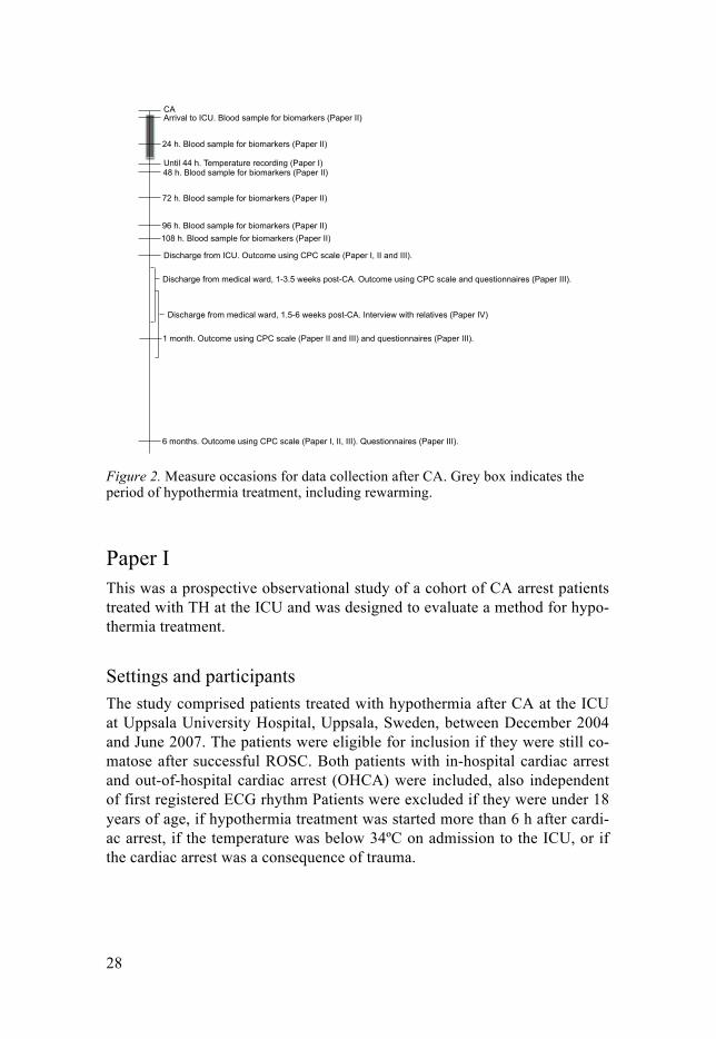
In Figure 2, an overview is provided of what time after the CA each data
collection occurred.
28
Figure 2. Measure occasions for data collection after CA. Grey box indicates the period of hypothermia treatment, including rewarming.
Paper I This was a prospective observational study of a cohort of CA arrest patients treated with TH at the ICU and was designed to evaluate a method for hypo-thermia treatment.
Settings and participants The study comprised patients treated with hypothermia after CA at the ICU at Uppsala University Hospital, Uppsala, Sweden, between December 2004 and June 2007. The patients were eligible for inclusion if they were still co-matose after successful ROSC. Both patients with in-hospital cardiac arrest and out-of-hospital cardiac arrest (OHCA) were included, also independent of first registered ECG rhythm Patients were excluded if they were under 18 years of age, if hypothermia treatment was started more than 6 h after cardi-ac arrest, if the temperature was below 34ºC on admission to the ICU, or if the cardiac arrest was a consequence of trauma.
Arrival to ICU. Blood sample for biomarkers (Paper II)
24 h. Blood sample for biomarkers (Paper II)
48 h. Blood sample for biomarkers (Paper II)
72 h. Blood sample for biomarkers (Paper II)
96 h. Blood sample for biomarkers (Paper II) 108 h. Blood sample for biomarkers (Paper II)
Discharge from medical ward, 1-3.5 weeks post-CA. Outcome using CPC scale and questionnaires (Paper III).
1 month. Outcome using CPC scale (Paper II and III) and questionnaires (Paper III).
6 months. Outcome using CPC scale (Paper I, II, III). Questionnaires (Paper III).
Until 44 h. Temperature recording (Paper I)
CA
Discharge from medical ward, 1.5-6 weeks post-CA. Interview with relatives (Paper IV)
Discharge from ICU. Outcome using CPC scale (Paper I, II and III).
29
Cooling method Hypothermia treatment was induced by infusion of a 4ºC intravenous saline, in a planned volume of 30 ml/kg at a rate of 100 ml/min via two peripheral intravenous catheters. To complete induction and for maintained cooling, ice packs were applied in the groins, axillae and along the neck and were changed when necessary to allow continuous cooling. The ice packs used were saline infusion bags of 250 ml, which were stored in the freezer of the emergency department or the ICU. The ice packs were covered with a pil-lowcase to prevent cold injury on the patient´s skin. During the cooling treatment, the temperature was adjusted by applying or removing ice packs. The planned duration of hypothermia treatment was 24 hours, and it was estimated that the first two hours after CA would be taken up by transporta-tion to hospital and decision-making regarding hypothermia treatment. The target temperature of 32-34ºC was therefore maintained for up to 26 hours after the estimated time of CA. Passive rewarming at 0.5ºC/hour started after 26 hours and it was expected that a temperature of 36ºC, which was consid-ered the normal core temperature, would be reached within 8 hours. If the temperature increased too rapidly, the patient was given a bolus injection of a sedative, the blankets were removed, and the ice packs were applied again.
Treatment protocol The patients were sedated and mechanically ventilated during TH. If the induction of TH went slowly or if there were any signs of shivering, the se-dation and analgesia were increased. Muscle relaxation was only used if necessary as a bolus injection and subsequently, if required, as an infusion. The goal was to discontinue administration of muscle relaxant when the target temperature for TH was reached. After rewarming, the sedation was terminated. The treatment protocol had defined goals for factors that are generally considered important for critically ill/CA patients, but was adjust-ed for TH.
Data collection and monitoring Core temperature was measured continuously in the urinary bladder and recorded on the patient´s chart every 15 min up to 44 h after the cardiac ar-rest. Furthermore patients were monitored routinely for continuous blood pressure, central venous pressures, ECG, respiration, oxygen saturation and diuresis. Arterial blood samples were drawn every 90-120 min for measure-ment and analysis of blood gases, glucose and electrolytes. Complete blood counts were taken every 24 h. Neurological evaluation was carried out with the Reaction Level Scale (RLS 85)156 or Glasgow Coma Scale (GCS)157 on arrival. The neurological outcome was assessed using the CPC scale (Table
30
1)123 at discharge from the ICU and 6 months after cardiac arrest. The as-sessment after 6 months was conducted by a journal review or a phone call.
Statistical analysis Descriptive statistics and demographic data are presented as means, standard deviations, ranges, percentages and numbers.
Paper II This paper was a prospective observational study performed in three ICU departments to investigate levels of biomarkers in CA patients.
Settings and participants The study was conducted in three hospitals, one university hospital and two general county hospitals, during the period from May 2008 to May 2012. Eligible for inclusion were CA patients with successful ROSC, systolic arte-rial blood pressure ≥ 80 mmHg > 5 min, unconscious with a GCS score157 < 8, age > 18 years for whom TH was induced. The decision to start TH was taken for all patients, irrespective of the first registered ECG rhythm or whether the CA occurred in or out of hospital. TH was induced by infusion of a 4°C intravenous saline, in a planned volume of 30-40 ml/kg. To main-tain cooling, all hospitals used external cooling, either ice packs or cooling suits depending on hospital-specific routines. Rewarming at a rate of 0.5°C/hour was used and 36°C was considered the normal core temperature. The patients were cooled to 32-34°C for 24 hours. Before cooling, all pa-tients were sedated, intubated and mechanically ventilated according to local ICU guidelines for severely ill patients adapted for TH after CA.
During the inclusion period, 242 CA patients were admitted to the ICUs, 209 underwent TH, and of these, 125 were included in the study. Sixty-three patients survived until hospital discharge, 38 died before ICU discharge and 24 died in the medical ward. The main cause of death was cerebral (n=39) and cardiac (n=16). Two patients died between one and six months, both in a new CA. One had reach CPC 1 and the other CPC 3.
Data collection Blood samples for biomarkers were collected as soon as possible upon arri-val to the ICU and at 24, 48, 72, 96 and 108 hours after CA from a peripher-al artery or vein. In patients who were provided with a catheter in the jugular bulb, blood samples were collected there at the same time points. The sam-
31
ples were stored in a -70° C freezer, and analysed at the same time after the study period.
Medical background variables, information about the CA and data on temperature management were retrieved from the medical chart. Functional outcome was assessed using the CPC scale (Table 1).123
Analysis of biomarkers Serum levels of NSE and S100B were measured using a Cobas e601 instru-ment and NSE and S100B reagent kits as described by the manufacturer (Roche Diagnostics, Penzberg, Germany). For detection of hemolysis, a serum measurement index of hemolysis (Sindh) was used. Serum GFAP was measured using the non-commercial Elecsys® GFAP prototype test on a Cobas e411 instrument (Roche, Penzberg, Germany). In the first step, biotin- and ruthenium-labeled monoclonal anti-GFAP antibodies are combined with 50µl of sample and incubated for 9 minutes. In the second step, streptavidin-coated magnetic microparticles are added and the mixture is incubated for an additional 9 minutes. After the second incubation step, the reaction mixture is transferred into the measuring cell where the beads are captured on the surface of an electrode by a magnet. The unbound label is removed by wash-ing the measuring cell. In the last step, voltage is applied to the electrode in the presence of a tri-propylamine (TPA)-containing buffer, the resulting electrochemiluminescent signal is recorded by a photomultiplier and the GFAP concentration is derived from a calibration curve. Because no acknowledged reference method is available at present, the method has been standardized by weighing pure human GFAP in analyte-free serum matrix. Three internal samples based on human serum matrix spiked with human GFAP low (< 5ng/ml), medium (20 – 30ng/ml) and high (> 50ng/ml) were used for quality control during the assay. The within- and between-run preci-sions were 1.1-1.9% and 2.7-4.2%, respectively.
Statistical analysis The scores were dichotomized into good (CPC 1-2) and poor (CPC 3-5) neurological outcome. The continuous data were not normally distributed, and therefore non-parametric statistics were used. Descriptive statistics were used to present participants’ demographic and medical characteristics. The Mann Whitney U-test (continuous variables) and Chi-squared test (categori-cal variables) were used to compare demographic and medical characteristics between the good and poor outcome groups. To test differences between the good and poor groups for GFAP, NSE and S100B at each time point, the Mann Whitney U-test was used. The incremental predictive value of GFAP, S100B and NSE was evaluated using logistic regression models and present-ed with area under the receiver operating characteristic (ROC) curve (AUC).
32
Sensitivity and specificity for specific cut-off values were derived using ROC. The Wilcoxon signed rank test was used for analysis of the difference between blood samples from a peripheral artery or vein and samples from the jugular bulb. P-values <0.05 (two-tailed) were defined as statistically significant. All statistical analyses were performed using SPSS version 21 (SPSS Inc. Chicago, IL, USA).
Paper III Paper III is a quantitative study designed to investigate HRQoL, anxiety and depression in CA patients who were treated with TH and survived until 6 months.
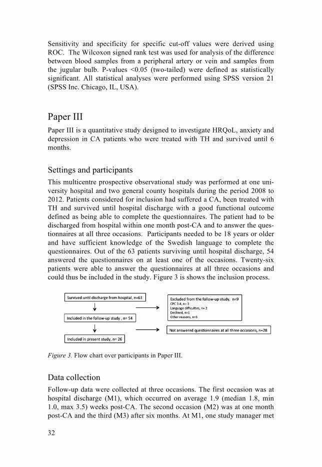
Settings and participants This multicentre prospective observational study was performed at one uni-versity hospital and two general county hospitals during the period 2008 to 2012. Patients considered for inclusion had suffered a CA, been treated with TH and survived until hospital discharge with a good functional outcome defined as being able to complete the questionnaires. The patient had to be discharged from hospital within one month post-CA and to answer the ques-tionnaires at all three occasions. Participants needed to be 18 years or older and have sufficient knowledge of the Swedish language to complete the questionnaires. Out of the 63 patients surviving until hospital discharge, 54 answered the questionnaires on at least one of the occasions. Twenty-six patients were able to answer the questionnaires at all three occasions and could thus be included in the study. Figure 3 is shows the inclusion process.
Figure 3. Flow chart over participants in Paper III.
Data collection Follow-up data were collected at three occasions. The first occasion was at hospital discharge (M1), which occurred on average 1.9 (median 1.8, min 1.0, max 3.5) weeks post-CA. The second occasion (M2) was at one month post-CA and the third (M3) after six months. At M1, one study manager met
33
the patient; at M2 the questionnaires were sent by post after telephone con-tact. At M3 a study manager again met with the patient. Functional outcome was assessed using the CPC scale (Table 1).123 The standardized question-naires used were the Hospital Anxiety and Depression Scale (HADS), the Euroqol (EQ5D) and the Short Form 12 (SF12). Medical variables were collected from the medical chart. Socio-demographic variables prior to the CA were collected by self-report at M1. Data on participants’ working situa-tion and whether they had been in contact with any health service were col-lected at M3.
HADS HADS is a 14-item standardized questionnaire designed to assess mood dis-orders in non-psychiatric hospital patients. The questionnaire is divided into two subscales measuring signs of anxiety and depression. The highest possi-ble score for each subscale is 21, higher scores representing more emotional problems. A score of 0-7 for each subscale indicates no anxiety or depres-sion, 8-10 mild to moderate anxiety or depression, and a score of 11 or high-er indicates risk for occurrence of anxiety disorders or depression requiring medical treatment.158
EQ5D The EQ-5D health status standardized questionnaire is a self-reporting ques-tionnaire and comprises 5 questions – each with 3 levels. Level one repre-sents no problems, level two some problems, and level three extreme prob-lems. The 5 questions correspond to 5 health domains: pain, mood, mobility, self-care and daily activities.159, 160 EQ5D can be converted into a single summary index, where the highest possible index for HRQoL in all domains is 1.00.160 The Euroqol visual analogue scale (EQ-VAS) is a measure of self-rated overall health status and ranges from 0-100.159, 160
SF12 The SF-12 is a validated standardized questionnaire composed of 12 items divided into two components, the mental component score (MCS) and the physical component score (PCS), which are calculated to summarize mental and physical status, respectively. The patients estimate their HRQoL during the past week. The components are standardized with scores ranging from 0 to 100, where a higher score represents better experienced HRQoL.161, 162 A norm-based standardized score with a mean of 50 and standard deviations of 10 has been computed for the general population in United States. A MCS or PCS summary score ≤40 represents moderate to low HRQoL.162 SF12 has been validated in a Swedish population and the mean values are MCS 52.8 and PCS 50.2.161
34
Statistical analysis Descriptive statistics were used to present participants’ demographic and medical characteristics as well as central tendencies and distributions of the variables covered in the questionnaires. To compare the study population with the excluded group that did not answer the questionnaires at all three occasions, Mann Whitney U-test and chi-squared test were used. Owing to the small sample size, Friedman’s test was used to conduct analysis of vari-ance across the three measurement occasions. The Wilcoxon signed rank test (two-tailed) was used for post-hoc analysis of the difference between two measurement occasions. Spearman’s rank correlation coefficient (two-tailed) was calculated to investigate relationships between anxiety and depression and HRQoL. Because multiple tests were used for comparison and correla-tion, a p-value ≤ 0.01 (two-tailed) was considered statistically significant. The reliability of the questionnaires was expressed as Cronbach´s α coeffi-cient. Data were recorded and statistical analyses were performed using SPSS version 21 (SPSS Inc. Chicago, IL, USA).
Paper IV In Paper IV, a qualitative methodology was used to describe relatives’ expe-riences during their next of kin’s hospital stay after surviving a CA.
Setting and participants The study took place at three hospitals in central Sweden, one university hospital and two general county hospitals. Twenty relatives of patients who had survived a CA and been treated with TH were included in the study dur-ing the period May 2008 to June 2010. Purposive sampling of relatives was based on age and gender to achieve as much variation as possible.163 Table 3 is shows demographic data on the participants.
Table 3. Demographic data on the relatives.
Sex Age Relation to the pa-tient
Working situation at the time of the CA
13 Women 20-70 years 10 Husband or wife 13 Worked full time 7 Men (mean 52) 3 Partners 4 Worked part time 6 Children
1 Parent 3 Retired or sick-listed
35
Data collection Data were collected using a semi-structured interview guide covering how the relatives experienced their next of kin’s CA and TH, support and infor-mation during the stay in hospital, how everyday life had changed and how they viewed the future. An interview guide was developed by consensus within the research group. A pilot interview was performed to test the usabil-ity of the interview guide, which was found to be feasible. Clarifying ques-tions were used such as “What do you mean?”, “How did you feel then?” and “What did you think then?” The interview was conducted at the time when the patient was discharged from hospital, 1.5 to 6 weeks post-CA. The interviews was recorded and lasted from 30 to 60 minutes. The interview took place in a separate room at the hospital or in the relative’s home, de-pending on the relative's wishes. Four of the interviews were conducted over the telephone because of the distance involved. Three ICU nurses performed the interviews, one had experience in the method and supervised the others in data collection.
Data analysis The interview text was systematically analysed using qualitative content analysis.163-166 The method was developed to describe the content of commu-nication and was originally a quantitative method.164 Qualitative content analysis is defined as a method for analysing a text in a systematic way and drawing replicable and valid inferences from the text to their context,166 fo-cusing on differences and similarities, thus demonstrating variability and diversity and giving the context a meaning. The descriptive level of qualita-tive content analysis, the manifest content, is described in categories or sub-categories, and the underlying meanings are used to create themes, repre-senting the latent content.164
In this study, the latent content was used and the process of the analysis was as following steps: 1. The interviews were transcribed verbatim. 2. The text was read several times in order to gain an overall impression. 3. The text was divided into meaning units in line with the aim of the
study. A meaning unit could consist of a few words or several sentences. 4. The meaning units were condensed and given a code. 5. Codes that expressed related meanings were grouped together into cate-
gories. 6. Categories with similar content and underlying meaning were sorted into
the same theme. Throughout the process, the authors went back and forth between the in-
terviews, codes, categories and themes to validate the findings. The first author continuously discussed the analysis process with the supervisor.
36
Ethical considerations The studies were performed according to the Declaration of Helsinki167 and reviewed and approved by The Regional Ethical Committee in Uppsala (Reg. no. 2004:M-207 and 2007/307). The participants were treated with respect and their interests were prioritized. Data collection was carried out with respect for integrity. The participants received written and oral infor-mation about the purpose of the study. They were told that participation was voluntary, that they were free to withdraw at any time and that full confiden-tiality was guaranteed. In Paper I-II, the patients were unconscious when they were included, so the information was given to and consent first ob-tained from a relative. Later, when the patients were considered competent, they received information and gave their informed consent, then they were also asked to participate in the study for Paper III. In Paper IV, the partici-pants gave their informed consent.
No medical risks were anticipated due to the study participation. The cooling method evaluated in Paper I had already been implemented in clini-cal practice. The samples of biomarkers were taken at the same time as ordi-nary blood samples and the amount of blood was small. The research team has extensive experience of conducting research with and caring for CA patients. This meant they had both the knowledge and preparedness to han-dle any questions or concerns the participants might have had. Because par-ticipants may have felt the researcher having access to their personal data was a violation of their personal integrity, the voluntary and confidential nature of the study was emphasized as well as the fact that the results would be presented at a group level and made anonymous.
37
Results
Paper I During the 30-months period of the study, 38 of the 45 patients treated with TH after CA were included. The mean age was 60 (22-82) years, and most were men (n=25, 66%). There were more frequently OHCA (n=30, 79%), and most were witnessed (n=28, 74%). First registered ECG rhythm was VF in 16 (42%) of the patients, 12 (32%) had asystole, 6 (16%) pulseless electric activity (PEA) and 4 (10%) other rhythms.
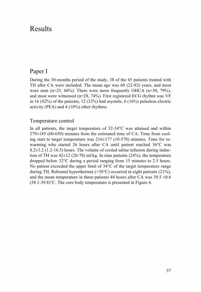
Temperature control In all patients, the target temperature of 32-34°C was attained and within 279±185 (60-650) minutes from the estimated time of CA. Time from cool-ing start to target temperature was 216±177 (10-570) minutes. Time for re-warming who started 26 hours after CA until patient reached 36°C was 8.2±3.2 (1.2-16.5) hours. The volume of cooled saline infusion during induc-tion of TH was 42±12 (20-70) ml/kg. In nine patients (24%), the temperature dropped below 32°C during a period ranging from 15 minutes to 2.5 hours. No patient exceeded the upper limit of 34°C of the target temperature range during TH. Rebound hyperthermia (>38°C) occurred in eight patients (21%), and the mean temperature in these patients 44 hours after CA was 38.5 ±0.6 (38.1-39.8)°C. The core body temperature is presented in Figure 4.

38
Figure 4. Core body temperature in degrees Celsius (C) expressed as mean ±SD during induction, maintenance and rewarming. Time 0 represents estimated time of CA.
Outcome At discharge from ICU, the neurological outcome was good in 10/38 (26%) participants and after 6 months in 17/38 (45%). Table 4 is shows neurologi-cal outcome from the study population, including the first registered ECG rhythm.
Table 4. Neurological outcome at discharge from ICU and after 6 months.
CPC 1-2 CPC 3-4 CPC 5 Discharge from ICU (n=38)
10 (26) 16 (42) 12 (32)
VF* (n=16) 6 (37) 7 (44) 3 (19) Asystole, PEA* (n=18)
3 (17) 7 (39) 8 (44)
6 months post-CA (n=38) VF* (n=16) Asystole, PEA* (n=18)
17 (45) 10 (63) 5 (28)
2 (5) - 2 (11)
19 (50) 6 (37) 11 (61)
*= First registered ECG rhythm. Values are reported as number of patients (%).
39
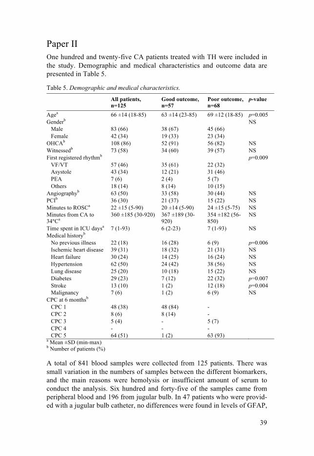
Paper II One hundred and twenty-five CA patients treated with TH were included in the study. Demographic and medical characteristics and outcome data are presented in Table 5.
Table 5. Demographic and medical characteristics.
All patients, n=125
Good outcome, n=57
Poor outcome, n=68
p-value
Agea 66 ±14 (18-85) 63 ±14 (23-85) 69 ±12 (18-85) p=0.005 Genderb NS Male 83 (66) 38 (67) 45 (66) Female 42 (34) 19 (33) 23 (34) OHCAb 108 (86) 52 (91) 56 (82) NS Witnessedb 73 (58) 34 (60) 39 (57) NS First registered rhythmb p=0.009 VF/VT 57 (46) 35 (61) 22 (32) Asystole 43 (34) 12 (21) 31 (46) PEA 7 (6) 2 (4) 5 (7) Others 18 (14) 8 (14) 10 (15) Angiographyb 63 (50) 33 (58) 30 (44) NS PCIb 36 (30) 21 (37) 15 (22) NS Minutes to ROSCa 22 ±15 (5-90) 20 ±14 (5-90) 24 ±15 (5-75) NS Minutes from CA to 34 Ca
360 ±185 (30-920) 367 ±189 (30-920)
354 ±182 (56-850)
NS
Time spent in ICU daysa 7 (1-93) 6 (2-23) 7 (1-93) NS Medical historyb No previous illness 22 (18) 16 (28) 6 (9) p=0.006 Ischemic heart disease 39 (31) 18 (32) 21 (31) NS Heart failure 30 (24) 14 (25) 16 (24) NS Hypertension 62 (50) 24 (42) 38 (56) NS Lung disease 25 (20) 10 (18) 15 (22) NS Diabetes 29 (23) 7 (12) 22 (32) p=0.007 Stroke 13 (10) 1 (2) 12 (18) p=0.004 Malignancy 7 (6) 1 (2) 6 (9) NS CPC at 6 monthsb CPC 1 48 (38) 48 (84) - CPC 2 8 (6) 8 (14) - CPC 3 5 (4) - 5 (7) CPC 4 - - - CPC 5 64 (51) 1 (2) 63 (93) a Mean ±SD (min-max) b Number of patients (%) A total of 841 blood samples were collected from 125 patients. There was small variation in the numbers of samples between the different biomarkers, and the main reasons were hemolysis or insufficient amount of serum to conduct the analysis. Six hundred and forty-five of the samples came from peripheral blood and 196 from jugular bulb. In 47 patients who were provid-ed with a jugular bulb catheter, no differences were found in levels of GFAP,
40
NSE and S100B between peripheral and jugular bulb samples. The following results are from peripheral blood samples.
GFAP There were differences between the good and poor outcome groups at 48 (p=0.016), 72 (p=0.003) and 96 (p=0.018) hours post-CA. High GFAP levels were more common in the poor outcome group. GFAP level at 0.83ng/mL was seen in one sample in the good outcome group, whereas the remaining samples had values ≤0.45ng/mL.
In the poor outcome group, GFAP levels above 0.83ng/mL were seen in 36 samples, ranging up to 445ng/mL. Of the patients with poor outcome, 18% (n=12) at 24 hours and 15 % (n=10) at 48 hours had GFAP levels above 0.83ng/mL. The ROC analysis showed the highest AUC at 72 hours (Table 6). The sensitivity and specificity of specific cut-off values for pre-dicting poor outcome are listed in Table 6.
NSE There were differences between good and poor outcome at all sampling times (p=<0.001) except in the acute phase upon arrival to ICU. An NSE above 0.22 µg/L at 72 hours post-CA resulted in 100% specificity for a poor outcome with a sensitivity of 50%, and 96 hours post-CA a value above 18 µg/L resulted in 100% specificity with a sensitivity of 55% (Table 6).
S100B Differences between good and poor outcome were significant at all sampling times (p=<0.05 and p=<0.001). The mean levels of S100B decreased in the good outcome group from the acute phase to 24 hours post-CA, but in the poor outcome group the mean levels instead increased between these time points. AUC value and sensitivity and specificity for S100B at different sampling times are presented in Table 6.
41
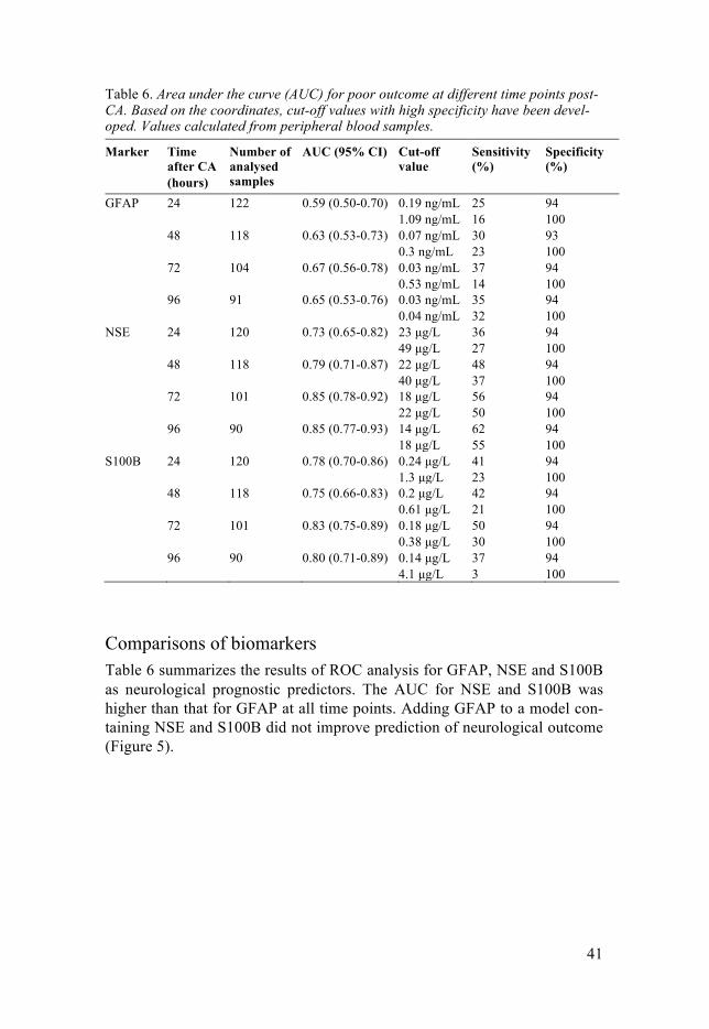
Table 6. Area under the curve (AUC) for poor outcome at different time points post-CA. Based on the coordinates, cut-off values with high specificity have been devel-oped. Values calculated from peripheral blood samples.
Marker Time after CA (hours)
Number of analysed samples
AUC (95% CI) Cut-off value
Sensitivity (%)
Specificity (%)
GFAP 24 122 0.59 (0.50-0.70) 0.19 ng/mL 25 94 1.09 ng/mL 16 100 48 118 0.63 (0.53-0.73) 0.07 ng/mL 30 93 0.3 ng/mL 23 100 72 104 0.67 (0.56-0.78) 0.03 ng/mL 37 94 0.53 ng/mL 14 100 96 91 0.65 (0.53-0.76) 0.03 ng/mL 35 94 0.04 ng/mL 32 100 NSE 24 120 0.73 (0.65-0.82) 23 µg/L 36 94 49 µg/L 27 100 48 118 0.79 (0.71-0.87) 22 µg/L 48 94 40 µg/L 37 100 72 101 0.85 (0.78-0.92) 18 µg/L 56 94 22 µg/L 50 100 96 90 0.85 (0.77-0.93) 14 µg/L 62 94 18 µg/L 55 100 S100B 24 120 0.78 (0.70-0.86) 0.24 µg/L 41 94 1.3 µg/L 23 100 48 118 0.75 (0.66-0.83) 0.2 µg/L 42 94 0.61 µg/L 21 100 72 101 0.83 (0.75-0.89) 0.18 µg/L 50 94 0.38 µg/L 30 100 96 90 0.80 (0.71-0.89) 0.14 µg/L 37 94 4.1 µg/L 3 100
Comparisons of biomarkers Table 6 summarizes the results of ROC analysis for GFAP, NSE and S100B as neurological prognostic predictors. The AUC for NSE and S100B was higher than that for GFAP at all time points. Adding GFAP to a model con-taining NSE and S100B did not improve prediction of neurological outcome (Figure 5).
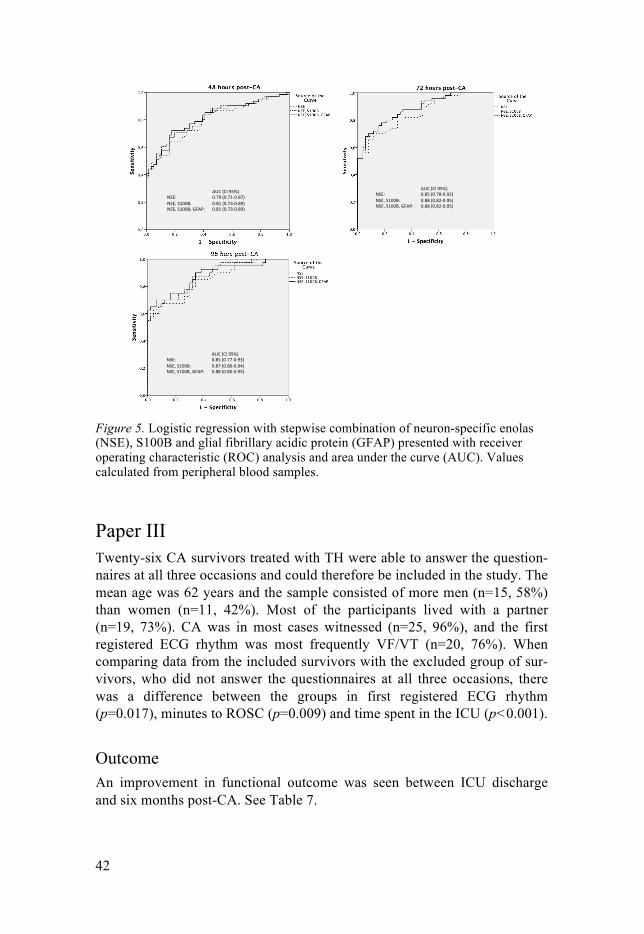
42
Figure 5. Logistic regression with stepwise combination of neuron-specific enolas (NSE), S100B and glial fibrillary acidic protein (GFAP) presented with receiver operating characteristic (ROC) analysis and area under the curve (AUC). Values calculated from peripheral blood samples.
Paper III Twenty-six CA survivors treated with TH were able to answer the question-naires at all three occasions and could therefore be included in the study. The mean age was 62 years and the sample consisted of more men (n=15, 58%) than women (n=11, 42%). Most of the participants lived with a partner (n=19, 73%). CA was in most cases witnessed (n=25, 96%), and the first registered ECG rhythm was most frequently VF/VT (n=20, 76%). When comparing data from the included survivors with the excluded group of sur-vivors, who did not answer the questionnaires at all three occasions, there was a difference between the groups in first registered ECG rhythm (p=0.017), minutes to ROSC (p=0.009) and time spent in the ICU (p<0.001).
Outcome An improvement in functional outcome was seen between ICU discharge and six months post-CA. See Table 7.
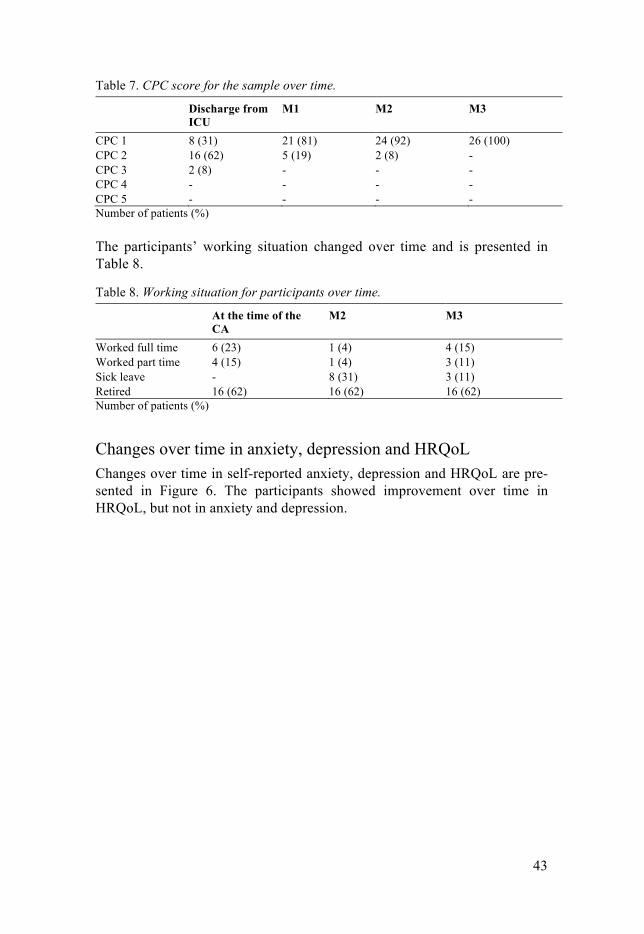
43
Table 7. CPC score for the sample over time.
Discharge from ICU
M1 M2 M3
CPC 1 8 (31) 21 (81) 24 (92) 26 (100) CPC 2 16 (62) 5 (19) 2 (8) - CPC 3 2 (8) - - - CPC 4 - - - - CPC 5 - - - - Number of patients (%) The participants’ working situation changed over time and is presented in Table 8.
Table 8. Working situation for participants over time.
At the time of the CA
M2 M3
Worked full time 6 (23) 1 (4) 4 (15) Worked part time 4 (15) 1 (4) 3 (11) Sick leave - 8 (31) 3 (11) Retired 16 (62) 16 (62) 16 (62) Number of patients (%)
Changes over time in anxiety, depression and HRQoL Changes over time in self-reported anxiety, depression and HRQoL are pre-sented in Figure 6. The participants showed improvement over time in HRQoL, but not in anxiety and depression.
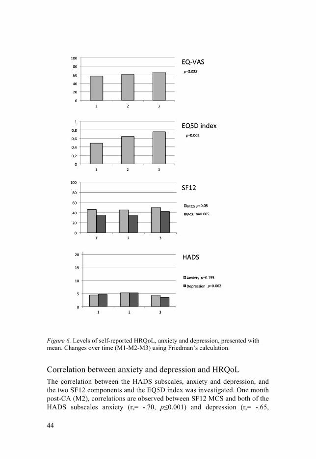
44
Figure 6. Levels of self-reported HRQoL, anxiety and depression, presented with mean. Changes over time (M1-M2-M3) using Friedman’s calculation.
Correlation between anxiety and depression and HRQoL The correlation between the HADS subscales, anxiety and depression, and the two SF12 components and the EQ5D index was investigated. One month post-CA (M2), correlations are observed between SF12 MCS and both of the HADS subscales anxiety (rs= -.70, p≤0.001) and depression (rs= -.65,
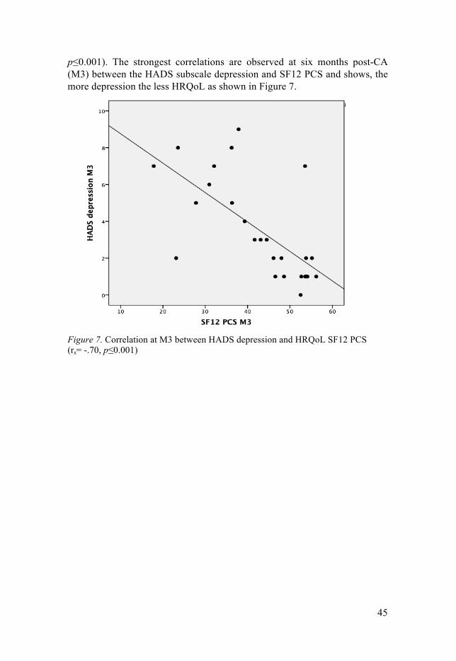
45
p#0.001). The strongest correlations are observed at six months post-CA (M3) between the HADS subscale depression and SF12 PCS and shows, the more depression the less HRQoL as shown in Figure 7.
Figure 7. Correlation at M3 between HADS depression and HRQoL SF12 PCS (rs= -.70, p#0.001)
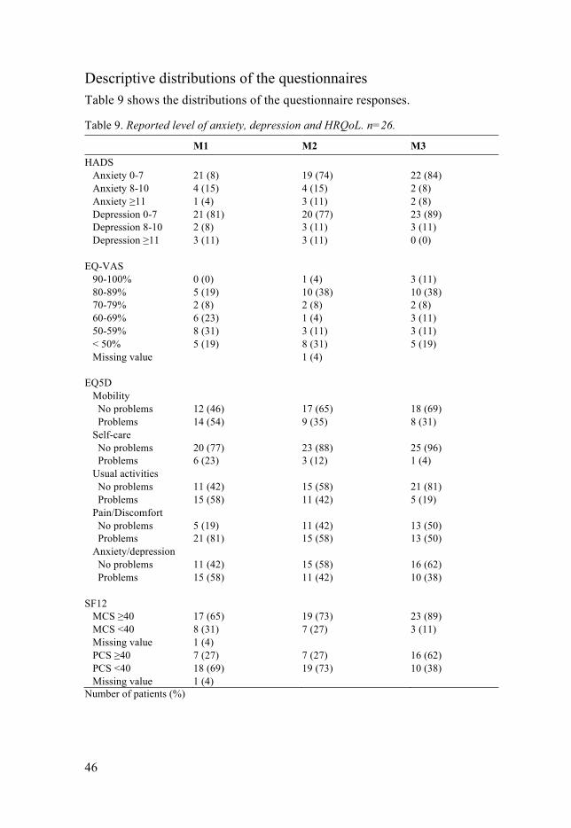
46
Descriptive distributions of the questionnaires Table 9 shows the distributions of the questionnaire responses.
Table 9. Reported level of anxiety, depression and HRQoL. n=26.
M1 M2 M3 HADS Anxiety 0-7 21 (8) 19 (74) 22 (84) Anxiety 8-10 4 (15) 4 (15) 2 (8) Anxiety ≥11 1 (4) 3 (11) 2 (8) Depression 0-7 21 (81) 20 (77) 23 (89) Depression 8-10 2 (8) 3 (11) 3 (11) Depression ≥11 3 (11) 3 (11) 0 (0) EQ-VAS 90-100% 0 (0) 1 (4) 3 (11) 80-89% 5 (19) 10 (38) 10 (38) 70-79% 2 (8) 2 (8) 2 (8) 60-69% 6 (23) 1 (4) 3 (11) 50-59% 8 (31) 3 (11) 3 (11) < 50% 5 (19) 8 (31) 5 (19) Missing value 1 (4) EQ5D Mobility No problems 12 (46) 17 (65) 18 (69) Problems 14 (54) 9 (35) 8 (31) Self-care No problems 20 (77) 23 (88) 25 (96) Problems 6 (23) 3 (12) 1 (4) Usual activities No problems 11 (42) 15 (58) 21 (81) Problems 15 (58) 11 (42) 5 (19) Pain/Discomfort No problems 5 (19) 11 (42) 13 (50) Problems 21 (81) 15 (58) 13 (50) Anxiety/depression No problems 11 (42) 15 (58) 16 (62) Problems 15 (58) 11 (42) 10 (38) SF12 MCS ≥40 MCS <40 Missing value
17 (65) 8 (31) 1 (4)
19 (73) 7 (27)
23 (89) 3 (11)
PCS ≥40 PCS <40 Missing value
7 (27) 18 (69) 1 (4)
7 (27) 19 (73)
16 (62) 10 (38)
Number of patients (%)
47
Paper IV Participants in the study underlying Paper IV are henceforth described as relatives and the person who had suffered the CA as the next of kin. The analysis revealed in ten categories and three themes through which the relatives described their experiences the first time after the next of kin’s CA. See Table 10.
Table 10. Overview of categories and themes.
Categories Themes Difficulties taking in what had happened The first period of chaos Feeling fear Waiting in uncertainty Painful to see a relative seriously ill Receiving support Feeling secure in a difficult situation Receiving information Feeling variations in the quality of care Feeling increased demands and responsibility Living in a changed existence Concerns for the future The next of kin has changed
The first period of chaos This theme describes how the relatives experienced the acute phase after their next of kin had suffered the CA. They described a sense of unreality for what had happened and a chaotic and shattering experience but with rational thoughts. The CA caused feelings of fear, panic and anxiety. The relatives found it difficult to see their next of kin seriously ill. They described the TH as unpleasant and the next of kin felt cold and almost dead. The time before the next of kin woke up was stressful because of the uncertainty as to wheth-er he/she had suffered any injuries, and if so, what kind.
Feeling secure in a difficult situation This theme illuminates the relatives’ need for support and information as well as the variations in the care their next of kin received. The relatives experienced the support from their family as the most important form of support. All of the relatives received information about their next of kin’s condition, but experienced difficulties in assimilating the information be-cause it was hard for them to concentrate. They wished there had been more written information about what caused the CA, the prognosis and infor-mation about prevention.
The relatives experienced the care in the ICU as good and safe. The per-sonnel behaved professionally and competently: they were kind and en-
48
gaged, treating all family members with consideration, but the need for a routine for how to taking care of relatives was emphasized. At the general ward, the relatives felt the staff were less supportive and engaged than staff at the ICU. The possibility to receive rehabilitation and care after hospital discharge was insufficient.
Living in a changed existence The impact on everyday life after their next of kin’s CA is illustrated in this theme. The relatives experienced increased workload and a responsibility to support other family members. They described a demand for keeping a con-tinuous contact with other family members and friends by phone calls, but felt they spent too much time by the phone. These feelings gave them a bad conscience. They felt responsible for their next of kin and wanted to support them to the outmost. The relatives felt great uncertainty and anxiety about the future. They were worried about their next of kin’s physical changes, but also hopeful and wished to see the future with confidence. They felt they had been changed by the incident, and thoughts about death and the meaning of life were common. The next of kin was still affected by the CA, experienc-ing problems with his/her short-term memory, speech, balance and walking as well as physical and mental complications.
49
Discussion
This work focused on CA patients who reached the ICU and the overall aim of this thesis was to study post-resuscitation care with focus on therapeutic hypothermia, outcome up to six months and relatives’ experiences. The find-ings showed that cold intravenous infusion combined with ice packs was useful with close monitoring of the body temperature in inducing and main-taining TH and also controlling rewarming. In Paper II we found that GFAP was significantly increased in the poor outcome group post-CA but NSE and S100B were shown to be better in predicting neurological outcome. HRQoL improved over time over the first six months post-CA. There were also rela-tively high negative correlations between anxiety, depression and HRQoL six months post-CA. The relatives described their experiences during the acute phase as a time of chaos. They found it difficult to assimilate the medi-cal information and wanted it in written form, and the information should be clear and honest. They lacked rehabilitation plans, felt uncertain about the future, but still hopeful.
Therapeutic hypothermia Therapeutic hypothermia for survivors of OHCA has been shown to improve both survival and long-term neurologic outcomes30, 31 and has been endorsed by American and European guidelines.7, 12 However, the optimal approach to providing TH is a topic of ongoing research. For example, a recent large trial demonstrated that targeting a temperature of 36°C provides similar results to a temperature goal of 33°C.16
In Paper I, we evaluated a method for TH with cold saline infusion com-bined with ice packs. All patients reached the target temperature of 32-34°C on average 4.4 hours from cardiac arrest and 3.4 hours from induction of TH. These results are in line with other studies.10, 30, 31, 41, 45, 54, 55, 58, 168 When making a retrospective comparison between the studied population and the patients for whom the same cooling method was used as for the participants from Paper II, the time to target temperature tended to increase (Table 11). There is no explanation for the increased time to target temperature: there was no difference in number of angiographies or PCIs between the groups, and the observations were performed at the same unit using the same guide-lines. Given the personnel’s longer experience of using the method, it is sur-
50
prising that the time increased. When Study I was underway, it was known among the personnel that the cooling method was being evaluated, which may have affected the results. However, a fast time to target temperature has not been shown to be associated with better outcomes.44, 168, 169 Time for re-warming increased between the two periods but might be explained by the different target of normal temperature, 36°C versus 36,5°C.
Table 11. Comparison between Paper I and patients cooled with the same method during the second inclusion period.
n=38, 2004-2007 n=75, 2008-2012 p-value Time from CA to target temperaturea 279 minutes ±185
(60-650) 342 minutes ±181 (30-920)
0.06
Time from cooling start to target tempera-turea
216 minutes ±177 (10-570)
274 minutes ±178 (0-865)
0.07
Time for rewarminga 8.2 hours* ±3.2 (1.2-16.5) (n=36)
14.9 hours** ±6.1 (5.0-36.5) (n=74)
0.00
Angiographyb 19 (50) 33 (44) 0.54 PCIb 13 (34) 17 (23) 0.19 a Mean ±SD (min-max) b Number of patients (%) * Started 26 hours after CA until patient reached 36°C ** Started 26 hours after CA until patient reached 36,5°C
In this study, the temperature remained stable during the maintenance phase. Cooling below the temperature range did occur but was not a major problem. In nine patients (24%), the temperature dropped below 32°C, and the lowest temperature, 31.3°C was noted in one patient. Problems with keeping the temperature stable have been reported when using ice packs as a cooling method, and temperatures below 32°C occurred in 10-63% of the patients.37,
170 In the current study, the temperature was monitored every 15 minutes and early information about changes in body temperature was obtained, which might have been a reason for the relatively stable temperature. We therefore consider that careful monitoring of body temperature during treatment is necessary to achieve good temperature control with the present method. For detection of changes in body temperature, the recommendation is to register the core temperature.7 In this study, bladder temperature measurement was used. It has been reported that the most accurate way to measure core tem-perature is in a central vein, which resulting in small variance even during the cooling phase.171
Rewarming can be performed using the same devise that is used for cool-ing. With the method used in the present study, careful monitoring of tem-perature was required in order to control the rate of temperature elevation. Additional sedation was given or ice packs re-applied in an effort to prevent too rapid rate of rewarming. The recommendation is to rewarm the patients after TH at a maximal rate of 0.2–0.5°C/h.12, 13 Rewarming in the present study continued for 8.2 hours, and was comparable to times reported from
51
other studies.31, 45, 55, 59 A high rewarming rate (>0.5°C/hour) has not been found to be associated with a higher risk for poor outcomes.38
Rebound hyperthermia (>38°C) has been registered in about one fifth of patients,37, 170 and was also seen in the present study. In the group of patients with rebound hyperthermia (n=8), four had good outcome and four poor outcome six months post-CA. In other studies, post-hypothermia fever oc-curred in half of the patients, and did not show any relation to outcome at hospital discharge172 or after six months.38 In another study, post-hypothermia fever was associated with mortality one month post-CA.15
There are several different cooling devices available on the market and all have their strengths and weaknesses. The method applied in this study could be initiated early and did not interfere with other interventions, which most frequently consisted of an immediate transfer from the emergency ward for coronary angiography or CT scan of the brain. Each unit should choose a cooling device that suits it logistically, and the personnel should be trained to use it.32, 64
Prognostication Predicting neurological outcome after CA is an important aspect of patient care and the information given to relatives. Early prognostication is difficult, as treatment with TH and sedation influences several of the prognostic tools. Biomarkers are likely to be independent of the effect of sedative drugs,77 but whether or not TH affects the levels of biomarkers is unclear, and TH may influence the time course of biomarkers.87 For prediction of neurological outcome based on biomarkers, it is important to have a method with high specificity.
In Paper II, GFAP, a relatively new biomarker in the field of CA, was evaluated as a neurological predictor, together with the more commonly studied biomarkers NSE and S100B. The result showed that GFAP was sig-nificantly increased in the poor outcome group at 48, 72 and 96 hours post-CA. However, NSE and S100B were shown to be better in predicting neuro-logical outcome.
In the poor outcome group, GFAP was increased, but not in all patients. We could not find any explanation why some in the poor outcome group had increased levels, while others were normal. Levels above 0.83ng/mL were only seen in the poor outcome group and were most frequently occurring at 24 and 48 hours post-CA. In contrast to the good outcome group, where only slightly elevated levels of GFAP were seen. Similar results have also been observed in previous studies on CA patients.89, 90, 173 In the present study, GFAP predicted poor neurological outcome with 100% specificity and 14-32% sensitivity at 24, 48, 72 and 96 hours post-CA. An earlier study on 12 CA patients treated with TH reported sensitivity between 37-75% for GFAP
52
at different time points when specificity was set to 100%.90 One problem with comparing biomarkers from different studies is the lack of standardiza-tion and the difference between assays and laboratories.
It has been difficult to found a reliable cut-off value for biomarkers.77 To predict poor neurological outcome using NSE, the best threshold in the pre-sent study was 18 µg/L 96 hours post-CA with specificity at 100% and sensi-tivity at 55% (Table 6). This seems to be a lower cut-off value than previous-ly described. S100B values of 0.18 and 0.21 µg/L have been reported as cut-off values 24 hours post-CA with specificity at 100%.77 The corresponding value in the present study was 1.3 µg/L 24 hours post-CA with 100% speci-ficity which is substantially higher (Table 6). In the present study, for GFAP levels to predict neurological outcome with 100% specificity, a cut-off value of 0.04 ng/ml 96 hours post-CA was found to be appropriate (Table 6). It should be noted, however, that because no certified reference material for assay calibration exists, it is difficult to compare values obtained using dif-ferent methods. The present method has not previously been used in CA patients.
In patients with traumatic brain injury, increased levels of GFAP have been reported.107, 108, 174 One study used a cut-off level of 1.5 µg/L with speci-ficity at 95% and sensitivity at 53% for predicting poor outcome,108 which is a considerably higher cut-off level than was found with 100% specificity in the present study. In stroke patients, the level of GFAP has been shown to correlate with the severity of brain damage and infarct volume.106 In a meta-analysis of the usefulness of GFAP as a marker after acute stroke, it was found that GFAP is effective to use in differential diagnosis. Specificity has varied between 70-98%, but different cut-off levels have been reported.175
In the study presented in Paper II, a logistic regression analysis was per-formed for stepwise combinations of NSE, S100B and GFAP. A combina-tion of current biomarkers did not increase the ability to predict neurological outcome. For prognostication, continuous evaluation of the patient’s progno-sis is needed and a combination of predictors is recommended.77, 176, 177
To our knowledge, biomarkers in the jugular bulb have not been studied in CA patients. In theory we had expected higher levels of biomarkers in the samples from the jugular bulb, given the direct drainage of blood from the brain. However, in the present study, levels of GFAP, NSE and S100B in peripheral blood and the jugular bulb did not differ.
Outcome In Paper I-III, the patients’ neurological outcomes have been evaluated using the CPC scale.123 About forty-five percent of the patients in both Paper I and II had scored a good outcome (CPC 1-2) after six months. There was clinical
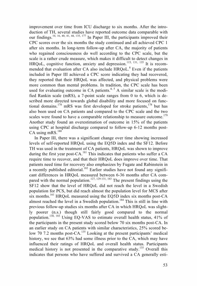
53
improvement over time from ICU discharge to six months. After the intro-duction of TH, several studies have reported outcome data comparable with our findings.10, 16, 40, 41, 44, 135, 177 In Paper III, the participants improved their CPC scores over the six months the study continued and all achieved CPC 1 after six months. In long-term follow-up after CA, the majority of patients who regained consciousness do well according to the CPC scale, but the scale is a rather crude measure, which makes it difficult to detect changes in HRQoL, cognitive function, anxiety and depression.115, 131, 135 It is recom-mended that evaluation after CA also include HRQoL.8 Even if the patients included in Paper III achieved a CPC score indicating they had recovered, they reported that their HRQoL was affected, and physical problems were more common than mental problems. In tradition, the CPC scale has been used for evaluating outcome in CA patients.4, 5 A similar scale is the modi-fied Rankin scale (mRS), a 7-point scale ranges from 0 to 6, which is de-scribed more directed towards global disability and more focused on func-tional domains.178 mRS was first developed for stroke patients,178 but has also been used on CA patients and compared to the CPC scale and the two scales were found to have a comparable relationship to measure outcome.179 Another study found an overestimation of outcome in 15% of the patients using CPC at hospital discharge compared to follow-up 6-12 months post-CA using mRS.180
In Paper III, there was a significant change over time showing increased levels of self-reported HRQoL using the EQ5D index and the SF12. Before TH was used in the treatment of CA patients, HRQoL was shown to improve during the first year post-CA.181 This indicates that patients who suffer a CA require time to recover, and that their HRQoL does improve over time. That patients need time for recovery also emphasizes by Fugate and Rabinstein in a recently published editorial.182 Earlier studies have not found any signifi-cant differences in HRQoL measured between 6-36 months after CA com-pared with the normal population.127, 129-131, 183 The present findings using the SF12 show that the level of HRQoL did not reach the level in a Swedish population for PCS, but did reach almost the population level for MCS after six months.161 HRQoL measured using the EQ5D index six months post-CA almost reached the level in a Swedish population.184 This is still in line with previous follow-up studies six months after CA in which HRQoL was slight-ly poorer (n.s.) though still fairly good compared to the normal population.130, 183 Using EQ-VAS to estimate overall health status, 41% of the participants in the present study scored below 70 six months post-CA. In an earlier study on CA patients with similar characteristics, 25% scored be-low 70 7.2 months post-CA.135 Looking at the present participants’ medical history, we see that 63% had some illness prior to the CA, which may have influenced their ratings of HRQoL and overall health status. Participants medical history is not presented in the comparative study.135 Overall this indicates that persons who have suffered and survived a CA generally esti-
54
mate their HRQoL as good. Life satisfaction and QoL are concepts that are closely to each other. In one study, most of the CA patients reported their satisfaction with “life as whole” to be good.134
In the study, presented in Paper III, the strongest correlations were shown between depression and HRQoL after six months, but correlations were also seen between both anxiety and depression and HRQoL at hospital discharge, after one month and after six months. Moulaert et al. found a correlation between anxiety and depression and HRQoL 36 months after CA when using the SF36 questionnaire.127 The present results and those of Moulaert et al. show a similar correlation, which remains a long time after the CA. The negative correlation between HRQoL and anxiety and depression shows that it is important to screen for and identify anxiety and depression problems to help patients to recover and be able to achieve a good QoL.182 In the present study, signs of anxiety and depression measured using HADS showed no significant changes over time. Nevertheless, the number of patients who scored high on anxiety disorders tended to increase at one month post-CA, before decreasing at six months. The measurement at one month was per-formed close in time after hospital discharge, which may have negatively affected the patients and caused more feeling of anxiety. This might be ex-plained by increased fatigue, loss of security at not having medical staff nearby and insight into what has happened to them. None of the participants had a previous history of anxiety or depression documented in their medical journal. In a recently published study, an intervention with consultations by a trained nurse was conducted to detect early emotional problems after CA, and both the patients and their caregivers positively evaluated the interven-tion.185 This demonstrates the importance of strategies for identifying and treating emotional problems such as anxiety and depression. The study pre-sented in Paper III was conducted in a small population with inclusion crite-ria excluding CA patients who needed a hospitalization up to one month or longer. This might have resulted in a selected group of patients, but in any case, patients had an influence on HRQoL and anxiety and depression.
The participants in Paper III had a higher frequency of VT/VF and fewer asystoli as the first registered ECG rhythm compared to the excluded group during the study period. This might have influenced the study results, as these factors are known to be associated with survival and morbidity.186 In Paper I, we also found a higher proportion of good outcomes in the VF/VT group.
A large number of different questionnaires have been used in studies to measure HRQoL after CA, and the time to follow-up varies.115, 117 This makes it difficult to compare findings between different studies.
55
Relative’s experiences The findings in Paper IV show that relatives of a person who has suffered a CA desired more information, especially written, on the cause of the CA, treatment, prognosis and prevention. Keeping contact with other family members and friends was described as demanding and the absence of a reha-bilitation plan after discharge from the medical ward was also a major issue.
One of the most important needs for the relatives of ICU patients is in-formation that is clear and honest.148, 149, 155, 187 In the present study, relatives wanted honest and clear information about their next of kin’s condition and prognosis. The relatives also found it difficult to assimilate the information provided. This has been confirmed in other studies.137, 150, 188 It appears that relatives cannot interpret and absorb the information given, and thus the information should be presented on several occasions.136, 148, 189, 190 Holm et al. found that partners of CA survivors were not prepared for the challenges that would arise when the next of kin was discharge from hospital.150 To reduce relatives’ anxiety and burden, it is important that they are informed and prepared, and that there is a plan for how continued care will be per-formed. The participants in Paper IV described the absence of a rehabilita-tion plan after discharge from the medical ward. Relatives felt uncertain about the future, but hopeful. It was important for them to feel hope even when the situation was critical and the prognosis uncertain. Studies have revealed the importance of hope when a next of kin is critically ill and shown that hope is of greater importance in cases of unexpected incidences than in chronically evolving illness.137, 138, 146, 148, 149 Relatives’ need for hope must be taken into account when staff meet with them. Because relatives desire honest and realistic information, it is important that staff encourage hopefulness without promoting unrealistic hope. Relatives experience am-bivalence, that is they are caught between wanting honest information and needing to remain hopeful.191, 192 The feeling of hope is complex, and hope has several dimensions. Even in moments of hopelessness, hope might sud-denly appear.193 Hope has been described as a way to cope with the situation when a close relative becomes critically ill.191, 192 In the present study, all relatives experienced support and the most important support came from other family members. Another study found that relatives prefer to talk to someone they know rather than to a hospital social worker or a priest.137 Having a social network has also been described as a way to cope with the situation.194
One surprising result was the demand placed on relatives to maintain tel-ephone contact with other family members and friends. This was time con-suming and instead they would have preferred to have time to relax and think. These feelings resulted in guilt and a bad conscience. Holm et al. de-scribed the demands as a burden, in that other family members and friends took time from the next of kin.150 Increased responsibility became apparent
56
in the form of supporting other family members and dealing with others’ worries, while being concerned about their next of kin. Concern for other family members has been described as a double worry in relation to how they will handle the situation.138 Six months after the next of kin’s CA, rela-tives still have reported increased demands, both in relation to the next of kin and other family members.155
In the present study, relatives described difficulties in understanding and absorbing what had happened, and the situation felt unreal. They protected themselves from unpleasant memories. Uncertainty was prevalent among relatives during the acute phase, and waiting in uncertainty was described as a time filled with anxiety and fear. The next of kin’s illness and thoughts about what kind of injuries the next of kin had suffered were powerful caus-es of stress. High levels of traumatic stress, anxiety and depression are de-scribed in relatives during the first days after a next of kin’s admission to an ICU.139, 195 This should be kept in mind in the care of relatives to CA pa-tients.
Intensive care is a highly technological ward, capable of providing ad-vanced treatment and nursing care for critically ill people. The care in the general ward is more focused on supporting, the monitoring is less important and staffing levels are lower. In the present study, relatives described a dif-ference between the ICU and the general ward in terms of effective interper-sonal relations, attitudes, and knowledge on the part of staff as well as lack of contact with staff. In line with this findings, ICU nurses and nurses in general ward have described a gap in care and differences in the environ-ment, differences in the nurses’ competence and in how to communicate.196 Transfer from the ICU to a general ward has been described as stressful and an issue that causes anxiety for patients and family members.197 Experiences depend on how the relatives and patients are treated by the staff and the in-formation received before, during and after transfer from an ICU to the gen-eral ward.198
Methodological considerations This thesis includes both quantitative and qualitative research designs. The research question and feasibility of data collection determined the choice of design. The advantage of using both quantitative and qualitative research designs is that the methods reinforce each other and different research ques-tions can be answered and a wider area investigated. This thesis also include different perspectives; medical care (Paper I and II), patients (Paper III) and relatives (Paper IV).
57
Paper I As our purpose was to describe a method of cooling, we employed a descrip-tive design. The study was intended for clinicians, treating CA patients with TH, to provide knowledge about the described cooling method used in the ICU. This was an observational study that did not involve comparisons with a normal-temperature control group or other established cooling devices. Not having any control group is a limitation, but because the purpose was to de-scribe rather than to compare, the design was preferable. The available pa-tients during the study period were consecutively included. The small sample size precluded any major assessment of the effect of this technique on the outcome and makes the results difficult to generalize. Five patients were excluded because the temperature protocol was not completed, and this may have biased our results.
Paper II The design used in Paper II was a prospective observational study. The pa-tients available during the study period were consecutively included. As the study had an exploratory purpose, no power calculation was performed. In the study, some data are missing due to hemolysis or insufficient amounts of serum to conduct the analysis, which could have affected the results. It is difficult to compare results across different studies due to the use of different assays for analysing biomarkers, which is a limitation in the area of bi-omarkers as regards finding reliable cut-off levels. Results on the biomarkers were not available to the caregivers and therefore could not have affected treatment or evaluation of the patient, which should be seen as a strength in the study.
Parts of the results are presented with sensitivity and specificity. Sensi-tivity is the ability to identify a condition correctly, in this study to identify patients with a possibility of achieving a good outcome. Specificity is the ability to identify non-cases correctly, thus in the present study to identify those with a poor outcome.163 In the present study, we pursued high specific-ity to avoid diagnosing poor outcome in patients with a possibility to recov-er, i.e. a low FPR. The accuracy of the test depends on how well the tests separate the good and poor outcome groups and is measured by AUC. The larger the area, the more accurate the test.163
Paper III This study prospectively investigated changes over time in HRQoL, anxiety and depression. Important strengths of the study are that all participants were followed up at the same time intervals from CA at M2 and M3, and followed over time. A limitation is the variation in hospital stay post-CA, which af-fected time from CA to M1 (min 1.0, max 3.5 weeks), resulting in a time from M1 to M2 of 1 week in 3 cases. The small study population is a limita-
58
tion that might have affected the results. Inclusion required participants to complete the questionnaires at three occasions, and thus they needed to have been discharged from hospital within one month after CA. This only applied to about half of the patients who survived CA and may have resulted in a selected group of CA patients.
All of the standardized questionnaires used in the study have been tested for validity and reliability. For two of the questionnaires, there are normative data available for the general Swedish population (SF12, EQ5D). The ques-tionnaires all involve self-reporting. Any cognitive impairment resulting from CA and also the risk of trying to please the investigators may have influenced participants’ answers.
Because multiple statistical tests were performed on the same data for comparison and correlation, a higher level of significance was used (p-value ≤ 0.01; two-tailed). This may have increased the risk of type II error, a false negative conclusion entailing failure to detect differences when they really exist.
The reliability of the questionnaires was expressed as a Cronbach’s α co-efficient. Cronbach’s α is used to test the reliability of an instrument and to ensure that all items in a test measure the same phenomenon. Cronbach’s α is a method of evaluating internal consistency. There is no standard for what an acceptable Cronbach’s α coefficient should be, but values ranging from 0.70-0.90 have been reported.163, 199 Low levels were seen at M1 and M3 in EQ5D, probably owing to the small number of items.
Paper IV The methodological approach in Paper IV was qualitative description and was influenced by Sandelowski’s work. Qualitative description is a method used for classification of experiences and to answer questions such as “who, what and where”.200, 201 Qualitative content analysis seemed an appropriate method, as our intention was to describe relatives’ experiences, with a focus on similarities and differences narrated in the interviews. An inductive ap-proach was used.163, 164
Trustworthiness is important in qualitative studies, and according to Graneheim and Lundman it comprises credibility, transferability and de-pendability.164 To achieve trustworthiness several aspects were considered during the process.
Credibility refers to confidence in the truth of the data and how well the data and processes of analysis describe the study area.163, 164 To achieve cred-ibility, participant selection aimed at variation in demographic characteris-tics, and a pilot interview was performed to test the interview guide and ex-amine whether the three researchers were focusing on the same topics. The interviews took place at about the same time after the next of kin’s CA, which made the participating relatives’ experiences comparable in this re-spect. The interviewers were familiar with the research field, and their pre-
59
understanding of the context may have contributed to trust and understand-ing among the relatives during the interviews as well as been helpful when analysing the data. Their pre-understanding could also be a weakness as it might have influenced the following-up questions, i.e., if the interviewer takes a given statement for granted. Limitations of this study are that only those who could understand and speak Swedish were included in the study. Because of the distance involved four of the interviews were conducted over telephone. According to Novick, face-to-face interviews are to preferable because the lack of visual cues during telephone interviews could lead to data loss or distortion.202 In the present study, the telephone interviews had a lighter and more limited content. However, this was compensated by the face-to-face interviews, which were more complex and exhaustive.
Dependability refers to the stability of data over time and condition, as well as how well the research process and analysis can be followed.163, 164 To enhance dependability an interview guide was used. During the analysis the authors continuously discussed the steps in the process. The last author is experienced in the method used in Paper IV and supervised the other authors involved in data collection and analysis.
Transferability describes to what extent the findings can be transferred to other settings and groups.163, 164 By offering a sufficient description of partic-ipants and including participants that varied in terms of age, sex and rela-tionship, transferability from the present study has been facilitated. Given the method used, the present results cannot be generalized, but they can be transferred to relatives in a similar situation.
60
Conclusions
This thesis focused on the care of CA survivors and their relatives. The re-sults have helped to improve the knowledge in the areas studied. The results from the studies present aspects that should be taken into account in the overall treatment of this group of patients, and they show the importance of developing an overall view and of having a chain of care from an individu-al’s CA until follow-up for both the patient and his/her relatives. • Use of cold intravenous saline infusion (4ºC) together with ice packs
placed in the groins, axillae and along the neck is a method for inducing and maintaining therapeutic hypothermia after CA that offers good tem-perature control even during the rewarming phase (I).
• GFAP in serum was increased in patients with poor outcome but did not show sufficient sensitivity for predicting neurological outcome after CA. Both NSE and S100B were more sensitive assessments in this area (II).
• A combination of NSE, S100B and GFAP did not increase the ability to predict neurological outcome (II).
• No differences were seen in levels of GFAP, NSE and S100B between peripheral and jugular bulb blood samples (II).
• Self-reported HRQoL after CA improved over the first six months. Pa-tients reported lower levels of HRQoL on the physical as compared to mental component (III).
• The less anxiety and depression patients perceive, the better HRQoL they have; time can be an important factor in recovery after CA (III).
• This group of relatives desired more information, especially written, on the cause of the CA, treatment, prognosis and prevention (IV).
• Maintaining contact with other family members and friends was de-scribed as demanding (IV).
• Relatives of a person who has suffered a CA described the absence of a rehabilitation plan for their next of kin (IV).
61
Clinical implications and future perspectives
This research began in my clinical practice in the ICU, caring for CA pa-tients and their relatives. Some suggestions for clinical practice, based on the findings in the thesis, have emerged. • The cooling method presented in Paper I is feasible in clinical practice at
low cost and should be considered as an alternative to other methods of planned hypothermia treatment.
• Routinely collecting jugular bulb samples for analysing biomarkers is not recommended based on the results in Paper II.
• Screening for anxiety, depression and HRQoL should be provided to identify those who are in need of additional support.
• Information about available support and booklets describing CA, the ICU stay and continuing care and rehabilitation directed at both the pa-tients and their relatives are needed. Current booklets used in the ICU need to be more developed and detailed. Follow-up visits for both pa-tients and relatives need to be considered.
• ICU staff must support relatives and encourage relatives to designate a contact person who can forward information to other family members and friends.
• Relatives and patients need to be prepared for the change in level of care and environment in the general ward before moving from the ICU.
• Hospitals should consider establishing a rehabilitation plan for this group of patients that is presented by a multidisciplinary team of healthcare professionals and that focuses on the individual’s situation, including the consequences of his/her heart disease and brain damage.
In a future perspective, additional knowledge about the care for CA patients and their relatives is needed. Because CA patients are a heterogeneous group with both heart and brain disease as well as different co-morbidities it might be difficult to get clear answers to the questions raised, and for this reason more research is needed. Here are some suggestions based on this thesis. • The cooling method, presented in Paper I, needs to be evaluated at dif-
ferent target temperatures. • Further evaluations of different prognostic tools are needed for accurate
and early prognostication after CA and TH. The role for biomarkers needs more evaluation, including the development of standardized as-sessments and reliable cut-off levels.
62
• An instrument for identifying and screening CA survivors is needed for physical and psychological follow-up.
• A follow-up study is recommended to further investigate relatives’ quality of life and posttraumatic stress.
63
Svensk sammanfattning (Swedish summary)
Antalet personer som överlever ett hjärtstopp har ökat det senaste årtiondet vilket innebär att även ökad kunskap behövs om hur vården av hjärtstopps-överlevare och deras närstående ska förbättras. Personer som återfår bärande cirkulation efter ett hjärtstopp är en patientgrupp med komplex sjukdomsbild vilket är en utmaning i vården av patienterna, både i den akuta fasen på in-tensivvårdsavdelningen och den fortsatta vården och rehabiliteringen.
Avhandlingens syfte var att undersöka vården av patienter som överlevt ett hjärtstopp med fokus på en metod för kylbehandling, bedömning av pro-gnos med användning av hjärnskademarkörer, hur patienterna skattar sin livskvalitet under de första sex månaderna och hur närstående upplever den första tiden efter hjärtstoppet.
Delarbete I I delarbete I evaluerades en metod för hypotermibehandlades efter hjärt-stopp. Den hypotermimetod som beskrevs var infusion av kall (4°C) intrave-nös vätska och kylklampar i armhålor, ljumskar och på halsen. Alla patienter nådde måltemperaturen på 32-34°C inom 279±185 minuter från hjärtstoppet. Under kylperioden var patientens temperatur stabil. Patienterna värmdes passivt under 8±3 timmar. Sammanfattningsvis visade denna studie att me-toden fungerar väl för att kyla och bibehålla temperaturen samt att uppvärm-ningen sker stabilt.
Delarbete II Prognostifiering och bedömning av neurologiskt utfall efter ett hjärtstopp med ischemisk hjärnskada är en utmaning för behandlande personal. I delar-bete II har hjärnskademarkören glial fibrillary acidic protein (GFAP) under-sökts som prognostiskt verktyg för att bedöma patienternas neurologiska utfall. Den har även jämförts med de mer dokumenterade hjärnskademarkö-rena neuron-specific enolas (NSE) och S100B. Resultatet visade att nivån av GFAP var högre hos de patienter som hade ett dåligt neurologiskt utfall jämfört med de som hade ett bra. Det gick däremot inte att visa att GFAP hade tillräcklig hög känslighet för att bedöma neurologiskt utfall efter hjärt-stopp. Både NSE och S100B var känsligare för bedömning av neurologiskt utfall.
64
Delarbete III I delarbete III har patienternas liv efter ett hjärtstopp undersökts. Patienterna har följts vid tre tillfällen upp till sex månader efter hjärtstoppet. De har be-svarat enkäter om ångest, depression och livskvalitet. Resultatet visar att återhämtning pågår flera månader efter hjärtstoppet och patienternas livskva-litet förbättrades över tid. Patienterna rapporterade större problem med den fysiska hälsan än den psykiska. Studien visade också att ju mindre problem med ångest och depression desto bättre livskvalitet.
Delarbete IV I det fjärde arbetet har närståendes upplevelser beskrivits den första tiden (1,5-6 veckor) efter en anhörig drabbats av hjärtstopp och hypotermibehand-lats. De närstående intervjuades och data analyserades med kvalitativ inne-hållsanalys. Närstående upplevde svårigheter att ta till sig information och önskade mer skriftlig information. Den information som gavs skulle vara tydlig och ärlig vad gällde den närståendes tillstånd och prognos. Det fram-kom att flera saknade en fortsatt rehabiliteringsplan efter utskrivning från sjukhus. De närstående upplevde en skyldighet att ha kontakt med flera familjemedlemmar, vänner och bekanta med många och långa telefonsamtal, denna känsla gav dem dåligt samvete. De kände en osäkerhet inför framtiden men var hoppfulla.
65
Acknowledgements
I would like to express my gratitude to every person who has contributed directly or indirectly to this thesis. Without your knowledge, support, and encouragement, I would not have been able to complete it. I especially want to thank:
All patients and their relatives in the studies for giving me your time and sharing your experiences.
Sten Rubertsson, my supervisor, for introducing me to scientific work, for your insightful comments, for entering new areas with enthusiasm and for having the courage to choose a nurse as PhD student.
Marja-Leena Kristofferzon, my co-supervisor, for your encouragement and support throughout this project, for useful insights and not least, for all the support you gave with the statistics.
Ewa Wallin, my friend, fellow PhD student and co-worker, thanks for “daily chats”, joyful times together, discussions and help. For collaboration and for being my support during both research and life in general. I am grateful for our time together.
Marie Sellert Rydberg, co-worker at the Intensive Car Unit, Falun, for your valuable help with inclusion of patients and data collection.
Henrik Zetterberg, co-author, for your expertise and insightful revision of our work, enthusiastic response to my questions and for sharing a part of the world of biomarkers.
Erik Mörtberg, Johanna Nordmark, David Smekal and Erik Lindgren for cooperation throughout the different projects.
Elisabeth Pettersson, for all your help with logistics and to finding things on the 3rd floor.
66
Joakim Engström, my colleague, fellow PhD student and roommate, for sharing an interest in research, discussions and for your invaluable Apple support.
Marie Thorén, head nurse at the Intensive Care Unit, Uppsala University Hospital, for your support, for creating conditions for research and for allow-ing me time off from clinical work.
Åsa Fredriksson for all your help with economic issues and changes in the schedule, always with a positive attitude.
Many thanks to all of my colleagues at CIVA throughout the years, for all your help and support with filling out protocols and obtaining blood sam-ples. You are the best colleagues ever.
Katja Andersson for all your assistance. Thank you!
Johann Valtysson, Göran Angergård and Torbjörn Karlsson, former and present heads of the Department of Anaesthesiology and Intensive Care, for providing me with research time.
The PhD-student network for Registered Nurses at Uppsala University Hos-pital for shared research interest, discussions, valuable support and encour-agement.
To Karin Norén, Ann-Sofi Yngveson, Elisabeth Jonasson and Ewa Wallin in the “Literature circle”, for all the years of sharing all kinds of books, con-versations and pleasant moments.
Ingegerd and Rolf Larsson, mina föräldrar, för kärlek, stöd och att ni trott på mig.
Anders, my brother, for sharing all memories from our childhood, and his family Carina and Olivia.
And last but not least, my closest family: Mikke, thank you for reading, giving constructive comments and supporting me in my scientific work. But most, for sharing my life. Hugo and Elsa, lastly but most importantly, for being who you are and for giving me an every-day joy. Big love!
67
References
1. Herlitz J. Svenska Hjärt- lungräddningsregistret. Årsrapport 2013. 2013. 2. Atwood C, Eisenberg MS, Herlitz J and Rea TD. Incidence of EMS-treated out-
of-hospital cardiac arrest in Europe. Resuscitation. 2005; 67: 75-80. 3. Berdowski J, Berg RA, Tijssen JG and Koster RW. Global incidences of out-of-
hospital cardiac arrest and survival rates: Systematic review of 67 prospective studies. Resuscitation. 2010; 81: 1479-87.
4. Cummins RO, Chamberlain DA, Abramson NS, et al. Recommended guidelines for uniform reporting of data from out-of-hospital cardiac arrest: the Utstein Style. A statement for health professionals from a task force of the American Heart Association, the European Resuscitation Council, the Heart and Stroke Foundation of Canada, and the Australian Resuscitation Council. Circulation. 1991; 84: 960-75.
5. Jacobs I, Nadkarni V, Bahr J, et al. Cardiac arrest and cardiopulmonary resuscitation outcome reports: update and simplification of the Utstein templates for resuscitation registries. A statement for healthcare professionals from a task force of the international liaison committee on resuscitation (American Heart Association, European Resuscitation Council, Australian Resuscitation Council, New Zealand Resuscitation Council, Heart and Stroke Foundation of Canada, InterAmerican Heart Foundation, Resuscitation Council of Southern Africa). Resuscitation. 2004; 63: 233-49.
6. Nolan J, Soar J and Eikeland H. The chain of survival. Resuscitation. 2006; 71: 270-1.
7. Peberdy MA, Callaway CW, Neumar RW, et al. Part 9: post-cardiac arrest care: 2010 American Heart Association Guidelines for Cardiopulmonary Resuscitation and Emergency Cardiovascular Care. Circulation. 2010; 122: S768-86.
8. Nolan JP, Neumar RW, Adrie C, et al. Post-cardiac arrest syndrome: epidemiology, pathophysiology, treatment, and prognostication. A Scientific Statement from the International Liaison Committee on Resuscitation; the American Heart Association Emergency Cardiovascular Care Committee; the Council on Cardiovascular Surgery and Anesthesia; the Council on Cardiopulmonary, Perioperative, and Critical Care; the Council on Clinical Cardiology; the Council on Stroke. Resuscitation. 2008; 79: 350-79.
9. Adrie C, Adib-Conquy M, Laurent I, et al. Successful cardiopulmonary resuscitation after cardiac arrest as a "sepsis-like" syndrome. Circulation. 2002; 106: 562-8.
10. Sunde K, Pytte M, Jacobsen D, et al. Implementation of a standardised treatment protocol for post resuscitation care after out-of-hospital cardiac arrest. Resuscitation. 2007; 73: 29-39.
11. Sunde K. SOPs and the right hospitals to improve outcome after cardiac arrest. Best practice & research Clinical anaesthesiology. 2013; 27: 373-81.
68
12. Deakin CD, Nolan JP, Soar J, et al. European Resuscitation Council Guidelines for Resuscitation 2010 Section 4. Adult advanced life support. Resuscitation. 2010; 81: 1305-52.
13. Castren M, Silfvast T, Rubertsson S, et al. Scandinavian clinical practice guidelines for therapeutic hypothermia and post-resuscitation care after cardiac arrest. Acta Anaesthesiol Scand. 2009; 53: 280-8.
14. Eastwood GM, Young PJ and Bellomo R. The impact of oxygen and carbon dioxide management on outcome after cardiac arrest. Curr Opin Crit Care. 2014; 20: 266-72.
15. Bro-Jeppesen J, Hassager C, Wanscher M, et al. Post-hypothermia fever is associated with increased mortality after out-of-hospital cardiac arrest. Resuscitation. 2013; 84: 1734-40.
16. Nielsen N, Wetterslev J, Cronberg T, et al. Targeted temperature management at 33 degrees C versus 36 degrees C after cardiac arrest. N Engl J Med. 2013; 369: 2197-206.
17. Zeiner A, Holzer M, Sterz F, et al. Hyperthermia after cardiac arrest is associated with an unfavorable neurologic outcome. Archives of internal medicine. 2001; 161: 2007-12.
18. Radovsky A, Safar P, Sterz F, Leonov Y, Reich H and Kuboyama K. Regional prevalence and distribution of ischemic neurons in dog brains 96 hours after cardiac arrest of 0 to 20 minutes. Stroke. 1995; 26: 2127-33; discussion 33-4.
19. Ames A, 3rd, Wright RL, Kowada M, Thurston JM and Majno G. Cerebral ischemia. II. The no-reflow phenomenon. The American journal of pathology. 1968; 52: 437-53.
20. Busl KM and Greer DM. Hypoxic-ischemic brain injury: pathophysiology, neuropathology and mechanisms. NeuroRehabilitation. 2010; 26: 5-13.
21. Lipton P. Ischemic cell death in brain neurons. Physiological reviews. 1999; 79: 1431-568.
22. Johansson J, Gedeborg R, Basu S and Rubertsson S. Increased cortical cerebral blood flow by continuous infusion of adrenaline (epinephrine) during experimental cardiopulmonary resuscitation. Resuscitation. 2003; 57: 299-307.
23. Mortberg E, Cumming P, Wiklund L, Wall A and Rubertsson S. A PET study of regional cerebral blood flow after experimental cardiopulmonary resuscitation. Resuscitation. 2007; 75: 98-104.
24. Hossmann KA. Reperfusion of the brain after global ischemia: hemodynamic disturbances. Shock. 1997; 8: 95-101; discussion 2-3.
25. Sundgreen C, Larsen FS, Herzog TM, Knudsen GM, Boesgaard S and Aldershvile J. Autoregulation of cerebral blood flow in patients resuscitated from cardiac arrest. Stroke. 2001; 32: 128-32.
26. Lorek A, Takei Y, Cady EB, et al. Delayed ("secondary") cerebral energy failure after acute hypoxia-ischemia in the newborn piglet: continuous 48-hour studies by phosphorus magnetic resonance spectroscopy. Pediatric research. 1994; 36: 699-706.
27. Polderman KH. Application of therapeutic hypothermia in the ICU: opportunities and pitfalls of a promising treatment modality. Part 1: Indications and evidence. Intensive Care Med. 2004; 30: 556-75.
28. Williams GR, Jr. and Spencer FC. The clinical use of hypothermia following cardiac arrest. Annals of surgery. 1958; 148: 462-8.
29. Leonov Y, Sterz F, Safar P, et al. Mild cerebral hypothermia during and after cardiac arrest improves neurologic outcome in dogs. J Cereb Blood Flow Metab. 1990; 10: 57-70.
69
30. Bernard SA, Gray TW, Buist MD, et al. Treatment of comatose survivors of out-of-hospital cardiac arrest with induced hypothermia. N Engl J Med. 2002; 346: 557-63.
31. Mild therapeutic hypothermia to improve the neurologic outcome after cardiac arrest. N Engl J Med. 2002; 346: 549-56.
32. Holzer M. Therapeutic hypothermia following cardiac arrest. Best practice & research Clinical anaesthesiology. 2013; 27: 335-46.
33. Yenari M, Kitagawa K, Lyden P and Perez-Pinzon M. Metabolic downregulation: a key to successful neuroprotection? Stroke. 2008; 39: 2910-7.
34. Froehler MT and Geocadin RG. Hypothermia for neuroprotection after cardiac arrest: mechanisms, clinical trials and patient care. J Neurol Sci. 2007; 261: 118-26.
35. Polderman KH and Herold I. Therapeutic hypothermia and controlled normothermia in the intensive care unit: Practical considerations, side effects, and cooling methods. Critical Care Medicine. 2009; 37: 1101-20.
36. Shinozaki K, Oda S, Sadahiro T, et al. Duration of well-controlled core temperature correlates with neurological outcome in patients with post-cardiac arrest syndrome. The American journal of emergency medicine. 2012; 30: 1838-44.
37. Merchant RM, Abella BS, Peberdy MA, et al. Therapeutic hypothermia after cardiac arrest: Unintentional overcooling is common using ice packs and conventional cooling blankets. Critical Care Medicine. 2006; 34 (12 Suppl). S490-4.
38. Bouwes A, Robillard LB, Binnekade JM, et al. The influence of rewarming after therapeutic hypothermia on outcome after cardiac arrest. Resuscitation. 2012; 83: 996-1000.
39. Sunde K and Soreide E. Therapeutic hypothermia after cardiac arrest: where are we now? Curr Opin Crit Care. 2011; 17: 247-53.
40. Busch M, Soreide E, Lossius HM, Lexow K and Dickstein K. Rapid implementation of therapeutic hypothermia in comatose out-of-hospital cardiac arrest survivors. Acta Anaesthesiologica Scandinavica. 2006; 50: 1277-83.
41. Oddo M, Schaller MD, Feihl F, Ribordy V and Liaudet L. From evidence to clinical practice: Effective implementation of therapeutic hypothermia to improve patient outcome after cardiac arrest. Critical Care Medicine. 2006; 34: 1865-73.
42. Belliard G, Catez E, Charron C, et al. Efficacy of therapeutic hypothermia after out-of-hospital cardiac arrest due to ventricular fibrillation. Resuscitation. 2007; 75: 252-9.
43. Castrejon S, Cortes M, Salto ML, et al. Improved prognosis after using mild hypothermia to treat cardiorespiratory arrest due to a cardiac cause: comparison with a control group. Revista espanola de cardiologia. 2009; 62: 733-41.
44. Nielsen N, Hovdenes J, Nilsson F, et al. Outcome, timing and adverse events in therapeutic hypothermia after out-of-hospital cardiac arrest. Acta Anaesthesiol Scand. 2009; 53: 926-34.
45. Wolff B, Machill K, Schumacher D, Schulzki I and Werner D. Early achievement of mild therapeutic hypothermia and the neurologic outcome after cardiac arrest. International Journal of Cardiology. 2009; 133: 223-8.
46. Che D, Li L, Kopil CM, Liu Z, Guo W and Neumar RW. Impact of therapeutic hypothermia onset and duration on survival, neurologic function, and neurodegeneration after cardiac arrest. Crit Care Med. 2011; 39: 1423-30.
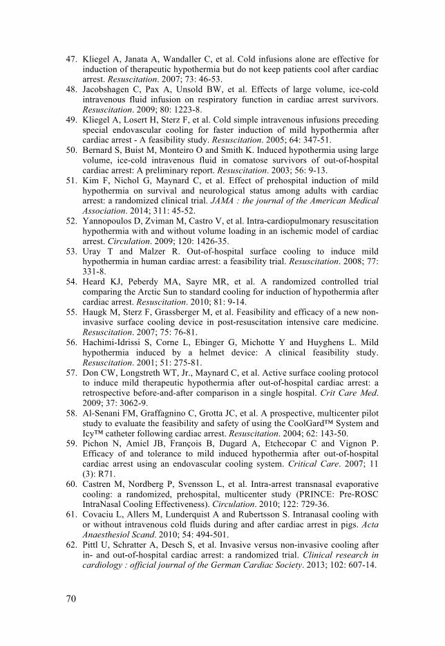
70
47. Kliegel A, Janata A, Wandaller C, et al. Cold infusions alone are effective for induction of therapeutic hypothermia but do not keep patients cool after cardiac arrest. Resuscitation. 2007; 73: 46-53.
48. Jacobshagen C, Pax A, Unsold BW, et al. Effects of large volume, ice-cold intravenous fluid infusion on respiratory function in cardiac arrest survivors. Resuscitation. 2009; 80: 1223-8.
49. Kliegel A, Losert H, Sterz F, et al. Cold simple intravenous infusions preceding special endovascular cooling for faster induction of mild hypothermia after cardiac arrest - A feasibility study. Resuscitation. 2005; 64: 347-51.
50. Bernard S, Buist M, Monteiro O and Smith K. Induced hypothermia using large volume, ice-cold intravenous fluid in comatose survivors of out-of-hospital cardiac arrest: A preliminary report. Resuscitation. 2003; 56: 9-13.
51. Kim F, Nichol G, Maynard C, et al. Effect of prehospital induction of mild hypothermia on survival and neurological status among adults with cardiac arrest: a randomized clinical trial. JAMA : the journal of the American Medical Association. 2014; 311: 45-52.
52. Yannopoulos D, Zviman M, Castro V, et al. Intra-cardiopulmonary resuscitation hypothermia with and without volume loading in an ischemic model of cardiac arrest. Circulation. 2009; 120: 1426-35.
53. Uray T and Malzer R. Out-of-hospital surface cooling to induce mild hypothermia in human cardiac arrest: a feasibility trial. Resuscitation. 2008; 77: 331-8.
54. Heard KJ, Peberdy MA, Sayre MR, et al. A randomized controlled trial comparing the Arctic Sun to standard cooling for induction of hypothermia after cardiac arrest. Resuscitation. 2010; 81: 9-14.
55. Haugk M, Sterz F, Grassberger M, et al. Feasibility and efficacy of a new non-invasive surface cooling device in post-resuscitation intensive care medicine. Resuscitation. 2007; 75: 76-81.
56. Hachimi-Idrissi S, Corne L, Ebinger G, Michotte Y and Huyghens L. Mild hypothermia induced by a helmet device: A clinical feasibility study. Resuscitation. 2001; 51: 275-81.
57. Don CW, Longstreth WT, Jr., Maynard C, et al. Active surface cooling protocol to induce mild therapeutic hypothermia after out-of-hospital cardiac arrest: a retrospective before-and-after comparison in a single hospital. Crit Care Med. 2009; 37: 3062-9.
58. Al-Senani FM, Graffagnino C, Grotta JC, et al. A prospective, multicenter pilot study to evaluate the feasibility and safety of using the CoolGard™ System and Icy™ catheter following cardiac arrest. Resuscitation. 2004; 62: 143-50.
59. Pichon N, Amiel JB, François B, Dugard A, Etchecopar C and Vignon P. Efficacy of and tolerance to mild induced hypothermia after out-of-hospital cardiac arrest using an endovascular cooling system. Critical Care. 2007; 11 (3): R71.
60. Castren M, Nordberg P, Svensson L, et al. Intra-arrest transnasal evaporative cooling: a randomized, prehospital, multicenter study (PRINCE: Pre-ROSC IntraNasal Cooling Effectiveness). Circulation. 2010; 122: 729-36.
61. Covaciu L, Allers M, Lunderquist A and Rubertsson S. Intranasal cooling with or without intravenous cold fluids during and after cardiac arrest in pigs. Acta Anaesthesiol Scand. 2010; 54: 494-501.
62. Pittl U, Schratter A, Desch S, et al. Invasive versus non-invasive cooling after in- and out-of-hospital cardiac arrest: a randomized trial. Clinical research in cardiology : official journal of the German Cardiac Society. 2013; 102: 607-14.
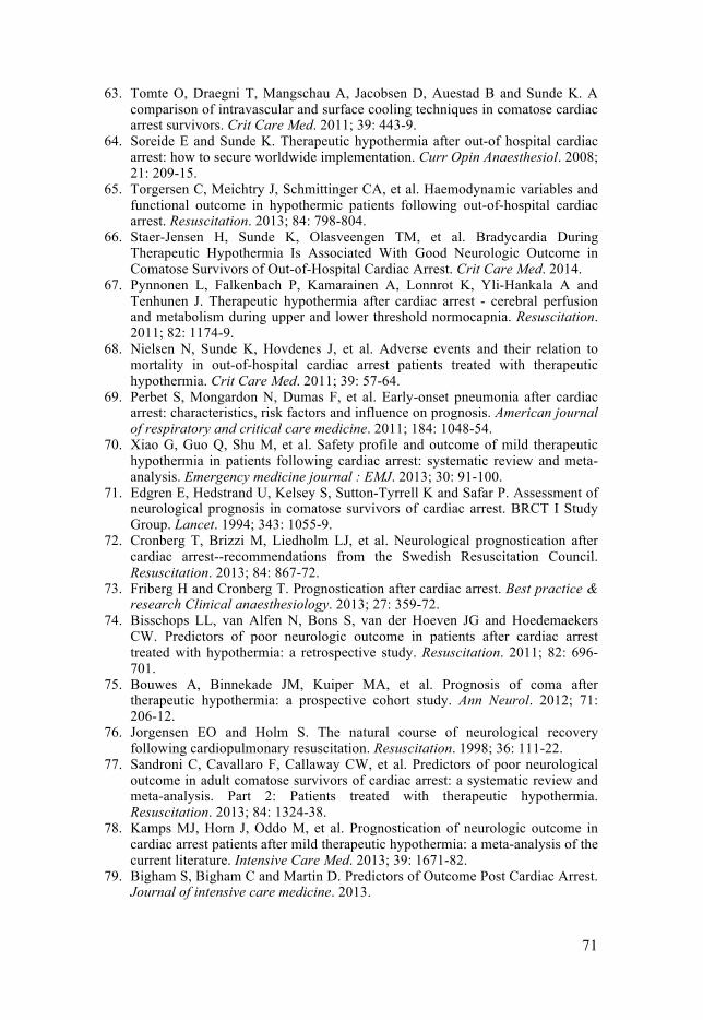
71
63. Tomte O, Draegni T, Mangschau A, Jacobsen D, Auestad B and Sunde K. A comparison of intravascular and surface cooling techniques in comatose cardiac arrest survivors. Crit Care Med. 2011; 39: 443-9.
64. Soreide E and Sunde K. Therapeutic hypothermia after out-of hospital cardiac arrest: how to secure worldwide implementation. Curr Opin Anaesthesiol. 2008; 21: 209-15.
65. Torgersen C, Meichtry J, Schmittinger CA, et al. Haemodynamic variables and functional outcome in hypothermic patients following out-of-hospital cardiac arrest. Resuscitation. 2013; 84: 798-804.
66. Staer-Jensen H, Sunde K, Olasveengen TM, et al. Bradycardia During Therapeutic Hypothermia Is Associated With Good Neurologic Outcome in Comatose Survivors of Out-of-Hospital Cardiac Arrest. Crit Care Med. 2014.
67. Pynnonen L, Falkenbach P, Kamarainen A, Lonnrot K, Yli-Hankala A and Tenhunen J. Therapeutic hypothermia after cardiac arrest - cerebral perfusion and metabolism during upper and lower threshold normocapnia. Resuscitation. 2011; 82: 1174-9.
68. Nielsen N, Sunde K, Hovdenes J, et al. Adverse events and their relation to mortality in out-of-hospital cardiac arrest patients treated with therapeutic hypothermia. Crit Care Med. 2011; 39: 57-64.
69. Perbet S, Mongardon N, Dumas F, et al. Early-onset pneumonia after cardiac arrest: characteristics, risk factors and influence on prognosis. American journal of respiratory and critical care medicine. 2011; 184: 1048-54.
70. Xiao G, Guo Q, Shu M, et al. Safety profile and outcome of mild therapeutic hypothermia in patients following cardiac arrest: systematic review and meta-analysis. Emergency medicine journal : EMJ. 2013; 30: 91-100.
71. Edgren E, Hedstrand U, Kelsey S, Sutton-Tyrrell K and Safar P. Assessment of neurological prognosis in comatose survivors of cardiac arrest. BRCT I Study Group. Lancet. 1994; 343: 1055-9.
72. Cronberg T, Brizzi M, Liedholm LJ, et al. Neurological prognostication after cardiac arrest--recommendations from the Swedish Resuscitation Council. Resuscitation. 2013; 84: 867-72.
73. Friberg H and Cronberg T. Prognostication after cardiac arrest. Best practice & research Clinical anaesthesiology. 2013; 27: 359-72.
74. Bisschops LL, van Alfen N, Bons S, van der Hoeven JG and Hoedemaekers CW. Predictors of poor neurologic outcome in patients after cardiac arrest treated with hypothermia: a retrospective study. Resuscitation. 2011; 82: 696-701.
75. Bouwes A, Binnekade JM, Kuiper MA, et al. Prognosis of coma after therapeutic hypothermia: a prospective cohort study. Ann Neurol. 2012; 71: 206-12.
76. Jorgensen EO and Holm S. The natural course of neurological recovery following cardiopulmonary resuscitation. Resuscitation. 1998; 36: 111-22.
77. Sandroni C, Cavallaro F, Callaway CW, et al. Predictors of poor neurological outcome in adult comatose survivors of cardiac arrest: a systematic review and meta-analysis. Part 2: Patients treated with therapeutic hypothermia. Resuscitation. 2013; 84: 1324-38.
78. Kamps MJ, Horn J, Oddo M, et al. Prognostication of neurologic outcome in cardiac arrest patients after mild therapeutic hypothermia: a meta-analysis of the current literature. Intensive Care Med. 2013; 39: 1671-82.
79. Bigham S, Bigham C and Martin D. Predictors of Outcome Post Cardiac Arrest. Journal of intensive care medicine. 2013.
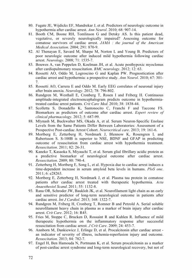
72
80. Fugate JE, Wijdicks EF, Mandrekar J, et al. Predictors of neurologic outcome in hypothermia after cardiac arrest. Ann Neurol. 2010; 68: 907-14.
81. Booth CM, Boone RH, Tomlinson G and Detsky AS. Is this patient dead, vegetative, or severely neurologically impaired? Assessing outcome for comatose survivors of cardiac arrest. JAMA : the journal of the American Medical Association. 2004; 291: 870-9.
82. Al Thenayan E, Savard M, Sharpe M, Norton L and Young B. Predictors of poor neurologic outcome after induced mild hypothermia following cardiac arrest. Neurology. 2008; 71: 1535-7.
83. Bouwes A, van Poppelen D, Koelman JH, et al. Acute posthypoxic myoclonus after cardiopulmonary resuscitation. BMC neurology. 2012; 12: 63.
84. Rossetti AO, Oddo M, Logroscino G and Kaplan PW. Prognostication after cardiac arrest and hypothermia: a prospective study. Ann Neurol. 2010; 67: 301-7.
85. Rossetti AO, Carrera E and Oddo M. Early EEG correlates of neuronal injury after brain anoxia. Neurology. 2012; 78: 796-802.
86. Rundgren M, Westhall E, Cronberg T, Rosen I and Friberg H. Continuous amplitude-integrated electroencephalogram predicts outcome in hypothermia-treated cardiac arrest patients. Crit Care Med. 2010; 38: 1838-44.
87. Scolletta S, Donadello K, Santonocito C, Franchi F and Taccone FS. Biomarkers as predictors of outcome after cardiac arrest. Expert review of clinical pharmacology. 2012; 5: 687-99.
88. Mlynash M, Buckwalter MS, Okada A, et al. Serum Neuron-Specific Enolase Levels from the Same Patients Differ Between Laboratories: Assessment of a Prospective Post-cardiac Arrest Cohort. Neurocritical care. 2013; 19: 161-6.
89. Mortberg E, Zetterberg H, Nordmark J, Blennow K, Rosengren L and Rubertsson S. S-100B is superior to NSE, BDNF and GFAP in predicting outcome of resuscitation from cardiac arrest with hypothermia treatment. Resuscitation. 2011; 82: 26-31.
90. Kaneko T, Kasaoka S, Miyauchi T, et al. Serum glial fibrillary acidic protein as a predictive biomarker of neurological outcome after cardiac arrest. Resuscitation. 2009; 80: 790-4.
91. Zetterberg H, Mortberg E, Song L, et al. Hypoxia due to cardiac arrest induces a time-dependent increase in serum amyloid beta levels in humans. PloS one. 2011; 6: e28263.
92. Mortberg E, Zetterberg H, Nordmark J, et al. Plasma tau protein in comatose patients after cardiac arrest treated with therapeutic hypothermia. Acta Anaesthesiol Scand. 2011; 55: 1132-8.
93. Rana OR, Schroder JW, Baukloh JK, et al. Neurofilament light chain as an early and sensitive predictor of long-term neurological outcome in patients after cardiac arrest. Int J Cardiol. 2013; 168: 1322-7.
94. Rundgren M, Friberg H, Cronberg T, Romner B and Petzold A. Serial soluble neurofilament heavy chain in plasma as a marker of brain injury after cardiac arrest. Crit Care. 2012; 16: R45.
95. Fries M, Stoppe C, Brucken D, Rossaint R and Kuhlen R. Influence of mild therapeutic hypothermia on the inflammatory response after successful resuscitation from cardiac arrest. J Crit Care. 2009; 24: 453-7.
96. Annborn M, Dankiewicz J, Erlinge D, et al. Procalcitonin after cardiac arrest - an indicator of severity of illness, ischemia-reperfusion injury and outcome. Resuscitation. 2013; 84: 782-7.
97. Engel H, Ben Hamouda N, Portmann K, et al. Serum procalcitonin as a marker of post-cardiac arrest syndrome and long-term neurological recovery, but not of
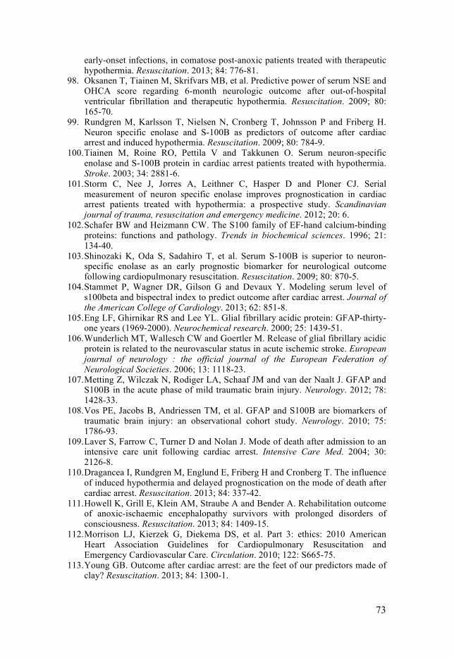
73
early-onset infections, in comatose post-anoxic patients treated with therapeutic hypothermia. Resuscitation. 2013; 84: 776-81.
98. Oksanen T, Tiainen M, Skrifvars MB, et al. Predictive power of serum NSE and OHCA score regarding 6-month neurologic outcome after out-of-hospital ventricular fibrillation and therapeutic hypothermia. Resuscitation. 2009; 80: 165-70.
99. Rundgren M, Karlsson T, Nielsen N, Cronberg T, Johnsson P and Friberg H. Neuron specific enolase and S-100B as predictors of outcome after cardiac arrest and induced hypothermia. Resuscitation. 2009; 80: 784-9.
100. Tiainen M, Roine RO, Pettila V and Takkunen O. Serum neuron-specific enolase and S-100B protein in cardiac arrest patients treated with hypothermia. Stroke. 2003; 34: 2881-6.
101. Storm C, Nee J, Jorres A, Leithner C, Hasper D and Ploner CJ. Serial measurement of neuron specific enolase improves prognostication in cardiac arrest patients treated with hypothermia: a prospective study. Scandinavian journal of trauma, resuscitation and emergency medicine. 2012; 20: 6.
102. Schafer BW and Heizmann CW. The S100 family of EF-hand calcium-binding proteins: functions and pathology. Trends in biochemical sciences. 1996; 21: 134-40.
103. Shinozaki K, Oda S, Sadahiro T, et al. Serum S-100B is superior to neuron-specific enolase as an early prognostic biomarker for neurological outcome following cardiopulmonary resuscitation. Resuscitation. 2009; 80: 870-5.
104. Stammet P, Wagner DR, Gilson G and Devaux Y. Modeling serum level of s100beta and bispectral index to predict outcome after cardiac arrest. Journal of the American College of Cardiology. 2013; 62: 851-8.
105. Eng LF, Ghirnikar RS and Lee YL. Glial fibrillary acidic protein: GFAP-thirty-one years (1969-2000). Neurochemical research. 2000; 25: 1439-51.
106. Wunderlich MT, Wallesch CW and Goertler M. Release of glial fibrillary acidic protein is related to the neurovascular status in acute ischemic stroke. European journal of neurology : the official journal of the European Federation of Neurological Societies. 2006; 13: 1118-23.
107. Metting Z, Wilczak N, Rodiger LA, Schaaf JM and van der Naalt J. GFAP and S100B in the acute phase of mild traumatic brain injury. Neurology. 2012; 78: 1428-33.
108. Vos PE, Jacobs B, Andriessen TM, et al. GFAP and S100B are biomarkers of traumatic brain injury: an observational cohort study. Neurology. 2010; 75: 1786-93.
109. Laver S, Farrow C, Turner D and Nolan J. Mode of death after admission to an intensive care unit following cardiac arrest. Intensive Care Med. 2004; 30: 2126-8.
110. Dragancea I, Rundgren M, Englund E, Friberg H and Cronberg T. The influence of induced hypothermia and delayed prognostication on the mode of death after cardiac arrest. Resuscitation. 2013; 84: 337-42.
111. Howell K, Grill E, Klein AM, Straube A and Bender A. Rehabilitation outcome of anoxic-ischaemic encephalopathy survivors with prolonged disorders of consciousness. Resuscitation. 2013; 84: 1409-15.
112. Morrison LJ, Kierzek G, Diekema DS, et al. Part 3: ethics: 2010 American Heart Association Guidelines for Cardiopulmonary Resuscitation and Emergency Cardiovascular Care. Circulation. 2010; 122: S665-75.
113. Young GB. Outcome after cardiac arrest: are the feet of our predictors made of clay? Resuscitation. 2013; 84: 1300-1.
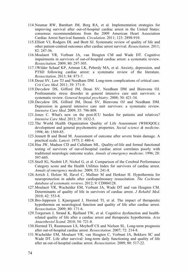
74
114. Neumar RW, Barnhart JM, Berg RA, et al. Implementation strategies for improving survival after out-of-hospital cardiac arrest in the United States: consensus recommendations from the 2009 American Heart Association Cardiac Arrest Survival Summit. Circulation. 2011; 123: 2898-910.
115. Elliott VJ, Rodgers DL and Brett SJ. Systematic review of quality of life and other patient-centred outcomes after cardiac arrest survival. Resuscitation. 2011; 82: 247-56.
116. Moulaert VR, Verbunt JA, van Heugten CM and Wade DT. Cognitive impairments in survivors of out-of-hospital cardiac arrest: a systematic review. Resuscitation. 2009; 80: 297-305.
117. 1Wilder Schaaf KP, Artman LK, Peberdy MA, et al. Anxiety, depression, and PTSD following cardiac arrest: a systematic review of the literature. Resuscitation. 2013; 84: 873-7.
118. Desai SV, Law TJ and Needham DM. Long-term complications of critical care. Crit Care Med. 2011; 39: 371-9.
119. Davydow DS, Gifford JM, Desai SV, Needham DM and Bienvenu OJ. Posttraumatic stress disorder in general intensive care unit survivors: a systematic review. General hospital psychiatry. 2008; 30: 421-34.
120. Davydow DS, Gifford JM, Desai SV, Bienvenu OJ and Needham DM. Depression in general intensive care unit survivors: a systematic review. Intensive Care Med. 2009; 35: 796-809.
121. Jones C. What's new on the post-ICU burden for patients and relatives? Intensive Care Med. 2013; 39: 1832-5.
122. The World Health Organization Quality of Life Assessment (WHOQOL): development and general psychometric properties. Social science & medicine. 1998; 46: 1569-85.
123. Jennett B and Bond M. Assessment of outcome after severe brain damage. A practical scale. Lancet. 1975; I: 480-4.
124. Hsu JW, Madsen CD and Callaham ML. Quality-of-life and formal functional testing of survivors of out-of-hospital cardiac arrest correlates poorly with traditional neurologic outcome scales. Annals of emergency medicine. 1996; 28: 597-605.
125. Stiell IG, Nesbitt LP, Nichol G, et al. Comparison of the Cerebral Performance Category score and the Health Utilities Index for survivors of cardiac arrest. Annals of emergency medicine. 2009; 53: 241-8.
126. Arrich J, Holzer M, Havel C, Mullner M and Herkner H. Hypothermia for neuroprotection in adults after cardiopulmonary resuscitation. The Cochrane database of systematic reviews. 2012; 9: CD004128.
127. Moulaert VR, Wachelder EM, Verbunt JA, Wade DT and van Heugten CM. Determinants of quality of life in survivors of cardiac arrest. J Rehabil Med. 2010; 42: 553-8.
128. Bro-Jeppesen J, Kjaergaard J, Horsted TI, et al. The impact of therapeutic hypothermia on neurological function and quality of life after cardiac arrest. Resuscitation. 2009; 80: 171-6.
129. Torgersen J, Strand K, Bjelland TW, et al. Cognitive dysfunction and health-related quality of life after a cardiac arrest and therapeutic hypothermia. Acta Anaesthesiol Scand. 2010; 54: 721-8.
130. Horsted TI, Rasmussen LS, Meyhoff CS and Nielsen SL. Long-term prognosis after out-of-hospital cardiac arrest. Resuscitation. 2007; 72: 214-8.
131. Wachelder EM, Moulaert VR, van Heugten C, Verbunt JA, Bekkers SC and Wade DT. Life after survival: long-term daily functioning and quality of life after an out-of-hospital cardiac arrest. Resuscitation. 2009; 80: 517-22.
75
132. Tiainen M, Poutiainen E, Kovala T, Takkunen O, Happola O and Roine RO. Cognitive and neurophysiological outcome of cardiac arrest survivors treated with therapeutic hypothermia. Stroke. 2007; 38: 2303-8.
133. Wilson M, Staniforth A, Till R, das Nair R and Vesey P. The psychosocial outcomes of anoxic brain injury following cardiac arrest. Resuscitation. 2014; 85: 795-800.
134. Wallin E, Larsson IM, Rubertsson S and Kristofferzon ML. Cardiac arrest and hypothermia treatment--function and life satisfaction among survivors in the first 6 months. Resuscitation. 2014; 85: 538-43.
135. Cronberg T, Lilja G, Rundgren M, Friberg H and Widner H. Long-term neurological outcome after cardiac arrest and therapeutic hypothermia. Resuscitation. 2009; 80: 1119-23.
136. Hughes F, Bryan K and Robbins I. Relatives' experiences of critical care. Nurs Crit Care. 2005; 10: 23-30.
137. Verhaeghe S, Defloor T, Van Zuuren F, Duijnstee M and Grypdonck M. The needs and experiences of family members of adult patients in an intensive care unit: a review of the literature. J Clin Nurs. 2005; 14: 501-9.
138. Engström Å and Söderberg S. The experiences of partners of critically ill persons in an intensive care unit. Intensive and Critical Care Nursing. 2004; 20: 299-308.
139. McAdam JL, Dracup KA, White DB, Fontaine DK and Puntillo KA. Symptom experiences of family members of intensive care unit patients at high risk for dying. Crit Care Med. 2010; 38: 1078-85.
140. American Psychiatric Association. and American Psychiatric Association. Task Force on DSM-IV. Diagnostic and statistical manual of mental disorders : DSM-IV-TR. 4th ed. Washington, DC: American Psychiatric Association, 2000, p.xxxvii, 943 p.
141. Paparrigopoulos T, Melissaki A, Efthymiou A, et al. Short-term psychological impact on family members of intensive care unit patients. J Psychosom Res. 2006; 61: 719-22.
142. Jones C, Skirrow P, Griffiths RD, et al. Post-traumatic stress disorder-related symptoms in relatives of patients following intensive care. Intensive Care Med. 2004; 30: 456-60.
143. Anderson WG, Arnold RM, Angus DC and Bryce CL. Posttraumatic stress and complicated grief in family members of patients in the intensive care unit. J Gen Intern Med. 2008; 23: 1871-6.
144. Young E, Eddleston J, Ingleby S, et al. Returning home after intensive care: a comparison of symptoms of anxiety and depression in ICU and elective cardiac surgery patients and their relatives. Intensive Care Med. 2005; 31: 86-91.
145. Olsen KD, Dysvik E and Hansen BS. The meaning of family members' presence during intensive care stay: a qualitative study. Intensive Crit Care Nurs. 2009; 25: 190-8.
146. Löf S, Sandström A and Engström Å. Patients treated with therapeutic hypothermia after cardiac arrest: Relatives' experiences. Journal of Advanced Nursing. 2010; 66: 1760-8.
147. Agard AS and Harder I. Relatives' experiences in intensive care--finding a place in a world of uncertainty. Intensive Crit Care Nurs. 2007; 23: 170-7.
148. McKiernan M and McCarthy G. Family members' lived experience in the intensive care unit: a phemenological study. Intensive Crit Care Nurs. 2010; 26: 254-61.
149. Wahlin I, Ek AC and Idvall E. Empowerment from the perspective of next of kin in intensive care. J Clin Nurs. 2009; 18: 2580-7.
76
150. Holm MS, Norekval TM, Falun N and Gjengedal E. Partners' ambivalence towards cardiac arrest and hypothermia treatment: a qualitative study. Nurs Crit Care. 2012; 17: 231-8.
151. Engstrom A and Soderberg S. Receiving power through confirmation: the meaning of close relatives for people who have been critically ill. J Adv Nurs. 2007; 59: 569-76.
152. McAdam JL, Arai S and Puntillo KA. Unrecognized contributions of families in the intensive care unit. Intensive Care Med. 2008; 34: 1097-101.
153. Ann-Britt T, Ella D, Johan H and Åsa AB. Spouses' experiences of a cardiac arrest at home: An interview study. European Journal of Cardiovascular Nursing. 2010; 9: 161-7.
154. Bremer A, Dahlberg K and Sandman L. Experiencing out-of-hospital cardiac arrest: significant others' lifeworld perspective. Qual Health Res. 2009; 19: 1407-20.
155. Wallin E, Larsson IM, Rubertsson S and Kristoferzon ML. Relatives' experiences of everyday life six months after hypothermia treatment of a significant other's cardiac arrest. J Clin Nurs. 2013; 22: 1639-46.
156. Starmark JE, Stalhammar D and Holmgren E. The Reaction Level Scale (RLS 85). Manual and guidelines. Acta Neurochirurgica. 1988; 91: 12-20.
157. Teasdale G and Jennett B. Assessment of coma and impaired consciousness. A practical scale. Lancet. 1974; 2: 81-4.
158. Snaith RP. The Hospital Anxiety And Depression Scale. Health Qual Life Outcomes. 2003; 1: 29.
159. EuroQol--a new facility for the measurement of health-related quality of life. The EuroQol Group. Health policy. 1990; 16: 199-208.
160. Rabin R OM, Oppe M. EQ-5D-3L User Guide; Basic information on how to use the EQ-5D-3L instrumentVersion 4.0: (2011).
161. Sullivan M KJ, Taft C. . Hälsoenkätsvensk manual (Swedish manual of SF-12 helthquestionarie). Gothenburg University. Gothenburg 1997.
162. Ware J, Jr., Kosinski M and Keller SD. A 12-Item Short-Form Health Survey: construction of scales and preliminary tests of reliability and validity. Medical care. 1996; 34: 220-33.
163. Polit DF and Beck CT. Nursing research : generating and assessing evidence for nursing practice. Ninth Edition. ed. Philadelphia: Wolters Kluwer Health/Lippincott Williams & Wilkins, 2011, p.p.
164. Graneheim UH and Lundman B. Qualitative content analysis in nursing research: Concepts, procedures and measures to achieve trustworthiness. Nurse Education Today. 2004; 24: 105-12.
165. Elo S and Kyngas H. The qualitative content analysis process. J Adv Nurs. 2008; 62: 107-15.
166. Krippendorff K. Content analysis : an introduction to its methodology. 2nd ed. Thousand Oaks, Calif.: Sage, 2004, p.xxiii, 413 p.
167. World Medical A. World Medical Association Declaration of Helsinki: ethical principles for medical research involving human subjects. JAMA : the journal of the American Medical Association. 2013; 310: 2191-4.
168. Haugk M, Testori C, Sterz F, et al. Relationship between time to target temperature and outcome in patients treated with therapeutic hypothermia after cardiac arrest. Crit Care. 2011; 15: R101.
169. Benz-Woerner J, Delodder F, Benz R, et al. Body temperature regulation and outcome after cardiac arrest and therapeutic hypothermia. Resuscitation. 2011.
77
170. Jarrah S, Dziodzio J, Lord C, et al. Surface cooling after cardiac arrest: effectiveness, skin safety, and adverse events in routine clinical practice. Neurocritical care. 2011; 14: 382-8.
171. Krizanac D, Stratil P, Hoerburger D, et al. Femoro-iliacal artery versus pulmonary artery core temperature measurement during therapeutic hypothermia: an observational study. Resuscitation. 2013; 84: 805-9.
172. Cocchi MN, Boone MD, Giberson B, et al. Fever After Rewarming: Incidence of Pyrexia in Postcardiac Arrest Patients Who Have Undergone Mild Therapeutic Hypothermia. Journal of intensive care medicine. 2013.
173. Hayashida H, Kaneko T, Kasaoka S, et al. Comparison of the predictability of neurological outcome by serum procalcitonin and glial fibrillary acidic protein in postcardiac-arrest patients. Neurocritical care. 2010; 12: 252-7.
174. Wiesmann M, Steinmeier E, Magerkurth O, Linn J, Gottmann D and Missler U. Outcome prediction in traumatic brain injury: comparison of neurological status, CT findings, and blood levels of S100B and GFAP. Acta neurologica Scandinavica. 2010; 121: 178-85.
175. Sun Y, Qin Q, Shang YJ, et al. The accuracy of glial fibrillary acidic protein in acute stroke differential diagnosis: A meta-analysis. Scandinavian journal of clinical and laboratory investigation. 2013; 73: 601-6.
176. Horn J, Cronberg T and Taccone FS. Prognostication after cardiac arrest. Curr Opin Crit Care. 2014; 20: 280-6.
177. Oddo M and Rossetti AO. Early multimodal outcome prediction after cardiac arrest in patients treated with hypothermia*. Crit Care Med. 2014; 42: 1340-7.
178. Rankin J. Cerebral vascular accidents in patients over the age of 60. II. Prognosis. Scottish medical journal. 1957; 2: 200-15.
179. Rittenberger JC, Raina K, Holm MB, Kim YJ and Callaway CW. Association between Cerebral Performance Category, Modified Rankin Scale, and discharge disposition after cardiac arrest. Resuscitation. 2011; 82: 1036-40.
180. Beesems SG, Wittebrood KM, de Haan RJ and Koster RW. Cognitive function and quality of life after successful resuscitation from cardiac arrest. Resuscitation. 2014; 85: 1269-74.
181. Lundgren-Nilsson A, Rosen H, Hofgren C and Sunnerhagen KS. The first year after successful cardiac resuscitation: function, activity, participation and quality of life. Resuscitation. 2005; 66: 285-9.
182. Fugate JE and Rabinstein AA. Life after cardiac arrest: better with time. Resuscitation. 2014; 85: 157-8.
183. van Alem AP, Waalewijn RA, Koster RW and de Vos R. Assessment of quality of life and cognitive function after out-of-hospital cardiac arrest with successful resuscitation. The American journal of cardiology. 2004; 93: 131-5.
184. Burström K. RC. Hälsorelaterad livskvalitet i Stockholmslän 2002. Resultat per åldersgrupp och kön, utbildningsnivå, födelseland samt sysselsättningsgrupp. Rapport 2006:1. Stockholm: Enheten för Socialmedicin och Hälsoekonomi. Centrum för Folkhälsa. FORUM för kunskap och gemensam utveckling. Stockholms läns landsting., 2006.
185. Moulaert VR, van Haastregt JC, Wade DT, van Heugten CM and Verbunt JA. 'Stand still ..., and move on' , an early neurologically-focused follow-up for cardiac arrest survivors and their caregivers: a process evaluation. BMC health services research. 2014; 14: 34.
186. Herlitz J, Engdahl J, Svensson L, Angquist KA, Young M and Holmberg S. Factors associated with an increased chance of survival among patients suffering from an out-of-hospital cardiac arrest in a national perspective in Sweden. Am Heart J. 2005; 149: 61-6.
78
187. Molter NC. Needs of relatives of critically ill patients: a descriptive study. Heart Lung. 1979; 8: 332-9.
188. Alvarez GF and Kirby AS. The perspective of families of the critically ill patient: their needs. Curr Opin Crit Care. 2006; 12: 614-8.
189. Verhaeghe ST, van Zuuren FJ, Defloor T, Duijnstee MS and Grypdonck MH. The process and the meaning of hope for family members of traumatic coma patients in intensive care. Qual Health Res. 2007; 17: 730-43.
190. Karlsson C, Tisell A, Engstrom A and Andershed B. Family members' satisfaction with critical care: a pilot study. Nurs Crit Care. 2011; 16: 11-8.
191. Schenker Y, White DB, Crowley-Matoka M, Dohan D, Tiver GA and Arnold RM. "It hurts to know... and it helps": exploring how surrogates in the ICU cope with prognostic information. Journal of palliative medicine. 2013; 16: 243-9.
192. Whitney SN, McCullough LB, Fruge E, McGuire AL and Volk RJ. Beyond breaking bad news: the roles of hope and hopefulness. Cancer. 2008; 113: 442-5.
193. Hammer K, Mogensen O and Hall EO. The meaning of hope in nursing research: a meta-synthesis. Scand J Caring Sci. 2009.
194. Johansson I, Hildingh C and Fridlund B. Coping strategies when an adult next-of-kin/close friend is in critical care: a grounded theory analysis. Intensive Crit Care Nurs. 2002; 18: 96-108.
195. Pochard F, Darmon M, Fassier T, et al. Symptoms of anxiety and depression in family members of intensive care unit patients before discharge or death. A prospective multicenter study. J Crit Care. 2005; 20: 90-6.
196. Haggstrom M, Asplund K and Kristiansen L. Struggle with a gap between intensive care units and general wards. International journal of qualitative studies on health and well-being. 2009; 4: 181-92.
197. Cullinane JP and Plowright CI. Patients' and relatives' experiences of transfer from intensive care unit to wards. Nurs Crit Care. 2013; 18: 289-96.
198. Forsberg AL, E; Engström A. Being transferred from an intensive care unit to a ward: Searching for the known in the unknown. International Journal of Nursing Practice. 2011; 17: 110-6.
199. Tavakol M DR. Making sense of Cronbach's alpha. International journal of medical education. 2011; 2: 53-5.
200. Sandelowski M. Whatever happened to qualitative description? Res Nurs Health. 2000; 23: 334-40.
201. Sandelowski M. What's in a name? Qualitative description revisited. Res Nurs Health. 2010; 33: 77-84.
202. Novick G. Is there a bias against telephone interviews in qualitative research? Res Nurs Health. 2008; 31: 391-8.
203.

Acta Universitatis UpsaliensisDigital Comprehensive Summaries of Uppsala Dissertationsfrom the Faculty of Medicine 1021
Editor: The Dean of the Faculty of Medicine
A doctoral dissertation from the Faculty of Medicine, UppsalaUniversity, is usually a summary of a number of papers. A fewcopies of the complete dissertation are kept at major Swedishresearch libraries, while the summary alone is distributedinternationally through the series Digital ComprehensiveSummaries of Uppsala Dissertations from the Faculty ofMedicine. (Prior to January, 2005, the series was publishedunder the title “Comprehensive Summaries of UppsalaDissertations from the Faculty of Medicine”.)
Distribution: publications.uu.seurn:nbn:se:uu:diva-229758
ACTAUNIVERSITATIS
UPSALIENSISUPPSALA
2014