Therapeutic hypothermia for neonatal hypoxic-ischemic encephalopathy

MEETING ABSTRACTS Open Access
Update on therapeutic temperature managementPortoroz, Slovenia. 7-9 June 2012
Edited by Gregor Broessner, Marlene Fischer, Gerrit Schubert, Bernhard Metzler and Erich Schmutzhard
Published: 7 June 2012
These abstracts are available online at http://ccforum.com/supplements/16/S2
INTRODUCTIONA1Update on therapeutic temperature managementGregor Broessner1*, Marlene Fischer1, Gerrit Schubert2, Bernhard Metzler3,Erich Schmutzhard11Department of Neurology, Medical University, Innsbruck, Austria;2Department of Neurosurgery, Medical University, Innsbruck, Austria;3Department of Cardiology, Medical University, Innsbruck, AustriaCritical Care 2012, 16(Suppl 2):A1
It is a pleasure to announce the 2nd Innsbruck Hypothermia Symposium.We are very happy that Critical Care has agreed to publish extendedabstracts submitted by invited renowned scientists from all over the world;that is, Europe, the Americas, Asia. Neuroprotection - potentially achievedby targeted temperature management (that is, therapeutic hypothermia orprophylactic controlled normothermia) - is essential in emergency andacute care management of various severe neurologic and cardiologicdiseases. Beyond neuroprotection - for this aim, therapeutic hypothermiahas been established after resuscitation of patients with cardiac arrest dueto a shockable arrhythmia and in neonatal asphyxic encephalopathy -therapeutic hypothermia and prophylactic controlled normothermia havebeen published in single case reports, retrospective, open, but also inprospective randomised controlled trials in many other emergencydisciplines in which both neuroprotection and protection of other organsand tissues are the target of our therapeutic endeavours. The MedicalUniversity Innsbruck, Austria, is happy to organise this conference ontemperature management, therapeutic hypothermia and prophylacticnormothermia respectively, to be held in Portoroz, Slovenia. In accordancewith the first Meeting on Hypothermia, which was held in Miami, Florida,USA (CHilling At the Beach), we are proud to suggest the acronym CHABstanding for take Care for Heart And Brain, characterising the major targetorgans of therapeutic and, possibly also, prophylactic temperaturemanagement. Again, we have been able to gather most renownedscientists, neurointensivists and intensivists, emergency physicians,cardiologists and other specialists to cover the entire scientific and clinicalspectrum of emergency temperature management, technical aspects ofcooling and management of potential complications including shivering,but also temperature management in neurology, neurosurgery, intensivecare medicine, in the operation theatre, cardiology, infectious diseases, andso forth. Beyond that we cross borders and discuss hypothermia andintracranial pressure, pharmacodynamics in hypothermic patients and theinfluence of hypothermia onto pharmacokinetics/pharmacodynamics,hypothermia in refractory status epilepticus or heat stroke, hypothermiaand advanced neuromonitoring, hypothermia and nutrition, shivering andthe critical issue of rewarming, amongst other topics.The aim of this symposium is to enhance the knowledge on temperaturemanagement, increase the readiness and stimulate the preparedness toinstitute therapeutic hypothermia and/or prophylactic controllednormothermia, respectively, in patients in need of tissue and organ
protection, uncontrolled body temperatures possibly adding - per se - toneuronal damage. Knowing the medical literature and knowing the issueof potentially life-threatening side effects and complications incurred bythis invasive therapeutic manoeuvre, it is the foremost aim of thissymposium and this supplementary issue of Critical Care to discuss allthese aspects of targeted temperature management in emergency, criticalcare and, in particular, neurocritical patients and conditions. For this reasonthe organisers have agreed that the discussion of these various issues,being so important for general critical care, neurocritical care andemergency medicine, must be distributed as widely as possible, making itavailable to critical care and neurocritical care specialists all over the world.Therefore we are extremely grateful to the Editors of Critical Care forproviding a forum for all of the extended abstracts of all invited speakers,covering the entire field of adult emergency and critical care medicine. Wedo hope and we are convinced that this supplementary issue will be asource of inspiration and knowledge, hopefully becoming a work ofreference for intensivists, neurologists, neurointensivists, cardiologists andall emergency physicians alike. It is the aim of the organisers to establish aseries of such symposia within the next years in order to keep up with allthe developments in this field and to maintain the highest possible level ofknowledge of targeted temperature management in the community ofemergency and intensive care physicians.
EMERGENCY TEMPERATUREMANAGEMENTA2Therapeutic hypothermia: the rationaleErich Schmutzhard*, Marlene Fischer, Anelia Dietmann, Gregor BrössnerDepartment of Neurology, Neurocritical Care Unit, Medical UniversityInnsbruck, AustriaCritical Care 2012, 16(Suppl 2):A2
For almost a century, therapeutic hypothermia - or as it was termed in theearly days: hibernation - has been discussed as a potential neuroprotectivemeasure, in particular in patients suffering from severe intracranial diseaseleading to impairment of consciousness, associated with fever [1-3].In a wide range of diseases, secondary damage to the brain or otherorgans follows the initial impact and may be responsible for aggravation ofdisease condition or clinical state, in particular neurological morbidity and/or mortality [4-11]. Therapeutic hypothermia, recently renamed targetedtemperature management, including prophylactic normothermia, has beenused to improve this secondary impact onto brain and other organ tissue.This holds true, in particular, for neurological and neurosurgical intensivecare patients since secondary brain and nervous tissue injury may precludea potentially benign course of disease. The mechanisms of action ofhypothermia are complex, not yet fully understood. Therapeutichypothermia/targeted temperature management aims to attenuate acascade of secondary injury mechanisms, which is started immediately
Critical Care 2012, Volume 16 Suppl 2http://ccforum.com/supplements/16/S2
© 2012 various authors, licensee BioMed Central Ltd. This is an Open Access article distributed under the terms of the CreativeCommons Attribution License (http://creativecommons.org/licenses/by/2.0), which permits unrestricted use, distribution, andreproduction in any medium, provided the original work is properly cited.

after the initial event (primary injury) and may last for hours and even days[4,6,12]. The majority of research has focused, so far, on these secondaryinjury processes being destructive to brain and nervous tissue. It may beexpected that any such protective effect can be replicated in other organsand tissues during therapeutic hypothermia/targeted temperaturemanagement. A wide range of side effects may negate and counteract itspositive initial effect; this implies side effects of hypothermia per se andside effects of rewarming or inconstant maintenance of temperature levels[13-17].This abstract limits itself to potential pathophysiological mechanisms ofactions, the risks of any such mechanism and side effects derived fromthem [4,5,10,12,16-18].The protective effect of hypothermia may be explained by several pathways.A decreased metabolism with less oxygen and energy consumption and carbondioxide production may prevent secondary injury when oxygen supply isinterrupted or, at least, impaired. However, it needs to be stressed that thereduction in metabolic rate, as seen in hypothermia, requires adjustment inventilator setup, insulin infusion rate, correct interpretation of electrolytes, inparticular low phosphate, magnesium and potassium levels. Of particularinterest are the rebound phenomena during rewarming or when,involuntarily, the temperature cannot be maintained at the targeted lowlevel.Following ischemia, hypoxia or direct trauma apoptotic processes may beinitiated in brain tissue and neuronal cells may even become necrotic. Inthese earliest stages these pathways may be blocked by hypothermia.However, little is known about the time frame and best window ofopportunity to use therapeutic hypothermia to prevent initiation ofapoptotic/necrotic processes.Any type of neuronal injury may provoke the neuro-excitatory cascade,starting off with excessive calcium influx, glutamate receptor activation,neuronal hyperexcitability, eventually leading to cell death even afterreperfusion and normalization of glutamate levels. It has been suggestedthat therapeutic hypothermia may reduce cellular/neuronal damagefollowing this neuro-excitatory cascade.It has been accepted that the release of free radicals may be deleterious toboth neuronal cells and the brain’s defense mechanisms alike. Whether thedirect impact or the ischemia reperfusion injury is overwhelminglyresponsible for the release/increase of free radicals oxidizing anddamaging neuronal cell components is both still a matter of discussionand of limited interest when therapeutic hypothermia comes into play.Hyperexcitability, cellular hyperactivity, mitochondrial dysfunction, ion-pump failure and reduction in cellular membrane integrity may lead tointracellular and, consequently, also intercellular/extracellular acidosis.Early initiation of hypothermia may improve this full spectrum of cellularfailure, improve brain glucose and energy metabolism and reduce lactateaccumulation; with this, intracellular and intercellular acidosis will improveand eventually metabolic recovery be enhanced [4,19-21].Any type of brain injury is capable of disrupting the blood brain barrierleading to enhanced vascular permeability, brain edema, vascularpermeability and perivascular hemorrhage. Brain edema, both afterischemia/hypoxia and traumatic injury peaks after 24 to 72 hours(sometimes reaching its highest peak even after this period of time) - thusopening widely the therapeutic window - allowing for therapeutichypothermia to reduce brain edema via stabilizing the disrupted blood-brain barrier and vascular permeability. After brain injury proinflammatorymediators are released, leucocytes cross the - already impaired - blood-brain barrier leading to an accumulation of inflammatory cells in the brain.This inflammatory response starts within 1 hour after injury and maypersist for up to 5 days, a fact which also suggests a widely opentherapeutic window for intervention. Hypothermia has been shown toreduce ischemia induced inflammatory and immune reactions [4,19-22].In healthy persons, brain temperature is around 0.5 to 1°C higher than corebody temperature. In any type of brain injury, in particular, in patients withfever or hyperpyrexia respectively, injured areas may be definitely hotter(up to 2°C post injury), most probably due to transitory cellularhyperactivity. Local brain edema might lead to cerebral thermopoolingadding to hyperthermia-related neuronal cellular injury [4,16,18-21].Cooling below 35°C has been shown to affect coagulation, it depends on theinitial type of brain injury whether a procoagulatory effect or ananticoagulatory effect is believed to be neuroprotective in an individual case.Targeted temperature management may influence the secretion ofvasoconstricting substances (for example, endothelin) or vasodilating
substances (for example, prostaglandins). Their balance is essential tomaintain homeostasis. Ischemic or traumatic conditions may increasevasoconstricting substances thus leading to reduced cerebral blood flow.Whether hypothermia is capable of regulating/improving cerebralperfusion is still a matter of investigation, pending the influence ofcerebral autoregulation and the quantity of secreted vasoactive mediatorsin brain-injured patients with cerebral ischemia or any other type ofinjury [10].Whether epileptic activity, in particular, subtle nonconvulsive statusepilepticus, accepted to indicate severe brain damage, can be positivelyinfluenced by therapeutic hypothermia still needs further research.However, it is accepted that a subtle nonconvulsive status epilepticusoccurring in the acute phase of brain injury is - per se - adding to neuronaldestruction [10,16].While many pathophysiological processes and cascades may be influencedby targeted temperature management/therapeutic hypothermia and/or evenprevention of fever through prophylactic normothermia, it is unclear whetherin all types of severely brain-injured patients (for whatever reason) thebenefits of this therapeutic hypothermia always outweigh its risks. It is nowfully accepted and of a high level of evidenced medicine that in cerebralhypoxia (in a patient with cardiac arrest due to a shockable arrhythmia) aswell as asphyxial encephalopathy a 24-hour therapeutic hypothermia (33 to34°C), irrespective of the type of cooling, improves neurological outcome;that is, morbidity but also mortality [7,10]. Whether therapeutic hypothermia/targeted temperature management or prophylactic normothermia mayimprove outcome in other diseases, as discussed in this meeting, is still notclear. It needs to be stressed that even such seemingly similar diseases asglobal hypoxia (in cardiac arrest due to a shockable arrhythmia), asphyxialencephalopathy and ischemic stroke have so few pathophysiologic cascadesin common. Therefore, they may not be treated all alike, in particular, withrespect to type, duration, speed and depth of hypothermia as well asrewarming management [23]. It has already been demonstrated that inhypoxic encephalopathy hypothermia for 24 hours may be sufficient.However, disease entities such as ischemic stroke, hemorrhagic stroke withformation of peri-hematomal edema, traumatic brain injuries with prolongedsecondary insult or the wide range of neuronal injuries after subarachnoidhemorrhage may present even more complex pathophysiologic mechanisms.Moreover, different pathologies such as encephalitis and bacterial meningitisor even spinal cord injury may all require a targeted and personalizedapproach to this adjunctive therapy. In some cases, prolonged hypothermiamay be equally necessary as in other cases mild hypothermia or even onlyprophylactic normothermia may suffice.It may be stated beyond doubt that the neuroprotective effect ofmoderate hypothermia (33 to 34°C) has been shown in cerebral hypoxiaand asphyxial encephalopathy. However, different neurocritical caredisease entities as discussed above have different mechanisms of primaryinsults as well as the mechanisms and cascades of secondary brain injuryand therefore require a different therapeutic approach in respect oftemperature management.Any type of therapeutic measure, still being the subject of research, mustnever harm the patient. Hypothermia-induced neurological signs andsymptoms must never be misinterpreted and as a matter of course thediagnosis of brain death can never be confirmed under hypothermicconditions [24].References1. Kellock B: The Fibreman, the Life Story of Dr Denis Burkitt Lion Publishers;
Oxford Oxfordshire OX2 7DH, UK, 1st 1985, p8.2. Zdravev P: Treatment of cerebral hernia following surgery of an
otogenous brain abscess. Ann Otolaryngol 1951, 68:201-205.3. Delgado BJ: Otogenous cerebral abscess treated by surgical intervention
and hibernation. Acta Otorinolaryngol Iber Am 1956, 7:212-220.4. Andrews PJ, Sinclair HL, Battison CG, et al: European society of intensive
care medicine study of therapeutic hypothermia (32°-35°C) forintracranial pressure reduction after traumatic brain injury (theEurotherm 3235 Trial). Trials 2011, 12:12-18.
5. Benz-Woerner J, Delodder F, Benz R, et al: Body temperature regulationand outcome after cardiac arrest and therapeutic hypothermia.Resuscitation 2012, 83:338-342.
6. Childs C: Human brain temperature: regulation, measurement andrelationship with cerebral trauma: part 1. Br J Neurosurg 2008, 22:486-496.
7. Delhaye C, Mahmoudi M, Waksman R: Hypothermia therapy neurologicaland cardiac benefits. J Am Coll Cardiol 2012, 59:197-210.
Critical Care 2012, Volume 16 Suppl 2http://ccforum.com/supplements/16/S2
Page 2 of 42

8. Dietrich WD, Cappuccino A, Cappuccino H: Systemic hypothermia for thetreatment of acute cervical spinal cord injury in sports. Curr Sports MedRep 2011, 10:50-54.
9. Dietrich WD: Therapeutic hypothermia for acute severe spinal cordinjury: ready to start large clinical trials? Crit Care Med 2012, 40:691-692.
10. Moore EM, Nichol AD, Bernard SA, Bellomo R: Therapeutic hypothermia:benefits, mechanisms and potential clinical applications in neurological,cardiac and kidney injury. Injury 2011, 42:843-854.
11. Rivera-Lara L, Zhang J, Muehlschlegel S: Therapeutic hypothermia foracute neurological injuries. Neurotherapeutics 2012, 9:73-86.
12. Polderman KH, Herold I: Therapeutic hypothermia and controllednormothermia in the intensive care unit: practical considerations, sideeffects, and cooling methods. Crit Care Med 2009, 37:1101-1120.
13. Broessner G, Beer R, Helbok R, et al: Prophylactic, endovascularly based,long-term normothermia in ICU patients with severe cerebrovasculardisease: bicenter prospective, randomized trial. Stroke 2009, 40:657-665.
14. Cueni-Villoz N, Devigili A, Delodder F, et al: Increased blood glucosevariability during therapeutic hypothermia and outcome after cardiacarrest. Crit Care Med 2011, 39:2225-2231.
15. Fischer M, Dietmann A, Lackner P, et al: Endovascular cooling andendothelial activation in hemorrhagic stroke patients. Neurocrit Care 2011in press.
16. Polderman KH: Mechanisms of action, physiological effects, andcomplications of hypothermia. Crit Care Med 2009, 38:186-202.
17. Polderman KH: Hypothermia, immune suppression and SDD; can wehave our cake and eat it? Crit Care 2011, 15:144.
18. Sacho RH, Childs C: The significance of altered temperature aftertraumatic brain injury: an analysis of investigations in experimental andhuman studies: part 2. Br J Neurosurg 2008, 22:497-507.
19. Broessner G, Lackner P, Fischer M, et al: Influence of prophylactic,endovascularly based normothermia on inflammation in patients withsevere cerebrovascular disease: a prospective, randomized trial. Stroke2010, 41:2969-2972.
20. Fischer M, Lackner P, Beer R, et al: Keep the brain cool - endovascularcooling in patients with severe traumatic brain injury: a case seriesstudy. Neurosurgery 2011, 68:867-873.
21. Childs C, Wieloch T, Lecky F, et al: Report of a consensus meeting onhuman brain temperature after severe traumatic brain injury: itsmeasurement and management during pyrexia. Front Neurol 2010, 1:146.
22. Todd MM, Hindman BJ, Clarke WR, et al: Perioperative fever and outcomein surgical patients with aneurysmal subarachnoid hemorrhage.Neurosurgery 2008, 64:897-908.
23. Perman SM, Kirkpatrick JN, Reitsma AM, et al: Timing ofneuroprognostication in postcardiac arrest therapeutic hypothermia. CritCare Med 2012, 40:719-724.
24. Webb AC, Samuels OB: Reversible brain death after cardiopulmonaryarrest and induced hypothermia. Crit Care Med 2011, 39:1538-1542.
A3Prehospital hypothermiaHans-Jörg Busch1*, Katrin Fink21Emergency Department, University Hospital Freiburg, Germany; 2Departmentof Cardiology and Angiology, University Hospital Freiburg, GermanyCritical Care 2012, 16(Suppl 2):A3
Mild hypothermia is widely used in the treatment of successfullyresuscitated patients after cardiac arrest [1]. Previous experimental andclinical studies have demonstrated beneficial effects of cooling aftercardiac arrest. Two clinical landmark studies in 2002 demonstrated the useof therapeutic hypothermia after cardiac arrest due to ventricularfibrillation decreases mortality and improves neurological outcome [2,3].This led the International Liaison Committee on Resuscitation and theAmerican Heart Association to recommend the use of therapeutichypothermia after cardiac arrest as soon as possible after the return ofspontaneous circulation (ROSC) [4].Despite major progress in intensive care medicine in the last decades,mortality rates after cardiac arrest remain unacceptably high [2,3]. The highmortality rates after cardiac arrest can be attributed to a uniquepathophysiological process [1,5,6]. The entity of the pathophysiologicalchanges after ROSC - for example, activation of the inflammatory system -can be summarized as the post-cardiac arrest syndrome [1,5-7].
Hypoxic encephalopathy, which is often a result of the initial hypoxicphase and/or the post-cardiac arrest syndrome, is one of the main causesfor mortality, disability and a need for permanent care in patients aftercardiac arrest [1].Pathophysiologically, the resuscitation period could be divided intodifferent time periods. After cessation of circulation, ischemia of differenttissues leads to necrotic cell death (hypoxia-induced cellular dysfunction)[7,8]. Reperfusion injury then follows after an imprecise period of timeonce oxygenated blood is returned to the ischemic tissues with thebeginning of mechanical resuscitation (reperfusion-induced cell death)[7,8]. From experimental and clinical studies, it is clear that the tissuedamage due to reperfusion occurs over several hours to days in the post-resuscitation phase [1,7,8].Several experimental studies have emphasized induction of therapeutichypothermia as soon as possible after ROSC or during cardiopulmonaryresuscitation [7-10]. These studies in the different animal modelsdemonstrate a beneficial effect, including attenuation of the cerebral injuryafter prolonged ischemia due to earlier cooling [7-10]. Recent experimentaldata in different animal models of cardiac arrest, stroke and myocardialinfarction suggest that warm reperfusion under normal or hyperthermicconditions could increase the deleterious effects of the reperfusion. For theeffective prevention and treatment of the reperfusion injury, reperfusionshould occur in temperature-controlled or cooled tissues.Nevertheless, prehospital induction of therapeutic hypothermia is stillunder discussion; consistent protocols are not present and human data arerare. In a retrospective clinical study, early achievement of the targettemperature appeared to reduce hypoxic brain injury and favor a goodneurologic outcome after successful resuscitation [11].On the other hand, a small retrospective, observational investigationfound a faster decline in body temperature to the target temperature islinked to a less favorable neurologic outcome in comatose patients aftercardiac arrest treated with therapeutic hypothermia [12]. However, thismay simply indicate a severe ischemic damage with consecutive impairedthermoregulation [12].In the PRINCE study, feasibility of preclinical transnasal cooling withevaporated perfluorcarbon that primarily leads to a prior selective coolingof the cerebrum was analyzed. In a subgroup of patients, intra-arresthypothermia via evaporated perfluorcarbon was beneficial [13,14]. Severalother studies show also safety and feasibility of prehospital hypothermia[15,16]. In summary, prehospital treatment of patients with a cardiac causeof the arrest may increase the rate of favorable outcome at hospitaldischarge. Further larger clinical investigations are needed to evaluate theeffects of prehospital cooling in cardiac arrest patients [7,8]. In a smallsurvey of emergency physicians in Germany, only a minority of patients isfrequently treated with hypothermia before hospital admission aftersuccessful resuscitation [7,8].However, taking the pathophysiological processes into consideration,induction of therapeutic hypothermia should not be limited to the ICUsbut should also be able in the field or in the emergency department.Different methods are available to achieve and maintain the targettemperature in the prehospital setting [7,8].References1. Nolan JP, Neumar RW, et al: Post-cardiac arrest syndrome: epidemiology,
pathophysiology, treatment, and prognostication. Resuscitation 2008, 79:350.2. Hypothermia After Cardiac Arrest Study Group: Mild therapeutic
hypothermia to improve the neurologic outcome after cardiac arrest.N Engl J Med 2002, 346:1756.
3. Bernard SA, Gray TW, Buist MD, et al: Treatment of comatose survivors ofout-of-hospital cardiac arrest with induced hypothermia. N Engl J Med2002, 346:557.
4. ECC Committee, Subcommittees and Task Forces of the American HeartAssociation: American Heart Association guidelines for cardiopulmonaryresuscitation and emergency cardiovascular care. Circulation 2005, 112(24Suppl IV):1-203.
5. Negovsky VA: The second step in resuscitation: the treatment of the‘post-resuscitation disease’. Resuscitation 1972, 1:1-7.
6. Fink K, Feldbrügge L, Schwarz M, et al: Circulating annexin V positivemicroparticles in patients after successful cardiopulmonary resuscitation.Crit Care 2011, 15:R251.
7. Taccone FS, Donadello K, Beumier M, et al: When, where and how toinitiate hypothermia after adult cardiac arrest. Minerva Anestesiol 2011,77:927-933.
Critical Care 2012, Volume 16 Suppl 2http://ccforum.com/supplements/16/S2
Page 3 of 42
8. Lampe JW, Becker LB: State of the art in therapeutic hypothermia. AnnuRev Med 2011, 62:79-93.
9. Boddicker KA, Zhang Y, Zimmerman MB, et al: Circulation 2005,111:3195-3201.
10. Zhao D, Abella BS, Beiser DG, et al: Resuscitation 2008, 77:242-249.11. Wolff B, Machill K, Schumacher D, et al: Early achievement of mild
therapeutic hypothermia and the neurologic outcome after cardiacarrest. Int J Cardiol 2009, 133:223-228.
12. Haugk M, Testori C, Sterz F, et al: Relationship between time to targettemperature and outcome in patients treated with therapeutichypothermia after cardiac arrest. Crit Care 2011 in press.
13. Castrén M, Nordberg P, Svensson L, et al: Intra-arrest transnasalevaporative cooling: a randomized, prehospital, multicenter study(PRINCE: Pre-ROSC IntraNasal Cooling Effectiveness). Circulation 2010,122:729-736.
14. Busch HJ, Eichwede F, Födisch M, et al: Safety and feasibility ofnasopharyngeal evaporative cooling in the emergency departmentsetting in survivors of cardiac arrest. Resuscitation 2010, 81:943-949.
15. Bernard SA, Smith K, Cameron P, et al: Rapid Infusion of Cold Hartmanns(RICH) Investigators: Induction of prehospital therapeutic hypothermiaafter resuscitation from nonventricular fibrillation cardiac arrest. Crit CareMed 2012, 40:747-753.
16. Busch HJ, Brendle V, Bode C, Koberne F, Schwab T: Prehospitalhypothermia after cardiac arrest a survey the in emergency physicianbased ambulance system in Baden-Wuerttemberg, Germany. NotfallRettungsmed 2011, 11:1474-1480.
A4Standard operating procedures: therapeutic hypothermia in CPR andpost-resuscitation careMarkus J Foedisch, Andreas ViehoeferDepartment of Anesthesia and Intensive Care Medicine, Evagelische KlinikenBonn, GermanyCritical Care 2012, 16(Suppl 2):A4
After two randomised studies published in 2002 [1,2] mild therapeutichypothermia treatment was internationally recommended as early andefficious treatment for comatose survivors after cardiac arrest (CA) not onlywith ventricular fibrillation, but also for patients suffering from CApresenting with other initial rhythms (asystole, PEA) and differentunderlying causes. Therapeutic hypothermia has been shown in theseinvestigations to improve not only survival significantly after CA butespecially the neurologic outcome after different courses of coolingtreatment. Nevertheless the in-hospital mortality of those patientsremained high.While several prehospital or pre-CPR factors contributing to the patients’outcome are well known and implemented in the BLS and ACLSguidelines, only little is known about the kind and impact of in-hospitalcontributing factors worsening the chance of surviving the event withgood neurological function. After return of spontaneous circulation, majorcardiovascular and haemodynamic disorders are widely common andassociated with a high rate of deaths within the first 24 hours after CPR.Sufficient post-resuscitation therapy has to include optimal treatmentstrategies of the cardiovascular and metabolic system, adaequateventilation support and strategies of neuroprotection [3]. In patientssurviving with a favourable outcome, haemodynamic and respiratorydisorders tend to normalise within the first 24 hours after ROSC.Several factors of hospital care are obviously important for survival of post-CA patients. Observational investigations done in Norway and Swedendetected severe differences in outcome of patients admitted to hospitalwith ROSC after out-of-hospital CA presenting survival rates between 33 to56% and 14 to 42% respectively [4-6]. There were no significant differencesin the prehospital management of those patients, but in-hospital factorslike blood glucose levels, seizures, body temperature and laboratorychanges could be related to outcome. A similar cohort study using amulticentre clinical ICU registry in the United States enrolled 4,674 patientsfrom 39 hospitals covering a 4-year hiatus showed the same interhospitalvariability in survival with an unadjusted mortality ranging from 41 to 81%.Those patients treated in centres with higher case volumes weresignificantly less likely to die in-hospital after ROSC independent of thelocation of the CA. As it was not possible to differentiate the effect of
specific therapies and interventions on survival in the post-CA period, theresults underlined the need for additional research to define optimal post-cardiac treatment strategies. The data underlined not only the volume-outcome relationship but also the necessity of implementing standardisedguidelines for optimal post-CA care in specialised centres.Based on this evidence a prospective observational study was performed inpatients admitted to hospital after regaining ROSC and treated using astandardised treatment protocol including instant onset of therapeutichypothermia, early reperfusion treatment with PCI, and protocol-based early-goal-directed therapy to restore adaequate arterial blood flow in thereperfusion period [7]. The observational group from the interventionalperiod was compared with controls from an earlier period in the samehospital. There were not only major differences in survival but also in thequality of neurologic outcome. After implementation of the standardisedtreatment protocol, survival improved from 31% to 56% in the interventionalperiod, 56% of the patients showed a favourable neurologic outcome (26%in the control period) at hospital discharge and were still alive after 1 year.With no changes in the algorithm of prehospital care in the years of theinvestigation, post-resuscitation care appeared to have a major effect onimproving not only survival but also the neurologic outcome after successfulCPR.Despite the fact that the level of evidence for many of the treatmentstrategies with the exception of therapeutic hypothermia in post-resuscitation care is weak, the quality of care after admission to the ICUor ED seems to be a somewhat missing link in the chain of survival. Thepost-resuscitation phase is associated with a sepsis-like syndrome [8] ofunknown time course causing or even intensifying global ischaemic braindamage and dysfunctional heart disease. Treatment of these disorders isthe main challenge after ROSC, but implementation of such strategies isoften slow and in a heterogeneous manner causing a widely variablestate of post-resuscitation care.Many factors (Table 1) may contribute to this phenomenon and show thecomplexity of treating patients after ROSC. This underlines the necessityof using protocol-driven care in those patients to help physicians andnurses to raise the level for the number of patients receiving standardtherapy. It is obvious that such protocols have to be adapted to localhospital specialities and logistic factors.In our hospital an early algorithm for therapeutic hypothermia based onthe standards used during the HACA trial [1] was designed andimplemented immediately after enrolling patients for that Europeanmulticentre study in 2001. All patients being successfully resuscitatedafter CA independent from localisation, initial rhythm and type of theevent were treated by therapeutic hypothermia and enrolled in our owndatabase (CoolBrain Registry Bonn) including EMS data, course andtechnique of cooling and following temperature management, neurologicoutcome at discharge and in a 1-year follow-up. Shortly afterimplementing the cooling protocol a special algorithm for general post-resuscitation care including therapeutic hypothermia and focusing on anearly goal-directed approach to cardiac function, normoventilation,seizure treatment and strict avoidance of high blood glucose levels wasdesigned and enabled physicians and nurses how to monitor and treatthose patients. Baseline data of heart and brain function using invasivecardiac output monitoring and brain damage markers were included inthe database as well. Both protocols and order sets are actualised to newguidelines and therapeutic standards based on actual science on aregular basis.
Table 1(Abstract A4) Inhospital factors influencingoutcome of CA patients
Lack of implementation of therapeutic hypothermia and temperaturemanagement
Missing standard operating procedures/protocols for post-resuscitationcare
Time lapse from ROSC to start of interventional phase
Treated case volumes of CA patients
Training and experience of personnel
Inadequate post-arrest treatment decisions
Critical Care 2012, Volume 16 Suppl 2http://ccforum.com/supplements/16/S2
Page 4 of 42
Unfortunately there is only limited access to outcome data of our patientstreated before implementation of these SOP protocols, so the data fromthe CoolBrain Registry are only observational and cannot be comparedwith a control group before using protocol-driven therapeutic standards.Neurologic outcome was not recorded before implementing therapeutichypothermia as standard care in 2001; survival rates after OHCA wererecorded between 1990 and 2000 as lower than 20%.Data enrolled from March 2001 to December 2011 (n = 276) presented ageneral survival rate of 51% independent of origin, initial rhythm andlocalisation of the cardiac arrest. Thirty-nine per cent of those patientsshowed a favourable neurologic outcome (CPC 0.5), 12% severeneurologic disability. In the group of survivors, 69.5% of patients withventricular fibrillation as the initial rhythm showed an excellentneurologic performance (CPC 1) and 35% of patients presenting withasystole/PEA as well at hospital discharge.Despite missing a control group before implementing therapeutichypothermia and standardised treatment protocols for post-resuscitationcare, these data show the effect of protocol-based treatment especially onneurologic outcome of those patients. This underlines that more attentionhas to be focused on optimising and standardising post-resuscitation careto improve survival and neurologic outcome. Treatment of patients withCA does not end with ROSC; despite the fact that there is only weakscientific level of evidence for some singular strategies of post-CAtreatment, the combination of an aggressive multifactorial therapeuticapproach including temperature management significantly improvesoutcome. Therefore further clinical trials of other post-resuscitationtherapies seem to be essential.References1. Hypothermia After Cardiac Arrest Study Group: Mild therapeutic
hypothermia to improve the neurologic outcome after cardiac arrest.N Engl J Med 2002, 346:549-563.
2. Bernard SA, Gray TW, Buist MD: Treatment of comatose survivors of out-of-hospital cardiac arrest with induced hypothermia. N Engl J Med 2002,346:557-563.
3. Gaieski DF, Band RA, Abella BS, et al: Early goal directed hemodynamicoptimization combined with hypothermia in comatose survivors of out-of-hospital cardiac arrest. Resuscitation 2009, 80:418-424.
4. Langhelle A, Tyvold SS, Lexow K, et al: In-hospial factors associated withimproved outcome after out-of-hospital cardiac arrest. A comparisonbetween for regions in Norway. Resuscitation 2003, 56:247-263.
5. Herlitz J, Engdahl J, Svensson L, et al: Major differences in 1 monthsurvival between hospitals in Sweden among initial survivors of out-of-hospital cardiac arrest. Resuscitation 2006, 70:404-409.
6. Carr BG, Kahn JM, Merchant RM, et al: Inter-hospital variability in post-cardiac arrest mortality. Resuscitation 2009, 80:30-34.
7. Sunde K, Pytte M, Jacobsen D, et al: Implementation of a standardisedtreatment protocol for post resuscitation care after out-of-hospitalcardiac arrest. Resuscitation 2007, 73:29-39.
8. Adrie C, Adip-Conquy M, Laurent I, et al: Successful cardiopulmonaryresuscitation after cardiac arrest as a ‘sepsis’-like’ syndrome. Circulation2002, 106:562-568.
A5In-hospital hypothermiaChristian StormDepartment of Nephrology and Medical Intensive Care Medicine, CharitéUniversitätsmedizin Berlin, Campus Virchow-Klinikum, Berlin, GermanyCritical Care 2012, 16(Suppl 2):A5
Introduction: Mild therapeutic hypothermia after cardiac arrest has becomestandard in post-resuscitation care in many hospitals as it is recommendedby current guidelines. The last update of guidelines by the EuropeanResuscitation Council on post-cardiac arrest treatment in 2010 recommendshypothermia for every patient after cardiac arrest who remains unconsciousafter cardiac arrest [1]. In addition to milestone trials [2-4], current publishedretrospective data from the large Finnish registry showed in a large group ofpatients a significant reduction of hospital mortality of survivors of out-of-hospital cardiac arrest after implementation of hypothermia [5].The mild therapeutic hypothermia procedure after cardiac arrest can bedivided into three phases: introduction, maintenance and rewarming. Thecooling techniques and devices to induce cooling of the cardiac arrest
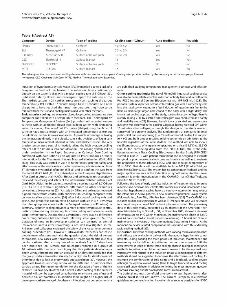
survivor can be separated into three main groups: conventional cooling(no device), non-invasive (surface) systems, and invasive (intravascular)systems (Table 1).Cooling techniques: Conventional cooling methods: The easiest wayto induce hypothermia after cardiac arrest is by using cold saline (forexample, 0.9% NaCl solution), crushed ice or ice bags. Kim and colleaguesreported the safety and efficacy of the administration of up to 2 litres of 4°Ccold saline to the patient after hospital admission [6]. Others published datausing 30 ml/kg body weight of saline 0.9% NaCl or Ringer’s lactatecombined sometimes with ice bags, which led to an acceptable reduction ofthe temperature [7-10]. Furthermore cold saline as well as other methodslike cooling caps and helmets have been evaluated for induction mainly inthe preclinical setting [4,11,12]. Kliegel and colleagues pointed out that coldinfusion alone is effective for induction but not for tight maintenance of thetarget temperature [13]. However, in at least one trial the combination ofcold saline and ice packs was proven to be effective even to maintaintemperature [7]. Focusing on the induction in the in-hospital setting, mostauthors rank cold saline and crushed ice more as effective adjuvantmethods to be combined with a computer-controlled cooling device [10].The big advantage of cold saline is its availability at almost every place inthe hospital if provided and the low costs. Following the data availableconcerning different amounts of saline administered to the patient, amedian amount of 1 to 2 litres of saline seems safe after cardiac arrest. Tomaintain target temperature with cold saline and ice bags seems to requirea high binding of personnel method without a very precise influence on thecentral body temperature.Surface cooling methods: Surface, non-invasive devices have to bedistinguished from intravascular, invasive devices. The range of availablecomputer-controlled surface devices with automatic temperature feedbackstretches from cooling blankets to be placed around the patient (BlanketrolIII, Cincinnati Sub-Zero; CritiCool, Medical ThermoRegulation Expertise) toadhesive cooling pads (Arctic Sun, Bard). Heard and colleagues comparedthe adhesive Arctic Sun surface-cooling system with normal cooling blanketscombined with ice bags. Although the reached target temperature within4 hours was not significantly different between the groups, the Arctic Sunsystem cooled more rapidly down to the target temperature [14]. A currentinvestigation from Norway compared the Arctic Sun surface (C.R. Bard)system (n = 92) with the invasive intravascular Coolgard (Alsius) system (n =75) in cardiac arrest survivors. The authors concluded no significantdifferences concerning neurological outcome and survival at discharge.A limitation for interpretation of the device efficacy (cooling rate/hour) is theadditional induction of cooling with cold saline and ice bags already in theemergency room [15]. A published case report described a severe skinpeeling during hypothermia with the Arctic Sun system without a knownhistory of skin problems or steroid therapy but with end-stage renal diseaseand coronary artery disease. This is the first severe adverse skin eventtowards the hydrogel pads known and the authors conclude that these skinlesions are very unusual as to be caused by the adhesive pads becauseexfoliative dermatitis is a rare syndrome and is often drug induced [16].Thus adverse skin reactions should not normally be expected using thismethod of cooling.Another surface feedback system using blankets is the CritiCool Pro systemby Medical ThermoRegulation Expertise (MTRE, Israel). The patient iswrapped into the body-shaped heat exchange garment resulting in amedian cooling rate of 0.7 ± 0.37°C/hour in a study by Laish-Farkash andcolleagues [17]. The Cincinnati Sub-Zero system has been compared withthe ArcticSun2000 (C.R. Bard) system by Mayer and colleagues for fevercontrol in neurocritical care patients. The authors conclude the ArcticSunsystem to be superior to the Cincinnati Sub-Zero system due to themaintenance of normothermia, a higher cooling rate and better feverreduction, although shivering occurred more frequently in the ArcticSungroup [18]. A surface cooling system without computer control andautomatic temperature feedback is the EMCOOLS cooling system. Theadhesive pads use a novel carbon cooling gel that has a high thermalconductivity resulting in a cooling rate of more than 3.5°C/hour. The FlexPad needs to be adapted to the body size and shape. The feasibility trial ofout-of-hospital surface cooling after return of spontaneous circulation(ROSC) in 15 survivors using the EMCOOLS system revealed a high mediancooling rate of 3.3°C/hour, the target temperature of 33°C was reachedapproximately within 70 minutes (55 to 106 minutes) after the start ofcooling and no skin lesions were observed [19]. A further novel system isthe Life Recovery ThermoSuit system, which was developed mainly for fast
Critical Care 2012, Volume 16 Suppl 2http://ccforum.com/supplements/16/S2
Page 5 of 42
induction of hypothermia by cold water (2°C) immersion due to a lack of atemperature feedback mechanism. The water circulates continuouslydirectly on the patient’s skin with a median cooling rate of 3°C/hour [20].Published data by Howes and colleagues report the safe use of theThermoSuit system in 24 cardiac arrest survivors reaching the targettemperature(<34°C) within 37 minutes (range 14 to 81 minutes) [21]. Afterthe patients have reached the target temperature, they have to beremoved from the suit and cooling maintained with other methods.Endovascular cooling: Intravascular closed-loop cooling systems are alsocomputer controlled with a temperature feedback. The Thermogard XPTemperature Management System (Zoll) provides both a central venouscatheter with an additional closed loop balloon system with circulatingwater for cooling. The InnerCool RTx device (Philips) using the Accutrolcatheter has a special feature with an integrated temperature sensor butno additional central intravascular access. A possible advantage of takingthe temperature directly in the bloodstream is the avoidance of lag in coretemperature measurement inherent in rectal and bladder sensors. The veryprecise temperature control is needed, taking the high average coolingrates of 4.0 to 5.0°C/hour into consideration. This cooling system will beunder evaluation in the Rapid Endovascular Catheter Core Coolingcombined with cold saline as an Adjunct to Percutaneous CoronaryIntervention for the Treatment of Acute Myocardial Infarction (CHILL-MI)study. This study was started in 2012 to further investigate the safety andeffectiveness of the endovascular cooling system in patients suffering fromST-elevation myocardial infarction (STEMI) and to confirm the data fromthe Rapid-MI-ICE trial [22]. In a subanalysis of the European HypothermiaAfter Cardiac Arrest trial (HACA), Holzer and colleagues retrospectivelyreviewed the efficacy and safety of the intravascular catheter system (CoolGard 3000, Alsius) in 56 patients, revealing a cooling rate of 1.2°C/hour(IQR 0.7 to 1.5) without significant differences to other techniquesconcerning adverse events [23]. A study by Gillies and colleagues reporteda good temperature control with endovascular cooling compared withconventional ice surface cooling [24]. After induction of cooling with coldsaline, one group was continued to be cooled with ice (n = 41) whereasthe other group was cooled with the Coolgard device (n = 42; Alsius). Insummary, catheter cooling provided a more precise temperature control,better control during rewarming, less overcooling and failure to reachtarget temperature. Despite these advantages there was no differenceconcerning outcome between both relatively small groups [24]. Theduration of time an intravascular catheter can be used as centralintravenous access after rewarming is not well investigated so far.Al-Senani and colleagues evaluated the safety of the Icy catheter during acooling procedure [25]. However, intravascular catheters can causebloodstream infections and raise the question about the risk of venousthrombosis. Few cases with thrombosis or thrombophlebitis due to acooling catheter after a using time of respectively 7 and 10 days havebeen published [26]. Simosa and colleagues reported in a group of10 patients with traumatic brain injury that five patients developing adepth venous thrombosis after an average of 5.4 days but concluded thatthe group under examination already had a high risk for development ofthrombosis due to lack of prophylactic anticoagulation [27]. However, theapproach towards anticoagulation will be different in survivors aftercardiac arrest. The recommendation for the duration of use of the Icycatheter is 4 days (Icy Quattro) but a novel surface coating of the cathetermaterial will soon be approved by authorities to enhance time of use anddecrease risk of thrombosis. In addition there might be a higher risk ofdeveloping catheter-related bloodstream infections but currently no data
are published studying temperature management catheters and infectionrates.Other cooling methods: The novel RhinoChill intranasal cooling devicewas able to demonstrate effective reduction of body temperature within thePre-ROSC Intranasal Cooling Effectiveness trial (PRINCE trial) [28]. Theportable system vaporises perfluorchlorcarbon gas with a catheter systeminto the nasal cavity leading to a fast induction of hypothermia first to thebrain as main target organ and second to the body with a slight delay. Theintra-arrest cooling approach of the study, starting induction of hypothermiaalready during CPR, by Castrén and colleagues was conducted as a safetyand feasibility study [28]. However, benefit towards survival and neurologicaloutcome was observed in the cooling subgroup, having received CPR within10 minutes after collapse, although the design of the study was notconceived for outcome analysis. The randomised trial compared in detailprehospital trans-nasal cooling (n = 83) with advanced cardiac live support(n = 99) and both groups received mild hypothermia on admission to thehospital regardless of the initial rhythm. This method was able to show asignificant decrease of tympanic temperature on arrival (34.2°C vs. 35.5°C).Due to the convincing data from the PRINCE trial, the PrehospitalResuscitation Intra Nasal Cooling Effectiveness Survival Study (PRINCESS)started in June 2010 with patient recruitment and is designed to evaluatefor good or poor neurological outcome and survival as well as to evaluatethe proportion of those achieving ROSC and time to target temperature of32 to 34°C. First data will be available in June 2013 (ClinicalTrial.govidentifier: NCT01400373). The system has no temperature feedback and themajor application area is the induction of hypothermia. Another novelapproach is under investigation in the CAMARO trial (ClinicalTrials.govidentifier: NCT01016236).Following the idea of early and fast induction of hypothermia to improveoutcome and decrease side effects after cardiac arrest and incorporate noveldata that hypothermia applied before a coronary intervention may reducethe infarct size in STEMI patients, a new automated peritoneal lavage system(Velomedix Inc., Palo Alto, USA) has been developed [22]. The CAMARO trialincludes cardiac arrest patients as well as STEMI patients who will be cooledto a target temperature of 34°C without prior resuscitation. The preliminarydata of this pilot study, presented as an abstract at the American HeartAssociation Meeting in Orlando, USA, in November 2011, showed a decreaseof temperature to 34°C within 9 minutes, the maintenance phase of 32.5°Cwas 24 hours in cardiac arrest patients (rewarming 16 hours) and 3 hoursmaintenance in myocardial infarction patients (rewarming 5 hours). At themoment no device-related complication has occurred with this extremelyrapid cooling method [29].Discussion: Different cooling methods with varying technical approachesand efficacy are available to deliver mild therapeutic hypothermia to ourpatients. During cooling the three phases of induction, maintenance andrewarming can be defined. Are different methods necessary to fulfil therequirements in each of these three cooling phases? Taking all mentionedmethods together, a combined approach seems to be the optimal way.Particularly with regard to the induction phase a combination of differentmethods should be suggested to increase the effectiveness of cooling, forexample the combination of cold saline and a feedback cooling device,although the optimal overall timing (time to target temperature and coolingrate) is still under debate. In addition to timing, the most important questionconcerns shivering and its prophylactic successful treatment.The optimal and most beneficial time point to start hypothermia aftercardiac arrest is still not known. The current European resuscitationguidelines recommend starting hypothermia as soon as possible after ROSC.
Table 1(Abstract A5)
Company Device Type of cooling Cooling rate (°C/hour) Auto feedback Reusable
Philips InnerCool RTx Catheter 4.0 to 5.0 Yes No
Zoll Thermogard XP Catheter 2.0 to 3.0 Yes No
C.R. Bard ArcticSun 5000 Surface adhesive pads 1.2 to 2.0 Yes No
CSZ Blanketrol III Surface blanket 1.5 Yes Yes
EMCOOLS FLEX.PAD Surface adhesive pads 3.5 No No
MTRE CritiCool Surface blanket 1.5 Yes no
The table gives the most common cooling devices with no claim to be complete. Cooling rates provided either by the company or at the company’s Internethomepage. CSZ, Cincinnati Sub-Zero; MTRE, Medical ThermoRegulation Expertise.
Critical Care 2012, Volume 16 Suppl 2http://ccforum.com/supplements/16/S2
Page 6 of 42

A recently published article by Sendelbach and colleagues revealed theimportance of avoiding any time delay of cooling to reach goodneurological outcome [30]. This ‘earlier is better’ strategy can be confirmedby animal data [31-34]. Following the ‘earlier is better’ strategy, some trialsexplored the possibility of inducing cooling during resuscitation or directlyafter ROSC, but data are controversial [35]. Induction of therapeutichypothermia during prehospital CPR using ice-cold intravenous fluid orintranasal cooling showed that it is feasible and is partially a benefit[28,36]. A major problem in predicting outcome and association withtiming and early cooling after cardiac arrest or even during resuscitationwith these data is the small sample size and the fact that prehospitalhypothermia was discontinued after admission to the hospital in many ofthese trials [37]. However, the analysis of data from the ScandinavianHypothermia Network including 986 patients after cardiac arrest byNielsen and colleagues showed no association of timing towardsneurological outcome [38].Certainly every ICU should provide 4°C cold saline to increase the coolingrate and to reach the target temperature as soon as possible. Theadministration of cold saline seems a feasible method in the preclinicalsetting as well as in addition to other preclinical devices available and afteradmission cold saline can be combined with a feedback device to speed upthe cooling. Furthermore, shivering is one of the most important side effectsthat can occur during hypothermia leading to an increased metabolic rate,high oxygen consumption and heat generation, and therefore needs to bekept in mind to be avoided and treated aggressively. The threshold for thisdefence mechanism of the thermoregulatory system is around ±35.5°C (1°Cbelow the vasoconstriction threshold) [39,40]. Therefore a fast induction tocross this threshold as quickly as possible seems indicated; additionaltreatment can include a sufficient analgosedation, magnesium andparalysation, but even the simple method of keeping the hands and feetwarm by wearing socks and gloves directly from the beginning of inductionof hypothermia can avoid shivering very reliably [41]. In patients withtraumatic brain injury undergoing temperature management, the benefit ofsurface counter warming concerning less shivering and improvement ofmetabolic profile was reported [42]. However, a high cooling rate duringinduction with a combination of a feedback-cooling device and severaladditional conventional cooling methods in combination with hand and feetcounter warming as described and a sufficient sedation level seems to bethe best way to avoid shivering. In addition, every temperaturemanagement procedure requires a reliable core temperature. The goldstandard is still the temperature taken directly in the bloodstream (forexample, pulmonary catheter) or directly by the cooling device itself aspossible with the Philips Accutrol endovascular catheter. Other commonplaces for temperature measurement are the bladder by Foley catheters,oeosophageal probes, tympanic and rectal temperature [43]. Moderntemperature management systems with high cooling rates lead to a fastinduction of hypothermia that can only be detected by most temperaturesensors with a time delay. The closed approach towards the gold standardmight be the oesophageal measurement with an approximately averagetime delay of 5 minutes (range 5 to 10 minutes) [40].Conclusion: A wide range of conventional and technical methods exists toapply mild therapeutic hypothermia after cardiac arrest. Hoedemaekers andcolleagues compared all described different methods (conventional coldinfusion/ice, water blankets, gel-coated pads, intravascular) in ICU patientsregarding the speed of cooling (°C/hour) and the reliability to maintain astable target temperature. The authors conclude that water-circulatingblankets, gel-coated pads and intravascular cooling are almost equallyefficient for induction but intravascular methods were superior formaintaining the target temperature [44]. Some performance data mighthave changed over the last years due to the industry having developed thenext generation of cooling devices. However, every method has its ownpartly limited, indication and a combination of an automatic computer-processed feedback device with conventional methods seems a good andsafe solution. The type of feedback device used in a hospital (invasive vs.non-invasive) depends on several factors but mainly on the personalpreference of the treating doctors, type of patients and the local standard aswell. In addition, the way of thinking is changing and it is no longer aquestion of making the patient cool as good as possible but rather hasevolved into a complex temperature management procedure with its ownrisks and pitfalls as well as benefits for the patient. It is a precondition toensure a precise and tight temperature control during all three treatmentphases. Especially during rewarming, which is a very critical phase of
temperature management, close temperature monitoring is necessary andcan be easily achieved with a computer-feedback cooling system. A passive,uncontrolled increase of temperature should be avoided in the moderntemperature management approach. However, the adoption rate andimplementation of hypothermia as part of standard post-arrest care is stillnot high enough. Reasons are manifold but the latest version of availablecooling devices may be able to help to increase the application rate bymaking the treatment safe and easy. If the hospital team feels confidentwith the topic of temperature management, numbers of operators mightincrease, even if the number of cardiac arrest patients treated in a hospital islow.The presentation of different temperature-management methods andinterpretation of their efficiency in the age of daily breaking news aboutmild hypothermia treatment and widening of the indication can only bea momentary snap-shot and cannot aspire to completeness.Competing interests: The author received financial support and materialresources from Medivance, Zoll, Philips, EMCOOL and C.R. Bard withindifferent projects and honorarium from Medivance, Zoll and Philips forlectures. This abstract has not been influenced by anyone in collection ofdata, analysis, interpretation and writing.References1. Nolan JP, Soar J, Zideman DA, Biarent D, Bossaert LL, Deakin C, Koster RW,
Wyllie J, Bottiger B: European Resuscitation Council guidelines forresuscitation 2010 Section 1. Executive summary. Resuscitation 2010,81:1219-1276.
2. Hypothermia After Cardiac Arrest Study Group: Mild therapeutichypothermia to improve the neurologic outcome after cardiac arrest. NEngl J Med 2002, 346:549-556.
3. Bernard SA, Gray TW, Buist MD, Jones BM, Silvester W, Gutteridge G,Smith K: Treatment of comatose survivors of out-of-hospital cardiacarrest with induced hypothermia. N Engl J Med 2002, 346:557-563.
4. Hachimi-Idrissi S, Corne L, Ebinger G, Michotte Y, Huyghens L: Mildhypothermia induced by a helmet device: a clinical feasibility study.Resuscitation 2001, 51:275-281.
5. Reinikainen M, Oksanen T, Leppanen P, Torppa T, Niskanen M, Kurola J:Mortality in out-of-hospital cardiac arrest patients has decreased in theera of therapeutic hypothermia. Acta Anaesthesiol Scand 2012, 56:110-115.
6. Kim F, Olsufka M, Carlbom D, Deem S, Longstreth WT, Hanrahan M,Maynard C, Copass MK, Cobb LA: Pilot study of rapid infusion of 2 L of 4°Cnormal saline for induction of mild hypothermia in hospitalized,comatose survivors of out-of-hospital cardiac arrest. Circulation 2005,112:715-719.
7. Larsson IM, Wallin E, Rubertsson S: Cold saline infusion and ice packsalone are effective in inducing and maintaining therapeutichypothermia after cardiac arrest. Resuscitation 2010, 81:15-19.
8. Spiel AO, Kliegel A, Janata A, Uray T, Mayr FB, Laggner AN, Jilma B, Sterz F:Hemostasis in cardiac arrest patients treated with mild hypothermiainitiated by cold fluids. Resuscitation 2009, 80:762-765.
9. Virkkunen I, Yli-Hankala A, Silfvast T: Induction of therapeutic hypothermiaafter cardiac arrest in prehospital patients using ice-cold Ringer’ssolution: a pilot study. Resuscitation 2004, 62:299-302.
10. Kliegel A, Losert H, Sterz F, Kliegel M, Holzer M, Uray T, Domanovits H: Coldsimple intravenous infusions preceding special endovascular cooling forfaster induction of mild hypothermia after cardiac arrest - a feasibilitystudy. Resuscitation 2005, 64:347-351.
11. Storm C, Schefold JC, Kerner T, Schmidbauer W, Gloza J, Krueger A,Jorres A, Hasper D: Prehospital cooling with hypothermia caps (PreCoCa):a feasibility study. Clin Res Cardiol 2008, 97:768-772.
12. Kim F, Olsufka M, Longstreth WT, Maynard C, Carlbom D, Deem S,Kudenchuk P, Copass MK, Cobb LA: Pilot randomized clinical trial ofprehospital induction of mild hypothermia in out-of-hospital cardiacarrest patients with a rapid infusion of 4 degrees C normal saline.Circulation 2007, 115:3064-3070.
13. Kliegel A, Janata A, Wandaller C, Uray T, Spiel A, Losert H, Kliegel M,Holzer M, Haugk M, Sterz F, Laggner AN: Cold infusions alone areeffective for induction of therapeutic hypothermia but do not keeppatients cool after cardiac arrest. Resuscitation 2007, 73:46-53.
14. Heard KJ, Peberdy MA, Sayre MR, Sanders A, Geocadin RG, Dixon SR,Larabee TM, Hiller K, Fiorello A, Paradis NA, O’Neil BJ: A randomizedcontrolled trial comparing the Arctic Sun to standard cooling forinduction of hypothermia after cardiac arrest. Resuscitation 2010,81:9-14.
Critical Care 2012, Volume 16 Suppl 2http://ccforum.com/supplements/16/S2
Page 7 of 42
15. Tomte O, Draegni T, Mangschau A, Jacobsen D, Auestad B, Sunde K: Acomparison of intravascular and surface cooling techniques in comatosecardiac arrest survivors. Crit Care Med 2011, 39:443-449.
16. Varon J, Acosta P, Wintz R, Mendoza N: Unusual side effect from hydrogelpads during therapeutic hypothermia. Resuscitation 2008, 78:248-249.
17. Laish-Farkash A, Matetzky S, Kassem S, Haj-Iahia H, Hod H: Therapeutichypothermia for comatose survivors after cardiac arrest. Israel Med AssocJ 2007, 9:252-256.
18. Mayer SA, Kowalski RG, Presciutti M, Ostapkovich ND, McGann E,Fitzsimmons BF, Yavagal DR, Du YE, Naidech AM, Janjua NA, Claassen J,Kreiter KT, Parra A, Commichau C: Clinical trial of a novel surface coolingsystem for fever control in neurocritical care patients. Crit Care Med 2004,32:2508-2515.
19. Uray T, Malzer R: Out-of-hospital surface cooling to induce mildhypothermia in human cardiac arrest: a feasibility trial. Resuscitation 2008,77:331-338.
20. Janata A, Weihs W, Bayegan K, Schratter A, Holzer M, Behringer W,Schock RB, Losert UM, Springler G, Schmidt P, Sterz F: Therapeutichypothermia with a novel surface cooling device improves neurologicoutcome after prolonged cardiac arrest in swine. Crit Care Med 2008,36:895-902.
21. Howes D, Ohley W, Dorian P, Klock C, Freedman R, Schock R, Krizanac D,Holzer M: Rapid induction of therapeutic hypothermia using convective-immersion surface cooling: safety, efficacy and outcomes. Resuscitation2010, 81:388-392.
22. Gotberg M, Olivecrona GK, Koul S, Carlsson M, Engblom H, Ugander M, vander Pals J, Algotsson L, Arheden H, Erlinge D: A pilot study of rapidcooling by cold saline and endovascular cooling before reperfusion inpatients with ST-elevation myocardial infarction. Circ Cardiovasc Interv2010, 3:400-407.
23. Holzer M, Mullner M, Sterz F, Robak O, Kliegel A, Losert H, Sodeck G, Uray T,Zeiner A, Laggner AN: Efficacy and safety of endovascular cooling aftercardiac arrest: cohort study and Bayesian approach. Stroke 2006,37:1792-1797.
24. Gillies MA, Pratt R, Whiteley C, Borg J, Beale RJ, Tibby SM: Therapeutichypothermia after cardiac arrest: a retrospective comparison of surfaceand endovascular cooling techniques. Resuscitation 2010, 81:1117-1122.
25. Al-Senani FM, Graffagnino C, Grotta JC, Saiki R, Wood D, Chung W,Palmer G, Collins KA: A prospective, multicenter pilot study to evaluatethe feasibility and safety of using the CoolGard System and Icy catheterfollowing cardiac arrest. Resuscitation 2004, 62:143-150.
26. Prunet B, Lacroix G, Bordes J, Poyet R, D’Aranda E, Goutorbe P: Catheterrelated venous thrombosis with cooling and warming catheters: twocase reports. Cases J 2009, 2:8857.
27. Simosa HF, Petersen DJ, Agarwal SK, Burke PA, Hirsch EF: Increased risk ofdeep venous thrombosis with endovascular cooling in patients withtraumatic head injury. Am Surg 2007, 73:461-464.
28. Castren M, Nordberg P, Svensson L, et al: Intra-arrest transnasalevaporative cooling: a randomized, prehospital, multicenter study(PRINCE: Pre-ROSC IntraNasal Cooling Effectiveness). Circulation 2010,122:729-736.
29. Polderman K, CAMARO trialists: CAMARO trial, preliminary results onsafety [abstract 13265]. Circulation 2011, 124.
30. Sendelbach S, Hearst MO, Johnson PJ, Unger BT, Mooney MR: Effects ofvariation in temperature management on cerebral performancecategory scores in patients who received therapeutic hypothermia postcardiac arrest. Resuscitation 2012 in press.
31. Kuboyama K, Safar P, Radovsky A, Tisherman SA, Stezoski SW, Alexander H:Delay in cooling negates the beneficial effect of mild resuscitativecerebral hypothermia after cardiac arrest in dogs: a prospective,randomized study. Crit Care Med 1993, 21:1348-1358.
32. Nozari A, Safar P, Stezoski SW, Wu X, Kostelnik S, Radovsky A, Tisherman S,Kochanek PM: Critical time window for intra-arrest cooling with coldsaline flush in a dog model of cardiopulmonary resuscitation. Circulation2006, 113:2690-2696.
33. Jia X, Koenig MA, Shin HC, Zhen G, Pardo CA, Hanley DF, Thakor NV,Geocadin RG: Improving neurological outcomes post-cardiac arrest in arat model: immediate hypothermia and quantitative EEG monitoring.Resuscitation 2008, 76:431-442.
34. Colbourne F, Corbett D: Delayed postischemic hypothermia: a six monthsurvival study using behavioral and histological assessments ofneuroprotection. J Neurosci 1995, 15:7250-7260.
35. Kamarainen A, Virkkunen I, Tenhunen J, Yli-Hankala A, Silfvast T: Prehospitaltherapeutic hypothermia for comatose survivors of cardiac arrest: arandomized controlled trial. Acta Anaesthesiol Scand 2009, 53:900-907.
36. Kamarainen A, Virkkunen I, Tenhunen J, Yli-Hankala A, Silfvast T: Prehospitalinduction of therapeutic hypothermia during CPR: a pilot study.Resuscitation 2008, 76:360-363.
37. Kamarainen A, Hoppu S, Silfvast T, Virkkunen I: Prehospital therapeutichypothermia after cardiac arrest - from current concepts to a futurestandard. Scand J Trauma Resusc Emerg Med 2009, 17:53.
38. Nielsen N, Hovdenes J, Nilsson F, Rubertsson S, Stammet P, Sunde K,Valsson F, Wanscher M, Friberg H: Outcome, timing and adverse events intherapeutic hypothermia after out-of-hospital cardiac arrest. ActaAnaesthesiol Scand 2009, 53:926-934.
39. Lopez M, Sessler DI, Walter K, Emerick T, Ozaki M: Rate and genderdependence of the sweating, vasoconstriction, and shivering thresholdsin humans. Anesthesiology 1994, 80:780-788.
40. Polderman KH, Herold I: Therapeutic hypothermia and controllednormothermia in the intensive care unit: practical considerations, sideeffects, and cooling methods. Crit Care Med 2009, 37:1101-1120.
41. van Zanten AR, Polderman KH: Blowing hot and cold? Skin counterwarming to prevent shivering during therapeutic cooling. Crit Care Med2009, 37:2106-2108.
42. Badjatia N, Strongilis E, Prescutti M, Fernandez L, Fernandez A, Buitrago M,Schmidt JM, Mayer SA: Metabolic benefits of surface counter warmingduring therapeutic temperature modulation. Crit Care Med 2009,37:1893-1897.
43. Hasper D, Nee J, Schefold JC, Krueger A, Storm C: Tympanic temperatureduring therapeutic hypothermia. Emerg Med J 2011, 28:483-485.
44. Hoedemaekers CW, Ezzahti M, Gerritsen A, van der Hoeven JG: Comparisonof cooling methods to induce and maintain normo- and hypothermia inintensive care unit patients: a prospective intervention study. Crit Care2007, 11:R91.
A6Pharmacodynamics in hypothermiaTorkjel Tveita1,21Anesthesia and Critical Care, Institute of Clinical Medicine, University ofTromsø, Norway; 2University Hospital of Northern Norway, Tromsø, NorwayCritical Care 2012, 16(Suppl 2):A6
Introduction: Guidelines for using inotropic drugs to support cardiovascularfunction at low core temperatures are not well characterized. The safeapplication of inotropic drugs during normothermic conditions, routinelyused to treat cardiovascular instability by effectively increasing cardiacoutput (CO) and improve end-organ perfusion associated with acute heartfailure [1], is based on a detailed understanding of pharmacodynamics andpharmacokinetics of these drugs. Detailed knowledge of temperature-dependent changes in pharmacodynamics and pharmacokinetics of suchcardioactive drugs is essential for establishing treatment guidelines.Therapeutic hypothermia: Over the last decade therapeutic hypothermiahas been established as a recognized intervention to increase survival andimprove neurologic outcome in adult comatose cardiac arrest survivors[2-6]. However, after return of spontaneous circulation (ROSC) and coronaryrevascularization, more than 50% of survivors suffer from acute heartfailure and need inotropic cardiac support to resume adequate circulatoryfunction [2] after induction of hypothermia when the core temperature isdeliberately lowered to 34 to 32ºC and maintained for 24 to 48 hours. Incontrast, in patients hospitalized for acute heart failure withouthypothermia a subgroup of about only 10% received inotropic drugs [7].Accidental hypothermia: Another group of patients in need of inotropicdrug therapy at low core temperature are accidental hypothermia patientsdisplaying hypothermia-induced cardiac failure during rewarming, rangingfrom mild reduction of CO to the fulminant circulatory shock termedrewarming shock [8-11]. Rewarming shock is a clinically descriptive term thatrefers to a pathophysiologic state of cardiovascular collapse taking placeduring or after rewarming from accidental hypothermia [12], recognized as aprogressive reduction of CO and a sudden fall in arterial blood pressure. Inorder to treat or prevent rewarming shock, cardioactive inotropic drugs arecommonly necessary to elevate a low CO.Research in experimental hypothermia has displayed a substantialdepression of LV myocardial function in earlier studies [8,11,13] as well as
Critical Care 2012, Volume 16 Suppl 2http://ccforum.com/supplements/16/S2
Page 8 of 42
in recent studies [9,13,14]. Based on the results from these studies,cellular calcium overload, disturbed calcium homeostasis, changes inmyocardial myofilament responsiveness to intracellular calcium as well asimpaired high-energy phosphate homeostasis could all be proposed asimportant factors leading to the changes observed in the hypothermicheart and contributing to failure of functional recovery during rewarming[15-18].β-receptor agonists: In the acutely failing heart postoperatively, onlydrugs such as epinephrine and NE provide positive inotropy and perfusionpressure. Epinephrine acts through stimulation via sarcolemmal b-adrenoceptors, causing phosphorylation of the sarcolemmal L-type Ca2+
channel via cyclic AMP and protein kinase A pathways. This phosphorylationincreases the open probability of the channel [19], allowing for greatertrans-sarcolemmal Ca2+ influx with each depolarization and producing, inpart, the positive inotropic effect of epinephrine.Vascular effects: Only a few experiments studying effects of epinephrineduring hypothermia or/and rewarming using in vivo animal models havebeen published. Some authors report that both b1-adrenoceptors and a-adrenoceptors increase their sensitivity to catecholamines duringhypothermia [18,20-22] as b1-adrenoceptor activity was potentiated by lowtemperature, and they claim the existence of hypothermia-inducedsupersensitivity and increased agonist activity for b1-adrenoceptors. Insupport of this view, a left shift of the concentration-response curve forepinephrine during hypothermia has been reported [23]. However, otherssuggest hypothermia-induced increase in sensitivity for both a1-adrenoceptors and a2-adrenoceptors during cooling [24], but that sensitivityof b1-adrenoceptors is not increased to the same extent as a-adrenoceptors.In contrast, other researchers have reported a hypothermia-inducedsupersensitivity of b1-adrenoceptors [18,20,21]. Rubinstein reported thathypothermia modified the vascular response to epinephrine [25]; that is, theepinephrine doses that induced vasodilation during normothermicconditions increased TPR at 25°C. He also claimed that myocardialcontractile effects of epinephrine is reduced at low temperatures (coveredbelow), a view also supported by others [25,25-27]. Experimental data showthat the sympathetic nervous system could be switched off at a thresholdtemperature about 29°C and hypotensive patients with temperatures belowthis may benefit from infusions of exogenous catecholamines [28]. Inaddition, if CO could be elevated pharmacologically, rewarming by anymeans becomes more efficient [17,29]. Some researchers recommendinfusion of low doses of catecholamines in patients who have lower bloodpressure than would be expected for that degree of hypothermia and whoare not responding to crystalloids and rewarming [29].It therefore seems that the use of vasoactive drugs during hypothermicconditions remains quite contradictory.Cardiac effects: Some sources claim that the hypothermic heart isunresponsive [30] or little responsive [31] to cardioactive drugs, and the lastreference as well as recommendation of the American Heart Association [26]refer to the potential hazard of overmedication, due to delayed drugmetabolism leading to accumulation to toxic levels in patients suffering fromdeep or severe hypothermia, if used repeatedly. The AHA recommends thefollowing algorithm for treatment of hypothermia: below 30°C i.v.epinephrine should not be given, but above 30°C epinephrine should begiven, if indicated, but at longer than standard intervals. To date, there areno prospective clinical studies to support the recommendation to avoidepinephrine during hypothermic CPR, but a preclinical work report majorside effects of repeated epinephrine administration during experimentalhypothermic CPR in pigs [32]. Even in the last recommendations from theAHA it was stated that treatment of severe hypothermia (temperature <30°C)in the field remains controversial [26].A reduction of CO and SV by inducing varying levels of hypercalcaemiaduring hypothermia (28ºC) was reported by Schiffmann and colleagues [18].Following infusion of epinephrine during these experimental conditions,CO and SV were even more depressed [18]. These findings shear similaritieswith findings in our intact animal models: infusion of epinephrine, whichtheoretically will induce opening of Ca2+ channels, increase calciuminflux and elevate intracellular calcium even further, caused a significantdepression of myocardial function during hypothermic as well as post-hypothermic conditions [13,33,34]. This made us conclude that hypothermiaand rewarming may cause alterations in the pharmacodynamic effects of a-receptor and b-receptor mediated drugs [13,33,35-37] or induce changes inreceptor affinity for these drugs. Further, we found that low-doseepinephrine managed to maintain positive inotropic effects on LV cardiac
function during cooling to 30ºC, but these effects vanished during coolingto 28ºC. If the epinephrine dose was increased at these low temperatures SVand CO were not elevated but a significant increase in afterload took place.Thus we conclude that if epinephrine is applied during hypothermia thetherapeutic window appears narrow with short distance to unwanted sideeffects. Further, the prehypothermic dose-dependent increase in LV functionin response to epinephrine was not present after rewarming in the groupthat had received epinephrine during hypothermia [13,33,35,38], and ifepinephrine was infused during rewarming vascular side effects ofepinephrine (vasoconstriction) dominated [13,38] without elevating COabove pre-hypothermic control values at any temperature.During cooling there is a reduction in Ca2+ sensitivity of troponin C due toprotein kinase A (PKA)-induced phosphorylation of troponin I [39], inaddition to the hypothermia-induced calcium overload [18,19]. Further, it isdocumented that PKA may inhibit the activity of adenylyl cyclase (AC)5 [40,41], predominantly expressed in the heart [42], which catalyzes ATPto cAMP. Based on this information, it is possible that lack of positiveinotropic effect on cardiac function of b-receptor stimulation duringhypothermia may be due to the inhibitory effects on AC 5 activity of PKAphosphorylation and/or a potential desensitization and internalization ofthe b-adrenoceptor. Another way of interpreting these results is that theefficacy of signal transduction through G-protein coupled receptorsis rapidly decreased through mechanisms like desensitization andinternalization, mechanisms that will avoid receptor overstimulation.However, during pathologic conditions, like acute heart failure followingacute coronary syndromes and cardiac standstill, or as a consequence ofexposure to accidental hypothermia, it is necessary for cardiac myocytes toproduce cAMP over the limitation of such adaptations.Dopamine: The still widespread use of DA in perioperative and intensivecare, the explicit recommendation for its use in accidental hypothermiaguidelines, and possible positive effects in hypothermia as reported fromexperimental studies all need evaluation. From experimental hypothermiaresearch we found that DA improved both systolic and diastolic function inhypothermia [43]. However, at 25°C no beneficial effect was seen on CI as SIdecreased with incrementing DA dosages. Increased SVRI at high-dose DA at25°C suggests a-adrenergic involvement not seen in normothermia.Properties of the low-flow, high-viscosity circulatory state, combined withserious alterations in the pharmacokinetics of DA, may explain lack ofbeneficial - and potentially harmful - effects from DA administration at 25°C.Milrinone: Milrinone is a phosphodiesterase (PDE) 3 inhibitor that isdominantly expressed in the heart and vascular tissues. The site of action ofmilrinone is cytosolic, and the administration of a PDE3 inhibitor increasescAMP only in the cardiovascular system, which subsequently enhancecardiac contraction and induce vasodilation during normothermicconditions [44]. In a recent experiment [45], using milrinone as a model drugof intracellular mode of action, the positive cardiac effects of this drug wasdemonstrated during normothermic conditions (30% increase in SV and CO)and remained during cooling to 15ºC. These effects of milrinone on cardiacfunction stay in essential contrast to those in our previous studies testingeffects of a-receptor and b-receptor mediated drugs [35,38] during coolingand rewarming.Conclusion: Taken together, the use of cardioactive drugs during hypo-thermic conditions remains quite contradictory. Therefore, pharmacologictreatment applied during the clinically challenging modalities, therapeutic aswell as accidental hypothermia, call upon written treatment protocols orguidelines that are so far largely missing or at least not properly updated.More research is needed to explore temperature-dependent changes inpharmacodynamics and pharmacokinetics of cardioactive drugs to writethese guidelines. Thus, due to significant hypothermia-induced alterations ofcardiac as well as vascular adrenoceptor sensitivity, the use of cardioactiveagents not affecting these receptor systems are advised during hypothermicconditions.References1. Petersen JW, Felker GM: Inotropes in the management of acute heart
failure. Crit Care Med 2008, 36:S106-S111.2. Bernard SA, Gray TW, Buist MD, Jones BM, Silvester W, Gutteridge G,
Smith K: Treatment of comatose survivors of out-of-hospital cardiacarrest with induced hypothermia. N Engl J Med 2002, 346:557-563.
3. Safar PJ, Kochanek PM: Therapeutic hypothermia after cardiac arrest.N Engl J Med 2002, 346:612-613.
4. Polderman KH: Mechanisms of action, physiological effects, andcomplications of hypothermia. Crit Care Med 2009, 37:S186-S202.
Critical Care 2012, Volume 16 Suppl 2http://ccforum.com/supplements/16/S2
Page 9 of 42

5. Polderman KH: Induced hypothermia and fever control for preventionand treatment of neurological injuries. Lancet 2008, 371:1955-1969.
6. Polderman KH: Hypothermia and neurological outcome after cardiacarrest: state of the art. Eur J Anaesthesiol Suppl 2008, 42:23-30.
7. Abraham WT, Adams KF, Fonarow GC, Costanzo MR, Berkowitz RL,LeJemtel TH, Cheng ML, Wynne J: In-hospital mortality in patients withacute decompensated heart failure requiring intravenous vasoactivemedications: an analysis from the Acute Decompensated Heart FailureNational Registry (ADHERE). J Am Coll Cardiol 2005, 46:57-64.
8. Blair E, Montgomery AV, Swan H: Posthypothermic circulatory failure.I. Physiologic observations on the circulation. Circulation 1956, 13:909-915.
9. Kondratiev TV, Flemming K, Myhre ES, Sovershaev MA, Tveita T: Is oxygensupply a limiting factor for survival during rewarming from profoundhypothermia? Am J Physiol Heart Circ Physiol 2006, 291:H441-H450.
10. Patton JF, Doolittle WH: Core rewarming by peritoneal dialysis followinginduced hypothermia in the dog. J Appl Physiol 1972, 33:800-804.
11. Tveita T, Skandfer M, Refsum H, Ytrehus K: Experimental hypothermia andrewarming: changes in mechanical function and metabolism of rathearts. J Appl Physiol 1996, 80:291-297.
12. Maclean D, Emslie-Smith D: Accidental Hypothermia Melbourne: BlackwellScientific 1977.
13. Kondratiev TV, Myhre ES, Simonsen O, Nymark TB, Tveita T: Cardiovasculareffects of epinephrine during rewarming from hypothermia in an intactanimal model. J Appl Physiol 2006, 100:457-464.
14. Filseth OM, How OJ, Kondratiev T, Gamst TM, Tveita T: Post-hypothermiccardiac left ventricular systolic dysfunction after rewarming in an intactpig model. Crit Care 2010, 14:R211.
15. Baller D, Wolpers HG, Schrader R, Hoeft A, Korb H, Rosick A,Bretschneider HJ, Hellige G: Paradoxical effects of catecholamines andcalcium on myocardial function in moderate hypothermia. ThoracCardiovasc Surg 1983, 31:131-138.
16. Belke DD, Wang LC, Lopaschuk GD: Effects of hypothermia on energymetabolism in rat and Richardson’s ground squirrel hearts. J Appl Physiol1997, 82:1210-1218.
17. Nicodemus HF, Chaney RD, Herold R: Hemodynamic effects of inotropesduring hypothermia and rapid rewarming. Crit Care Med 1981, 9:325-328.
18. Schiffmann H, Gleiss J, von HA, Schroder T, Kahles H, Hellige G: Effects ofepinephrine on the myocardial performance and haemodynamics of theisolated rat heart during moderate hypothermia - importance of calciumhomeostasis. Resuscitation 2001, 50:309-317.
19. Bers DM: Ca regulation in cardiac muscle. Med Sci Sports Exerc 1991,23:1157-1162.
20. Dundee JW, Clarke RS: pharmacology of hypothermia. Int Anesthesiol Clin1964, 2:857-872.
21. Williams RG, Broadley KJ: Responses mediated via beta 1, but not beta 2-adrenoceptors, exhibit hypothermia-induced supersensitivity. Life Sci1982, 31:2977-2983.
22. Chess Williams RG, Broadley KJ, Duncan C: A fundamental temperature-dependent difference between beta-adrenoceptor agonists andantagonists. Life Sci 1984, 35:1091-1099.
23. Riishede L, Nielsen-Kudsk F: Myocardial effects of adrenaline, isoprenalineand dobutamine at hypothermic conditions. Pharmacol Toxicol 1990,66:354-360.
24. Gomez B, Borbujo J, Garcia-Villalon AL, Nava-Hernandez E, Valle J, Garcia JL,Dieguez G: Alpha 1- and alpha 2-adrenergic response in human isolatedskin arteries during cooling. Gen Pharmacol 1991, 22:341-346.
25. Rubinstein EH: Vascular responses to adrenaline, noradrenaline andangiotensin in hypothermic dogs. Acta Physiol Lat Am 1961, 11:30-37.
26. Guidelines for cardiopulmonary resuscitation and emergency cardiaccare. Emergency Cardiac Care Committee and Subcommittees, AmericanHeart Association. Part IV. Special resuscitation situations. JAMA 1992,268:2242-2250.
27. Omar SA, Hammad D, Varma S: Reduced beta adrenergic responsivenessin isolated rabbit atria during hypothermia. Indian J Physiol Pharmacol1979, 23:199-203.
28. Chernow B, Lake CR, Zaritsky A, Finton CK, Casey L, Rainey TG, Fletcher JR:Sympathetic nervous system ‘switch off’ with severe hypothermia. CritCare Med 1983, 11:677-680.
29. Danzl DF, Pozos RS: Accidental hypothermia. N Engl J Med 1994,331:1756-1760.
30. Lloyd EL: Accidental hypothermia. Resuscitation 1996, 32:111-124.31. Murray P, Hall J: Principles of Critical Care New York: McGraw-Hill 1998.
32. Kornberger E, Lindner KH, Mayr VD, Schwarz B, Rackwitz KS, Wenzel V,Krismer AC, Mair P: Effects of epinephrine in a pig model of hypothermiccardiac arrest and closed-chest cardiopulmonary resuscitation combinedwith active rewarming. Resuscitation 2001, 50:301-308.
33. Kondratiev TV, Tveita T: Effects of sympathetic stimulation during coolingon hypothermic as well as posthypothermic hemodynamic function. CanJ Physiol Pharmacol 2006, 84:985-991.
34. Kondratiev TV, Wold RM, Aasum E, Tveita T: Myocardial mechanicaldysfunction and calcium overload following rewarming fromexperimental hypothermia in vivo. Cryobiology 2008, 56:15-21.
35. Han YS, Tveita T, Kondratiev TV, Prakash YS, Sieck GC: Changes incardiovascular beta-adrenoceptor responses during hypothermia.Cryobiology 2008, 57:246-250.
36. Mortensen E, Berntsen R, Tveita T, Lathrop DA, Refsum H: Changes inventricular fibrillation threshold during acute hypothermia: a model forfuture studies. J Basic Clin Physiol Pharmacol 1993, 4:313-319.
37. Bokenes L, Alexandersen TE, Osterud B, Tveita T, Mercer JB: Physiologicaland haematological responses to cold exposure in the elderly. Int JCircumpolar Health 2000, 59:216-221.
38. Tveita T, Sieck GC: The physiologic responses to epinephrine duringcooling and after rewarming in vivo. Crit Care 2011, 15:R225.
39. Han YS, Tveita T, Prakash YS, Sieck GC: Mechanisms underlyinghypothermia-induced cardiac contractile dysfunction. Am J Physiol HeartCirc Physiol 2010, 298:H890-H897.
40. Iwami G, Kawabe J, Ebina T, Cannon PJ, Homcy CJ, Ishikawa Y: Regulation ofadenylyl cyclase by protein kinase A. J Biol Chem 1995, 270:12481-12484.
41. Chen Y, Harry A, Li J, Smit MJ, Bai X, Magnusson R, Pieroni JP, Weng G,Iyengar R: Adenylyl cyclase 6 is selectively regulated by protein kinase Aphosphorylation in a region involved in Galphas stimulation. Proc NatlAcad Sci USA 1997, 94:14100-14104.
42. Iwatsubo K, Okumura S, Ishikawa Y: Drug therapy aimed at adenylylcyclase to regulate cyclic nucleotide signaling. Endocr Metab ImmuneDisord Drug Targets 2006, 6:239-247.
43. Filseth OM, How OJ, Kondratiev T, Gamst TM, Sager G, Tveita T: Changes incardiovascular effects of dopamine in response to graded hypothermiain vivo. Crit Care Med 2012, 40:178-186.
44. Iwatsubo K, Okumura S, Ishikawa Y: Drug therapy aimed at adenylylcyclase to regulate cyclic nucleotide signaling. Endocr Metab ImmuneDisord Drug Targets 2006, 6:239-247.
45. Tveita , Sieck GC: Effects of milrinone on left ventricular cardiac functionduring cooling in an intact animal model. Cryobiology 2012 in press.
HOT AND COLDA7Pathogenetic mechanisms of heatstroke and novel therapiesAbderrezak BouchamaKing Abdullah International Medical Research Center, Riyadh, Kingdom ofSaudi ArabiaCritical Care 2012, 16(Suppl 2):A7
Heatstroke, a life-threatening condition defined by a rapidly increasing coretemperature greater than 40°C and multiple organ system dysfunction, is aleading cause of morbidity and mortality during heat waves [1]. The heatwave that affected Europe during August 2003 led to an unprecedented70,000 excess deaths of which up to 40% were confirmed to be due toheatstroke [2]. Sophisticated climate models predict increasing frequencyand severity of heat waves and so the incidence of heat-related death couldrise if proactive measures and novel therapy to address this threat are notadopted [3].The mechanisms of multiple organ system dysfunction in heatstroke are notfully understood and include direct tissue injury and cell death resultingfrom heat cytotoxicity together with delayed organ dysfunction anddamage secondary to activation of inflammatory and coagulation pathways.Histopathological changes include endothelial injury, disseminatedintravascular thrombosis, neutrophils infiltration and apoptosis [4,5]. Despitecooling and optimal treatment in intensive care the overall mortality fromheatstroke exceeds 60%, due in large part to the fact that no specifictreatment is available [6,7].Experimental evidence from rodent models of heatstroke suggest thatimmunomodulation of the host response may alter the clinical course of
Critical Care 2012, Volume 16 Suppl 2http://ccforum.com/supplements/16/S2
Page 10 of 42
heatstroke and thereby improve outcome [8-10]. Heatstroke in inducessystemic and local (central nervous system) production of TNFa and IL-1,and severe coagulopathy. This is associated with multiple organ systemdysfunction including severe neuronal injury, and high mortality.Administration of an IL-1 receptor antagonist [8], corticosteroids [9] oractivated protein C [10] before the onset of heatstroke attenuates themultiple organ system dysfunction and improves survival. However,extrapolation of these data from these small animal models to humans isproblematic because of inter-species differences.Baboons represent the most appropriate model for the study of the hostinflammatory and hemostatic responses to heat stress and their relation tocellular injury and death as well as for testing novel therapy, which targetsthese pathways. Moreover, the findings may have direct applicability tohuman heatstroke, and could represent the basis for clinical trial. In thisreview, we show that baboons subjected to heat stress reproduce both theclinical and immunological changes similar to those seen in humans [11].Using this experimental model, we report that immunomodulating the hostsystemic inflammatory and coagulation responses failed to translateinto improved survival, suggesting that further basic research on thepathogenesis of heatstroke is required [12-14].References1. Bouchama A, Knochel JP: Heat stroke. N Engl J Med 2002, 346:1978-1988.2. Robine JM, Cheung SL, Le Roy S, Van Oyen H, Griffiths C, Michel JP,
Herrmann FR: Death toll exceeded 70,000 in Europe during the summerof 2003. C R Biol 2008, 331:171-178.
3. Easterling DR, Meehl GA, Parmesan C, Changnon SA, Karl TR, Mearns LO:Climate extremes: observations, modeling, and impacts. Science 2000,289:2068-2074.
4. Malamud N, Haymaker W, Custer R: Heatstroke: a clinico-pathologic studyof 125 fatal cases. Milit Surg 1946, 99:397-449.
5. Chao TC, Sinniah R, Pakiam JE: Acute heat stroke deaths. Pathology 1981,13:145-156.
6. Misset B, De Jonghe B, Bastuji-Garin S, Gattolliat O, Boughrara E, Annane D,Hausfater P, Garrouste-Orgeas M, Carlet J: Mortality of patients withheatstroke admitted to intensive care units during the 2003 heat wavein France: a national multiple-center risk-factor study. Crit Care Med 2006,34:1087-1092.
7. Argaud L, Ferry T, Le QH, Marfisi A, Ciorba D, Achache P, Ducluzeau R,Robert D: Short- and long-term outcomes of heatstroke following the2003 heat wave in Lyon, France. Arch Intern Med 2007, 167:2177-2183.
8. Lin MT, Liu HH, Yang YL: Involvement of interleukin-1 receptormechanisms in development of arterial hypotension in rat heatstroke.Am J Physiol 1997, 273(4 Pt 2):H2072-H2077.
9. Liu CC, Chien CH, Lin MT: Glucocorticoids reduce interleukin-1concentration and result in neuroprotective effects in rat heatstroke.J Physiol 2000, 527(Pt 2):333-343.
10. Chen CM, Hou CC, Cheng KC, Tian RL, Chang CP, Lin MT: Activated proteinC therapy in a rat heat stroke model. Crit Care Med 2006, 34:1960-1966.
11. Bouchama A, Roberts G, Al Mohanna F, El-Sayed R, Lach B, Chollet-Martin S,Ollivier V, Al Baradei R, Loualich A, Nakeeb S, et al: Inflammatory,hemostatic, and clinical changes in a baboon experimental model forheatstroke. J Appl Physiol 2005, 98:697-705.
12. Bouchama A, Kwaasi A, Dehbi M, Al Mohanna F, Eldali A, El-Sayed R,Tbakhi A, Alzahrani AS, Roberts AG: Glucocorticoids do not protect againstthe lethal effects of experimental heatstroke in baboons. Shock 2007,27:578-583.
13. Bouchama A, Kunzelmann C, Dehbi M, Kwaasi A, Eldali A, Zobairi F,Freyssinet JM, de Prost D: Recombinant activated protein C attenuatesendothelial injury and inhibits procoagulant microparticles release inbaboon heatstroke. Arterioscler Thromb Vasc Biol 2008, 28:1318-1325.
14. Bouchama A, Al-Mohanna F, Assad L, Baturcam E, Eldali A, Owaidah T, et al:Tissue factor/factor VIIa pathway mediates coagulation activation ininduced-heat stroke in the baboon. Crit Care Med 2011, 40:1229-1236.
A8Is therapeutic hypothermia immunosuppressive?Kees H PoldermanDepartment of Critical Care Medicine, University of Pittsburgh MedicalCenter, Pittsburgh, PA, USACritical Care 2012, 16(Suppl 2):A8
Introduction: The answer to the question in the title of this article wouldappear obvious. Even the term ‘to catch a cold’ is partly based on thewidely-held assumption that lower temperatures or a chill can decreasethe resistance to certain viral infections such as the common cold [1].Furthermore, a number of studies have clearly demonstrated thatinadvertent decreases in temperature in the perioperative setting, and insome other situations, can significantly increase infection risk.However, the question is not as easy to answer in cases where hypothermiais applied deliberately and with various precautions, which is the case intherapeutic cooling. In these cases potentially harmful responses such asshivering and tachycardia are carefully controlled, body temperatures arenot allowed to drop below a predefined setpoint, and every effort is madeto prevent side effects [2,3]. Moreover, even the evidence that hypothermiaplays a role in the development of the common cold is mixed, and the fewstudies performed to address this issue did not support the popular beliefthat colds are associated with exposure to a cold environment [1,4].The relationship between hypothermia and immune function are brieflydiscussed below.In vitro studies and animal experiments: In most types of acute braininjury there is a significant and protracted inflammatory response in thehours following the acute event, whether this is ischemic, traumatic, or acombination of both. Proinflammatory mediators such as TNFa and IL-1are released in large quantities by astrocytes, microglia, and endothelialcells following an episode of ischemia and reperfusion [2]. The levels ofthese mediators begin to rise around 1 hour after injury and remainelevated for up to 5 days [2,5,6]. This in turn stimulates the chemotaxis ofactivated leucocytes across the blood-brain barrier and leads to anaccumulation of inflammatory cells in the injured brain, as well as theemergence of adhesion molecules on leukocytes and endothelial cells.Simultaneously activation of the complement system occurs, beginning inthe very early stages after brain injury. This further stimulates the passageof neutrophils and (in later stages) monocytes/macrophages [6]. Theseinflammatory and immunological responses occur especially duringreperfusion and are accompanied by free radical production (see below).These inflammatory responses can cause significant (additional) injurythrough the phagocytic actions of macrophages, synthesis of toxicproducts, and further stimulation of immune reactions in a vicious cycle.Thus it can be argued that, at least in the initial stages of acute brain injury,a hyperinflammatory state exists. As explained in some more detail belowhypothermia attenuates this proinflammatory state, but this in itself doesnot constitute immune suppression. On the other hand, it should be realizedthat the proinflammatory response outlined above is to some extentphysiological; there is evidence suggesting that some inflammatorymediators have neuroprotective properties, although many others areneurotoxic [6-9]. Thus attenuating the response could have protectiveeffects, but also some detrimental ones [2,8,9].On balance there is persuasive evidence suggesting that the production ofcytokines and leukocyte infiltration is disproportionate and harmful, and cansignificantly increase the risk and extent of brain cell injury and infarction[6-15]. Especially, the IL-1 family appears to be important in this regard [15].The destructive aspects of inflammation appear to outweigh the potentialbenefits especially in the later stages of injury [2,6-9]. Thus there is apotential time window for therapeutic interventions to block or mitigate thisprocess before it causes permanent injury.Many animal experiments and in vitro studies have shown that mildhypothermia can suppress harmful inflammatory reactions that damagepotentially viable nerve cells and astrocytes [10-12]. Hypothermia can alsodecrease production of leukotrienes and nitric oxide, prevent reperfusion-related DNA injury and lipid peroxidation, and impair neutrophil andmacrophage function [2,3]. Thus on balance the protective effects of coolingare likely to outweigh the potential negative effects. However, this does notmean that there are no negative effects, especially if the temperature dropsbelow 32°C. It should be kept in mind that the suppression of inflammatoryresponses will occur in all organs, not just the injured ones; this is one of thereasons why inhibition of the immune response can lead to increasedinfection risk. Moreover, the effect is present regardless of whether a local orgeneral hyperinflammatory state exists.The systemic effect on the immune system can be enhanced by a decreasein the white blood cell count, which can begin at temperatures below 32 to33°C (although major decreases usually occur only at temperatures below 30to 31°C). Biggar and coworkers reported a drop in WBC count from 6.0 ± 0.6to 2.3 ± 0.3 when hypothermia of 29°C was induced in pigs [16]. They did
Critical Care 2012, Volume 16 Suppl 2http://ccforum.com/supplements/16/S2
Page 11 of 42

not observe significant changes in the number of circulating mature orimmature neutrophils, and reported that neutrophil demargination afteradministration of intravenous catecholamines was similar at 37°C and 29°C.However, they also observed that administration of corticosteroids and,importantly, of endotoxins failed to stimulate the normal release ofneutrophils from the bone marrow [16]. Failure of this response couldsignificantly impact infection risks [2].Most of these observations were made in animal experiments, but thehypothermia-induced suppression of the hyperinflammatory responsesand decrease in WBC count have been confirmed in clinical reports inhumans with traumatic brain injury [10,11,17].Thus the very mechanisms that provide tissue protection couldsimultaneously impair the patients’ ability to fight infections. In addition,lowering body temperature can lead to a decrease in insulin secretion andto induction of insulin resistance [2,3]. This can lead to hyperglycaemia,which can in turn impair leucocyte function and further increase infectionrisks. Finally, hypothermia can cause vasoconstriction in the skin, whichcould increase the risk for bedsores and surgical wound infections.In summary, based on the in vitro evidence we would expect hypothermiato inhibit the mostly harmful neuroinflammatory response and amelioratethe hyperinflammatory state that occurs after acute injury, but at the priceof increasing infection risk.Clinical evidence: Clinical studies reporting the infection risks associatedwith therapeutic cooling in different categories of patients with acutebrain injury have produced divergent results; studies in patients whodevelop accidental hypothermia have mostly reported higher infectionrisks.The link between accidental hypothermia in the perioperative setting anda higher incidence of surgical wound infections was first demonstrated byKurz and coworkers in 1996 [18], and has since been confirmed innumerous studies in various categories of surgical patients [19-26]. Themost recent example is a study by Seamon and coworkers, who found thatintraoperative hypothermia (below 35°C) was independently associatedwith surgical site infection rates after trauma laparotomy [26]. Local factorssuch as hypothermia-induced vasoconstriction in the skin may add to theunderlying immunosuppressive effect to further increase the rate ofwound infections. Recently, Laupland and colleagues reported that severehypothermia (<32°C, but not 32 to 35.9°C) was associated with significantincreases in risk for infections acquired in ICU [27].The link between hypothermia and infections is far less clear when mildhypothermia is induced under controlled circumstances. Numerous studiesin patients with post-hypoxic brain injury following cardiac arrest have notreported significant increases in rates of infections, although some havereported trends in that direction [28]. Seven multicentered trials in newbornbabies treated with neonatal asphyxia for 48 to 72 hours also did not reportconsistent increases in infection risks [28].In contrast, clinical studies in patients with ischemic stroke and TBI havetended to find higher risks of infection, especially pneumonia, in patientstreated with hypothermia. For example, Hemmen and coworkers reported arate of pneumonia of 50% in patients with ischemic stroke treated withhypothermia and thrombolysis, compared to 10% in controls [29]. Althoughthe overall outcome was better in hypothermia patients in spite of the highinfection rate, this indicates that use of hypothermia in these patients maypresent significant difficulties.Some studies using hypothermia in patients with severe traumatic braininjury have also reported high infection rates [28]. There is evidence thatthis can be prevented by a combination of preventive measures, perhapsincluding use of antibiotic prophylaxis such as selective decontaminationof the digestive tract (SDD) [28,30,31].In one example, Kamps and coworkers reported on their use of prolongedtherapeutic cooling to control intracranial pressure in patients with severetraumatic brain injury, in a setting where SDD was routinely used, andreported that infection rates were 20% in patients treated with hypothermiaand 34.4% for matched controls [31]. Most notably, the risk of ventilator-associated pneumonia was the same in patients treated with hypothermiacompared with matched controls.Conclusion: Hypothermia impairs immune function and inhibits variousinflammatory responses. This is inherent to the treatment, and impairmentof harmful inflammatory reactions in the brain may be one of themechanisms through which hypothermia can exert protective effects.Hypothermia-induced insulin resistance and hyperglycemia may furtherincrease infection risks. In clinical studies, hypothermia has been most
clearly linked to infection risk in the context of accidental hypothermia;controlled therapeutic cooling appears to carry a lower risk, especially ifhypothermia is used for limited periods of time (<48 hours). The risk appearsto increase with prolonged use, and careful monitoring is required in thesepatients. Prophylactic antibiotics may be considered in high-risk patientswho are cooled for prolonged periods.References1. Heikkinen T, Järvinen A: The common cold. Lancet 2003, 361:51-59.2. Polderman KH: Mechanisms of action, physiological effects, and
complications of hypothermia. Crit Care Med 2009, 37:S186-S202.3. Polderman KH, Herold I: Therapeutic hypothermia and controlled
normothermia in the intensive care unit: practical considerations, sideeffects, and cooling methods. Crit Care Med 2009, 37:1101-1120.
4. Douglas RG Jr, Lindgren KM, Couch RB: Exposure to cold environmentand rhinovirus common cold: failure to demonstrate effect. N Engl J Med1968, 279:742-747.
5. Small DL, Morley P, Buchan AM: Biology of ischemic cerebral cell death.Prog Cardiovasc Dis 1999, 42:185-207.
6. Schmidt OI, Heyde CE, Ertel W, Stahel PF: Closed head injury - aninflammatory disease? Brain Res Brain Res Rev 2005, 48:388-399.
7. Asensio VC, Campbell IL: Chemokines in the CNS: plurifunctionalmediators in diverse states. Trends Neurosci 1999, 22:504-512.
8. Merrill JE, Benveniste EN: Cytokines in inflammatory brain lesions: helpfuland harmful. Trends Neurosci 1996, 19:331-338.
9. Morganti-Kossmann MC, Rancan M, Stahel PF, Kossmann T: Inflammatoryresponse in acute traumatic brain injury: a double-edged sword. CurrOpin Crit Care 2002, 8:101-105.
10. Aibiki M, Maekawa S, Ogura S, Kinoshita Y, Kawai N, Yokono S: Effect ofmoderate hypothermia on systemic and internal jugular plasma IL-6 levelsafter traumatic brain injury in humans. J Neurotrauma 1999, 16:225-232.
11. Kimura A, Sakurada S, Ohkuni H, Todome Y, Kurata K: Moderatehypothermia delays proinflammatory cytokine production of humanperipheral blood mononuclear cells. Crit Care Med 2002, 30:1499-1502.
12. Suehiro E, Fujisawa H, Akimura T, Ishihara H, Kajiwara K, Kato S, Fujii M,Yamashita S, Maekawa T, Suzuki M: Increased matrix metalloproteinase-9in blood in association with activation of interleukin-6 after traumaticbrain injury: influence of hypothermic therapy. J Neurotrauma 2004,21:1706-1711.
13. Dietrich WD, Chatzipanteli K, Vitarbo E, Wada K, Kinoshita K: The role ofinflammatory processes in the pathophysiology and treatment of brainand spinal cord trauma. Acta Neurochir Suppl 2004, 89:69-74.
14. Schoettle RJ, Kochanek PM, Magargee MJ, Uhl MW, Nemoto EM: Earlypolymorphonuclear leukocyte accumulation correlates with thedevelopment of posttraumatic cerebral edema in rats. J Neurotrauma1990, 7:207-217.
15. Patel HC, Boutin H, Allan SM: Interleukin-1 in the brain: mechanisms ofaction in acute neurodegeneration. Ann NY Acad Sci 2003, 992:39-47.
16. Biggar WD, Bohn D, Kent G: Neutrophil circulation and release from bonemarrow during hypothermia. Infect Immun 1983, 40:708-712.
17. Ishikawa K, Tanaka H, Shiozaki T, Takaoka M, Ogura H, Kishi M, Shimazu T,Sugimoto H: Characteristics of infection and leukocyte count in severelyhead-injured patients treated with mild hypothermia. J Trauma 2000,49:912-922.
18. Kurz A, Sessler DI, Lenhardt R, Study of Wound Infection and TemperatureGroup: Perioperative normothermia to reduce the incidence of surgical-wound infection and shorten hospitalization. N Engl J Med 1996,334:1209-1215.
19. McAnally HB, Cutter GR, Ruttenber AJ, Clarke D, Todd JK: Hypothermia as arisk factor for pediatric cardiothoracic surgical site infection. Pediatr InfectDis J 2001, 20:459-462.
20. Melling AC, Ali B, Scott EM, Leaper DJ: Effects of preoperative warming onthe incidence of would infection after clean surgery: a randomizedcontrolled trial. Lancet 2001, 358:876-880.
21. Flores-Maldonado A, Medina-Escobedo CE, Ríos-Rodríguez HM, Fernández-Domínguez R: Mild perioperative hypothermia and risk of woundinfection. Arch Med Res 2001, 32:227-231.
22. Hedrick TL, Heckman JA, Smith RL, Sawyer RG, Friel CM, Foley EF: Efficacyof protocol implementation on incidence of wound infection incolorectal operations. J Am Coll Surg 2007, 205:432-438.
23. Wong PF, Kumar S, Bohra A, Whetter D, Leaper DJ: Randomized clinicaltrial of perioperative systemic warming in major elective abdominalsurgery. Br J Surg 2007, 94:421-426.
Critical Care 2012, Volume 16 Suppl 2http://ccforum.com/supplements/16/S2
Page 12 of 42
24. Moretti B, Larocca AM, Napoli C, Martinelli D, Paolillo L, Cassano M,Notarnicola A, Moretti L, Pesce V: Active warming systems to maintainperioperative normothermia in hip replacement surgery: a therapeuticaid or a vector of infection? J Hosp Infect 2009, 73:58-63.
25. Sumer BD, Myers LL, Leach J, Truelson JM: Correlation betweenintraoperative hypothermia and perioperative morbidity in patients withhead and neck cancer. Arch Otolaryngol Head Neck Surg 2009, 135:682-686.
26. Seamon MJ, Wobb J, Gaughan JP, Kulp H, Kamel I, Dempsey DT: Theeffects of intraoperative hypothermia on surgical site infection: ananalysis of 524 trauma laparotomies. Ann Surg 2012 in press, doi: 10.1097/SLA.0b013e31824b7e35.
27. Laupland KB, Zahar JR, Adrie C, Minet C, Vésin A, Goldgran-Toledano D,Azoulay E, Garrouste-Orgeas M, Cohen Y, Schwebel C, Jamali S, Darmon M,Dumenil AS, Kallel H, Souweine B, Timsit JF: Severe hypothermia increasesthe risk for intensive care unit-acquired infection. Clin Infect Dis 2012 inpress, doi: 10.1093/cid/cir1033.
28. Polderman KH: Induced hypothermia and fever control for preventionand treatment of neurological injuries. Lancet 2008, 371:1955-1969.
29. Hemmen TM, Raman R, Guluma KZ, Meyer BC, Gomes JA, Cruz-Flores S,Wijman CA, Rapp KS, Grotta JC, Lyden PD, ICTuS-L Investigators:Intravenous thrombolysis plus hypothermia for acute treatment ofischemic stroke (ICTuS-L): final results. Stroke 2010, 41:2265-2270.
30. Polderman KH, Tjong Tjin Joe R, Peerdeman SM, Girbes AR: Effects oftherapeutic hypothermia on intracranial pressure and outcome inpatients with severe head injury. Intensive Care Med 2002, 28:1563-1573.
31. Kamps M, Bisschops LA, van der Hoeven JG, Hoedemaekers CW:Hypothermia does not increase the risk of infection: a case controlstudy. Crit Care 2011, 15:R48.
A9Shivering: scores and protocolsNeeraj BadjatiaNeurological Institute of New York, Columbia University College of Physiciansand Surgeons, New York, USACritical Care 2012, 16(Suppl 2):A9
Shivering is both an anticipated consequence and, potentially, a majoradverse effect of therapeutic hypothermia. Even mild hypothermia can elicita vigorous thermoregulatory defense to maintain body temperature at thehypothalamic set point. In healthy humans, peripheral vasoconstriction istriggered at 36.5°C and shivering at 35.5°C. Temperature thresholds forvasoconstriction and shivering are often higher than normal in brain-injuredpatients; therefore, these thermoregulatory defenses may occur morevigorously and at higher temperatures in these individuals. Control ofshivering is essential for effective cooling, as shivering fights the coolingprocess, makes attaining target temperature difficult, is extremelyuncomfortable, and can trigger massive increases in systemic and cerebralenergy consumption and metabolic demand. The first step in treatment isadequate tools to recognize shivering. The Bedside Shivering AssessmentScale is a simple, validated four-point scale that enables repeatedquantification of shivering at the bedside. Therapy for shivering shouldideally stop or suppress the central thermoregulatory reflex rather than justuncoupling this response from skeletal muscle contraction, as the latterapproach does not mitigate the ongoing cerebral and systemic stressresponse. Analgo-sedation with opioids, a2-receptor agonists, or propofol isalmost always effective as a last resort to prevent shivering. However,nonpharmacological strategies as first-line interventions for shiveringminimize the risk of excessive sedation, which can make neurologicalexamination difficult and increase the risk of complications. The ColumbiaAnti Shivering protocol has been developed with these strategies in mind,and we base our approach on prospectively collected cooling data on 213patients who underwent 1,388 patient-days of temperature modulation.Eighty-nine patients underwent hypothermia and 124 patients underwentinduced normothermia. In 18% of patients and 33% of the total patient-days, only none-sedating baseline interventions were needed. The firstagent used was most commonly dexmeditomidine half the time, followedby opiates and increased doses of propofol. Younger patients, men, andlower body surface area were factors associated with increased number ofanti-shivering interventions. As noted by this protocol, a significantproportion of patients undergoing temperature modulation can beeffectively treated for shivering without oversedation and paralysis.
Patients at higher risk for needing more interventions are younger men withdecreased body surface area.References1. Choi HA, Badjatia N, Mayer SA: Hypothermia for acute brain injury-
mechanisms and practical aspects. Nat Rev Neurol 2012 in press, doi:10.1038/nrneurol.2012.21.
2. Choi HA, Ko SB, Presciutti M, Fernandez L, Carpenter AM, Lesch C,Gilmore E, Malhotra R, Mayer SA, Lee K, Claassen J, Schmidt JM, Badjatia N:Prevention of shivering during therapeutic temperature modulation: theColumbia anti-shivering protocol. Neurocrit Care 2011, 14:389-394.
3. Badjatia N: Hyperthermia and fever control in brain injury [review]. CritCare Med 2009, 37(7 Suppl):S250-S257.
4. Badjatia N, Strongilis E, Prescutti M, Fernandez L, Fernandez A, Buitrago M,Schmidt JM, Mayer SA: Metabolic benefits of surface counter warmingduring therapeutic temperature modulation. Crit Care Med 2009,37:1893-1897.
5. Badjatia N, Strongilis E, Gordon E, Prescutti M, Fernandez L, Fernandez A,Buitrago M, Schmidt JM, Ostapkovich ND, Mayer SA: Metabolic impact ofshivering during therapeutic temperature modulation: the BedsideShivering Assessment Scale. Stroke 2008, 39:3242-3247.
6. Badjatia N, Kowalski RG, Schmidt JM, Voorhees ME, Claassen J,Ostapkovich ND, Presciutti M, Connolly ES, Palestrant D, Parra A, Mayer SA:Predictors and clinical implications of shivering during therapeuticnormothermia. Neurocrit Care 2007, 6:186-191.
A10Controlled prophylactic normothermiaGregor Broessner*, Marlene Fischer, Bettina Pfausler, Erich SchmutzhardNeurologic Intensive Care Unit, Medical University Innsbruck, AustriaCritical Care 2012, 16(Suppl 2):A10
Introduction: Fever is a very frequent complication of intensive caretreatment and an independent predictor of unfavourable outcome andmortality in most patients with an acute severe neurologic injury. Today notonly treatment but even more prevention of fever has become the focus ofintensivists.Preliminary animal data for the beneficial neuroprotective effect oftherapeutic hypothermia could not satisfyingly be reproduced in patientsraising questions about the possible side effects of hypothermia. Controlledprophylactic normothermia (36.5°C) prevents secondary injury throughconsequent treatment of fever and limits dose-dependent side effectsthrough therapeutic hypothermia. Novel endovascular and gel-pad surfacecooling measures have shown to be feasible and efficacious in inducing andmaintaining even long-term controlled normothermia.Rationale for treatment of fever: Fever is one of the most frequentcomplications of intensive care treatment. Up to 90% of patients developat least one febrile episode within 7 days after being admitted to an ICU[1]. First of all, fever has always to be interpreted as a sign of an infection.Thus temperature modulation by any means has to include a strictprotocol identifying any source of infection followed by a straightforwardtreatment approach. There is widespread consensus that fever alone isassociated with unfavourable outcome. This consensus is a result of animaland human data over the past decades. In a meta-analysis conducted byGreer and colleagues including more than 14,000 patients, fever alone wasa significant and independent predictor of morbidity and mortality acrosssuch different diseases entities as ischaemic stroke, haemorrhagic strokeand traumatic brain injury [2]. Increasing evidence from animal and humanstudies suggests that fever, irrespective of its cause, can directly andadversely affect neurological outcome in various types of neurologicalinjury [3].The pathophysiological mechanisms by which fever affects patient outcomeare discussed, controversially comprising increase of metabolic demand(under circumstances of reduced supply), production of free radicals, localthermopooling, disruption of the blood-brain barrier, intracranial pressure(ICP) elevation, increased enzymatic inhibition of protein kinases, andworsened cytoskeletal proteolysis [1,3].Concept of controlled prophylactic normothermia: The aggressivetreatment of fever in any patient with a severe acute neurologic injury hasbecome increasingly important and is now the focus of many prospectivestudies including such patients. Whether the reduction of hyperthermiaalone or even controlled hypothermia should be the treatment goal is still
Critical Care 2012, Volume 16 Suppl 2http://ccforum.com/supplements/16/S2
Page 13 of 42
under debate and there are pros and cons for either approach. Theenthusiastic preliminary results from animal hypothermia studies could notbe satisfyingly reproduced when implemented in human controlled trials,shifting the focus on the possible side effects of hypothermia. Today thereis a whole body of evidence that the potentially neuroprotective effects ofhypothermia can be significantly diminished by its inherent side effects. Itcould be shown that hypothermia may lead to increased rate of infections,hypotension, shivering, disturbances in blood clotting, rewarming injuriesand significant changes in pharmacokinetics and pharmacodynamicspossibly limiting outcome effects of the treated patients [4-8]. Aggressivetreatment of fever in the ICU without risk elevation through the sideeffects of therapeutic hypothermia led to the concept of controlledprophylactic normothermia. This concept is based upon strict control ofbody core temperature with a target of 36.5°C beginning as early aspossible with the goal of complete fever prevention. Prophylacticcontrolled normothermia can therefore not be compared with thenormothermia control group of most randomised trials since this novelapproach aims to control temperature prophylactically and is therefore notonly treatment of fever.Experiences from clinical trials: Controlled prophylactic normothermiacannot be achieved through conventional temperature control measuresincluding NSAIDS and conventional cooling blankets [5,9]. In a controlledtrial conducted by our study group, reduction of fever burden (that is, bodycore temperature >37.9°C) was significantly higher in the endovascularcooling group than in the control group although strictly following apredefined fever management protocol including NSAIDS, opioids andconventional cooling blankets in patients with severe cerebrovasculardiseases [5]. In this trial an endovascular cooling catheter was placed in thesubclavian vein and prophylactic normothermia was maintained over168 hours in patients with ischaemic stroke and intracerebral haemorrhageand over 336 hours in patients with spontaneous subarachnoidhaemorrhage[5]. Safety evaluation revealed no relevant increase in directdevice-related adverse events in the endovascular group.Although there was significant decrease of the fever burden in the devicegroup no difference could be found in the long-term, 6-month, patientfollow-up. This lack of outcome efficacy may be attributed due to theincreased rate of infectious complications in the device group againpointing out that state-of-the-art temperature modulation has to becombined with a standardised surveillance of infections [6].In patients with severe traumatic brain injury (GCS ≤8) direct measurementof brain temperature together with ICP is possible [10]. Since fever maydeteriorate elevated ICP, prophylactic controlled normothermia should beevaluated in this patient population. In a small pilot trial, brain temperatureunder endovascular cooling showed that even under normothermia (36.5°C)brain temperature almost reaches body core temperature [11]. This is ofutmost interest as brain temperature exceeds body core temperature evenunder physiological circumstances and even more under fever with a peakgap >2°C [10]. As the brain is the target tissue of all neuroprotectants, it isnow evident that even controlled endovascular normothermia cansignificantly lower brain temperature in TBI patients.Others could achieve normothermia through a novel surface coolingdevice using gel-coated energy transfer pads, directly applied to the skinin patients with spontaneous subarachnoid haemorrhage and severetraumatic brain injury [9,12].Questions to be addressed in future trials: What is the optimal targettemperature and duration for temperature modulation in various diseaseentities? What is the appropriate approach to inherent complications suchas shivering, infections, pharmacodynamic and pharmacokineticdisturbances? Are surface and endovascular treatment approachesequivalent?Conclusion: Controlled prophylactic normothermia is not only feverreduction but aims to strictly maintain the body temperature at 36.5°C.Induced as early as possible the duration should cover the acute phase ofthe neurological injury, minimising secondary additional neurologicalimpairment though prevention of fever. Endovascular and surface coolingmeasures using gel-coated pads have shown to be efficacious and feasiblein inducing and maintaining normothermia whereas conventionaltemperature control measures including NSAIDs and conventional coolingblankets are not sufficient to prevent fever. Side effects known fromtherapeutic hypothermia such as shivering and increased rate of infectionsalso may occur under controlled prophylactic normothermia, although to alesser extent. Nevertheless, a consequent prevention of those limiting
factors should be kept in mind when applying controlled prophylacticnormothermia.References1. Marion DW: Controlled normothermia in neurologic intensive care. Crit
Care Med 2004, 32(Suppl):S43-S45.2. Greer DM, Funk SE, Reaven NL, et al: Impact of fever on outcome in
patients with stroke and neurologic injury: a comprehensive meta-analysis. Stroke 2008, 39:3029-3035.
3. Polderman KH: Induced hypothermia and fever control for preventionand treatment of neurological injuries. Lancet 2008, 371:1955-1969.
4. van den Broek MP, Groenendaal F, Egberts AC, et al: Effects ofhypothermia on pharmacokinetics and pharmacodynamics: a systematicreview of preclinical and clinical studies. Clin Pharmacokinet 2010,49:277-294.
5. Broessner G, Beer R, Lackner P, et al: Prophylactic, endovascularly based,long-term normothermia in ICU patients with severe cerebrovasculardisease: bicenter prospective, randomized trial. Stroke 2009, 40:e657-e665.
6. Broessner G, Lackner P, Fischer M, et al: Influence of prophylactic,endovascularly based normothermia on inflammation in patients withsevere cerebrovascular disease: a prospective, randomized trial. Stroke2010, 41:2969-2972.
7. Clifton GL, Miller ER, Choi SC, et al: Lack of effect of induction ofhypothermia after acute brain injury. N Engl J Med 2001, 344:556-563.
8. Clifton GL, Valadka A, Zygun D, et al: Very early hypothermia induction inpatients with severe brain injury (the National Acute Brain Injury Study:Hypothermia II): a randomised trial. Lancet Neurol 2011, 10:131-139.
9. Oddo M, Frangos S, Milby A, et al: Induced normothermia attenuatescerebral metabolic distress in patients with aneurysmal subarachnoidhemorrhage and refractory fever. Stroke 2009, 40:1913-1916.
10. Rumana CS, Gopinath SP, Uzura M, et al: Brain temperature exceedssystemic temperature in head-injured patients. Crit Care Med 1998,26:562-567.
11. Fischer M, Dietmann A, Lackner P, et al: Endovascular cooling andendothelial activation in hemorrhagic stroke patients. Neurocrit Care 2011in press.
12. Oddo M, Frangos S, Maloney-Wilensky E, et al: Effect of shivering on braintissue oxygenation during induced normothermia in patients withsevere brain injury. Neurocrit Care 2010, 12:10-16.
COOLING IN NEUROLOGY ANDNEUROSURGERYA11Therapeutic hypothermia in traumatic brain injuryJonathan KJ RhodesUniversity of Edinburgh, Department of Anaesthesia, Critical Care and PainMedicine, Intensive Care Unit, Western General Hospital, Edinburgh, UKCritical Care 2012, 16(Suppl 2):A11
Hypothermia has profound effects on the brain function but importantly ispotentially protective against both focal and global injuries. Aspects of thebiochemical response to acute ischaemia and trauma, which are associatedwith poor outcome, can be inhibited by cooling. Unlike many pharmaco-logical treatments that tend to antagonise a single neurochemical process,hypothermia offers a simple method of inhibiting multiple pathologicalprocesses simultaneously. It therefore has the potential, if applied correctly, toimprove outcomes after acute brain injuries, where drug trials have so farfailed.The systemic cooling of patients after acute brain injury is an establishedtreatment modality in many neuro-ICUs. It is a strategy for protecting theinjured brain that makes intuitive sense and can reduce both intracranialpressure and the potential for ischaemic secondary insults. Basic scienceevidence also suggests that cooling can attenuate many secondarybiochemical cascades that are activated after acute injury.However, despite these multiple lines of supportive evidence there is asyet no confirmation from a high-quality randomised controlled trial thatprophylactic hypothermia improves outcome or reduces mortality.This talk will look at the potentially beneficial effects of hypothermia onthe biochemistry of acute brain injury, consider the reasons for the failureto demonstrate clinical efficacy and review the supportive data from
Critical Care 2012, Volume 16 Suppl 2http://ccforum.com/supplements/16/S2
Page 14 of 42

meta-analysis, suggesting how hypothermia might be best delivered.Finally I will discuss EuroTherm3235, a European Society of Intensive CareMedicine funded multicentre randomised controlled trial investigatingprophylactic hypothermia in traumatic brain injury, which draws on thelessons from the available literature.References1. Clifton GL, Valadka A, Zygun D, et al: Very early hypothermia induction in
patients with severe brain injury (the National Acute Brain Injury Study:Hypothermia II): a randomised trial. Lancet Neurol 2011, 10:131-139.
2. Andrews PA, Sinclair HL, Battison CG, et al: European Society of IntensiveCare Medicine study of therapeutic hypothermia (32-35° C) forintracranial pressure reduction after traumatic brain injury (theEurotherm 3235 trial). Trials 2011, 12:8.
A12Hypothermia in spinal cord injuryGuadalupe A Castillo-Abrego1,21Neurocritical Care Unit, Caja de Seguro Social Hospital, Panamá City,Panamá; 2Medical Staff Critical Care Unit, Punta Pacifica Hospital: JohnHopkins Affiliated Hospital, San Fernando Hospital, Centro Médico Paitilla,Hospital Nacional, Panamá City, PanamáCritical Care 2012, 16(Suppl 2):A12
Historical considerations: In 1862 Edwin Smith, Egyptologist, acquired apapyrus that was thought to be between 2,500 and 3,000 years old. It wastranslated in 1930 by James Breasted, and was found to contain informationon medical therapies performed on 48 patients, including six cases ofcervical spine trauma. In the papyrus these injuries were described as‘dislocation of the vertebrae of the neck with unconscious arms and legs,where urine was drained and the phallus was erect, and it was determinedthat this cannot be treated’ [1].Other physicians like Hippocrates and Claudius Galen made historicalreference to the management of spinal cord injury. Galen studied injuries ingladiators, and described ‘that injury of the spinal cord caused paralysis andloss of sensation below the level of injury ... and determined that high spinalcord injuries are incompatible with life’ [2,3].Chaulic Guy (1300 to 1360) carried out innovations in the management oftraumatic bone injuries by introducing boards and suspensions to treatthese fractures; however, he gave a pessimistic view on spinal injuries[4,5].Ambrose Pare (1510 to 1590) developed spine surgery, and along withHildanus Fabricius (1560 to 1634) used traction techniques, reduction andutilization of wood frames to treat cervical spine fractures [6].Louis Pasteur’s (1822 to 1895) surgical innovations and discoveries and theadvent of asepsis and antisepsis by Semmelweis (1816 to 1868) andothers, and primarily the application of general anesthesia by Morton(1819 to 1868) and others gave a new impulse to this field. Together thesedevelopments helped reduce surgical mortality and infections [5,7].In the nineteenth century, anatomy and physiology of the central nervoussystem were studied. The neuron doctrine was developed by SantiagoRamon y Cajal (1852 to 1934), who demonstrated the individuality of nervecells and the connections that they have with each other [8,9].Alfred R Allen between 1908 and 1911 developed a reproducible andquantifiable model that allowed induction of a uniform traumatic injury tothe spine, and also explained the pathophysiological changes seen inspinal trauma, including key aspects of secondary spinal cord injury. In1972 Tarlov showed that symptoms caused by compression of the spinalcord for more than 12 hours could sometimes still be reversed. During thisperiod ‘progressive central hemorrhagic necrosis’ was identified, whichconsists of bleeding from the gray matter of the spinal cord centralnecrosis and destruction of white matter, with subsequent cavitations[10,11]. Finally, one of the possible immunomodalities of neuroprotectionwas suggested in 1968 by Albin and White who applied local hypothermiawith favorable results in animals [12]. However, use of hypothermia waslimited due to fear of side effects [13-17].These preliminary studies were difficult to interpret due to the limitednumber of patients, lack of controls, concomitant surgical procedures andthe concomitant use of drugs such as methylprednisolone [18]. In recentyears there has been a renewed interest in the use of moderate therapeutichypothermia due to its demonstrated neuroprotective effects in other areas.In Panama, we have been administering therapeutic hypothermia in
selected cases with severe spinal cord injury. More frequently we apply fevermanagement through endovascular, surface pads or medicalthermomodulation protocols.Epidemiological aspects of spinal cord injury: No accurate numbers onspinal cord injury are available for Panama and Central America. In theUnited States there are around 12,000 new cases of traumatic spinal cordinjury each year, not including those who die at the scene of the accident[19]. Approximately 1.3 million Americans have some type of chronicparalysis resulting from spinal cord injury [20,21]. The leading causes aremotor vehicle accidents (41.3%), falls (27.3%), acts of violence (15%) andrecreational activities (7.9%). Apart from the medical and personalconsequences for the patient, the economic impact in terms of ability towork is significant.Spinal cord injury, neurophysiological aspects - how can therapeutichypothermia help?: We will provide a concise description of thepathophysiological changes that occur after a spinal injury that might justifyuse of therapeutic hypothermia to mitigate ongoing destructive processes.Spinal cord injury is a process that can be divided into three phases [22,23].The primary mechanism refers to the direct mechanical damage. This mayinclude compression, stretching, persistent concussion, contusion,compression and laceration of the spinal cord [22,23]. The secondarymechanism involves a cascade of events at the cellular level triggered by theprimary mechanism. These biochemical events are responsible for increasedtissue damage and promote the apoptotic cascade. These include thoselisted in Table 1. The healing mechanism (third mechanism) begins in thedays after injury and can last for months or years. This can paradoxicallyincrease the neurological damage. Healing in the primary injury takes placeby neutrophils, macrophages and lymphocytes, reactive astrocytes, Schwanncells, meningeal fibroblasts, and microglia invasion. Scar formation forms abarrier for cellular and molecular axonal regeneration.Therapeutic hypothermia has been used in various types of neurologicalinjury such as stroke, post-anoxic encephalopathy, and traumatic braininjury. Some of these results have been extrapolated to spinal cordtrauma patients.Moderate therapeutic hypothermia has been shown to affect the apoptoticcascade as well as other destructive mechanisms, ranging from improvedenergy balance, reduction of mitochondrial dysfunction, decreased vascularpermeability and capillary leakage, mitigation of cell membrane injury,improvement of intracellular acidosis, mitigation of DNA injury, reduction inmetabolic demand, and a decrease in proinflammatory cytokine and freeradical release [24-28].It is important to realize that there is a window time, perhaps several hoursto days after the injury, during which this treatment modality can influencethe course of events [29].Experimental studies and supporting evidence: Yu and colleaguesused therapeutic hypothermia (33°C) after 30 minutes of ischemic injury tothe thoracic spinal cord in rats, and reported improvement of motorfunction at the microscopic level associated with cooling [30]. Othersreported improvements in functional outcome in various animal modelsassociated with cooling [30-32]. In an animal study of spinal cord ischemia,hypothermia increased the duration of ischemia required to produceneurological deficits [31].Moderate hypothermia has also shown potential benefits in invasiveprocedures including aortic clamp cross-clamping during thoracic surgery[33,34]. This can be regarded as indirect evidence that hypothermia couldprotect the spinal cord from ischemic injury, although the mechanisms intraumatic injury may be different from surgical trauma and ischemia.Fever is a common complication in patients with spinal cord injury [35-37].Of the identifiable etiologies the most common cause is infection (especiallypneumonia and urinary tract infections). However, fever of unknown originis the most frequent diagnosis, occurring in 66% of patients. It should berealized that patients with spinal cord injury have a high incidence ofthermoregulatory problems, which can contribute to the high incidence ofFUO [38,39].Of note, Yu and colleagues demonstrated in an experimental study in ratsthat post-traumatic hyperthermia in thoracic spinal cord injury worsenedbehavioral and histopathological damage compared with normothermia,and was associated with an increase of overall contusion volume byincreasing the vulnerability of both gray and white matter structurescompared with normothermia [40].Role of moderate hypothermia in the clinical scenario of patientswith spinal cord injury: Early clinical studies with irrigation to induce
Critical Care 2012, Volume 16 Suppl 2http://ccforum.com/supplements/16/S2
Page 15 of 42

local hypothermia produced conflicting results [41]. However, the adventof more reliable cooling devices that can better maintain bodytemperature within a predetermined range have improved our ability todeliver targeted therapy.Hypothermia has been used to protect the spinal cord and preventparaplegia during high aortic cross surgery. In one study with long-termfollow-up, the incidence of spinal cord injury in patients undergoing high-risk thoracoabdominal aneurysm repair under hypothermia over a 10-yearperiod the risk of SCI was 18% in hypothermic patients compared with29% in historical controls (P <0.01) [29,33].Interest in hypothermia for spinal cord injury received a boost when in 2007a high-profile case was reported. An NFL player suffered a complete AIS Acervical spine injury, and was treated immediately on the football field withmoderate hypothermia. This individual had a much better than expectedoutcome and this led to a spate of publications on the potential use ofhypothermia for spinal cord injury [40,42-44]. Of course, such anecdotalevidence cannot prove the benefit of any therapy, and professionalorganizations such as the American Association of neurological surgeons,Neurological and Spine Surgery Joint Sections and Joint Section of Traumaconcluded that there was currently not enough evidence available torecommend or discourage the practice of therapeutic hypothermia as atreatment for spinal cord injury [42,45-47].In 2009 Levi and colleagues successfully tested the safety and feasibility ofsystemic hypothermia induction in spinal cord injury with an endovascularcooling device [48]. The authors treated 14 patients with AIS A spinal cordinjury with moderate hypothermia. At a median follow-up of 1 year theyfound an improved conversion rate: in 6/14 cooled patients there was animprovement of the neurological examination (three patients improved toAIS B, two patients improved to AIS C and one patient improved to AIS D).This represents an improvement rate of 42.8%, higher than the 12.5 to 20%found in various other studies where patients had not been treated withhypothermia [49,50]. Complications associated with cooling were mostlyrespiratory issues (atelectasis and pneumonia) but these rates were similar inthe other studies where cooling had not been used. Adverse events such ascoagulopathy, deep venous thrombosis and pulmonary embolism were notreported in the patients treated with hypothermia. This is the firstinvestigation on the safety systemic cooling in acute spinal cord injury [48,51].To determine efficacy will require randomized controlled clinical studies.Currently there are plans to organize such a trial, which will involve 17centers to determine whether moderate hypothermia improves outcome ina larger population of patients with acute spinal cord injury. Details can befound at online: http://www.miamiproject.org. The protocol calls forinduction of hypothermia within 6 hours of injury, to be maintained byendovascular cooling, where they will evaluate the safety of differentdurations of hypothermia, outcomes and risks [47].Fever in patients with spinal cord injury, if not controlled promptly, maylead to increased morbidity and mortality because of hyperthermicdamage to cells. Therefore controlling fever is an important goal of care inthese patients. Preliminary data suggest that endovascular cooling can beused effectively for this purpose [52,53].There are no proven treatments with high grades of scientific evidencefor the devastating consequences of spinal cord injury. In Panama,selected patients with ASIA A lesions are treated with therapeutic cooling
for a period of 24 to 48 hours. This can be done in either mechanicallyventilated or nonventilated patients. Other aspects of treatment includekeeping adequate medullar perfusion pressure, normothermia throughouttheir ICU stay (accomplished by pharmacological interventions,mechanical cooling either with a surface cooling or endovascular device),early enteral immunonutrition, and various tests such as somatosensoryevoked potentials.Conclusion: So far there are no proven therapeutic interventions thatimprove outcome in severe spinal cord injury. Hypothermia appears to bea promising treatment in this population, and needs to be studied inprospective clinical trials. Fever control should be a goal of care in thesepatients.Acknowledgements: The author thanks Dr Kees Polderman, DrGuillermo Castillo, Dr Waltter Kravcio, Dr Javier Alvarado and FranklinPaulino for their support.References1. Wilkins RH: Neurosurgical classic XVII. Edwin Smith surgical papyrus.
J Neurosurg 1964, 24:240-244.2. Marketos SG, Skiadas PK: Hippocrates. The father of spine surgery. Spine
1999, 24:1381-1387.3. Marketos SG, Skiadas PK: Galen: a pioneer of spine research. Spine 1999,
24:2358-2362.4. Goodrich JT: History of spine surgery in the ancient and medieval
worlds. Neurosurg Focus 2004, 16:1-3.5. Lifshutz J, Colohan A: A brief history of therapy for traumatic spinal cord
injury. Neurosurg Focus 2004, 16:1-8.6. Sontang VKH: History of degenerative and traumatic disease of the
spine. A History of Neurosurgery in its Scientific and Professional ContextsParke Ridge, IL: American Association of Neurological Surgeons: GreenblattSH 1997, 355-371.
7. Knoeller SM, Seifried C: Historical perspective. History of spinal surgery.Spine 2000, 25:2838-2843.
8. McHenry LC: Garrison’s History of Neurology Springfield, IL: Charles C.Thomas 1960, 265-424.
9. Naderi S, Ture U, Pait TG: History of spinal cord localization. NeurosurgFocus 2004, 16:1-6.
10. Collins WF: Historical perspective on spinal cord injury. Neurotrauma NewYork: McGraw Hill: Narayan RK, Wilberger JE, Povlishock JT 1995, 1041-1047.
11. Tarlov I: Acute spinal cord compression paralysis. J Neurosurg 1972,36:10-20.
12. Albin MD, White RJ, Acosta Rua G, et al: Study of functional recoveryproduced by delayed localized cooling after spinal cord injury inprimates. J Neurosurg 1968, 29:113-120.
13. Bricolo A, Ore GD, Da Pian R, et al: Local cooling in spinal cord injury. SurgNeurol 1976, 6:01-106.
14. Demian YK, White RJ, Yashon D, et al: Anesthesia for laminectory andlocalized cord cooling in acute cervical spine injury. Report of threecases. Br J Anaesth 1971, 43:973-979.
15. Koons D, Gildenberg PL, Dohn DF, et al: Local hypothermia in thetreatment of spinal cord injuries. Report of seven cases. Clev Clin Q 1972,39:109-117.
16. Selker RG: Icewater irrigation of the spinal cord CH. Surg Forum 1971,22:411-413.
Table 1(Abstract A12)
1. After the death of an oligodendrocyte, a progressive Walerian degeneration takes place from the site of primary injury, compromising the integrityof axons and mitochondria. Simultaneously caspase activation and apoptosis occur, leading finally to demyelination and release of nerve growthfactor.
2. Vascular ischemia, impaired autoregulation, neurogenic shock, radial and axial hemorrhage, compromised microcirculation, collapsed veins, venousobstruction, vasospasm and thrombosis.
3. Ionic alterations: intracellular and extracellular calcium overload, increase of sodium and potassium.
4. Accumulation of neurotransmitters such as dopamine, norepinephrine, serotonin and glutamate at the extracellular level.
5. Release of arachidonic acid, production of free radicals and lipid eicosanoid peroxidation.
6. Endogenous opioid activation
7. Formation of cytotoxic and vasogenic edema
8. (Hyper)inflammatory response
9. Failure of ATP-dependent intracellular processes
Critical Care 2012, Volume 16 Suppl 2http://ccforum.com/supplements/16/S2
Page 16 of 42

17. Tator CH, Deecke L: Value of normothermic perfusion, hypothermicperfusion and durotomy in the treatment of experimental acute spinalcord trauma. J Neurosurg 1973, 39:52-64.
18. Hansebout RR, Kuchner EF: Effect of local hypothermia and of steroidsupon recovery from experimental spinal cord injuries compressioninjury. Surg Neurol 1975, 4:531-536.
19. Spinal cord injury, facts and figures at a glance. J Spinal Cord Med 2008,31:357-358.
20. Paralysis and Spinal Cord Injury in the United States Short Hills, NJ:Christopher and Dana Reeve Foundation 2009.
21. Spinal cord injury facts and figures at a glance. J Spinal Cord Med 2010,33:439-440.
22. Hagg T, Oudega M: Degenerative and spontaneous regenerative processafter spinal cord injury. J Neurotrauma 2006, 23:248-261.
23. Mc Donald J, Belegu V: De myelization and remyelination after spinalcord injury. J Neurotrauma 2006, 23:345-359.
24. Polderman KH: Therapeutic hypothermia in the intensive care unit:problems, pitfalls and opportunities (review). Part 1: indications andevidence. Intensive Care Med 2004, 30:556-575.
25. Mayer SA, Sessler DI: Therapeutic Hypothermia New York: Marcel Dekker, 1 2005.26. Hayashi N, Dietrich DW: Brain Hypothermia Treatment Tokyo: Springer-
Verlag, 1 2004.27. Polderman KH: Mechanism and potential side effects of therapeutic mild
hypothermia. Cardiac Arrest: The Science and Practice of ResuscitationMedicine Cambridge: Cambridge University Press: Paradis N, Halperin H,Kern K, Wenzel V, Chamberlain D , 2 2007, 859-870.
28. Edwards P, Arango M, Balica L, et al: CRASH trial collaborators. Finalresults of MRC CRASH, a randomized placebo controlled trial ofintravenous corticosteroid in adult with head injury - outcomes at6 months. Lancet 2005, 365:1957-1959.
29. Polderman KH: Induced hypothermia and fever control for preventionand treatment of neurological injuries. Lancet 2008, 371:1955-1969.
30. Yu GC, Jimenez O, Marcillo AE, et al: Beneficial effects of modest systemichypothermia on locomotors function and histopathological damagefollowing contusion-induced spinal cord injury in rats. J Neurosurg 2000,93:85-93.
31. Pearse DD, Lo TP Jr, Cho KS, et al: Histopathological and behavioralcharacterization of a novel cervical spinal cord displacement contusioninjury in the rat. J Neurotrauma 2005, 22:680-702.
32. Lo TP, Cho KS, Grag MS, et al: Systemic hypothermia improveshistological and functional outcome after cervical spinal cord contusionin rats. J Comp Neurol 2009, 514:433-448.
33. Hartemink K, Wisselink W, Rauwerda J, Girbes A, Polderman K: Novelapplications of therapeutic hypothermia: report of three cases. Crit Care2004, 8:R343-R346.
34. Fehrenbacher JW, Hart DW, Huddleston E, et al: Optimal end organprotection for thoracic and thoracoabdominal aortic aneurysm repairusing deep hypothermia circulatory arrest. Ann Thorac Surg 2007,83:1041-1046.
35. Mc Kinley W, McNamee S, Meade M, Kandra K, et al: Incidence, etiologyand risk factors for fever following acute spinal cord injury. J Spinal CordMed 2006, 29:501-506.
36. Ulger F, Dilek A, Karakaya D, Senel A, et al: Fatal fever of unknown originin acute cervical spinal cord injury: five cases. J Spinal Cord Med 2009,32:343-348.
37. Scott SR, Matthew MJ, Philip MS, William LB, et al: Fatal malignanthyperpyrexia in a cervical spine-injured patient. J Trauma 2005,58:375-377.
38. Montgomerie JZ: Infections in patients with spinal cord injuries. Clin InfectDis 1997, 25:1285-1290.
39. Sugarman B, Brown D, Musher D: Fever and infection in spinal cord injurypatients. JAMA 1982, 248:66-70.
40. Yu CG, Jagid J, Ruenes G, et al: Detrimental effects of systemichyperthermia on locomotor function and histophatological outcomeafter traumatic spinal cord injury in the rat. Neurosurgery 2001, 49:152-159.
41. Kwon BK, Mann C, Sohn HM, et al: hypothermia for spinal cord injury.Spine 2008, 371:1955-1969.
42. Dietrich WD: Presidential address. Presented at 34th Annual Meeting of theCervical Spine Research Society; 30 November 2006; Florida, USA.
43. Goldstein J: Lower Inf Body Temperature Shows Promise for TraumaTreatment. [http://sci-info-pages.com/2006/05/lowering-body-temp-shows-promise-for.html].
44. Hartemink KJ, Wisselink W, Rauweda JA, et al: Novel applications oftherapeutic hypothermia: report of three cases. Crit Care 2004, 8:R343-R346.
45. Cappuccino A, Bisson LJ, Carpenter B, et al: The use of systemichypothermia for the treatment of an acute cervical spinal cord injury ina professional football player. Spine 2010, 35:E57-E62.
46. Dietrich DW: Therapeutic hypothermia for spinal cord injury. Crit CareMed 2009, 12:183-187.
47. Dietrich D, Cappucino A, Capuccino H: Systemic hypothermia for thetreatment of acute cervical spinal cord injury in sports. Curr Sports MedRep 2011, 10:50-54.
48. Levi A, Barth Green, Wang M, et al: Clinical application of modesthypothermia after spinal cord injury. J Neurotrauma 2009, 26:407-415.
49. Geisler FH, Coleman WP, Crieco G, Poonian D, Sygen Study Group: TheSygen multicenter acute spinal cord injury study. Spine 2001, 26(24Suppl):S87-S98.
50. Van Middendorp JJ, Hosman AJ, Pouw MH, EM-SCI Study Group: ASIAimpairment scale conversion in traumatic SCI: is it related with theability to walk? A descriptive comparison with functional ambulationoutcome measures in 273 patients. Spinal Cord 2009, 47:555-560.
51. Levi A, Casella G, et al: Clinical outcome using modest intravascularhypothermia after acute cervical spinal cord injury. Neurosurgery 2010,66:670-677.
52. Marion DW: Controlled normothermia in neurologic intensive care. CritCare Med 2004, 32:S43-S51.
53. Tripathy W, Whitehead C: Endovascular cooling for severe hyperthermiain cervical spine injury. Neurocrit Care 2011, 15:525-528.
A13Phase 2/3 study of intravenous thrombolysis and hypothermia foracute treatment of ischemic stroke (ICTuS 2/3)Thomas Hemmen1*, Karen Rapp1, Rema Raman1, Mauricio Concha2,Gregor Brössner3, Erich Schmutzhard3, Gilda Tafreshi4, Vivek Misra5,Salvador Cruz-Flores6, Rainer Kollmar7, David Brown8, Irfan Altafullah9,Patrik Michel10, Andrei Alexandrov11, Carlos Smith12, Julie Jurf1,Mary Jane Hess5, James Grotta5, Patrick D Lyden131UCSD Medical Center, San Diego, CA, USA; 2Sarasota Memorial Hospital,Sarasota, FL, USA; 3University Hospital Innsbruck, Austria; 4Scripps MercyHospital, San Diego, CA, USA; 5University of Texas, Houston, TX, USA; 6StLouis University, St Louis, MO, USA; 7University of Erlangen, Germany; 8HoagHospital, Newport Beach, CA, USA; 9North Memorial Hospital, Robbinsdale,MN, USA; 10University of Lausanne, Switzerland; 11University of Alabama,Birmingham, AL, USA; 12St Joseph Hospital, Tampa, FL, USA; 13Cedars SinaiMedical Center, Los Angeles, CA, USACritical Care 2012, 16(Suppl 2):A13
Background: The ICTuS trial showed feasibility of endovascularhypothermia for acute ischemic stroke [1]; ICTuS L confirmed safety andfeasibility of endovascular hypothermia during thrombolysis [2]. The ICTuS2/3 study seeks to determine whether the combination of thrombolysis andhypothermia is superior to thrombolysis alone for the treatment of acuteischemic stroke. A phase 2 study will include 450 patients to assess thesafety of various protocol changes, to demonstrate sufficient recruitment,and to allow an interim analysis for futility. If pre-specified milestones areachieved the study will be enlarged to a 1,800-patient phase 3 efficacystudy.Methods: This is a prospective, randomized, single-blind, multicenter phase2/3 study. We aim to include ischemic stroke patients treated within 3 hoursof symptom onset with IV tPA (according to FDA or EMEA protocol), NIHSS≥7 and ≤20, age 22 to 80. Patients are randomly assigned to eitherhypothermia permissively targeted to 33°C or normothermia. Favorableoutcome is defined as a 90-day Modified Rankin score (mRS) of 0 or 1.Secondary outcome measures are: 90-day NIHSS, Barthel Index (BI),mortality, shift analysis of the mRS, global odds ratio of mRS, BI, NIHSS,incidence of symptomatic intracranial hemorrhage and 90-day MontrealCognitive Assessment. An interim analysis for futility is planned near the endof phase 2. In addition to futility, analyses will assess the frequency of targettemperature reached within 6 hours from symptom, pneumonia rate, safetyprofile of iced saline infusion and study-wide average enrollment rate.Results: The study team initiated 11 study sites in the USA and two inEurope. Enrolment began in December 2010. Currently, 37 subjects are
Critical Care 2012, Volume 16 Suppl 2http://ccforum.com/supplements/16/S2
Page 17 of 42
enrolled. A full DSMB review of experience to date allowed the study tocontinue enrollment. A safety review after the first 50 patients isexpected in late 2012.Conclusions: ICTuS 2/3 is the largest trial of endovascular hypothermia foracute stroke currently running. There appear to be no safety or feasibilityconcerns.Acknowledgements: NINDS grants P50NS5044148 (UCSD) andP50NS044227 (Houston).References1. Lyden PD, Allgren RL, Ng K, Akins P, Meyer B, Al-Sanani F, Lutsep H,
Dobak J, Matsubara BS, Zivin J: Intravascular Cooling in the Treatment ofStroke (ICTuS): early clinical experience. J Stroke Cerebrovasc Dis 2005,14:107-114.
2. Hemmen TM, Raman R, Guluma KZ, Meyer BC, Gomes JA, Cruz-Flores S,Wijman CA, Rapp KS, Grotta JC, Lyden PD: Intravenous thrombolysis plushypothermia for acute treatment of ischemic stroke (ICTuS-L): finalresults. Stroke 2010, 41:2265-2270.
A14Therapeutic hypothermia decreases growth of perihemorrhagic edemaand prevents critical increase of intracranial pressure in largeintracerebral haemorrhageRainer Kollmar*, Stefan Schwab, Dimitre StaykovDepartment of Neurology, Friedrich-Alexander-University of Erlangen-Nuernberg, Erlangen, GermanyCritical Care 2012, 16(Suppl 2):A14
Background: Intracerebral hemorrhage (ICH) accounts up to 15% of all first-ever strokes and is associated with high mortality, morbidity, and disability[1,2]. Main factors contributing to poor outcome within the first days aftersymptom onset are hematoma size, early hematoma growth, the presenceof intraventricular hemorrhage, and the size of perihemorrhagic edema(PHE) [3]. PHE leads to secondary injury by a complex pathophysiologicalcascade following ICH. Above all, mass effects of PHE can lead to criticalincrease of intracranial pressure and subsequent herniation. Since thevolume of PHE increases within the first days after ICH and is correlated toICH volume, PHE represents a meaningful target for interventions [3].Therapeutic hypothermia (TH) is a promising candidate to treat or evenprevent PHE. Experimental data indicate that TH is neuroprotective afteracute brain injury including ICH and reduces PHE [4]. In a proof of conceptstudy, we investigated the effects of mild TH of 35°C over a period of10 days in patients suffering from large (>25 ml) ICH and compared thesepatients with a historical control group [5]. Even with standard treatment,ICH of this size has an extremely high mortality and almost never leads toacceptable neurological outcome [2]. In our study, TH prevented theincrease of PHE and led to a superb in-hospital survival rate and anacceptable long-term survival and grade of neurological deficits [5,6].Because of these promising results, we established an institutional protocolin the Department of Neurology at University Hospital Erlangen and treatedpatients with large ICH with mild TH. Here, we report data of 20 patientswith large ICH treated in our neuro-ICU.Materials and methods: All patients with large ICH were treated by adetailed institutional protocol that is in line with our ongoing prospectivestudy [7]. Patients aged over 18 years with primary ICH at the level of thebasal ganglia or thalamus and a hematoma volume of over 25 ml on initialCCT were treated by TH. Patients have been treated within the first 12 hoursafter symptom onset if they had a score on the Glasgow Coma Scale (GCS)of ≤8 at presentation or early worsening by 2 points with subsequentendotracheal intubation and neurointensive care treatment. All patientsreceived invasive ICP measurement by external ventricular drainage or aparenchymal probe. Relatives were informed about the treatment andapproved this regimen. Patients have not been treated by TH if any clinicalsigns of herniation such as pupillomotoric defects or bilateral signs of thepyramidal tract at baseline could be observed or the treating team agreedto a do-not-resuscitate order. Laboratory exclusion criteria included aninternational normalized ratio >1.5, a thrombocyte count below 70,000/μl orleukocytosis >20,000/μl on admission. The presence of intraventricularhemorrhage has not been an exclusion criterion, since we have astandardized protocol, including external ventricular drainage,intraventricular clot lysis and the use of lumbar drainage. Patients who have
been randomized to the control arm of our multicenter randomizedcontrolled trial CINCH [7] are not reported here.Intervention of therapeutic hypothermia: Patients were treated with anendovascular catheter-based cooling system (ICY catheter; Zoll Medical,USA) positioned in the femoral vein as described previously [5]. The targettemperature has been set to 35.0°C. The body core temperature has beenmeasured by a urinary bladder catheter. As soon as body core temperaturedrops below 36.0°C, patients are covered by a warming blanket to avoidshivering. Ten days after initiation of TH, patients received slowly,controlled rewarming by 0.1°C/hour. The catheter has been changed atleast once during the treatment period, preferably on day 4 ± 0.5 after ICH,or if clinically indicatedOutcome analysis: Patients have been analysed for in-hospital mortality,mortality and functional outcome (modified Ranking Scale and BarthelIndex) on days 90 and 180 after ICH.Semiautomatic assessment of ICH and PHE volume: The SiemensLeonardo V software for semiautomatic CT volumetry has been used forassessment of hematoma and perihemorrhagic edema volumes. Theprocedure has been described in detail before [3].Statistical analysis: Statistical tests were performed with SPSS 16.0software package. Data are given as mean ± standard deviation, if notindicated differently. Normality of distribution was tested using the Shapiro-Wilk and Kolmogorov-Smirnov tests. Absolute edema values were notdistributed normally. Accordingly, single comparisons of absolute edemabetween the two groups at different time points were performed using thenonparametric Mann-Whitney U test. All other data were distributednormally. The unpaired t test was used for single comparisons of ICH valuesbetween the two groups. P < 0.05 was considered significant.Results: Patient characteristics: A total of 20 patients have been treatedwith mild hypothermia so far. There have been no significant differencesbetween these patients and our historic control group (n = 25; Table 1).Overall, the volume of ICH in the initial CT was large with 57 ± 25 ml forthe hypothermia group and 59 ± 31 ml for the control group. The meanvolume of PHE after ICH has been calculated from cranial CT. Day 1indicates the CT before start of hypothermia or standard treatment.Rewarming was started at day 10. A significant difference (P < 0.05) of thevolumes at the specific day between the control and hypothermia groupwas found [5].Volume of PHE assessed in CT: While there was no significant differencebetween the PHE on days 1 and 2, we found significantly higher volumesof PHE in the historic control group on days 3, 6, 11, and 14. Importantly,PHE did not increase after rewarming at days 11 and 14.Complications of therapeutic hypothermia: ICP crisis: none of thepatients in the hypothermia group had ICP crisis, in contrast to 11 (44%)patients in the control group.We detected deep venous thrombosis in one patient in the hypothermiagroup. Shivering appeared in nine (45%) patients and was treatedsufficiently by medication including meperidine and, if needed, musclerelaxants. Pneumonia was the most common complication in hypothermiapatients. Nineteen (95%) of the patients developed pneumonia, threesuffered from ARDS and sepsis. In contrast, pneumonia appeared in 79% ofthe control patients. Two (10%) patients in the hypothermia group had aventriculitis during external ventricular drainage. Five (25%) of thehypothermia patients showed a decrease of thrombocytes. However, thethrombocyte count never dropped below 80,000/μl and no complicationsassociated to this decrease could be detected. Four patients (20%)developed bradycardia of below 40 beats per minute, but only one patientreceived treatment due to bradycardia. One patient (5%) had a pulmonaryembolism during hypothermia. However, we could not detect deep venousthrombosis or any other source for PE.Clinical outcome: Eighteen patients survived the first 90 days after ICH.Two showed a mRS of 3, seven had a mRS of 4, and eight a mRS of 5 after90 days. Follow-up assessment after 1a showed that 17 patients survivedICH. Of the surviving patients, 10 patients had a mRS of 3, 12 had a mRS of4, and two had a mRS of 5.Discussion: PHE develops early after ICH, causes an additional mass effectafter ICH, and contributes to secondary brain injury [3,4]. This mass effectcan be critical especially in large ICH and leads frequently to ICP crisis andbrain herniation [3,5,6]. Therefore, PHE is an important target fortherapeutic interventions. Animal experiments and clinical data show thatTH is a promising candidate for edema and ICP control [4-6]. We couldshow in a previously published case series that TH decreased PHE and led
Critical Care 2012, Volume 16 Suppl 2http://ccforum.com/supplements/16/S2
Page 18 of 42

to acceptable short-term and long-term survival [5,6]. Therefore, weimplicated a TH as the standard care for large ICH in our institution and weinitiated a German-Austrian controlled multicenter trial to overcome theshortcomings of our historical control group [7].Here, we report results of the routine use of prolonged mild therapeutichypothermia in patients suffering from large ICH at the level of the basalganglia or thalamus. TH prevented the increase of PHE, prevented ICP crisisand lead to an acceptable long-term outcome compared with the historicalcontrol group. Importantly, rewarming did not lead to rebound edema asmeasured by CT. However, TH was also associated with complications, ofwhich infections were the most frequent ones. As this study underscores thepromising results of our proof-of-concept study, we are looking forward tothe results of the CINCH study and other clinical data investigating theeffects of temperature and temperature management of PHE in large ICH.References1. Broderick J, Connolly S, Feldmann E, et al: Guidelines for the management
of spontaneous intracerebral hemorrhage in adults: 2007 update: aguideline from the American Heart Association/American StrokeAssociation Stroke Council, High Blood Pressure Research Council, andthe Quality of Care and Outcomes in Research Interdisciplinary WorkingGroup. Stroke 2007, 38:2001-2023.
2. Broderick JP, Brott TG, Duldner JE, et al: Volume of intracerebralhemorrhage: a powerful and easy-to-use predictor of 30-day mortality.Stroke 1993, 24:987-993.
3. Staykov D, Wagner I, Volbers B, Hauer EM, Doerfler AD, et al: Natural courseof perihemorrhagic edema after intracerebral hemorrhage. Stroke 2011,42:2625-2629.
4. MacLellan CL, Davies LM, Fingas MS, Colbourne F: The influence ofhypothermia on outcome after intracerebral hemorrhage in rats. Stroke2006, 37:1266-1270.
5. Kollmar R, Staykov D, Dorfler A, Schellinger PD, Schwab S, Bardutzky J:Hypothermia reduces perihemorrhagic edema after intracerebralhemorrhage. Stroke 2010, 41:1684-1689.
6. Staykov D, Schwab S, Dörfler A, Kollmar R: Hypothermia reducesperihemorrhagic edema after intracerebral hemorrhage: but does itinfluence functional outcome and mortality? Ther Hypotherm TempManage 2011, 1:105-106.
7. Kollmar R, Juettler E, Huttner HB, Dörfler A, Staykov D, Kallmuenzer B,Schmutzhard E, Schwab S, Broessner G: Cooling in intracerebralhemorrhage (CINCH) trial: protocol of a randomized German-Austrianclinical trial. Int J Stroke 2012, 7:168-172.
COOLING IN INTENSIVE CARE ANDINTRAOPERATIVE MANAGEMENTA15Hypothermia in burns intensive care: use of the intravenoustemperature management system Thermogard XP®Meyyappan Nachiappan*, Dilnath Gurusinghe, Sameer BhandariYorkshire Regional Burns Centre, Pinderfields General Hospital, Mid YorkshireHospitals NHS Trust, West Yorkshire, UKCritical Care 2012, 16(Suppl 2):A15
Introduction: The care of the patient with major burns in the ICU is acomplex and challenging task. They differ from the other critical carepatient groups in several ways. One of the major challenges faced isconfronting their hypermetabolic state and temperature management [1].
It is widely known that major burn injury is associated with the mostprofound of hypermetabolic responses to a pathological state. Hyperthermiathat is non-infectious is a feature of the systemic inflammatory response tothis. In burns intensive care, the disproportionate increase in metabolic rateto small rises in core temperature can have significant impact onresuscitation and prognosis. The pathophysiology of the hyperthermicresponse in major burn injury is poorly understood. It could be secondary toan infective etiology or a metabolic response to the systemic inflammation.Irrespective of the reason, sustained hyperthermia above 40°C can culminatein cellular injury and death [2].The hypermetabolic response starts within the first 5 days of the majorburn and can last for a year after the injury. Because of the ongoingsystemic inflammatory stimulation, patients with major burns often havepyrexia and their thermoregulatory system reset at a higher baselinetemperature around 38.5°C [3].While therapeutic cooling is widely used in neuro intensive care in themanagement of hyperthermic brain-injured patients and in patients afterout-of-hospital cardiac arrests, there is very scarce literature available on themanagement of hyperthermia in burns intensive care. We, in this article,would like to share our experience of using the intravascular temperaturemanagement system (IVTM) Thermoguard XP® in our unit to managerefractory hyperthermia in patients with major burns. We report theresponses of two major burns patients to core intravascular thermoregulationduring periods of severe hyperthermia (>40°C).Case 1: A 24-year-old male had sustained 80% mixed depth, total bodysurface area (TBSA) flame burns following a road traffic accident. He had nosignificant past medical history. Initial resuscitation including endotrachealintubation and fluid resuscitation was instituted in the nearby districtgeneral hospital and was transferred over to our burns ICU without muchdelay. On admission to the ICU, detailed assessment of the burn injuriesrevealed second-degree and third-degree burns involving the trunk,abdomen, back, upper and lower extremities. Initial temperature recordedwas 34°C. He responded to external warming, which included nursing in awarm environment, use of warm air blanket and warm fluids.The patient underwent extensive escharatomies on the day of admission asa part of his initial resuscitation. He developed multiorgan failure requiringhigh inotropic support, renal replacement therapy and high FiO2. Hedeveloped hyperpyrexia (temperature >42°C) on day 11 post burn.Relevant microbiology investigations had demonstrated no obvious focusof ongoing infection. The hyperpyrexia was resistant to conventionalactive cooling (bladder/gastric lavage, hemofiltration, external coolingwith cooling blanket). The hyperpyrexia was associated with markedtachycardia (heart rate >150 beats/minute) with increasing oxygendemands and hypotension with escalating inotropic support. Forced corethermoregulation was commenced due to instability attributed to highcore temperature.The Thermogard XP® was inserted in the femoral vein, the targettemperature was set at 37°C. Within 2 hours of initiating the IVTM, the corebody temperature dropped by 3°c down to 39°C. It took a further 3 hours tostabilise at the target temperature of 37°C.The IVTM system was used for a period of 6 days. The objectivemeasurements of pulse rate, blood pressure, respiratory rate and urineoutput were seen to improve in the presence of a normothermic state(Figure 1). After a protracted and convoluted stay in the ICU, the patient wasdischarged to a ward after 38 days.Case 2: A 34-year-old female, a known intravenous drug abuser, admittedto burns ICU following an attempt of deliberate self-harm. The patient,heavily drunk, allegedly grasped a high-voltage (400 kV) live wire on apylon and was found 15 feet away; she had sustained polytrauma
Table 1(Abstract A14) Patient characteristics (from [5])
Therapeutic hypothermia (n = 20) Historic control (n = 25)
Age (years) 62 ± 9 67 ± 7
GCS 5 (3 to 10) 8 (3 to 10)
ICH volume on admission (ml) 57 ± 25 59 ± 31
PHE on admission (ml) 49 ± 40 40 ± 28
Number of patients with intraventricular hemorrhage 13 14
ICH, intracerebral hemorrhage; PHE, perihemorrhagic edema.
Critical Care 2012, Volume 16 Suppl 2http://ccforum.com/supplements/16/S2
Page 19 of 42
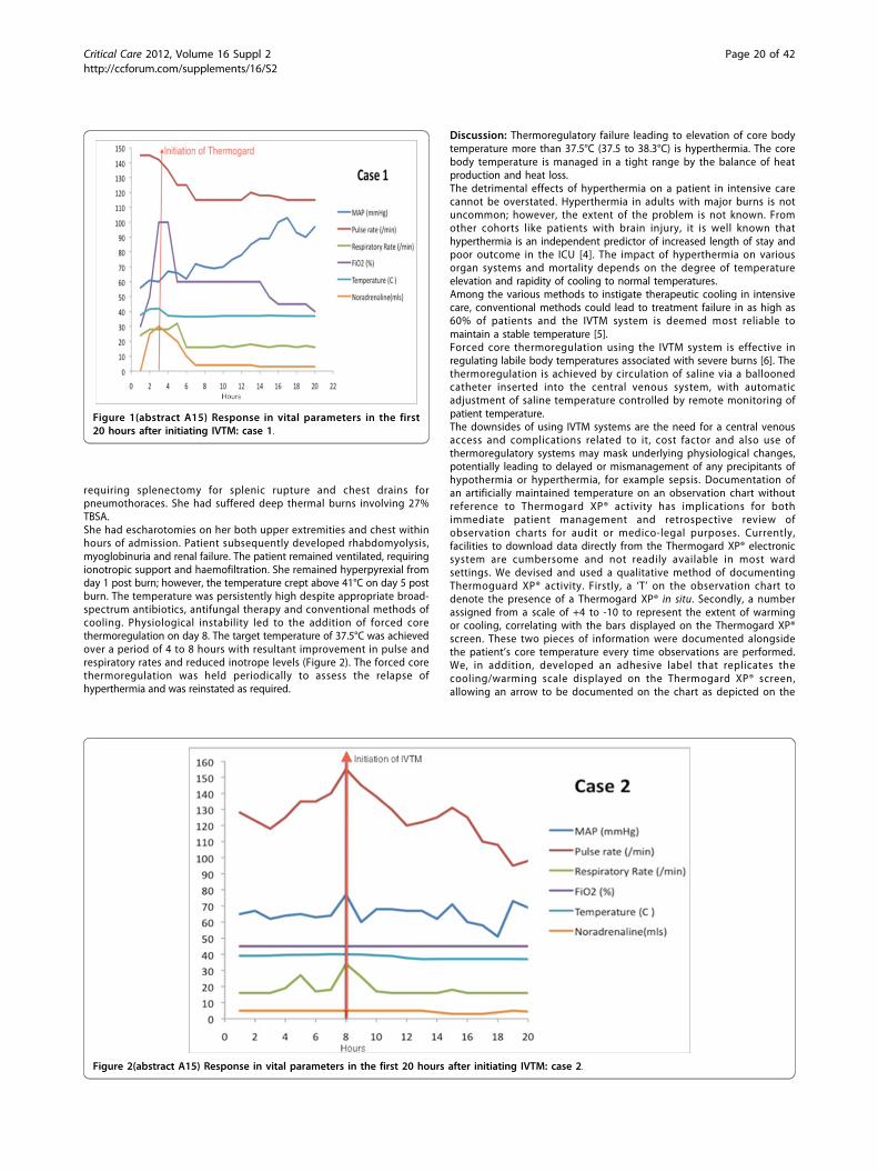
requiring splenectomy for splenic rupture and chest drains forpneumothoraces. She had suffered deep thermal burns involving 27%TBSA.She had escharotomies on her both upper extremities and chest withinhours of admission. Patient subsequently developed rhabdomyolysis,myoglobinuria and renal failure. The patient remained ventilated, requiringionotropic support and haemofiltration. She remained hyperpyrexial fromday 1 post burn; however, the temperature crept above 41°C on day 5 postburn. The temperature was persistently high despite appropriate broad-spectrum antibiotics, antifungal therapy and conventional methods ofcooling. Physiological instability led to the addition of forced corethermoregulation on day 8. The target temperature of 37.5°C was achievedover a period of 4 to 8 hours with resultant improvement in pulse andrespiratory rates and reduced inotrope levels (Figure 2). The forced corethermoregulation was held periodically to assess the relapse ofhyperthermia and was reinstated as required.
Discussion: Thermoregulatory failure leading to elevation of core bodytemperature more than 37.5°C (37.5 to 38.3°C) is hyperthermia. The corebody temperature is managed in a tight range by the balance of heatproduction and heat loss.The detrimental effects of hyperthermia on a patient in intensive carecannot be overstated. Hyperthermia in adults with major burns is notuncommon; however, the extent of the problem is not known. Fromother cohorts like patients with brain injury, it is well known thathyperthermia is an independent predictor of increased length of stay andpoor outcome in the ICU [4]. The impact of hyperthermia on variousorgan systems and mortality depends on the degree of temperatureelevation and rapidity of cooling to normal temperatures.Among the various methods to instigate therapeutic cooling in intensivecare, conventional methods could lead to treatment failure in as high as60% of patients and the IVTM system is deemed most reliable tomaintain a stable temperature [5].Forced core thermoregulation using the IVTM system is effective inregulating labile body temperatures associated with severe burns [6]. Thethermoregulation is achieved by circulation of saline via a balloonedcatheter inserted into the central venous system, with automaticadjustment of saline temperature controlled by remote monitoring ofpatient temperature.The downsides of using IVTM systems are the need for a central venousaccess and complications related to it, cost factor and also use ofthermoregulatory systems may mask underlying physiological changes,potentially leading to delayed or mismanagement of any precipitants ofhypothermia or hyperthermia, for example sepsis. Documentation ofan artificially maintained temperature on an observation chart withoutreference to Thermogard XP® activity has implications for bothimmediate patient management and retrospective review ofobservation charts for audit or medico-legal purposes. Currently,facilities to download data directly from the Thermogard XP® electronicsystem are cumbersome and not readily available in most wardsettings. We devised and used a qualitative method of documentingThermoguard XP® activity. Firstly, a ‘T’ on the observation chart todenote the presence of a Thermogard XP® in situ. Secondly, a numberassigned from a scale of +4 to -10 to represent the extent of warmingor cooling, correlating with the bars displayed on the Thermogard XP®screen. These two pieces of information were documented alongsidethe patient’s core temperature every time observations are performed.We, in addition, developed an adhesive label that replicates thecooling/warming scale displayed on the Thermogard XP® screen,allowing an arrow to be documented on the chart as depicted on the
Figure 1(abstract A15) Response in vital parameters in the first20 hours after initiating IVTM: case 1.
Figure 2(abstract A15) Response in vital parameters in the first 20 hours after initiating IVTM: case 2.
Critical Care 2012, Volume 16 Suppl 2http://ccforum.com/supplements/16/S2
Page 20 of 42

screen (Figure 3.) It provided a more visual representationof Thermogard XP® activity and was used as an adjunct to the abovenumerical scale. All members of the multidisciplinary team couldobserve our method of documenting trends in core body temperature/Thermoguard XP® activity at a glance. It is reproducible in any unitcaring for critically ill burns patients.Conclusion: The directed response of hyperthermia in febrile andnonfebrile states has physiological merits when considering therequirements of inflammatory mediators and cells. Forced corethermoregulation has aided us in the early management of unstableintensive care patients with refractory hyperthermia.The use of an IVTMsystem and subsequent documentation of artificially maintainedtemperature can be misleading and has implications for patientmanagement and retrospective review.References1. Gunn SW, Dilbo SA: Metabolic implications of severe burn injuries and
their management - a systematic review of the literature. World J Surg2008, 32:1857.
2. Lepock JR: Cellular effects of hyperthermia: relevance to the minimumdose for thermal damage. Int J Hyperthermia 2003, 19:252-266.
3. Greenhalgh DG, Saffle JR, Holmes JH, et al: American Burn Associationconsensus con ference to define sepsis and infection in burns. J BurnCare Res 2007, 28:776-790.
4. Diringer MN, Reaven NL, Funk SE, Uman GC: Elevated body temperatureindependently contributes to increased length of stay in neurologicintensive care unit patients. Crit Care Med 2004, 32:1489-1495.
5. Hoedemaekers CW, Ezzahti M, Gerritsen A, van der Hoeven JG: Comparisonof cooling methods to induce and maintain normo- and hypothermia inintensive care unit patients: a prospective intervention study. Crit Care2007, 11:R91.
6. Mavrogordato AE, Wagstaff MJD, Fletcher AJP, Wilson DI, Jayamaha JEL: Anovel method to treat hyperthermia in a burns case: use of a catheter-based heat exchange system. Burns 2009, 35:141-145.
A16Hypothermia after aneurysmal subarachnoid hemorrhageMartin Seule*, Emanuela KellerNeurointensive Care Unit, Department of Neurosurgery, University HospitalZurich, SwitzerlandCritical Care 2012, 16(Suppl 2):A16
Introduction: Aneurysmal subarachnoid hemorrhage (SAH) is a commonand frequently devastating condition, accounting for 1 to 7% of all strokeswith an incidence of 9.1 per 100,000 [1]. Major advances in SAHmanagement over the past three decades have decreased case fatality by0.8% per year, but it is still 40% and many survivors have long-termdisabilities [2]. The most important and potentially treatable complication isdevelopment of delayed cerebral ischemia (DCI), which can progress tocerebral infarction associated with poor outcome.The pathogenesis of DCI is multifactorial and assumed to be initiated inthe early phase of SAH [3]. The onset of SAH is characterized by a short-lasting and cerebral perfusion pressure (CPP)-dependent decrease incerebral blood flow (CBF) leading to global cerebral ischemia [4].Elevated intracranial pressure (ICP) and acute cerebral ischemia are themain factors for early disruption of the blood-brain barrier as well asimpairment of autoregulation associated with brain edema and brainswelling. Important pathogenic mechanisms of CPP-independenthypoperfusion include acute vasoconstriction, cortical spreadingischemia, and activation of the inflammatory response. The release ofoxyhemoglobin and endothelin-1 (ET-1) are the key factors for corticalspreading ischemia, reduced nitric oxide (NO) availability, and secondarycytotoxic edema formation. Cerebral vasospasm (CVS) is a delayedmorphological narrowing of cerebral arteries, occurring 4 to 10 daysafter SAH. Although CVS have been associated with DCI, it is generallyaccepted that CVS is not solely responsible for DCI [5]. In fact, DCImay occur in the absence of CVS and vice versa and the distribution of
Figure 3(abstract A15) Adhesive label that replicates the cooling/warming scale displayed on the Thermogard XP® screen, allowing an arrowto be documented on the chart.
Critical Care 2012, Volume 16 Suppl 2http://ccforum.com/supplements/16/S2
Page 21 of 42

CVS may fail to reliably predict the subsequent pattern of cerebralinfarction [6].Neuroprotective strategies to prevent DCI have been mainly focused ontreatment of CVS, but despite extensive research, effective and/or causativeprophylaxis and treatment are not available [7]. So far, oral nimodipine is theonly drug that can reduce the incidence of DCI and poor outcome, but thereis no beneficial effect on CVS [8]. Hypothermia (HT) treatment exertingnumerous protective effects such as a decrease in cerebral metabolism [9],stabilization of the blood-brain barrier [10], reduction of cerebral edema [11],suppression of excitatory neurotransmitter concentrations [12] andinflammatory reactions [13] seems to be well suited as a neuroprotectivestrategy. In the following, the clinical application of HT after SAH ispresented by reviewing the existing literature.Hypothermia during aneurysm surgery: In the past, promising studieson intraoperative HT during aneurysm surgery as an attempt to reduceischemic injury have been published [14-18]. The IntraoperativeHypothermia for Aneurysm Surgery Trial (IHAST) applied HT in a randomizedstudy in 1,001 patients with good-grade SAH (WFNS 1 to 3); however, itfound no improvement in neurological outcome 3 months after surgery[19]. Post-hoc analysis demonstrated no difference in the incidence ofcognitive impairment between hypothermic and normothermic groups [20].Furthermore, there was no evidence for the benefit of intraoperative HT on24-hour and 3-month outcome in patients who underwent temporaryclipping [21]. It has to be noted that these results only apply to good-gradeSAH patients and may not be extrapolated to the general SAH population.This suggests that only a carefully selected subgroup of patients, withspecific complications induced by SAH, may benefit from HT treatment at aparticular time and for certain duration.Hypothermia in patients with poor-grade subarachnoid hemorrhage:Several experimental studies have demonstrated that HT is effective inminimizing neuronal damage, if induced before or early after the SAH.Kawamura and colleagues found a reduced expression of c-jun and hsp-70mRNA, indicating a reduced stress response that may otherwise manifestas necrosis or apoptosis [22]. Thome and colleagues and Schubert andcolleagues demonstrated that early induction of HT, up to 60 minutes afterSAH, reversed CPP-independent hypoperfusion and brain edema formation,preserved cerebral autoregulation, and reduced accumulation of lactateand glutamate [11,12,23]. On the other hand, delayed induction of HT, upto 180 minutes after SAH, failed to reduce brain edema formation, thusindicating a limited time window of HT application [24]. However, HT waseffective in reducing ICP and associated with improved neurologicalrecovery. These studies encourage early induction of HT in poor-grade SAHpatients, for example, as soon as signs of brain swelling are seen on CTscans.So far, only few retrospective case series have investigated the effect of mildHT during the acute phase of SAH. Table 1 presents an overview of thesestudies applying HT within 24 hours after SAH to patients with poor-gradeSAH (WFNS 4 to 5). The duration of HT varied between 2 and 14 days.Overall outcome results were unsatisfactory with a mortality rate of 47.4%and favorable outcome in less than one-quarter of cases. Yasui andcolleagues evaluated seven patients admitted within 6 hours from symptomonset and treated with HT for 48 hours [25]. PET studies during HT revealeda reduction in cerebral oxygen metabolism exceeding the decrease in CBF,thus indicating a state of luxury perfusion. However, according to the
Barthel Index only two patients were independent, one was partiallydependent, able to walk without assistance, and more than one-half of thepatients bedridden at 12 months after SAH. Anei and colleagues comparedoutcome results before and after introduction of HT treatment to theirinstitution and found no significant difference in the mortality rate with56.3% and 57.9%, respectively [26]. The authors noted that post-HT fevercan be a serious complication resulting in brain swelling and unfavorableoutcome. In contrast to these studies, Gasser and colleagues showed morepromising outcome results in 21 patients with poor-grade SAH (WFNS 4 to5) and induction of HT after developing intracranial hypertension (>15mmHg) that was refractory to conventional treatment. HT was induced onaverage 4.2 ± 3.3 days after SAH and maintained for 4.3 ± 3.9 days.Prolonged HT (>72 hours) was associated with an increased risk of systemiccomplications, but 10 patients (47.6%) showed favorable outcome (GOS 4 to5) and five patients died (23.8%).Hypothermia in patients with delayed cerebral ischemia/cerebralvasospasm: Recently, a SAH-CVS model in dogs demonstrated that HTcan attenuate the degree of CVS up to 14 days after SAH, possibly byregulating the levels of ET-1 and NO [27]. The duration of HT was directlyproportional to the duration of relieving CVS. Table 2 presents anoverview of clinical studies applying HT to patients with symptomaticCVS leading to DCI. Nagao and colleagues treated five patients withgood-grade SAH (H&H I to III), starting HT either during delayedaneurysm clipping or if CVS was refractory to hyperdynamic andendovascular therapy [28]. Four patients survived with favorable outcomeand one patient was severely disabled. In a follow-up study, Nagao andcolleagues included eight patients with good-grade SAH (H&H II to III),and seven patients had favorable outcome and one survived severelydisabled [29]. According to SPECT studies, HT was associated withdecreased CBF levels in all patients. Nakamura and colleagues reported areduction in arterio-jugular oxygen difference (AVDO2) during HT in fivepatients (H&H III to IV), thus indicating a reduced metabolic demand [30].All patients received hyperdynamic therapy before and during HT, butoutcome results were unsatisfactory. Possible contributing factors of pooroutcome include a higher grade of SAH and it is unclear whetherendovascular treatment was applied or not. In our series of 100 SAHpatients treated with HT, 28 patients had symptomatic CVS refractory tohypertensive therapy and endovascular treatment [31]. HT was combinedwith barbiturate coma in 23 of 28 patients and maintained until CVSresolved or severe side effects occurred (mean duration 5.7 ± 3.3 days).Although the majority of patients had poor-grade SAH (H&H 4 to 5 in57.1%, Fisher 3 to 4 in 85.7%), favorable outcome (GOS 4 to 5) wasachieved in 57.1%. In patients with intracranial hypertension (>20 mmHg)with and without refractory CVS, favorable outcome was obtained in only25.0% and 26.5%, respectively. Systemic side effects possibly caused fromHT and/or barbiturate coma included pneumonia in 52.0%,thrombocytopenia (<100,000/µl) in 47.0%, septic shock syndrome in40.0%, and acute respiratory distress syndrome in 16.0%. In a subgroupof seven patients with combined HT and barbiturate coma, daily levels ofIL-6, IL-1b, TNFa, and leukocyte count in the cerebrospinal fluid andplasma were quantified [13]. IL-6 levels in the cerebrospinal fluid andsystemic IL-1b levels were significantly lower compared with patientsreceiving barbiturate coma alone (n = 8), thus indicating HT-relatedattenuation of the inflammatory response.
Table 1(Abstract A16) Studies applying hypothermia on day of aneurysm rupture to patients with poor-grade SAH(WFNS 4 to 5 or H&H IV to V)
Study Number ofpatients (n)
Target temperature(°C)
Duration of hypothermia(days)
Favorable outcome*,% (n)
Mortality rate, %(n)
Nagao and colleagues(2000)
9 32 to 34 5 to 14 44.4% (4) 55.6% (5)
Yasui and colleagues(2002)
7 33 to 34 2 42.8% (3)* 0.0% (0)
Nakamura and colleagues(2002)
3 32 to 34 5 to 14 0.0% (0) 66.7% (2)
Anei and colleagues (2010) 19 34 2 5.3% (1) 57.9% (11)
Total 38 21.1% (8) 47.4% (18)
*GOS ≥4 or mRS ≤3.
Critical Care 2012, Volume 16 Suppl 2http://ccforum.com/supplements/16/S2
Page 22 of 42

Conclusion: So far, the evidence of HT on improved outcome after SAHis limited. Intraoperative HT has been abandoned based on therandomized Intraoperative Hypothermia Study on Aneurysm Surgery ingood-grade SAH patients. The available data suggest that HT mayimprove outcome in a carefully selected subgroup of patients developingintracranial hypertension and/or symptomatic CVS that are refractory toconventional treatment. Further evaluation of cerebral hemodynamicsand oxygenation during HT treatment is required to obtain importantinsights in the effects of HT and to identify patients who may benefitmost.References1. Feigin VL, Lawes CM, Bennett DA, Anderson CS: Stroke epidemiology: a
review of population-based studies of incidence, prevalence, and case-fatality in the late 20th century. Lancet Neurol 2003, 2:43-53.
2. Nieuwkamp DJ, Setz LE, Algra A, Linn FH, de Rooij NK, Rinkel GJ: Changesin case fatality of aneurysmal subarachnoid haemorrhage over time,according to age, sex, and region: a meta-analysis. Lancet Neurol 2009,8:635-642.
3. Ostrowski RP, Colohan AR, Zhang JH: Molecular mechanisms of earlybrain injury after subarachnoid hemorrhage. Neurol Res 2006,28:399-414.
4. Bederson JB, Germano IM, Guarino L: Cortical blood flow and cerebralperfusion pressure in a new noncraniotomy model of subarachnoidhemorrhage in the rat. Stroke 1995, 26:1086-1091, discussion 1091-1092.
5. Vergouwen MD, Ilodigwe D, Macdonald RL: Cerebral infarction aftersubarachnoid hemorrhage contributes to poor outcome by vasospasm-dependent and -independent effects. Stroke 2011, 42:924-929.
6. Rabinstein AA, Weigand S, Atkinson JL, Wijdicks EF: Patterns of cerebralinfarction in aneurysmal subarachnoid hemorrhage. Stroke 2005,36:992-997.
7. Muroi C, Seule M, Mishima K, Keller E: Novel treatments for vasospasmafter subarachnoid hemorrhage. Curr Opin Crit Care 2012, 18:119-126.
8. Castanares-Zapatero D, Hantson P: Pharmacological treatment of delayedcerebral ischemia and vasospasm in subarachnoid hemorrhage. AnnIntensive Care 2011, 1:12.
9. Erecinska M, Thoresen M, Silver IA: Effects of hypothermia on energymetabolism in mammalian central nervous system. J Cereb Blood FlowMetab 2003, 23:513-530.
10. Karibe H, Zarow GJ, Graham SH, Weinstein PR: Mild intraischemichypothermia reduces postischemic hyperperfusion, delayedpostischemic hypoperfusion, blood-brain barrier disruption, brainedema, and neuronal damage volume after temporary focal cerebralischemia in rats. J Cereb Blood Flow Metab 1994, 14:620-627.
11. Schubert GA, Poli S, Schilling L, Heiland S, Thome C: Hypothermia reducescytotoxic edema and metabolic alterations during the acute phase ofmassive SAH: a diffusion-weighted imaging and spectroscopy study inrats. J Neurotrauma 2008, 25:841-852.
12. Schubert GA, Poli S, Mendelowitsch A, Schilling L, Thome C: Hypothermiareduces early hypoperfusion and metabolic alterations during the acutephase of massive subarachnoid hemorrhage: a laser-Doppler-flowmetryand microdialysis study in rats. J Neurotrauma 2008, 25:539-548.
13. Muroi C, Frei K, El Beltagy M, Cesnulis E, Yonekawa Y, Keller E: Combinedtherapeutic hypothermia and barbiturate coma reduces interleukin-6 in
the cerebrospinal fluid after aneurysmal subarachnoid hemorrhage.J Neurosurg Anesthesiol 2008, 20:193-198.
14. Hindman BJ, Todd MM, Gelb AW, Loftus CM, Craen RA, Schubert A,Mahla ME, Torner JC: Mild hypothermia as a protective therapy duringintracranial aneurysm surgery: a randomized prospective pilot trial.Neurosurgery 1999, 44:23-32, discussion 32-33.
15. Karibe H, Sato K, Shimizu H, Tominaga T, Koshu K, Yoshimoto T:Intraoperative mild hypothermia ameliorates postoperative cerebralblood flow impairment in patients with aneurysmal subarachnoidhemorrhage. Neurosurgery 2000, 47:594-599, discussion 599-601.
16. Ogilvy CS, Carter BS, Kaplan S, Rich C, Crowell RM: Temporary vesselocclusion for aneurysm surgery: risk factors for stroke in patientsprotected by induced hypothermia and hypertension and intravenousmannitol administration. J Neurosurg 1996, 84:785-791.
17. Sato K, Yoshimoto T: Systemic and cerebral haemodynamics duringcraniotomy under mild hypothermia in patients with acutesubarachnoid haemorrhage. Acta Neurochir (Wien) 2000, 142:1013-1019,discussion 1019-1020.
18. Kimme P, Fridrikssen S, Engdahl O, Hillman J, Vegfors M, Sjoberg F:Moderate hypothermia for 359 operations to clip cerebral aneurysms. BrJ Anaesth 2004, 93:343-347.
19. Todd MM, Hindman BJ, Clarke WR, Torner JC: Mild intraoperativehypothermia during surgery for intracranial aneurysm. N Engl J Med2005, 352:135-145.
20. Anderson SW, Todd MM, Hindman BJ, Clarke WR, Torner JC, Tranel D,Yoo B, Weeks J, Manzel KW, Samra S: Effects of intraoperativehypothermia on neuropsychological outcomes after intracranialaneurysm surgery. Ann Neurol 2006, 60:518-527.
21. Hindman BJ, Bayman EO, Pfisterer WK, Torner JC, Todd MM: No associationbetween intraoperative hypothermia or supplemental protective drugand neurologic outcomes in patients undergoing temporary clippingduring cerebral aneurysm surgery: findings from the IntraoperativeHypothermia for Aneurysm Surgery Trial. Anesthesiology 2010, 112:86-101.
22. Kawamura Y, Yamada K, Masago A, Katano H, Matsumoto T, Mase M:Hypothermia modulates induction of hsp70 and c-jun mRNA in the ratbrain after subarachnoid hemorrhage. J Neurotrauma 2000, 17:243-250.
23. Thome C, Schubert G, Piepgras A, Elste V, Schilling L, Schmiedek P:Hypothermia reduces acute vasospasm following SAH in rats. ActaNeurochir Suppl 2001, 77:255-258.
24. Torok E, Klopotowski M, Trabold R, Thal SC, Plesnila N, Scholler K: Mildhypothermia (33°C) reduces intracranial hypertension and improvesfunctional outcome after subarachnoid hemorrhage in rats. Neurosurgery2009, 65:352-359, discussion 359.
25. Yasui N, Kawamura S, Suzuki A, Hadeishi H, Hatazawa J: Role ofhypothermia in the management of severe cases of subarachnoidhemorrhage. Acta Neurochir Suppl 2002, 82:93-98.
26. Anei R, Sakai H, Iihara K, Nagata I: Effectiveness of brain hypothermiatreatment in patients with severe subarachnoid hemorrhage:comparisons at a single facility. Neurol Med Chir (Tokyo) 2010, 50:879-883.
27. Wang ZP, Chen HS, Wang FX: Influence of plasma and cerebrospinal fluidlevels of endothelin-1 and NO in reducing cerebral vasospasm aftersubarachnoid hemorrhage during treatment with mild hypothermia, in adog model. Cell Biochem Biophys 2011, 61:137-143.
Table 2(Abstract A16) Studies applying hypothermia to patients with symptomatic vasospasm refractory toconventional treatment
Study Number ofpatients (n)
Target temperature(°C)
Duration of hypothermia(days)
Favorable outcome*,% (n)
Mortality rate, %(n)
Nagao and colleagues(2000)
5 32 to 34 5 to 14 80.0% (4) 0.0% (0)
Nakamura and colleagues(2002)
5 32 to 34 5 to 14 40.0% (2) 60.0% (3)
Nagao and colleagues(2003)
8 32 to 34 5 to 12 87.5% (7) 0.0% (0)
Seule and colleagues(2009)
28 33 to 34 1 to 16 57.1% (16) 28.6% (8)
Total 46 63.0% (29) 26.1% (12)
Note: *GOS ≥4 or mRS ≤3.
Critical Care 2012, Volume 16 Suppl 2http://ccforum.com/supplements/16/S2
Page 23 of 42

28. Nagao S, Irie K, Kawai N, Kunishio K, Ogawa T, Nakamura T, Okauchi M:Protective effect of mild hypothermia on symptomatic vasospasm: apreliminary report. Acta Neurochir Suppl 2000, 76:547-550.
29. Nagao S, Irie K, Kawai N, Nakamura T, Kunishio K, Matsumoto Y: The use ofmild hypothermia for patients with severe vasospasm: a preliminaryreport. J Clin Neurosci 2003, 10:208-212.
30. Nakamura T, Tatara N, Morisaki K, Kawakita K, Nagao S: Cerebral oxygenmetabolism monitoring under hypothermia for severe subarachnoidhemorrhage: report of eight cases. Acta Neurol Scand 2002, 106:314-318.
31. Seule MA, Muroi C, Mink S, Yonekawa Y, Keller E: Therapeutic hypothermiain patients with aneurysmal subarachnoid hemorrhage, refractoryintracranial hypertension, or cerebral vasospasm. Neurosurgery 2009,64:86-92, discussion 92-93.
A17Hypothermia in the operating theatreAndreas Gruber1*, Wilhelm Behringer2, Engelbert Knosp11Department of Neurosurgery, Medical University of Vienna, Austria;2Department of Emergency Medicine, Medical University of Vienna, AustriaCritical Care 2012, 16(Suppl 2):A17
Background: When addressing neuroprotective effects and possibleindications of therapeutic hypothermia, several scenarios have to bedistinguished. First, therapeutic hypothermia can be classified based on thedepth of cooling from normal body temperature into mild hypothermia(32 to 35°C), moderate hypothermia (28 to 32°C), deep hypothermia (28 to17°C), and profound (<17°C). Second, therapeutic hypothermia can beinitiated prior to the insult, for preservation of tissue during the insult, or forthe reduction of reperfusion injury after an insult. Third, animal studies andclinical trials have addressed the neuroprotective effects of therapeutichypothermia under conditions of both global and focal ischaemia. Forth,additional parameters, for example, rewarming rate, duration of ischaemia,and extent of reperfusion, influence the effects of therapeutic hypothermia.Fifth, different outcome measures have been used to describe the effects ofhypothermia, for example, infarct size, extent of cellular death, andneurologic condition at different time intervals after ischemia. Therefore,whenever discussing procedures involving therapeutic hypothermia, theaforementioned parameters deserve clarification.Cerebral ischaemia: Cerebral ischaemia results from a reduction orcomplete loss of cerebral blood flow (CBF) and lack of cerebral oxygenation,followed by depletion of ATP, dysfunction of ATP-dependent membranepumps and subsequently occurrence of anoxic depolarisation. A largeamount of glutamate is released from the intracellular space into theextracellular space, causing excitotoxic injury by stimulating N-methyl-D-aspartate (NMDA) receptors and triggering calcium influx. Increasedintracellular calcium levels per se amplify injury by increasing calciumpermeability and glutamate release via second messenger mechanisms.These acute cascades lead to necrotic neuronal death by interfering withthe mitochondrial respiratory chain. Ischaemia and reperfusion furtherenhance excitotoxicity by providing oxygen as a substrate for severalenzymatic oxidation reactions, thereby generating products of reactiveoxygen species in large quantities. These free radicals enhance proteinoxidation and lipid membrane disintegration and in conjunction with blood-brain barrier (BBB) disruption further contribute to ischaemic necrosis.Apoptosis also occurs in cerebral ischemia, with antiapoptotic proteins beingselectively upregulated in surviving neurons and proapoptotic proteinsbeing highly expressed in dying cells.Hypothermia in cerebral ischaemia: The first controlled attempts to coolthe human brain were undertaken by the neurosurgeon Temple Fay in 1938[1]. Irrigating the brain directly with ice water and sometimes achieving solidparenchymal freezing, he claimed ‘extremely gratifying results’ in a paper on‘local and generalized refrigeration of the human brain’. Over time, manymechanisms have been proposed regarding the neuroprotective effect ofhypothermia. First, hypothermia results in a temperature-dependentdecrease of oxygen and glucose metabolism; that is, a 10°C decrease intemperature reduces ATP consumption and the cerebral metabolic rate(CMR) of oxygen, glucose, and lactate twofold to fourfold [2]. Second, intra-ischaemic hypothermia exerts inhibitory effects on many of the detrimentalischaemic cascades, thereby retarding the initial ATP depletion, preservingmetabolic stores, delaying anoxic depolarisation, reducing ischaemia-induced excitotoxic neurotransmitter release and intracellular calcium levels,
changing glutamate receptor regulation, and limiting BBB breakdown. Bustoand coworkers in 1987 reported that even 1 to 2°C temperature reductionswere sufficient to protect against experimental ischemic stroke [3], therebydemonstrating that the aforementioned mechanisms can exceed the effectsof temperature-induced reductions in CMR, and in turn providing thepathophysiologic foundation for mild and moderate therapeutichypothermia in the management of cerebral ischaemia.Indications for therapeutic hypothermia: The importance of therapeutichypothermia has recently been emphasized by randomised trials in patientswith global cerebral ischaemia from out-of-hospital cardiac arrest (OHCA) [4]and in neonates with perinatal hypoxic-ischaemic encephalopathy (HIE) [5].Clinical experience with patients suffering traumatic brain injuries (TBI) andischaemic strokes are not as convincing, although mild therapeutichypothermia was sufficient to control intracranial hypertension in thispopulation.The use of therapeutic hypothermia in today’s OR theatres differssignificantly from the scenarios outlined in the context of OHCA, HIE, TBI,or stroke. First, in the OR theatre hypothermia is induced beforehand inexpectation of a severe cerebral ischaemic challenge caused by thesurgical procedure. Second, underexperienced neuro-anaesthesiologist andcardio-anaesthesiologic management, highly invasive procedures includingdeep hypothermic cardiac arrest (DHCA) and selective cerebral perfusioncan be employed. In other words, whereas outside the OR mild tomoderate hypothermia is used to treat patients in the post-ischaemicperiod, deep intra-ischaemic hypothermia can be used in cardiothoracicand neurosurgery for the management of congenital heart disease,thoracic aneurysms, and intracranial aneurysms; that is, interventionsprovoking global cerebral ischaemia and otherwise resulting in devastatingintraoperative strokes. Moreover, mild hypothermia is used duringtemporary parent artery clipping in cerebral aneurysm surgery, closelyresembling a state of mild intra-ischaemic hypothermia in a condition oftransient focal cerebral ischaemia.DHCA in global cerebral ischaemia: Although DHCA is being used inboth cardiothoracic and neurosurgery and usually involves the sameprinciples of extracorporeal circulation under cardiopulmonary bypass(CPB), the rationale behind these procedures is very different.Hypothermic CPB and DHCA are established strategies of cerebroprotectionduring cardiothoracic surgery. Cerebral circulatory standstill is an undesiredbyproduct of this procedure and limits the possible safe duration of surgery.To overcome this problem, methods of retrograde and antegrade cerebralperfusion have been established [6]. Retrograde cerebral perfusion (RCP)was initially considered to extend the safe operative time by both, backwardperfusion of the brain via the superior cava vein at pressures of 20 to30 mmHg, and selective cooling of the brain parenchyma. It has becomeevident that RCP indeed provides inadequate cerebral perfusion and exertsneuroprotective actions mainly by providing additional cerebral cooling. Incontrast, antegrade cerebral perfusion (ACP) is obtained by intermittentinfusion of cooled blood directly into cerebral arteries. In detail, selectiveACP allows bihemispheric perfusion through direct cannulation of at leasttwo aortic arch vessels, whereas nonselective, hemispheric ACP uses theaxillary canula that is used for systemic perfusion as a route for ACP. Fromthe neurosurgical standpoint, not surprisingly, insufficient crossflow acrossthe communicating arteries at the circle of Willis with inadequate perfusionof the contralateral hemisphere has been identified as an importantlimitation for nonselective ACP. Although a final cooling temperature of20°C or below with a long cooling time and gradual rewarming arecommonly advocated, promising results have been reported by somegroups using selective ACP with moderate hypothermia [7].From the neurosurgical standpoint, the most important information from thecardiothoracic surgical experience is the recommendation that the DHCAtime in sole application should not exceed 20 to 25 minutes and in everycase with expected DHCA time >25 minutes, ACP or RCP supplement shouldbe performed. In the field of neurosurgery, DHCA combines advancedcerebroprotection with optimal surgical conditions; that is, a blood-free no-flow surgical field and a collapsed aneurysm dome. Since the surgicalprocedure requires cerebral exsanguination for rapid aneurysm repair, ACPand RCP cannot be employed to dilate the time of DHCA, limiting the safeno-flow period to 20 to 30 minutes.Management algorithms for intracranial aneurysm surgery under DHCAhave been published previously [8-11]. Such advanced cerebrovascularprocedures require multimodality neuromonitoring and are performedunder barbiturate-induced EEG burst suppression. The decision to use
Critical Care 2012, Volume 16 Suppl 2http://ccforum.com/supplements/16/S2
Page 24 of 42

DHCA is usually made intraopertively; that is, hypothermia and cardiacstandstill are employed only after microsurgical exploration has proventhat safe aneurysm repair is impossible without these adjunctivemeasures. In this case, brain retractor placement is adjusted, avoidingfurther retractor repositioning - with the risk of contusional parenchymalhaemorrhage - after systemic heparinisation. Thereafter femoro-femoralpercutaneous cannulation is performed and after systemic heparinisation(300 to 400 IU/kg) heart-lung extracorporeal circulation with a heatexchanger and oxygenator is started. Once adequate CPB flow is achieved,systemic hypothermia is induced. Cooling during extracorporeal circulationis continued until a desired brain temperature of 14 to 18°C is reached.Hypothermia results in ventricular fibrillation below 28°C and circulatoryarrest at 18 to 22°C. At this point CPB is stopped and blood is activelydrained into the venous reservoir, rendering the operative field blood freeand the aneurysm collapsed. The duration of this circulatory standstill islimited to the duration of surgical aneurysm repair. In the largest reportedseries [9], the mean duration of circulatory arrest was 21.8 minutes (range2 to 72 minutes) with a mean temperature during circulatory arrest of17.2°C (range 12 to 20°C). The decompressed aneurysm can now bedissected circumferentially, adjacent vital perforating branches can beseparated from the aneurysm fundus, and in partially thrombosed lesionsthe sac can be opened and evacuated. Some centres use short butrepetitive periods of circulatory standstill followed by periods ofreperfusion, thereby reducing the length of a single ischaemic period andchecking for bleedings in the operative field [12]. After aneurysm repair,circulation is restored and under slow rewarming the heart starts tofibrillate spontaneously and will either convert to sinus rhythm or willrequire cardioversion. Pharmacologic peripheral vasodilation is used tofacilitate reperfusion, the blood previously drained is reinfused, andheparin is reversed with protamine. Only after reperfusion, rewarming, andreversal of heparin is adequate hemostasis possible in the operative field.DHCA was used to treat intracranial aneurysms in the 1960s, butshortcomings of CPB and hypothermia-induced coagulopathies resulted inoperative morbidity and declining use of the procedure. In the late 1980s,refined microsurgical and anaesthesiologic techniques allowed the use ofDHCA for the management of most difficult intracranial aneurysms inselected neurosurgical centres of excellence [8-11]. With the development ofnew diagnostic and therapeutic strategies, however, DHCA is only rarelyneeded for the management of cerebral aneurysms today. Possibleexplanations include the following. First, easy access to improvedneuroradiologic imaging techniques has resulted in the detection ofcerebral aneurysms at an earlier stage; that is, before they can grow to giantsize requiring the aforementioned surgical procedures. Second,endovascular treatment, for example, flow diverter implantation [13] andtechniques of flow reversal, has become an alternative to surgery andshould in some instances be considered the first-line treatment; for example,complex and giant posterior circulation aneurysms as well as patients withsignificant co-morbidities. Third, techniques of cerebral revascularisation -that is, parent artery occlusion under bypass protection - have proven highlyeffective in the management of complex aneurysms [14]. Forth, alternativestrategies for intraoperative aneurysm decompression have beendiscovered. Adenosine-induced cardiac asystole has emerged as a possiblealternative, resulting in brief repeated periods of cardiac arrest (5 to15 seconds) to facilitate aneurysm clipping [15]. Others have used selectivebrain cooling with extracorporeal femoral to carotid artery perfusion in giantaneurysm surgery [16], a technique differing from DHCA in so far as thepatient’s body core temperature is maintained at 35°C and aneurysmdecompression is achieved by temporary clipping (trapping) of the parentartery under selective brain cooling to 22°C. Another group has reportedsuccessful treatment of a giant basilar artery aneurysm using moderatehypothermia and extracorporeal circulation [12].Even in expert hands, the procedural complication rate of aneurysm surgeryunder DHCA is significantly higher than in ordinary aneurysm surgery. Thelargest published series [9] reports a perioperative mortality of 14% and acombined rate of permanent treatment-related morbidity and mortality of32%. This recent study, however, reported a contemporary populationof patients harbouring complex aneurysms not lending themselves to oneof the aforementioned alternative treatment forms. In view of the otherwiseunfavourable natural course of these aneurysms, the reported 63% goodoutcome rate justifies the procedure in a small subset of highly selectedpatients. For the time being, DHCA should be considered a procedure of lastresort for otherwise untreatable aneurysms.
Intra-ischaemic mild hypothermia in focal cerebral ischaemia: Thesituation of temporary parent artery clipping during cerebral aneurysmsurgery closely resembles animal models of transient focal ischaemia,wherein intra-ischaemic hypothermia was more effective than delayedpost-ischaemic cooling and was more effective in transient ischaemia andreperfusion models [17-23]. The IHAST trial included 1,001 good grade(WFNS grades I to III) subarachnoid haemorrhage (SAH) patientsundergoing aneurysm surgery and randomised them to intraoperativenormothermia (36.5°C) versus mild intraoperative hypothermia (33°C).Surprisingly, intraoperative hypothermia did not improve neurologicoutcome [24]. Theoretically, the group randomised to mild hypothermiashould have enjoyed the cerebroprotective effects of mild intra-ischaemichypothermia intraoperatively, especially during the ischaemic challenge oftemporary clipping. It was subsequently speculated that possibly not allIHAST patients had experienced a significant intraoperative ischaemicchallenge and in turn a subgroup analysis of those 441 patients whoindeed required temporary clipping was performed [25]. Among thesepatients, the duration of focal ischaemia - that is, temporary clipping - wasthe most important determinant of outcome, with those patients requiringtemporary clipping of >20 minutes faring significantly worse. Even in thisselected subpopulation, mild intra-ischaemic hypothermia did notsignificantly alter neurologic outcome after intraoperative focal ischaemia.The IHAST results raise the question of how to document relevantintraoperative focal ischaemic challenges and the beneficial effects ofneuroprotective interventions in future studies. Even after SAH, gentlesurgical manipulation and brief temporary clipping in expert hands wereapparently not adequate ischaemic challenges to resemble animal modelsof temporary focal cerebral ischaemia and reperfusion.References1. Fay T: Early experiences with local and generalized refrigeration of the
human brain. J Neurosurg 1959, 16:239-260.2. Illievich UM, Spiess CK: Hypothermic therapy for the injured brain. Curr
Opin Anaesth 1994, 7:394-400.3. Busto R, Dietrich WD, Globus MY, Valdes I, Scheinberg P, Ginsberg MD: Small
differences in intraischemic brain temperature critically determine theextent of ischemic neuronal injury. J Cereb Blood Flow Metab 1987, 7:729-738.
4. Holzer M, Bernard SA, Hachimi-Idrissi S, Roine RO, Sterz F, Müllner M,Collaborative Group on Induced Hypothermia for Neuroprotection afterCardiac Arrest: Hypothermia for neuroprotection after cardiac arrest:systematic review and individual patient data meta-analysis. Crit CareMed 2005, 33:414-418.
5. Shankaran S, Laptook AR, Poole WK: Whole body hypothermia forneonates with hypoxic ischemic encephalopathy. N Engl J Med 2005,353:1574-1584.
6. Stein LH, Elefteriades JA: Protecting the brain during aortic artery surgery:an enduring debate with unanswered questions. J Cardiothorac VascAnesth 2010, 24:316-321.
7. Minatoya K, Ogino H, Matsuda H: Evolving selective cerebral perfusion foraortic arch replacement: high flow rate with moderate hypothermiccirculattory arrest. Ann Thorac Surg 2008, 86:1827-1831.
8. Mack WJ, Ducruet AF, Angevine PD, Komotar RJ, Shrebnick DB,Edwards NM, Smith CR, Heyer EJ, Monyero L, Connolly ES, Solomon RA:Deep hypothermic circulatory arrest for complex cerebral aneurysms:lessons learned. Neurosurgery 2007, 60:815-827.
9. Ponce FA, Spetzler RF, Han PP, Wait SD, Killory BD, Nakaji P, Zabramski JM:Cardiac standstill for cerebral aneurysms in 103 patients: an update on theexperience at the Barrow Neurological Institute. J Neurosurg 2011,114:877-884.
10. Schebesch KM, Proescholdt M, Ullrich OW, Camboni D, Moritz S,Wiesenack C, Brawanski A: Circulatory arrest and deep hypothermia forthe treatment of complex intracranial aneurysms - results from a singleEuropean center. Acta Neurochir (Wien) 2010, 152:783-792.
11. Spetzler RF, Hadley MN, Rigamonti D, Carter LP, Raudzens PA, Shedd SA,Wilkinson E: Aneurysms of the basilar artery treated with circulatoryarrest, hypothermia, and barbiturate cerebral protection. J Neurosurg1988, 68:868-879.
12. Yamada M, Nishikawa F, et al: Anesthetic management for clipping agiant basilar artery aneurysm with moderate hypothermia,extracorporeal circulation assistance, and propofol infusion. J NeurosurgAnesthesiol 2003, 15:274-277.
13. Szikora I, Berentei Z, Kulcsar Z, Marosfoi M, Vajda ZS, Lee W, Berez A,Nelson PK: Treatment of intracranial aneurysms by functional
Critical Care 2012, Volume 16 Suppl 2http://ccforum.com/supplements/16/S2
Page 25 of 42

reconstruction of the parent artery: the Budapest experience with thepipeline embolization device. AJNR Am J Neuroradiol 2010, 31:1139-1147.
14. Sughrue ME, Saloner D, Rayz VL, Lawton MT: Giant intracranial aneurysms:evolution of management in a contemporary surgical series.Neurosurgery 2011, 69:1261-1271.
15. Bendok BR, Gupta DK, Dahme EJ, Eddleman CS, Adel JG, Sherma AK,Surdell DL, Bebawy JF, Koht A, Batjer HH: Adenosine for temporary flowarrest during intracranial aneurysm surgery: a single center retrospectivereview. Neurosurgery 2011, 69:815-821.
16. Lownie SP, Menkis AH, Craen RA, Mezon B, MacDonald J, Steinman DA:Extracorporeal femoral to carotid artery perfusion in selective braincooling for a giant aneurysm. J Neurosurg 2004, 100:343-347.
17. Andres RH, Steinberg GK, Guzman R: Intraoperative hypothermia duringvascular neurosurgical procedures. Neurosurg Focus 2009, 26:1-6.
18. Mackensen GB, McDonagh DL, Warner DS: Perioperative hypothermia: useand therapeutic implications. J Neurotrauma 2009, 26:342-358.
19. Huh PW, Belayev L, Zhao W, Koch S, Busto R, Ginsberg MD: Comparativeneuroprotective effects of prolonged moderate intraischemic andpostischemic hypothermia in focal cerebral ischemia. J Neurosurg 2000,92:91-99.
20. Baker CJ, Fiore AJ, Frazzini VI, Choudhri TF, Zubay GP, Solomon RA:Intraischemic hypothermia decreases the release of glutamate in thecores of permanent focal cerebral infarcts. Neurosurgery 1995,36:994-1002.
21. Kader A, Brisman MH, Maraire N, Huh JT, Solomon RA: The effect of mildhypothermia on permanent focal ischemia in the rat. Neurosurgery 1992,31:1056-1061.
22. Goto Y, Kassell NF, Hiramatsu KI, Soleau SW, Lee KS: Effects ofintraischemic hypothermia on cerebral damage in a model of reversiblefocal ischemia. Neurosurgery 1993, 32:980-985.
23. Maier CM, Sun GH, Kunis D, Yenari MA, Steinberg GK: Delayed inductionand long term effects of mild hypothermia in a focal model of transientcerebral ischemia: neurological outcome and infarct size. J Neurosurg2001, 94:90-96.
24. Todd MM, Hindman BJ, Clarke WR, Torner JC, Intraoperative Hypothermiafor Aneurysm Surgery Trial: Mild intraoperative hypothermia duringsurgery for intracranial aneurysms. N Engl J Med 2005, 352:135-145.
25. Hindman BJ, Bayman EO, Pfisterer W, Torner JC, Todd MM, IHASTinvestigators: No association between intraoperative hypothermia orsupplemental protective drug and neurologic outcomes in patientsundergoing temporary clipping during cerebral aneurysm surgery.Anesthesiology 2010, 112:86-101.
A18Temperature management in central nervous infectionErich Schmutzhard*, Peter Lackner, Ronny Beer, Marlene Fischer,Anelia Dietmann, Bettina PfauslerDepartment of Neurology, Neurocritical Care Unit, Medical UniversityHospital Innsbruck, AustriaCritical Care 2012, 16(Suppl 2):A18
Introduction: Brian Kellock quotes in his book The Fibreman the seven-year-old (that is, in the year 1918) Denis Burkitt discussing respectfully hisuncle Dr Roland Burkitt’s legendary reputation amongst his patients inKenya, Africa, as: ‘... he believed in curing fevers, in particular in deliriouspatients, by artificially lowering the patients’ temperature ...’ [1].Obviously, Dr Roland Burkitt, the uncle of the legendary Prof. Dr DenisBurkitt, after whom the African lymphoma and its association with Epstein-Barr virus was named, had realized that lowering the body temperature inpatients with high fevers, in Kenya highly likely to being due to cerebralmalaria, meningitis or encephalitis, might influence positively the course ofsuch potentially - at this time, in many instances, definitely - life-threatening disease [1]. Zdravev stipulated that otogenous brain abscessesnot only need to be surgically evacuated but, when causing cerebralherniation, may benefit from lowering body temperature. He was the firstto conclude that high body temperature in patients with increasedintracranial pressure may be a deleterious association [2].The wide variety of mechanisms of injury that are exaggerated byhyperthermia and may be ameliorated by moderate hypothermia aredescribed elsewhere in this supplement (see abstract A1). They includemechanisms of neuroexcitotoxicity [3], release of free radicals, changes in
blood-brain barrier and vascular permeability, the release ofproinflammatory mediators, drawing leucocytes across the blood-brainbarrier, increasing the number of inflammatory cells in the brain tissue andthe passage of neutrophils, phagocytes, monocytes and macrophages intothe brain, additionally injuring neuronal cells by stimulating furtherimmune reactions [4]. Whether interfering with these mechanisms bymoderate hypothermia may reduce secondary insult onto neuronal cellsand brain tissue is still a matter of discussion. What has been known forlong time is that increased intracranial pressure, as frequently seen in viralencephalitis, severe bacterial meningitis and/or brain abscesses, may bemodified by therapeutic hypothermia/targeted temperature management[5]. In addition, long-term morbidity and mortality in CNS infection mayadditionally deteriorate if bacterial meningitis is complicated by cerebralinfarction, occurring during the course of disease [6]. Thus, preventingcerebral infarction may be an important tool in reducing both morbidityand mortality in adults with community-acquired bacterial meningitis.Prandini and colleagues have shown that mild hypothermia reducesremarkably polymorphonuclear leucocyte infiltration in induced braininflammation in rats compared with those without hypothermia [4].After any type of brain injury, in particular, ischemic and ischemiareperfusion injury, the reduced brain oxygen supply quickly leads to adecrease in ATP and phosphocreatine levels, thus initiating a complexcascade of events involving excessive calcium influx into brain cells,excessive glutamate receptor activation and neuronal hyperexcitability;that is, triggering off the excitotoxic cascade [3]. Exactly this excitotoxiccascade may be responsible for provoking overt or subtle epilepticseizures in patients with CNS infection, thereby deteriorating the prognosisof these patients. Irazuzta and colleagues could nicely show that in rabbitshypothermia decreases excitatory neurotransmitter release in bacterialmeningitis, in particular, the release of glutamate and aspartate, known tobe involved in the pathogenesis of neuronal injury in meningitis,suggesting that hypothermia may attenuate excess neuronal stress in thisdisease [3].Febrile refractory status epilepticus may be caused by presumedencephalitis carrying a very high morbidity and mortality rate [7]. Inaddition, it is believed that hyperthermia aggravates brain damage due tocontinuing epileptic activity. In a retrospective analysis Nakagawa andcolleagues found a significant improvement of outcome in children withsuch febrile refractory status epilepticus who were treated by therapeutichypothermia with subsequent prophylactic normothermia compared withthose children with the same disease but treated only by conventionaltherapy without fever management [7]. The authors conclude thattreatment with therapeutic hypothermia plus subsequent prophylacticnormothermia may reduce neurological damage in such patients withsevere acute encephalopathy due to refractory febrile convulsive statusepilepticus. Another Japanese group treated an adult patient withinfluenza type A virus-associated encephalopathy with routine therapeuticsas oseltamivir and methylprednisolone pulse therapy. Because of severebrain swelling, hypothermia was added. This combination treatment led tofull recovery without neurologic sequelae [8]. A similar observation hasbeen reported in 43 children with acute inflammatory encephalopathyand/or encephalitis, out of whom 27 were treated with mild hypothermia.These 27 children were compared with 16 similar patients who were caredfor at normothermia levels [9]. The effect of therapeutic hypothermia forchildren with acute encephalopathy/encephalitis was observed to bedependent on the timing when cooling was initiated. Early cooling (within12 hours of initial neurological signs of encephalopathy/encephalitis)improved outcome whereas delayed cooling (after 12 hours) tended to beeven deleterious [9].In adults, therapeutic hypothermia in CNS infection has been limited tosingle case reports and case series. Cuthbertson and colleagues reportedinduced hypothermia, together with barbiturate coma, in a meningitispatient associated with intractable raised intracranial pressure. Throughoutthe entire period of mild hypothermia (35°C), achieved by thiopental andexternal cooling, cerebral perfusion pressure was maintained above70 mmHg. Barbiturate-induced coma and induced hypothermia werecontinued for 48 hours when complications (pancreatitis, ventilator-associated pneumonia) prompted one to stop these two therapy modalities.However, the patient remained without any ICP rebound, eventually beingweaned from the ventilator and making a rather uneventful recovery, beingdischarged home with reportedly no neurologic deficits 3 weeks after thedisease had began [5]. Similarly, a case with space occupying herpes
Critical Care 2012, Volume 16 Suppl 2http://ccforum.com/supplements/16/S2
Page 26 of 42

simplex virus encephalitis has been reported by Wagner and colleagues. Theauthors correctly state that conventional intracranial pressure-loweringmodalities are limited in such severe herpes simplex virus encephalitispatients and more aggressive treatment options are needed. They inducedmoderate hypothermia (33°C), resulting in fast and sustained control ofintracranial pressure. Eventually the patient regained a relatively goodfunctional outcome, being able to walk unassisted (GOS 4). It needs to benoted that rewarming was done very slowly (1°C/day); that is, over a periodof 4 days [10].Very recently two larger cases series have been published on the use oftherapeutic hypothermia both in adult viral meningoencephalitis and inadult community-acquired meningitis by the same Croatian group:Therapeutic hypothermia in adult viral meningoencephalitis [11]:Eleven adult patients with viral or presumed viral encephalitis (two TBE, twoHSV, one VZV, six unknown) pathogen were treated with mild hypothermia,rectal temperature aimed to be 32 to 34°C. The authors, however, do notelaborate on rewarming, speed of rewarming and any adverse effects ofrewarming. Remarkably only one patient (1/11 = 9%) died, 3/11 recoveredfully, 2/11 with only minor impairment and 5/11 with moderate to severeresidual neurological deficit. All patients showed severely impaired GCS atadmission (median GCS 8 (range 3 to 10)) and median Acute Physiology andChronic Health Evaluation score (APACHE) being 24 (range 12 to 32).A major drawback of this study is the lack of ICP monitoring, sonographicmeasurement of optic nerve sheath diameter having been used assurrogate markers for ICP, both methods not being as reliable as continuousICP and CPP monitoring. The mortality rate of 9% in this case seriescompared with 29% in patients with viral meningoencephalitis treated intheir unit before implementation of the therapeutic hypothermia protocol.The authors suggest that in carefully selected patients, carefully regardingvasoreactivity status, and, of course, only in those with initially reducedcerebral perfusion pressure, therapeutic hypothermia may be a tool toreduce long-term morbidity and mortality.Therapeutic hypothermia in bacterial meningitis [12]: The sameCroatian group presented a series of 10 patients with severe bacterialmeningitis (nine patients: pneumococci, one patient: Escherichia coli) withan initial median GCS of 6 (range 3 to 9), APACHE II ranging from 22 to34 (median 31). They employed the same protocol of non-invasive ICPmonitoring (using transcranial Doppler sonography, optic nerve sheathdiameter sonography and jugular bulb oximetry). Hypothermia wasinduced by intravenous infusion of cold isotonic saline and maintainedwith continuous venovenous hemofiltration at 32 to 34°C. Nothing is saidabout rewarming, rewarming speed and duration of hypothermia. Twopatients died within 48 hours from admission because of refractoryintracranial hypertension, two more patients with severe residualneurological deficits (GOS 2) died later on, after discharge from the ICU,because of late-onset nosocomial sepsis; in total, a rather high mortalityrate [13,14]. The surviving six patients had a mean ICU stay of 22 days(range 8 to 36), two had a severe and two a moderate residualneurologic deficit. Two of the entire group of 10 patients with bacterialmeningitis had a complete neurological recovery (GOS 5).The authors discuss hypothermia as a neuroprotective measure in severebacterial meningitis. In nine (out of 10) patients pneumococci were isolatedas the causative organisms; however, the authors do not elaborate whetherthese patients - qualifying early for dexamethasone treatment, in particular,since they showed severe clinical signs and symptoms of increasedintracranial pressure - had been given dexamethasone or not. They discussthat corticosteroids have not substantially changed disease outcomesamong patients with most severe pneumococcal meningitis. Exactly thisaspect is in contrast to the widely accepted knowledge and level ofevidence respectively. Recently a meta-analysis has clearly shown thatEuropean patients with pneumococcal bacterial meningitis (aged >55 years)definitely benefit from the adjunctive application of dexamethasone [15].Only one single patient would not have qualified for this definition (47-year-old female being severely immunocompromised). This might explain thehigh mortality rate despite the administration of the adjunctive therapy withmoderate hypothermia. The authors correctly claim that any adjunctivetherapy should be continued beyond the very first hours and days since ithas been known that in severe bacterial meningitis vasculopathy leading toischemic stroke may ensue even days after onset. Adequate maintenance ofsufficient cerebral perfusion pressure until recovery of CO2 reactivity (not tobe expected before day 4 after initiation of antibiotic treatment) is essentialand requires therapeutic hypothermia to be maintained for this period
of time. However, the results of this pilot trial in 10 consecutive patients donot justify one to recommend therapeutic hypothermia as adjunctivetherapy in bacterial (pneumococcal) meningitis. Whether selected patientswith either intractable ICP, dangerously low CPP or infection-associatedrefractory status epilepticus benefit from moderate hypothermia needs tobe elucidated; today this is still a case to case decision.References1. Kellock B: The Fibreman, the Life Story of Dr. Denis Burkitt Lion Publishers;
Oxford Oxfordshire OX2 7DH, UK, 1st 1985, p8.2. Zdravev P: Treatment of cerebral hernia following surgery of an
otogenous brain abscess. Ann Otolaryngol 1951, 68:201-205.3. Irazuzta JE, Olson J, Kiefaber MP, Wong H: Hypothermia decreases
excitatory neurotransmitter release in bacterial meningitis in rabbits.Brain Res 1999, 847:143-148.
4. Prandini MN, Neves Filho A, Lapa AJ, Stavale JN: Mild hypothermiareduces polymorphonuclear leukocytes infiltration in induced braininflammation. Arq Neuropsiquiatr 2005, 63:779-784.
5. Cuthbertson BH, Dickson R, Mackenzie A: Intracranial pressuremeasurement, induced hypothermia and barbiturate coma in meningitisassociated with intractable raised intracranial pressure. Anaesthesia 2004,59:908-911.
6. Schut ES, Lucas MJ, Brouwer MC, et al: Cerebral infarction in adults withbacterial meningitis. Neurocrit Care 2011 in press.
7. Nakagawa T, Fujita K, Saji Y, Maruyama A, Nagase H: Induced hypothermia/normothermia with general anesthesia prevents neurological damage infebrile refractory status epilepticus in children. No To Hattatsu 2011,43:459-463.
8. Sakurai T, Kimura A, Tanaka Y, Hozumi I, Ogura S, Inzuka T: Case of adultinfluenza type A virus-associated encephalopathy successfully treated withprimary multidisciplinary treatments. Rinsho Shinkeigaku 2007, 47:639-643.
9. Kawano G, Iwata O, Iwata S, et al: Determinants of outcomes followingacute child encephalopathy and encephalitis: pivotal effect of early anddelayed cooling. Arch Dis Child 2011, 96:936-941.
10. Wagner I, Staykov D, Volbers B: Therapeutic hypothermia for space-occupying Herpes simplex virus encephalitis. Minerva Anestesiol 2011,77:371-374.
11. Kutlesa M, Barsic B, Lepur D: Therapeutic hypothermia for adult viralmeningoencephalitis. Neurocrit Care 2011, 15:151-155.
12. Lepur D, Kutlesa M, Barsic B: Induced hypothermia in adult community-acquired bacterial meningitis - more than just a possibility? J Infect 2011,62:172-177.
13. Branco RG, Tasker RC: Meningococcal meningitis. Curr Treat Options Neurol2010, 12:464-474.
14. Weisfelt M, van de Beek D, Spanjaard L, Reitsma JB, de Gans J: A risk scorefor unfavorable outcome in adults with bacterial meningitis. Ann Neurol2008, 63:90-97.
15. van de Beek D, Farrar JJ, de Gans J, et al: Adjunctive dexamethasone inbacterial meningitis: a meta-analysis of individual patient data. LancetNeurol 2010, 9:254-263.
A19Complications of hypothermia: infectionsGregor Broessner*, Marlene Fischer, Peter Lackner, Bettina Pfausler,Erich SchmutzhardNeurologic Intensive Care Unit, Medical University Innsbruck, AustriaCritical Care 2012, 16(Suppl 2):A19
Background: Therapeutic hypothermia (TH) is a very elegant way ofinducing short-term and long-term neuroprotection in various diseaseentities. It has become the standard of care after cardiac resuscitationwith an impressive outcome improvement in prospective randomisedtrials. However, by a broader use of this sophisticated measure the criticalcare community has become aware of potential side effects limiting itseffect on patient outcome. Among others, an increased rate of infectionsis observed under therapeutic hypothermia and controlled normothermia.The pathophysiological considerations by which TH increases infectiouscomplications comprise reduced inflammatory response and suppressionof leukocyte migration and phagocytosis. All together, these observationsjustify a high vigilance towards infectious manifestations if temperaturemodulation measures, namely therapeutic hypothermia and controllednormothermia, are used in critical care patients.
Critical Care 2012, Volume 16 Suppl 2http://ccforum.com/supplements/16/S2
Page 27 of 42

Pathophysiological considerations about hypothermia and infections:Intriguing data derived from animal models showing a potent neur-oprotective effect in various disease entities induced by hypothermia gaveway to a broader use of this method in humans. Thus, therapeutichypothermia has become the standard of care after cardiac resuscitation asstudies demonstrated its strong neuroprotective effect and neurologicoutcome improvement [1,2]. It is now recommended by the EuropeanResuscitation Council and the International Liaison Committee onResuscitation in cases of comatose adults with spontaneous circulationafter out-of-hospital cardiac arrest (OHCA) [3]. However, in indications otherthan resuscitation, such promising results could not be achieved inprospective trials shifting the scientific focus on possible side effects of TH[4-6]. Rewarming injury, shivering, electrolyte dysbalance, pharmacologicaland pharmacodynamic alterations, cardiovascular effects includingarrhythmia, insulin resistance and infections have recently been attributedas limitations occurring in a dose-dependent fashion under TH [7]. Takentogether, maximal reduction of these side effects should be a treatmentgoal if dealing with temperature control measures irrespective of the targettemperature. Today, infectious complications are thought to be one ofthe major contributors limiting the effects of hypothermia [7-12]. Thusadvancing diagnostic approach, prevention and treatment of theseinfectious complications is a great concern of the scientific critical carecommunity and need to be addressed in future prospective trials. Invarious studies enrolling patient populations suffering from such differentdiseases as traumatic brain injury, ischaemic stroke or resuscitation postcardiac arrest, an increased rate of infections under TH was observed[4,6,11]. Whether this negative effect has to be attributed to a specificcooling measure remains under debate; however, this hypothesis isunlikely as increased infections are found under both endovascular andsurface cooling measures.The biological interpretation of the pathophysiological backgrounds ischallenging as temperature modulation inhibits various inflammatoryresponses on different levels that are only partly understood today [7,8].Hypothermia impairs the secretion of proinflammatory cytokines andsuppresses leukocyte migration and phagocytosis [7,8]. Recently it hasbeen speculated that hypothermia may induce insulin resistance leadingto hyperglycaemia possibly promoting infection onset [8,13].However, increased rate of infections has also been observed not onlyunder TH but also under endovascularly controlled prophylacticnormothermia in patients with severe cerebrovascular disease [10,14].A significant increase of infectious complications was observed in theendovascular treatment group although TH was strictly avoided.Importantly in this study from our group, analysis of the inflammatoryparameters revealed a significant increase of C-reactive protein (CRP) inthe prophylactic normothermia group whereas procalcitonin (PCT) andwhite blood cell count were not elevated [10]. This is a crucial point as itmight indicate that temperature modulation may influence the prognosticvalue of inflammatory parameters [10].In a retrospective review by Mongardon and coworkers including 421patients being treated after cardiac arrest, in 281 patients (67%) aninfectious complication was diagnosed [11]. Pneumonia was the mostfrequent, followed by bloodstream infections and catheter-related infections[11]. Gram-negative bacteria were the most frequently isolated infectiousgerms, but the main pathogen detected was Staphylococcus aureus [11]. Thehigh rate of reported pneumonia raises the question of whether intubationat an early stage should be considered in patients under TH to minimise therisk of aspiration.Is this surcharge too much and how can we minimise it in clinicalroutine?: Hospital-acquired infections lead to secondary injury inpatients and are responsible for a considerable cost increase especiallyin critical care patients [15,16]. An increased rate of infections undercontrolled normothermia and therapeutic hypothermia has beendescribed in patients suffering from ischaemic stroke, traumatic braininjury, spontaneous subarachnoid haemorrhage, post resuscitation andintracerebral haemorrhage [6,9,10]. If this observation is associated withsignificantly impaired outcome or even mortality is under debate[6,9-11]. However, there is general consensus that infections lead toprolonged ICU treatment, secondary injury and lastly to cost increasewith significant global economic burden [15]. Therefore, the critical carecommunity has to find effective strategies to minimise the risk ofinfection complications.
Temperature modulation by any means has to be combined with astandard operation procedure including: routine microbiologicalsurveillance including blood, urine, respiratory specimen work-up;radiological pneumonia surveillance; daily monitoring of inflammatoryparameters (CRP, PCT, leukocytes); routine check of catheter insertionsites and timely catheter replacement; avoidance of hyperglycaemia(under TH) and hypoglycaemia (while rewarming!); and monitorperformance of cooling device as a high cooling power/rate mightindicate fever even if the body core temperature is normal or even <36°C.Conclusion: These observations justify a high level of vigilance towardsinfectious manifestations if temperature modulation measures, namelytherapeutic hypothermia and controlled normothermia, are used incritical care patients. Whether, at all, the early use of antibiotics in case ofsuspected infections under TH is justified has to be addressed in futureprospective trials.References1. Mild therapeutic hypothermia to improve the neurologic outcome after
cardiac arrest. N Engl J Med 2002, 346:549-556.2. Bernard SA, Gray TW, Buist MD, et al: Treatment of comatose survivors of
out-of-hospital cardiac arrest with induced hypothermia. N Engl J Med2002, 346:557-563.
3. Nolan JP, Morley PT, Vanden Hoek TL, et al: Therapeutic hypothermia aftercardiac arrest: an advisory statement by the advanced life support taskforce of the International Liaison Committee on Resuscitation. Circulation2003, 108:118-121.
4. Clifton GL, Miller ER, Choi SC, et al: Lack of effect of induction ofhypothermia after acute brain injury. N Engl J Med 2001, 344:556-563.
5. Clifton GL, Valadka A, Zygun D, et al: Very early hypothermia induction inpatients with severe brain injury (the National Acute Brain Injury Study:Hypothermia II): a randomised trial. Lancet Neurol 2011, 10:131-139.
6. Hemmen TM, Raman R, Guluma KZ, et al: Intravenous thrombolysis plushypothermia for acute treatment of ischemic stroke (ICTuS-L): finalresults. Stroke 2010, 41:2265-2270.
7. Polderman KH, Herold I: Therapeutic hypothermia and controllednormothermia in the intensive care unit: practical considerations, sideeffects, and cooling methods. Crit Care Med 2009, 37:1101-1120.
8. Polderman KH: Induced hypothermia and fever control for preventionand treatment of neurological injuries. Lancet 2008, 371:1955-1969.
9. Broessner G, Beer R, Lackner P, et al: Prophylactic, endovascularly based,long-term normothermia in ICU patients with severe cerebrovasculardisease: bicenter prospective, randomized trial. Stroke 2009, 40:e657-e665.
10. Broessner G, Lackner P, Fischer M, et al: Influence of prophylactic,endovascularly based normothermia on inflammation in patients withsevere cerebrovascular disease: a prospective, randomized trial. Stroke2010, 41:2969-2972.
11. Mongardon N, Perbet S, Lemiale V, et al: Infectious complications in out-of-hospital cardiac arrest patients in the therapeutic hypothermia era.Crit Care Med 2011, 39:1359-1364.
12. Kamps M, Bisschops LA, van der Hoeven JG, et al: Hypothermia does notincrease the risk of infection: a case control study. Crit Care 2011, 15:R48.
13. van den Berghe G, Wouters P, Weekers F, et al: Intensive insulin therapy incritically ill patients. N Engl J Med 2001, 345:1359-1367.
14. Broessner G, Lackner P, Hoefer C, et al: Influence of red blood celltransfusion on mortality and long-term functional outcome in 292patients with spontaneous subarachnoid hemorrhage. Crit Care Med2009, 37:1886-1892.
15. Farmer JC: Therapeutic hypothermia: is there an unintended surcharge?Crit Care Med 2011, 39:1570-1571.
16. Stone PW, Braccia D, Larson E: Systematic review of economic analyses ofhealth care-associated infections. Am J Infect Control 2005, 33:501-509.
A20Hypothermia and coagulationKees H PoldermanDepartment of Critical Care Medicine, University of Pittsburgh MedicalCenter, Pittsburgh, PA, USACritical Care 2012, 16(Suppl 2):A20
Introduction: The effects of hypothermia on coagulation may represent atwo-edged sword in patients with acute brain injury who are treated with
Critical Care 2012, Volume 16 Suppl 2http://ccforum.com/supplements/16/S2
Page 28 of 42

therapeutic cooling. On the one hand inhibition of coagulation can havepositive effects, such as improvements in the microcirculation andinhibition of the formation of harmful microthrombi in the brain [1]. Onthe other hand this could lead to increased bleeding risk and therebycause harm to patients, especially if they have suffered traumatic injuriesor are actively bleeding for other reasons. This manuscript will brieflydiscuss what is known about the effects of hypothermia on cooling.In vitro and experimental data: The effects of hypothermia oncoagulation have been studied mostly in vitro. Very mild hypothermia(down to 35°C) has no effect on any part of the coagulation cascade.Temperatures below 35°C can in some cases (but not in all patients, seebelow) induce mild platelet dysfunction and sometimes a mild decreasein platelet count. When temperatures drop below 33°C other steps in thecoagulation cascade, such as the synthesis and kinetics of clottingenzymes and plasminogen activator inhibitors, can also be affected [2-8].Recently, Ruzicka and coworkers performed a study in which theyprecisely measured thromboelastography in healthy subjects at atemperature range starting at 38°C all the way down to 12°C [9]. Theyreported that decreasing temperatures led to a progressive delay in theinitiation of thrombus formation, as well as a decrease in the speed ofclot creation and growth. However, significant effects of hypothermia onthis parameter began only at 30°C, progressing rapidly below thistemperature but reaching statistical significance only at 24°C [9]. Theseauthors also found that once clot formation had been completed, thestability of the clot could no longer be influenced by hypothermia; thatis, the clots once formed remained stable regardless of temperature. Ofnote, there was significant interindividual variability in the response ofthe coagulation parameters to cooling [9].Hanke and coworkers [10,11] reported that the anticoagulatory effects ofhypothermia were markedly increased if acidosis was present, that theeffects of hypothermia could be effectively reversed by administeringDDAVP and fibrinogen, but that these drugs worked well only if acidosiswas corrected [10].Finally, a number of animal studies have looked at the effects ofhypothermia on hematoma formation in models for intracranialhemorrhage and subdural hematoma [12-16]. These studies have notfound any evidence for increased hematoma growth or bleeding riskassociated with mild hypothermia; in fact, the opposite effect (decreasedhematoma volume and vascular brain edema) was observed in most ofthese studies [12-16]. Indeed a randomized clinical trial is currently beingorganized to test the safety and efficacy of cooling in patients withintracranial hemorrhage [17].Clinical studies: The clinical effects of mild hypothermia on bleedingappear to be minor, and clinical studies suggest that the risk of severebleeding associated with mild hypothermia is very low or even absent.None of the large studies in cardiac arrest, stroke, or traumatic braininjury have reported significant increase in bleeding risks associated withtherapeutic cooling, although it should be emphasized that activelybleeding patients were excluded from these studies [12].Preliminary data suggest that hypothermia can even be used safely incombination with thrombolytic therapy. Hemmen and colleaguesperformed a prospective controlled clinical trial in 58 patients with acuteischemic stroke, 28 of whom were treated with hypothermia (33°C)combined with thrombolytic therapy [18]; they found that the risk ofhemorrhagic conversion did not increase in patients treated with bothhypothermia and TPA compared to those treated with TPA alone, and infact the risk of symptomatic ICH trended to be lower in cooled patients[18].Spiel and colleagues studied the effect of hypothermia induced by coldfluid infusion in cardiac arrest patients; they reported only slightlyprolonged clotting time as measured by rotation thrombelastography[19].Storm and coworkers compared risk and severity of bleeding in cardiacarrest patients treated with mild hypothermia and thrombolysis tomatched historical patients treated with thrombolysis only [20]. Theyfound that the incidence of bleeding was not increased by hypothermia,although there was a trend towards more red blood cell units beingrequired to reach target hematocrit in hypothermic patients who diddevelop bleeding complications. As neurological outcomes weresignificantly better in patients treated with both thrombolytics andhypothermia, even in those who developed bleeding complications, the
authors concluded that bleeding risks should not be viewed as a reasonto withhold hypothermia treatment [20].Tuma and colleagues performed a retrospective analysis from a traumaregistry database and identified patients who had developed cardiac arrestand had been treated with hypothermia [21]. The number of patients wassmall but they found no increased complication rate from hypothermia, inparticular bleeding, in their patients. This is noteworthy as hypothermiahas gained a negative reputation among those treating multi-traumatizedpatients, and is seen as one of the factors in the ‘lethal triad’ of shock,acidosis and hypothermia [22]. The discrepancy may be explained by theinterplay between acidosis and hypothermia, as explained above; theeffects of (deep) hypothermia on coagulation are pronounced, and difficultto reverse, mainly if there is simultaneous acidosis [22]. However, in theabsence of acidosis the effects of hypothermia are much less pronounced,and far more easily controllable and reversible. Most patients undergoingelective treatment with mild hypothermia will not have severe acidosis,and therefore effects of hypothermia on coagulation will be minimal.These clinical data and the physiological effects of cooling should be takeninto account when decisions are made on whether or not to usehypothermia in patients who are actively bleeding, or who are at high riskfor bleeding. If possible the (potential) source of bleeding should be(surgically) controlled before cooling is initiated. If this is not possible therisks of bleeding should be weighed against the benefits of cooling, andcareful consideration should be given to the depth of hypothermiainduced in that patient. Because very mild hypothermia (35°C) does notaffect coagulation in any patient this temperature can be safely inducedeven in patients with very high bleeding risk. At temperatures below 35°Csome patients may begin to have mild platelet dysfunction (although inmost patients this will occur only at lower temperatures). These effects willbe significantly magnified in the presence of acidosis.In my hospital patients who are admitted after cardiac arrest will usuallybe cooled to 32 or 33°C for a period of 24 hours, followed by slowrewarming over 16 to 24 hours. If such a patient has concomitant severetraumatic injury (for example, blunt abdominal trauma) and/or hasinjuries that pose a high bleeding risk that cannot easily be surgicallycontrolled (for example, liver laceration), this patient typically would becooled to 35°C, and cooling would be continued for 48 or 72 hoursinstead of our current standard of 24 hours. Similarly, if a patientdevelops severe uncontrollable bleeding during hypothermia therapy (forexample, major upper GI bleed with hemodynamic instability) we wouldconsider rewarming this patient to 35°C (or maintaining 33°C and givingplatelet transfusion). In mildly elevated bleeding risk we will cool thepatient to 33°C and will treat with platelet transfusion and DDAVP ifbleeding complications occur (unless there are severe counter-indicationssuch as recent stent placement).Of note, it is unknown whether decreasing the intensity of cooling whileextending the duration of therapy will provide similar benefits as applyingtreatment according to published guidelines [23]. Therefore, of coursethose not at high bleeding risk should be cooled to between 32 and 34°Cas current guidelines recommend [23].Conclusion: Although mild to moderate hypothermia has some effect onthe coagulation system the clinical risk of bleeding associated with coolingappears to be very low. This will, however, increase significantly if thepatient has moderate-to-severe acidosis. No effects of hypothermia oncoagulation occur in any patient as long as temperature is ≥35°C, andpatients at very high bleeding risk can safely be cooled to this temperature.References1. Polderman KH: Mechanisms of action, physiological effects, and
complications of hypothermia. Crit Care Med 2009, 37:S186-S202.2. Michelson AD, MacGregor H, Barnard MR, Kestin AS, Rohrer MJ, Valeri CR:
Hypothermia-induced reversible platelet dysfunction. Thromb Haemost1994, 71:633-640.
3. Watts DD, Trask A, Soeken K, Perdue P, Dols S, Kaufmann C: Hypothermiccoagulopathy in trauma: effect of varying levels of hypothermia onenzyme speed, platelet function, and fibrinolytic activity. J Trauma 1998,44:846-854.
4. Valeri CR, MacGregor H, Cassidy G, Tinney R, Pompei F: Effects oftemperature on bleeding time and clotting time in normal male andfemale volunteers. Crit Care Med 1995, 23:698-704.
5. Patt A, McCroskey B, Moore E: Hypothermia-induced coagulopathies intrauma [review]. Surg Clin North Am 1988, 68:775-785.
Critical Care 2012, Volume 16 Suppl 2http://ccforum.com/supplements/16/S2
Page 29 of 42

6. Ferrara A, MacArthur JD, Wright HK, Modlin IM, McMillen MA: Hypothermiaand acidosis worsen coagulopathy in the patients requiring massivetransfusion. Am J Surg 1990, 160:515-518.
7. Reed RL, Bracey AW, Hudson JD, Miller TA, Fischer RP: Hypothermia andblood coagulation: dissociation between enzyme activity and clottingfactor levels. Circ Shock 1990, 32:141-152.
8. Valeri CR, Feingold H, Cassidy G, Ragno G, Khuri S, Altschule MD: Hypothermia-induced reversible platelet dysfunction. Ann Surg 1987, 205:175-181.
9. Ruzicka J, Stengl M, Bolek L, Benes J, Matejovic M, Krouzecky A:Hypothermic anticoagulation: testing individual responses to gradedsevere hypothermia with thromboelastography. Blood Coagul Fibrinolysis2012 in press, doi: 10.1097/MBC.0b013e328351885a.
10. Hanke AA, Dellweg C, Schöchl H, Weber CF, Jüttner B, Johanning K,Görlinger K, Rahe-Meyer N, Kienbaum P: Potential of whole bloodcoagulation reconstitution by desmopressin and fibrinogen underconditions of hypothermia and acidosis - an in vitro study usingrotation thrombelastometry. Scand J Clin Lab Invest 2011, 71:292-298.
11. Hanke AA, Dellweg C, Kienbaum P, Weber CF, Görlinger K, Rahe-Meyer N:Effects of desmopressin on platelet function under conditions ofhypothermia and acidosis: an in vitro study using multiple electrodeaggregometry. Anaesthesia 2010, 65:688-691.
12. Polderman KH: Induced hypothermia and fever control for preventionand treatment of neurological injuries. Lancet 2008, 371:1955-1969.
13. Yao C, Wei G, Lu XC, Yang W, Tortella FC, Dave JR: Selective brain coolingin rats ameliorates intracerebral hemorrhage and edema caused bypenetrating brain injury: possible involvement of heme oxygenase-1expression. J Neurotrauma 2011, 28:1237-1245.
14. Kawanishi M, Kawai N, Nakamura T, Luo C, Tamiya T, Nagao S: Effect ofdelayed mild brain hypothermia on edema formation after intracerebralhemorrhage in rats. J Stroke Cerebrovasc Dis 2008, 17:187-195.
15. Okauchi M, Kawai N, Nakamura T, Kawanishi M, Nagao S: Effects of mildhypothermia and alkalizing agents on brain injuries in rats with acutesubdural hematomas. J Neurotrauma 2002, 19:741-751.
16. Kawai N, Nakamura T, Okauchi M, Nagao S: Effects of hypothermia onintracranial hemodynamics and ischemic brain damage-studies in therat acute subdural hematoma model. Acta Neurochir Suppl 2000,76:529-533.
17. Kollmar R, Juettler E, Huttner HB, Dörfler A, Staykov D, Kallmuenzer B,Schmutzhard E, Schwab S, Broessner G, CINCH investigators: Cooling inintracerebral hemorrhage (CINCH) trial: protocol of a randomizedGerman-Austrian clinical trial. Int J Stroke 2012, 7:168-172.
18. Hemmen TM, Raman R, Guluma KZ, Meyer BC, Gomes JA, Cruz-Flores S,Wijman CA, Rapp KS, Grotta JC, Lyden PD, ICTuS-L Investigators:Intravenous thrombolysis plus hypothermia for acute treatment ofischemic stroke (ICTuS-L): final results. Stroke 2010, 41:2265-2270.
19. Spiel AO, Kliegel A, Janata A, Uray T, Mayr FB, Laggner AN, Jilma B, Sterz F:Hemostasis in cardiac arrest patients treated with mild hypothermiainitiated by cold fluids. Resuscitation 2009, 80:762-765.
20. Storm C, Schefold JC, Kerner T, Schmidbauer W, Gloza J, Krueger A,Jörres A, Hasper D: Prehospital cooling with hypothermia caps (PreCoCa):a feasibility study. Clin Res Cardiol 2008, 97:768-772.
21. Tuma MA, Stansbury LG, Stein DM, McQuillan KA, Scalea TM: Inducedhypothermia after cardiac arrest in trauma patients: a case series.J Trauma 2011, 71:1524-1527.
22. Martini WZ: Coagulopathy by hypothermia and acidosis: mechanisms ofthrombin generation and fibrinogen availability. J Trauma 2009,67:202-208.
23. Peberdy MA, Callaway CW, Neumar RW, Geocadin RG, Zimmerman JL,Donnino M, Gabrielli A, Silvers SM, Zaritsky AL, Merchant R, VandenHoek TL, Kronick SL: Part 9: post-cardiac arrest care: 2010 American HeartAssociation Guidelines for Cardiopulmonary Resuscitation andEmergency Cardiovascular Care. Circulation 2010, 122:S768-S786.
COOLING IN CARDIOLOGYA21Hypothermia in cardiogenic shockJesper van der PalsDepartment of Cardiology, Lund University, Skane University Hospital,SwedenCritical Care 2012, 16(Suppl 2):A21
Cardiogenic shock is a state of inadequate systemic tissue perfusion,despite adequate left ventricular filling pressure. It is caused by extensivemyocardial damage and appears to be aggravated by a systemicinflammatory response [1-4]. The result is hypotension with metabolicacidosis and often a fatal outcome. The condition affects approximately5% of the patients with myocardial infarction, and carries a dismalprognosis if it prevails after reperfusion.Therapeutic hypothermia has several properties of potential benefit incardiogenic shock: Experiments with isolated myofibrils, papillary musclesand cross-circulated hearts have demonstrated that mild hypothermiaincreases myocardial contractility [5-7]. In the in vivo heart, mild hypothermiahas been found to increase stroke volume and cardiac output [6,8].The increase in contractility is considered to be mediated by an increasedmyofilament sensitivity to existing Ca2+, without a corresponding increase inmyocardial oxygen consumption [9]. Moreover, hypothermia reduces themetabolic rate with 5 to 7%/°C [10,11], thereby reducing the demand on thecirculation from the peripheral tissues. In an experimental setting, it also hasthe ability to reduce infarct size if applied prior to reperfusion [12,13].In dog-based and porcine-based models of cardiogenic shock secondary toischemia, therapeutic hypothermia has improved hemodynamic andmetabolic parameters, and reduced mortality [14,15]. No randomizedcontrolled trials of therapeutic hypothermia in cardiogenic shock in humansexist, but case series indicate that the effects observed in animalexperiments can be reproduced [16-19].In conclusion, therapeutic hypothermia is a promising treatment optionfor patients in cardiogenic shock that warrants further investigation.References1. Hochman JS: Cardiogenic shock complicating acute myocardial
infarction: expanding the paradigm. Circulation 2003, 107:2998-3002.2. Kaluski E, Hendler A, Blatt A, Uriel N: Nitric oxide synthase inhibitors in
post-myocardial infarction cardiogenic shock - an update. Clin Cardiol2006, 29:482-488.
3. Cotter G, Berger PB: Cardiogenic shock - beyond the large infarction. CritCare Med 2006, 34:2234-2235.
4. Geppert A, Dorninger A, Delle-Karth G, Zorn G, Heinz G, Huber K: Plasmaconcentrations of interleukin-6, organ failure, vasopressor support, andsuccessful coronary revascularization in predicting 30-day mortality ofpatients with cardiogenic shock complicating acute myocardialinfarction. Crit Care Med 2006, 34:2035-2042.
5. Bjornstad H, Tande PM, Refsum H: Mechanisms for hypothermia-inducedincrease in contractile force studied by mechanical restitution and post-rest contractions in guinea-pig papillary muscle. Acta Physiol Scand 1993,148:253-264.
6. Weisser J, Martin J, Bisping E, Maier LS, Beyersdorf F, Hasenfuss G, Pieske B:Influence of mild hypothermia on myocardial contractility andcirculatory function. Basic Res Cardiol 2001, 96:198-205.
7. Mattheussen M, Mubagwa K, Van Aken H, Wusten R, Boutros A, Flameng W:Interaction of heart rate and hypothermia on global myocardialcontraction of the isolated rabbit heart. Anesth Analg 1996, 82:975-981.
8. Nishimura Y, Naito Y, Nishioka T, Okamura Y: The effects of cardiac coolingunder surface-induced hypothermia on the cardiac function in the insitu heart. Interact Cardiovasc Thorac Surg 2005, 4:101-105.
9. Suga H, Goto Y, Igarashi Y, Yasumura Y, Nozawa T, Futaki S, Tanaka N:Cardiac cooling increases Emax without affecting relation between O2
consumption and systolic pressure-volume area in dog left ventricle. CircRes 1988, 63:61-71.
10. Polderman KH: Mechanisms of action, physiological effects, andcomplications of hypothermia. Critical Care Med 2009, 37:S186-S202.
11. Polderman KH: Application of therapeutic hypothermia in the intensivecare unit. Opportunities and pitfalls of a promising treatment modality -Part 2: practical aspects and side effects. Intensive Care Med 2004,30:757-769.
12. Gotberg M, Olivecrona GK, Koul S, Carlsson M, Engblom H, Ugander M, vander Pals J, Algotsson L, Arheden H, Erlinge D: A pilot study of rapidcooling by cold saline and endovascular cooling before reperfusion inpatients with ST-elevation myocardial infarction. Circ Cardiovasc Interv2010, 3:400-407.
13. Götberg M M, Olivecrona GK, Engblom H, Ugander M, van der Pals J,Heiberg E, Arheden H, Erlinge D: Rapid short-duration hypothermia withcold saline and endovascular cooling before reperfusion reducesmicrovascular obstruction and myocardial infarct size. BMC CardiovascDisord 2008, 8:7.
Critical Care 2012, Volume 16 Suppl 2http://ccforum.com/supplements/16/S2
Page 30 of 42

14. Gotberg M, van der Pals J, Olivecrona GK, Koul S, Erlinge D: Mildhypothermia reduces acute mortality and improves hemodynamicoutcome in a cardiogenic shock pig model. Resuscitation 2010,81:1190-1196.
15. Boyer NH, Gerstein MM: Induced hypothermia in dogs with acutemyocardial infarction and shock. J Thorac Cardiovasc Surg 1977,74:286-294.
16. Skulec R, Kovarnik T, Dostalova G, Kolar J, Linhart A: Induction of mildhypothermia in cardiac arrest survivors presenting with cardiogenicshock syndrome. Acta Anaesthesiol Scand 2008, 52:188-194.
17. Hovdenes J, Laake JH, Aaberge L, Haugaa H, Bugge JF: Therapeutichypothermia after out-of-hospital cardiac arrest: experiences withpatients treated with percutaneous coronary intervention andcardiogenic shock. Acta Anaesthesiol Scand 2007, 51:137-142.
18. Yahagi N, Kumon K, Watanabe Y, Tanigami H, Haruna M, Hayashi H,Imanaka H, Takeuchi M, Ohashi Y, Takamoto S: Value of mild hypothermiain patients who have severe circulatory insufficiency even after intra-aortic balloon pump. J Clin Anesth 1998, 10:120-125.
19. Moriyama Y, Iguro Y, Shimokawa S, Saigenji H, Toyohira H, Taira A:Successful application of hypothermia combined with intra-aorticballoon pump support to low-cardiac-output state after open heartsurgery. Angiology 1996, 47:595-599.
A22Reperfusion injury in acute myocardial infarctionGregor Simonis*, Ruth H Strasser, Bernd EbnerDepartment of Medicine/Cardiology, Heart Center, Dresden University ofTechnology, Dresden, GermanyCritical Care 2012, 16(Suppl 2):A22
Background: For many years it has been shown that the size of amyocardial infarction is not only determined by ischemic damage, but alsoby reperfusion itself. This reperfusion injury contributes to up to 50% of thefinal infarct size. Mechanical postconditioning using short periods ofischemia immediately after reperfusion, pharmacologic postconditioningtargeted to prevent opening of the mitochondrial permeability transitionpore, and hypothermia have been shown to prevent reperfusion injury inanimals and to reduce infarct size in smaller studies. Larger studies targetedto reduce heart failure and mortality are eagerly awaited.Introduction: Since the early 1970s, infarct size has been identified as amajor predictor of prognosis after myocardial infarction: infarct size parallelswith arrhythmia, heart failure, and mortality [1]. Apart from this finding,infarct size is not only determined by the area at risk; that is, themyocardium perfused by the infarct-related artery. In a seminal work,Reimer and coworkers could show that irreversible myocardial injury, asdetermined by cardiac myocyte necrosis, progresses as a wavefront fromthe subendocardium towards the subepicardium [2]. This observation led tothe concept that early reperfusion therapy can salvage myocardium at riskfrom injury. Early reperfusion therapy within the first 3 to 6 hours after theonset of ischemia was, in consequence, rapidly introduced into clinicalroutine and is now standard treatment of patients with acute myocardialinfarction.In parallel, it was recognized that reperfusion of temporarily nonperfusedmyocardium itself has effects on cellular integrity [2]. Reperfusion savesviable myocytes. However, it accelerates the disruption of irreversibly injuredmyocytes, and thereby permits the process of inflammation, phagocytosis,and infarct repair to begin quickly. It leads to interstitial hemorrhage fromvessels that are injured by ischemia but are reperfusable at the time ofreflow; that is, in the border zone of the infarction. The hemorrhage itselfincreases the interstitial pressure, which in consequence worsens the tissueperfusion again. Moreover, reperfusion induces severe morphologicalterations of the myocardium such as cardiac myocytes swelling,mitochondrial damage, hypercontracture, and loss of myofibrillarorganization [3]. The observation that reperfusion itself influences infarctsize has led to the concept of lethal reperfusion injury [2,3]. It becamequickly clear that reperfusion injury is not only determined by mechanicalfactors such as hemorrhage or interstitial pressure. Reperfusion leads to theactivation of many signaling pathways that contribute independently toboth apoptotic and necrotic tissue injury and thus decrease the amount ofviable myocardium (reviewed in [4,5]). The concept of additional myocardialdamage is induced by lethal reperfusion injury has been supported by the
observation that interventions started before reperfusion can reduce infarctsize, as discussed below [4,5]. Studies in animals suggest that lethalreperfusion injury accounts to up to 50% of the final size of a myocardialinfarction.Clinically, reperfusion injury may be seen in four different types of cardiacdysfunction: myocardial stunning - that is persistent mechanical dysfunctiondespite restored blood flow which is usually reversible within weeks; the no-reflow phenomenon after opening of an infarcted coronary artery;reperfusion arrhythmia; and lethal, irreversible injury of the myocardium. Inthe recent years, rapid revascularization was instituted to preventreperfusion injury. From a clinical view, currently there seems to be nopotential to further reduce infarct size by faster restoration of blood flow.Therefore other than mechanical strategies to reduce reperfusion injury andin consequence infarct size are highly welcome to improve the outcome ofthe patients. To further understand potential strategies, the molecularmechanisms contributing to reperfusion injury are of importance.Signals contributing to reperfusion injury: The signals contributing toreperfusion injury have been recently reviewed in detail [5]. Majorcontributors are as follows.Oxidative stress [6]: which is discussed as the oxygen paradox sincethe reoxygenation of ischemia generates a myocardial injury thatexceeds the injury of ischemia alone. Oxidative stress diminishes thecardioprotective effects of nitric oxide, leading to more neutrophilactivation, increased levels of superoxide radicals, and diminishedmyocardial blood flow.Increased intracellular calcium: occurring secondary to the ischemicdamage of the sarcolemmal membranes and to the oxidative stress-induceddysfunction of the sarcoplasmic reticulum [7]. The calcium excess inducescardiac myocytes death by hypercontracture and opening of themitochondrial permeability transition pore (mPTP), a molecule recentlyidentified as one of the most important targets in reperfusion injury [8].The rapid restoration of physiologic pH during reperfusion: whichfollows the washout of lactic acid. This leads to the activation of thesodium-hydrogen exchanger, finally leading to mPTP opening [9].Inflammation: leading to neutrophil accumulation and their transmigrationinto the myocardial tissue. These neutrophils cause vascular plugging andrelease degradative enzymes and reactive oxygen species [10].In addition to these more traditional factors, the reperfusion injury salvagekinase (RISK) pathway and its effector, the mPTP, have been currentlypostulated to be centrally involved in reperfusion injury [8,11,12]. Openingof the mPTP, which releases calcium from the mitochondria and leads tointracellular calcium overload, does not occur in ischemia but is a keydeterminant of the first few minutes of myocardial reperfusion. Someauthors discuss that this pathway could serve as the final effector of theabove discussed contributors to reperfusion injury.Treatment in humans: Whereas in animal studies many agents weresuccessful to reduce reperfusion injury, the translation of these results intothe clinical setting has been disappointing for many years [5]. Severalgroups tried to target oxidative stress with antioxidants or nitric oxidesupplementation. These trials had negative results [5,13,14]. Comparably,trials of inhibition of the sodium-hydrogen exchanger, which have beensuccessful in animals by reducing the intracellular calcium overload inreperfusion and by delaying pH normalization in the reperfusedmyocardium, failed in humans [15], as have various measures to reduce theinflammatory damage induced by neutrophils [5]. Trials with metabolicmodulation such as glucose-insulin-potassium infusions or magnesiumtherapy had inconclusive results. Together, these measures addressing thetraditionally seen contributors to reperfusion injury were, at least in humans,not successful to clearly reduce myocardial damage.Some attempts have clinically been done to treat the no-reflowphenomenon. The administration of platelet glycoprotein GP IIb/IIIa blockersor thrombus aspiration improves reflow after vessel reopening, with limitedinfluence of infarct size. Adenosine is widely used to treat slow-flowphenomena in the infarct-related artery. Of note, the administration ofadenosine as an anti-inflammatory and vasodilatatory agent duringreperfusion could reduce infarct size in humans, making adenosine currentlythe only clinically used drug which improves both reflow and infarct size.Adenosine, however, did not reduce clinical end points, which was theprimary end point of this study [16].Three strategies are currently discussed as innovative treatmentmodalities for reperfusion injury in acute myocardial infarction:mechanical postconditioning, pharmacological postconditioning with
Critical Care 2012, Volume 16 Suppl 2http://ccforum.com/supplements/16/S2
Page 31 of 42

substances influencing the RISK pathway and the mPTP, andhypothermia.Mechanical postconditioning: In 2003, Zhao and coworkers coulddemonstrate that short periods of ischemia and reperfusion appliedimmediately after reopening of an infarct-related artery could reducereperfusion injury and infarct size, a concept referred to as ischemicpostconditioning [17]. In animal studies, ischemic postconditioning involvesall major contributors to reperfusion injury including the RISK pathway andthe mPTP. Various smaller clinical studies showed in humans that repetitiveinflations of an angioplasty balloon after reopening of the infarct-relatedartery reduces infarct size, wall-motion scores and comparable end points[18-20]. A related strategy, which has also shown to be effective in animalsand humans, is the remote ischemic postconditioning. Here, transientepisodes of ischemia and reperfusion in a remote organ - that is, skeletalmuscle - protect the heart from reperfusion injury. A very elegantrandomized trial could show that the simple, non-invasive, repetitiveinflation of a standard blood pressure cuff in patients with acute myocardialinfarction reduces infarct size when applied before the reopening of theinfarct-related artery [21]. Given the confirmation of this finding in largertrials, mechanical, remote postconditioning could be an easy and safemeasure to reduce reperfusion injury.Pharmacologic postconditioning: Extensive preclinical evidence showedthat pharmacologic activation of the RISK pathway or prevention of mPTPopening reduces reperfusion injury. Some drugs addressing the RISKpathway have been tested in proof-of-concept studies in patients. Of those,inhibition of the protein kinase C-isoform delta with the substance KAI-9803given into the infarct-related area immediately before reperfusion [22] andadministration of high-dose atorvastatin before PCI [23] are the mostpromising, both showing favorable effects in about 150 randomizedpatients. Another clinical trial addressed directly the mPTP. In a small pilottrial including 58 patients, the i.v. administration of cyclosporine (2.5 mg/kg),known to inhibit mPTP opening, reduced infarct size and the release ofcardiac markers [24]. Due to the limited numbers of patients, however, it isnot clear whether those interventions result in the reduction of clinicalendpoints.Hypothermia: For a couple of years, it has been known that in animalshypothermia instituted before the reopening of an infarct-related coronaryartery reduces reperfusion injury and thus infarct size [25]. For infarct sizereduction, hypothermia has to be instituted before reperfusion [26],whereas cooling directly after vessel reopening is unsuccessful in mostcases [26]. It is discussed that hypothermia prevents the reactivehyperemia in early reperfusion.Few studies examined the effects of hypothermia in patients with acutemyocardial infarction. Dixon and colleagues could show that therapeutichypothermia is safe in myocardial infarction patients, however, withoutinfluencing outcomes [27]. Wolfrum and colleagues showed in patientswith myocardial infarction subjected to CPR that institution of therapeutichypothermia before revascularization did not prolong door-to-balloontimes [28]. Two yet unpublished studies, the COOL-MI study and the ICE-ITstudy, showed no effect on infarct size in the total population. However, inpatients with anterior myocardial infarction cooled to <35°C at the time ofreperfusion, infarct sizes were roughly halved [29]. The treatment of awakepatients not subject to CPR is, however, limited due to counter-regulatoryprocesses such as shivering, which leads to increased oxygen demand andworkload of the heart. Future studies will test hypothermia in patients withmyocardial infarction, using a straightforward adjunctive treatment withbuspirone and meperidine to prevent shivering. In this setting, cleartreatment protocols are necessary to avoid prolongation of the door-to-balloon time in patients with myocardial infarction.Problems still to face: Signaling: Some conditions have been identifiedto inhibit salvage from reperfusion damage like myocardial hypertrophy,diabetes, age, and hypercholesterolemia (reviewed in [30]). All theseconditions are associated with limited ability to successfully activate theRISK pathway. Either they increase the threshold of mechanicalpostconditioning or they abolish myocardial salvage at all. As theseconditions are highly present in the patient population, further researchmay be of relevance to reestablish susceptibility to modified reperfusionstrategies.Timing: All proposed strategies to prevent reperfusion damage assume arelevant amount of reperfusable myocardium that is waiting to besalvaged. All planned measures to reduce reperfusion damage beforerevascularization should preferably be applied in a very short time.
Infarct size: Hard clinical endpoints can only be met when there is asignificant clinical benefit to be achieved. Posterior infarcts or smallerinfarcts of the anterior region only affecting small or medium-sizedvessels do not go along with high mortality and therefore there are notmany lives to be saved by an optimal reperfusion strategy.Conclusion: Reperfusion injury contributes to up to 50% of the totalmyocardial damage. In spite of many successful results in animals, thetranslation into the clinical setting has been disappointing for many years.Recently, mechanical, remote ischemic postconditioning as well aspharmacologic postconditioning with substances addressing the RISKpathway and the mPTP have shown positive effects on surrogate end pointsin patients. Hypothermia may be another option in the treatment ofreperfusion injury. Larger studies exploring the potential of thesetherapeutic options to influence clinically relevant endpoints are eagerlyawaited. Currently, the evidence is insufficient to permit a widespreadclinical use of those interventions.References1. Sobel BE, Breshnan GF, Shell WE, Yoder RD: Estimation of infarct size in
man and its relation to prognosis. Circulation 1972, 46:640-648.2. Reimer KA, Lowe JE, Rasmussen MM, Jennings RB: The wavefront
phenomenon of ischemic cell death. 1. Myocardial infarct size vsduration of coronary occlusion in dogs. Circulation 1977, 56:786-794.
3. Jennings RB, Sommers HM, Smyth GA, Flack HA, Linn H: Myocardialnecrosis induced by temporary occlusion of a coronary artery in thedog. Arch Pathol 1960, 70:68-78.
4. Ovize M, Baxter GF, Di Lisa F, Ferdinandy P, Garcia-Dorado D, Hausenloy DJ,et al: Postconditioning and protection from reperfusion injury: where dowe stand? Cardiovasc Res 2010, 87:406-423.
5. Yellon DM, Hausenloy DJ: Myocardial reperfusion injury. N Engl J Med2007, 357:1121-1135.
6. Zweier JL: Measurement of superoxide-derived free radicals in thereperfused heart. Evidence for a free radical mechanism of reperfusioninjury. J Biol Chem 1988, 263:1353-1357.
7. Piper HM, Garcia-Dorado D, Ovize M: A fresh look at reperfusion injury.Cardiovasc Res 1998, 38:291-300.
8. Hausenloy DJ, Yellon DM: The mitochondrial permeability transition pore:its fundamental role in mediating cell death during ischaemia andreperfusion. J Mol Cell Cardiol 2003, 35:339-341.
9. Kim JS, Jin Y, Lemasters JJ: Reactive oxygen species, but not Ca2+
overloading, trigger pH- and mitochondrial permeability transition-dependent death of adult rat myocytes after ischemia-reperfusion. Am JPhysiol Heart Circ Physiol 2006, 290:H2024-H2034.
10. Vinten-Johansen J: Involvement of neutrophils in the pathogenesis oflethal myocardial reperfusion injury. Cardiovasc Res 2004, 61:481-497.
11. Hausenloy DJ, Tsang A, Mocanu MM, Yellon DM: Ischemic preconditioningprotects by activating pro-survival kinases at reperfusion. Am J PhysiolHeart Circ Physiol 2005, 288:H971-H976.
12. Hausenloy DJ, Yellon DM: New directions for protecting the heart againstischaemia/reperfusion injury: targeting the Reperfusion Injury SalvageKinase (RISK)-pathway. Cardiovasc Res 2004, 61:448-460.
13. Effect of 48-h intravenous trimetazidine on short- and long-termoutcomes of patients with acute myocardial infarction, with and withoutthrombolytic therapy. A double-blind, placebo-controlled, randomizedtrial. Eur Heart J 2000, 21:1537-1546.
14. Flaherty JT, Pitt B, Gruber JW, Heuser RR, Rothbaum DA, Burwell LR, et al:Recombinant human superoxide dismutase (h-SOD) fails to improverecovery of ventricular function in patients undergoing coronaryangioplasty for acute myocardial infarction. Circulation 1994,89:1982-1991.
15. Zeymer U, Suryapranata H, Monassier JP, Opolski G, Davies J, Rasmanis G,et al: The Na+/H+ exchange inhibitor eniporide as an adjunct to earlyreperfusion therapy for acute myocardial infarction: Results of theevaluation of the safety and cardioprotective effects of eniporide inacute myocardial infarction (ESCAMI) trial. JACC 2001, 38:1644-1650.
16. Ross AM, Gibbons RJ, Stone GW, Kloner RA, Alexander RW: A randomized,double-blinded, placebo-controlled multicenter trial of adenosine as anadjunct to reperfusion in the treatment of acute myocardial infarction(AMISTAD-II). JACC 2005, 45:1775-1780.
17. Zhao ZQ, Corvera JS, Halkos ME, Kerendi F, Wang NP, Guyton RA, et al:Inhibition of myocardial injury by ischemic postconditioning duringreperfusion: comparison with ischemic preconditioning. Am J PhysiolHeart Circ Physiol 2003, 285:H579-H588.
Critical Care 2012, Volume 16 Suppl 2http://ccforum.com/supplements/16/S2
Page 32 of 42

18. Laskey WK: Brief repetitive balloon occlusions enhance reperfusionduring percutaneous coronary intervention for acute myocardialinfarction: a pilot study. Catheter Cardiovasc Interv 2005, 65:361-367.
19. Ma X, Zhang X, Li C, Luo M: Effect of postconditioning on coronary bloodflow velocity and endothelial function and LV recovery after myocardialinfarction. J Intervent Cardiol 2006, 19:367-375.
20. Staat P, Rioufol G, Piot C, Cottin Y, Cung TT, L Huillier I, et al:Postconditioning the human heart. Circulation 2005, 112:2143-2148.
21. Botker HE, Kharbanda R, Schmidt MR, Bottcher M, Kaltoft AK, Terkelsen CJ,et al: Remote ischaemic conditioning before hospital admission, as acomplement to angioplasty, and effect on myocardial salvage inpatients with acute myocardial infarction: a randomised trial. Lancet2010, 375:727-734.
22. DELTA Investigators: Intracoronary KAI-9803 as an adjunct to primarypercutaneous coronary intervention for acute ST-segment elevationmyocardial infarction. Circulation 2008, 117:886-896.
23. Patti G, Pasceri V, Colonna G, Miglionico M, Fischetti D, Sardella G, et al:Atorvastatin pretreatment improves outcomes in patients with acutecoronary syndromes undergoing early percutaneous coronaryintervention: results of the ARMYDA-ACS randomized trial. JACC 2007,49:1272-1278.
24. Piot C, Croisille P, Staat P, Thibault H, Rioufol G, Mewton N, et al: Effect ofcyclosporine on reperfusion injury in acute myocardial infarction. N EnglJ Med 2008, 359:473-481.
25. Miki T, Liu G-S, Cohen MV, Downey JM: Mild hypothermia reduces infarctsize in the beating rabbit heart: a practical intervention for acutemyocardial infarction? Basic Res Cardiol 1998, 93:372-383.
26. Götberg M, Olivecrona G, Engblom H, Ugander M, van der Pals J, Heiberg E,et al: Rapid short-duration hypothermia with cold saline andendovascular cooling before reperfusion reduces microvascularobstruction and myocardial infarct size. BMC Cardiovasc Disord 2008, 8:7.
27. Dixon SR, Nguyen TT, O Neill WW, Whitbourn RJ, Dae MW, Grube E, et al:Induction of mild systemic hypothermia with endovascular coolingduring primary percutaneous coronary intervention for acute myocardialinfarction. JACC 2002, 40:1928-1934.
28. Wolfrum S, Pierau C, Radke PW, Schunkert H, Kurowski V: Mild therapeutichypothermia in patients after out-of-hospital cardiac arrest due to acuteST-segment elevation myocardial infarction undergoing immediatepercutaneous coronary intervention. Crit Care Med 2008, 36:1780-1786.
29. Tissier R, Chenoune M, Ghaleh B, Cohen MV, Downey JM, Berdeaux A: Thesmall chill: mild hypothermia for cardioprotection? Cardiovasc Res 2010,88:406-414.
30. Przyklenk K: Efficacy of cardioprotective postconditioning strategies inaging and diabetic cohorts: the co-morbidity conundrum. Drugs Aging2011, 28:331-341.
COOLING: CROSSING BORDERSA23Intracranial pressure and hypothermiaClaudius ThoméDepartment of Neurosurgery, Innsbruck Medical University, Innsbruck, AustriaCritical Care 2012, 16(Suppl 2):A23
Although the neuroprotective potential of hypothermia is well known andhas been established experimentally, its clinical use is limited to selectedindications [1], as large trials have yielded disappointing results [2]. This hasbeen mainly attributed to the side effects of hypothermia in critically illpatients and problems with rewarming.Intracranial hypertension is a major problem in neurocritical care andparticularly in patients with subarachnoid hemorrhage (SAH) and traumaticbrain injury (TBI), causing death if uncontrolled. As trials on prophylactichypothermia, for example, for TBI have not been successful in improvingoutcome, its routine use can currently not be recommended. However,there are many literature reports demonstrating enormous efficacy ofhypothermia to reduce elevated intracranial pressure (ICP). Mechanisms ofaction are thought to be the reduction of metabolism and perfusion and thereduction of edema besides others. Studies have indicated that therapeuticefficacy is sufficient for ICP control at mild hypothermia of 35°C, thusminimizing detrimental effects. In desperate clinical situations hypothermiais used to control intracranial hypertension both for TBI and SAH, but it has
recently been applied only as a last resort. Other second-tier therapiesand surgical maneuvers like decompressive craniectomy have beenpopularized instead, although their efficacy is still questioned as well. Theknowledge and experience with therapeutic hypothermia has advanced inrecent years and the problems of side effects and most importantlyrewarming can be better addressed [3]. The latter has been a tremendousproblem in patients with uncontrollable ICP, as despite its initial efficacy ICPproblems recurred, if hypothermia was stopped prematurely. This goes inline with a recent metaanalysis that stressed the importance of prolongedhypothermia (48 hours to 5 days) and of slow rewarming (<1°C/4 hours).As a consequence the Eurotherm3235Trial was initiated to investigatethe effect of hypothermia particularly for intracranial pressure reduction [4].It has to be awaited whether this will foster the use of hypothermia to treatelevated ICP or whether we will stick with the policy of controllednormothermia.References1. Rivera-Lara L, Zhang J, Muehlschlegel S: Therapeutic hypothermia for
acute neurological injuries. Neurotherapeutics 2012, 9:73-86.2. Clifton GL, Miller ER, Choi SC, Levin HS, McCauley S, Smith KR Jr,
Muizelaar JP, Wagner FC Jr, Marion DW, Luerssen TG, Chesnut RM,Schwartz M: Lack of effect of induction of hypothermia after acute braininjury. N Engl J Med 2001, 344:556-563.
3. Polderman KH, Herold I: Therapeutic hypothermia and controllednormothermia in the intensive care unit: practical considerations, sideeffects, and cooling methods. Crit Care Med 2009, 37:1101-1120.
4. Andrews PJ, Sinclair HL, Battison CG, et al: European society of intensivecare medicine study of therapeutic hypothermia (32°-35°C) forintracranial pressure reduction after traumatic brain injury (theEurotherm3235 Trial). Trials 2011, 12:12-18.
A24Rewarming: facts and myths from the neurological perspectivesErich Schmutzhard*, Marlene Fischer, Anelia Dietmann, Raimund Helbok,Gregor BroessnerDepartment of Neurology, Neurocritical Care Unit, Medical UniversityHospital Innsbruck, AustriaCritical Care 2012, 16(Suppl 2):A24
Standard operating procedures both for scientific study protocols as wellas in routine daily practice stress the importance of slow rewarming aftertargeted temperature management/moderate therapeutic hypothermia(32 to 34°C) [1-3]. The recommended rewarming speed ranges from 0.1 to0.4°C/hour, the former allowing a minimum of 24 hours to reachnormothermia levels whereas in case of the latter the normothermictemperature levels are reached within 6 to 8 hours. It has been well knownand it is widely accepted that abrupt temperature changes, withinsufficient temperature control methods, cause change in energyexpenditure and intracranial pressure and a negative effect onto thecerebral perfusion pressure and cerebral blood flow. In avalanche survivorsthe so-called afterdrop has been described to be an additional potentiallydangerous condition [4,5]; hypercapnia has been shown to increase thecore temperature cooling rate in snow burial victims; furthermore,shivering influences both cooling and rewarming and the coolingafterdrop [4,5].In the rat model the speed of rewarming plays a crucial role in thedevelopment of acute lung injury in intestinal ischemia treated withtherapeutic hypothermia. Reactive oxygen species have been shown toplay an important role in the pathogenesis of various injuries, includingbrain injury and lung injury [6]. Besides this, inflammatory reaction andnitric oxide levels are clearly influenced both by therapeutic hypothermiaand speed of rewarming. It might be surmised that more gradualrewarming and the addition of anti-inflammatory drugs during this periodof rewarming [7,8] might add to or even enhance the neuroprotectiveeffect of therapeutic hypothermia/targeted temperature management[9,10]. This assumption is underlined by the findings that biogenic aminesthat have been demonstrated to protect cells from apoptotic cell death(for example, serotonin and dopamine) protect cells against rewarming-induced active oxygen species formation and apoptosis [11]. Thus, it iscorrectly hypothesized that adding drugs containing these biogenicamines or releasing endogenously serotonin and dopamine might help toprevent potential negative side effects of rewarming [11].
Critical Care 2012, Volume 16 Suppl 2http://ccforum.com/supplements/16/S2
Page 33 of 42

Most of the knowledge of post-hypothermic rewarming effects has beengained in experimental settings [1,12,13]. More than 10 years ago theneuroprotective effect visualized by amyloid precursor protein positiveaxonal swellings, quantifying them per unit area and, thus, serving asmarker of both pathophysiologic and neuroprotective effect respectively,has been shown to be reversed completely when normothermia wasachieved very rapidly within 20 minutes [3,14]. The originally documentedaxonal protection was not only eliminated by this rapid rewarming but, infact, the overall burden of axonal damage was dramatically increased,reaching a virtual doubling of the numbers of damaged axons seen perunit area [3]. Not only amyloid precursor protein accumulation but alsoneurofilament compaction was seen to be exacerbated by rapidrewarming [3].Besides this and other neuropathological effects there is now clearevidence that cerebral microcirculation, both in traumatic brain injury andin other severe intracranial diseases, is structurally and functionallyperturbed and that this perturbation can be attenuated by hypothermiafollowed by slow rewarming [2,15]. Similar to the situation with axonalinjury, discussed above, the hypothermic protection against cerebralmicrocirculation impairment can be reversed or the injury evenexacerbated by rapid post-hypothermic rewarming [2]. In addition,compelling evidence has been found that traumatically induced orhypoxia-induced generation of oxygen radicals significantly contributes tosecondary insult onto brain cells but also damaging endothelium andsmooth muscle cells [13]. When employing hypothermic interventionfollowed by slow rewarming, significant vascular protection was provided;in particular, the generation of oxygen radicals was reduced [14]. Theprotective effect of hypothermia, once again, is not only a function of timeof initiating target temperature and overall duration of hypothermia. Evenmore importantly, all these positive effects are reversed by rapidrewarming enhancing vascular dysfunction following neuronal injury [10].Povlishock and Wei clearly state that in the context of traumaticallyinduced axonal damage, microvascular dysfunction, and cerebralcontusion, any potentially beneficial effect of hypothermic intervention isconsistently reversed and the lesion even exacerbated when post-traumatic hypothermia is followed by rapid rewarming [2]. Most likely,both direct and indirect mitochondrial perturbation together withprocesses mediated by free radicals influence the ensuing biology andpathophysiology. Post-hypothermic rewarming rates and the potentialadverse consequences of rapid rewarming are closely observed in the fieldof transplantation medicine wherein hypothermic organ maintenance andrewarming are integral to successful organ viability and subsequenttransplantation. Rapid rewarming is highly damaging in case of livertransplantation, most likely due to rapid ATP depletion, energy failure andoxygen radical production associated with hepatic mitochondrial damage.Extrapolation from this body of experimental and human medicalknowledge clearly indicates/allows the statement that any type oftargeted temperature management/therapeutic hypothermia needs to befollowed both by very slow rewarming on the one hand andmaintenance of normothermia after reaching the normothermia level onthe other hand [16-19]. Exactly this aspect of slow/very slow rewarminghas been partially neglected in most recently published studies [19,20]; inparticular, on therapeutic hypothermia in traumatic brain injury [21]. Inview of the ongoing pathophysiologic processes leading to secondaryneuronal damage over a period of more than 5 days post trauma, notonly that the duration of therapeutic hypothermia but also the speed ofrewarming (0.4°C/hour) need to be questioned and might be interpretedas the major cause of the negative study outcome.Every researcher, when compiling a study protocol, as well as every clinicianis strongly advised to accept the impact of duration of hypothermia andspeed/rate of rewarming as being crucial elements both in research andclinical practice. Although it might correctly be surmised that the duration oftherapeutic hypothermia is associated with the incidence and intensity ofcomplications, the same might hold true for too short a duration and tooquick a rewarming rate. Further studies are needed to evaluate variousdurations (1 day vs. 3 or 5 days, vs. even longer) both in traumatic braininjury but also in other neurologic diseases that trigger off secondarypathophysiological processes such as ischemic stroke (concept ofpenumbra), spontaneous intracerebral hemorrhage (peri-hematoma edema),traumatic brain injury or spontaneous aneurysmatic subarachnoidhemorrhage with pathophysiologic processes going on for days and evenweeks after the acute ictus. In view of these considerations, prolonging
therapeutic hypothermia and, in particular, slowing the rate of rewarmingmight be the crucial clue to achieve the best possible benefit from targetedtemperature management/moderate therapeutic hypothermia withoutcounteracting these benefits by too speedy a rewarming rate. In addition,maintaining normothermia after hypothermia and rewarming seems to beessential in order to avoid the negative rebound effects [22,23].Finally, it needs again to be noted that a too rapid rewarming might lead tovasodilatation, thus aggravating intracranial pressure, reducing cerebralperfusion pressure and leading to a negative effect on overall neurologicaloutcome [1,19].References1. Alzaga AG, Cerdan M, Varon J: Therapeutic hypothermia. Resuscitation
2006, 70:369-380.2. Povlishock JT, Wei EP: Posthypothermic rewarming considerations
following traumatic brain injury. J Neurotrauma 2009, 26:333-340.3. Suehiro E, Ueda Y, Wei EP, Kontos HA, Povlishock JT: Posttraumatic
hypothermia followed by slow rewarming protects the cerebralmicrocirculation. J Neurotrauma 2003, 20:381-390.
4. Grissom CH, Harmston CH, McAlpine JC, et al: Spontaneous endogenouscore temperature rewarming after cooling due to snow burial. WildernessEnviron Med 2010, 21:229-235.
5. Muller MD: ’Rewarming’ an important issue from the cold: simulatedavalanche survival and the physiology of afterdrop. Wilderness EnvironMed 2011, 22:98-99.
6. Kim K, Jo YH, Rhee JE, et al: Effect of speed of rewarming andadministration of anti-inflammatory or anti-oxidant agents on acutelung injury in an intestinal ischemia model treated with therapeutichypothermia. Resuscitation 2010, 81:100-105.
7. Bisschops LL, Hoedemaekers CM, Mollnes TE, van der Hoeven JG:Rewarming after hypothermia after cardiac arrest shifts theinflammatory balance. Crit Care Med 2012, 40:1136-1142.
8. Kamps M, Bisschops LA, van der Hoeven JG, Hoedemaekers CW:Hypothermia does not increase the risk of infection: a case controlstudy. Crit Care 2011, 15:R48.
9. Broessner G, Lackner P, Fischer M, et al: Influence of prophylactic,endovascularly based normothermia on inflammation in patients withsevere cerebrovascular disease: a prospective, randomized trial. Stroke2010, 41:2969-2972.
10. Suehiro E, Povlishock JT: Exacerbation of traumatically induced axonalinjury by rapid posthypothermic rewarming and attenuation of axonalchange by cyclosporine A. J Neurosurg 2001, 94:493-498.
11. Talaei F, Bouma HR, Van der Graaf AC, et al: Serotonin and dopamineprotect from hypothermia/rewarming damage through the CBS/H2Spathway. PLoS One 2011, 6:e22568.
12. Ma M, Matthews BT, Lampe JW, et al: Immediate short-durationhypothermia provides long-term protection in an in vivo model oftraumatic axonal injury. Exp Neurol 2009, 215:119-127.
13. Wei EP, Hamm RJ, Baranova Al, Povlishock JT: The long-termmicrovascular and behavioural consequences of experimentaltraumatic brain injury after hypothermic intervention. J Neurotrauma2009, 26:527-537.
14. Ueda Y, Suehiro E, Wei EP, Kontos HA, Povlishock JT: Uncomplicated rapidposthypothermic rewarming alters cerebrovascular responsiveness.Stroke 2004, 35:601-601.
15. Bisschops LL, Hoedemaekers CW, Simons KS, van der Hoeven JG: Preservedmetabolic coupling and cerebrovascular reactivity during mildhypothermia after cardiac arrest. Crit Care Med 2010, 38:1542-1547.
16. Bisschops LL, van Alfen N, Bons S, van der Hoeven JG, Hoedemaekers CW:Predictors of poor neurologic outcome in patients after cardiac arresttreated with hypothermia: a retrospective study. Resuscitation 2011,82:696-701.
17. Burnsed J, Quigg M, Zanelli S, Goodkin HP: Clinical severity, rather thanbody temperature, during the rewarming phase of therapeutichypothermia affect quantitative EEG in neonates with hypoxic ischemicencephalopathy. J Clin Neurophysiol 2011, 28:10-14.
18. Davies AR: Hypothermia improves outcome from traumatic brain injury.Crit Care Resusc 2005, 7:238-243.
19. Jiang JY, Yang XF: Current status of cerebral protection with mild-to-moderate hypothermia after traumatic brain injury. Curr Opin Crit Care2007, 13:153-155.
20. Clifton GL, Miller ER, Choi SC, et al: Lack of effect of induction ofhypothermia after acute brain injury. N Engl J Med 2001, 344:556-563.
Critical Care 2012, Volume 16 Suppl 2http://ccforum.com/supplements/16/S2
Page 34 of 42

21. Clifton GL, Valadka A, Zygun D, et al: Very early hypothermia induction inpatients with severe brain injury (the National Acute Brain Injury Study:Hypothermia II): a randomized trial. Lancet Neurol 2011, 10:131-139.
22. Broessner G, Beer R, Lackner P, et al: Prophylactic, endovascularly based,long-term normothermia in ICU patients with severe cerebrovasculardisease: bicenter prospective, randomized trial. Stroke 2009, 40:e657-e665.
23. Fischer M, Lackner P, Beer R, et al: Keep the brain cool - endovascularcooling in patients with severe traumatic brain injury: a case seriesstudy. Neurosurgery 2011, 68:867-873.
A25Rewarming: facts and myths from the systemic perspectiveV Scaravilli1*, D Bonacina1, G Citerio21Department of Experimental Medicine, University of Milano-Bicocca,Ospedale San Gerardo, Monza, Italy; 2Neuroanesthesia and NeurointensiveCare Unit, Department of Perioperative Medicine and Intensive Care,Ospedale San Gerardo, Monza, ItalyCritical Care 2012, 16(Suppl 2):A25
Introduction: Rewarming is a delicate phase of therapeutic hypothermia(TH). Adverse consequences of rewarming on the whole body mayseriously limit the protective effects of hypothermia, leading to secondaryinjury. Thus, understanding, predicting, and managing possible systemicside effects of rewarming is important for guaranteeing TH efficacy. Theaim of this brief report is to describe rewarming effects from a systemicperspective.Hemodynamics and imbalance in oxygen consumption and delivery:TH linearly decreases the metabolic rate of homeothermic organisms.During the cooling process, tissue oxygen consumption (VO2) slows byroughly 6%/°C reduction in body temperature [1,2], obeying the van’tHoff-Arrhenius law, which states that the rate of a biochemical reaction ishalved for each 10°C decrease in temperature. The reduction in brainmetabolism is similar [3].In contrast, during rewarming, the possible appearance of a mismatchbetween total body oxygen demand and oxygen delivery (DO2) [4] has beenrecognized since the pioneer works of Hegnauer and colleagues on dogs [5]and Bigelow on humans [6]. Bigelow has described this side effect ofrewarming as rewarming shock: ‘This syndrome of acute acidosis, orrewarming shock, was characterized by a progressive decline in blood pH[...] associated with respiratory inadequacy [...]. A fall in blood pressure andtachycardia were features in some cases’. In more recent studies, rewarmingshock after moderate TH seems to be a more infrequent eventuality,probably because TH management has been completely changed by theadvent of ICUs and a far less hypothermic regimen. The mismatch betweenoxygen supply and consumption during rewarming could depend onnumerous factors, including metabolic rate, abnormalities in oxygenextraction, cardiac output (CO), circulating blood volume, regional bloodflow, pH, blood viscosity, and a shift in the hemoglobin dissociation curve.The physiopathology of this side effect of rewarming is not known.Rewarming from hypothermia is such a complex and metabolism-pervasiveprocess to alter all of the possible determinants of a VO2/DO2 mismatch. Ofthe possible determinants of VO2/DO2, cardiac dysfunction has been themost investigated. Cooling determines a proportional decrease in cardiacoutput [1], heart rate, and mean arterial blood pressure, with no change instroke volume and increased peripheral vascular resistance. During themaintenance stage of TH, the decrease in metabolic rate is equal to orgreater than the decrease in cardiac output, and alteration of oxygendelivery is not a matter of concern. Preliminary clinical studies [7] and arecent meta-analysis [8] have shown a decrease in myocardial ischemicinjury. Many of the alterations in the cardiovascular system occurring duringhypothermia completely reverse during rewarming. Therefore, therewarming phase could lead to a permanent deterioration of myocardialfunction and cardiac output. The pathophysiological mechanism underlyingcardiac dysfunction induced by hypothermia rewarming has been studiedby Tveita’s group, first in vitro using a rat left ventricular papillary muscle [9]and then in vivo [10] in an intact rat model. These studies showed how post-rewarming systolic left ventricular dysfunction can be related to decreasedmyofibrillar Ca2+ sensitivity due to increased troponin C phosphorylation.In addition, Blair and colleagues [11] and Morray and Pavlin [12] documentedan increase in total oxygen consumption to values above prehypothermiccontrols in a dog model of rewarming after deep hypothermia. The authors
suggested many possible explanations for this event. First, heterogeneousblood flow distribution [13] during hypothermia may determine areas ofoxygen debt, with decreased or absent perfusion, that become hypoxic andgenerate lactate. During rewarming, these areas are reperfused and lactatere-enters normal oxidative pathways, consuming oxygen in the process.Second, with a return to normothermia, free radical oxidation [14,15] andinflammatory response to injury [16,17] could resume, leading tononrespiratory utilization of oxygen and an increase of VO2 over pre-injurycontrol. Third, shivering can occur during rewarming as a response todeviations from the temperature set point. The shivering response tomaintain a constant core temperature is a concerted reaction involvingskeletal muscle contraction and peripheral vasoconstriction. When shiveringoccurs during rewarming, it is associated with increased VO2 [18,19] andhemodynamic instability [20].Cain and Bradley 21] and Schumacker and colleagues [22] have describedabnormalities of peripheral oxygen extraction in dogs during hypothermia,even with adequate oxygen delivery. An alteration in the temperaturetransition of oxidative phosphorylation has been documented in an animalmodel. Leducq and colleagues presented evidence for an abnormalpattern of oxidative phosphorylation control that correlated with atransition in mitochondrial permeability and persisted after rewarming[23]. This phenomenon may cause alterations in oxygen utilization duringand after rewarming.Kondratiev and colleagues addressed the problem of oxygen supply in arat model of deep hypothermia (15°C) and rewarming [24]. The experimentdemonstrated a reduction in cardiac output and oxygen delivery afterprolonged deep hypothermia (15°C for 5 hours) compared with lessprolonged exposure. The rewarming-related rightward shift of the oxygenhemoglobin saturation curve, which facilitates oxygen dissociation at thetissue level, compensated for compromised peripheral oxygen transport,leading to a stable oxygen supply. Knowing the events causing VO2/DO2
mismatch during rewarming is important in this phase of TH formonitoring and assuring adequate cerebral and whole body oxygendelivery. Low oxygen delivery accounts for the development of secondaryinjury, which limits the safety and effectiveness of TH. With thisperspective in mind, we can suggest various measures to limit VO2/DO2
mismatch during rewarming.First, rewarming after TH should be done slowly and in a controlledmanner [25]. Eshel, in a rat model of TH, showed how rapid rewarmingfrom moderate hypothermia is associated with more acute hemodynamicalterations compared with slow rewarming [25]. Similar effects weredescribed in humans [26] and pediatric patients [27] undergoing TH forhypoxic ischemic encephalopathy and deep intraoperative hypothermia(27°C), respectively, as well as in the work of Hanhela and colleagues [28]on adults undergoing cardiopulmonary bypass for cardiac surgery.Second, controlling pain, sedation, and preventing shivering should limitoxygen consumption. Michenfelder and colleagues [29], Rodriguez andcolleagues [30], and Zwischenberger and colleagues [31] demonstrated thatthe suppression of shivering by neuromuscular blockade is an effectivemethod for diminishing VO2. More recently, Badjata and colleagues [32]proposed a simple shivering grading tool, the Bedside Shivering AssessmentScale (BSAS), developed by assessing the correlation of bedside shiveringand systemic metabolic stress quantified by indirect calorimetry. Usingclinical observation of muscle involvement, the BSAS provides an accuraterepresentation of shivering-related oxygen consumption. Accurately definingshivering intensity assures the possibility of a stepwise treatment forshivering. We recommend initially managing shivering with nonsedatinginterventions, such as correcting hypomagnesemia, or a serotonin (5-TH) 1Apartial agonist like buspirone or meperidine. Meperidine has beendemonstrated to effectively reduce VO2 augmentation associated withpostoperative shivering at a dosage that does not cause respiratorydepression [33]. When these first line interventions fail, sedation with short-acting sedative agents and neuromuscular blockade can be used.Third, oxygen content and transport should be optimized. Anemia andarterial desaturation must be avoided during rewarming. To date, noclinical trials have examined hemodynamic optimization in patients thathave undergone TH, least of all during rewarming, and no evidence iscurrently available to indicate the best strategy for hemodynamic supportin such a critical phase. We suggest a strict control of hemodynamics, withthe aim of guaranteeing adequate oxygen delivery and avoiding VO2/DO2
mismatch, using at least continuous arterial pressure monitoring, volumebalance and urine output surveillance, and frequent serum lactate
Critical Care 2012, Volume 16 Suppl 2http://ccforum.com/supplements/16/S2
Page 35 of 42
measurements. In the case of hemodynamic instability, advancedmonitoring capable of finer management could be useful. Thus, in thiscontext, echocardiography and goal-directed hemodynamic optimization[34] may have a place. Treatment of systolic left ventricular impairmentpresents additional concerns. Pharmacological therapy with catechola-mines presents substantial limitations [35,36], as the decreasedmyofilament Ca2+ sensitivity during rewarming significantly diminishes b-adrenoceptor effects. In addition, catecholamines determine elevatedmyocardial oxygen consumption and arrhythmogenesis. A recent study byRungatscher and colleagues [37] tested the efficacy of levosimendan inimproving myocardial dysfunction after rewarming from deep hypo-thermia in a rat model. Levosimendan, as a Ca2+ sensitizer, demonstratedbetter inotropic and lusitropic effects than epinephrine.Glycemic homeostasis: Animal models have shown that hypothermiainduces alterations in blood glucose homeostasis via several mechanisms:reduced glucose utilization [38], decreased endogenous insulin secretion[39-41], and increased resistance to exogenous insulin [42,43]. In a recentlypublished prospective observational study dealing with glycemichomeostasis during TH after cardiac arrest (CA), Cueni-Villoz and colleaguesfound a significantly higher mean blood glucose concentration, bloodglucose variability, and insulin dose during TH compared with thenormothermia that follows passive rewarming [44]. Because the doses ofadrenergic agents did not change significantly between the two steps, theauthors advocated lower endogenous insulin levels and the development ofinsulin resistance as an explanation for the findings. The improvement inglycemic control observed during normothermia, despite lower insulininfusion, suggests that progressive recovery towards normal glycemichomeostasis occurred during rewarming. The rate of hypoglycemic episodescorrelated with poor neurological outcome and was similar during TH (8%)and normothermia (7.5%), but more frequently in patients who presentedwith higher blood glucose variability during TH. These data highlight theimportance of progressive tapering of insulin doses to avoid hypoglycemiaduring passive rewarming from TH after CA, especially for patients whoexhibited abrupt glucose shifts during TH.The importance of glycemic control is further outlined in a recent work bySmith and colleagues. The alteration of blood glucose homeostasis isassociated with increased ICU morbidity and poor outcome [45]. Passiverewarming from TH increases insulin sensitivity, but active rewarming fromcardiopulmonary bypass decreases it. In both settings, rewarming ischaracterized by a dynamic insulin/glucose ratio; glucose should be checkedfrequently and insulin requirements promptly adapted to achieve optimalglycemic control.Electrolytes: Mild hypothermia shifts potassium inside the cells andpredisposes the patient to hypokalemia, as well as hypocalcemia,hypomagnesemia, and hypophosphatemia. During rewarming, reboundincreases in these electrolytes (particularly potassium) may occur,especially if they were replaced excessively during the cooling period [46].Hyperkalemia can be prevented by slow and controlled rewarming,allowing the kidney to excrete the excess potassium. In patients withsevere oliguria or anuria, renal replacement therapy should be startedbefore rewarming to avoid hyperkalemia.Systemic inflammation: TH has been shown to suppress ischemia-inducedcerebral and systemic inflammation after traumatic brain injury (TBI) inpreclinical [47-51] and clinical settings [16]. Following CA and reperfusion,TH is the only effective therapy for increasing survival and decreasingmorbidity [52], probably by impairing harmful inflammatory reactions, whichcharacterize systemic ischemia-reperfusion syndrome [53]. In a recent study,Bisschops and colleagues measured the kinetics of inflammatory mediatorsduring TH and rewarming after CA [16]. Proinflammatory IL-6 increasedduring the TH phase, but values were surprisingly lower after rewarming.Anti-inflammatory IL-10 and IL-1RA did not significantly change over time.Complement and adhesion molecules, an index of endothelial activation,were elevated at admission, fell to low values during TH, and increasedagain after rewarming, confirming the hypothesis that inflammatoryprocesses reactivate with the increase in temperature [17]. Interestingly, nosignificant differences were found between artery and jugular samples,confirming that the ischemia-reperfusion phenomenon is not confined tothe brain, but affects the whole organism.Fast rewarming rates have been shown to predict worse outcomes inanimal models [49,54,55] due to the rapid reactivation of the inflammatoryprocesses that were set-off by TH. Even if experimental evidence showsthe advantages of controlled rewarming, additional clinical studies are
needed to determine the optimal rewarming rate and strategy. Severaldrugs have also been tested recently in cell culture, tissue, and animalmodels to check their ability to mitigate the detrimental effects ofrewarming. Data from Schmitt and colleagues suggest that pretreatmentwith methylprednisolone increases cerebral cell survival after deephypothermia [50], but also suppresses important neuroprotective andregenerative processes induced by the proinflammatory cytokine IL-6.Diestel and colleagues focused on endothelial cells [56], which maintainsystemic inflammation via cytokine production. Only combinedpretreatment with methylprednisolone and tacrolimus inhibited IL-6secretion. A specific p38 inhibitor was demonstrated to downregulate theunwanted release of IL-6 after cooling and rewarming most effectively. In arat model of intestinal ischemia, gradual rewarming and administration ofdexamethasone improved [48] survival and attenuated ALI after intestinalischemia/reperfusion injury treated with TH in rats. Alva and colleagues[15] found that metabolic acidosis induced by rewarming was preventedby fructose 1,6-biphosphate (F1,6BP) administration in rats. F1,6BP alsoprotected against oxidative stress induced after rewarming by decreasinglipid peroxidation in the plasma and potentiating antioxidant enzymeactivities in erythrocytes. These results may be due to an increase inplasma nitric oxide and leukocytosis after the F1,6BP bolus. Otherpreclinical studies dealing with the anti-inflammatory safety and efficacy ofthese drugs are needed before a clinical study can start.Infections: The most described infectious complication during TH ispneumonia. As most of the pneumonia diagnoses are made duringrewarming or after achieving normothermia [57,58], several authors haveclaimed that rewarming itself should be considered a high risk forinfection. However, as the studies cited above adopted TH for 24 hoursand achieved normothermia in the following 12 to 24 hours, theoccurrence of pneumonia diagnosis during or after rewarming could alsobe due to latency from inoculation to the clinical manifestation ofinfection. Moreover, as hypothermia causes an impaired inflammatoryresponse, clinical signs of infection leading to the diagnosis may be fullydetectable only after reactivation of the immune system during or afterrewarming. Whether more gradual controlled rewarming can reduce thefrequency of pneumonia is unclear. In a small case series [59], even veryslow controlled rewarming (0.1°C/hour) was associated with a highfrequency of pulmonary infection, perhaps because slow rewarmingprolongs the total duration of hypothermia.Coagulation: Mild platelet dysfunction occurs at temperatures <35°C,and some inhibition of the coagulation cascade develops at temperatures<33°C. In TH after TBI [58] and stroke [57], the platelet count can alsodecrease, which persists during and after rewarming. In neonatal coldinjury, death occurring during rewarming has been attributed to massivethrombosis from platelet hyperaggregation [60].All of these data suggest that, in the clinical setting, attention must bepaid to rewarming rates and attaining the target temperature to assurethe optimal effects of hypothermia. The rewarming rate is an importantvariable; slower rewarming rates should be routinely employed to avoidsystemic side effects.References1. Wong KC: Physiology and pharmacology of hypothermia. West J Med
1983, 138:227-232.2. Bacher A, Illievich UM, Fitzgerald R, Ihra G, Spiss CK: Changes in
oxygenation variables during progressive hypothermia in anesthetizedpatients. J Neurosurg Anesthesiol 1997, 9:205-210.
3. Michenfelder JD, Theye RA: Hypothermia: effect on canine brain andwhole-body metabolism. Anesthesiology 1968, 29:1107-1112.
4. Lubarsky DA, Smith LR, Sladen RN, Mault JR, Reed RL: Defining therelationship of oxygen delivery and consumption: use of biologicsystem models. J Surg Res 1995, 58:503-508.
5. Hegnauer AH, Flynn J, D’Amato H: Cardiac physiology in dog duringrewarming from deep hypothermia. Am J Physiol 1951, 167:69-75.
6. Bigelow WC: Methods for inducing hypothermia and rewarming. Ann N YAcad Sci 1959, 80:522-532.
7. Dixon SR, Whitbourn RJ, Dae MW, Grube E, Sherman W, Schaer GL, et al:Induction of mild systemic hypothermia with endovascular coolingduring primary percutaneous coronary intervention for acute myocardialinfarction. J Am Coll Cardiol 2002, 40:1928-1934.
8. Mottillo S, Sharma K, Eisenberg MJ: Therapeutic hypothermia in acutemyocardial infarction: a systematic review. Can J Cardiol 2011,27:555-561.
Critical Care 2012, Volume 16 Suppl 2http://ccforum.com/supplements/16/S2
Page 36 of 42

9. Han YS, Tveita T, Prakash YS, Sieck GC: Mechanisms underlyinghypothermia-induced cardiac contractile dysfunction. Am J Physiol HeartCirc Physiol 2010, 298:H890-H897.
10. Tveita T, Myklebust R, Ytrehus K: Changes in myocardial ultrastructureinduced by cooling as well as rewarming. Res Exp Med (Berl) 1998,197:243-254.
11. Blair E, Montgomery AV, Swan H: Posthypothermic circulatory failure. I.Physiologic observations on the circulation. Circulation 1956, 13:909-915.
12. Morray JP, Pavlin EG: Oxygen delivery and consumption duringhypothermia and rewarming in the dog. Anesthesiology 1990, 72:510-516.
13. Steen PA, Milde JH, Michenfelder JD: The detrimental effects of prolongedhypothermia and rewarming in the dog. Anesthesiology 1980, 52:224-230.
14. Talaei F, Bouma HR, Van der Graaf AC, Strijkstra AM, Schmidt M, Henning RH:Serotonin and dopamine protect from hypothermia/rewarming damagethrough the CBS/H2S pathway. PLoS One 2011, 6:e22568.
15. Alva N, Carbonell T, Roig T, Bermudez J, Palomeque J: Fructose 1,6biphosphate administration to rats prevents metabolic acidosis andoxidative stress induced by deep hypothermia and rewarming. Eur JPharmacol 2011, 659:259-264.
16. Bisschops LL, Hoedemaekers CW, Mollnes TE, van der Hoeven JG:Rewarming after hypothermia after cardiac arrest shifts theinflammatory balance. Crit Care Med 2012, 40:1136-1142.
17. Hildebrand F, van Griensven M, Giannoudis P, Luerig A, Harwood P,Harms O, et al: Effects of hypothermia and re-warming on theinflammatory response in a murine multiple hit model of trauma.Cytokine 2005, 31:382-393.
18. Horvath SM, Spurr GB, Hutt BK, Hamilton LH: Metabolic cost of shivering. JAppl Physiol 1956, 8:595-602.
19. Bay J, Nunn JF, Prys-Roberts C: Factors influencing arterial PO2 duringrecovery from anaesthesia. Br J Anaesth 1968, 40:398-407.
20. Ralley FE, Wynands JE, Ramsay JG, Carli F, MacSullivan R: The effects ofshivering on oxygen consumption and carbon dioxide production inpatients rewarming from hypothermic cardiopulmonary bypass. Can JAnaesth 1988, 35:332-337.
21. Cain SM, Bradley WE: Critical O2 transport values at lowered bodytemperatures in rats. J Appl Physiol 1983, 55:1713-1717.
22. Schumacker PT, Rowland J, Saltz S, Nelson DP, Wood LD: Effects ofhyperthermia and hypothermia on oxygen extraction by tissues duringhypovolemia. J Appl Physiol 1987, 63:1246-1252.
23. Leducq N, Delmas-Beauvieux MC, Bourdel-Marchasson I, Dufour S, Gallis JL,Canioni P, et al: Mitochondrial permeability transition duringhypothermic to normothermic reperfusion in rat liver demonstrated bythe protective effect of cyclosporin A. Biochem J 1998, 336(Pt 2):501-506.
24. Kondratiev TV, Flemming K, Myhre ES, Sovershaev MA, Tveita T: Is oxygensupply a limiting factor for survival during rewarming from profoundhypothermia? Am J Physiol Heart Circ Physiol 2006, 291:H441-H450.
25. Polderman KH, Callaghan J: Equipment review: cooling catheters toinduce therapeutic hypothermia? Crit Care 2006, 10:234.
26. Thoresen M, Whitelaw A: Cardiovascular changes during mild therapeutichypothermia and rewarming in infants with hypoxic-ischemicencephalopathy. Pediatrics 2000, 106(1 Pt 1):92-99.
27. Saleh M, Barr TM: The impact of slow rewarming on inotropy, tissuemetabolism, and ‘after drop’ of body temperature in pediatric patients. JExtra Corpor Technol 2005, 37:173-179.
28. Hanhela R, Mustonen A, Korhonen I, Salomaki T: The effects of tworewarming strategies on heat balance and metabolism after coronaryartery bypass surgery with moderate hypothermia. Acta AnaesthesiolScand 1999, 43:979-988.
29. Michenfelder JD, Uihlein A, Daw EF, Theye RA: Moderate hypothermia inman: haemodynamic and metabolic effects. Br J Anaesth 1965,37:738-745.
30. Rodriguez JL, Weissman C, Damask MC, Askanazi J, Hyman AI, Kinney JM:Physiologic requirements during rewarming: suppression of theshivering response. Crit Care Med 1983, 11:490-497.
31. Zwischenberger JB, Kirsh MM, Dechert RE, Arnold DK, Bartlett RH:Suppression of shivering decreases oxygen consumption and improveshemodynamic stability during postoperative rewarming. Ann Thorac Surg1987, 43:428-431.
32. Badjatia N, Strongilis E, Gordon E, Prescutti M, Fernandez L, Fernandez A,et al: Metabolic impact of shivering during therapeutic temperaturemodulation: the Bedside Shivering Assessment Scale. Stroke 2008,39:3242-3247.
33. Macintyre PE, Pavlin EG, Dwersteg JF: Effect of meperidine on oxygenconsumption, carbon dioxide production, and respiratory gas exchangein postanesthesia shivering. Anesth Analg 1987, 66:751-755.
34. Jones AE, Shapiro NI, Kilgannon JH, Trzeciak S: Goal-directedhemodynamic optimization in the post-cardiac arrest syndrome: asystematic review. Resuscitation 2008, 77:26-29.
35. Tveita T, Sieck GC: The physiologic responses to epinephrine duringcooling and after rewarming in vivo. Crit Care 2011, 15:R225.
36. Weiss SJ, Muniz A, Ernst AA, Lippton HL: The physiological response tonorepinephrine during hypothermia and rewarming. Resuscitation 1998,39:189-195.
37. Rungatscher A, Linardi D, Tessari M, Menon T, Luciani GB, Mazzucco A, et al:Levosimendan is superior to epinephrine in improving myocardialfunction after cardiopulmonary bypass with deep hypothermiccirculatory arrest in rats. J Thorac Cardiovasc Surg 2012, 143:209-214.
38. Escolar JC, Hoo-Paris R, Castex C, Sutter BC: Effect of low temperatures onglucose-induced insulin secretion and glucose metabolism in isolatedpancreatic islets of the rat. J Endocrinol 1990, 125:45-51.
39. Blackard WG, Nelson NC, Labat JA: Insulin secretion in hypothermic dogs.Am J Physiol 1967, 212:1185-1187.
40. Escolar JC, Hoo-Paris R, Castex C, Sutter BC: Effect of low temperatures onglucose-induced insulin secretion and ionic fluxes in rat pancreaticislets. J Endocrinol 1987, 115:225-231.
41. Sasaki Y, Takahashi H, Aso H, Ohneda A, Weekes TE: Effects of coldexposure on insulin and glucagon secretion in sheep. Endocrinology1982, 111:2070-2076.
42. Helman A, Gilbert M, Pfister-Lemaire N, Reach G, Assan R: Glucagon andinsulin secretion and their biological activities in hypothermic rats.Endocrinology 1984, 115:1722-1728.
43. Torlinska T, Perz M, Madry E, Hryniewiecki T, Nowak KW, Mackowiak P:Effect of hypothermia on insulin-receptor interaction in different rattissues. Physiol Res 2002, 51:261-266.
44. Cueni-Villoz N, Devigili A, Delodder F, Cianferoni S, Feihl F, Rossetti AO,et al: Increased blood glucose variability during therapeutic hypothermiaand outcome after cardiac arrest. Crit Care Med 2011, 39:2225-2231.
45. Smith FG, Sheehy AM, Vincent JL, Coursin DB: Critical illness-induceddysglycaemia: diabetes and beyond. Crit Care 2010, 14:327.
46. Polderman KH, Peerdeman SM, Girbes AR: Hypophosphatemia andhypomagnesemia induced by cooling in patients with severe headinjury. J Neurosurg 2001, 94:697-705.
47. Alam HB, Rhee P, Honma K, Chen H, Ayuste EC, Lin T, et al: Does the rateof rewarming from profound hypothermic arrest influence theoutcome in a swine model of lethal hemorrhage? J Trauma 2006,60:134-146.
48. Kim K, Jo YH, Rhee JE, Kim TY, Lee JH, Kwon WY, et al: Effect of speed ofrewarming and administration of anti-inflammatory or anti-oxidantagents on acute lung injury in an intestinal ischemia model treated withtherapeutic hypothermia. Resuscitation 2010, 81:100-105.
49. Gordan ML, Kellermann K, Blobner M, Nollert G, Kochs EF, Jungwirth B: Fastrewarming after deep hypothermic circulatory arrest in rats impairshistologic outcome and increases NFkappaB expression in the brain.Perfusion 2010, 25:349-354.
50. Schmitt KR, Kern C, Berger F, Ullrich O, Hendrix S, Abdul-Khaliq H:Methylprednisolone attenuates hypothermia- and rewarming-inducedcytotoxicity and IL-6 release in isolated primary astrocytes, neurons andBV-2 microglia cells. Neurosci Lett 2006, 404:309-314.
51. Vaagenes P, Gundersen Y, Opstad PK: Rapid rewarming after mildhypothermia accentuates the inflammatory response after acute volumecontrolled haemorrhage in spontaneously breathing rats. Resuscitation2003, 58:103-112.
52. Bernard SA, Buist M: Induced hypothermia in critical care medicine: areview. Crit Care Med 2003, 31:2041-2051.
53. Scumpia PO, Sarcia PJ, Kelly KM, DeMarco VG, Skimming JW: Hypothermiainduces anti-inflammatory cytokines and inhibits nitric oxide andmyeloperoxidase-mediated damage in the hearts of endotoxemic rats.Chest 2004, 125:1483-1491.
54. Maxwell WL, Watson A, Queen R, Conway B, Russell D, Neilson M, et al:Slow, medium, or fast re-warming following post-traumatic hypothermiatherapy? An ultrastructural perspective. J Neurotrauma 2005, 22:873-884.
55. Berger C, Xia F, Kohrmann M, Schwab S: Hypothermia in acute stroke -slow versus fast rewarming an experimental study in rats. Exp Neurol2007, 204:131-137.
Critical Care 2012, Volume 16 Suppl 2http://ccforum.com/supplements/16/S2
Page 37 of 42

56. Diestel A, Roessler J, Pohl-Schickinger A, Koster A, Drescher C, Berger F,et al: Specific p38 inhibition in stimulated endothelial cells: a possiblenew anti-inflammatory strategy after hypothermia and rewarming.Vascul Pharmacol 2009, 51:246-252.
57. Schwab S, Schwarz S, Spranger M, Keller E, Bertram M, Hacke W: Moderatehypothermia in the treatment of patients with severe middle cerebralartery infarction. Stroke 1998, 29:2461-2466.
58. Metz C, Holzschuh M, Bein T, Woertgen C, Frey A, Frey I, et al: Moderatehypothermia in patients with severe head injury: cerebral andextracerebral effects. J Neurosurg 1996, 85:533-541.
59. Steiner T, Friede T, Aschoff A, Schellinger PD, Schwab S, Hacke W: Effectand feasibility of controlled rewarming after moderate hypothermia instroke patients with malignant infarction of the middle cerebral artery.Stroke 2001, 32:2833-2835.
60. Diephuis JC, Balt J, van Dijk D, Moons KG, Knape JT: Effect of rewarmingspeed during hypothermic cardiopulmonary bypass on cerebralpressure-flow relation. Acta Anaesthesiol Scand 2002, 46:283-288.
A26Hypothermia in refractory status epilepticusAndrea O RossettiService de Neurologie, CHUV-BH07, Lausanne, SwitzerlandCritical Care 2012, 16(Suppl 2):A26
Introduction: Status epilepticus (SE) is a neurological emergency withpotentially important mortality and morbidity. After refractoriness to generalanesthetics, several pharmacological and nonpharmacological options havebeen described more or less anecdotally. In this context, and despite animaldata supporting neuroprotective actions of brain hypothermia and showingits efficacy in SE models, hypothermia targeting a core temperature of about33°C for at least 24 hours together with pharmacological sedation has beenscarcely reported in adults and children. It seems that this approach rarelyallows a sustained control of SE, as seizures tend to recur in normothermicconditions. Conversely, hypothermia has a high evidence level and isincreasingly used in postanoxic encephalopathy, both in newborns andadults. Due to the thin available clinical evidence, prospective studies areneeded to define the value of hypothermia in SE.Refractory status epilepticus and its treatment: SE represents thesecond most frequent neurological emergency after acute stroke, and bearssignificant risks of morbidity and mortality [1]. SE persisting despiteadequate doses of benzodiazepines and at least one antiepileptic drug(AED) is considered refractory (RSE) [2,3]; this develops in 23 to 43% ofpatients with SE. RSE is associated with acute, severe and potentially fatalunderlying etiologies, such as encephalitis, large stroke, or rapidlyprogressive primary brain tumors, and may be accompanied by coma; thesefactors, together with increasing age, represent the most importantoutcome predictors [1].After securing pulmonary and cardiac functions, intravenous administrationof a sequence of three groups of drugs represents the mainstay ofmanagement [4]: benzodiazepines (the only clearly evidence-based step);classical antiepileptic drugs (AED, mostly phenytoin, valproate, orlevetiracetam); and general anaesthetics for RSE. Among anaesthetics,midazolam, propofol, or barbiturates represents the most popular agents,without any hard evidence favoring one specific compound [1]. Anesthetictreatment may lead to various complications (infections, metabolicdisturbances, ileus, neuropathy, myopathy, thromboembolic events) [5]; it istherefore necessary to balance these risks with the benefit of rapid seizurecontrol. Generalized convulsive RSE should be treated rapidly with generalanesthetics, given the danger of systemic and neurological injury withongoing convulsions; conversely, nonconvulsive SE without markedconsciousness impairment can be approached more conservatively, as theseforms are probably not associated with the same risk of injury [1,2,6].RSE that proves refractory to a first course of general anesthetics implies a(repeated) careful search of the underlying etiology. This condition may bemanaged in several ways, which mostly rely on small series or case reports[1,7]. Briefly, pharmacological options may include further use of anesthetics(the three aforementioned, ketamine, isofluorane), other AED (for example,topiramate, lacosamide), or ketogenic diet. Reported nonpharmacologicalapproaches span from resective surgery, through vagus-nerve,electroconvulsive or transcranial magnetic stimulations, to mild therapeutichypothermia (TH).
While the benefits of hypothermia on patients with head injury werealready described by Hippocrates [8], TH enjoys an only evidence-basedstatus in the setting of adult and pediatric postanoxic encephalopathy,and reduction of intracranial pressure [9]. Its indication for the treatmentof other acute brain disorders, including SE and traumatic brain injury, isessentially anecdotic.Animal data on hypothermia: Low brain temperature exerts beneficialeffects on the cascades involved in acute cerebral injuries; several seminalstudies have been recently reviewed [9,10]. Hypothermia reduces brainmetabolism and ATP consumption, and leads to decrease of glutamaterelease, free radicals, oxidative stress, mitochondrial dysfunction and calciumoverload. Conversely, brain-derived neurotrophic factor increases; as a result,apoptosis is inhibited. Furthermore, hypothermia reduces reperfusion injury,permeability of the blood-brain barrier, and inflammatory reactions. Of note,most of these mechanisms are involved in the pathophysiology of SEleading to neuronal injury [11]. Various rodent models of SE support theneuroprotective effects of TH, showing (with concomitant benzodiazepines)a reduction of seizure severity in SE triggered by electrical stimulation oflimbic structures [12], and mitigation of seizures, brain edema, and cognitivedeficits in kainate-induced SE [13]. Temperature lowering before pilocarpineinjections (a proconvulsant) protects against SE and apopotosis [14].However, TH has received very little attention in clinical settings.Experience in patients with refractory status epilepticus: More than twodecades ago, three children with generalized SE were successfully treatedwith TH (30 to 31°C) and thiopental [15]. Four adults with SE triggered bylimbic encephalitis (two patients), hepatic encephalopathy (one patient)and of unknown origin (one patient) were treated with TH (31 to 35°C) co-administered with midazolam; SE was controlled in all, but two patientslater died [16]. Shivering was managed by neuromuscular blockade; veinthrombosis and pulmonary embolism were the reported side effects.Another adult with cryptogenic SE was treated with TH (34°C) andthiopental, but she developed paralytic ileus requiring emergency intestinalresection; she survived after further treatment of her RSE [5]. Ongoingseizures in an infant with a severe developmental disorder were controlledby TH at 35 to 36°C, together with ketamine; subsequently, hemisphere-ctomy had to be carried out [17]. Finally, an abstract in Japanese describesan improved functional outcome in 12 children treated with TH (34 to36°C) and general anesthesia for febrile SE, as compared with 16 treatedwith conventional therapy, in a retrospective assessment [18]. Reported THdurations are variable, between 20 hours and several days. These casestudies suggest that hypothermia may contribute to seizure control.However, its efficacy seems to be only transient: seizures tend to recur innormothermia. TH may thus represent an option in severely refractory SE,but rather to gain some time as to definitively control seizures.Recently, it has been recognized that postanoxic SE, even with earlymyoclonus, does not imply an invariably dismal outcome. It seems that SEoccurring during TH, mostly as a seizure suppression EEG pattern, doesreflect an extremely severe brain damage, and patients are extremelyunlikely to survive [19,20]; conversely, SE arising after return of normothermiaand in presence of a reactive EEG background, and preservation of brainstemreflexes and early cortical somatosensory evoked potentials, may besuccessfully treated with the usual therapeutic armamentarium; those casesrepresent at most 10% of patients with postanoxic SE, and a good functionaloutcome can be reached [21]. This actually suggests that TH (with moderatemidazolam or propofol doses) can be sufficient to transitorily control benignpostanoxic SE (corroborating its antiepileptic properties), while it does notprevent a poor outcome in more severe forms.Conclusion: As there is a lack of clinical evidence, mild TH (32 to 36°C) mayrepresent a therapeutic option for RSE, albeit on a patient by patient basis.Barbiturates should be avoided because of the risk of paralytic ileus (thusfavoring midazolam or propofol), and mild hypothermia should beadministered for 24 to 48 hours. Repeated controls of cardiovascular indices,coagulation parameters and lactate (metabolic acidosis following severeinfections or intestinal necrosis), and clinical surveillance (vein thrombosis)are mandatory. A well-designed, prospective trial appears necessary toassess the exact role of TH in SE.References1. Rossetti AO, Lowenstein DH: Management of refractory status epilepticus
in adults: still more questions than answers. Lancet Neurol 2011,10:922-930.
2. Holtkamp M: Treatment strategies for refractory status epilepticus. CurrOpin Crit Care 2011, 17:94-100.
Critical Care 2012, Volume 16 Suppl 2http://ccforum.com/supplements/16/S2
Page 38 of 42

3. Novy J, Logroscino G, RossettiAO : Refractory status epilepticus: aprospective observational study. Epilepsia 2010, 51:251-256.
4. Meierkord H, et al: EFNS guideline on the management of statusepilepticus in adults. Eur J Neurol 2010, 17:348-355.
5. Cereda C, Berger MM, Rossetti AO: Bowel ischemia: a rare complication ofthiopental treatment for status epilepticus. Neurocrit Care 2009,10:355-358.
6. Shorvon S: The treatment of status epilepticus. Curr Opin Neurol 2011,24:165-170.
7. Shorvon S, Ferlisi M: The treatment of super-refractory status epilepticus:a critical review of available therapies and a clinical treatment protocol.Brain 2011, 134(Pt 10):2802-2818.
8. Harris OA, et al: The role of hypothermia in the management of severebrain injury: a meta-analysis. Arch Neurol 2002, 59:1077-1083.
9. Polderman KH: Induced hypothermia and fever control for preventionand treatment of neurological injuries. Lancet 2008, 371:1955-1969.
10. Holzer M: Targeted temperature management for comatose survivors ofcardiac arrest. N Engl J Med 2010, 363:1256-1264.
11. Chen JW, Wasterlain CG: Status epilepticus: pathophysiology andmanagement in adults. Lancet Neurol 2006, 5:246-256.
12. Schmitt FC, et al: Anticonvulsant properties of hypothermia inexperimental status epilepticus. Neurobiol Dis 2006, 23:689-696.
13. Wang Y, Liu PP, Li LY, Zhang HM, Li T: Hypothermia reduces brain edema,spontaneous recurrent seizure attack, and learning memory deficits inthe kainic acid treated rats. CNS Neurosci Ther 2011, 17:271-280.
14. Yu L, Zhou Y, Chen W, Wang Y: Mild hypothermia pretreatment protectsagainst pilocarpine-induced status epilepticus and neuronalapoptosis inimmature rats. Neuropathology 2011, 31:144-151.
15. Orlowski JP, et al: Hypothermia and barbiturate coma for refractorystatus epilepticus. Crit Care Med 1984, 12:367-372.
16. Corry JJ, et al: Hypothermia for refractory status epilepticus. Neurocrit Care2008, 9:189-197.
17. Elting JW, Naalt J, Fock JM: Mild hypothermia for refractory focal statusepilepticus in an infant with hemimegalencephaly. Eur J Paediatr Neurol2010, 14:452-455.
18. Nakagawa T, et al: [Induced hypothermia/normothermia with generalanesthesia prevents neurological damage in febrile refractory statusepilepticus in children]. No To Hattatsu 2011, 43:459-463.
19. Rossetti AO, et al: Prognostic value of continuous EEG monitoring duringtherapeutic hypothermia after cardiac arrest. Crit Care 2010, 14:R173.
20. Thenayan EA, et al: Electroencephalogram for prognosis after cardiacarrest. J Crit Care 2010, 25:300-304.
21. Rossetti AO, et al: Predictors of awakening from postanoxic statusepilepticus after therapeutic hypothermia. Neurology 2009, 72:744-749.
A27Hypothermia and advanced neuromonitoringRaimund Helbok*, Alois Schiefecker, Marlene Fischer, Anelia Dietmann,Erich SchmutzhardDepartment of Neurology, Neurocritical Care Unit, Medical UniversityHospital Innsbruck, AustriaCritical Care 2012, 16(Suppl 2):A27
Therapeutic hypothermia (TH) improves short-term neurologic outcomeand reduces mortality after cardiac arrest (CA) [1]. Neuroprotectivemechanisms comprise reduced cerebral metabolic demand [2,3],decreased excitotoxicity [4-7], cell membrane stabilization [8,9], inhibitionof spreading depolarizations [10] and cytokine release [11], andpreservation of cerebral autoregulation. This concept of neuroprotectionled to clinical trials using TH for the prevention of secondary brain injury inpatients with traumatic brain injury, subarachnoid hemorrhage (SAH) andischemic stroke; however, they failed to show any benefit in clinicaloutcomes [12-15]. This may reflect our lack of understanding of the exactpathophysiologic processes induced by TH and the potential harmthrough hypothermia and rewarming injury [16].Advanced neuromonitoring techniques allow online measurement of brainmetabolism, cerebral autoregulation, brain tissue oxygenation, andcerebral blood flow (CBF) and provide information about brain function,energy supply and demand [17-19]. Here we will summarize currentknowledge about the effects of TH on brain homeostasis after acute braininjury using advanced neuromonitoring techniques and call for more
observational trials investigating pathophysiologic mechanisms during THand rewarming to maximize the benefits of this emerging therapeuticmodality.TH effectively decreases intracranial pressure (ICP) by up to 10 mmHg[12,20-22], whereby mild hypothermia (35°C) seems to be as effective as33°C [21,23]. Mechanisms include the reduction in metabolic demand andthe inhibitory effect on inflammation and free radical productionstabilizing the blood-brain barrier and decreasing vasogenic edema[20,24-26].Cerebral microdialysis allows bedside monitoring of cerebral metabolicchanges from the extracellular fluid of the brain [27]. Decreased energysupply, increased demand or mitochondrial dysfunction may result in brainmetabolic distress and/or brain hypoglycemia (brain glucose <0.7 mmol/l).Cerebral microdialysis is feasible during TH and sensitive to detectsecondary energy failure as indicated by an increase in the lactate-pyruvate ratio (LPR) [28,29]. Therapeutic hypothermia reduces brainmetabolic demand for oxygen and glucose and preserves ATP supply tothe brain decreasing the risk of secondary energy failure [29-32]. Moreover,extensive cerebral lactate accumulation is inhibited by TH, whichameliorates the deleterious effects on cell membranes and the blood-brainbarrier [33]. Increased brain glucose may be found during TH [6]; however,an increased blood glucose variability during hypothermia has beenreported [34]. This may negatively affect brain glucose as suddendecreases in systemic glucose have been associated with brain metabolicdistress and worse outcome [35]. Therefore monitoring of brain glucose isimportant to detect brain hypoglycemia and prevent further neuronaldamage during TH and the rewarming phase. An additional beneficialeffect of hypothermia that has been extensively studied in vitro and inanimal models is the reduction in excitotoxic neurotransmitter release[4-7,28], thereby inhibiting nitric oxide synthesis and apoptotic pathways[16,36]. A decrease of extracellular glutamate was observed during TH aftercardiac arrest and in ischemic stroke patients using cerebral microdialysis[28,37]. In summary, cerebral microdialysis allows monitoring of themetabolic effects of TH after acute brain injury.Another mechanism how TH may reduce secondary energy failure is adecrease in oxygen consumption through diminished metabolism [2,3],resulting in increased brain tissue oxygenation [25,38]. Brain tissueoxygenation reflects the net effect of oxygen delivery, diffusion andconsumption and can be assessed by positron emission tomography,magnetic resonance spectroscopy, near-infrared spectroscopy or by invasivePbtO2 probes [39,40]. Therapeutic hypothermia below 35°C may impair braintissue oxygenation through a left-shift of the oxygen dissociation curve,therefore enhancing the affinity of oxygen to hemoglobin, or by decreasingdelivery of oxygen to the brain [21,41]. Jugular bulb oxygen saturation(jSvO2) is a global measurement of brain oxygen extraction and is increasedduring mild TH [31,41], reflecting a reduction in cerebral metabolic rate ofoxygen.Shivering is frequently observed during TH and may abolish theneuroprotective effect of temperature modulation through increase inmetabolic demand and systemic and cerebral energy consumption [42].A shivering-associated reduction in PbtO2 seems to correlate with theintensity of therapeutic cooling and potentially increases the risk of brainhypoxia [43]. These results imply that the neuroprotective effect of THmay be most beneficial at a temperature not lower than 35°C andshivering should be assessed at the bedside and effectively treated bypharmacological and nonpharmacological means [42].It is important to note that CO2 reactivity may be preserved during TH [44].Therapeutic hyperventilation as TH is used as rescue therapy to decreaseraised ICP and unintentional hypocapnia is also commonly observed inpatients with acute brain injury [45], which increases the risk of brain tissuehypoxia [38]. Monitoring of brain tissue oxygen or jugular venous oxygensaturation (jSvO2) is recommended for patients with acute traumatic braininjury, where therapeutic hyperventilation is used [46] and is importantespecially during TH. Preserved cerebral autoregulation seems not to bedisturbed during TH and early induction of hypothermia after SAH led tofaster restoration of cerebrovascular reactivity in vivo [6,47,48].The rewarming has been considered as the vulnerable phase following TH[42,49,50]. Rapid rewarming and timing in vulnerable phases of the injuredbrain may abolish the neuroprotective effects of TH through ICP increase,excitotoxicity, increased metabolic demand and derangement ofcerebrovascular reactivity [42,51-53]. A report of four patients treated withTH after CA observed an increase in LPR in all patients during rewarming
Critical Care 2012, Volume 16 Suppl 2http://ccforum.com/supplements/16/S2
Page 39 of 42

indicating brain ischemia [28]. Slow and controlled rewarming aftermoderate hypothermia may prevent ICP increase and glutamate releaseand stabilize infarct volume [53]. Close monitoring of cerebral metabolism,ICP, CBF and brain tissue oxygenation can help to define the optimalrewarming rate to avoid increases in ICP (recommended rate of 0.1°C) andto early detect an imbalance in energy supply and demand.Conclusion: In the clinical setting there is still need to further explore thebest induction and maintenance method, optimal duration and targetedtemperature of therapeutic hypothermia. Due to the complexity ofpathophysiologic mechanisms during hypothermia and rewarming,combining different advanced monitoring techniques seems mandatory.Multimodal neuromonitoring guidance may then help to definetherapeutic targets and to establish clinical protocols to maximize thebenefits of this emerging therapeutic modality.References1. Mild therapeutic hypothermia to improve the neurologic outcome after
cardiac arrest. N Engl J Med 2002, 346:549-556.2. Rosomoff HL, Holaday DA: Cerebral blood flow and cerebral oxygen
consumption during hypothermia. Am J Physiol 1954, 179:85-88.3. Chopp M, Knight R, Tidwell CD, Helpern JA, Brown E, Welch KM: The
metabolic effects of mild hypothermia on global cerebral ischemia andrecirculation in the cat: comparison to normothermia and hyperthermia.J Cereb Blood Flow Metab 1989, 9:141-148.
4. Busto R, Globus MY, Dietrich WD, Martinez E, Valdes I, Ginsberg MD: Effectof mild hypothermia on ischemia-induced release of neurotransmittersand free fatty acids in rat brain. Stroke 1989, 20:904-910.
5. Winfree CJ, Baker CJ, Connolly ES, Fiore AJ, Solomon RA: Mild hypothermiareduces penumbral glutamate levels in the rat permanent focal cerebralischemia model. Neurosurgery 1996, 38:1216-1222.
6. Schubert GA, Poli S, Mendelowitsch A, Schilling L, Thome C: Hypothermiareduces early hypoperfusion and metabolic alterations during the acutephase of massive subarachnoid hemorrhage: a laser-Doppler-flowmetryand microdialysis study in rats. J Neurotrauma 2008, 25:539-548.
7. Shima H, Fujisawa H, Suehiro E, Uetsuka S, Maekawa T, Suzuki M: Mildhypothermia inhibits exogenous glutamate-induced increases in nitricoxide synthesis. J Neurotrauma 2003, 20:1179-1187.
8. Fischer S, Renz D, Wiesnet M, Schaper W, Karliczek GF: Hypothermiaabolishes hypoxia-induced hyperpermeability in brain microvesselendothelial cells. Brain Res Mol Brain Res 1999, 74:135-144.
9. Jiang JY, Lyeth BG, Kapasi MZ, Jenkins LW, Povlishock JT: Moderatehypothermia reduces blood-brain barrier disruption following traumaticbrain injury in the rat. Acta Neuropathol 1992, 84:495-500.
10. Ueda M, Watanabe N, Ushikubo Y, Kasai K, Tsuzuki T, Aoki K, Yamazaki Y,Samejima H: [The effect of hypothermia on CSD propagation in rats]. NoShinkei Geka 1997, 25:523-528.
11. Marion DW, Penrod LE, Kelsey SF, Obrist WD, Kochanek PM, Palmer AM,Wisniewski SR, DeKosky ST: Treatment of traumatic brain injury withmoderate hypothermia. N Engl J Med 1997, 336:540-546.
12. Clifton GL, Miller ER, Choi SC, Levin HS, McCauley S, Smith KR, Muizelaar JP,Wagner FC, Marion DW, Luerssen TG, et al: Lack of effect of induction ofhypothermia after acute brain injury. N Engl J Med 2001, 344:556-563.
13. Linares G, Mayer SA: Hypothermia for the treatment of ischemic andhemorrhagic stroke. Crit Care Med 2009, 37:S243-S249.
14. Todd MM, Hindman BJ, Clarke WR, Torner JC: Mild intraoperativehypothermia during surgery for intracranial aneurysm. N Engl J Med2005, 352:135-145.
15. Hutchison JS, Ward RE, Lacroix J, Hebert PC, Barnes MA, Bohn DJ, Dirks PB,Doucette S, Fergusson D, Gottesman R, et al: Hypothermia therapy aftertraumatic brain injury in children. N Engl J Med 2008, 358:2447-2456.
16. Warren DE, Bickler PE, Clark JP, Gregersen M, Brosnan H, McKleroy W,Gabatto P: Hypothermia and rewarming injury in hippocampal neuronsinvolve intracellular Ca2+ and glutamate excitotoxicity. Neuroscience 2012,207:316-325.
17. Stuart RM, Schmidt M, Kurtz P, Waziri A, Helbok R, Mayer SA, Lee K,Badjatia N, Hirsch LJ, Connolly ES, Claassen J: Intracranial multimodalmonitoring for acute brain injury: a single institution review of currentpractices. Neurocrit Care 2010, 12:188-198.
18. Stover JF: Actual evidence for neuromonitoring-guided intensive carefollowing severe traumatic brain injury. Swiss Med Wkly 2011, 141:w13245.
19. Vajkoczy P, Roth H, Horn P, Lucke T, Thome C, Hubner U, Martin GT,Zappletal C, Klar E, Schilling L, Schmiedek P: Continuous monitoring of
regional cerebral blood flow: experimental and clinical validation of anovel thermal diffusion microprobe. J Neurosurg 2000, 93:265-274.
20. Schubert GA, Poli S, Schilling L, Heiland S, Thome C: Hypothermia reducescytotoxic edema and metabolic alterations during the acute phase ofmassive SAH: a diffusion-weighted imaging and spectroscopy study inrats. J Neurotrauma 2008, 25:841-852.
21. Tokutomi T, Morimoto K, Miyagi T, Yamaguchi S, Ishikawa K, Shigemori M:Optimal temperature for the management of severe traumatic braininjury: effect of hypothermia on intracranial pressure, systemic andintracranial hemodynamics, and metabolism. Neurosurgery 2003,52:102-111, discussion 111-112.
22. Schreckinger M, Marion DW: Contemporary management of traumaticintracranial hypertension: is there a role for therapeutic hypothermia?Neurocrit Care 2009, 11:427-436.
23. Tokutomi T, Miyagi T, Takeuchi Y, Karukaya T, Katsuki H, Shigemori M: Effectof 35°C hypothermia on intracranial pressure and clinical outcome inpatients with severe traumatic brain injury. J Trauma 2009, 66:166-173.
24. Shiozaki T, Sugimoto H, Taneda M, Yoshida H, Iwai A, Yoshioka T,Sugimoto T: Effect of mild hypothermia on uncontrollable intracranialhypertension after severe head injury. J Neurosurg 1993, 79:363-368.
25. Soukup J, Zauner A, Doppenberg EM, Menzel M, Gilman C, Young HF,Bullock R: The importance of brain temperature in patients after severehead injury: relationship to intracranial pressure, cerebral perfusionpressure, cerebral blood flow, and outcome. J Neurotrauma 2002,19:559-571.
26. Polderman KH: Mechanisms of action, physiological effects, andcomplications of hypothermia. Crit Care Med 2009, 37:S186-S202.
27. Hillered L, Persson L, Nilsson P, Ronne-Engstrom E, Enblad P: Continuousmonitoring of cerebral metabolism in traumatic brain injury: a focus oncerebral microdialysis. Curr Opin Crit Care 2006, 12:112-118.
28. Nordmark J, Rubertsson S, Mortberg E, Nilsson P, Enblad P: Intracerebralmonitoring in comatose patients treated with hypothermia after acardiac arrest. Acta Anaesthesiol Scand 2009, 53:289-298.
29. Berger C, Kiening K, Schwab S: Neurochemical monitoring of therapeuticeffects in large human MCA infarction. Neurocrit Care 2008, 9:352-356.
30. Steen PA, Newberg L, Milde JH, Michenfelder JD: Hypothermia andbarbiturates: individual and combined effects on canine cerebral oxygenconsumption. Anesthesiology 1983, 58:527-532.
31. Nordmark J, Enblad P, Rubertsson S: Cerebral energy failure followingexperimental cardiac arrest Hypothermia treatment reduces secondarylactate/pyruvate-ratio increase. Resuscitation 2009, 80:573-579.
32. Erecinska M, Thoresen M, Silver IA: Effects of hypothermia on energymetabolism in Mammalian central nervous system. J Cereb Blood FlowMetab 2003, 23:513-530.
33. Jiang JY, Liang YM, Luo QZ, Zhu C: Effect of mild hypothermia on braindialysate lactate after fluid percussion brain injury in rodents.Neurosurgery 2004, 54:713-717, discussion 717-718.
34. Cueni-Villoz N, Devigili A, Delodder F, Cianferoni S, Feihl F, Rossetti AO,Eggimann P, Vincent JL, Taccone FS, Oddo M: Increased blood glucosevariability during therapeutic hypothermia and outcome after cardiacarrest. Crit Care Med 2011, 39:2225-2231.
35. Helbok R, Schmidt JM, Kurtz P, Hanafy KA, Fernandez L, Stuart RM,Presciutti M, Ostapkovich ND, Connolly ES, Lee K, et al: Systemic glucoseand brain energy metabolism after subarachnoid hemorrhage. NeurocritCare 2010, 12:317-323.
36. Tseng EE, Brock MV, Lange MS, Troncoso JC, Blue ME, Lowenstein CJ,Johnston MV, Baumgartner WA: Glutamate excitotoxicity mediatesneuronal apoptosis after hypothermic circulatory arrest. Ann Thorac Surg2010, 89:440-445.
37. Berger C, Schabitz WR, Georgiadis D, Steiner T, Aschoff A, Schwab S: Effectsof hypothermia on excitatory amino acids and metabolism in strokepatients: a microdialysis study. Stroke 2002, 33:519-524.
38. Zhang S, Zhi D, Lin X, Shang Y, Niu Y: Effect of mild hypothermia onpartial pressure of oxygen in brain tissue and brain temperature inpatients with severe head injury. Chin J Traumatol 2002, 5:43-45.
39. Maloney-Wilensky E, Le Roux P: The physiology behind direct brainoxygen monitors and practical aspects of their use. Childs Nerv Syst 2010,26:419-430.
40. Rosenthal G, Hemphill JC, Sorani M, Martin C, Morabito D, Obrist WD,Manley GT: Brain tissue oxygen tension is more indicative of oxygendiffusion than oxygen delivery and metabolism in patients withtraumatic brain injury. Crit Care Med 2008, 36:1917-1924.
Critical Care 2012, Volume 16 Suppl 2http://ccforum.com/supplements/16/S2
Page 40 of 42
41. Gupta AK, Al-Rawi PG, Hutchinson PJ, Kirkpatrick PJ: Effect of hypothermiaon brain tissue oxygenation in patients with severe head injury. Br JAnaesth 2002, 88:188-192.
42. Choi HA, Badjatia N, Mayer SA: Hypothermia for acute brain injury -mechanisms and practical aspects. Nat Rev Neurol 2012 in press.
43. Oddo M, Frangos S, Maloney-Wilensky E, Andrew Kofke W, Le Roux PD,Levine JM: Effect of shivering on brain tissue oxygenation duringinduced normothermia in patients with severe brain injury. NeurocritCare 2010, 12:10-16.
44. Bisschops LL, Hoedemaekers CW, Simons KS, van der Hoeven JG: Preservedmetabolic coupling and cerebrovascular reactivity during mildhypothermia after cardiac arrest. Crit Care Med 2010, 38:1542-1547.
45. Neumann JO, Chambers IR, Citerio G, Enblad P, Gregson BA, Howells T,Mattern J, Nilsson P, Piper I, Ragauskas A, et al: The use of hyperventilationtherapy after traumatic brain injury in Europe: an analysis of the BrainITdatabase. Intensive Care Med 2008, 34:1676-1682.
46. Bratton SL, Chestnut RM, Ghajar J, McConnell Hammond FF, Harris OA,Hartl R, Manley GT, Nemecek A, Newell DW, Rosenthal G, et al: Guidelinesfor the management of severe traumatic brain injury. XIV.Hyperventilation. J Neurotrauma 2007, 24(Suppl 1):S87-S90.
47. Preisman S, Marks R, Nahtomi-Shick O, Sidi A: Preservation of static anddynamic cerebral autoregulation after mild hypothermiccardiopulmonary bypass. Br J Anaesth 2005, 95:207-211.
48. Verhaegen MJ, Todd MM, Hindman BJ, Warner DS: Cerebral autoregulationduring moderate hypothermia in rats. Stroke 1993, 24:407-414.
49. Nakamura T, Miyamoto O, Yamagami S, Hayashida Y, Itano T, Nagao S:Influence of rewarming conditions after hypothermia in gerbils withtransient forebrain ischemia. J Neurosurg 1999, 91:114-120.
50. Jiang JY, Xu W, Li WP, Gao GY, Bao YH, Liang YM, Luo QZ: Effect of long-term mild hypothermia or short-term mild hypothermia on outcome ofpatients with severe traumatic brain injury. J Cereb Blood Flow Metab2006, 26:771-776.
51. Lavinio A, Timofeev I, Nortje J, Outtrim J, Smielewski P, Gupta A,Hutchinson PJ, Matta BF, Pickard JD, Menon D, Czosnyka M:Cerebrovascular reactivity during hypothermia and rewarming. Br JAnaesth 2007, 99:237-244.
52. Povlishock JT, Wei EP: Posthypothermic rewarming considerationsfollowing traumatic brain injury. J Neurotrauma 2009, 26:333-340.
53. Berger C, Xia F, Kohrmann M, Schwab S: Hypothermia in acute stroke -slow versus fast rewarming an experimental study in rats. Exp Neurol2007, 204:131-137.
A28Hypothermia and nutrition: at present more questions than answers?Ronny Beer*, Marlene Fischer, Anelia Dietmann, Bettina Pfausler,Erich SchmutzhardNeurological Intensive Care Unit, Department of Neurology, InnsbruckMedical University, Innsbruck, AustriaCritical Care 2012, 16(Suppl 2):A28
Therapeutic hypothermia (TH) has been recently accepted as a powerfulmedical intervention for providing neuroprotection to patients sustainingcardiac arrest and hypoxic-ischemic encephalopathy. Further, TH has alsobeen explored as a potential treatment strategy for patients with traumaticbrain injury and ischemic or hemorrhagic stroke (for review see [1]). Withthe widespread propagation of TH as treatment for intracranial hypertensionin patients who are refractory to standard interventions [2], induction andmaintenance of TH is no longer restricted to the early period (that is, thefirst 12 to 24 hours) of acute brain injuries but has expanded over severaldays until rewarming [3]. Therefore, it is evident that, in addition to thedisease process itself, TH has a major impact on ICU management such asanalgesia and sedation, ventilator therapy, cardiocirculatory support, orartificial nutrition.Importantly, the significance of nutrition therapy in critically ill patientscannot be overstated [4]. Critical illness typically induces a catabolic stressstate proportional to the severity of injury, predisposing patients toserious nutritional deficits coupled with multiple organ dysfunction,delayed recovery and disproportionate mortality [5]. Effective nutritiontherapy can play a major role in attenuating the catabolic response andavoiding the harmful effects of prolonged hypermetabolism. International
guidelines provide recommendations for timing until initiation of artificialnutrition, administration route, energy targets and type of nutrients incritically ill patients [4,6]. Starting early nutrition within 24 to 48 hours,primarily using the enteral route, is a proactive strategy to reduce energydeficit [7]. Importantly, underfeeding and overfeeding must be avoided,which implies monitoring nutritional delivery to timely identify anincreasing energy gap or excess administration [8]. Energy requirementsare most accurately assessed by measuring resting energy expenditureusing indirect calorimetry; however, this method is not widely available[9]. Instead, guideline targets could be applied, with a cautious initialenergy requirement of 20 to 25 kcal/kg/day increasing thereafter in therecovery phase. Enteral nutrition has the advantage of preventingadverse structural and functional alterations of the gut barrier and ofimproving mesenteric blood flow as well as enhancing local and systemicimmune responsiveness [10]. On the other hand, critically ill patients mayoften have intolerance to gastric feeding [11]. Prokinetic medications mayhelp restore gastric emptying and promote gastrointestinal motility [12].Persistent intolerance to enteral nutrition selects patients who will requiresupplemental parenteral nutrition [6]. Another issue in the context ofmetabolic consequences of nutrition therapy that deserves attention isglycemic control. Both hyperglycemia and hypoglycemia are of particularconcern in critically ill patients [13]. However, to date the goal for bloodglucose level and whether intensive or a moderate insulin therapy shouldbe employed in these patients, especially with acute brain injuries, is stilluncertain [14]. Implementation of a protocol to promote control of serumglucose when providing nutrition therapy is advocated, and a range of110 to 150 mg/dl (6.1 to 8.3 mmol/l) may be most appropriate [4].Whether the aforementioned recommendations on artificial nutrition ofthe general intensive care patient population can also be applied on equalterms to patients undergoing TH is still a matter of debate. Studiesaddressing the various issues of nutrition therapy in TH are scarce, asevidenced by a contemporary PubMed search including bibliographies ofpublished reviews. So far, data are available on the course of energyexpenditure in patients treated with TH suffering from ischemic orhemorrhagic stroke [15,16] and severe traumatic brain injury [2]. Insedated, ventilated patients receiving muscle relaxants a significantdecrease in resting energy expenditure ranging at approximately 75 to85% of baseline values during TH could be demonstrated. Downregulationof cerebral and overall metabolism as well as muscle relaxation has beendiscussed as major factors of the reduction in energy expenditure [15]. Incontrast, shivering, an anticipated consequence and potentially majoradverse effect of TH, has been shown to be strongly associated withgraded increases in systemic metabolism [16]. This clearly indicates theneed for an individualized determination of the optimal amount of energyto be delivered during TH. Further, one has to bear in mind that other(patho)physiological changes might occur in patients treated with TH.During the maintenance period of TH, electrolyte replacement is oftenneeded because of a decrease in serum levels of potassium, magnesiumand phosphate. With rewarming, these electrolytes are released fromintracellular stores and move to extracellular spaces. Therefore, care mustbe taken to avoid rebound hyperkalemia [17]. In addition, insulinresistance, which can lead to hyperglycemia, may occur during TH. Again,in the rewarming period insulin sensitivity may increase rapidly, resultingin sometimes marked hypoglycemia if the insulin dose is not adjustedappropriately [17].In conclusion, many questions regarding the nutrition therapy of patientstreated with TH remain to be answered. Further studies that focus on theoptimal amount of caloric intake, timing, preferred route of administrationand monitoring of nutrition delivery during targeted temperaturemanagement are urgently needed.Competing interests: RB received financial support as speaker’s feesfrom Baxter Austria and Fresenius-Kabi Austria.References1. Choi HA, Badjatia N, Mayer SA: Hypothermia for acute brain injury-
mechanisms and practical aspects. Nat Rev Neurol 2012 in press,doi:10.1038/nrneurol.2012.21.
2. Tokutomi T, Miyagi T, Takeuchi Y, et al: Effect of 35°C hypothermia onintracranial pressure and clinical outcome in patients with severetraumatic brain injury. J Trauma 2009, 66:166-173.
3. Kollmar R, Juettler E, Huttner HB, et al: Cooling in intracerebralhemorrhage (CINCH) trial: protocol of a randomized German-Austrianclinical trial. Int J Stroke 2012, 7:168-172.
Critical Care 2012, Volume 16 Suppl 2http://ccforum.com/supplements/16/S2
Page 41 of 42
4. McClave SA, Martindale RG, Vanek VW, et al: Guidelines for the provisionand assessment of nutrition support therapy in the adult critically illpatient: Society of Critical Care Medicine (SCCM) and American Societyfor Parenteral and Enteral Nutrition (A.S.P.E.N.). JPEN J Parenter EnteralNutr 2009, 33:277-316.
5. Alberda C, Gramlich L, Jones N, et al: The relationship between nutritionalintake and clinical outcomes in critically ill patients: results of aninternational multicenter observational study. Intensive Care Med 2009,35:1728-1737.
6. Singer P, Berger MM, Van den Berghe G, et al: ESPEN guidelines onparenteral nutrition: intensive care. Clin Nutr 2009, 28:387-400.
7. Berger MM, Pichard C: Best timing for energy provision during criticalillness. Crit Care 2012, 16:215.
8. Stapleton RD, Jones N, Heyland DK: Feeding critically ill patients: what isthe optimal amount of energy? Crit Care Med 2007, 35:S535-S540.
9. Singer P, Anbar R, Cohen J, et al: The tight calorie control study(TICACOS): a prospective, randomized, controlled pilot study ofnutritional support in critically ill patients. Intensive Care Med 2011,37:601-609.
10. McClave SA, Heyland DK: The physiologic response and associatedclinical benefits from provision of early enteral nutrition. Nutr Clin Pract2009, 24:305-315.
11. Nguyen NQ, Chapman MJ, Fraser RJ, et al: The effects of sedation ongastric emptying and intra-gastric meal distribution in critical illness.Intensive Care Med 2008, 34:454-460.
12. Dickerson RN, Mitchell JN, Morgan LM, et al: Disparate response tometoclopramide therapy for gastric feeding intolerance in traumapatients with and without traumatic brain injury. JPEN J Parenter EnteralNutr 2009, 33:646-655.
13. NICE-SUGAR Study Investigators, Finfer S, Chittock DR, et al: Intensiveversus conventional glucose control in critically ill patients. N Engl J Med2009, 360:1283-1297.
14. Oddo M, Schmidt JM, Carrera E, et al: Impact of tight glycemic control oncerebral glucose metabolism after severe brain injury: a microdialysisstudy. Crit Care Med 2008, 36:3233-3238.
15. Bardutzky J, Georgiadis D, Kollmar R, Schwab S: Energy expenditure inischemic stroke patients treated with moderate hypothermia. IntensiveCare Med 2004, 30:151-154.
16. Badjatia N, Strongilis E, Gordon E, et al: Metabolic impact of shiveringduring therapeutic temperature modulation: the Bedside ShiveringAssessment Scale. Stroke 2008, 39:3242-3247.
17. Polderman KH: Mechanisms of action, physiological effects, andcomplications of hypothermia. Crit Care Med 2009, 37:S186-S202.
Cite abstracts in this supplement using the relevant abstract number,e.g.: Beer et al.: Hypothermia and nutrition: at present more questionsthan answers?. Critical Care 2012, 16(Suppl 2):A28
Critical Care 2012, Volume 16 Suppl 2http://ccforum.com/supplements/16/S2
Page 42 of 42