The Metatarsophalangeal Joints (MR Anatomy and Pathology)
123
The Metatarsophalangeal Joints (MR Anatomy and Pathology)
Transcript of The Metatarsophalangeal Joints (MR Anatomy and Pathology)

The Metatarsophalangeal Joints (MR Anatomy and Pathology)

ANATOMY OF THE GREAT TOE MTP JOINT

Articular anatomy
- Metatarsophalangeal
- Metatarsosesamoid (tibial, fibular)

Capsuloligamentous complex Fibrous capsule
Redundant; attachments to MT head/neck junction, proximal phalangeal base
Collateral ligamentous complex (CLC)
Main collateral ligaments (medial, lateral): MT head -> base proximal phalanx
Sesamoid-metatarsal ligaments (medial, lateral); aka “sesamoid ligaments”
Common proximal attachment (depressions in sides of MT head)
Sesamoid-phalangeal ligaments (medial, lateral)
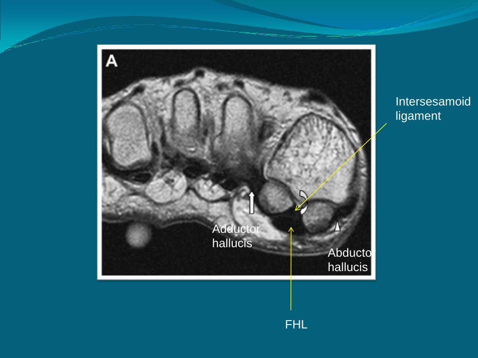
Intersesamoid ligament
Plantar plate
Fibrocartilagenous structure at the plantar aspect of the 1st MTP joint
Proximally, blends with intersesamoid ligament, fibrous capsule
Distal attachment is plantar aspect proximal phalangeal base
Extensor hood (sagittal band)
Extends from common extensor tendons to peripheral aspects of both sesamoids

Additional supporting structures Flexor hallucis brevis tendons (medial, lateral heads)
Origin: cuboid, lateral cuneiform
Insertion: medial, lateral sesamoids
Adductor hallucis tendon (transverse, oblique heads)
Transverse head originates from capsules of 2nd-5th MTP joints, deep transverse lig
Oblique head originates from 2nd-4th MT bases, long plantar ligament
Insertion: Lateral aspect of lateral sesamoid, lateral/plantar aspect proximal phalanx, (blends with the joint capsule)
Abductor hallucis tendon
Origin: medial aspect of the calcaneal tuberosity
Insertion: medial aspect of medial sesamoid, medial/plantar aspect of proximal phalanx (blends with joint capsule)
Flexor hallucis longus tendon
Runs between sesamoids in the groove formed by the intersesamoid ligament/plantar capsular tissue; inserts on the plantar aspect of the distal phalanx
Extensor hallucis brevis, longus tendons
EHB inserts on dorsal aspect of proximal phalanx
EHL inserts on dorsal aspect of distal phalanx

First MTP joint structures (coronal plane) 1 cm proximal to the sesamoid bones

First MTP joint structures at the level of the sesamoid bones

First MTP joint at the level of the proximal phalangeal base

First MTP joint structures in the transverse plane at the level of the sesamoid bones


Extensor Hallucis LongusExtensor Hallucis Brevis
Flexor Hallucis BrevisFlexor Hallucis Longus
J Comput Assist Tomogr, Vol. 26, No. 5, 2002

Distal recess of plantar plate
EHLJoint capsule
FHL
J Comput Assist Tomogr, Vol. 26, No. 5, 2002

Sesamoid-
phalangeal
ligament
MT-
sesamoid
ligament
FHB
insertion

Abductor
hallucis
Adductor
hallucis
Flexor hallucis brevis (medial & lateral heads)
FHL
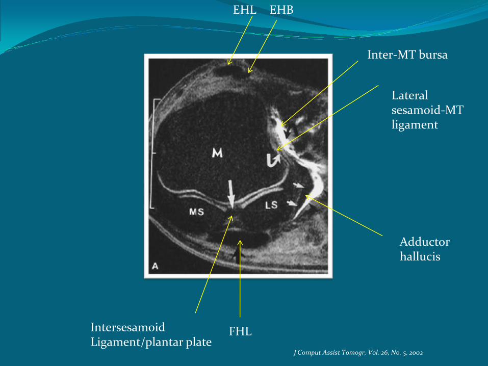
Inter-MT bursa
Lateral sesamoid-MTligament
Adductor hallucis
FHLIntersesamoidLigament/plantar plate
EHL EHB
J Comput Assist Tomogr, Vol. 26, No. 5, 2002

J Comput Assist Tomogr, Vol. 26, No. 5, 2002

Adductor Hallucis
Abductor Hallucis
Lateral main collateral ligament
Bursa
J Comput Assist Tomogr, Vol. 26, No. 5, 2002

EHL
EHB
Abductor Hallucis
FHLAdductor Hallucis
Deep transverse metatarsal ligament
Adductor Hallucis (transverse head)
J Comput Assist Tomogr, Vol. 26, No. 5, 2002
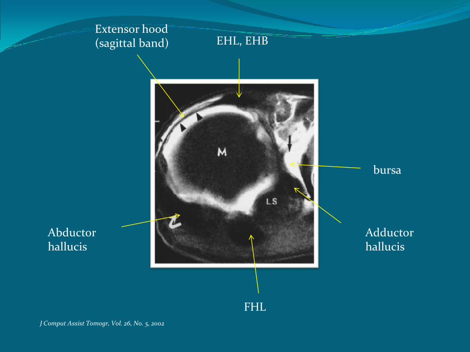
Extensor hood (sagittal band) EHL, EHB
bursa
Adductor hallucis
Abductor hallucis
FHL
J Comput Assist Tomogr, Vol. 26, No. 5, 2002

Adductor hallucis
Lateral sesamoid MT ligament
Abductor hallucis
Intersesamoid ligament/ plantar plate
FHL

EHL
Sagittal band
Lateral sesamoid-MT ligament
Add hallucis
FHLIS ligament/plantar plate
Abd hallucis
J Comput Assist Tomogr, Vol. 26, No. 5, 2002

Abductor hallucis
Adductor hallucis
FHL
J Comput Assist Tomogr, Vol. 26, No. 5, 2002

ANATOMY OF THE LESSER MTP JOINTS

Capsuloligamentous complex
Fibrous capsule
Collateral ligamentous complex
Main collateral ligaments (attach to sides of phalangeal base)
Accessory collateral ligaments (attach to sides of plantar plate)
Common proximal attachment to dorsal tubercle of MT heads
Plantar plate
Fibrocartilagenous structure at plantar aspect of joint
runs between metatarsal head, proximal phalanx

Additional structures
Flexor digitorum longus and brevis
Extensor digitorum longus and brevis
Extensor expansion/hood
Flexor digiti minimi brevis
Abductor digiti minimi
Interosseous muscles
Lumbricals
Deep transverse metatarsal ligament
Superficial transverse metatarsal ligament
Neurovascular bundles
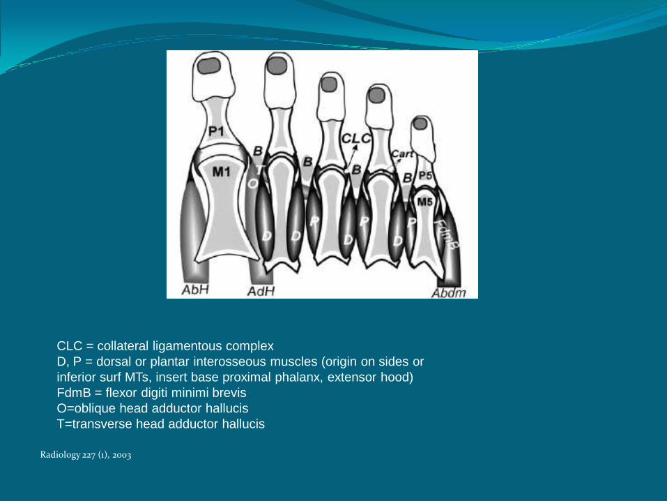
CLC = collateral ligamentous complex
D, P = dorsal or plantar interosseous muscles (origin on sides or
inferior surf MTs, insert base proximal phalanx, extensor hood)
FdmB = flexor digiti minimi brevis
O=oblique head adductor hallucis
T=transverse head adductor hallucis
Radiology 227 (1), 2003

EHL EHB EDL EDB
Extensor hoodJoint capsule
Abductor
hallucis
FHL
Deep transverse
MT ligament (connects
plantar plates)
NV
bundle
Superficial
transverse MT
ligament
Abductor digiti
minimi
Flexor digiti
minimi brevis
Pl fasciaFDL, FDB
L FDL
Radiology 227 (1), 2003
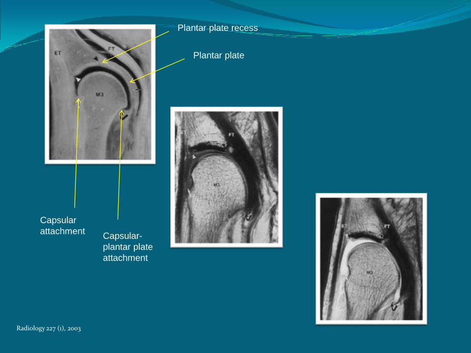
Capsular
attachmentCapsular-
plantar plate
attachment
Plantar plate
Plantar plate recess
Radiology 227 (1), 2003

Extensor hood
Fibrous capsule
Interosseous tendonCLC
Plantar plateRadiology 227 (1), 2003

Main collateral
ligament
Interosseous
tendon
bursa
Radiology 227 (1), 2003
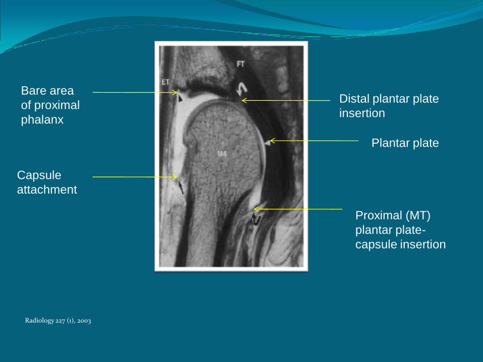
Plantar plate
Distal plantar plate
insertion
Proximal (MT)
plantar plate-
capsule insertion
Capsule
attachment
Bare area
of proximal
phalanx
Radiology 227 (1), 2003

MTP JOINT PATHOLOGY

MR protocol Dedicated extremity coil
Triplanar nonfat suppressed PD for anatomy
Triplanar PD FS or STIR for acute pathology
PD FS has better resolution, anatomic detail
STIR preferred if homogenous fat suppression cannot be obtained secondary to field inhomogeneity
FOV 10-14 cm
3 mm slice thickness

Painful conditions affecting the MTP joints
Trauma
Degenerative
Arthritis
Infection
Osteonecrosis
Neoplastic /non-neoplastic masses
Sesamoid dysfunction

Trauma
Capsuloligamentous injury
Turf toe
Skimboarder’s toe
Acute fracture
Stress fracture
Fatigue
Insufficency

TURF TOE
Sprain of the plantar
capsuloligamentous complex of
the great toe MTP joint
#1 mechanism is
hyperextension
Called “turf toe” after advent
of artificial playing surfaces in
late 1960’s led to increased use
of flexible footwear with less
plantar support
MC in athletes who
participate in cutting or pivoting
sports, especially football

Capsuloligamentous
complex:
Plantar plate
fibrous capsule
collateral ligamentous
complex
• main collateral ligaments
• sesamoid-MT ligaments
sesamoid-phalangeal
ligaments
intersesamoid ligament
Spectrum of injury also
encompasses: Osseous/osteochondral
injury:
• sesamoid injury
(fx, diastasis, diastasis
of bipartitie sesamoid)
• 1st MT fx, chondral injury
FHB, abd/add hallucis strain

Football injury: Complete tear of medial sesamoid phalangeal ligament
with proximal retraction of the medial sesamoid, FHB strain

NORMAL ANATOMY
Sesamoid-
phalangeal
ligament
MT-
sesamoid
ligament
FHB
insertion

Football injury: Bilateral sesamoid phalangeal ligament tears with
edema, hemorrhage; plantar plate is outlined by edema and
hemorhage deep to FHL.

NORMAL ANATOMY
SPSP
FHL
CRISTA

Football injury: Distal metatarsosesamoid ligament tear, FHB
strain; intact sesamoid phalangeal ligament

Intersesamoid ligament rupture with sesamoid diastasis
Abductor
hallucis
Adductor
hallucis
Intersesamoid
ligament
FHL

Complete tear of MCL and partial tear of LCL

Normal MCL, LCL (main collateral ligaments)

FHB medial head strain

Abductor
hallucis
Adductor
hallucis
Flexor hallucis brevis (medial & lateral heads)
FHL

Capsular and tendinous avulsion from the medial margin of the
medial sesamoid with periosteal stripping (subacute injury)

51-year-old man with recent injury of left great toe


Turf toe with diastasis of fractured sesamoids

Bipartitie sesamoid diastasis

Normal plantar plate for
comparison
Turf toe injury in a 24-year-old
professional football player:
disruption of plantar plate with
associated edema and
osteochondral injury1st MT head

Disrupted plantar plate at the 2nd
MTP joint in a 48-year-old woman
who presented with foot pain (no
history of injury)

Classification of injury Grade I: sprain of the plantar capsular complex with
pain, tenderness, swelling
Grade II: capsular disruption with bruising, decreased ROM
Grade III: Chronic injury; results in decreased ROM, OA

Treatment Low grade injury usually treated conservatively
High consideration for surgery if:
Extensive capsular tearing with instability
Sesamoid fx
Significant sesamoid retration
Sesamoid diastasis
Osteochondral lesions
Intra-articular bodies
High level athletes
Goal of surgery = repair and restore anatomy

Preoperative exam: proximal rupture of
the sesamoid phalangeal ligament

Post-operative examination showing primary repair
of the sesamoid phalangeal ligament

Complications/sequelae Chondromalacia of 1st MT head
Osteoarthritis 1st MTP
Hallux valgus
Hallux rigidus (dorsal osteophytosis)

SKIMBOARDER’S TOE
Skimboarding is a beachside
sport in which the athlete stands
on the shore, drops the board on
the ground, and jumps on it in
very shallow water
Skimboarder’s toe =
hyperdorsiflexion injury of the
MTP joints
Unlike in turf toe, the injured
capsuloligamentous structures
are dorsal, rather than plantar

MECHANISM:
Skimboarder uses toes to grip board;
if board slips posteriorly in relation to
skimboarder, hyperdorsiflexion at the
MTP joints may occur
If the toe is violently hyperextended,
forces apply to the EHL/EDL in a dorsal
direction, potentially disrupting the
extensor expansion
May be a/w avulsion fx proximal
phalanx
Theory as to why anatomic
distribution of injury differs from turf toe
despite similar mechanism:
skimboarding is done barefoot,
rendering extensor longus tendons
more apt to dorsiflex and tear the
extensor expansion
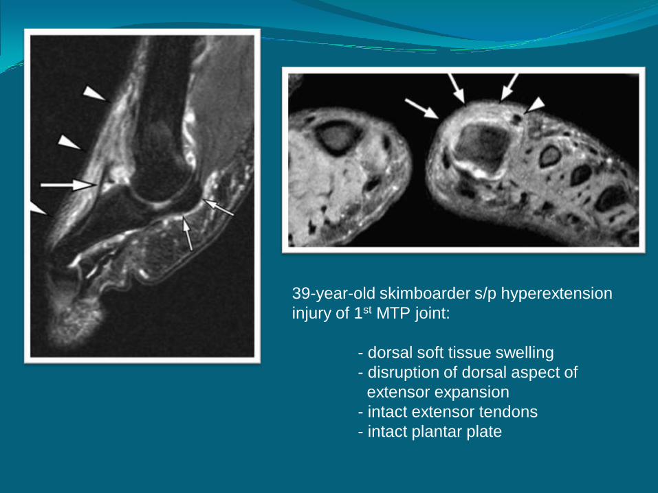
39-year-old skimboarder s/p hyperextension
injury of 1st MTP joint:
- dorsal soft tissue swelling
- disruption of dorsal aspect of
extensor expansion
- intact extensor tendons
- intact plantar plate

Hyperextension injury of the 2nd MTP:
- dorsal soft tissue swelling
- lax/wavy, discontinuous extensor hood
- marrow edema proximal phalanx
- normal plantar plate

Metatarsal fracture Acute fx
Stress fx
Fatigue
Runners, military recruits, gymnasts
Especially mid-distal 2nd-4th MTs
Insufficiency

Metatarsal stress fracture

Insufficiency fracture of 2nd MT head

Insufficiency fracture of 2nd MT head

Insufficiency fracture of 2nd MT head

Freiberg’s infraction MC in adolescents, young women
MC in 2nd MT head
Cause is controversial, likely multifactorial
Popular theory: traumatic insult (acute or repetitive) leading to vascular compromise
Radiographs show flattening, increased density, cystic lucent areas; ultimately leading to deformity and enlargement, secondary degenerative changes

Freiberg’s infraction

Hallux valgus/Bunion
Static subluxation of 1st MTP joint characterized by valgus deviation of the great toe and vaurs deviation of the 1st metatarsal
Etiology is multifactorial; higher frequency in women (constrictive footwear); other predisposing factors include metatarsus primus varus, pronation of the foot, rheumatoid arthritis, neuromuscular disease
Sesamoids maintain their relation with the other metatarsal bones; therefore they become laterally located with respect to 1st MT head
Overgrowth of median eminence of 1st MT head, which has an irregular appearance; may contain prominent cystic areas simulating the appearance of gout
Adjacent soft tissue swelling
Complications:
OA (1st MTP and sesamoid-MT), dorsal osteophytosis
Stress fx sesamoids, medial margin of proximal phalangeal base
Rx = medianl eminence shaving, 1st MT osteotomy
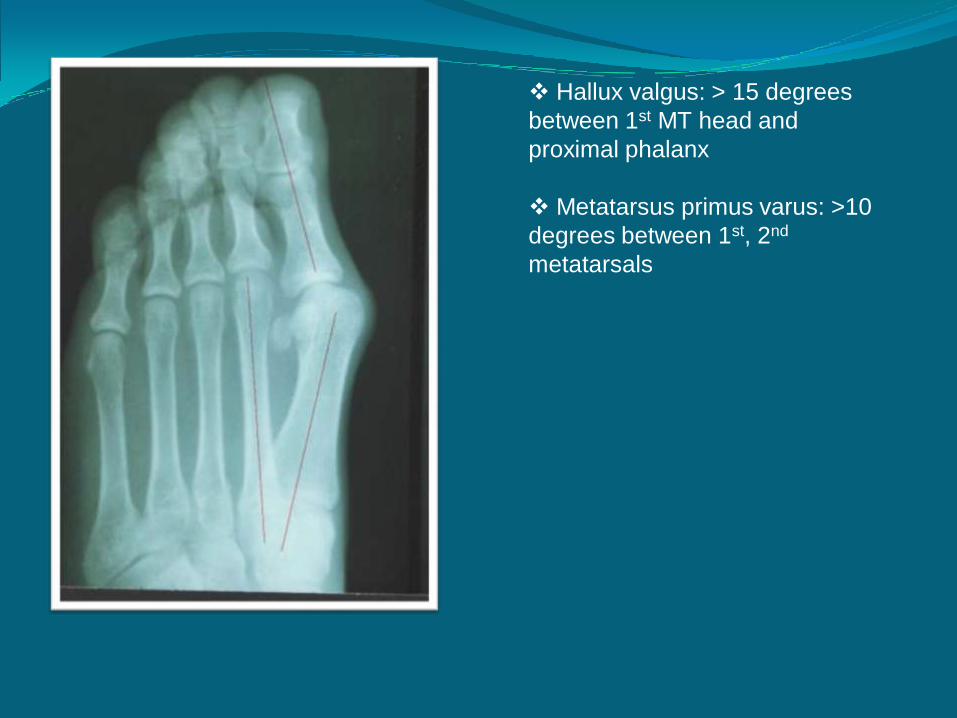
Hallux valgus: > 15 degrees
between 1st MT head and
proximal phalanx
Metatarsus primus varus: >10
degrees between 1st, 2nd
metatarsals

HALLUX VALGUS,
BUNION

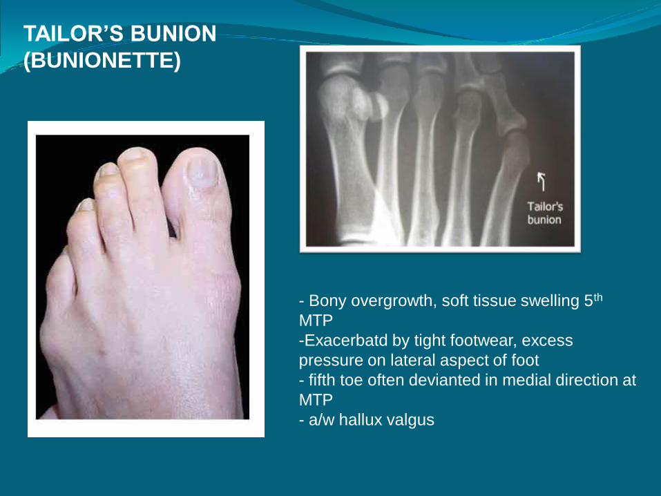
TAILOR’S BUNION
(BUNIONETTE)
- Bony overgrowth, soft tissue swelling 5th
MTP
-Exacerbatd by tight footwear, excess
pressure on lateral aspect of foot
- fifth toe often devianted in medial direction at
MTP
- a/w hallux valgus

Arthritis OA
RA
Gout
CPPD
Reactive arthritis
Neuropathic arthropathy

OSTEOARTHRITIS

Severe OA, bulky dorsal osteophytosis
HALLUX RIGIDUS

GOUT
-1st MTP joint is MC location
- well-defined erosions with overhanging edge
- soft tissue tophi
- normal bone mineralization
- late joint space narrowing

Tophaceous gout in a
56-year-old man with
hyperuricemia,
presenting with foot
pain and swelling
-low signal intensity
tophi , with post
contrast enhancement
- adjacent periarticular
erosions with
characteristic
overhanging edges
- Non-specific MR
features; correlate with
lab values to
distinguish from RA,
septic arthritis,
neoplasm

Rheumatoid arthritis: hallux valgus, erosions, soft tissue swelling,
joint space narrowing, lateral subluxation of sesamoids

Rheumatoid arthritis

Rheumatoid arthritis: marginal erosions, joint space narrowing, synovitis

Reiter’s disease: enthesopathy or
whiskering of the sesamoids, MTP

Osteomyelitis MC in diabetics
Usually from transcutaneous spread of infection
Cutaneous ulcers develop at pressure points; esp under 1st, 5th MT heads
MR non-specific: T1 hypointensity, T2 hyperintensity, enhancement (ddx = neuropathic arthropahty)
+/- abscess, sinus tract, bony destruction
Distinguishing factors: location, ulcer, abscess/phlegmon, sinus tract
Septic arthritis: joint effusion, synovitis, marrow edema

Osteomyelitis of 1st MT head, septic arthritis of 1st MTP joint

Morton’s neuroma Fibrotic response in and about plantar digital nerves (digital branches
of medial, lateral plantar nerves)
Likely on the basis of mechanical impingement
MC in women, repetitive stress such as in ballet or running, etc
# 1 location is between the 3rd & 4th MT heads (3rd interspace), #2 location is 2nd interspace
Clinical:
Pain at level of MTP joint that may radiate into toes
May be asx
MR: T1 - hypo, T2 SE – iso to hypo, T2 FSE FS hyperintense; ++ enhancement
Appear larger when foot imaged prone
Often associated with intermetatarsal bursitis
1st intersapce = Joplin’s neuroma

Morton’s neuroma

Bursitis Intermetatarsal or adventitial (beneath MT heads)
May be mechanical, post-traumatic, infections, inflammatory
NOTE: small fluid collections within first 3 inter-metatarsal bursae with transverse diameter <3mm are common and of doubtful clinical significance

Adventitial bursal formation (submetatarsal)

Sub-metatarsal Fibrosis

Benign soft tissue masses
Ganglion cyst (#1)
Plantar fibromatosis
Hemangioma
Lipoma
Giant cell tumor tendon sheath
Nerve sheath tumor
Foreign body granuloma
Inflammatory mass (i.e. gouty tophus)

Ganglion cyst
Plantar fibromatosis
Common condition associated with fibrous proliferation and replacement of portions of the plantar aponeurosis
All age groups
MC central cord > medial cord
May be solitary or multiple, can enlarge
Usually asx, nodules usually found on palpation
Patterns of abnormality
Focal nodule/soft tissue mass
Small fusiform and tapered thickenings, usually involging the central cord, often in its distal portions
MR: low signal on T1 (similar to muscle), low to intermediate on T2 (though can also be T2/STIR hyperintense), variable enhancement

Plantar fibromatosis


Plantar fascia
Central cord
largest
originates from medial tuberosity of calcaneus
adheres to subjacent flexor digitorum brevis muscle
Broadens as it extends distally; near MT heads, divides into 5 processes, each with superficial and deep components , each extending to one toe
Lateral cord
originates from the lateral margin of the medial tuberosity of the calcaneus
blends with fascia of abductor digiti minimi
Attaches to 5th MT base
Medial cord
very thin, hard to identify proximally
forms the investing fascia of abductor hallucis muscle
becomes more substantial distally, passing medially and obliquely to join the dorsal fascia of the foot


Foreign body granuloma
Malignant soft tissue masses Less common than benign tumors
< 45 yrs
Synovial sarcoma (heterogeneous mass with fluid levels)
Rhabdomyosarcoma
> 45 yrs
MFH
KS
Leiomyosarcoma
Liposarcoma

Leiomyosarcoma

The Sesamoid bones
Assist with weight bearing (especially tibial sesamoid)
Improve mechanical advantage of FHL tendon
Critical for high level athletic function

Sesamoid dysfunction Congenital
Traumatic
Articular disease
Infection
Osteonecrosis

Bipartite/Multipartite sesamoids
Can simulate pathology
33% sesamoids are bipartite; LC multipartite
Usually tibial (medial) sesamoid – 85%
Often bilateral
May be more susceptible to injury cf complete sesamoid
Cleft is usually transverse, smooth, rounded with well-corticated margins
Usually no uptake on bone scan or marrow edema on MR
Usually asx, though can occasinally be a/w abnormal motion between the fragments with pain, marrow edema

BIPARTITE TIBIAL SESAMOID

Bipartite sesamoids: well-corticated, smooth
margins, no uptake on bone scan

Sesamoid trauma “Sesamoiditis”
Stress fracture
Acute fracture
Turf toe
Fracture
Diastasis
Diastasis of bipartitie sesamoid
Dislocation

“sesamoiditis”
Controversial, generic/non-specific term, usually applied when other conditions have been excluded
Described as a painful inflammatory condition related to injury, such as pressure from football cleats, stepping on rocks, etc
Overlap with “stress response”
May be difficult to distinguish from osteonecrosis
MC in medial sesamoid
Imaging may be NL or may see marrow edema on MR, increased density, sclerosis, fragmentation; increased uptake on bone scan
Usually self-limiting

“Sesamoiditis” in a 28-year-old with pain and swelling
beneath the great toe and no h/o trauma to this area

Sesamoiditis (vs stress response) in a 24-year-old female kickboxer

Sesamoid fracture MC in tibial sesamoid
Unlike bipartite sesamoid =>Jagged, irregular margins w/o sclerotic edge, associated with soft tissue swelling, + bone scan, marrow edema on MR
Stress fracture
Ballet dancers, sprinters
Forced propulsion off dorsiflexed toe
More gradual onset of sxs c/w acute fracture

Sesamoid fractures

Sesamoid fracture (subacute) with resorption at
the fracture site

Sesamoid arthritis 1st MTP joint usually also affected
OA
RA
Gout
CPPD
Reactive arthritis

Sesamoid infection MC in diabetics
Clinical: elevated ESR, leukocytosis, fever

Osteomyelitis of the
medial sesamoid

Osteonecrosis Controversial; some maintain that the changes are
related to prior trauma or chronic repetitive injury
MC in adolescents, young women
Gradual onset of pain, worse with weightbearing
Non-specific imaging appearaince: fragmentation, irregularity, mottling, cyst formation; progressing to sclerosis, collapse, enlargement of sesamoid
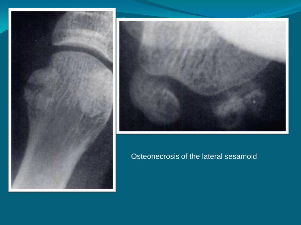
Osteonecrosis of the lateral sesamoid

SUMMARY Pain in the region of the MTP joints is a common
clinical complaint
Causes are numerous
Detailed knowledge of the complex anatomy is important for accurate diagnosis

References1. Crain J, Phancao J. MR Imaging of Turf Toe. Magn Reson Imaging Clin N Am
2008.
2. Resnick D. Internal Derangement of Joints, 2nd ed.
3. Theumann N, Pfirrmann C, et al. Metatarsophalangeal Joint of the Great Toe: Normal MR, MR Arthrographic, and MR Bursographic Findings in Cadavers. JCAT 2002.
4. Theumann N, Mohana-Borges A, Chung C, Resnick D. Lesser Metatarsophalangeal Joints: Standard MR Imaging, MR Arthrography, and MR Bursography – Initial Results in 48 Cadaveric Joints. Radiology 2003.
5. Ashman C, Klecker R. Forefoot Pain Involving the Metatarsal Region: Differential Diagnosis with MR Imaging. Radiographics 2001.
6. Donnelley L, Betts J. Skimboarder’s Toe: Findings on High Field MRI: AJR 2005.
7. Taylor J, Sartoris D. Painful Conditions Affecting the First Metatarsal Sesamoid Bones. Radiographics 1993.
8. Gentili A. The Advanced Imaging of Gouty Tophi. Current Rheumatology Reports 2006.