TEG Wake Med - ICU Bootcampicubootcamp.com/wp-content/uploads/2017/10/TEG-Wake-Med... · TEG! and!...
18
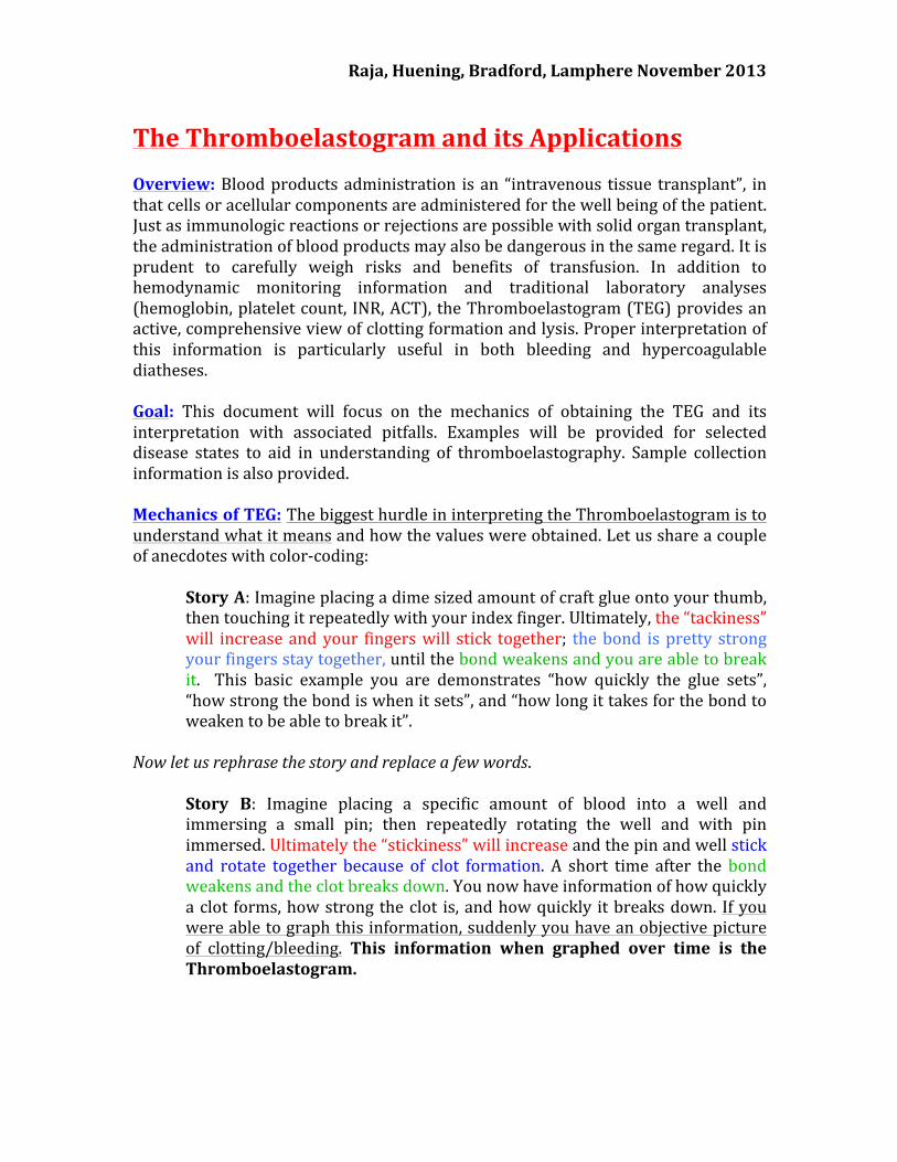
Raja, Huening, Bradford, Lamphere November 2013 The Thromboelastogram and its Applications Overview: Blood products administration is an “intravenous tissue transplant”, in that cells or acellular components are administered for the well being of the patient. Just as immunologic reactions or rejections are possible with solid organ transplant, the administration of blood products may also be dangerous in the same regard. It is prudent to carefully weigh risks and benefits of transfusion. In addition to hemodynamic monitoring information and traditional laboratory analyses (hemoglobin, platelet count, INR, ACT), the Thromboelastogram (TEG) provides an active, comprehensive view of clotting formation and lysis. Proper interpretation of this information is particularly useful in both bleeding and hypercoagulable diatheses. Goal: This document will focus on the mechanics of obtaining the TEG and its interpretation with associated pitfalls. Examples will be provided for selected disease states to aid in understanding of thromboelastography. Sample collection information is also provided. Mechanics of TEG: The biggest hurdle in interpreting the Thromboelastogram is to understand what it means and how the values were obtained. Let us share a couple of anecdotes with colorcoding: Story A: Imagine placing a dime sized amount of craft glue onto your thumb, then touching it repeatedly with your index finger. Ultimately, the “tackiness” will increase and your fingers will stick together; the bond is pretty strong your fingers stay together, until the bond weakens and you are able to break it. This basic example you are demonstrates “how quickly the glue sets”, “how strong the bond is when it sets”, and “how long it takes for the bond to weaken to be able to break it”. Now let us rephrase the story and replace a few words. Story B: Imagine placing a specific amount of blood into a well and immersing a small pin; then repeatedly rotating the well and with pin immersed. Ultimately the “stickiness” will increase and the pin and well stick and rotate together because of clot formation. A short time after the bond weakens and the clot breaks down. You now have information of how quickly a clot forms, how strong the clot is, and how quickly it breaks down. If you were able to graph this information, suddenly you have an objective picture of clotting/bleeding. This information when graphed over time is the Thromboelastogram.
Transcript of TEG Wake Med - ICU Bootcampicubootcamp.com/wp-content/uploads/2017/10/TEG-Wake-Med... · TEG! and!...
Raja, Huening, Bradford, Lamphere November 2013 The Thromboelastogram and its Applications Overview: Blood products administration is an “intravenous tissue transplant”, in that cells or acellular components are administered for the well being of the patient. Just as immunologic reactions or rejections are possible with solid organ transplant, the administration of blood products may also be dangerous in the same regard. It is prudent to carefully weigh risks and benefits of transfusion. In addition to hemodynamic monitoring information and traditional laboratory analyses (hemoglobin, platelet count, INR, ACT), the Thromboelastogram (TEG) provides an active, comprehensive view of clotting formation and lysis. Proper interpretation of this information is particularly useful in both bleeding and hypercoagulable diatheses. Goal: This document will focus on the mechanics of obtaining the TEG and its interpretation with associated pitfalls. Examples will be provided for selected disease states to aid in understanding of thromboelastography. Sample collection information is also provided. Mechanics of TEG: The biggest hurdle in interpreting the Thromboelastogram is to understand what it means and how the values were obtained. Let us share a couple of anecdotes with color-‐coding:
Story A: Imagine placing a dime sized amount of craft glue onto your thumb, then touching it repeatedly with your index finger. Ultimately, the “tackiness” will increase and your fingers will stick together; the bond is pretty strong your fingers stay together, until the bond weakens and you are able to break it. This basic example you are demonstrates “how quickly the glue sets”, “how strong the bond is when it sets”, and “how long it takes for the bond to weaken to be able to break it”.
Now let us rephrase the story and replace a few words.
Story B: Imagine placing a specific amount of blood into a well and immersing a small pin; then repeatedly rotating the well and with pin immersed. Ultimately the “stickiness” will increase and the pin and well stick and rotate together because of clot formation. A short time after the bond weakens and the clot breaks down. You now have information of how quickly a clot forms, how strong the clot is, and how quickly it breaks down. If you were able to graph this information, suddenly you have an objective picture of clotting/bleeding. This information when graphed over time is the Thromboelastogram.
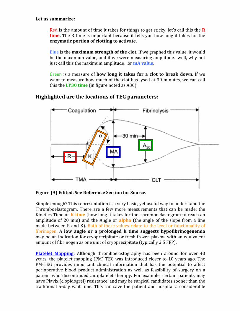
Let us summarize:
Red is the amount of time it takes for things to get sticky, let’s call this the R time. The R time is important because it tells you how long it takes for the enzymatic portion of clotting to activate.
Blue is the maximum strength of the clot. If we graphed this value, it would be the maximum value, and if we were measuring amplitude…well, why not just call this the maximum amplitude…or mA value.
Green is a measure of how long it takes for a clot to break down. If we want to measure how much of the clot has lysed at 30 minutes, we can call this the LY30 time (in figure noted as A30).
Highlighted are the locations of TEG parameters:
Figure (A) Edited. See Reference Section for Source. Simple enough? This representation is a very basic, yet useful way to understand the Thromboelastogram. There are a few more measurements that can be made: the Kinetics Time or K time (how long it takes for the Thromboelastogram to reach an amplitude of 20 mm) and the Angle or alpha (the angle of the slope from a line made between R and K). Both of these values relate to the level or functionality of fibrinogen. A low angle or a prolonged k time suggests hypofibrinogenemia may be an indication for cryoprecipitate or fresh frozen plasma with an equivalent amount of fibrinogen as one unit of cryoprecipitate (typically 2.5 FFP). Platelet Mapping: Although thromboelastography has been around for over 40 years, the platelet mapping (PM) TEG was introduced closer to 10 years ago. The PM-‐TEG provides important clinical information that has the potential to affect perioperative blood product administration as well as feasibility of surgery on a patient who discontinued antiplatelet therapy. For example, certain patients may have Plavix (clopidogrel) resistance, and may be surgical candidates sooner than the traditional 5-‐day wait time. This can save the patient and hospital a considerable

amount of money for an inpatient stay and allow the patient to have surgery sooner. A gene study for CYP2C19 expressed in liver metabolism could be obtained, however the test has to be outsourced, is expensive, takes 5 days. Even then, it may not yield functional or applicable information since it may not account for all gene variants. Furthermore, a gene test does not necessarily translate to gene function. PM-‐TEG on the other hand specifically tests for platelet suppression as a result of drugs affecting Arachidonic Acid pathway (such as Aspirin), or ADP pathway (such as clopidogrel, ticlopidine etc.). An mA-‐ADP greater than 50 is considered safe for surgery. It should be noted that one must also take thrombocytopenia into account. Examples below of what a normal platelet mapping TEG looks like (Tracing Courtesy Tobin Timmons, Haemonetics).
PM-‐TEG ADP Assay (above)
PM-‐TEG AA Assay (above)
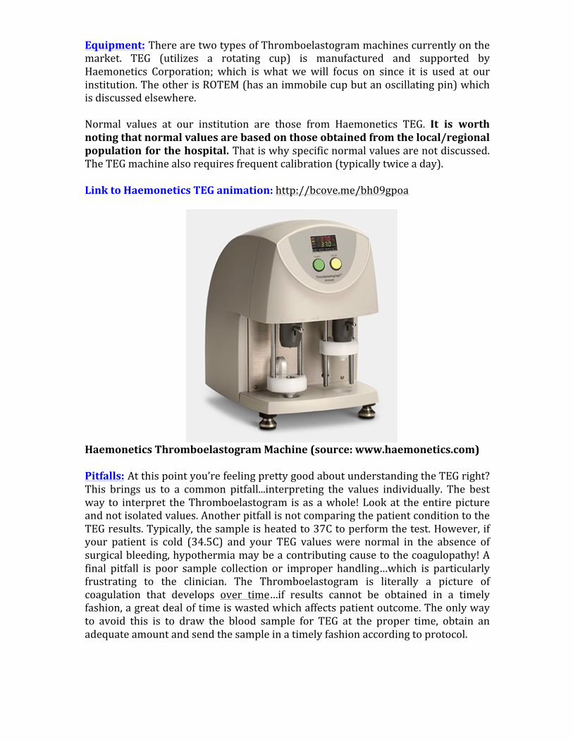
Equipment: There are two types of Thromboelastogram machines currently on the market. TEG (utilizes a rotating cup) is manufactured and supported by Haemonetics Corporation; which is what we will focus on since it is used at our institution. The other is ROTEM (has an immobile cup but an oscillating pin) which is discussed elsewhere. Normal values at our institution are those from Haemonetics TEG. It is worth noting that normal values are based on those obtained from the local/regional population for the hospital. That is why specific normal values are not discussed. The TEG machine also requires frequent calibration (typically twice a day). Link to Haemonetics TEG animation: http://bcove.me/bh09gpoa
Haemonetics Thromboelastogram Machine (source: www.haemonetics.com) Pitfalls: At this point you’re feeling pretty good about understanding the TEG right? This brings us to a common pitfall...interpreting the values individually. The best way to interpret the Thromboelastogram is as a whole! Look at the entire picture and not isolated values. Another pitfall is not comparing the patient condition to the TEG results. Typically, the sample is heated to 37C to perform the test. However, if your patient is cold (34.5C) and your TEG values were normal in the absence of surgical bleeding, hypothermia may be a contributing cause to the coagulopathy! A final pitfall is poor sample collection or improper handling…which is particularly frustrating to the clinician. The Thromboelastogram is literally a picture of coagulation that develops over time…if results cannot be obtained in a timely fashion, a great deal of time is wasted which affects patient outcome. The only way to avoid this is to draw the blood sample for TEG at the proper time, obtain an adequate amount and send the sample in a timely fashion according to protocol.
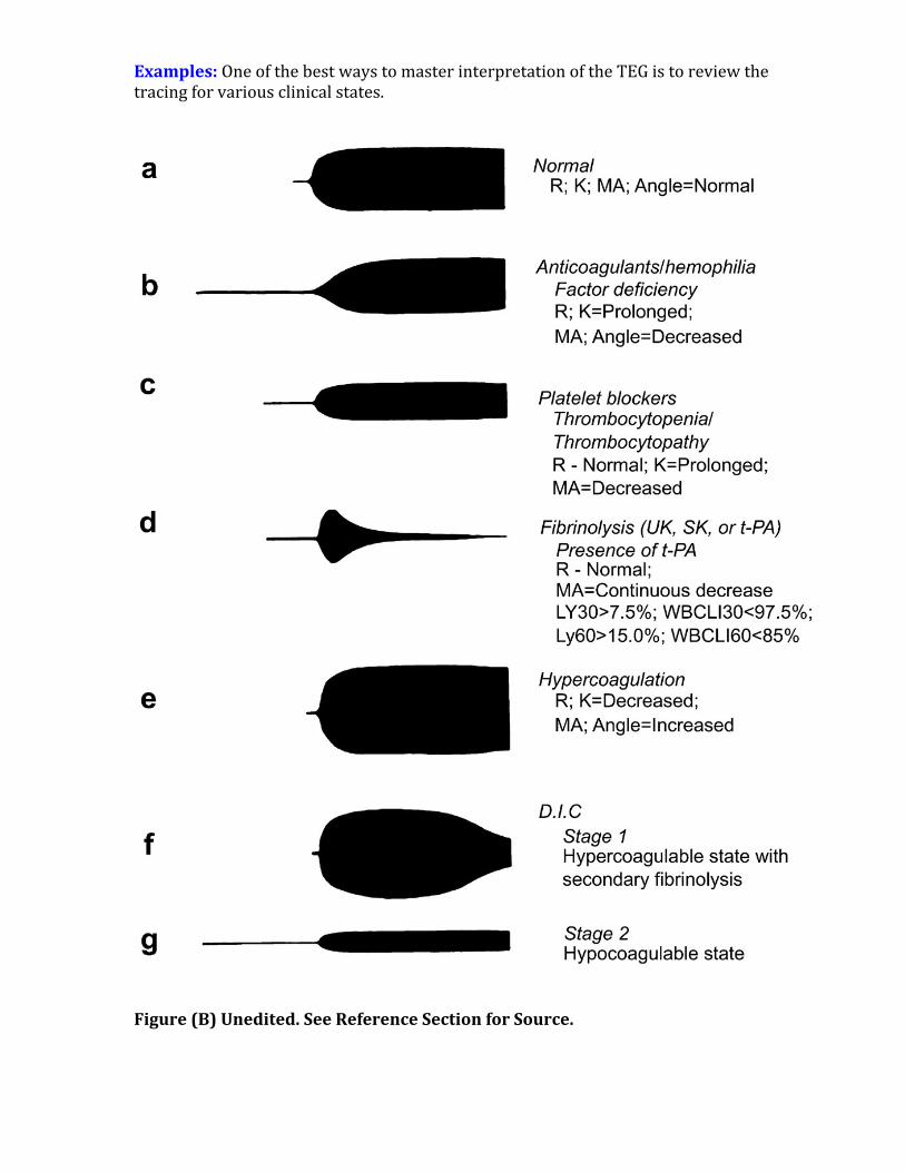
Examples: One of the best ways to master interpretation of the TEG is to review the tracing for various clinical states.
Figure (B) Unedited. See Reference Section for Source.

Protocol for Cardiac Surgery Patient scheduled for __________:
Elective Cardiac Surgery – Obtain Platelet Mapping TEG Intraoperative, prior to Operative Incision from Arterial line, if patient on Aspirin or Clopidogrel.
Urgent Cardiac Surgery – Obtain Platelet Mapping TEG while patient in hospital 3 days prior to planned cardiac surgery if suspicion for platelet dysfunction (clopidogrel, or thrombocytopenia). AA component does not need to be measured unless patient on aspirin. ADP assay should be obtained. If normalized or platelet inhibition less than 30%, proceed to operating room. If abnormal, draw another Platelet Mapping TEG on day of surgery (intraoperative, prior to incision). Emergent Cardiac Surgery – Obtain Platelet Mapping TEG (ADP and AA assays) at earliest possible opportunity intraoperatively mainly to unmask drug related platelet dysfunction. Do not obtain if patient already on cardiopulmonary bypass or on ventricular assist device/ECMO. If intra-‐aortic balloon pump (IABP) in place, ok to draw sample to differentiate drug related vs. mechanical platelet dysfunction only if patient on Plavix/ASA immediately prior to placement of IABP.
Overview of Sample Acquisition for Cardiac Surgery

Sample Draw Instructions (Pre-‐operative) using standard cup unless patient on heparin:
1) Platelet Mapping Thromboelastogram ordered by Cardiothoracic Surgical team in consultation with Cardiology.
2) RN calls TEG lab tech directly in anticipation of sample. Blood is drawn (green top AND light blue top tubes) from non-‐diluted source (limb contralateral to functioning peripheral IV). If patient has two peripheral IVs, sample is drawn from limb that has non-‐critical IV that has been held for 15-‐30 minutes. Great care taken not to pull excessive vacuum that will lyse platelets.
3) Sample hand delivered by RN or tubed to lab within 10 minutes of draw time. 4) Lab accepts or rejects sample on the spot. Standard (non-‐heparinase) cup is
to be used. 5) Results of Platelet Mapping Thromboelastogram made available. If abnormal,
lab calls cardiothoracic surgical team.
Use ONE Citrate Tube (BD)
Use ONE Gel and Lithium Heparin (BD)

Sample Draw Instructions/Protocol (Intraoperative On-‐CPB, excluding TAVR) using heparinase cup:
1) Draw sample at 33-‐34 Celsius during active re-‐warming. (Note: the TEG results are most applicable to a fully warmed patient since the sample analysis occurs between 36-‐37 Celsius. Our machine is calibrated to 37C). For Off-‐pump CABG surgery, send TEG, platelet count, and fibrinogen level 5-‐10 minutes following administration of protamine. A heparinase cup is to be used. An ACT should be drawn concurrently.
2) CRNA calls TEG lab tech directly from OR and notifies them this is an on bypass sample. The lab will use a heparinase cup. Ordering physician is Attending Anesthesiologist for case. Circulating RN calls OR tech to room specifying: “a TEG sample needs to be sent”.
3) CRNA draws off 12mL blood, then draws TEG sample (blue top) and ensure fill of blood slightly over tube line. Also sent is a Platelet count (do not send full CBC) and a Fibrinogen level (do not send a DIC panel). Return “waste” arterial blood intraVenously to patient.
4) Sample is provided to OR Tech (PCT), with TEG lab slip, who then takes sample directly to lab or via tube system within 10 minutes of draw time. Sample is accepted or rejected on the spot. Lab will call the OR directly if sample has been rejected, at which point another sample is immediately drawn and sent.
5) 15 minutes after the sample has been sent, circulating RN will pull up the Thromboelastogram in progress. This tracing program window will not be closed until the patient leaves the room to the ICU.
6) The Attending Anesthesiologist is notified by the CRNA once 30 minutes have elapsed from sample start time. The Thromboelastogram is evaluated and a blood product management strategy is determined in conjunction with the surgeon and perfusionist. (See blood products protocol)
7) Following separation from cardiopulmonary bypass, draw a second TEG sample with the post-‐protamine ACT (repeat steps 1-‐4) ONLY IF any of the following conditions hold true:
a. If pre-‐separation TEG, Fibrinogen and/or platelet values were abnormal. Delay draw of second TEG until planned blood products have been administered.
b. Significant on-‐going surgical bleeding (greater than 100mL/min loss). Clotting factors are being lost and TEG values will change.
c. Double valve repair/replacement d. Aortic root or aortic arch surgery involving deep hypothermic
circulatory arrest. e. If intra-‐operative core patient temperature was below 28C at any
point during the surgery. f. Intra-‐cardiac tumor resection, or presence of intra-‐cardiac clot g. End-‐Stage renal disease or high-‐velocity lesion such as critical aortic
stenosis or ASD/VSD. h. Mechanical ventricular assist device (includes IABP). i. Extremes of age (age > 70) or patient with poor tissue quality.
**Perfusionist to monitor second TEG tracing if drawn intraoperatively and CRNA occupied with resuscitation of patient. Notify anesthesiologist and surgeon for any residual abnormal values to decide next step.
Sample Draw Instructions/Protocol (ICU Post-‐operative) using standard cup
1) If post-‐separation TEG sample was not sent from the operating room, a sample will be sent upon arrival to the ICU with first set of labs.
2) If the intra-‐operative post-‐protamine TEG was abnormal AND patient is still bleeding significantly, cardiothoracic surgery team will order TEG from the ICU.
3) ICU RN or Charge RN will call TEG tech in anticipation of sample. 4) Sample drawn after 12mL blood “waste” (returned intravenously) is drawn
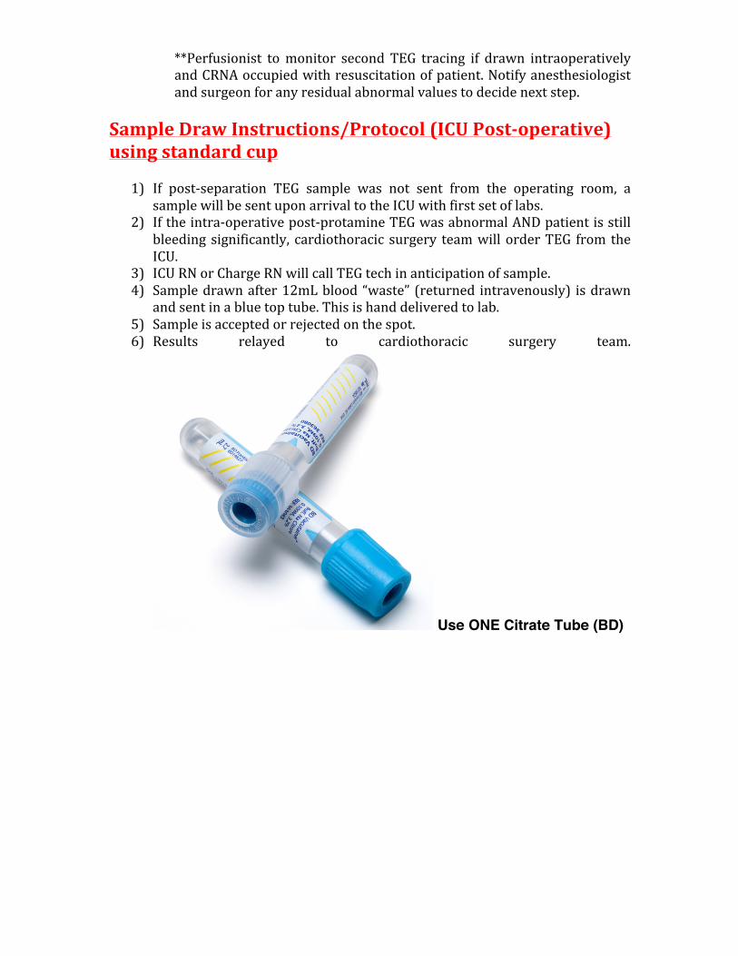
and sent in a blue top tube. This is hand delivered to lab. 5) Sample is accepted or rejected on the spot. 6) Results relayed to cardiothoracic surgery team.
Use ONE Citrate Tube (BD)

Recommended Intra-‐operative Blood Component Therapy for Cardiac Surgery in Bleeding Patients
• In this section, detailed scenarios are provided (common things first followed by less common). See algorithm for condensed view.
• Note: if values are abnormal and patient is not bleeding, strongly
consider holding off transfusion unless clinically necessary. Tailor to appropriate scenario.
• Unless otherwise specified, fibrinogen means traditional lab based
fibrinogen assay. Functional fibrinogen will be specifically noted. Scenario A R time is normal, mA is normal, fibrinogen and platelet count are normal (heparinase cup, with ABNORMAL ACT)
-‐ There is difficulty getting the coagulation cascade started -‐ The patient has a normal TEG, but has a prolonged ACT value Solution: Give protamine (post CPB separation only)
Scenario B R time is prolonged, mA is normal, fibrinogen and platelet count are normal (heparinase cup, with a normal ACT)
-‐ There is difficulty getting the coagulation cascade started -‐ The patient is low on coagulation factors Solution: Give FFP
Scenario C R time is prolonged, mA is low, fibrinogen is normal, platelet count is low (heparinase cup)
-‐ There is difficulty getting the coagulation cascade started -‐ The clot strength is low -‐ The patient is low on coagulation factors and platelets Solution: Give FFP and platelets.
Scenario D R time is prolonged, mA is low, fibrinogen is low, platelet count is normal (heparinase cup)
-‐ There is difficulty getting the coagulation cascade started -‐ The clot strength is low -‐ Also, unique to low fibrinogen or low functional fibrinogen, you will
notice a decreased angle and prolonged K. -‐ The patient is low on fibrinogen and possibly other factors. -‐ Even with a normal platelet count, the platelets may be dysfunctional
(especially if the platelet mapping was abnormal from the start). Solution: Give cryoprecipitate, OR if currently on CPB and is major cardiac surgery (double valve, deep hypothermic circulatory arrest, high risk cardiac surgery on age > 70, re-‐do sternotomy etc.) consider 4 FFP on pump at 34-‐
35C with hemoconcentration performed by the perfusionist. If very high risk with a TEG that is grossly abnormal, consider 6 FFP with hemoconcentration on pump (rare). Note: 3 FFP have approximately the same content of fibrinogen as 1 unit of cryoprecipitate.
Scenario E R time is normal, mA is low, fibrinogen is low, platelet count is normal (heparinase cup)
-‐ Coagulation onset is normal -‐ The clot strength is low -‐ Most likely the fibrinogen is dysfunctional and low in quantity. Angle will
be decreased and k time will be prolonged! -‐ Platelet mapping was normal Solution: Give cryoprecipitate only, and send another TEG. If fibrinogen is < 200, give 1 unit cryoprecipitate (cryo). If less than 150, give 2 units cryo. Can also elect to run functional fibrinogen TEG at this time instead of sending a standard fibrinogen level. Results available in 5 minutes instead of 45 minutes.
Scenario F R time is normal, mA is low, fibrinogen is normal, platelet count is low or abnormal pre-‐operative platelet mapping (heparinase cup).
-‐ Coagulation onset is normal -‐ The clot strength is low -‐ Most likely there are not enough platelets or they are not working
properly (see platelet mapping) Solution: Give platelets and ddAVP (0.3mg/kg) following protamine. If Platelet count less than 150, give 1 platelet. If less than 100, give 2 platelets. If less than 50, give 3 platelets. **Proper platelet handling: Platelets are very delicate! Do not give platelets while on CPB! Do not shake the bag, warm the platelets or cool them. Only ask for them when you are ready to transfuse within 1 hour of arrival into the OR. Do not mechanically force them into the patient with a syringe. Do not transfuse them using a rapid transfusion device. Do not administer them with a dedicated medication line. Administer them slowly and watch PA pressures carefully to avoid an unnoticed platelet reaction. Increase the flow rate once platelets are deemed safe.
Scenario G R time is prolonged, mA is low, fibrinogen is low, platelet count is low and abnormal platelet mapping pre-‐operatively (heparinase cup)
-‐ All lab parameters are grossly abnormal, either operation was long, hypothermia was profound, the patient has a mechanical device through which blood is flowing a high speeds, and/or the patient may be in a fibrinolytic state or DIC Stage 2.
Solution: If the patient is still on CPB, strongly consider administration of 4-‐6FFP while on CPB after patient is fully re-‐warmed, and hemoconcentrate. Re-‐evaluate anti-‐fibrinolytic therapy and re-‐dose if safe. Otherwise, in such an abnormal profile send a functional fibrinogen. Order cryo and platelets to room prior to separation from CPB, as they will be administered. Pay particular attention to angle (if low, indicated fibrinogen deficiency). If despite blood component therapy, the patient still has an abnormal TEG, and is continuing to bleed, consideration for novo-‐7 should be made, although this should be the last resort.
Scenario H R time is abnormally short, mA is normal or high, Fibrinogen is normal, Platelet count is normal and platelet mapping is normal.
-‐ This scenario is unlikely to occur during a cardiac case requiring sternotomy, but may be seen after novo-‐7 has been administered.
Solution: Monitor and examine patient closely for signs of thrombosis or signs of DIC. Consider discontinuation of antifibrinolytic and carefully starting heparin to avoid coronary thrombosis. This is an unlikely scenario with the potential to occur in the rare cases.
SPECIAL NOTE regarding clot degradation: The lysis time value (LY 30) can provide you additional information regarding clot stability. If the clotting occurs within a normal time period (R time) and has good strength (normal mA), then consider administering antifibrinolytic or additional fibrinogen in the form of cryoprecipitate for an elevated LY 30. Additional pearls: In patients with a normal TEG, ACT, Platelet Count (with normal platelet mapping), and Fibrinogen one should have a strong suspicion for iatrogenic bleeding. In patients with high velocity/stenotic lesions, particularly aortic stenosis or ESRD patients, consider administration of ddAVP (0.3mg/kg). Do not empirically give ddAVP to all patients due to the risk of hypotension. Low blood viscosity or hemodilution can play a major factor in bleeding, particularly in elderly patients, redo-‐sternotomy or very complex cardiac procedures. If blood is viscous (hemoglobin of 10g/dL), blood speed decreases which promotes increased contact time to allow the coagulation cascade to occur. Hypocalcemia can also pre-‐dispose a patient to bleeding, as Ca++ is a critical component for the coagulation cascade. Finally, hypothermia causes coagulopathy, so warm up the room, or increase active body warming to the maximally allowed safe level. Blood pH should also be normalized. Post operative The same scenario/principles above may be utilized, with corresponding therapies, although it should be noted that a non-‐heparinase cup should be used for post operative TEG. If there is suspicion that residual heparin is present and is the source of coagulopathy, both a heparinase and non-‐heparinase sample should be obtained with the expectation of a normalized R time in the heparinase cup. A prolonged R time on a TEG with an abnormal ACT can also clue you into the same problem, although this is less specific. If persistent heparin is causing a prolonged R time and

coagulopathy, start the patient on a protamine infusion 50mg/hr for a maximum of 5 hours (stop earlier if hemostasis is achieved and maintained). The typical scenario for this case is in the patient in whom a supra-‐normal dose of heparin was administered, due to pre-‐procedure anti-‐thrombin III deficiency. In such patients, following administration of FFP or anti-‐thrombin III concentrate, there is a surplus of heparin that continues to leach out of the peripheral circulation (post-‐procedure). This rebound heparin phenomenon is easily treatable with protamine rather than blood products saving the patient from unnecessary blood product exposure.

The next two pages can be printed separately for reference purposes as a guide. Important notes: Since great variation exists in thromboelastography, it is best to adopt an approach for understanding the parameters rather than rote memorization of the algorithm. The algorithm is a guide/tool to help you accomplish that goal. When using the algorithm, circle the relative values (Normal, Increased, Decreased) starting from the top, working downward. Match the appropriate letter to the therapy. If TEG is abnormal, first verify that the patient is not hypothermic, that blood pH is corrected, heparin has been reversed (unless on CPB) and serum calcium is at an appropriate level. If patient is on heparin or CPB, make sure TEG sample assay was performed using a TEG heparinase cup. For patients with high velocity lesions, long CPB time, deep hypothermic circulatory arrest, moderate to severe renal dysfunction, ventricular assist devices, consider administering ddAVP and also ensuring that blood viscosity is adequate (typically a Hemoglobin of 10 gm/dL or higher). For blood products administered prior to separation from CPB, consider hemoconcentration techniques to increase blood viscosity and reduce acellular volume. This often helps avoid bleeding from suture hole sites without the need of transfusion. If the patient is not bleeding, DO NOT transfuse for an abnormal TEG value unless clinically necessary. On the same note, appropriate blood component therapy earlier in patient course typically reduces overall transfusion. The team taking care of the patient should exercise final clinical judgment and therapy. Author of this document assumes no responsibility, and no guarantees have been made. Refer to TEG manufacturer literature and relevant thromboelastography critically reviewed evidence based literature for most up-‐to-‐date information.

TEG Therapy Algorithm for Clotting Dysfunction
A, B – TEG is normal. Do not transfuse clotting components. Bleeding is anatomic/surgical. Consider transfusion of PRBC if profound anemia and blood viscosity is low. Correct pH, temperature and/or Calcium if abnormal. C, D – TEG is normal, with supra-‐normal clot strength. Do not transfuse clotting components. Bleeding is anatomic/surgical. Consider transfusion of PRBC if profound anemia and blood viscosity is low. Correct pH, temperature and/or Calcium if abnormal. E – TEG is abnormal. Clotting start time is normal, but the clot is weak. Rate of clot formation is normal, the patient is either thrombocytopenic or the platelets are dysfunctional. Transfuse platelets (If < 150 give 1 pack, If < 100 give 2 packs, If < 50 give 3 packs). If the patient has a high velocity lesion, or has ESRD, transfuse ddAVP. Otherwise for platelet dysfunction, give 2 packs of platelets. F – TEG is abnormal. Clotting start time is normal, but the clot is weak and rate of clotting is slow. Follow same as ‘E’ (above); however if fibrinogen is less than 200, also transfuse 1 pack cryo. G, H – TEG is abnormal. Clotting start time is delayed, but the clot is strong. Ensure that heparin has been fully reversed with protamine. If ACT is normal, then consider transfusion of 1-‐2 FFP. If fibrinogen is low, also consider transfusion of 1 pack cryo. I – TEG is abnormal. Clotting start time is delayed, but clot strength is supra normal. Do not transfuse! Strongly suspect residual heparin. Review total heparin dose. If patient BMI > 40, or received anti-‐thrombin III intraoperatively following very large doses of heparin, consider a protamine infusion of 50mg/hr for a maximum of 5 hours following the bolus dose of protamine. Also verify correction of pH, temperature and hypocalcaemia. J – TEG is abnormal. Clotting start time is delayed and the clot is weak. Transfuse 2 FFP and transfuse platelets (If < 150 give 1 pack, If < 100 give 2 packs, If < 50 give 3 packs). If the patient has a high velocity lesion, or has ESRD, transfuse ddAVP. Otherwise for platelet dysfunction, give 2 packs of platelets. K – TEG is abnormal. Clotting start time is delayed and clot is weak. Follow same as J; also if fibrinogen is low give 1 unit of cryo. L, M, O – TEG is abnormal. Patient is hypercoagulable. Do not transfuse clotting components. If DIC, or HIT, consult hematologist as needed. If patient on CPB, on ECMO, or has VAD, verify adequate heparinization. Patient may need AT III if no response with escalating heparin doses. P, Q – TEG is abnormal. Clotting start time is fast, however clot is weak. Likely von Willebrand factor deficiency. Administer ddAVP. If thrombocytopenia, transfuse platelets (If < 150 give 1 pack, If < 100 give 2 packs, If < 50 give 3 packs).

Normal Thromboelastogram (TEG) Values R Time 5-‐10 min
MA 50 -‐ 70 millimeters K Time 1-‐3 min Angle 53-‐72 degrees LY30 0-‐8 %

References: Bolliger D, Seeberger MD, Tanaka KA. Principles and practice of thromboelastography in clinical coagulation management and transfusion practice. Transfus Med Rev. 2012 Jan;26(1):1-‐13. Bowbrick VA, Mikhailidis DP, Stansby G. The use of citrated whole blood in thromboelastography. Anesth Analg 2000, 90:1086–88. Chen L, Bracey AW, Radovancevic R, et al. Clopidogrel and bleeding in patients undergoing elective coronary artery bypass grafting. J Thorac Cardiovasc Surg 2004;128:425-‐31. Collyer TC, Gray DJ, Sandhu R, Berridge J, Lyons G. Assessment of platelet inhibition secondary to clopidogrel and aspirin therapy in preoperative acute surgical patients measured by Thrombelastography Platelet Mapping. Br J Anaesth 2009;102:492-‐8. Ganter MT, Hofer CK. Coagulation monitoring: current techniques and clinical use of viscoelastic point-‐of-‐care coagulation devices. Anesth Analg. 2008 May;106(5):1366-‐75. Harr JN et. al. Functional fibrinogen assay indicates that fibrinogen is critical in correcting abnormal clot strength following trauma. Shock 2013, 39(1): 45–9. Levrat A et. al. Evaluation of rotation thrombelastography for the diagnosis of hyperfibrinolysis in trauma patients. Br J Anaesth 2008, 100:792–7. Kaur J, Jones N, and Mallett S. Thrombelastography Platelet Mapping is a useful preoperative tool in surgical patients taking antiplatelet medication. Br. J. Anaesth. (2009) 103 (2): 304-‐5. Mahla E, Suarez TA, et.al. Platelet function measurement-‐based strategy to reduce bleeding and waiting time in clopidogrel-‐treated patients undergoing coronary artery bypass graft surgery: the timing based on platelet function strategy to reduce clopidogrel-‐associated bleeding related to CABG (TARGET-‐CABG) study. Circ Cardiovasc Interv. 2012;5:261-‐69. Ronald A et al. Can the use of TEG predict and decrease bleeding and blood product requirements in adult patients undergoing cardiac surgery? Inter CV and Thor Surg 4 (2005) 456-‐63. Shore-‐Lesserson L et. al. Thromboelastography-‐guided transfusion algorithm reduces transfusions in complex cardiac surgery. Anesth Analg. 1999 Feb;88(2):312-‐9. Spiess BD et. al. Thromboelastography as an indicator of post-‐cardiopulmonary bypass coagulopathies. J Clin Monit. 1987 Jan;3(1):25-‐30.
Scarpelini S et.al. Normal range values for thromboelastography in healthy adult volunteers. Braz J Med Biol Res 2009, 42(12):1210–17. Tuman KJ et al. Evaluation of coagulation during cardiopulmonary bypass with a heparinase modified thromboelastographic assay. J Cardiothoracic Vasc Anesth 1994; 8:144-‐9. Weber CF, Klages M, Zacharowski K. Perioperative coagulation management during cardiac surgery. Curr Opin Anesthesiol 2013, 26:60–64. Figures: Figure (A) The characteristic signature waveform output generated by the TEG analyzer (with permission from Elsevier Limited). Kouerinis I A et al. Interact CardioVasc Thorac Surg 2008;7:560-‐56 Figure (B) Unedited. Typical TEG waveforms for blood samples displaying various conditions (with permission from Elsevier Limited). Kouerinis I A et al. Interact CardioVasc Thorac Surg 2008;7:560-‐563 Images, as noted, from Haemonetics and BD. This document may not be copied or duplicated without permission of author. Usage of information herein is at sole discretion of reader and/or clinician. No guarantees for outcome or safety have been made. Information provided is best available evidence/information for presentation of concepts. Refer to Haemonetics Company for most up-‐to-‐date information including but not limited to warnings and precautions. Patients must be treated individually and transfusion decisions must be made based on clinical scenario at hand. If patient is not bleeding, even if TEG lab values are abnormal, do not transfuse unless clinically necessary.