Gram-Positive Anaerobic Cocci - Clinical Microbiology Reviews
Upload
mark-harveyCategory
view
229download
0
Streptococcus Gram+ cocciIn chains
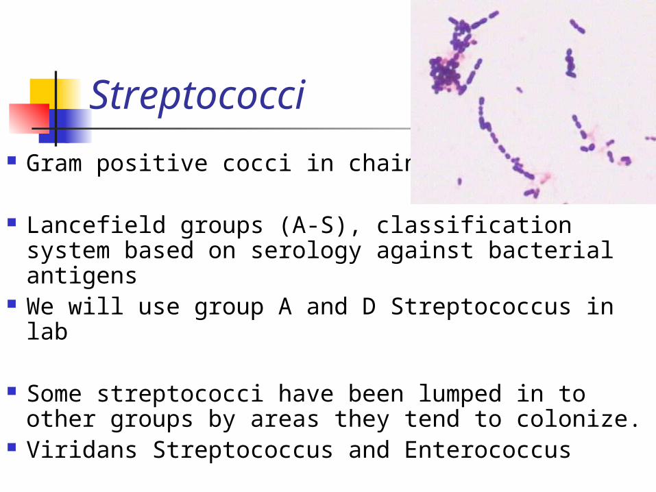
Streptococci
Gram positive cocci in chains
Lancefield groups (A-S), classification system based on serology against bacterial antigens
We will use group A and D Streptococcus in lab
Some streptococci have been lumped in to other groups by areas they tend to colonize.
Viridans Streptococcus and Enterococcus
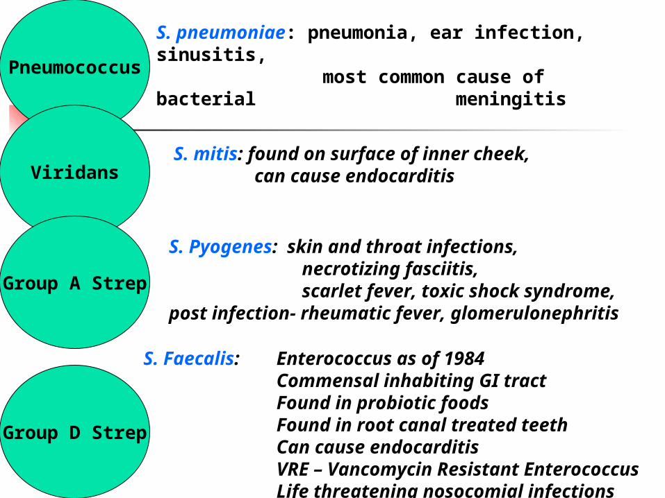
Pneumococcus
Viridans
Group A Strep
Group D Strep
S. pneumoniae: pneumonia, ear infection, sinusitis,
most common cause of bacterial meningitis
S. mitis: found on surface of inner cheek, can cause endocarditis
S. Pyogenes: skin and throat infections, necrotizing fasciitis,scarlet fever, toxic shock syndrome,
post infection- rheumatic fever, glomerulonephritis
S. Faecalis: Enterococcus as of 1984Commensal inhabiting GI tractFound in probiotic foodsFound in root canal treated teethCan cause endocarditisVRE – Vancomycin Resistant EnterococcusLife threatening nosocomial infections
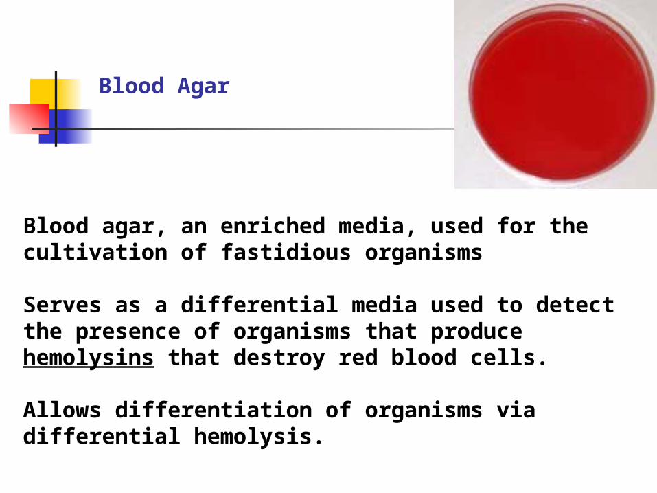
Blood agar, an enriched media, used for the cultivation of fastidious organisms
Serves as a differential media used to detect the presence of organisms that produce hemolysins that destroy red blood cells.
Allows differentiation of organisms via differential hemolysis.
Blood Agar
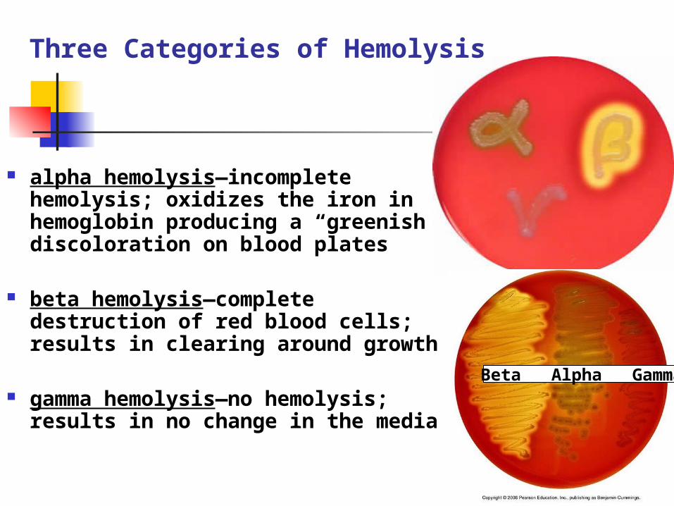
alpha hemolysis—incomplete hemolysis; oxidizes the iron in hemoglobin producing a “greenish” discoloration on blood plates
beta hemolysis—complete destruction of red blood cells; results in clearing around growth
gamma hemolysis—no hemolysis; results in no change in the media
Beta Alpha Gamma
Three Categories of Hemolysis
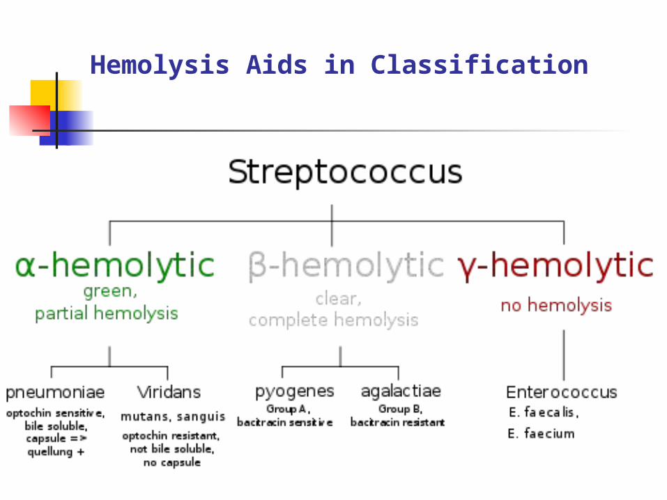
Hemolysis Aids in Classification
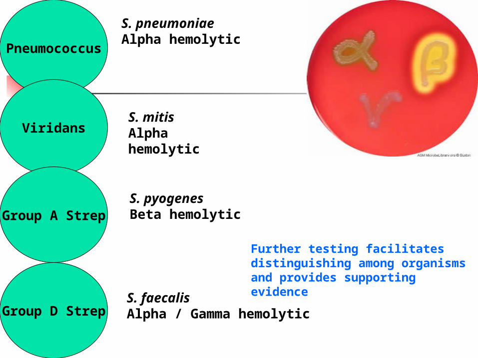
Pneumococcus
Viridans
Group A Strep
Group D Strep
S. pneumoniae Alpha hemolytic
S. mitisAlpha hemolytic
S. pyogenesBeta hemolytic
S. faecalisAlpha / Gamma hemolytic
Further testing facilitates distinguishing among organisms and provides supporting evidence
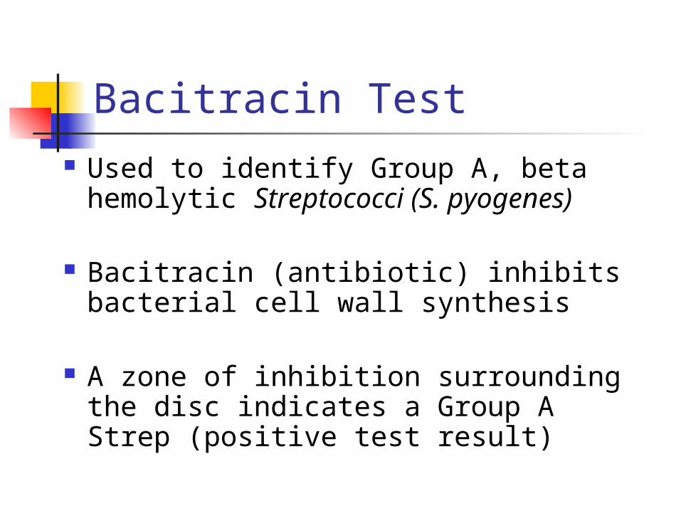
Bacitracin Test Used to identify Group A, beta
hemolytic Streptococci (S. pyogenes)
Bacitracin (antibiotic) inhibits bacterial cell wall synthesis
A zone of inhibition surrounding the disc indicates a Group A Strep (positive test result)
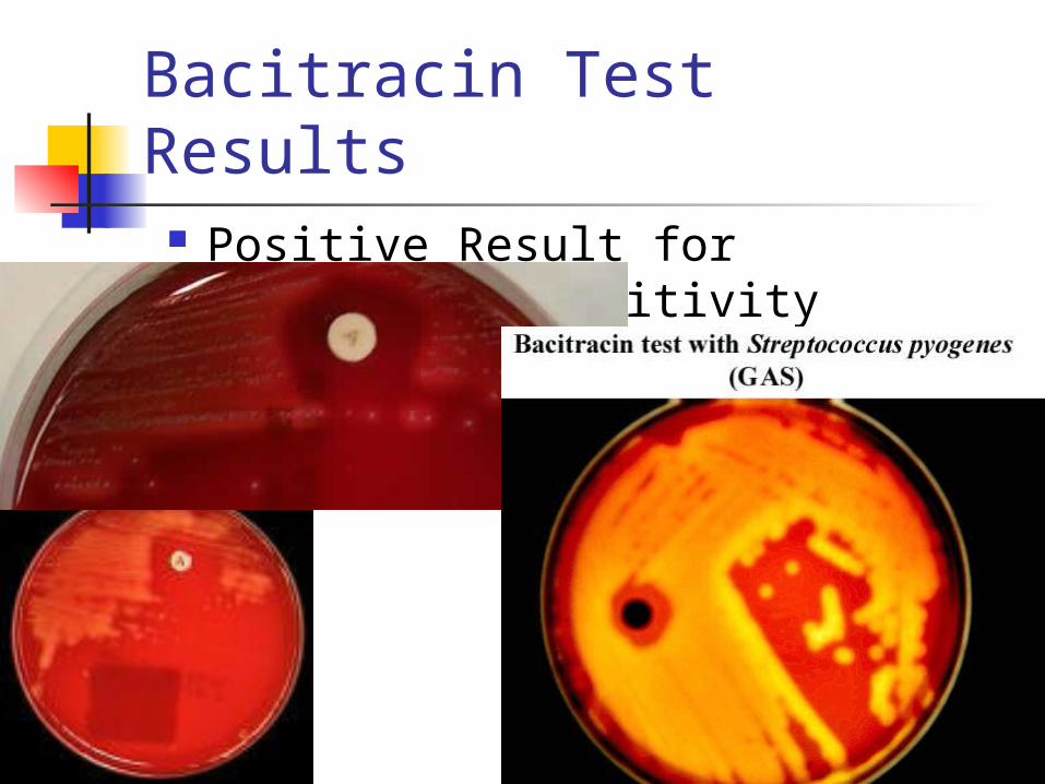
Bacitracin Test Results Positive Result for Bacitracin
Sensitivity
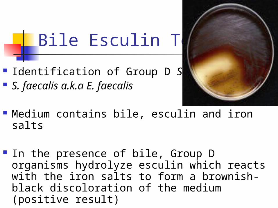
Bile Esculin Test
Identification of Group D Streptococci S. faecalis a.k.a E. faecalis
Medium contains bile, esculin and iron salts
In the presence of bile, Group D organisms hydrolyze esculin which reacts with the iron salts to form a brownish-black discoloration of the medium (positive result)
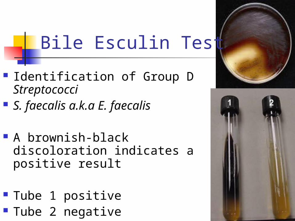
Bile Esculin Test
Identification of Group D Streptococci
S. faecalis a.k.a E. faecalis
A brownish-black discoloration indicates a positive result
Tube 1 positive Tube 2 negative
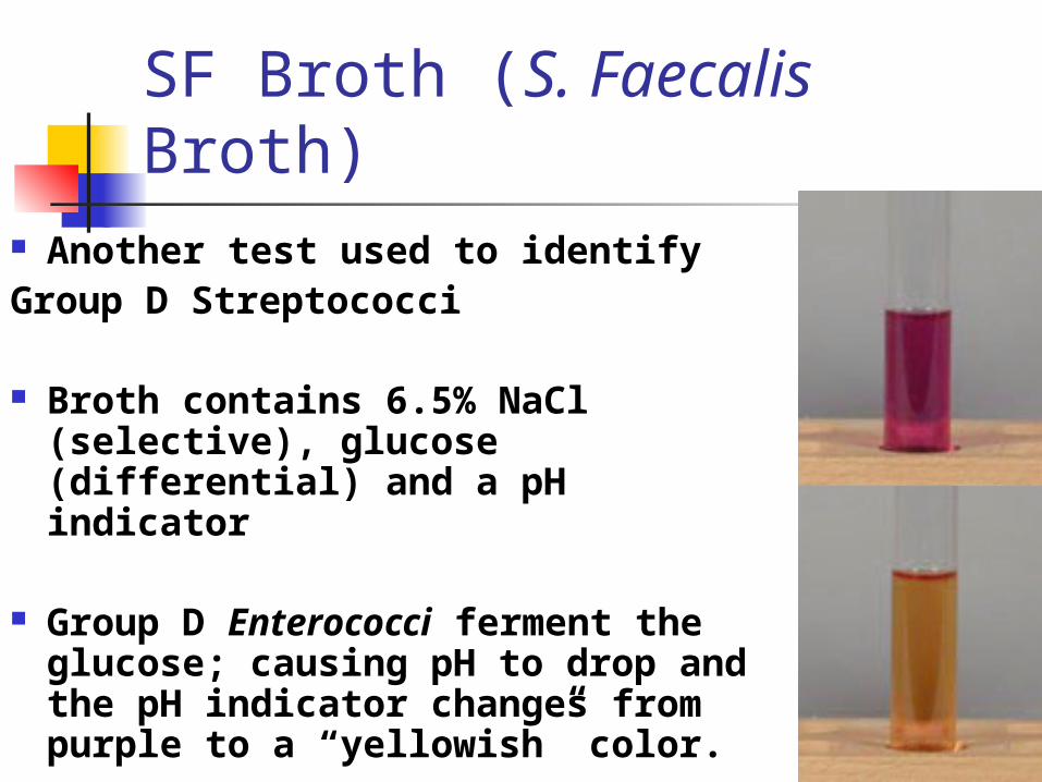
SF Broth (S. Faecalis Broth)
Another test used to identify Group D Streptococci
Broth contains 6.5% NaCl (selective), glucose (differential) and a pH indicator
Group D Enterococci ferment the glucose; causing pH to drop and the pH indicator changes from purple to a “yellowish” color.
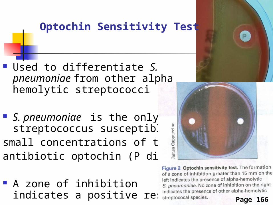
Used to differentiate S. pneumoniae from other alpha hemolytic streptococci
S. pneumoniae is the only streptococcus susceptible to
small concentrations of the antibiotic optochin (P disk)
A zone of inhibition indicates a positive result
Optochin Sensitivity Test
Page 166

Pneumococcus
Viridans
Group A Strep
Group D Strep
S. pneumoniae Alpha hemolyticOptochin sensitive
S. mitisAlpha hemolyticOptochin resistant
S. pyogenesBeta hemolyticBacitracin sensitive
S. faecalisAlpha / Gamma hemolytic+ Bile Esculin Test+ SF