SorLA regulates the activity of lipoprotein lipase by...
11
Research Article 1095 Introduction SorLA (sortilin-related receptor; also known as and hereafter referred to as SorLA), sortilin and SorCS1, SorCS2 and SorCS3 constitute the mammalian Vps10p-domain (Vps10p-D) receptor family. The common feature of this receptor family is the luminal Vps10p-D, which is dominated by a large ligand-binding ten- bladed b-propeller (Mazella et al., 1998; Petersen et al., 1997; Quistgaard et al., 2009). In addition to the Vps10p-D, the luminal parts of SorCS1, 2 and 3 also have short leucine-rich sequences (Hermey et al., 2004). SorLA is the largest Vps10p-D receptor and is far more complex. Apart from the Vps10p-D, the most prominent structural elements of SorLA are the 11 low-density lipoprotein (LDL)-receptor class A (LA) repeats and a b-propeller domain known from the LDL receptor family (Jacobsen et al., 1996). SorLA is therefore also known as LDL receptor 11 (LR11) (Yamazaki et al., 1997). The Vps10p-D carries a propeptide that is removed in the late trans Golgi network (TGN) by the proprotein convertase furin (Munck Petersen et al., 1999). Although ligand binding to the Vps10p-D in both sortilin and SorLA is impaired by the propeptide, binding of ligands to the LA repeats in SorLA is not affected (Westergaard et al., 2004). Sortilin and SorLA are multi-ligand-binding receptors, and their Vps10p domains are targeted by several growth factors and peptides. In addition, the LA repeats of SorLA interact with, for example, components of the plasminogen-activating system, platelet-derived growth factor, receptor-associated protein (RAP), apolipoprotein E and apolipoprotein AV (Gliemann et al., 2004; Jacobsen et al., 1996; Nilsson et al., 2008; Nilsson et al., 2007). SorLA is expressed at substantial levels in many tissues, such as kidney, testis, ovary, lymph nodes, vascular smooth muscle cells (SMCs) and in various parts of the nervous system. Sortilin and SorLA are multifunctional receptors, and they both mediate endocytic activity and trafficking among intracellular vesicles. For instance, we have shown that sortilin and SorLA shuttles between the TGN and late endosomes (Nielsen et al., 2007; Nielsen et al., 2001). This type of trafficking involves interactions between the cytoplasmic domain of the receptors and various adaptors, including adaptor-protein-1 (AP1), Golgi- localized, ear-containing, ARF-binding proteins (GGA1, GGA2 and GGA3) and the retromer complex (Nielsen et al., 2007). One known function of SorLA is to protect the amyloid precursor protein (APP) from proteolysis into soluble APPb and the insoluble amyloid b-peptide (Ab) (Rogaeva et al., 2007). In fact, SorLA has been genetically associated with late-onset Alzheimer’s disease (Andersen et al., 2005). SorLA has a different role in the vascular system, where a secreted soluble form of SorLA enhances intimal migration of SMCs, by interaction with urokinase-type plasminogen activator receptor (Gliemann et al., 2004; Ohwaki et al., 2007). Lipoprotein lipase (LPL) is differentially expressed in adipose tissue, heart and skeletal muscles cells, macrophages, the mammary gland, regions of the nervous system and in several other tissues (Braun and Severson, 1992). LPL hydrolyzes triacylglycerols in circulating lipoproteins, but can also function as a bridging molecule Summary Many different tissues and cell types exhibit regulated secretion of lipoprotein lipase (LPL). However, the sorting of LPL in the trans Golgi network has not, hitherto, been understood in detail. Here, we characterize the role of SorLA (officially known as SorLA-1 or sortilin-related receptor) in the intracellular trafficking of LPL. We found that LPL bound to SorLA under neutral and acidic conditions, and in cells this binding mainly occurred in vesicular structures. SorLA expression changed the subcellular distribution of LPL so it became more concentrated in endosomes. From the endosomes, LPL was further routed to the lysosomes, which resulted in a degradation of newly synthesized LPL. Consequently, an 80% reduction of LPL activity was observed in cells that expressed SorLA. By analogy, SorLA regulated the vesicle-like localization of LPL in primary neuronal cells. Thus, LPL binds to SorLA in the biosynthetic pathway and is subsequently transported to endosomes. As a result of this SorLA mediated-transport, newly synthesized LPL can be routed into specialized vesicles and eventually sent to degradation, and its activity thereby regulated. Key words: Intracellular trafficking, Lipoprotein lipase, SorLA (SORL1), Vps10p-domain receptor Accepted 6 October 2010 Journal of Cell Science 124, 1095-1105 © 2011. Published by The Company of Biologists Ltd doi:10.1242/jcs.072538 SorLA regulates the activity of lipoprotein lipase by intracellular trafficking Stine C. Klinger 1 , Simon Glerup 1 , Merete K. Raarup 2 , Muriel C. Mari 3 , Mette Nyegaard 4 , Gerbrand Koster 5 , Thaneas Prabakaran 1 , Stefan K. Nilsson 6 , Maj M. Kjaergaard 2 , Oddmund Bakke 5 , Anders Nykjær 1 , Gunilla Olivecrona 6 , Claus Munck Petersen 1 and Morten S. Nielsen 1, * 1 The MIND-Center, Department of Medical Biochemistry, University of Aarhus, Ole Worms Allé 1170, DK 8000 Aarhus C, Denmark 2 The MIND-Center, Stereology and Electron Microscopy Research Laboratory, University of Aarhus, Ole Worms Allé 1185, DK 8000, Aarhus C, Denmark 3 Department of Cell Biology, UMCU, Utrecht, AZU, Heidelberglaan 100, 3584 CX Utrecht, The Netherlands 4 Department of Haematology, Aalborg Hospital, Mølleparkvej 4, Postboks 561, 9100 Aalborg, Denmark 5 Department of Molecular Biosciences, University of Oslo, Postbox 1041 Blindern, N-0316 Oslo, Norway 6 Department of Medical Biosciences, Umea University, SE-901 87 Umeå, Sweden *Author for correspondence ([email protected]) Journal of Cell Science
Transcript of SorLA regulates the activity of lipoprotein lipase by...

Research Article 1095
IntroductionSorLA (sortilin-related receptor; also known as and hereafterreferred to as SorLA), sortilin and SorCS1, SorCS2 and SorCS3constitute the mammalian Vps10p-domain (Vps10p-D) receptorfamily. The common feature of this receptor family is the luminalVps10p-D, which is dominated by a large ligand-binding ten-bladed b-propeller (Mazella et al., 1998; Petersen et al., 1997;Quistgaard et al., 2009). In addition to the Vps10p-D, the luminalparts of SorCS1, 2 and 3 also have short leucine-rich sequences(Hermey et al., 2004). SorLA is the largest Vps10p-D receptor andis far more complex. Apart from the Vps10p-D, the most prominentstructural elements of SorLA are the 11 low-density lipoprotein(LDL)-receptor class A (LA) repeats and a b-propeller domainknown from the LDL receptor family (Jacobsen et al., 1996).SorLA is therefore also known as LDL receptor 11 (LR11)(Yamazaki et al., 1997). The Vps10p-D carries a propeptide that isremoved in the late trans Golgi network (TGN) by the proproteinconvertase furin (Munck Petersen et al., 1999). Although ligandbinding to the Vps10p-D in both sortilin and SorLA is impaired bythe propeptide, binding of ligands to the LA repeats in SorLA isnot affected (Westergaard et al., 2004). Sortilin and SorLA aremulti-ligand-binding receptors, and their Vps10p domains aretargeted by several growth factors and peptides. In addition, theLA repeats of SorLA interact with, for example, components of theplasminogen-activating system, platelet-derived growth factor,receptor-associated protein (RAP), apolipoprotein E andapolipoprotein AV (Gliemann et al., 2004; Jacobsen et al., 1996;
Nilsson et al., 2008; Nilsson et al., 2007). SorLA is expressed atsubstantial levels in many tissues, such as kidney, testis, ovary,lymph nodes, vascular smooth muscle cells (SMCs) and in variousparts of the nervous system.
Sortilin and SorLA are multifunctional receptors, and theyboth mediate endocytic activity and trafficking among intracellularvesicles. For instance, we have shown that sortilin and SorLAshuttles between the TGN and late endosomes (Nielsen et al.,2007; Nielsen et al., 2001). This type of trafficking involvesinteractions between the cytoplasmic domain of the receptors andvarious adaptors, including adaptor-protein-1 (AP1), Golgi-localized, ear-containing, ARF-binding proteins (GGA1, GGA2and GGA3) and the retromer complex (Nielsen et al., 2007). Oneknown function of SorLA is to protect the amyloid precursorprotein (APP) from proteolysis into soluble APPb and theinsoluble amyloid b-peptide (Ab) (Rogaeva et al., 2007). In fact,SorLA has been genetically associated with late-onset Alzheimer’sdisease (Andersen et al., 2005). SorLA has a different role in thevascular system, where a secreted soluble form of SorLA enhancesintimal migration of SMCs, by interaction with urokinase-typeplasminogen activator receptor (Gliemann et al., 2004; Ohwakiet al., 2007).
Lipoprotein lipase (LPL) is differentially expressed in adiposetissue, heart and skeletal muscles cells, macrophages, the mammarygland, regions of the nervous system and in several other tissues(Braun and Severson, 1992). LPL hydrolyzes triacylglycerols incirculating lipoproteins, but can also function as a bridging molecule
SummaryMany different tissues and cell types exhibit regulated secretion of lipoprotein lipase (LPL). However, the sorting of LPL in the transGolgi network has not, hitherto, been understood in detail. Here, we characterize the role of SorLA (officially known as SorLA-1 orsortilin-related receptor) in the intracellular trafficking of LPL. We found that LPL bound to SorLA under neutral and acidic conditions,and in cells this binding mainly occurred in vesicular structures. SorLA expression changed the subcellular distribution of LPL so itbecame more concentrated in endosomes. From the endosomes, LPL was further routed to the lysosomes, which resulted in adegradation of newly synthesized LPL. Consequently, an 80% reduction of LPL activity was observed in cells that expressed SorLA.By analogy, SorLA regulated the vesicle-like localization of LPL in primary neuronal cells. Thus, LPL binds to SorLA in thebiosynthetic pathway and is subsequently transported to endosomes. As a result of this SorLA mediated-transport, newly synthesizedLPL can be routed into specialized vesicles and eventually sent to degradation, and its activity thereby regulated.
Key words: Intracellular trafficking, Lipoprotein lipase, SorLA (SORL1), Vps10p-domain receptor
Accepted 6 October 2010Journal of Cell Science 124, 1095-1105 © 2011. Published by The Company of Biologists Ltddoi:10.1242/jcs.072538
SorLA regulates the activity of lipoprotein lipase byintracellular traffickingStine C. Klinger1, Simon Glerup1, Merete K. Raarup2, Muriel C. Mari3, Mette Nyegaard4, Gerbrand Koster5,Thaneas Prabakaran1, Stefan K. Nilsson6, Maj M. Kjaergaard2, Oddmund Bakke5, Anders Nykjær1,Gunilla Olivecrona6, Claus Munck Petersen1 and Morten S. Nielsen1,*1The MIND-Center, Department of Medical Biochemistry, University of Aarhus, Ole Worms Allé 1170, DK 8000 Aarhus C, Denmark2The MIND-Center, Stereology and Electron Microscopy Research Laboratory, University of Aarhus, Ole Worms Allé 1185, DK 8000, Aarhus C,Denmark3Department of Cell Biology, UMCU, Utrecht, AZU, Heidelberglaan 100, 3584 CX Utrecht, The Netherlands4Department of Haematology, Aalborg Hospital, Mølleparkvej 4, Postboks 561, 9100 Aalborg, Denmark5Department of Molecular Biosciences, University of Oslo, Postbox 1041 Blindern, N-0316 Oslo, Norway6Department of Medical Biosciences, Umea University, SE-901 87 Umeå, Sweden*Author for correspondence ([email protected])
Jour
nal o
f Cel
l Sci
ence
between lipoproteins and cellular membrane receptors or heparansulfate proteoglycans. In the brain, LPL is believed to be involvedin synaptic remodeling following injury by transporting cholesteroland lipids (Blain et al., 2004; Paradis et al., 2004). Newlysynthesized LPL is glycosylated and assembled into a homodimerin the endoplasmic reticulum (ER), where the enzyme also obtainsits catalytic activity by a lipase maturation factor 1 (LMF1)-dependent process (Peterfy et al., 2007). After passing through theGolgi network, LPL is secreted, accumulated in vesicles or sent tolysosomes for degradation (Cupp et al., 1987; Vannier and Ailhaud,1989). Many physiological conditions are known to affect LPLactivity by post-translational mechanisms. As an example, LPLactivity is rapidly downregulated during fasting in adipose tissue,by a mechanism that is independent of the level of LPL mRNA(Bergo et al., 2002). Although the transcriptional control of LPLhas been well described, the post-translational mechanismsregulating the level of secreted active LPL in several physiologicalstates are far from understood (Preiss-Landl et al., 2002; Wang andEckel, 2009).
As SorLA is engaged in intracellular trafficking, we haveexamined the influence of SorLA on the intracellular sorting andturnover of LPL. We demonstrate that SorLA binds LPL inintracellular vesicles and mediates a more vesicular localizationof LPL in transfected cells, as well as in primary neuronal andglial cultures. Moreover, we show that SorLA mediates a directtransport of LPL from the TGN to the endosomes, from whichLPL is delivered to the lysosomes for degradation. Through thistransport mechanism, SorLA is a post-translational regulator ofLPL activity.
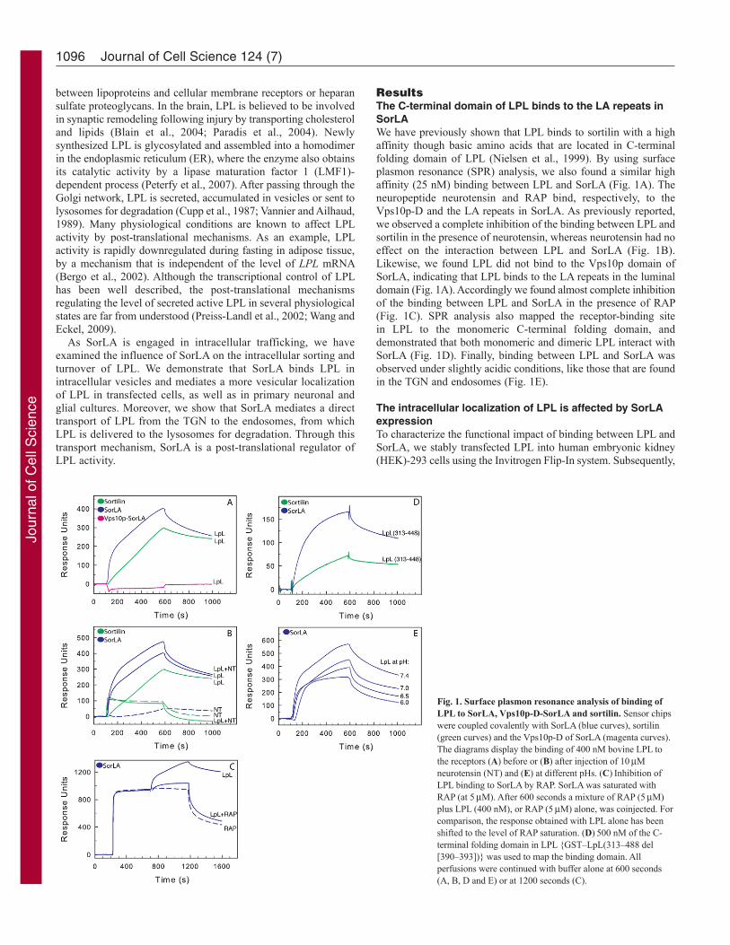
ResultsThe C-terminal domain of LPL binds to the LA repeats inSorLAWe have previously shown that LPL binds to sortilin with a highaffinity though basic amino acids that are located in C-terminalfolding domain of LPL (Nielsen et al., 1999). By using surfaceplasmon resonance (SPR) analysis, we also found a similar highaffinity (25 nM) binding between LPL and SorLA (Fig. 1A). Theneuropeptide neurotensin and RAP bind, respectively, to theVps10p-D and the LA repeats in SorLA. As previously reported,we observed a complete inhibition of the binding between LPL andsortilin in the presence of neurotensin, whereas neurotensin had noeffect on the interaction between LPL and SorLA (Fig. 1B).Likewise, we found LPL did not bind to the Vps10p domain ofSorLA, indicating that LPL binds to the LA repeats in the luminaldomain (Fig. 1A). Accordingly we found almost complete inhibitionof the binding between LPL and SorLA in the presence of RAP(Fig. 1C). SPR analysis also mapped the receptor-binding sitein LPL to the monomeric C-terminal folding domain, anddemonstrated that both monomeric and dimeric LPL interact withSorLA (Fig. 1D). Finally, binding between LPL and SorLA wasobserved under slightly acidic conditions, like those that are foundin the TGN and endosomes (Fig. 1E).
The intracellular localization of LPL is affected by SorLAexpressionTo characterize the functional impact of binding between LPL andSorLA, we stably transfected LPL into human embryonic kidney(HEK)-293 cells using the Invitrogen Flip-In system. Subsequently,
1096 Journal of Cell Science 124 (7)
Fig. 1. Surface plasmon resonance analysis of binding ofLPL to SorLA, Vps10p-D-SorLA and sortilin. Sensor chipswere coupled covalently with SorLA (blue curves), sortilin(green curves) and the Vps10p-D of SorLA (magenta curves).The diagrams display the binding of 400 nM bovine LPL tothe receptors (A) before or (B) after injection of 10Mneurotensin (NT) and (E) at different pHs. (C)Inhibition ofLPL binding to SorLA by RAP. SorLA was saturated withRAP (at 5M). After 600 seconds a mixture of RAP (5M)plus LPL (400 nM), or RAP (5M) alone, was coinjected. Forcomparison, the response obtained with LPL alone has beenshifted to the level of RAP saturation. (D)500 nM of the C-terminal folding domain in LPL {GST–LpL(313–488 del[390–393])} was used to map the binding domain. Allperfusions were continued with buffer alone at 600 seconds(A, B, D and E) or at 1200 seconds (C).
Jour
nal o
f Cel
l Sci
ence

this cell line was stably transfected with mutated or wild-typeSorLA, or sortilin. Western blotting confirmed the presence ofexpressed LPL and SorLA in the transfected cells (Fig. 2A).Furthermore, quantitative real-time PCR (qRT-PCR) of LPL mRNAverified that expression of LPL was similar in SorLA- and sortilin-expressing and non-expressing HEK-293 cell lines, and was notaffected by clonal selection (Table 1).
Immunofluorescent staining of LPL indicated that SorLA affectsits overall cellular localization. LPL showed a strongly increasedvesicular localization in cells expressing SorLA, whereas a less-disperse and perinuclear LPL localization was observed in cellsexpressing only LPL (Fig. 2B). To evaluate the increased vesicularstaining observed by immunofluorescence, automated quantificationof vesicular structures in LPL-transfected, and LPL and SorLA co-transfected, cells was performed using an Olympus scan^R imagingstation. Before fixation, 20 units of heparin/ml was added to themedium, as it is well-known that heparin causes release of activeLPL from the cell surface and thereby prevents any cellular uptakeof LPL (Makoveichuk et al., 2004). As the cells are about 4-mhigh and the LPL vesicles are distributed throughout the cell, amaximal intensity projection of a z-stack of four layers (1 mapart) was used for quantification for each of the 121 recordedpositions. We found 23.8 LPL-positive vesicles (s.e.±0.82, n1580)in SorLA transfectants, but only 12.02 (s.e.±0.62, n1054) in cellswithout SorLA, demonstrating a significantly higher amount ofvesicular LPL in the presence of SorLA (P<0.0001) (see theMaterials and Methods section for further description).
The changed subcellular localization of LPL in the presence ofSorLA was also observed by using subcellular fractionation. Aftersize separation in a velocity gradient (fractionated into 11 fractions),TGN46- and LAMP1-positive fractions (fractions 7–9) wereseparated further, according to buoyant density, by equilibriumgradient centrifugation. LPL was mainly concentrated in fractions2–3 and 7–10 in LPL-expressing cells, but peaked in fractions 4–10 when coexpressed with SorLA (Fig. 2C). A changed subcellularpattern was also seen upon co-transfection of a LPL-expressingconstruct with a construct expressing a SorLA variant that exhibitsimpaired sorting due to a C-terminal truncation [SorLA-MVIA;the MVIA motif mediates binding to the three GGA adaptors, andit was recently demonstrated that this interaction is involved in theTGN-to-endosome sorting of SorLA (Jacobsen et al., 2002; Nielsenet al., 2007)].
Intracellular localization of SorLA and LPLThe nature of the SorLA-positive vesicles was analyzed byimmunoelectron microscopy. As expected, we mainly found SorLAstaining in the ER, Golgi and endosomes, but occasionally we also
1097TGN-to-endosome trafficking of LPL
Table 1. Relative mRNA expression levels of LPL and SORL1in transfected cell lines
Relative RNA expression (fold change)
Cell line LPL SORL1
LPL 0.97±0.03 0.003±0.0008LPL plus SorLA 0.91±0.26 1.00±0.43LPL plus SorLA-MVIA 1.02±0.20 1.18±0.31LPL plus sortilin 0.98±0.05 0.002±0.0002
The endogenous housekeeping gene HPRT1 was used to normalize for thenumber of cells. The mean expression levels (±s.d.) for each cell line werecalculated from four biological quadruplicates (n4).
Fig. 2. The intracellular localization of LPL in stably transfected HEK-293 cells. (A)A western blot of 25g of total cell lysate, demonstrating thepresence of LPL and SorLA in stably transfected cells (LpL/SorLA, cellscotransfected with LPL and SorLA). (B)Cells were fixed and stained withchicken anti-LPL antibody (green) or mouse anti-SorLA antibody (red).(C)Subcellular fractionation of transfected cells. Fractions 7–9 of the velocitygradient (VG) were subjected to equilibrium-gradient centrifugation (EG).Fractions were analyzed by western blotting with the mouse anti-LPLantibody. The fractions containing the indicated intracellular markers aremarked by the bars. Scale bars: 5m.
Jour
nal o
f Cel
l Sci
ence

found staining on the plasma membrane (Fig. 3, top panel). Todifferentiate between early endosomes (EEs) and multi-vesicularbodies/late endosomes (MVB/LEs), we use the definition describedby Mari and colleagues in which endosomes with up to eightintraluminal vesicles are defined as EEs, whereas endosomes withnine or more intraluminal vesicles are defined as MVB/LEs (Mariet al., 2008). According to this definition, we observed intensestaining of SorLA in MVB/LEs and, probably, also in EEs,represented by endosomes with fewer than nine intraluminalvesicles (Fig. 3, lower panel). However, as electron-dense tubulesor vesicles are known to preferentially surround EEs, labeling invesicles surrounded by these structures supports the presence ofSorLA in EEs (arrows Fig. 3). Moreover, the SorLA-positiveelectron-dense tubules or vesicles resemble the endosome-to-TGN
recycling compartments that are positive for sortilin, mannose-6-phosphate receptor (MPR) and sorting nexin-1 (SNX1), asdescribed previously (Mari et al., 2008). As we did not succeed inacquiring electron microscope images of LPL, the presence of LPLin SorLA-positive vesicles was probed with fluorescence andcrosslinking experiments.
Fluorescence studies show a punctated colocalization of LPLand SorLA in the cytoplasm (Fig. 4A). This observation wasverified by use of a proximity ligation assay (PLA) that is basedon antibodies tagged with circular DNA probes. Probes locatedless than 40 nm from each other can hybridize and subsequentlybe amplified with a polymerase (Soderberg et al., 2006). Interactingprobes can be detected when fluorescently labeled oligonucleotidesare used. Although this technique is not suitable for subcellularlocalization studies, Fig. 4B shows positive punctated staining inLPL and SorLA co-transfected cells, representing structures withSorLA and LPL in close proximity. For comparison, PLA using thesame probes resulted in almost zero background in LPL-expressingcells devoid of SorLA (Fig. 4B, middle panel). To control for thespecificity of this method, we also performed this experiment withan antibody against MPR300, which colocalizes with SorLA in theTGN. As we observed no positive interaction using PLA with anti-MPR300 and anti-LPL primary antibodies (Fig. 4B, right-handpanel), we conclude that LPL and SorLA are tightly colocalized.
Finally, in biolabeled HEK-293 cells expressing LPL orcoexpressing LPL and SorLA, cellular proteins were crosslinkedwith the membrane-permeable cleavable crosslinker DSP[dithiobis(succinimidyl propionate)]. The 55-kDa LPL protein wasprecipitated using a rabbit anti-LPL antibody. In cells expressingSorLA, a 250-kDa protein, which corresponds to the size of SorLA,was co-precipitated with LPL (Fig. 4C, lane 4). Other, but muchweaker, bands were present on the gel and might represent tracesof dimerized LPL as well as traces of nonspecific proteininteractions. No substantial bands were obtained after precipitationwith beads without antibodies (Fig. 4C, lanes 1 and 3). Althoughthe 250-kDa band was only present in cells expressing SorLA, andnot in cells expressing only LPL, we confirmed further that the250-kDa band represented SorLA by precipitating with an anti-SorLA antibody (Fig. 4D, top panel). As a negative control fornonspecific crosslinking between SorLA and other proteins in thebiosynthetic pathway, crosslinked cell lysate from LPL and SorLAco-transfectants was immunoprecipitated with LMF1. This proteinwas mainly located in the ER, and did not result in SorLAprecipitation (Fig. 4D, lower panel).
From these fluorescence and crosslinking experiments, weconclude that intracellular LPL and SorLA colocalize and thatthere is probably a physical interaction between these two proteins.
LPL is directly routed from the TGN to endosomesWe have previously reported that SorLA can reach endosomeseither by retrograde trafficking from the cell surface or by directtransport from the TGN (Nielsen et al., 2007). To clarify whetherLPL is transported directly to endosomes from the TGN, we firstblocked protein synthesis in HEK-293 transfectants withcycloheximide. This resulted in a markedly reduced LPL stainingin intracellular vesicles after 2 hours (Fig. 5A, upper row). After 3hours vesicular LPL was almost absent, showing that vesicular-located LPL is either secreted or degraded within 3 hours. Whencycloheximide was added to the cells together with the lysosomalproteinase inhibitors leupeptin and pepstatin, vesicular stainingwas still present even after 6 hours (Fig. 5A, lower row). Although
1098 Journal of Cell Science 124 (7)
Fig. 3. Electron microscopy of SorLA intracellular localization. Ultrathincryosections of HEK-293 SorLA cells were immuno-gold-labeled with themouse anti-SorLA antibody. The gold size used for the immuno-labeling isindicated by the superscript number. The upper panel shows an overview ofSorLA labeling in transfected HEK-293 cells. SorLA staining is primarilyfound in the Golgi and endosomes. The lower panels show enlarged pictures ofendosomes. EEs and MVB/LEs have been defined as described previously(Mari et al., 2008). SorLA is found in the limiting membrane and inside EEsand late endosomes (LE), and in dense tubular structures in close vicinity toEEs (arrows). Scale bars: 100 nm. L, lysosomes; M, mitochondria; PM,plasma membrane.
Jour
nal o
f Cel
l Sci
ence

there was very little LPL staining after 3 hours in the presence ofcycloheximide, we wanted to exclude the possibility that LPL issimply secreted and then subsequently re-endocytosed toendosomes. It has been demonstrated and widely accepted thatLPL undergoes receptor-mediated endocytosis and that heparinblocks this uptake by releasing LPL from the cell surface.Accordingly, we also found that exogenous LPL is endocytosed inHEK-293 cells expressing SorLA, and that this uptake is blockedby heparin (Fig. 5B, columns 1 and 2). The weak surface stainingthat is observed in the presence of heparin probably represents a
1099TGN-to-endosome trafficking of LPL
Fig. 4. Cellular binding of LPL to SorLA. (A)HEK-293 cells coexpressingLPL and SorLA (LpL/SorLA), stained with rabbit anti-SorLA and mouse anti-LPL antibodies, indicates a large degree of colocalization between SorLA andLPL. As secondary antibodies, goat anti-(mouse Ig) and goat-anti-(rabbit Ig)antibodies, conjugated to Alexa-Fluor-488 and Alexa-Fluor-568, respectively,were used. The insets show a magnified region with colocalization.(B)Duolink in situ proximity ligation assay (PLA) performed on LPL andSorLA coexpressing and LPL-expressing cells. Chicken anti-LPL and mouseanti-SorLA antibodies were used as primary antibodies, and as secondary plusand minus probes we used PLA–anti-(mouse Ig) and PLA–anti-(chicken Ig),respectively. Positive reactions were detected with a 563-nm-fluorescence-label detection kit. As a control, mouse-anti-LPL and rabbit-anti-MPR300were used as primary antibodies. (C)LPL (55 kDa) from the crosslinkedlysates of biolabelled HEK-293 LPL-expressing (lanes 1 and 2) andLpL/SorLA cells (lane 3 and 4) was precipitated with a polyclonal anti-LPLantibody or blank beads. In cells coexpressing SorLA (250 kDa), the receptorwas co-precipitated. (D)Upper panel: SorLA from the crosslinked lysates ofbiolabeled LpL/SorLA cells was precipitated with rabbit-anti-LPL, rabbit-anti-SorLA or blank beads to verify that the precipitated 250 kDa band representsSorLA. Lower panel: western blot, probed with rabbit-anti-SorLA antibody, ofcross-linked LPL and LpL/SorLA transfectants. Lane 1, crude LpL/SorLA celllysate (input); lane 2, LPL transfectants, cross-linked with DSP andimmunoprecipitated with mouse-anti-LPL; lane 3, LpL/SorLA transfectants,crosslinked with DSP and immunoprecipitated with mouse-anti-LPL antibody;lane 4, LpL/SorLA transfectants, cross-linked with DSP andimmunoprecipitated with rabbit-anti-LMF1 antibody. Scale bars: 5m.
Fig. 5. Sorting of LPL in HEK-293 cells. HEK-293 cells expressing LPL,SorLA or both (LpL/SorA) were treated with cycloheximide, leupeptin andpepstatin (Leu/Pep) and/or heparin, as indicated. After incubation, the cellswere fixed and stained with mouse anti-LPL antibody. (A)Upper panel:blocking protein synthesis with cycloheximide demonstrates that the lifetimeof vesicular LPL is less than 3 hours. Lower panel: cycloheximide usedtogether with cellular proteinase inhibitors prolongs the lifetime of vesicularLPL to more than 6 hours. (B)Blocking endocytosis of LPL with heparinshows that vesicular LPL is observed over the whole incubation period,indicating that newly synthesized LPL is transported directly to vesicles. Scalebars: 5m.
Jour
nal o
f Cel
l Sci
ence
fraction of monomerized LPL, which is known to be insensitive toheparin (Makoveichuk et al., 2004). Column 3 in Fig. 5B showsthat heparin itself does not induce vesicular LPL staining ofendogenously expressed LPL. As endosomes are depleted for LPLafter 3 hours when protein synthesis and LPL uptake are blocked,we concluded that the LPL-positive endosomes seen after 6 hoursmust be due to a direct transport of newly synthesized LPL fromthe TGN, as uptake from the cell surface was blocked by heparin(Fig. 5B, column 4).
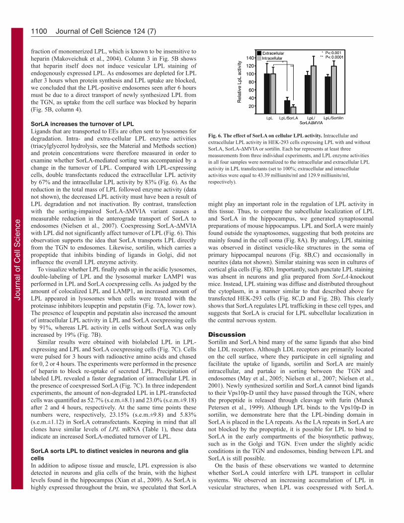
SorLA increases the turnover of LPLLigands that are transported to EEs are often sent to lysosomes fordegradation. Intra- and extra-cellular LPL enzyme activities(triacylglycerol hydrolysis, see the Material and Methods section)and protein concentrations were therefore measured in order toexamine whether SorLA-mediated sorting was accompanied by achange in the turnover of LPL. Compared with LPL-expressingcells, double transfectants reduced the extracellular LPL activityby 67% and the intracellular LPL activity by 83% (Fig. 6). As thereduction in the total mass of LPL followed enzyme activity (datanot shown), the decreased LPL activity must have been a result ofLPL degradation and not inactivation. By contrast, transfectionwith the sorting-impaired SorLA-MVIA variant causes ameasurable reduction in the anterograde transport of SorLA toendosomes (Nielsen et al., 2007). Coexpressing SorLA-MVIAwith LPL did not significantly affect turnover of LPL (Fig. 6). Thisobservation supports the idea that SorLA transports LPL directlyfrom the TGN to endosomes. Likewise, sortilin, which carries apropeptide that inhibits binding of ligands in Golgi, did notinfluence the overall LPL enzyme activity.
To visualize whether LPL finally ends up in the acidic lysosomes,double-labeling of LPL and the lysosomal marker LAMP1 wasperformed in LPL and SorLA coexpressing cells. As judged by theamount of colocalized LPL and LAMP1, an increased amount ofLPL appeared in lysosomes when cells were treated with theproteinase inhibitors leupeptin and pepstatin (Fig. 7A, lower row).The presence of leupeptin and pepstatin also increased the amountof intracellular LPL activity in LPL and SorLA coexpressing cellsby 91%, whereas LPL activity in cells without SorLA was onlyincreased by 19% (Fig. 7B).
Similar results were obtained with biolabeled LPL in LPL-expressing and LPL and SorLA coexpressing cells (Fig. 7C). Cellswere pulsed for 3 hours with radioactive amino acids and chasedfor 0, 2 or 4 hours. The experiments were performed in the presenceof heparin to block re-uptake of secreted LPL. Precipitation oflabeled LPL revealed a faster degradation of intracellular LPL inthe presence of coexpressed SorLA (Fig. 7C). In three independentexperiments, the amount of non-degraded LPL in LPL-transfectedcells was quantified as 52.7% (s.e.m.±8.1) and 23.0% (s.e.m.±9.18)after 2 and 4 hours, respectively. At the same time points thesenumbers were, respectively, 23.15% (s.e.m.±9.8) and 5.83%(s.e.m.±1.12) in SorLA cotransfectants. Keeping in mind that allclones have similar levels of LPL mRNA (Table 1), these dataindicate an increased SorLA-mediated turnover of LPL.
SorLA sorts LPL to distinct vesicles in neurons and gliacellsIn addition to adipose tissue and muscle, LPL expression is alsodetected in neurons and glia cells of the brain, with the highestlevels found in the hippocampus (Xian et al., 2009). As SorLA ishighly expressed throughout the brain, we speculated that SorLA
might play an important role in the regulation of LPL activity inthis tissue. Thus, to compare the subcellular localization of LPLand SorLA in the hippocampus, we generated synaptosomalpreparations of mouse hippocampus. LPL and SorLA were mainlyfound outside the synaptosomes, suggesting that both proteins aremainly found in the cell soma (Fig. 8A). By analogy, LPL stainingwas observed in distinct vesicle-like structures in the soma ofprimary hippocampal neurons (Fig. 8B,C) and occasionally inneurites (data not shown). Similar staining was seen in cultures ofcortical glia cells (Fig. 8D). Importantly, such punctate LPL stainingwas absent in neurons and glia prepared from SorLA-knockoutmice. Instead, LPL staining was diffuse and distributed throughoutthe cytoplasm, in a manner similar to that described above fortransfected HEK-293 cells (Fig. 8C,D and Fig. 2B). This clearlyshows that SorLA regulates LPL trafficking in these cell types, andsuggests that SorLA is crucial for LPL subcellular localization inthe central nervous system.
DiscussionSortilin and SorLA bind many of the same ligands that also bindthe LDL receptors. Although LDL receptors are primarily locatedon the cell surface, where they participate in cell signaling andfacilitate the uptake of ligands, sortilin and SorLA are mainlyintracellular, and partake in sorting between the TGN andendosomes (May et al., 2005; Nielsen et al., 2007; Nielsen et al.,2001). Newly synthesized sortilin and SorLA cannot bind ligandsto their Vps10p-D until they have passed through the TGN, wherethe propeptide is released through cleavage with furin (MunckPetersen et al., 1999). Although LPL binds to the Vps10p-D insortilin, we demonstrate here that the LPL-binding domain inSorLA is placed in the LA repeats. As the LA repeats in SorLA arenot blocked by the propeptide, it is possible for LPL to bind toSorLA in the early compartments of the biosynthetic pathway,such as in the Golgi and TGN. Even under the slightly acidicconditions in the TGN and endosomes, binding between LPL andSorLA is still possible.
On the basis of these observations we wanted to determinewhether SorLA could interfere with LPL transport in cellularsystems. We observed an increasing accumulation of LPL invesicular structures, when LPL was coexpressed with SorLA.
1100 Journal of Cell Science 124 (7)
Fig. 6. The effect of SorLA on cellular LPL activity. Intracellular andextracellular LPL activity in HEK-293 cells expressing LPL with and withoutSorLA, SorLA-MVIA or sortilin. Each bar represents at least threemeasurements from three individual experiments, and LPL enzyme activitiesin all four samples were normalized to the intracellular and extracellular LPLactivity in LPL transfectants (set to 100%; extracellular and intracellularactivities were equal to 43.39 milliunits/ml and 129.9 milliunits/ml,respectively).
Jour
nal o
f Cel
l Sci
ence

Using the scan^R screening system we found approximately twiceas many LPL-positive vesicles in cells coexpressing SorLA. Theeffect of SorLA on the intracellular localization of LPL was also
evident when cellular vesicles were separated by use ofultracentrifugation. The effect was most clear when LPL wasexpressed with a variant of SorLA that lacks the last four amino
1101TGN-to-endosome trafficking of LPL
Fig. 7. Degradation of LPL. (A)HEK-293 LPL and SorLA coexpressing cells(LpL/SorLA) were treated with leupeptin and pepstatin (+ leu/pep) for 24hours (medium replaced every 6 hours) and compared with untreated cells.After fixation, the cells were stained with chicken anti-LPL (green) and mouseanti-LAMP1 (a lysosomal marker; red) as primary antibodies. (B)IntracellularLPL activity in HEK-293 cells measured with and without leupeptin andpepstatin in cells expressing LPL and/or SorLA. The values are LPL enzymeactivity given in milliunits per ml of total cell extract with a proteinconcentration of 1.3 mg/ml. (C)Cells were biolabeled in the presence ofbrefeldin A and chased for 0, 2 or 4 hours. Labeled LPL was then precipitatedfrom the medium and lysates with polyclonal rabbit anti-LPL antibody, andanalyzed by reducing SDS-PAGE and autoradiography. The intensity of thecellular fractions (average of several experiments) is shown in the histogram.Error bars show the s.e.m. Scale bars: 5m.
Fig. 8. SorLA regulates LPL trafficking in the central nervous system.(A)Synaptosomal preparations of mouse hippocampus, analyzed by westernblotting, showing that the majority of SorLA and LPL is localized close to thesoma of hippocampal neurons and outside of synaptosomes. Fractions werealso probed using antibodies against the synaptic markers synaptophysin andPSD-95, and the purity of postsynaptic densities was validated by the absenceof synaptophysin and the presence of PSD-95. P1 and S1, initial pellet andsupernatant, respectively; P2, a crude synaptosomal preparation; S2, lightmembrane fraction; P3, synaptosomal membrane fraction; SPM, synapticplasma membrane; SVP, synaptic vesicle preparation; PSD, postsynapticdensities. (B)Primary cultures of hippocampal neurons prepared from wild-type mice were stained using antibodies against LPL (green) and the neuron-specific marker Tuj1 (red). (C)Hippocampal neurons prepared from wild-type(+/+) and SorLA-knockout mice (–/–) were stained for LPL (green). Nucleiwere visualized using DAPI (blue). (D)Similar staining of primary corticalglia cells. Both cell types have a high endogenous expression of SorLA, asshown by western blotting (gels on the right-hand side).
Jour
nal o
f Cel
l Sci
ence

acids in the cytoplasmic domain (SorLA-MVIA). From previouswork, we know that this variant of SorLA shuttles substantiallyless between the TGN and MVB/LEs, because MVIA is importantfor binding to the cytosolic adaptors GGA1, 2 and 3. Electronmicroscope images of SorLA demonstrate the presence of SorLAin EEs, MVB/LEs and electron-dense tubules or vesiclespreferentially surrounding the EEs. Similar tubular endosomalstructures surrounding the EEs have previously been demonstratedto be an endosome-to-TGN recycling compartment for sortilin andMPR300 (Mari et al., 2008). This recycling was suggested toinvolve SNX1, which is a subunit of the retromer complex andwhich is also located in the recycling compartment. As we knowthat SorLA is transported in an SNX1-dependent manner back tothe TGN (Nielsen et al., 2007), it is tempting to speculate that theSorLA-positive electron-dense tubules also function as endosome-to-TGN recycling compartments for SorLA. Fluorescent double-labeling of LPL and SorLA, and the PLA, demonstrated a tightcolocalization between LPL and SorLA. Moreover, using anintracellular crosslinker, coimmunoprecipitation of SorLA withLPL was demonstrated. We therefore conclude that intracellularLPL is located in close proximity to SorLA and that a physicalinteraction is likely to take place.
As SorLA internalizes ligands and also facilitates anterogradetransport from TGN to endosomes, the increased amount of LPL-positive peripheral punctated structures could result from eithertransport route. In our experiments, the lifetime for LPL in thesepunctated structures, which probably correspond to endosomes,was less than 3 hours. Furthermore, when blocking receptor-mediated endocytosis for up to 6 hours, LPL-positive vesicle-likestructures were still observed. Thus, we believe that a fraction ofnewly synthesized LPL is transported directly from the TGN to theendosomal system. This is the first time it has been demonstratedthat SorLA can bring cargo by way of anterograde transport fromthe TGN to the endosomal system (Fig. 9).
To evaluate the consequences of this SorLA-mediatedintracellular transport, we measured LPL activity in cells with andwithout SorLA and sortilin. The experiment showed thatintracellular LPL activity and mass was reduced ~80% in cellscoexpressing SorLA. As the LPL mRNA level was not affected byclonal selection, we infer that SorLA mediates LPL degradation.Furthermore, coexpressing LPL with the MVIA-deleted SorLAreceptor restored the LPL activity levels similar to those found incells without wild-type SorLA. As the MVIA motif only influencesthe cellular TGN-to-endosome transport, and not the endocytosis,this finding supports the conclusion that SorLA transports LPLdirectly from the TGN to endosomes. We assumed that the increaseddegradation of LPL took place in the lysosomes, and therefore werepeated the activity measurements in the presence of the proteinaseinhibitors leupeptin and pepstatin. In this setup, intracellular LPLactivity in the SorLA cells was increased by over 90% comparedwith less than 20% in cells expressing only LPL. Furthermore,LPL colocalized with the lysosomal marker LAMP1 in the presenceof lysosomal proteinase inhibitors. We therefore conclude thatSorLA enhances the degradation of LPL by routing it to lysosomes.
LPL is regulated in a tissue-specific manner, and the regulationtakes place on every step from transcriptional to post-transcriptionallevels (Wang and Eckel, 2009). The first step in the post-transcriptional regulation is in the ER, where LMF1 tightly regulatesthe activation of LPL (Fig. 9). LMF1 is ubiquitously expressed,and lack of functional LMF1 results in severe hypertriglyceridemia(Peterfy et al., 2007). Here, we demonstrate that SorLA might be
an important post-transcriptional regulator of LPL in the TGN.However, SorLA is only expressed in a subset of LPL-producingcells, and we therefore suspect that SorLA is one of the proteinsinvolved in tissue-specific LPL regulation. Both SorLA and LPLare highly expressed in the hippocampus, and our synaptosomalpreparations of hippocampi from wild-type mice suggest that themajority of SorLA and LPL are localized in the soma of neurons.The presence of LPL in the cell soma was confirmed in primaryhippocampal neurons and cortical glia cells by usingimmunofluorescence, showing that LPL was localized in distinctvesicle-like structures. Of particular importance was the findingthat no such punctate LPL staining was observed in culturesprepared from SORL1-knockout mice. Taken together, our findingsdemonstrate that SorLA plays a crucial role for LPL trafficking incells of the central nervous system. Interestingly, we observed thata minor fraction of LPL was found in presynaptic vesicles but thatit was absent from postsynaptic densities. LPL-deficient micedisplay impaired memory and this phenotype is attributed to apresynaptic defect in the hippocampus (Xian et al., 2009). Hence,it is tempting to speculate that SorLA regulates the routing of LPLto presynaptic vesicles and thereby affects general cognition. Otherevidence indicates that LPL is involved in the distribution ofcholesterol and other lipids in the brain. The need for lipids afteran injury fits with the observation of Paradis et al. (Paradis et al.,2004), who demonstrate that LPL is upregulated after ischemia.
In this study, we have demonstrated that SorLA can transportLPL directly from the biosynthetic pathway to the endosomalsystem, from which LPL is forwarded to lysosomes (Fig. 9). Thisis the first time a direct intracellular transport mechanism from theearly biosynthetic system has been demonstrated for SorLA. Wehave also shown that the Vps10p-D receptor sortilin cannot act asan intracellular-sorting receptor for LPL in the early compartmentsof the biosynthetic pathway, because the Vps10p-D is blocked bythe propeptide. This finding contributes to the understanding of thecomplex trafficking and regulation of LPL in the brain, wherealtered LPL levels are observed in relation to cerebral strokes andAlzheimer’s disease.
1102 Journal of Cell Science 124 (7)
Fig. 9 Model of LPL trafficking. LPL achieves its dimeric active form in theER through an LMF1-dependent process. After passage through the Golgi,LPL can be secreted by the constitutive pathway (CP) or it can bind to SorLAin the TGN and be sorted to late endosomes (LEs). From the LEs, SorLAreturns to the TGN, and LPL continues to lysosomes (LS). Sortilin (andreceptors of the LDL receptor family) can facilitate receptor-mediated uptakeof secreted LPL, whereby LPL also ends up in lysosomes for degradation.
Jour
nal o
f Cel
l Sci
ence

Materials and MethodsSurface plasmon resonanceThe soluble domains of SorLA and sortilin and the Vps10p domain of SorLA werepurified from stably transfected CHO cells, as described previously (Jacobsen etal., 2001; Tauris et al., 1998). Bovine LPL was purified from bovine milk on aheparin column (Bengtsson-Olivecrona and Olivecrona, 1991). Measurementswere performed on a BIAcore 3000 instrument and kinetic parameters weredetermined using the BIAevaluation 4.1 software. The BIAcore 3000 instrumentwas equipped with CM5 sensor chips maintained at 4°C. The sensor chip wasactivated by injection of 0.2 M N-ethyl-N-(3-dimethylaminopropyl)carbodiimideand 0.05 M N-hydroxy-succinimide in water. A 10 mM sodium acetate solution of15–35 g/ml purified receptor was injected over flow cells 2–4, giving densitiesof 88 fmol/mm2, 64 fmol/mm2 and 23 fmol/mm2 for sortilin, SorLA and Vps10p-SorLA, respectively. Remaining binding sites were blocked with 1 M ethanolamine.Flow cell 1 was activated and blocked without protein and used as a reference. Thesamples were injected in 10 mM HEPES pH 7.4, 150 mM NaCl, 1.5 mM CaCl2,1 mM EGTA and 0.010% surfactant P20, and this was also used as the runningbuffer. To obtain the relative response units, the response from flow cell 1 wassubtracted from the responses from flow cells 2–4. The sensor chip was regeneratedby injection of 10 mM glycin pH 4.0, 20 mM EDTA, 500 mM NaCl and 0.005%surfactant P20.
DNA and cell culturesSorLA (GenBank accession number NM_003105) and sortilin (GenBank accessionnumber NM_002959) full-length constructs were made as described previously(Nielsen et al., 2007). Human LPL (GenBank accession number NM_000237) wascloned into the pcDNA5/FRT/Hyg vector (Invitrogen). HEK-293 Flp-In cells(Invitrogen) were transfected with the pcDNA5/FRT/Hyg construct, containing full-length LPL, together with the pOG44 vector. Cells were transfected using FuGENE6 transfection reagent (Roche), and stably transfected clones were selected with 500g/ml hygromycin. Clones were selected for further stable transfection withpcDNA3.1(–)zeo constructs containing full-length or mutant SorLA, or full-lengthsortilin. For selection, clones were cultured with 500 g/ml zeocin and 500 g/mlhygromycin.
Cortical glia cells were prepared from P0 (postnatal day 0) pups. In brief, isolatedcortex was digested with papain (Worthington, Lakewood, NJ) followed by treatmentwith 100 g/ml DNaseI (Sigma). Then the tissue was triturated in Dulbecco’smodified Eagle’s medium (DMEM) containing 10% fetal bovine serum (FBS) and0.1 mg/ml primocin (Amaxa; Lonza Cologne, Germany), and seeded onto laminin-coated coverslips. After 2 hours in the incubator, cold medium (4°C) was added inorder to kill the neurons and leave only glia cells. Hippocampal neurons wereprepared similarly from isolated P0 hippocampus and cultured in neurobasal medium(Gibco, Invitrogen) supplemented with Glutamax, 2% B27 supplement (Invitrogen),20 M 5-fluorodeoxyuridine (Sigma) and 0.1 mg/ml primocin.
qRT-PCRcDNA was generated from quadruplicates (n4) of each cell type using a FastLaneCell cDNA Kit and a mixture of oligo-dT and random hexamers (Qiagen). qRT-PCRwas performed in a 96-well format using a LightCycler 480 instrument (Roche) andthe SYBR green method, according to the manufacturer’s instructions (Roche). PCRprimer pairs are listed in supplementary material Table S1. For all primer pairs, theamplification efficiency was determined from a serial dilution and found to be above95%. Samples were analyzed in triplicates and the mean expression levels,corresponding to SORL1 and LPL mRNA expression were normalized to HPRT1 (anendogenous housekeeping gene) mRNA levels using the Genex software(http://www.multid.se). Controls without reverse transcriptase for all cell-typesshowed no amplification, demonstrating no genomic DNA contamination in theRNA samples.
ImmunofluorescenceCells growing on coverslips were fixed in 4% formaldehyde and permeabilized in0.5% saponin. For secondary labeling, Alexa-Fluor-488 and -568-conjugatedantibodies (Invitrogen) were used. Cells were analyzed using an LSM510-METAlaser-scanning confocal microscope using a 40 or 63� C-Apochromat waterimmersion objective, NA 1.2 (Carl Zeiss, Göttingen, Germany). For inhibition oflysosomal hydrolases, 50 g/ml leupeptin and pepstatin (Sigma) were added (andreplaced every 6 hours) to the medium at 24 hours before fixation.
High-content imaging and quantitative analysis was performed with the Olympusscan^R imaging station. Images were acquired with a 40� objective, a triple-bandemission filter for Hoechst 33258, Alexa-Fluor-488 and Alexa-Fluor-568, and aHamamatsu camera (C8484-05G). For image analysis, z-stack-projected imageswere background-subtracted, after which an edge-detection algorithm was used forsegmentation of nuclei and LPL-containing vesicles. Vesicles were detected usingthis algorithm, which makes use of the gradient of the intensity in the image. If aclosed connecting line (edge) can be drawn around an object, the object is segmented.Objects were recognized and selected, independent of their shape, with the onlylimitation that they should be smaller than 3 m in diameter. About 95% of thedetected objects were smaller than 1 m in diameter and the average diameter of thedetected vesicles was ~500 nm. After gating out the images with zero cells or with
imaging artifacts, the mean number of LPL vesicles per cell was determined bycalculating for each image the ratio of vesicles to nuclei.
The antibody against LPL was from the egg yolk of chickens immunized withbovine LPL, mouse anti-LPL and rabbit anti-LAMP1 antibodies were generous giftsfrom John D. Brunzell and Sven Carlsson. Rabbit anti-LPL, mouse anti-SorLA andrabbit anti-SorLA antibodies were produced in-house. Anti-TGN46 antibody wasfrom AbD Serotec (Hamar, Norway) and anti-LMF1 antibody was from Sigma.
Subcellular fractionationFor preparation of a post-nuclear supernatant (PNS), HEK-293 Flp-In cells werewashed in Tris-buffered saline (TBS) and harvested in TBS with 0.4 mM PMSF at583 g for 10 minutes. The cells were resuspended in 10 ml of homogenizationbuffer (0.25 M sucrose, 1 mM EDTA, 1 mM magnesium acetate, 0.4 mM PMSFand 10 mM HEPES-KOH pH 7.2) and centrifuged at 583 g for 7 minutes. The cellswere resuspended in 700 l of homogenization buffer and disrupted by sevenpassages though a 21-gauge needle followed by five passages through a steel cellcracker (EMBL, Heidelberg, Germany) with a 9-m gap. The PNS was obtainedafter removal of nuclei and unbroken cells by centrifugation at 1843 g for 7minutes.
For subcellular fractionation, the PNS was subjected to density-gradientcentrifugation (SW41Ti rotor, 25,000 rpm, for 18 minutes) on a linear 0.3–1.2 Msucrose gradient prepared by mixing at angles of 45° for 10 minutes and at 80° for1 minute using a BioComp gradient master (BioComp, Fredericton, Canada).Fractions of 1 ml were collected from the top using a BioComp piston gradientfractionator. Selected fractions were pooled and subjected to equilibrium-gradientcentrifugation on a 0.8–1.2 M non-linear sucrose gradient with 5% increments(SW41Ti rotor, 25,000 rpm, overnight). Fractions were collected and analyzed bywestern blotting.
Electron microscopyHEK-293 SorLA-expressing cells, grown to 80% confluence, were fixed by addingfreshly prepared 4% formaldehyde in 0.1 M phosphate buffer (pH 7.4) to an equalvolume of culture medium for 10 minutes, followed by post-fixation in 4%formaldehyde at 4°C overnight. Ultrathin cryosectioning and immuno-gold labelingwas performed as previously described (Liou et al., 1996; Slot et al., 1991).
Proximity ligation assayThe proximity ligation assay (PLA) has been described in detail previously (Soderberget al., 2006). Briefly, cells were fixed with 4% formaldehyde for 20 minutes,permeabilized with 0.5% saponin and incubated with chicken anti-LPL plus mouseanti-SorLA antibodies, or mouse anti-LPL plus rabbit anti-SorLA antibodies for 90minutes. As secondary antibodies, conjugated with oligonucleotides, we usedPLA(+)–anti-(chicken Ig) plus PLA(–)–anti-(mouse Ig), and PLA(–)–anti-(mouseIg) plus PLA(+)–anti-(rabbit Ig), respectively. After hybridization and ligation,interacting probes were amplified with polymerase. Finally, a 563-nm-fluorescence-label detection kit (source??) was used to detect interactions. All incubations wereperformed at 37°C, using the incubation times and buffers given in the manufacturer’sinstructions (Olink Bioscience, Uppsala, Sweden).
Metabolic labeling, affinity precipitation and crosslinkingTransfected HEK-293 Flp-In cells were cultured in poly-L-lysine-coated six-welltrays (Sigma) to ~60% confluence. The cells were washed twice and preincubatedfor 20 minutes in cysteine- and methionine-free modified Eagle’s medium (Sigma)with 2% dialyzed FBS. Biolabeling was performed in the same medium supplementedwith 10 g/ml brefeldin A (Sigma) using 11.1 MBq/ml [35S]L-cysteine and [35S]L-methionine (pro-mix, GE Healthcare) for 3 hours at 37°C. For chase experiments,the cells were washed twice and incubated in complete DMEM with 20 units/mlheparin for 2–4 hours before the medium was harvested. Alternatively, the mediumwas harvested immediately after the labeling period. The cells were washed withPBS (4°C) and subsequently lyzed for 10 minutes in 1% Triton X-100 (in 20 mMTris-HCl, pH 8.0, 10 mM EDTA and 150 mM NaCl).
For immunoprecipitation, the cell lysates and medium (1 ml) were preincubatedwith 50 l of uncoupled CNBr-activated Sepharose 4B beads (GE Healthcare) (for2 hours at 4°C) before incubation with 75 l of GammaBind-G–Sepharose beads(GE Healthcare) coated with a rabbit polyclonal anti-LPL antibody (2 h, 4°C). Thebeads were washed five times in PBS pH 7.4 with 0.05% Tween 20, and preparedfor SDS-PAGE by boiling for 3 minutes in 100 l of reducing sample buffer (10 mMdithiothreitol and 2.5% SDS).
For crosslinking, labeled cells were washed with PBS and incubated for 30minutes with 2 mM DSP (Thermo Scientific) at room temperature, after which 100mM Tris-HCl, pH 7.5, was added for 15 minutes. Cells were washed in twice in PBSand lysed for 10 minutes as described above. Immunoprecipitation was afterwardsperformed with various antibodies.
LPL sortingHEK-293 Flp-In cells expressing LPL and SorLA were incubated for 1, 3 and 6hours in medium containing 20 units of heparin (SAD, Copenhagen, Denmark), for3 and 6 hours in medium containing 10 g/ml cycloheximide and 50 g/ml leupeptinand pepstatin, or for 1, 2 and 3 hours in medium with 10 g/ml cycloheximide only.
1103TGN-to-endosome trafficking of LPL
Jour
nal o
f Cel
l Sci
ence

The sorting of LPL was tested with HEK-293 cells expressing SorLA, LPL or both.The cells were incubated for 1, 3 and 6 hours in medium containing 50 nM LPLand/or 20 units heparin. Owing to the half-life of LPL and heparin, the medium wasreplaced every 3 hours.
LPL activity measurementsSamples were incubated with 200 l of a medium containing a phospholipid-stabilized emulsion of soybean triacylglycerols, with the same composition asIntralipid, into which 3H-labeled triolein had been incorporated by sonication(Fresenius-KABI, Uppsala, Sweden). The medium also contained 6% BSA, 16units/ml of heparin, to stabilize the lipase, 5% PMSF-treated heat-inactivated frozenrat serum, as a source of apoCII, 0.1 M NaCl and 0.15 M Tris-HCl, pH 8.5.Incubations were performed in a water-bath at 25°C and exact time was noted. Thereactions were stopped by addition of 2 ml of an isopropanol, heptane and sulfuricacid mixture (40:48:3.1), with 0.5 ml water. After vortexing and centrifugation, theupper heptane phase was transferred into a new tube containing alkaline-ethanol.Heptane was added followed by vortexing and centrifugation. The upper phase wasremoved and the step repeated. Finally, the amount of labeled fatty acids in the lowerphase was measured in a scintillation counter (WinSpectra, Wallac, Germany).Samples were assayed in triplicates. The activity was corrected for variations in theamount of cells by measuring the protein concentration of the lysates. One milliunitof lipase activity corresponds to the release of 1 nmol of fatty acids per minute.
Hippocampal synaptosomal preparationsThe procedure for fractionation of hippocampi from wild-type mice intosynaptosomes, synaptic vesicle fractions, synaptic plasma membrane and postsynapticdensities was performed essentially as described previously (Blackstone et al., 1992).Briefly, the hippocampus was isolated from the brains of 12 wild-type mice (8–12weeks of age) and homogenized in 0.32 M sucrose and 4 mM HEPES pH 7.4,containing proteinase inhibitors. P1 and S1 are the pellet and supernatant, respectively,after centrifugation for 10 minutes at 1000 g. S1 was further centrifuged for 15minutes at 10,000 g to obtain supernatant S2 and pellet P2, a crude synaptosomalpreparation. Solubilized P2 was centrifuged for 15 minutes at 10,000 g and theresulting pellet was solubilized in ice-cold water and centrifuged again at 25,000 gfor 20 minutes, generating the synaptosomal membrane fraction P3. The supernatantwas further centrifuged at 165,000 g for 2 hours generating pellet SVP, enriched inpresynaptic vesicles. Solubilized P3 was applied to a discontinuous gradientcontaining 0.8, 1.0, and 1.2 M sucrose and centrifuged at 150,000 g for 2 hours. Thefraction between the 1.0 and 1.2 M sucrose layer was recovered and diluted to give0.32 M sucrose, after which it was centrifuged again at 150,000 g for 30 min.,resulting in pellet SPM, containing the synaptic plasma membrane. ResuspendedSPM was centrifuged at 35,000 g for 20 minutes to obtain the pellet PSDI, containingpostsynaptic densities. This pellet was again solubilized and centrifuged at 200,000g for 20 minutes, to obtain the concentrated postsynaptic-density fraction PSDII. Allcentrifugation steps were performed at 4°C. Total protein concentration in fractionswas determined using a bicinchoninic acid kit (Sigma), and equal amounts ofproteins of each fraction were separated by reducing SDS-PAGE and analyzed bywestern blotting.
Use of animalsHousing, breeding and experimentation with mice complied with approved standardsfor humane treatment of vertebrate animals (Ministry of Justice, Denmark: permissionnumber J.2006/561-1206).
We thank Inger Andersen (Department of Medicinal Biochemistry,Aarhus, Denmark), Marc van Peski (Department of Cell Biology,Utrecht, The Netherlands) and Solveig Nilsson (Department of MedicalBiosciences, Umeå, Sweden) for technical assistance. The MIND-center is sponsored by The Lundbeck Foundation and support was alsoobtained from the Swedish Research Council (grant no 12203). Theuse of the Normic-UIO imaging platform at the Department ofMolecular Biosciences, University of Oslo is acknowledged. G.K. wassupported by grants from the Norwegian Research Council.
Supplementary material available online athttp://jcs.biologists.org/cgi/content/full/124/7/1095/DC1
ReferencesAndersen, O. M., Reiche, J., Schmidt, V., Gotthardt, M., Spoelgen, R., Behlke, J.,
von Arnim, C. A., Breiderhoff, T., Jansen, P., Wu, X. et al. (2005). Neuronalsorting protein-related receptor sorLA/LR11 regulates processing of the amyloidprecursor protein. Proc. Natl. Acad. Sci. USA 102, 13461-13466.
Bengtsson-Olivecrona, G. and Olivecrona, T. (1991). Phospholipase activity of milklipoprotein lipase. Methods Enzymol. 197, 345-356.
Bergo, M., Wu, G., Ruge, T. and Olivecrona, T. (2002). Down-regulation of adiposetissue lipoprotein lipase during fasting requires that a gene, separate from the lipasegene, is switched on. J. Biol. Chem. 277, 11927-11932.
Blackstone, C. D., Moss, S. J., Martin, L. J., Levey, A. I., Price, D. L. and Huganir,R. L. (1992). Biochemical characterization and localization of a non-N-methyl-D-aspartate glutamate receptor in rat brain. J. Neurochem. 58, 1118-1126.
Blain, J. F., Paradis, E., Gaudreault, S. B., Champagne, D., Richard, D. and Poirier,J. (2004). A role for lipoprotein lipase during synaptic remodeling in the adult mousebrain. Neurobiol. Dis. 15, 510-519.
Braun, J. E. and Severson, D. L. (1992). Regulation of the synthesis, processing andtranslocation of lipoprotein lipase. Biochem. J. 287, 337-347.
Cupp, M., Bensadoun, A. and Melford, K. (1987). Heparin decreases the degradationrate of lipoprotein lipase in adipocytes. J. Biol. Chem. 262, 6383-6388.
Gliemann, J., Hermey, G., Nykjaer, A., Petersen, C. M., Jacobsen, C. and Andreasen,P. A. (2004). The mosaic receptor sorLA/LR11 binds components of the plasminogen-activating system and platelet-derived growth factor-BB similarly to LRP1 (low-density lipoprotein receptor-related protein), but mediates slow internalization ofbound ligand. Biochem. J. 381, 203-212.
Hermey, G., Plath, N., Hubner, C. A., Kuhl, D., Schaller, H. C. and Hermans-Borgmeyer, I. (2004). The three sorCS genes are differentially expressed and regulatedby synaptic activity. J. Neurochem. 88, 1470-1476.
Jacobsen, L., Madsen, P., Moestrup, S. K., Lund, A. H., Tommerup, N., Nykjaer,A., Sottrup-Jensen, L., Gliemann, J. and Petersen, C. M. (1996). Molecularcharacterization of a novel human hybrid-type receptor that binds the alpha2-macroglobulin receptor-associated protein. J. Biol. Chem. 271, 31379-31383.
Jacobsen, L., Madsen, P., Jacobsen, C., Nielsen, M. S., Gliemann, J. and Petersen,C. M. (2001). Activation and functional characterization of the mosaic receptorSorLA/LR11. J. Biol. Chem. 276, 22788-22796.
Jacobsen, L., Madsen, P., Nielsen, M. S., Geraerts, W. P., Gliemann, J., Smit, A. B. andPetersen, C. M. (2002). The sorLA cytoplasmic domain interacts with GGA1 and -2 anddefines minimum requirements for GGA binding. FEBS Lett. 511, 155-158.
Liou, W., Geuze, H. J. and Slot, J. W. (1996). Improving structural integrity of cryosectionsfor immunogold labeling. Histochem. Cell Biol. 106, 41-58.
Makoveichuk, E., Castel, S., Vilaro, S. and Olivecrona, G. (2004). Lipoprotein lipase-dependent binding and uptake of low density lipoproteins by THP-1 monocytes andmacrophages: possible involvement of lipid rafts. Biochim. Biophys. Acta 1686, 37-49.
Mari, M., Bujny, M. V., Zeuschner, D., Geerts, W. J., Griffith, J., Petersen, C. M.,Cullen, P. J., Klumperman, J. and Geuze, H. J. (2008). SNX1 defines an earlyendosomal recycling exit for sortilin and mannose 6-phosphate receptors. Traffic 9,380-393.
May, P., Herz, J. and Bock, H. H. (2005). Molecular mechanisms of lipoproteinreceptor signalling. Cell. Mol. Life Sci. 62, 2325-2338.
Mazella, J., Zsurger, N., Navarro, V., Chabry, J., Kaghad, M., Caput, D., Ferrara,P., Vita, N., Gully, D., Maffrand, J. P. et al. (1998). The 100-kDa neurotensinreceptor is gp95/sortilin, a non-G-protein-coupled receptor. J. Biol. Chem. 273, 26273-26276.
Munck Petersen, C., Nielsen, M. S., Jacobsen, C., Tauris, J., Jacobsen, L., Gliemann,J., Moestrup, S. K. and Madsen, P. (1999). Propeptide cleavage conditionssortilin/neurotensin receptor-3 for ligand binding. EMBO J. 18, 595-604.
Nielsen, M. S., Jacobsen, C., Olivecrona, G., Gliemann, J. and Petersen, C. M.(1999). Sortilin/neurotensin receptor-3 binds and mediates degradation of lipoproteinlipase. J. Biol. Chem. 274, 8832-8836.
Nielsen, M. S., Madsen, P., Christensen, E. I., Nykjaer, A., Gliemann, J., Kasper, D.,Pohlmann, R. and Petersen, C. M. (2001). The sortilin cytoplasmic tail conveysGolgi-endosome transport and binds the VHS domain of the GGA2 sorting protein.EMBO J. 20, 2180-2190.
Nielsen, M. S., Gustafsen, C., Madsen, P., Nyengaard, J. R., Hermey, G., Bakke, O.,Mari, M., Schu, P., Pohlmann, R., Dennes, A. et al. (2007). Sorting by thecytoplasmic domain of the amyloid precursor protein binding receptor SorLA. Mol.Cell. Biol. 27, 6842-6851.
Nilsson, S. K., Lookene, A., Beckstead, J. A., Gliemann, J., Ryan, R. O. andOlivecrona, G. (2007). Apolipoprotein A-V interaction with members of the lowdensity lipoprotein receptor gene family. Biochemistry 46, 3896-3904.
Nilsson, S. K., Christensen, S., Raarup, M. K., Ryan, R. O., Nielsen, M. S. andOlivecrona, G. (2008). Endocytosis of apolipoprotein AV by members of the lowdensity lipoprotein receptor and the Vps10p domain receptor families. J. Biol. Chem.283, 25920-25927.
Ohwaki, K., Bujo, H., Jiang, M., Yamazaki, H., Schneider, W. J. and Saito, Y.(2007). A secreted soluble form of LR11, specifically expressed in intimal smoothmuscle cells, accelerates formation of lipid-laden macrophages. Arterioscler. Thromb.Vasc. Biol. 27, 1050-1056.
Paradis, E., Clavel, S., Julien, P., Murthy, M. R., de Bilbao, F., Arsenijevic, D.,Giannakopoulos, P., Vallet, P. and Richard, D. (2004). Lipoprotein lipase andendothelial lipase expression in mouse brain: regional distribution and selectiveinduction following kainic acid-induced lesion and focal cerebral ischemia. Neurobiol.Dis. 15, 312-325.
Peterfy, M., Ben-Zeev, O., Mao, H. Z., Weissglas-Volkov, D., Aouizerat, B. E.,Pullinger, C. R., Frost, P. H., Kane, J. P., Malloy, M. J., Reue, K. et al. (2007).Mutations in LMF1 cause combined lipase deficiency and severe hypertriglyceridemia.Nat. Genet. 39, 1483-1487.
Petersen, C. M., Nielsen, M. S., Nykjaer, A., Jacobsen, L., Tommerup, N., Rasmussen,H. H., Roigaard, H., Gliemann, J., Madsen, P. and Moestrup, S. K. (1997).Molecular identification of a novel candidate sorting receptor purified from humanbrain by receptor-associated protein affinity chromatography. J. Biol. Chem. 272,3599-3605.
1104 Journal of Cell Science 124 (7)
Jour
nal o
f Cel
l Sci
ence

Preiss-Landl, K., Zimmermann, R., Hammerle, G. and Zechner, R. (2002).Lipoprotein lipase: the regulation of tissue specific expression and its role in lipid andenergy metabolism. Curr. Opin. Lipidol. 13, 471-481.
Quistgaard, E. M., Madsen, P., Groftehauge, M. K., Nissen, P., Petersen, C. M. andThirup, S. S. (2009). Ligands bind to Sortilin in the tunnel of a ten-bladed beta-propeller domain. Nat. Struct. Mol. Biol. 16, 96-98.
Rogaeva, E., Meng, Y., Lee, J. H., Gu, Y., Kawarai, T., Zou, F., Katayama, T.,Baldwin, C. T., Cheng, R., Hasegawa, H. et al. (2007). The neuronal sortilin-relatedreceptor SORL1 is genetically associated with Alzheimer disease. Nat. Genet. 39,168-177.
Slot, J. W., Geuze, H. J., Gigengack, S., Lienhard, G. E. and James, D. E. (1991).Immuno-localization of the insulin regulatable glucose transporter in brown adiposetissue of the rat. J. Cell Biol. 113, 123-135.
Soderberg, O., Gullberg, M., Jarvius, M., Ridderstrale, K., Leuchowius, K. J.,Jarvius, J., Wester, K., Hydbring, P., Bahram, F., Larsson, L. G. et al. (2006).Direct observation of individual endogenous protein complexes in situ by proximityligation. Nat. Methods 3, 995-1000.
Tauris, J., Ellgaard, L., Jacobsen, C., Nielsen, M. S., Madsen, P., Thogersen, H. C.,Gliemann, J., Petersen, C. M. and Moestrup, S. K. (1998). The carboxy-terminal
domain of the receptor-associated protein binds to the Vps10p domain of sortilin.FEBS Lett. 429, 27-30.
Vannier, C. and Ailhaud, G. (1989). Biosynthesis of lipoprotein lipase in culturedmouse adipocytes. II. Processing, subunit assembly, and intracellular transport. J.Biol. Chem. 264, 13206-13216.
Wang, H. and Eckel, R. H. (2009). Lipoprotein lipase: from gene to obesity. Am. J.Physiol. Endocrinol. Metab. 297, E271-E288.
Westergaard, U. B., Sorensen, E. S., Hermey, G., Nielsen, M. S., Nykjaer, A.,Kirkegaard, K., Jacobsen, C., Gliemann, J., Madsen, P. and Petersen, C. M.(2004). Functional organization of the sortilin Vps10p domain. J. Biol. Chem. 279,50221-50229.
Xian, X., Liu, T., Yu, J., Wang, Y., Miao, Y., Zhang, J., Yu, Y., Ross, C., Karasinska,J. M., Hayden, M. R. et al. (2009). Presynaptic defects underlying impaired learningand memory function in lipoprotein lipase-deficient mice. J. Neurosci. 29, 4681-4685.
Yamazaki, H., Bujo, H. and Saito, Y. (1997). A novel member of the LDL receptorgene family with eleven binding repeats is structurally related to neural adhesionmolecules and a yeast vacuolar protein sorting receptor. J. Atheroscler. Thromb. 4,20-26.
1105TGN-to-endosome trafficking of LPL
Jour
nal o
f Cel
l Sci
ence