Shuning Li HHS Public Access Jie Chen Katherine S. Kula ...
17
Comparison of movement rate with different initial moment-to- force ratios Shuning Li a , Jie Chen b,c , Katherine S. Kula c a. Department of Engineering Technology, Indiana University Purdue University Indianapolis b. Department of Mechanical Engineering, Indiana University Purdue University Indianapolis c. Department of Orthodontics and Oral Facial Genetics, Indiana University Abstract Introduction: The objective of this clinical prospective study was to evaluate the effect of the two treatment strategies, translation or controlled tipping followed by root correction, on canine retraction efficiency, specifically canine movement rate. Methods: Twenty-one patients who needed bilateral maxillary canine retraction to close extraction space as part of their treatment plan were selected for this study. Segmental T-loops designed for controlled tipping or for translation were applied randomly to each side. Two digital maxillary dental casts (taken pre- and post-treatment) were used to measure the tooth displacements of each patient. The coordinate system located at the center of canine crown on the pre-treatment model with the three axes defined in the mesial-distal (M-D), buccal-lingual (B-L), and occlusal-gingival (O-G) directions was used to express the six tooth displacement components. The movement rates on the occlusal plane and in the M-D direction were computed. Movement rates were calculated by dividing the M-D displacements or the resultant displacement on the occlusal plane with the corresponding treatment time. Results: T-loops for controlled tipping moved canines faster (33.3% on occlusal plane and 38.5% in the M-D direction) than T-loops for translation. The differences are statistically significant (p = 0.041 on the occlusal plane and 0.020 in the M-D direction). Conclusion: 1. Moment-to-force ratio (M/F) impacts on the canine movement rate in a maxillary canine retraction treatment with segmented T-loop mechanism. 2. Within the neighborhood of the ratio for translation, lower M/F moves canine faster than higher M/F both on occlusal plane and in the M-D direction. Corresponding author: Shuning Li, PhD, Assistant Professor, Department of Engineering Technology, Indiana University Purdue University Indianapolis, 799 W. Michigan St. ET201, Indianapolis, IN 46202, U.S., Tel: 317-278-4415, [email protected]. Publisher's Disclaimer: This is a PDF file of an unedited manuscript that has been accepted for publication. As a service to our customers we are providing this early version of the manuscript. The manuscript will undergo copyediting, typesetting, and review of the resulting proof before it is published in its final citable form. Please note that during the production process errors may be discovered which could affect the content, and all legal disclaimers that apply to the journal pertain. HHS Public Access Author manuscript Am J Orthod Dentofacial Orthop. Author manuscript; available in PMC 2020 August 01. Published in final edited form as: Am J Orthod Dentofacial Orthop. 2019 August ; 156(2): 203–209. doi:10.1016/j.ajodo.2018.10.016. Author Manuscript Author Manuscript Author Manuscript Author Manuscript
Transcript of Shuning Li HHS Public Access Jie Chen Katherine S. Kula ...

Comparison of movement rate with different initial moment-to-force ratios
Shuning Lia, Jie Chenb,c, Katherine S. Kulac
a.Department of Engineering Technology, Indiana University Purdue University Indianapolis
b.Department of Mechanical Engineering, Indiana University Purdue University Indianapolis
c.Department of Orthodontics and Oral Facial Genetics, Indiana University
Abstract
Introduction: The objective of this clinical prospective study was to evaluate the effect of the
two treatment strategies, translation or controlled tipping followed by root correction, on canine
retraction efficiency, specifically canine movement rate.
Methods: Twenty-one patients who needed bilateral maxillary canine retraction to close
extraction space as part of their treatment plan were selected for this study. Segmental T-loops
designed for controlled tipping or for translation were applied randomly to each side. Two digital
maxillary dental casts (taken pre- and post-treatment) were used to measure the tooth
displacements of each patient. The coordinate system located at the center of canine crown on the
pre-treatment model with the three axes defined in the mesial-distal (M-D), buccal-lingual (B-L),
and occlusal-gingival (O-G) directions was used to express the six tooth displacement
components. The movement rates on the occlusal plane and in the M-D direction were computed.
Movement rates were calculated by dividing the M-D displacements or the resultant displacement
on the occlusal plane with the corresponding treatment time.
Results: T-loops for controlled tipping moved canines faster (33.3% on occlusal plane and 38.5%
in the M-D direction) than T-loops for translation. The differences are statistically significant (p =
0.041 on the occlusal plane and 0.020 in the M-D direction).
Conclusion: 1. Moment-to-force ratio (M/F) impacts on the canine movement rate in a maxillary
canine retraction treatment with segmented T-loop mechanism. 2. Within the neighborhood of the
ratio for translation, lower M/F moves canine faster than higher M/F both on occlusal plane and in
the M-D direction.
Corresponding author: Shuning Li, PhD, Assistant Professor, Department of Engineering Technology, Indiana University Purdue University Indianapolis, 799 W. Michigan St. ET201, Indianapolis, IN 46202, U.S., Tel: 317-278-4415, [email protected].
Publisher's Disclaimer: This is a PDF file of an unedited manuscript that has been accepted for publication. As a service to our customers we are providing this early version of the manuscript. The manuscript will undergo copyediting, typesetting, and review of the resulting proof before it is published in its final citable form. Please note that during the production process errors may be discovered which could affect the content, and all legal disclaimers that apply to the journal pertain.
HHS Public AccessAuthor manuscriptAm J Orthod Dentofacial Orthop. Author manuscript; available in PMC 2020 August 01.
Published in final edited form as:Am J Orthod Dentofacial Orthop. 2019 August ; 156(2): 203–209. doi:10.1016/j.ajodo.2018.10.016.
Author M
anuscriptA
uthor Manuscript
Author M
anuscriptA
uthor Manuscript

Introduction
Tooth movement rate is one of the most important orthodontic treatment outcomes. It has
been commonly used to evaluate effectiveness of treatment strategies and orthodontic
appliances. Accurate quantification of the movement rates is critical for the evaluation.
Tooth movement rate has been estimated in previous studies. The relationship between the
magnitude of the orthodontic force and tooth movement rate was investigated in animal
studies1–4. A mathematic model to describe the relationship was also developed based on the
experimental studies in beagle dogs.5 In human studies, movement rates were used to
compare the effects of force magnitudes,6–8 treatment efficiency among different appliances,9–11 and different types of anchorage.12 In these studies, tooth movement rate was quantified
by using either digital caliper13–16 or two-dimensional (2D) cephalometric analysis.17, 18
These methods could only provide linear or 2D measurements between landmarks. The
point-to-point measurement did not provide information on tooth movement type, translation
and rotation. The off-plane movement and rotations were not assessed. These components
are equally important because they characterize the side-effects. Therefore, three-
dimensional (3D) tooth movement needs to be quantified.
With the development of 3D imaging and modeling techniques, digital alternatives are
available to calculate the 3D tooth movements. The clinical 3D displacements can be
calculated using cone-beam computed tomography (CBCT) images.16 Digital dental casts
can also be used to quantify the 3D tooth displacements.19–23
Canine retraction after first premolar extraction is a treatment stage of space closure. Two
treatment strategies, translation or controlled tipping followed by root correction, are
commonly used. Different canine movement patterns (i.e. translation and controlled tipping)
were achieved by implementing different treatment strategies characterized by
corresponding moment-to-force ratios (M/F).24 Three-dimensional clinical evaluation has
not been reported on the effect of M/F on canine retraction efficiency, specifically canine
movement rate, which is the objective of this study.
Materials and Methods
Twenty-one patients (nine male and twelve female) participated in this split-mouth
prospective trial study. The study was approved by the Indiana University’s Institutional
Review Board and signed consent form was documented for each patient. The average age
of the patients was 21±8.8 years old. These patients were planned for bilateral maxillary
canine retractions (symmetrical pattern) after the upper dental arch was bracketed, leveled,
and aligned with sequential archwires to close first pre-molar extraction spaces. Canine roots
were radiographically evaluated by the clinician for typical dental anatomy (average root
sizes, support and shape). No palatal expansion was performed during the treatment, and no
metal or ceramic restorations were on the teeth of interest or their adjacent teeth. All patients
were allocated into two groups using the fixed allocation randomization method. Therefore,
equal numbers of the canines were treated under either controlled tipping (CT) or translation
(TR) strategy. The CT and TR strategies were accomplished by using specifically designed
Li et al. Page 2
Am J Orthod Dentofacial Orthop. Author manuscript; available in PMC 2020 August 01.
Author M
anuscriptA
uthor Manuscript
Author M
anuscriptA
uthor Manuscript

segmental T-loops, which could provide a distal retraction force of 124 cN along the
maxillary arch 25, an anti-tipping and an anti-rotation moment along with minimized force
and moment components in other directions.24 The average desired M/F ratios for 21
patients were 10.69 for TR, and 8.06 for CT. The details of the wire design and orthodontic
force verification were reported previously.24 Patients were scheduled with regular
appointments every 5-6 weeks and normally underwent multiple treatment intervals (TIs). A
TI was defined as a time interval when the inter-bracket distance between the canine and the
2nd molar on one side reduced over 1 mm. When a TI ended, a pair of new T-loops was re-
designed, fabricated, and reactivated consistent with the designated treatment strategies. The
study was completed when either CT or TR side finished the canine retraction with the
canine in the upright position judged by the clinicians using visual evaluation. Patients’ post-
treatment Cone beam computed tomography (CBCT) images were used to validate the final
positions of the CT side canines.
Two dental casts were used for each patient in this study. The first cast was taken before
starting the canine retraction, which was called the pre-treatment cast; and the second one
was taken before the last TI started, which was considered as the post-treatment cast. The
movements from the last TI were not included because tooth movements might stop
sometimes within the period, which was not recorded. The two casts were scanned and
digitized with an OPTIX 400S (3D Digital Corp, Sandy Hook, Conn) 3D laser scanner. The
resolution of the scanner is 60 μm. The corresponding 3D digital models were reconstructed
by using RapidForm® (INUS Technology Inc., Seoul, South Korea). The digital models
were used to calculate the overall canine displacement in treatment.
For each canine, the origin of the coordinate system was set at the crown center on the pre-
treatment model. The positive x-axis was directed buccally, y-axis distally, and z-axis
apically for the left side canine; and the positive y-axis is directed mesially, for the right
side.26 To be consistent, the displacement components on the right side were converted to be
expressed on the left side. The x and y axes formed a plane that was parallel to the posterior
occlusal plane. 26
The two digital models were superimposed by overlapping the 3D palatal (rugae) area. After
the superimposition, the post-treatment model was in a comparable position with the pre-
treatment model. Next, the crowns of the canine in the two locations were aligned using the
entire crown surface points. Then the canine movement’s six components in terms of
translation along and rotation about the three coordinate axes were computed from the
entries of the transformation matrix between the two crowns. The details of the
superimposition and calculation methods were reported in a previous publication.26
Two of the displacements were used to calculate the movement rate, the resultant
displacement and the displacement in the distal direction, y. The resultant displacements
represent the total movement rate on the occlusal plane (XY plane); while the mesial-distal
direction data were used to show the movement rate of the desired major movement in a
space closure case. The rate of tooth movement considered in this study was determined by
dividing total actual canine movement except the movement in the last TI by the treatment
time in days (also excluding the days of the last TI). The resultant movement rate on
Li et al. Page 3
Am J Orthod Dentofacial Orthop. Author manuscript; available in PMC 2020 August 01.
Author M
anuscriptA
uthor Manuscript
Author M
anuscriptA
uthor Manuscript

occlusal plane (XY plane), RG, was defined in Eq.1, where x, and y are the two translation
components and T is the treatment time. The corresponding movement direction, θ, was
defined in Eq. 2. It is the angle between the resultant movement direction on occlusal plane
(XY plane) and the distal direction (Figure 1).
RG = x2 + y2
TEq. 1
θ = tan−1 xy Eq. 2
To find the movement rate in the desired distal direction, the y component was used. The
movement rate, RC, was defined in Eq. 3, which was directed distally.
RC = yT Eq. 3
The intrusion (+) and extrusion (−) were represented by the z-components in the coordinate
system.
The means and standard deviations of the resultant movement rates, the movement direction
angle, and movement rates in the distal direction were calculated. Differences in canine
movement rates between controlled tipping side and translation side were compared with
paired t-tests, since there was only one observation on each side for each subject. The equal-
variance and normality assumptions were checked using normal probability plots.
Differences were considered to be significant at p<0.05.
The angle between the crown vestibular long axis of control tipping side canine and the
occlusal plane (Figure 2) was measured using each patient’s post-treatment CBCT image to
ensure that the control tipping side canine was in its upright position when the treatment
stopped.
Results
The resultant movement rates are shown in Figure 3. The rate for controlled tipping side
canine ranged between 0.000 and 0.041 mm/day; and for translation side ranged between
0.001 and 042 mm/day. The movement rate difference varies among patients (between 0.000
and 0.029 mm/day).
Distal movement rates are shown in Figure 4. The rate for controlled tipping ranged between
0.000 and 0.040 mm/day; and for translation ranged between 0.000 and 0.035 mm/day. The
movement rate difference varies among patients (between 0.000 and 0.026 mm/day).
The means and standard deviations of movement rates were shown in Table 1. Both on
occlusal plane (XY plane) and in M-D direction, controlled tipping T-loops moved canines
Li et al. Page 4
Am J Orthod Dentofacial Orthop. Author manuscript; available in PMC 2020 August 01.
Author M
anuscriptA
uthor Manuscript
Author M
anuscriptA
uthor Manuscript

33.3% (0.005/0.015) and 38.5% (0.005/0.013) faster than translation T-loops, respectively.
In general, differences were statistically significant.
In both the occlusal plane (XY plane) data and the distal direction data, most patients had
larger movement rate on their tipping side than the rate on their translation side. The
differences were calculated by using Eq. 4.
Difference = Tipping Side Rate − Translation Side Rate Eq. 4
Six patients, Patient 2, 3, 4, 7, 16, and 17 had larger translation movement rate on occlusal
plane and also in the distal direction (Figure 5 and 6). There is one patient, Patient 19, who
has the same tipping and translation movement rate on the occlusal plane (Figure 5); and one
patient, Patient 9, who has the same tipping and translation movement rate in the distal
direction (Figure 6). The average difference is 0.005 mm/day on the occlusal plane and also
in the distal direction.
The resultant movement directions on the occlusal plane (XY plane) varied from −60.3° to
58.7° with an average being 16.64° ± 26.583°. Positive moving direction angle implied that
tooth moved in the buccal and distal direction; while negative angle implied that tooth
moved in the lingual and distal direction. Four tipping side and five translation side canines
had the magnitude of the angles greater than 45°.
The means and standard deviations of intrusion and extrusion movement (z-axis) rates were
shown in Table 2. Intrusions are positive, and extrusions are negative.
The average angle between the control tipping side canine’s crown long axis and the
occlusal plane was 93.611° ± 1.158° among all 21 patients, which indicated that the control
tipping side canines returned to their upright positions when the treatments completed.
Discussion
The tipping and translation here represented treatment intentions, which were implemented
using different M/F, higher M/F for translation and lower M/F for tipping. They did not
represent clinical displacement patterns due to the fact that the T-Loop’s M/F was very
sensitive to the inter-bracket distance change due to tooth movement so that it changed as the
tooth moves.24
This study measures the absolute displacement relative to the established coordinate
systems. Using absolute displacement provides a more accurate description of the tooth
movement in response to an orthodontic force than using relative displacement. Previous
studies quantified relative displacement between two landmarks on the moving and
anchorage teeth, respectively. Anchorage loss occurred clinically, which affected the
measurement accuracy. Furthermore, the displacement was commonly described on a point.
Choosing different points resulted in different displacement especially when a large tipping
was involved. These factors would affect accuracy and make comparisons unreliable.
Li et al. Page 5
Am J Orthod Dentofacial Orthop. Author manuscript; available in PMC 2020 August 01.
Author M
anuscriptA
uthor Manuscript
Author M
anuscriptA
uthor Manuscript

Reported maxillary canine retraction rates varied greatly. For the frictionless mechanics, the
largest value reported was more than 2.5 mm/month among ten patients by using a calibrated
sectional archwire,27 while the smallest one was about 0.6 mm/month measured from six
patients using a vertical loop, which would cause the force to rapidly decline.28 The average
value was about 1.6 mm/month.18, 22, 29–31 In this study, the average movement rate on the
occlusal plane calculated to 0.56 mm/month for the tipping side and 0.42 mm/month for the
translation side (28 days per month). The values were relatively smaller compared to
previous studies, likely due to different displacement quantification methods. The absolute
3D displacement of the crown center was calculated and only the movements in the occlusal
plane were used (Eq. 1 and 3) in this study while others did point-to-point measurements,
which might not be in the occlusal plane and in the directions clearly defined in this study,
and might likely use relative displacement.
Intuitively, the same orthodontic load system would produce consistent clinical tooth
movement rate. The phenomenon was not observed in this study, meaning that load system
is not the only dominant factor controlling the tooth movement rate. The movement rate
varied significantly among patients, which might be caused by multiple factors, such as bone
quality, functional habit, and genotype.
Description of tooth movement with respect to a static reference helps clinician to quantify
real tooth displacement and effects of various treatment strategies. Many previous studies
reported tooth movement relative to an anchorage tooth, which might move during the
treatment. Thus, the absolute movement was not obtained. In this study, movements of the
moving and anchorage teeth were described relative to the rugae area. They were decoupled
so that they could be analyzed separately. Thus, the reported movement would be the true
response to the applied orthodontic load for a specified tooth. Furthermore, the intrusion/
extrusion was reliably quantified, which was not possible from the relative displacement. On
the other hand, clinicians could still use distal displacement to evaluate clinical outcomes in
the space closure case. For this reason, the distal displacement, y, was used to calculate the
closing movement rate.
Our previous study showed that M/F could not uniquely determine the pattern of clinical
tooth movement when segmental T-loop was used.26 As canine moved during treatment,
M/F changed which caused the change of canine movement pattern. However, the results in
this study showed that initial M/F still had important impact on the efficiency of tooth
movement in terms of movement rate. Based on the segmental T-loop design for each
patient, the initial M/F for the controlled tipping side was lower than the initial M/F for the
translation side.24 The results of this study showed that the canine with lower initial M/F
moved statistically faster on the occlusal plane and in the distal direction for 71.4% patients
(Figure 4 and 5, Table 1).
There were discrepancies between movement rates in the distal and on the occlusal plane.
The difference was due to the coupled movement in either lingual or buccal direction. In this
canine retraction treatment, with well-controlled orthodontic load system, ideally canines
should move in the distal direction to close the space, thus both movement rates should be
close. In reality, teeth had significant movements along other directions, i.e. buccal-lingual
Li et al. Page 6
Am J Orthod Dentofacial Orthop. Author manuscript; available in PMC 2020 August 01.
Author M
anuscriptA
uthor Manuscript
Author M
anuscriptA
uthor Manuscript

(B-L) or occlusal-gingival (O-G) direction. The variation of the movement directions was
large, indicating the well-controlled orthodontic load was not the only dominant factor
controlling the tooth movement direction. The large variation in angle, indicated large inter-
patient variation in the movement direction. There were also coupled movements in the O-G
direction. The average movement rate was 0.004mm, which was very slow comparing with
the movement rate in the occlusal plane (20%) or distal direction. This observation showed
that no trend on intrusion or extrusion could be found.
Conclusion
1. Moment-to-force ratio (M/F) impacts on the canine movement rate in a maxillary
canine retraction treatment with segmented T-loop mechanism.
2. Within the neighborhood of the ratio for translation, lower M/F moves canine
faster than higher M/F both on occlusal plane and in the M-D direction.
References
[1]. Pilon JJ, Kuijpers-Jagtman AM, Maltha JC: Magnitude of orthodontic forces and rate of bodily tooth movement. An experimental study. Am J Orthod Dentofacial Orthop 1996, 110:16–23. [PubMed: 8686673]
[2]. Kohno T, Matsumoto Y, Kanno Z, Warita H, Soma K: Experimental tooth movement under light orthodontic forces: rates of tooth movement and changes of the periodontium. J Orthod 2002, 29:129–35. [PubMed: 12114463]
[3]. Van Leeuwen EJ, Kuijpers-Jagtman AM, Von den Hoff JW, Wagener FA, Maltha JC: Rate of orthodontic tooth movement after changing the force magnitude: an experimental study in beagle dogs. Orthod Craniofac Res 2010, 13:238–45. [PubMed: 21040467]
[4]. van Leeuwen EJ, Maltha JC, Kuijpers-Jagtman AM: Tooth movement with light continuous and discontinuous forces in beagle dogs. Eur J Oral Sci 1999, 107:468–74. [PubMed: 10625106]
[5]. Ren Y, Maltha JC, Van ‘t Hof MA, Kuijpers-Jagtman AM: Optimum force magnitude for orthodontic tooth movement: a mathematic model. Am J Orthod Dentofacial Orthop 2004, 125:71–7. [PubMed: 14718882]
[6]. Owman-Moll P, Kurol J, Lundgren D: Effects of a doubled orthodontic force magnitude on tooth movement and root resorptions. An inter-individual study in adolescents. Eur J Orthod 1996, 18:141–50. [PubMed: 8670926]
[7]. Owman-Moll P, Kurol J, Lundgren D: The effects of a four-fold increased orthodontic force magnitude on tooth movement and root resorptions. An intra-individual study in adolescents. Eur J Orthod 1996, 18:287–94. [PubMed: 8791892]
[8]. Yee JA, Turk T, Elekdag-Turk S, Cheng LL, Darendeliler MA: Rate of tooth movement under heavy and light continuous orthodontic forces. Am J Orthod Dentofacial Orthop 2009, 136:150 e1–9; discussion -1.
[9]. Hayashi K, Uechi J, Murata M, Mizoguchi I: Comparison of maxillary canine retraction with sliding mechanics and a retraction spring: a three-dimensional analysis based on a midpalatal orthodontic implant. Eur J Orthod 2004, 26:585–9. [PubMed: 15650067]
[10]. Mezomo M, de Lima ES, de Menezes LM, Weissheimer A, Allgayer S: Maxillary canine retraction with self-ligating and conventional brackets. Angle Orthod 2011, 81:292–7. [PubMed: 21208082]
[11]. Dholakia KD, Bhat SR: Clinical efficiency of nonconventional elastomeric ligatures in the canine retraction phase of preadjusted edgewise appliance therapy: an in-vivo study. Am J Orthod Dentofacial Orthop 2012, 141:715–22. [PubMed: 22640673]
Li et al. Page 7
Am J Orthod Dentofacial Orthop. Author manuscript; available in PMC 2020 August 01.
Author M
anuscriptA
uthor Manuscript
Author M
anuscriptA
uthor Manuscript

[12]. Thiruvenkatachari B, Ammayappan P, Kandaswamy R: Comparison of rate of canine retraction with conventional molar anchorage and titanium implant anchorage. Am J Orthod Dentofacial Orthop 2008, 134:30–5. [PubMed: 18617100]
[13]. Dahlberg G: Statistical methods for medical and biological students. London,: G. Allen & Unwin ltd., 1940.
[14]. Lee H, Hong H: Hybrid surface- and voxel-based registration for MR-PET brain fusion. Lect Notes Comput Sc 2005, 3617:930–7.
[15]. Bjork A: Variations in the growth pattern of the human mandible: longitudinal radiographic study by the implant method. J Dent Res 1963, 42(1)Pt 2:400–11. [PubMed: 13971295]
[16]. Chen J, Li S, Fang S: Quantification of tooth displacement from cone-beam computed tomography images. Am J Orthod Dentofacial Orthop 2009, 136:393–400. [PubMed: 19732674]
[17]. Park TJ, Lee SH, Lee KS: A method for mandibular dental arch superimposition using 3D cone beam CT and orthodontic 3D digital model. Korean journal of orthodontics 2012, 42:169–81. [PubMed: 23112948]
[18]. Tai K, Park JH, Mishima K, Hotokezaka H: Using superimposition of 3-dimensional cone-beam computed tomography images with surface-based registration in growing patients. The Journal of clinical pediatric dentistry 2010, 34:361–7. [PubMed: 20831142]
[19]. Commer P, Bourauel C, Maier K, Jager A: Construction and testing of a computer-based intraoral laser scanner for determining tooth positions. Medical engineering & physics 2000, 22:625–35. [PubMed: 11259931]
[20]. Ashmore JL, Kurland BF, King GJ, Wheeler TT, Ghafari J, Ramsay DS: A 3-dimensional analysis of molar movement during headgear treatment. Am J Orthod Dentofacial Orthop 2002, 121:18–29; discussion -30. [PubMed: 11786867]
[21]. Keilig L, Piesche K, Jager A, Bourauel C: Applications of surface-surface matching algorithms for determination of orthodontic tooth movements. Computer methods in biomechanics and biomedical engineering 2003, 6:353–9. [PubMed: 14675956]
[22]. Cha BK, Lee JY, Jost-Brinkmann PG, Yoshida N: Analysis of tooth movement in extraction cases using three-dimensional reverse engineering technology. Eur J Orthod 2007, 29:325–31. [PubMed: 17513876]
[23]. Burstone CJ, Pryputniewicz RJ: Holographic determination of centers of rotation produced by orthodontic forces. American journal of orthodontics 1980, 77:396–409. [PubMed: 6928741]
[24]. Xia Z, Chen J, Jiangc F, Li S, Viecilli RF, Liu SY: Load system of segmental T-loops for canine retraction. Am J Orthod Dentofacial Orthop 2013, 144:548–56. [PubMed: 24075663]
[25]. Viecilli RF, Katona TR, Chen J, Hartsfield JK, Roberts WE: Three-dimensional mechanical environment of orthodontic tooth movement and root resorption. American Journal of Orthodontics and Dentofacial Orthopedics 2008, 133:791.e11–.e26.
[26]. Li S, Xia Z, Liu SS, Eckert G, Chen J: Three-dimensional canine displacement patterns in response to translation and controlled tipping retraction strategies. Angle Orthod 2015, 85:18–25. [PubMed: 24885592]
[27]. Tanne K, Inoue Y, Sakuda M: Biomechanical behavior of the periodontium before and after orthodontic tooth movement. Angle Orthod 1995, 65:123–8. [PubMed: 7785803]
[28]. Daskalogiannakis J, McLachlan KR: Canine retraction with rare earth magnets: an investigation into the validity of the constant force hypothesis. Am J Orthod Dentofacial Orthop 1996, 109:489–95. [PubMed: 8638593]
[29]. Hasler R, Schmid G, Ingervall B, Gebauer U: A clinical comparison of the rate of maxillary canine retraction into healed and recent extraction sites--a pilot study. Eur J Orthod 1997, 19:711–9. [PubMed: 9458604]
[30]. Lee BW: The force requirements for tooth movement, Part I: Tipping and bodily movement. Aust Orthod J 1995, 13:238–48. [PubMed: 8975662]
[31]. Darendeliler MA, Darendeliler H, Uner O: The drum spring (DS) retractor: constant and continuous force for canine retraction. Eur J Orthod 1997, 19:115–30. [PubMed: 9183061]
Li et al. Page 8
Am J Orthod Dentofacial Orthop. Author manuscript; available in PMC 2020 August 01.
Author M
anuscriptA
uthor Manuscript
Author M
anuscriptA
uthor Manuscript

Highlights
• The objective is to provide 3D clinical evaluation for canine movement rate.
• Both controlled tipping and translation strategies are studied.
• Movement rates quantified both on occlusal plane and in mesial-distal
direction.
• Results show that moment-to-force ratios have impact on canine movement
rates.
Li et al. Page 9
Am J Orthod Dentofacial Orthop. Author manuscript; available in PMC 2020 August 01.
Author M
anuscriptA
uthor Manuscript
Author M
anuscriptA
uthor Manuscript

Figure 1. Movement Directions on Occlusal Plane (XY Plane)
Li et al. Page 10
Am J Orthod Dentofacial Orthop. Author manuscript; available in PMC 2020 August 01.
Author M
anuscriptA
uthor Manuscript
Author M
anuscriptA
uthor Manuscript

Figure 2. Angle between Canine Long Axis and the Occlusal Plane
Li et al. Page 11
Am J Orthod Dentofacial Orthop. Author manuscript; available in PMC 2020 August 01.
Author M
anuscriptA
uthor Manuscript
Author M
anuscriptA
uthor Manuscript

Figure 3. Canine Movement Rate Results on Occlusal Plane (XY Plane)
Li et al. Page 12
Am J Orthod Dentofacial Orthop. Author manuscript; available in PMC 2020 August 01.
Author M
anuscriptA
uthor Manuscript
Author M
anuscriptA
uthor Manuscript

Figure 4. Canine Movement Rate Results in Distal-mesial Direction
Li et al. Page 13
Am J Orthod Dentofacial Orthop. Author manuscript; available in PMC 2020 August 01.
Author M
anuscriptA
uthor Manuscript
Author M
anuscriptA
uthor Manuscript

Figure 5. Movement Rate Difference (Tipping Side Rate - Translation Side Rate) on Occlusal Plane
(XY Plane)
Li et al. Page 14
Am J Orthod Dentofacial Orthop. Author manuscript; available in PMC 2020 August 01.
Author M
anuscriptA
uthor Manuscript
Author M
anuscriptA
uthor Manuscript
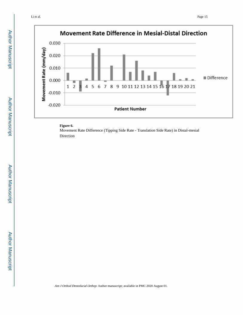
Figure 6. Movement Rate Difference (Tipping Side Rate - Translation Side Rate) in Distal-mesial
Direction
Li et al. Page 15
Am J Orthod Dentofacial Orthop. Author manuscript; available in PMC 2020 August 01.
Author M
anuscriptA
uthor Manuscript
Author M
anuscriptA
uthor Manuscript

Author M
anuscriptA
uthor Manuscript
Author M
anuscriptA
uthor Manuscript
Li et al. Page 16
Table 1
Movement Rates Statistical Analysis Results (mm/day)
Distance (mm) Tipping Translation Difference p-value
Mean ± SD Mean ± SD Mean ± SD
Occlusal Plane 0.020 ± 0.011 0.015 ± 0.011 0.005 ± 0.010 0.041
Mesial-Distal 0.018 ± 0.011 0.013 ± 0.010 0.005 ± 0.010 0.020
Am J Orthod Dentofacial Orthop. Author manuscript; available in PMC 2020 August 01.

Author M
anuscriptA
uthor Manuscript
Author M
anuscriptA
uthor Manuscript
Li et al. Page 17
Table 2
Movement rate of Intrusion or Extrusion (mm)
Tipping Translation Overall
Mean ± SD Mean ± SD Mean ± SD
−0.002 ± 0.013 −0.006 ± 0.016 −0.004 ± 0.014
Am J Orthod Dentofacial Orthop. Author manuscript; available in PMC 2020 August 01.


![kula world [english] - ia601008.us.archive.org](https://static.fdocuments.in/doc/165x107/620e4eb78ac281663c45d856/kula-world-english-.jpg)