s3. file · Web viewSkeletal Joints Study Guide. 1) ... cartilaginous and synovial...
11
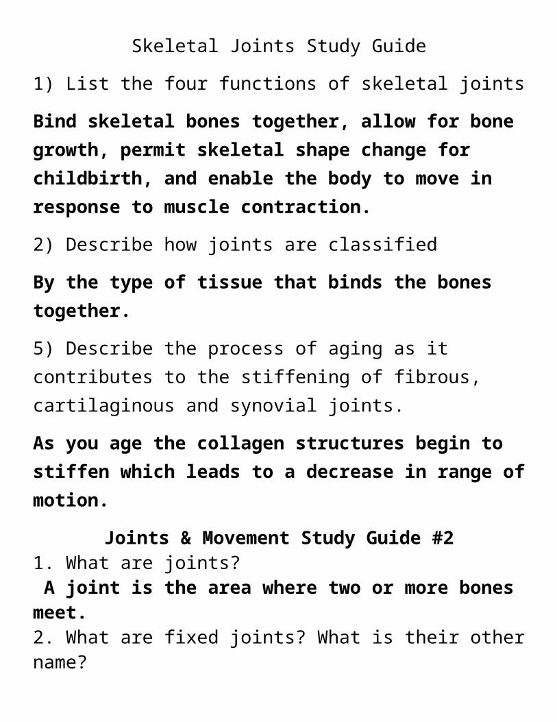
Skeletal Joints Study Guide 1) List the four functions of skeletal joints Bind skeletal bones together, allow for bone growth, permit skeletal shape change for childbirth, and enable the body to move in response to muscle contraction. 2) Describe how joints are classified By the type of tissue that binds the bones together. 5) Describe the process of aging as it contributes to the stiffening of fibrous, cartilaginous and synovial joints. As you age the collagen structures begin to stiffen which leads to a decrease in range of motion. Joints & Movement Study Guide #2 1. What are joints? A joint is the area where two or more bones meet. 2. What are fixed joints? What is their other name?
Transcript of s3. file · Web viewSkeletal Joints Study Guide. 1) ... cartilaginous and synovial...
Skeletal Joints Study Guide
1) List the four functions of skeletal joints
Bind skeletal bones together, allow for bone growth, permit skeletal shape change for childbirth, and enable the body to move in response to muscle contraction.
2) Describe how joints are classified
By the type of tissue that binds the bones together.
5) Describe the process of aging as it contributes to the stiffening of fibrous, cartilaginous and synovial joints.
As you age the collagen structures begin to stiffen which leads to a decrease in range of motion.
Joints & Movement Study Guide #21. What are joints? A joint is the area where two or more bones meet.2. What are fixed joints? What is their other name? Fixed joints are joints where there is no movement between the bones. Fixed joints are also called immovable joints or fibrous joints 3. What are slightly moveable joints? What is their other name? Slightly movable joints allow a slight amount of movement. They are also called cartilaginous joints 5. What are freely moveable joints? What is their other name? Freely moveable joints allow a wide range of movements.
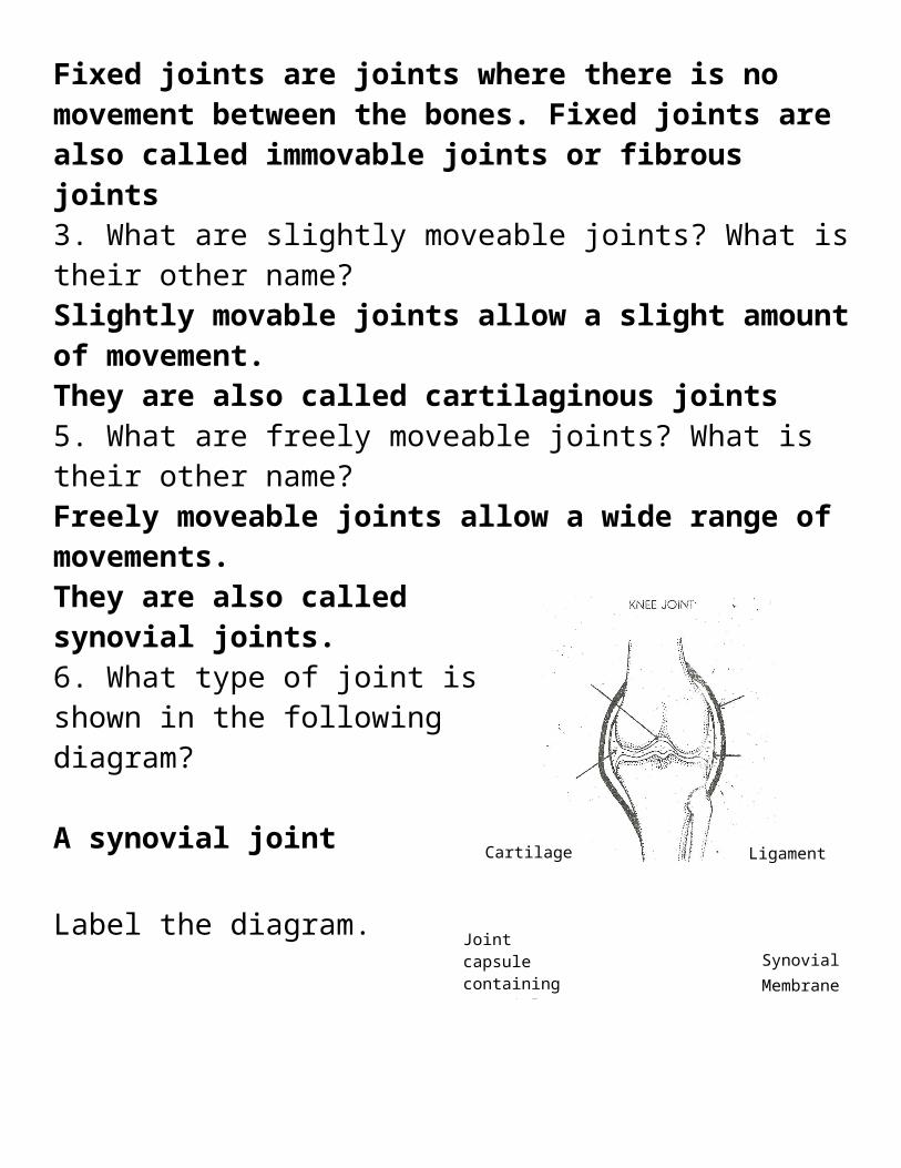
They are also called synovial joints. 6. What type of joint is shown in the following diagram?
A synovial joint Label the diagram.
7. How many types of freely moveable joints are there? There are six types of synovial joints 8. Under each picture write the names of the synovial joints shown.
a. Hinge joint b. Saddle joint c. Ball and socket joint
Cartilage Ligament
Joint capsule containing synovial fluid
Synovial Membrane
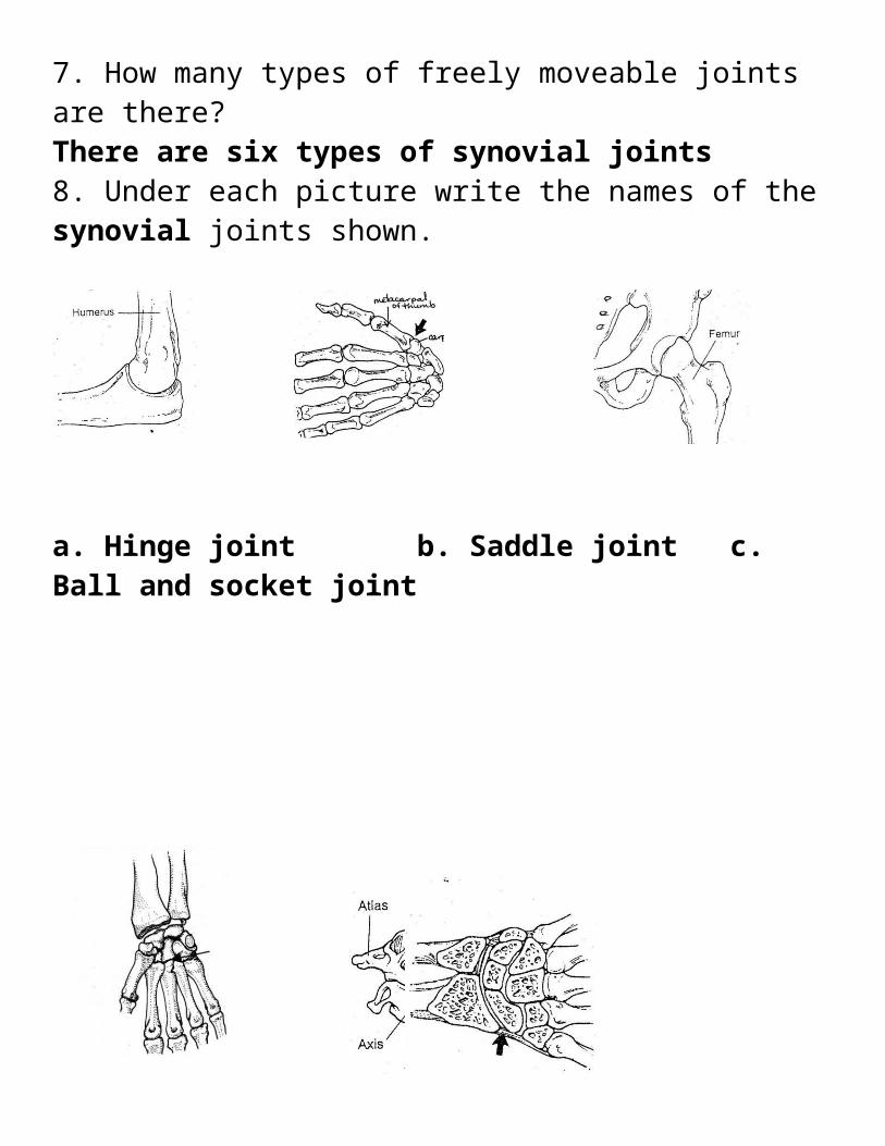
d. gliding joint e. pivot joint f. condyloid
Identify one area in the body where the following joints can be found. (6 marks) Ball and socket: hips and shoulder
Hinge: elbow, knee
Pivot: neck(where the, atlas & axis meet), upper forearm(where the radius& ulna meet)
Condyloid: wrist
Cartilaginous: spine(vertebral column), between ribs and sternum
Fixed: skull
Complete the following sentences

Ligaments link bones together and limit the range of movement of a joint.
Cartilage protects bones and prevents them from wearing each other down.
**Muscle to bone is a tendonDescribe the following types of movement. Flexion is a movement which bends, thereby decreasing the angle at the joint between the bones
Extension is a movement which straightens, thereby increasing the angle at the joint between the bones.
Abduction is a sideways movement of a bone/ limb away from the center line of the body.
Adduction is a sideways movement towards the centre line of the body.
Circumduction is a movement where the end of the bone makes a circle.
Rotation is a turning movement around an imaginary line or central axis.
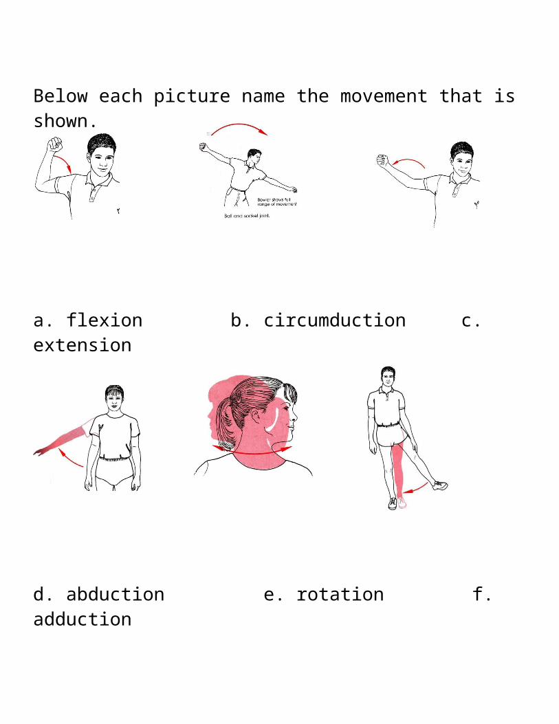
Below each picture name the movement that is shown.
a. flexion b. circumduction c. extension
d. abduction e. rotation f. adduction
Muscular System Study Guide3. List the 5 functions of the muscular system
1) Movement (walking, talking, breathing, etc.)2) Provide muscle tone3) Propel body fluids and food4) Generate heartbeat5) Distribute Heat
5. Thick filament fibers in myofibrils are myonsin while thin filaments are called actin.
6. For each step in the diagram describe the state of contraction for thesarcomere.a) Sarcomere is relaxedb) Sarcomere is beginning to shorten
actin slides toward the center, as it is pulled by the myosin crossbridgesc) Sarcomere is contracted
7. Acetylcholine is the neurotransmitter that is released to stimulate the muscle.
8. What two ions are exchanged when the neurotransmitter opens the gates on the membrane of the muscle fiber? ADP & Phosphate
9. What ion is stored by the sarcoplasmic reticulum and starts muscle contraction? calcium10. Place the following steps in order of how a muscle contracts
1 a) neuron releases a neurotransmitter to stimulate the muscle3b) myosin heads attach to the actin filaments4c) ATP is converted to ADP when the myosin head change shape pulling the actin filament5d) Myosin heads pull the actin filament together2 e) Sarcoplasmic reticulum release calcium ions
12. Match the following terms using the word bank:a. connective tissues that joins muscle to bone Tendonb. another name for muscle cell muscle fiberc. type of protein filament (2 answers) myonsin, actind. movement of muscles in a circular motion Circumductione. movement of muscles towards the midline adductionf. movement of muscles away from the midline abductiong. cell membrane of the muscle sarcolemmah. muscle responsible for primary movement prime mover

13. Identify if this would be anaerobic (fermentation), aerobic respiration
1. Sprint 60 meters in 5 10 sec ‐ anaerobic2. Walking 45 minutes aerobic3. Creates only 2 ATP anaerobic4. Creates 36 ATP aerobic5. Lactic acid is created anaerobic
14. (Aerobic/ Anaerobic) exercise would create the most amount of energy for muscle contraction.
15. The depletion of oxygen causes muscles to fatigue.16. Muscles produce different amounts of force because of different numbers of muscle fibers contracting. (True or False)
17. An inherited disease that causes muscles to degenerate and atrophy is calledMuscular dystrophy.
18. A strain involves the overextension of a tendon while a sprain injures the ligament.
19. What is the best way to maintain our muscle mass and strength as one ages? exercise
20. The end of the muscle that is attached to the moving bone is called the insertion, while the muscle that is attached to the non‐moving bone is called the origin.
21. Identify if these muscle pairs are antagonist or synergistica. flexor digitorum and extensor digitorum antagonistb. gastrocnemius and tibialis anterior antagonistc. biceps brachii and triceps brachii antagonistd. rectus abdominius and external oblique synergistic

22. Extensor muscles cause bones to (straighten/bend) at the joint while flexors (straighten/bend).
23. Muscle work in antagonistic pairs.Ex: biceps flexes and triceps extends when elbow is flexed.24. List the ways muscles are named. (at least 3)Direction of fibers, location, size, number of origins, action, shape, origin and insertion
25. Match each description with the correct muscle:hamstrings 1. bending of the knee would flex the _______Inner thigh adductor group 2. muscle group that adducts legBiceps brachii 3. bending of elbow flexes the _______Triceps brachii 4. bending the elbow extends the ________sternocleidomastoid 5. turns the headfrontalis 6. raises the eyebrowmasseter 7. closes jaw and allows for chewingrectus abdominus 8. muscles of 6 pack stomach‐gastrocnemius 9. muscle that flexes for “dancers toes”deltoid 10 adducts arm at shoulder pointOrbicularis oculi 11. closes the eyeOrbicularis oris 12. kissing muscle


![file · Web viewskeletal muscle tissue only. ... Into each sentence below, copy a term from the word . bank that . correctly . completes . the. sentence. [bone] [nervous] [skeletal]](https://static.fdocuments.in/doc/165x107/5ab6f9957f8b9ab7638e5209/web-viewskeletal-muscle-tissue-only-into-each-sentence-below-copy-a-term.jpg)