Research Article How to Evaluate Adenomyosis in Patients Affected...
8
Research Article How to Evaluate Adenomyosis in Patients Affected by Endometriosis? Nadine Di Donato and Renato Seracchioli Minimally Invasive Gynaecological Surgery Unit, S. Orsola-Malpighi Hospital, University of Bologna, Via Massarenti 13, 40138 Bologna, Italy Correspondence should be addressed to Nadine Di Donato; [email protected] Received 7 June 2014; Accepted 31 July 2014; Published 12 August 2014 Academic Editor: Peng Hui Wang Copyright © 2014 N. Di Donato and R. Seracchioli. is is an open access article distributed under the Creative Commons Attribution License, which permits unrestricted use, distribution, and reproduction in any medium, provided the original work is properly cited. Objective. e aim of the study is to evaluate adenomyosis in patients undergoing surgery for different type of endometriosis. It is an observational study including women with preoperative ultrasound diagnosis of adenomyosis. Demographic data and symptoms were recorded (age, body mass index, parity, history of previous surgery, dysmenorrhea, dyspareunia, dyschezia, dysuria, and abnormal uterine bleeding). Moreover a particular endometrial shape “question mark sign” linked to the presence of adenomyosis was assessed. Results. From 217 patients with ultrasound diagnosis of adenomyosis, we found 73 with ovarian histological confirmation of endometriosis, 92 with deep infiltrating endometriosis, and 52 patients who underwent surgery for infertility. Women with adenomyosis alone represented the oldest group of patients (37.8 ± 5.18 years, = 0.02). Deep endometriosis patients were nulliparous more frequently ( < 0.0001), had history of previous surgery ( = 0.004), and complained of more intense pain symptoms than other groups. Adenomyosis alone was significantly associated with abnormal uterine bleeding ( < 0.0001). e question mark sign was found to be strongly related to posterior deep infiltrating endometriosis ( = 0.01). Conclusion. Our study confirmed the strong relationship between adenomyosis and endometriosis and evaluated demographic aspects and symptoms in patients affected by different type of endometriosis. 1. Introduction Adenomyosis is a benign condition of the uterus, defined by the presence of endometrial glands and stroma within the myometrium. It is known as a histological diagnosis but it has a clinical dignity showing symptoms (dysmenorrhea, dyspareunia, abnormal uterine bleeding, and infertility) and sharing some pathogenic mechanisms with endometriosis [1]. Most of the major authors of the first half of the past cen- tury dealing with the disease considered pelvic endometriosis and uterine adenomyosis as variants of the same disease process [2, 3]. Also, Sampson (1927), although focusing mainly on the aetiology of the pelvic dissemination of the disease, mentioned uterine adenomyosis and referred to it as “primary endometriosis” [4]. Bazot and colleagues reported that 27% of women with endometriosis had concomitant ade- nomyosis [5]. Moreover, a 42.76% prevalence of adenomyosis in patients with endometriosis has been recently identified in patients reporting severe or incapacitating dysmenorrhea and deep dyspareunia and in patients with endometriosis of the rectosigmoid [6]. A common pathogenesis for adenomyosis and endometriosis has been hypothesized [7–9] and it was argued that endometrial stroma being in direct contact with the underlying myometrium allows communication and interaction, thus facilitating endometrial invagination or invasion of a structurally weakened myometrium dur- ing periods of regeneration, healing, and reepithelization [10]. Mechanical damage [11] to and/or physical disruption of the endometrial-myometrial interface may be due to dysfunctional uterine hyperperistalsis and/or dysfunctional contractility of the subendometrial myometrium. Finally, considering adenomyosis as consequence of infil- tration of basal endometrium into the underlying myom- etrium [8, 12], a correlation between the stage of endometrio- sis and the depth of adenomyotic infiltration has been reported [13]. Up to the present, in literature there is a lack Hindawi Publishing Corporation Minimally Invasive Surgery Volume 2014, Article ID 507230, 7 pages http://dx.doi.org/10.1155/2014/507230
Transcript of Research Article How to Evaluate Adenomyosis in Patients Affected...
Research ArticleHow to Evaluate Adenomyosis in PatientsAffected by Endometriosis?
Nadine Di Donato and Renato Seracchioli
Minimally Invasive Gynaecological Surgery Unit, S. Orsola-Malpighi Hospital, University of Bologna,Via Massarenti 13, 40138 Bologna, Italy
Correspondence should be addressed to Nadine Di Donato; [email protected]
Received 7 June 2014; Accepted 31 July 2014; Published 12 August 2014
Academic Editor: Peng Hui Wang
Copyright © 2014 N. Di Donato and R. Seracchioli. This is an open access article distributed under the Creative CommonsAttribution License, which permits unrestricted use, distribution, and reproduction in any medium, provided the original work isproperly cited.
Objective. The aim of the study is to evaluate adenomyosis in patients undergoing surgery for different type of endometriosis.It is an observational study including women with preoperative ultrasound diagnosis of adenomyosis. Demographic data andsymptoms were recorded (age, body mass index, parity, history of previous surgery, dysmenorrhea, dyspareunia, dyschezia,dysuria, and abnormal uterine bleeding). Moreover a particular endometrial shape “question mark sign” linked to the presenceof adenomyosis was assessed. Results. From 217 patients with ultrasound diagnosis of adenomyosis, we found 73 with ovarianhistological confirmation of endometriosis, 92 with deep infiltrating endometriosis, and 52 patients who underwent surgeryfor infertility. Women with adenomyosis alone represented the oldest group of patients (37.8 ± 5.18 years, 𝑃 = 0.02). Deependometriosis patients were nulliparousmore frequently (𝑃 < 0.0001), had history of previous surgery (𝑃 = 0.004), and complainedof more intense pain symptoms than other groups. Adenomyosis alone was significantly associated with abnormal uterine bleeding(𝑃 < 0.0001). The question mark sign was found to be strongly related to posterior deep infiltrating endometriosis (𝑃 = 0.01).Conclusion. Our study confirmed the strong relationship between adenomyosis and endometriosis and evaluated demographicaspects and symptoms in patients affected by different type of endometriosis.
1. Introduction
Adenomyosis is a benign condition of the uterus, definedby the presence of endometrial glands and stroma withinthe myometrium. It is known as a histological diagnosis butit has a clinical dignity showing symptoms (dysmenorrhea,dyspareunia, abnormal uterine bleeding, and infertility) andsharing some pathogenic mechanisms with endometriosis[1]. Most of the major authors of the first half of the past cen-tury dealingwith the disease considered pelvic endometriosisand uterine adenomyosis as variants of the same diseaseprocess [2, 3]. Also, Sampson (1927), although focusingmainly on the aetiology of the pelvic dissemination of thedisease, mentioned uterine adenomyosis and referred to it as“primary endometriosis” [4]. Bazot and colleagues reportedthat 27%ofwomenwith endometriosis had concomitant ade-nomyosis [5]. Moreover, a 42.76% prevalence of adenomyosisin patients with endometriosis has been recently identified in
patients reporting severe or incapacitating dysmenorrhea anddeep dyspareunia and in patients with endometriosis of therectosigmoid [6]. A common pathogenesis for adenomyosisand endometriosis has been hypothesized [7–9] and it wasargued that endometrial stroma being in direct contactwith the underlying myometrium allows communicationand interaction, thus facilitating endometrial invaginationor invasion of a structurally weakened myometrium dur-ing periods of regeneration, healing, and reepithelization[10]. Mechanical damage [11] to and/or physical disruptionof the endometrial-myometrial interface may be due todysfunctional uterine hyperperistalsis and/or dysfunctionalcontractility of the subendometrial myometrium.
Finally, considering adenomyosis as consequence of infil-tration of basal endometrium into the underlying myom-etrium [8, 12], a correlation between the stage of endometrio-sis and the depth of adenomyotic infiltration has beenreported [13]. Up to the present, in literature there is a lack
Hindawi Publishing CorporationMinimally Invasive SurgeryVolume 2014, Article ID 507230, 7 pageshttp://dx.doi.org/10.1155/2014/507230
2 Minimally Invasive Surgery
of studies evaluating specifically demographic characteris-tics and symptoms in patients with adenomyosis and dif-ferent type of endometriosis. In this view, we consideredin our paper women with diagnosis of adenomyosis andendometriosis undergoing surgery and we recorded theirdemographic and clinical characteristics.
2. Materials and Methods
2.1. Design and Patients. For this observational study, ethicalapproval was obtained from the local ethics committee(198/2013/O/OssN). From January 2010 to December 2012,in the Minimally Invasive Gynecological Surgery Unit ofthe Department of Gynecology, S. Orsola-Malpighi Hospital,University of Bologna, women with ultrasound diagnosisof adenomyosis and endometriosis undergoing surgery forendometriosis and patients with ultrasound diagnosis ofadenomyosis undergoing surgery for infertility were enrolled.Patients were divided according to the type of endometriosis(superficial, ovarian, and deep infiltrating endometriosis).This anatomic classification based on depth of infiltration ofendometriotic nodules has been largely accepted by manyauthors [14, 15]. Deep infiltrating endometriosis was consid-ered as a particular form of endometriosis that penetrates>5 mm under the peritoneal surface [15]. In case more thanone condition was found, we considered the more advanceddisease to classify women in the three different types ofendometriosis. In endometriosis groups, histological confir-mation of endometriosis was considered as an inclusion cri-terion.We excluded women who had previously undergone ahysterectomy, those who were pregnant women, those withmalignant gynaecological disease, and those who receivedhormonal therapy (gonadotropin-releasing hormone agonist,levonorgestrel intrauterine system) in the preceding 3monthsbefore surgery.
The following demographic data and symptoms wererecorded: age, body mass index (BMI), history of previoussurgery, dysmenorrhea, dyspareunia, dyschezia, dysuria, andabnormal uterine bleeding. The level of patient’s discomfortand pain was evaluated by the visual analog scale (VAS)system, utilizing a 10 cm line with the extreme points 0and 10 corresponding to “no pain” and “maximum pain,”respectively. Menstrual bleeding was assessed with the useof the pictorial blood loss assessment chart (PBAC), [16] avalidated method used to objectively estimate blood loss;monthly scores range from 0 to more than 500, with highernumbers indicating more bleeding, and menorrhagia wasdefined as a PBAC > 100 during 1 menstrual period, whichcorresponds to a blood loss of more than 80mL.
A transvaginal ultrasound scan was then done beforesurgery by a single operator (G.M.) and other diagnostictests, such as magnetic resonance imaging (MRI) and com-puted tomography (CT), were performed when indicatedto evaluate the presence, the localization, and the extensionof endometriosis lesions. Sonographic examinations wereperformed in a systematic manner by a single sonographer,using high-quality ultrasound machine (Voluson S8, GE
Milwaukee, WI, USA) equipped with a transvaginal wide-band 5.0–9.0MHz transducer, which ensured a consistentapproach to data collection and ultrasound examination.First, the uterus was examined in the longitudinal planeto identify the endometrium. The probe was then rotated90∘ anticlockwise and cervical canal and the uterine cavitywere visualized in the transverse plane. The myometriumwas systematically examined for the sonographic featuresassociated with adenomyosis. Diagnosis of adenomyosis wasmade when 3 or more of the following sonographic featureswere present: heterogeneous myometrial echotexture (pres-ence of an indistinctly myometrial area with decreased orincreased echogenicity), globular-appearing uterus (regularenlarged uterus), asymmetrical thickness of anteroposteriorwall of the myometrium, subendometrial myometrial cysts(round anechoic areas of 1–7mm diameter), subendometrialechogenic linear striations (radiate pattern of thin acousticshadowing not arising from echogenic foci), or poor defini-tion of the endometrial-myometrial junction, according toprevious studies [17–19].
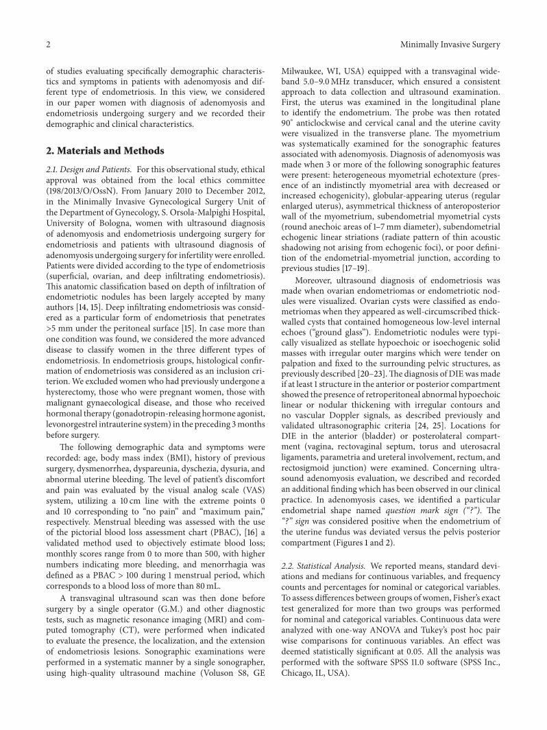
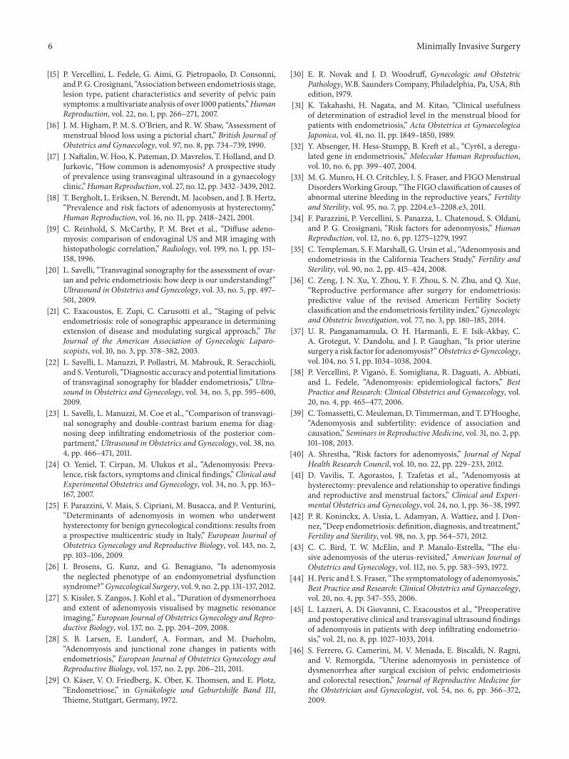
Moreover, ultrasound diagnosis of endometriosis wasmade when ovarian endometriomas or endometriotic nod-ules were visualized. Ovarian cysts were classified as endo-metriomas when they appeared as well-circumscribed thick-walled cysts that contained homogeneous low-level internalechoes (“ground glass”). Endometriotic nodules were typi-cally visualized as stellate hypoechoic or isoechogenic solidmasses with irregular outer margins which were tender onpalpation and fixed to the surrounding pelvic structures, aspreviously described [20–23].The diagnosis of DIEwasmadeif at least 1 structure in the anterior or posterior compartmentshowed the presence of retroperitoneal abnormal hypoechoiclinear or nodular thickening with irregular contours andno vascular Doppler signals, as described previously andvalidated ultrasonographic criteria [24, 25]. Locations forDIE in the anterior (bladder) or posterolateral compart-ment (vagina, rectovaginal septum, torus and uterosacralligaments, parametria and ureteral involvement, rectum, andrectosigmoid junction) were examined. Concerning ultra-sound adenomyosis evaluation, we described and recordedan additional finding which has been observed in our clinicalpractice. In adenomyosis cases, we identified a particularendometrial shape named question mark sign (“?”). The“?” sign was considered positive when the endometrium ofthe uterine fundus was deviated versus the pelvis posteriorcompartment (Figures 1 and 2).
2.2. Statistical Analysis. We reported means, standard devi-ations and medians for continuous variables, and frequencycounts and percentages for nominal or categorical variables.To assess differences between groups of women, Fisher’s exacttest generalized for more than two groups was performedfor nominal and categorical variables. Continuous data wereanalyzed with one-way ANOVA and Tukey’s post hoc pairwise comparisons for continuous variables. An effect wasdeemed statistically significant at 0.05. All the analysis wasperformed with the software SPSS 11.0 software (SPSS Inc.,Chicago, IL, USA).
Minimally Invasive Surgery 3
Figure 1: Question mark sign.
3. Results
Demographic and clinical characteristics of women includedin the study are summarized in Table 1.
A total of 268 women were enrolled. Of the 268 women,51 were excluded from the data analysis as 21 patients had nohistological diagnosis of endometriosis, 26 had intolerance totransvaginal ultrasound examination, and 4 had previouslyundergone a hysterectomy.
From 217 patients included in the study with ultrasounddiagnosis of adenomyosis, we found 66 with ovarian histo-logical confirmation of endometriosis, 92 with DIE, and 52patients who underwent surgery for infertility. Only sevenwomen were found with superficial endometriosis alone.For the analysis of data, we considered them together withthe ovarian group. Nine women during laparoscopic surgeryfor endometriosis underwent hysterectomy. For all of themadenomyosis diagnosis was histologically confirmed.
Women with adenomyosis alone represented the oldestgroup of patients (37.8 ± 5.18 years, 𝑃 = 0.02); instead, BMIdid not differ between groups. Concerning the characteristicsrecorded, we found that patients with DIE were nulliparousmore frequently than other groups (𝑃 < 0.0001) and com-paring to endometriosis patients (87/92, 94.6% DIE; 54/73,73.9% ovarian endometriosis), only 27 (27/52 51.9%) womenwith adenomyosis alone were nulliparous. Moreover, inwomen with one or more births adenomyosis alone wassignificantly more frequent than in the others (𝑃 < 0.0001).
We evaluated the history of previous surgical proceduresand we found that women with adenomyosis and DIE hadmore frequently a history of previous surgery (𝑃 = 0.004).Regarding symptoms, DIE group complained ofmore intensepain symptoms (dysmenorrhea, chronic pelvic pain, dyspare-unia, dysuria, and dyschezia) than other groups. Moreover,patients with only adenomyosis were more likely to haveabnormal uterine bleeding (𝑃 < 0.0001) (Table 2).
Describing the particular endometrial shape mentionedabove, we found a strong relationship between this sono-graphic sign and adenomyosis associated deep infiltratingendometriosis. It was significantly (𝑃 < 0.0001) more presentin DIE group (34/92 [37%]) than in the others (ovarian7/73 [9.6%] and adenomyosis alone 4/52 [7.7%]). Specifically,
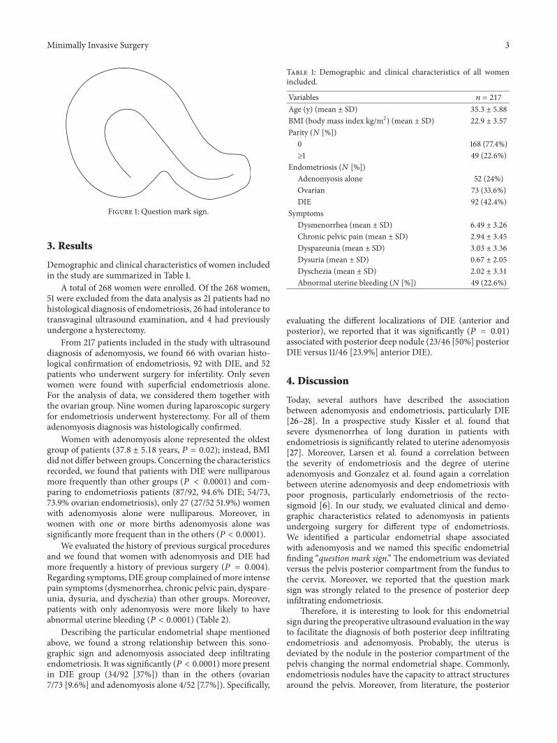
Table 1: Demographic and clinical characteristics of all womenincluded.
Variables 𝑛 = 217
Age (y) (mean ± SD) 35.3 ± 5.88
BMI (body mass index kg/m2) (mean ± SD) 22.9 ± 3.57
Parity (𝑁 [%])0 168 (77.4%)≥1 49 (22.6%)
Endometriosis (𝑁 [%])Adenomyosis alone 52 (24%)Ovarian 73 (33.6%)DIE 92 (42.4%)
SymptomsDysmenorrhea (mean ± SD) 6.49 ± 3.26
Chronic pelvic pain (mean ± SD) 2.94 ± 3.45
Dyspareunia (mean ± SD) 3.03 ± 3.36
Dysuria (mean ± SD) 0.67 ± 2.05
Dyschezia (mean ± SD) 2.02 ± 3.31
Abnormal uterine bleeding (𝑁 [%]) 49 (22.6%)
evaluating the different localizations of DIE (anterior andposterior), we reported that it was significantly (𝑃 = 0.01)associated with posterior deep nodule (23/46 [50%] posteriorDIE versus 11/46 [23.9%] anterior DIE).
4. Discussion
Today, several authors have described the associationbetween adenomyosis and endometriosis, particularly DIE[26–28]. In a prospective study Kissler et al. found thatsevere dysmenorrhea of long duration in patients withendometriosis is significantly related to uterine adenomyosis[27]. Moreover, Larsen et al. found a correlation betweenthe severity of endometriosis and the degree of uterineadenomyosis and Gonzalez et al. found again a correlationbetween uterine adenomyosis and deep endometriosis withpoor prognosis, particularly endometriosis of the recto-sigmoid [6]. In our study, we evaluated clinical and demo-graphic characteristics related to adenomyosis in patientsundergoing surgery for different type of endometriosis.We identified a particular endometrial shape associatedwith adenomyosis and we named this specific endometrialfinding “question mark sign.” The endometrium was deviatedversus the pelvis posterior compartment from the fundus tothe cervix. Moreover, we reported that the question marksign was strongly related to the presence of posterior deepinfiltrating endometriosis.
Therefore, it is interesting to look for this endometrialsign during the preoperative ultrasound evaluation in thewayto facilitate the diagnosis of both posterior deep infiltratingendometriosis and adenomyosis. Probably, the uterus isdeviated by the nodule in the posterior compartment of thepelvis changing the normal endometrial shape. Commonly,endometriosis nodules have the capacity to attract structuresaround the pelvis. Moreover, from literature, the posterior

4 Minimally Invasive Surgery
Table 2: Demographic and clinical characteristics in the different patient groups.
Variables DIE + adenomyosis (92) Ovarian + adenomyosis (73) Adenomyosis alone (52) 𝑃 valueAge (y) (mean ± SD) 34.4 ± 5.34 34.7 ± 6.36 37.8 ± 5.18 𝑃 = 0.021
BMI 22.6 ± 3.87 23.2 ± 3.81 23.0 ± 2.54 𝑃 = 0.541
Parity (𝑁 [%])0 87 (94.6%) 54 (73.9%) 27 (51.9%) 𝑃 < 0.0001
≥1 5 (5.4%) 19 (26.1%) 25 (48.1%) 𝑃 < 0.0001
History of previous surgery (≥1) 26 (28.3%) 26 (35.6%) 5 (9.6%) 𝑃 = 0.004
SymptomsDysmenorrhea (mean ± SD) 7.04 ± 3.47 6.00 ± 3.46 6.21 ± 2.39 𝑃 = 0.096
Chronic pelvic pain (mean ± SD) 3.79 ± 3.58 2.83 ± 3.55 1.59 ± 2.54 𝑃 = 0.001
Dyspareunia (mean ± SD) 3.47 ± 3.54 3.35 ± 3.64 1.84 ± 2.15 𝑃 = 0.013
Dysuria (mean ± SD) 1.84 ± 2.15 0.50 ± 1.70 1.18 ± 2.68 𝑃 = 0.002
Dyschezia (mean ± SD) 3.32 ± 3.86 1.83 ± 2.98 0.01 ± 0.01 𝑃 < 0.0001
Abnormal uterine bleeding (𝑁 [%]) 5 (5.4%) 23 (31.5%) 21 (40.4%) 𝑃 < 0.0001
Figure 2: Ultrasound image of question mark sign.
wall of the uterus was predominantly affected by adeno-myosis [29, 30]. However, no data are available that showan increased mechanical stress of the posterior uterine walldue to chronic uterine peristalsis and hyperperistalsis and arelationship of the site of predilection of adenomyosis withante- or retroflection of the uterus [29, 30]. There is indirectevidence of an archimetral hyperestrogenism in women withendometriosis that interferes with the ovarian control of uter-ine peristaltic activity resulting in uterine hyperperistalsis [31,32]. Moreover, it was argued that endometrial stroma beingin direct contact with the underlying myometrium allowscommunication and interaction, thus facilitating endome-trial invagination or invasion of a structurally weakenedmyometrium during periods of regeneration, healing, andreepithelization [10, 11].
From our results, we found that patients with DIE andadenomyosis were nulliparous more frequently and com-plained of more intense pain symptoms (dysmenorrhea,chronic pelvic pain, dyspareunia, dysuria, and dyschezia)than other groups with the exception of abnormal uterinebleeding which was complained by the majority of womenwith only adenomyosis. Heavy bleeding may be positivelyrelated to the depth of penetration of individual adenomyoticglands into the myometrium and to the density of deependometrial glands within the myometrium [33].
In concordance with previous clinical evidence, wefound that a high percentage of women with adenomyosiswere multiparous [11, 18, 34, 35]. Instead, patients affectedby endometriosis are frequently associated with infertilitybecause of anatomical and immunological alterations causedby endometriosis. Generally, endometriosis is common inwomen with subfertility and can affect fertility at differentlevel, from the induction of a local inflammation and decreasein endometrial receptivity to mechanical obstruction andaltered sexual function [36].
There are some potential mechanisms associated withparity and adenomyosis pathology. First, pregnancy mightfacilitate formation of adenomyosis by allowing adenomyoticfoci to be included in the myometrium due to the invasivenature of the trophoblast on the extension of myometrialfibers. Second, the possibility of Cesarean sectionmay lead toiatrogenic adenomyosis [11, 37]. Third, the hormonal milieuof pregnancy may favour the development of islands ofectopic endometrium [38]. A recent review in 2013 defineda number of factors that encourage the development ofadenomyosis (spontaneous miscarriage, curettage, hystero-scopic resection of the endometrium, uterine myomectomy,caesarean section, and taking Tamoxifen) and underlinedthat the main factor is having had more than one preg-nancy [39]. In agreement with our data, Shrestha found that

Minimally Invasive Surgery 5
the frequency of adenomyosis was higher in parous womenin comparison with nulliparous [40]. Moreover, Vercelliniand colleagues suggested that parity may be associated withan increased frequency of adenomyosis [38]. Other studiesreported the same relationship between parity and adeno-myosis [34, 41].
Concerning symptoms, DIE is the most severe form ofendometriosis, associated with infertility or pain symptoms,including chronic pelvic pain, dysmenorrhea, dyspareunia,dysuria, and dyschezia [42]. Nowadays, it is clarified thatthere is an overlap in the symptom complexes of bothendometriosis and adenomyosis. In endometriosis, the pres-ence of adenomyosis is found to be a risk factor for dys-menorrhea severity [6]. As described before, dysmenorrheaincreases with greater depth of penetration of the adenomy-otic process and to the density of deep endometrial glandsinto myometrium [43]; instead dyspareunia and chronicpain are not considered constant symptoms in patients withadenomyosis [5, 44]. Lazzeri and colleagues showed a persis-tence of dyspareunia in women after surgery for DIE whenadenomyosis was present [45]. Another study reported painrecurrence in DIE, despite the radicality of the surgery [46].Consequently, a correct counselling about pain recurrence isrequired in case of coexistence of both pathological entities.Surgery for posterior DIE with coexistence of adenomyosiscould be not resolved for postoperative pain relief because ofadenomyosis persistence.This information allows the chanceof a correct counselling before surgery and should help todevelopmore effective treatment strategies inwomen affectedby DIE and adenomyosis.
The strength of our study is the large number ofwomen with endometriosis histologically confirmed andthat it was conducted in a tertiary care university hospital,by clinicians highly skilled in endometriosis management.Moreover, sonographic examinations were performed in asystematic manner by a single sonographer, using high-quality ultrasoundmachine.The present study has alsomajorlimitations. First, the lack of histological confirmation ofadenomyosis for the majority of patients included. How-ever, recently transvaginal ultrasound has reached a highlevel of accuracy and many authors have reported the highagreement between ultrasound diagnosis of adenomyosis andhistological findings [17, 47]. A recent review [48] reportedthat transvaginal ultrasound should be the primary toolfor the diagnosis of adenomyosis, with MRI being usedwhen transvaginal ultrasound is inconclusive or when largefibroids are present. Secondly, being patients included in thestudy with a mean age of 35 years old, it can be a possiblebias for evaluation of the natural history of adenomyosispathology. Since the adenomyotic nodules communicatewiththe uterine cavity, pathophysiologically a continuous processfrom initial to deep infiltration must exist. Authors reportedthat endometriosis-associated adenomyosis progresses withage [13, 49].
In conclusion, our data confirmed the strong relation-ship between adenomyosis and endometriosis and evaluateddemographic aspects and symptoms in patients affected bydifferent type of endometriosis. Our findings suggest thatadenomyosis and endometriosis share same symptoms with
important difference linked to the type of endometriosis andhelp to better understand the endometriosis-adenomyosisrelationship and their associated factors.
Conflict of Interests
The authors declare that they have no conflict of interests.
References
[1] G. Leyendecker, L. Wildt, and G. Mall, “The pathophysiologyof endometriosis and adenomyosis: tissue injury and repair,”Archives of Gynecology and Obstetrics, vol. 280, no. 4, pp. 529–538, 2009.
[2] R. Meyer, “Uber den stand der frage der adenomyositis undadenomyome im allgemeinen und insbesondere uber adeno-myositis seroepithelialis und adenomyometritis sarcomatosa,”Zentralbl Gynakol, vol. 36, pp. 745–750, 1919.
[3] T. S. Cullen, “The distribution of adenomyoma containinguterine mucosa,” Archives of Surgery, vol. 1, pp. 215–283, 1920.
[4] J. A. Sampson, “Peritoneal endometriosis due to the menstrualdissemination of endometrial tissue into the peritoneal cavity,”American Journal of Obstetrics & Gynecology, vol. 14, pp. 422–429, 1927.
[5] M. Bazot, O. Fiori, and E. Darai, “Adenomyosis in endo-metriosis—prevalence and impact on fertility. Evidence frommagnetic resonance imaging,”Human Reproduction, vol. 21, no.4, pp. 1101–1102, 2006.
[6] M. Gonzales, L. A. de Matos, M. O. D. C. Goncalves et al.,“Patients with adenomyosis are more likely to have deependometriosis,”Gynecological Surgery, vol. 9, no. 3, pp. 259–264,2012.
[7] G. Leyendecker, M. Herbertz, G. Kunz, and G. Mall, “Endo-metriosis results from the dislocation of basal endometrium,”Human Reproduction, vol. 17, no. 10, pp. 2725–2736, 2002.
[8] G. Leyendecker, G. Kunz, M. Noe, M. Herbertz, and G. Mall,“Endometriosis: a dysfunction and disease of the archimetra,”Human Reproduction Update, vol. 4, no. 5, pp. 752–762, 1998.
[9] G. Leyendecker, G. Kunz, L. Wildt, D. Beil, and H. Deininger,“Uterine hyperperistalsis and dysperistalsis as dysfunctionsof the mechanism of rapid sperm transport in patients withendometriosis and infertility,”Human Reproduction, vol. 11, no.7, pp. 1542–1551, 1996.
[10] A. Ferenczy, “Pathophysiology of adenomyosis,”Human Repro-duction Update, vol. 4, no. 4, pp. 312–322, 1998.
[11] M. Levgur, M. A. Abadi, and A. Tucker, “Adenomyosis: symp-toms, histology, and pregnancy terminations,” Obstetrics &Gynecology, vol. 95, no. 5, pp. 688–691, 2000.
[12] M. Noe, G. Kunz, M. Herbertz, G. Mall, and G. Leyendecker,“The cyclic pattern of the immunocytochemical expression ofoestrogen and progesterone receptors in human myometrialand endometrial layers: characterisation of the endometrial-subendometrial unit,” Human Reproduction, vol. 14, no. 1, pp.190–197, 1999.
[13] G. Kunz, D. Beil, P. Huppert, M. Noe, S. Kissler, and G. Ley-endecker, “Adenomyosis in endometriosis—prevalence andimpact on fertility: evidence from magnetic resonance imag-ing,” Human Reproduction, vol. 20, no. 8, pp. 2309–2316, 2005.
[14] RCOG, “The investigation and management of endometriosisguideline,” no. 24, 2006.
6 Minimally Invasive Surgery
[15] P. Vercellini, L. Fedele, G. Aimi, G. Pietropaolo, D. Consonni,andP.G.Crosignani, “Association between endometriosis stage,lesion type, patient characteristics and severity of pelvic painsymptoms: amultivariate analysis of over 1000 patients,”HumanReproduction, vol. 22, no. 1, pp. 266–271, 2007.
[16] J. M. Higham, P. M. S. O’Brien, and R. W. Shaw, “Assessment ofmenstrual blood loss using a pictorial chart,” British Journal ofObstetrics and Gynaecology, vol. 97, no. 8, pp. 734–739, 1990.
[17] J. Naftalin,W.Hoo, K. Pateman, D.Mavrelos, T. Holland, andD.Jurkovic, “How common is adenomyosis? A prospective studyof prevalence using transvaginal ultrasound in a gynaecologyclinic,”HumanReproduction, vol. 27, no. 12, pp. 3432–3439, 2012.
[18] T. Bergholt, L. Eriksen, N. Berendt,M. Jacobsen, and J. B. Hertz,“Prevalence and risk factors of adenomyosis at hysterectomy,”Human Reproduction, vol. 16, no. 11, pp. 2418–2421, 2001.
[19] C. Reinhold, S. McCarthy, P. M. Bret et al., “Diffuse adeno-myosis: comparison of endovaginal US and MR imaging withhistopathologic correlation,” Radiology, vol. 199, no. 1, pp. 151–158, 1996.
[20] L. Savelli, “Transvaginal sonography for the assessment of ovar-ian and pelvic endometriosis: how deep is our understanding?”Ultrasound in Obstetrics and Gynecology, vol. 33, no. 5, pp. 497–501, 2009.
[21] C. Exacoustos, E. Zupi, C. Carusotti et al., “Staging of pelvicendometriosis: role of sonographic appearance in determiningextension of disease and modulating surgical approach,” TheJournal of the American Association of Gynecologic Laparo-scopists, vol. 10, no. 3, pp. 378–382, 2003.
[22] L. Savelli, L. Manuzzi, P. Pollastri, M. Mabrouk, R. Seracchioli,and S. Venturoli, “Diagnostic accuracy and potential limitationsof transvaginal sonography for bladder endometriosis,” Ultra-sound in Obstetrics and Gynecology, vol. 34, no. 5, pp. 595–600,2009.
[23] L. Savelli, L. Manuzzi, M. Coe et al., “Comparison of transvagi-nal sonography and double-contrast barium enema for diag-nosing deep infiltrating endometriosis of the posterior com-partment,”Ultrasound in Obstetrics and Gynecology, vol. 38, no.4, pp. 466–471, 2011.
[24] O. Yeniel, T. Cirpan, M. Ulukus et al., “Adenomyosis: Preva-lence, risk factors, symptoms and clinical findings,”Clinical andExperimental Obstetrics and Gynecology, vol. 34, no. 3, pp. 163–167, 2007.
[25] F. Parazzini, V. Mais, S. Cipriani, M. Busacca, and P. Venturini,“Determinants of adenomyosis in women who underwenthysterectomy for benign gynecological conditions: results froma prospective multicentric study in Italy,” European Journal ofObstetrics Gynecology and Reproductive Biology, vol. 143, no. 2,pp. 103–106, 2009.
[26] I. Brosens, G. Kunz, and G. Benagiano, “Is adenomyosisthe neglected phenotype of an endomyometrial dysfunctionsyndrome?”Gynecological Surgery, vol. 9, no. 2, pp. 131–137, 2012.
[27] S. Kissler, S. Zangos, J. Kohl et al., “Duration of dysmenorrhoeaand extent of adenomyosis visualised by magnetic resonanceimaging,” European Journal of Obstetrics Gynecology and Repro-ductive Biology, vol. 137, no. 2, pp. 204–209, 2008.
[28] S. B. Larsen, E. Lundorf, A. Forman, and M. Dueholm,“Adenomyosis and junctional zone changes in patients withendometriosis,” European Journal of Obstetrics Gynecology andReproductive Biology, vol. 157, no. 2, pp. 206–211, 2011.
[29] O. Kaser, V. O. Friedberg, K. Ober, K. Thomsen, and E. Plotz,“Endometriose,” in Gynakologie und Geburtshilfe Band III,Thieme, Stuttgart, Germany, 1972.
[30] E. R. Novak and J. D. Woodruff, Gynecologic and ObstetricPathology, W.B. Saunders Company, Philadelphia, Pa, USA, 8thedition, 1979.
[31] K. Takahashi, H. Nagata, and M. Kitao, “Clinical usefulnessof determination of estradiol level in the menstrual blood forpatients with endometriosis,” Acta Obstetrica et GynaecologicaJaponica, vol. 41, no. 11, pp. 1849–1850, 1989.
[32] Y. Absenger, H. Hess-Stumpp, B. Kreft et al., “Cyr61, a deregu-lated gene in endometriosis,” Molecular Human Reproduction,vol. 10, no. 6, pp. 399–407, 2004.
[33] M. G. Munro, H. O. Critchley, I. S. Fraser, and FIGOMenstrualDisordersWorkingGroup, “TheFIGOclassification of causes ofabnormal uterine bleeding in the reproductive years,” Fertilityand Sterility, vol. 95, no. 7, pp. 2204.e3–2208.e3, 2011.
[34] F. Parazzini, P. Vercellini, S. Panazza, L. Chatenoud, S. Oldani,and P. G. Crosignani, “Risk factors for adenomyosis,” HumanReproduction, vol. 12, no. 6, pp. 1275–1279, 1997.
[35] C. Templeman, S. F. Marshall, G. Ursin et al., “Adenomyosis andendometriosis in the California Teachers Study,” Fertility andSterility, vol. 90, no. 2, pp. 415–424, 2008.
[36] C. Zeng, J. N. Xu, Y. Zhou, Y. F. Zhou, S. N. Zhu, and Q. Xue,“Reproductive performance after surgery for endometriosis:predictive value of the revised American Fertility Societyclassification and the endometriosis fertility index,”Gynecologicand Obstetric Investigation, vol. 77, no. 3, pp. 180–185, 2014.
[37] U. R. Panganamamula, O. H. Harmanli, E. F. Isik-Akbay, C.A. Grotegut, V. Dandolu, and J. P. Gaughan, “Is prior uterinesurgery a risk factor for adenomyosis?”Obstetrics&Gynecology,vol. 104, no. 5 I, pp. 1034–1038, 2004.
[38] P. Vercellini, P. Vigano, E. Somigliana, R. Daguati, A. Abbiati,and L. Fedele, “Adenomyosis: epidemiological factors,” BestPractice and Research: Clinical Obstetrics and Gynaecology, vol.20, no. 4, pp. 465–477, 2006.
[39] C. Tomassetti, C.Meuleman,D. Timmerman, andT.D’Hooghe,“Adenomyosis and subfertility: evidence of association andcausation,” Seminars in Reproductive Medicine, vol. 31, no. 2, pp.101–108, 2013.
[40] A. Shrestha, “Risk factors for adenomyosis,” Journal of NepalHealth Research Council, vol. 10, no. 22, pp. 229–233, 2012.
[41] D. Vavilis, T. Agorastos, J. Tzafetas et al., “Adenomyosis athysterectomy: prevalence and relationship to operative findingsand reproductive and menstrual factors,” Clinical and Experi-mental Obstetrics and Gynecology, vol. 24, no. 1, pp. 36–38, 1997.
[42] P. R. Koninckx, A. Ussia, L. Adamyan, A. Wattiez, and J. Don-nez, “Deep endometriosis: definition, diagnosis, and treatment,”Fertility and Sterility, vol. 98, no. 3, pp. 564–571, 2012.
[43] C. C. Bird, T. W. McElin, and P. Manalo-Estrella, “The elu-sive adenomyosis of the uterus-revisited,” American Journal ofObstetrics and Gynecology, vol. 112, no. 5, pp. 583–593, 1972.
[44] H. Peric and I. S. Fraser, “The symptomatology of adenomyosis,”Best Practice and Research: Clinical Obstetrics and Gynaecology,vol. 20, no. 4, pp. 547–555, 2006.
[45] L. Lazzeri, A. Di Giovanni, C. Exacoustos et al., “Preoperativeand postoperative clinical and transvaginal ultrasound findingsof adenomyosis in patients with deep infiltrating endometrio-sis,” vol. 21, no. 8, pp. 1027–1033, 2014.
[46] S. Ferrero, G. Camerini, M. V. Menada, E. Biscaldi, N. Ragni,and V. Remorgida, “Uterine adenomyosis in persistence ofdysmenorrhea after surgical excision of pelvic endometriosisand colorectal resection,” Journal of Reproductive Medicine forthe Obstetrician and Gynecologist, vol. 54, no. 6, pp. 366–372,2009.

Minimally Invasive Surgery 7
[47] M. Dueholm, E. Lundorf, E. S. Hansen, J. S. Sorensen, S. Leder-toug, and F. Olesen, “Magnetic resonance imaging and trans-vaginal ultrasonography for the diagnosis of adenomyosis,”Fertility and Sterility, vol. 76, no. 3, pp. 588–594, 2001.
[48] M. Dueholm, “Transvaginal ultrasound for diagnosis of adeno-myosis: a review,” Best Practice and Research: Clinical Obstetricsand Gynaecology, vol. 20, no. 4, pp. 569–582, 2006.
[49] B. Kunz, D. Beil, P. Huppert, and G. Leyendecker, “Structuralabnormalities of the uterine wall in women with endometriosisand infertility visualized by vaginal sonography and magneticresonance imaging,”Human Reproduction, vol. 15, no. 1, pp. 76–82, 2000.

Submit your manuscripts athttp://www.hindawi.com
Stem CellsInternational
Hindawi Publishing Corporationhttp://www.hindawi.com Volume 2014
Hindawi Publishing Corporationhttp://www.hindawi.com Volume 2014
MEDIATORSINFLAMMATION
of
Hindawi Publishing Corporationhttp://www.hindawi.com Volume 2014
Behavioural Neurology
EndocrinologyInternational Journal of
Hindawi Publishing Corporationhttp://www.hindawi.com Volume 2014
Hindawi Publishing Corporationhttp://www.hindawi.com Volume 2014
Disease Markers
Hindawi Publishing Corporationhttp://www.hindawi.com Volume 2014
BioMed Research International
OncologyJournal of
Hindawi Publishing Corporationhttp://www.hindawi.com Volume 2014
Hindawi Publishing Corporationhttp://www.hindawi.com Volume 2014
Oxidative Medicine and Cellular Longevity
Hindawi Publishing Corporationhttp://www.hindawi.com Volume 2014
PPAR Research
The Scientific World JournalHindawi Publishing Corporation http://www.hindawi.com Volume 2014
Immunology ResearchHindawi Publishing Corporationhttp://www.hindawi.com Volume 2014
Journal of
ObesityJournal of
Hindawi Publishing Corporationhttp://www.hindawi.com Volume 2014
Hindawi Publishing Corporationhttp://www.hindawi.com Volume 2014
Computational and Mathematical Methods in Medicine
OphthalmologyJournal of
Hindawi Publishing Corporationhttp://www.hindawi.com Volume 2014
Diabetes ResearchJournal of
Hindawi Publishing Corporationhttp://www.hindawi.com Volume 2014
Hindawi Publishing Corporationhttp://www.hindawi.com Volume 2014
Research and TreatmentAIDS
Hindawi Publishing Corporationhttp://www.hindawi.com Volume 2014
Gastroenterology Research and Practice
Hindawi Publishing Corporationhttp://www.hindawi.com Volume 2014
Parkinson’s Disease
Evidence-Based Complementary and Alternative Medicine
Volume 2014Hindawi Publishing Corporationhttp://www.hindawi.com