Anti-platelet therapy holds promises in treating adenomyosis ...
16
RESEARCH Open Access Anti-platelet therapy holds promises in treating adenomyosis: experimental evidence Bo Zhu 1† , Yumei Chen 1† , Xiaolu Shen 1 , Xishi Liu 2,3 and Sun-Wei Guo 2,3* Abstract Background: Recently emerging evidence indicates that endometriotic lesions are wounds undergoing repeated tissue injury and repair (ReTIAR), and platelets induce epithelial-mesenchymal transition (EMT), fibroblast-to- myofibroblast transdifferentiation (FMT), leading ultimately to fibrosis. Due to the commonality of cyclic bleeding as in endometriosis, adenomyotic lesions are also wounds that undergo ReTIAR, and we have recently provided evidence corroborating platelet-induced EMT, FMT and fibrogenesis in adenomyosis. This study sought to evaluate the effect of antiplatelet therapy in a mouse model of adenomyosis. Methods: Adenomyosis was induced in 57 female ICR mice with neonatal dosing of tamoxifen, while another 12 (group C) were dosed with solvent only, serving as a blank control. Starting from 4 weeks after birth, hotplate test was administrated to all mice every 4 weeks. At the 16th week, all mice with induced adenomyosis were randomly divided into 6 groups: untreated, low- and high-dose Ozagrel, low- and high-dose anti-mouse GPIbα polyclonal IgG antibody to deplete platelets, and isotype-matched inert IgG non-immune antibody. Group C received no treatment. After 3 weeks of treatment, they were hotplate tested again, their uterine horns and brains were harvested, and a blood sample was taken to measure the plasma corticosterone level by ELISA. The left uterine horn was used for immunohistochemistry analysis. The brainstem nucleus raphe magnus (NRM) sections were subjected to immunofluorescence staining for GAD65. The depth of myometrial infiltration and uterine contractility were evaluated. Results: We found that both Ozagrel treatment and platelet depletion dose-dependently suppressed myometrial infiltration, improved generalized hyperalgesia, reduced uterine contractility, and lowered plasma corticosterone levels, improved the expression of some proteins known to be involved in adenomyosis and slowed down the process of fibrogenesis. It also elevated the number of GAD65-expressing neurons in the brainstem NRM, possibly boosting the GABAergic inhibition of pain due to adenomyosis. Conclusion: This study further provides evidence that platelets play important roles in the development of adenomyosis. Anti-platelet treatment is efficacious in suppression of myometrial infiltration, improving generalized hyperalgesia, reducing uterine hyperactivity and systemic corticosterone levels. Collectively, these results demonstrate that anti-platelet therapy seems to be promising for treating adenomyosis. Keywords: Adenomyosis, Generalized hyperalgesia, Hotplate latency, Mouse, Ozagrel, Platelet, Uterine contractility * Correspondence: [email protected] † Equal contributors 2 Shanghai Key Laboratory of Female Reproductive Endocrine-Related Diseases, Shanghai 200011, China 3 Shanghai Obstetrics and Gynecology Hospital, Fudan University, 419 Fangxie Road, Shanghai 200011, China Full list of author information is available at the end of the article © 2016 The Author(s). Open Access This article is distributed under the terms of the Creative Commons Attribution 4.0 International License (http://creativecommons.org/licenses/by/4.0/), which permits unrestricted use, distribution, and reproduction in any medium, provided you give appropriate credit to the original author(s) and the source, provide a link to the Creative Commons license, and indicate if changes were made. The Creative Commons Public Domain Dedication waiver (http://creativecommons.org/publicdomain/zero/1.0/) applies to the data made available in this article, unless otherwise stated. Zhu et al. Reproductive Biology and Endocrinology (2016) 14:66 DOI 10.1186/s12958-016-0198-1
Transcript of Anti-platelet therapy holds promises in treating adenomyosis ...

RESEARCH Open Access
Anti-platelet therapy holds promises intreating adenomyosis: experimentalevidenceBo Zhu1†, Yumei Chen1†, Xiaolu Shen1, Xishi Liu2,3 and Sun-Wei Guo2,3*
Abstract
Background: Recently emerging evidence indicates that endometriotic lesions are wounds undergoing repeatedtissue injury and repair (ReTIAR), and platelets induce epithelial-mesenchymal transition (EMT), fibroblast-to-myofibroblast transdifferentiation (FMT), leading ultimately to fibrosis. Due to the commonality of cyclic bleeding asin endometriosis, adenomyotic lesions are also wounds that undergo ReTIAR, and we have recently providedevidence corroborating platelet-induced EMT, FMT and fibrogenesis in adenomyosis. This study sought to evaluatethe effect of antiplatelet therapy in a mouse model of adenomyosis.
Methods: Adenomyosis was induced in 57 female ICR mice with neonatal dosing of tamoxifen, while another 12(group C) were dosed with solvent only, serving as a blank control. Starting from 4 weeks after birth, hotplate testwas administrated to all mice every 4 weeks. At the 16th week, all mice with induced adenomyosis were randomlydivided into 6 groups: untreated, low- and high-dose Ozagrel, low- and high-dose anti-mouse GPIbα polyclonal IgGantibody to deplete platelets, and isotype-matched inert IgG non-immune antibody. Group C received notreatment. After 3 weeks of treatment, they were hotplate tested again, their uterine horns and brains wereharvested, and a blood sample was taken to measure the plasma corticosterone level by ELISA. The left uterinehorn was used for immunohistochemistry analysis. The brainstem nucleus raphe magnus (NRM) sections weresubjected to immunofluorescence staining for GAD65. The depth of myometrial infiltration and uterine contractilitywere evaluated.
Results: We found that both Ozagrel treatment and platelet depletion dose-dependently suppressed myometrialinfiltration, improved generalized hyperalgesia, reduced uterine contractility, and lowered plasma corticosteronelevels, improved the expression of some proteins known to be involved in adenomyosis and slowed down theprocess of fibrogenesis. It also elevated the number of GAD65-expressing neurons in the brainstem NRM, possiblyboosting the GABAergic inhibition of pain due to adenomyosis.
Conclusion: This study further provides evidence that platelets play important roles in the development ofadenomyosis. Anti-platelet treatment is efficacious in suppression of myometrial infiltration, improving generalizedhyperalgesia, reducing uterine hyperactivity and systemic corticosterone levels. Collectively, these resultsdemonstrate that anti-platelet therapy seems to be promising for treating adenomyosis.
Keywords: Adenomyosis, Generalized hyperalgesia, Hotplate latency, Mouse, Ozagrel, Platelet, Uterine contractility
* Correspondence: [email protected]†Equal contributors2Shanghai Key Laboratory of Female Reproductive Endocrine-RelatedDiseases, Shanghai 200011, China3Shanghai Obstetrics and Gynecology Hospital, Fudan University, 419Fangxie Road, Shanghai 200011, ChinaFull list of author information is available at the end of the article
© 2016 The Author(s). Open Access This article is distributed under the terms of the Creative Commons Attribution 4.0International License (http://creativecommons.org/licenses/by/4.0/), which permits unrestricted use, distribution, andreproduction in any medium, provided you give appropriate credit to the original author(s) and the source, provide a link tothe Creative Commons license, and indicate if changes were made. The Creative Commons Public Domain Dedication waiver(http://creativecommons.org/publicdomain/zero/1.0/) applies to the data made available in this article, unless otherwise stated.
Zhu et al. Reproductive Biology and Endocrinology (2016) 14:66 DOI 10.1186/s12958-016-0198-1
BackgroundAdenomyosis is a common gynecologic disorder with apoorly understood pathogenesis [1]. As in endometriosis,it is characterized by the ectopic deposition and growthof endometrial glands and stroma deep and haphazardlyinto the myometrium [1]. It shares with endometriosismany similarities in terms of estrogen-dependency, pro-gesterone resistance, symptomology, and many molecu-lar aberrations but differs in risk factors, age at onsetand, possibly, etiology [2]. Similar to endometriosis, ourcurrent knowledge of its pathophysiology is still woefullyinadequate. Consequently, treatment of adenomyosis hasbeen a challenge [3], with hysterectomy being the treat-ment of choice for severe symptomatic adenomyosis.Thus, medical treatment of adenomyosisis still an unmetmedical need.Adenomyosis is first and foremost viewed as an
estrogen-dependent disease, featuring increased localproduction of estrogen [4]. It also displays signs ofinflammation, characterized by the constitutive activa-tion of NF-kB [5], increased macrophage infiltration[6], and elevated expression of COX-2, a rate-limitingenzyme in catalyzing prostaglandin (PG) E2 (PGE2)[7], and increased production of proinflammatory cy-tokines and chemokines [8]. All existing therapeuticsfor adenomyosis are hormonal drugs.As with endometriosis, the ectopic endometrium in ade-
nomyosis also experience cyclic bleeding. Yet bleeding, anindication of vascular injury, is a cardinal hallmark of awound or tissue damage. Consequently, a physiologicalprocess, called wound healing or tissue repair, ensues. Assuch, platelets must be involved, as shown recently forendometriosis [9]. In fact, based on serial immunohisto-chemistry analyses of ectopic endometrium in a mousemodel of adenomyosis, we recently report that activatedplatelets coincide with TGF-β1 release and the induction ofTGF-β/Smad signaling pathway in adenomyosis, as well asevidence of epithelial-mesenchymal transition (EMT)and fibroblast-to-myofibroblast transdifferentiation(FMT), resulting ultimately in fibrosis [10] and alsosmooth muscle metaplasia (Shen et al., unpublisheddata). These observations are confirmed in humanadenomyosis [11]. Therefore, due to the commonalityshared with endometriosis, i.e., cyclic bleeding, adeno-myotic lesions behave just like endometriotic lesions,which are essentially wounds that undergo repeatedtissue injury and repair (ReTIAR) [9, 12, 13].In light of the important roles that platelets play in the
development of endometriosis [9, 14, 15] and adenomyosis[10, 11], one may wonder as whether anti-platelet therapymay have any potential in treating adenomyosis. This studywas undertaken to test the hypothesis that anti-platelettreatment, by either platelet depletion or administration ofOzagrel, indeed has potential for therapeutic purposes.
MethodsChemicalsOzagrel, a thrmboxane A2 (TXA2) synthese inhibitor[16], was purchased from YaoDa Pharmacology IndustryCompany (Shenyang, China) and was dissolved in 0.9 %normal saline for intraperitoneal administration. The ratanti-mouse GPIbα polyclonal IgG antibody and itsisotype-matched non-immune rat anti-mouse IgG anti-body were purchased from Emfret Analytics (Eibelstadt,Germany). Tamoxifen citrate was purchased from FudanForward Pharmaceutical Company (Shanghai, China).All other chemicals were purchased from Sigma unlessstated otherwise.
Animals and the procedure for induction of adenomyosisFour pregnant ICR mice with a gestational age of 15–16days were purchased from Shanghai Laboratory AnimalCorporation (Shanghai, China) and each of them washoused in a single cage during the rest of the gestationperiod and the ensuing birth and nursing period. Theirpups (1 day after birth) were sexed and the female pupswere selected for use in this study. The same litter ofpups and the dam were housed in the same cage untilweaned. All mice were housed in an animal care facilityunder controlled conditions (20 °C, 12:12 light/darkcycle with lights on at 6:00 AM) and had free access tochow and fresh water.Following Parrott et al. [17, 18], and as reported previ-
ously [2, 19, 20], adenomyosis was induced by orallydosing female neonatal mice with 1 mg/kg tamoxifensuspended in peanut oil/lecithin/condensed milk mix-ture (2:0.2:3, by volume) at a dose volume of 5 μl/gbodyweight from day 2 to day 5 after birth. Female con-trol neonatal mice, selected randomly, were fed similarlywith the same amount of solvent, without tamoxifen.When these female mice reached 3 weeks of age, theywere weaned and separated from the dams.All experiments were performed under the guidelines
of the National Research Council’s Guide for the Careand Use of Laboratory Animals [21] and approved bythe institutional experimental animals review board ofShanghai OB/GYN Hospital, Fudan University.
Experimental protocolThis experiment was conducted side-by-side with anotherexperiment evaluating the efficacy of epigallocatechin-3-gallate (EGCG) in treating adenomyosis in mice, as re-ported in [20]. Fifty-six female neonatal pups were orallydosed with tamoxifen from day 2 to day 5 after birth,while another 12 were dosed in similar fashion with thesolvent only (control group, or group C). Starting from4 weeks after birth, hotplate test was administered to allmice every 4 weeks, as described previously [2, 19] (seeAdditional file 1 for full description). At the 16th week
Zhu et al. Reproductive Biology and Endocrinology (2016) 14:66 Page 2 of 16

after birth, all mice dosed with tamoxifen were randomlydivided into6 groups of roughly equal size, Group U (n =9), or the untreated group, received the vehicle only.Another two groups received, for 3 weeks, daily intraperi-toneal (i.p.) administration of either low-dose (12.5 μg/g)Ozagrel (Group L, n = 10) or of high-dose (25 μg/g)Ozagrel (Group H, n = 10). The Ozagrel doses were deter-mined based on the dosage given to an adult female andthen converted from human to mouse based on body sur-face area and further adjusted based on each mouse’sbodyweight measured every day before Ozagrel adminis-tration. The mice in group C received no treatment at alland served as a blank control. The other three groupswere intravenously (i.v.) administrated with different dosesof rat anti-mouse GPIbα polyclonal IgG and non-immunerat anti-mouse IgG: Group LD (n = 10) received a low-dose (1 μg/g) rat anti-mouse GPIbα polyclonal IgG treat-ment; Group HD (n = 9) received a high-dose (2 μg/g) ratanti-mouse GPIbα polyclonal IgG treatment; Group NI (n= 8) received 1 μg/g non-immune (NI) rat anti-mouse IgGisotope-matched with the anti-GPIbα antibody; The micein group C were treated the same as above. The dosagesof rat anti-mouse GPIbα polyclonal IgG were determinedbased on instructions provided by Emfret Analytics.After the 3-week-long treatment period (at the 19th
week), the final hotplate test was administered to all themice with or without induced adenomyosis, after body-weight measurement. A 0.5 ml blood sample was takenbetween 9:00 and 15:00 of the dayfrom each mouse, andwas used for the measurement of plasma corticosteronelevels by enzyme linked immunosorbent assay (ELISA,see Additional file 1 for more details). After the bloodsamples were taken, all mice were sacrificed by perfusingthe heart with formalin. For each mouse, both uterinehorns were harvested and the uterine weight was re-corded. The left uterine horn was used for uterine con-tractility measurement (described in Additional file 1),and the right one was fixed in 4 % paraformaldehyde im-mediately after collection and then embedded in paraf-fin. The brains of all mice were harvested and analyzed(described below). The experiment design is shownschematically in Fig. 1.We evaluated the depth of myometrial infiltration of ec-
topic endometrium following the criteria of Bird et al.[22], as reported previously [2]. Briefly, Grade 1 was de-fined to be the case where penetration of the ectopicendometrium into superficial myometrium; Grade 2,penetration into mid-myometrium; and Grade 3, penetra-tion beyond mid-myometrium. For ease of statistical ana-lysis, Grade 0 was recorded when there was a completeabsence of any ectopic endometrium in the myometrium.For histological examination, serial 4-μm sections were
obtained from each paraffin-embedded tissue block, andthen 3 randomly selected sections were chosen for H&E
staining to confirm pathologic diagnosis, as describedpreviously [2, 19]. If endometrial glands and stroma wereseen to be infiltrated into in myometrium, the diagnosisof adenomyosis was made.
Histochemistry and immunohistochemistry analysesAs described above, the right horn of uterus was fixed in4 % formalin, and then embedded in paraffin. From eachtissue block, serial 4-μm sections were obtained andsubjected to H&E staining to confirm pathologic diagno-sis of adenomyosis, which was characterized by the pres-ence of endometrial glands and stroma that arecompletely enveloped by myometrium and discontinu-ous with the endometrial cavity [23, 24].The rabbit polyclonal antibodies against progester-
one receptor isoform B (PR-B, ab2765; Abcam, HongKong, China), phosphorylated-p65 (ab30623; Abcam),cyclooxygenase 2 (COX-2, #4842; CST, Boston, USA),oxytocin receptor (OTR, ab115664; Abcam), transientreceptor potential cation channel, subfamily V, mem-ber 1 (TRPV1, ab31895;Abcam), collagen I (ab292;Abcam), collagen IV (ab6586; Abcam), and the ratmonoclonal antibody against F4/80 (MCA497G; AbD-Serotec, Cambridge, England), CD41(ab33661), dilutedto1:50,1:80,1:200, 1:100, 1:1000,1:100, 1:500, 1:200 and1:100, respectively, were used as primary antibodies.Serial 4-μm sections were made from paraffin-
embedded tissue blocks. After routine deparaffinizationand dehydration, some sections were randomly selectedto be heat-retrieved with Tis-EDTA buffer (0.5 mol/LPH 9.0) over 98 °C for a total of 30 min for immuno-staining for PR-B, p-p65, COX-2, OTR and TRPV1, andthe others were heat-retrieved with citric acid (0.01 mol/L pH 6.0) over 98 °C for a total of 30 min for immuno-histochemistry analysis of collagen I, collagen IV, andF4/80. Then all sections were cooled naturally at roomtemperature, and then incubated with the primary anti-bodies at 4 °C overnight. After the sections were rinsedwith PBS, they were incubated with the secondary anti-body (Sunpoly-HII, BioSunTechnclogy, Shanghai, China)for half an hour, or, for F4/80, with the goat anti-rat anti-body (AbDSerotec) for 1 hour. The bound antibodycomplexes were stained for 3–5 minor until appropriatefor microscopic examination with diaminobenzidine(DAB) (BioSunTechnclogy Co., Ltd) and then counter-stained with hematoxylin and mounted.Images were obtained with the microscope (Olympus
BX51, Olympus, Tokyo, Japan) fitted with a digital camera(Olympus DP70, Olympus). Five randomly selected im-ages from 10 to 12 images on 2–4 sections of each mousewere taken for each immunostaining marker to obtain amean optional density value by Image Pro-Plus 6.0 (MediaCybernetics, Inc., Bethesda, MD, USA) as reported in [25].Staining was defined via color intensity, and a color mask
Zhu et al. Reproductive Biology and Endocrinology (2016) 14:66 Page 3 of 16

was made. The mask was then applied equally to all im-ages, and measurement readings were obtained. Immuno-histochemical parameters assessed in the area detectedincluded (a) integrated optical density (IOD); (b) totalstained area (S); and (c) mean optical density (MOD),which is defined as MOD= IOD/S, equivalent to the meanintensity of stain in all positive cells.For F4/80, we counted the number of F4/80-positive
macrophages from five randomly selected images andcalculated their average. Myometrial OTR stainingscores were calculated by multiplying the percentage ofpositive OTR cells per section (0–100 %) by a semi-quantitative classifier for immunohistochemical stainingintensity, which was scored as 0 if there was a completeabsence of any staining, 1 for weak staining, 2 for mod-erate staining, and 3 for strong staining. Consequently,the staining scores ranged from a minimum of 0 to amaximum of 300. We used the mean score averagedover 5 randomly selected images.For all markers, the staining levels were scored on ec-
topic endometrium in mice with induced adenomyosis.For mice in the control group or in mice without ectopicendometrium due to treatment, they were scored in theendometrium. We counted the number of macrophages(F4/80-positive) infiltrated into the ectopic endometriumin the entire focal field for mice with adenomyosis. Forthe mice in control group or mice that had none suchlesion due to treatment, the number of F4/80-positivemacrophages in endometrium was counted.The mouse spleen tissue was used for the positive im-
munostaining of macrophages, while breast cancer tissuesections were used for positive control for other
markers, Negative control sections were processed simi-larly, but using a non-immune rabbit or rat IgG insteadof the primary antibody, or by omitting the primary anti-body from the incubation medium. No positive reactionwas observed under these conditions. All sections wereinspected a single investigator (BZ) without the know-ledge of the group identity. The positive and negativecontrols are shown in Additional file 1: Figure S2 ofSupplemental Information.
GAD65 immunofluorescence staining of neurons inbrainstem nucleus raphe magnusThe procedure has been reported previously in [26].Briefly, the mouse brains containing the nucleus raphemagnus (NRM) were harvested and immediately embed-ded in O.C. T. compound in liquid phlegm after themice were sacrificed. The NRM sections were between5.68 and 6.48 mm to the bregma and the NRM was lo-cated 1.72–2.68 mm interaurally, as described previously[26]. Serial 6-μm sections were performed on a cryostatfor each block and stored at the temperature below−20 °C until use. The sections were incubated in goatanti mouse serum (BioSunTechnclogy) for 10 min andthen incubated in mouse anti primary antibody againstGAD65 (ab26113, Abcam; 1:1,000) at 4 °C overnight.GAD65 is expressed in the cytoplasm of presynapticneurons. The sections were rinsed with PBS (pH 7.4)and incubated in secondary antibody mixed withDyLight649 (E032610, Earthox, San Francisco, USA) forone hour, and then rinsed with PBS (pH 7.4) again andmounted.
Fig. 1 Schematic illustration of the experiment design of this study. Untreated: mice that received no treatment; Lo-dose Ozagrel (Oza): micetreated with 12.5 μg/g Oza (low-dose); Hi-dose Oza: mice treated with 25 μg/g Oza (high-dose). Lo-dose PD: mice treated with 1 μg/g rat anti-mouseGPIbα polyclonal IgG antibody for platelet depletion (PD); Hi-dose PD: mice treated with 2 μg/g rat anti-mouse GPIbα polyclonal IgG antibody; Non-immune: received 1 μg/g non-immune (NI) rat anti-mouse IgG isotope-matched with the anti-GPIbα antibody See text on experiment protocol formore details
Zhu et al. Reproductive Biology and Endocrinology (2016) 14:66 Page 4 of 16

Images were obtained with a microscope (OlympusBX51) fitted with a digital camera (Olympus DP70). Fiverandomly selected images out of 6–7 sections of eachmouse were taken for each immunostained parameter tocount the numbers of GAD65-positive (red) cells in theNRM, located between 1.72 mm and 2.68 mm interau-rally, and the mean was calculated.
Statistical analysisComparison of the distributions among two or moregroups of continuous variables was made using the Wil-coxon and Kruskal-Wallis tests, respectively, and thepaired Wilcoxon test was used when the before-aftercomparison was made for the same group of subjects.Pearson’s or Spearman’s rank correlation coefficient wasused when evaluating correlations between two variableswhen both variables were continuous or when at leastone variable was ordinal. To see whether Ozagrel treat-ment or platelet depletion and other possible factorswere responsible for the change in hotplate latency be-fore and after the treatment, a multiple linear regressionmodel was used. To see whether there is trend in immu-nostaining levels as a function of the depth of myome-trial infiltration, Jonckheere trend was used.To determine correlates of depth of myometrial infil-
tration, we used the Cox proportional odds logistic re-gression model. This model assumes, implicitly, that thedata were ordered categorical data, with an implicitunderlying order (scale of severity) in the data [27], with4 categories corresponding to Grade 0, I, II, and IIIinfiltration.P values of less than 0.05 were considered statistically
significant. All computations were made with R statisticssoftware system version 3.3.1 [28].
ResultsConsistent with Parrott et al. [17, 18] and as previouslyreported [2, 19], we found that adenomyosis was suc-cessfully induced in all (100 %) mice dosed with tamoxi-fen but none in un-dosed mice.Ozagrel was well-tolerated, as no mice in either LO or
HO group died, and we found nothing unusual in thesemice. In HD group, however, 1 mouse died after it re-ceived the 4th injection of the depletion antibody, and 2appeared to be lethargic. In the LD group, no mice diedand nothing appeared unusual. There was no differencein platelet counts between the mice in groups UT, NI,LO, and HO at the end of the experiment. However, theplatelet count in mice in both LD and HD groups wasreduced by 99.6 and 99.7 % as compared with those inthe NI group, demonstrating the effectiveness of plateletdepletion in these two groups.
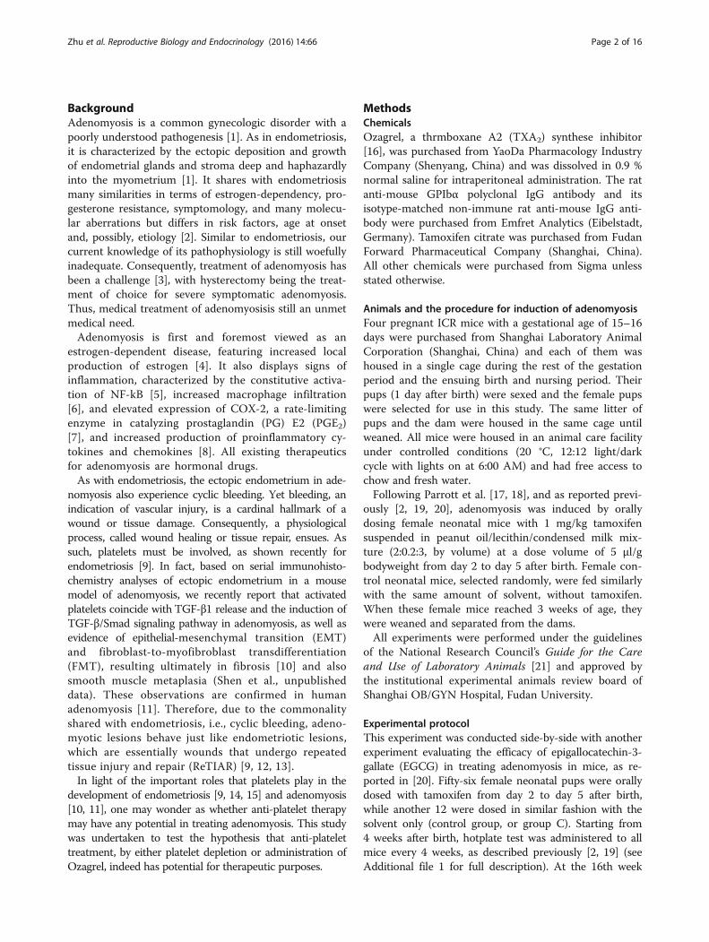
Treatment effect on the depth of myometrial infiltration,hotplate latency, and uterine and bodyweightWe first evaluated the effect of Ozagrel treatment or plate-let depletion on the depth of myometrial infiltration. Wefound that, compared with untreated mice, mice treatedwith either low- or high-dose Ozagrel had significantly lessinfiltration (both p-values <0.001; Fig. 2a). Compared withNI mice, mice in either LD or HD group also had signifi-cantly less infiltration (both p-values <0.001; Fig. 2a). Micein HO and HD groups appeared to have less infiltrationthan those in the LO or LD group (Fig. 2a).The multiple linear regression analysis suggested that
both Ozagrel treatment and platelet depletion significantlyand dose-dependently reduced the depth of myometrialinfiltration (regression coefficient β = −0.956, p = 3.2×10⁻⁷,and β = −0.627, p = 1.5×10⁻⁶, respectively, R2 = 0.62), butNI mice had deeper infiltration (β = 0.856, p = 0.015). TheCox regression analysis yielded similar results (all three p-values <0.028).We found that there is a significant difference in uterine
weight vs. bodyweight ratio among the 7 groups of mice(p < 0.001; Fig. 2b). Using the ratio as a dependent variableand the bodyweight after treatment, the induction of ade-nomyosis, dose of Ozagrel, the non-immune IgG injectionor not, and the dose of antibody used in platelet depletionas covariates, we found, via a linear multiple regression,that the non-immune IgG was positively associated withthe ratio (p < 0.01; R2 = 0.60; Fig. 2b) while both Ozagreland anti-platelet doses were negatively associated with theratio (both p-values <0.001). Similar results were obtainedfor the uterine weight (data not shown).The induction of adenomyosis was significantly asso-
ciated with reduced latency just 4 weeks after birth or23 days after the completion of tamoxifen dosing (p <0.05; Fig. 2c). At 8 weeks after birth, the difference inhotplate latency amount the four groups of mice be-came very pronounced (p < 0.001; Fig. 2c), with themice with induced adenomyosis all having reduced la-tency (p < 0.001). At 12 and 16 weeks after birth, themice with induced adenomyosis had further progres-sively reduced hotplate latency (both p-values <0.001;Fig. 2c). In all mice with induced adenomyosis, the la-tency evaluated at week 8, 12 and 16 was allsignificantly decreased as compared with the previouslyevaluated latency (all p-values <0.001; Fig. 2c).After platelet depletion or treatment with Ozagrel
for 3 weeks, however, the hotplate latency was signifi-cantly improved in a dose-dependent fashion (β =4.008, p < 0.001, and β = 1.792, p < 0.001, respectively,in a multiple linear regression analysis, R2 = 0.73;Fig. 2c). In contrast, the presence of adenomyosis andthe injection of the dummy antibody were associatedwith decrease in hotplate latency (p < 0.001 and p <0.01, respectively).
Zhu et al. Reproductive Biology and Endocrinology (2016) 14:66 Page 5 of 16
While there was no difference in bodyweight amongthe 7 groups of mice at 4 and 8 weeks after birth (bothp-values >0.05; Fig. 2d), the difference became statisti-cally and progressive significant starting from week 12(p < 0.05 and p < 0.01), and was significant at the end ofthe experiment (p < 0.001; Fig. 2d). The multiple linear re-gression analysis using pre-treatment bodyweight, pres-ence of adenomyosis, and the dose of Ozagrel, the non-immune IgG injection or not, and the dose of antibodyused in platelet depletion as covariates, we found that onlythe induction of adenomyosis was negatively associatedwith the bodyweight (p < 0.001; R2 = 0.66) while the pre-treatment bodyweight was positively associated with thebodyweight (p < 0.001). In other words, neither Ozagreltreatment nor platelet depletion had any impact on body-weight, but the induction of adenomyosis had a negativeimpact due, possibly, to adenomyosis-associated pain and/or pain-induced suppression of appetite.
Treatment effect on uterine contractilityThere was a significant difference in the amplitude of uter-ine contractility after drug treatment among the 7 groups(p < 0.001; Fig. 3a). In particular, both untreated and NImice had a significantly higher amplitude as compared withthe mice without adenomyosis (both p-values < 0.001;Fig. 3A). Regressing the amplitude on the Ozagreldose, the presence of adenomyosis, the non-immuneIgG injection or not, and the dose of antibody used inplatelet depletion indicated that, while Ozagrel treat-ment and platelet depletion were both negatively asso-ciated with the amplitude in a dose-dependent fashion(both p-values < 0.001), the induction of adenomyosisand the IgG injection were positively and significantlyassociated with increased amplitude (p < 0.001, andp < 0.01, respectively; R2 = 0.60).Similarly, there was a significant difference in the fre-
quency of uterine contractility after drug treatment (p <
Fig. 2 Some summary results of the experiment. a Boxplot of the depth of myometrial infiltration among different groups of mice with inducedadenomyosis. b Boxplot of uterine vs. body weight ratio at the end of 3-week-long RSV treatment among different groups of mice. c Kineticchanges in average hotplate latency among different treatment groups. d Kinetics of mean bodyweight among different treatment groups. UT:Untreated group; CT: Blank control group; LO: low-dose Ozagrel group; HO: high-dose Ozagrel group; LD: platelet depletion using low-dose anti-body; HD: platelet depletion group using high-dose antibody; NI: mock depletion using non-immune antibody; Tx: Treatment; Exp’t: Experiment.The arrows showing different tests are the administrated hotplate tests. The blue line in (c) and (d) indicates the duration of the treatment. In (a) and (b),the statistical significance of the difference between the testing group and the comparison group was indicated, and “***” means that the p-value is lessthan 0.001. In (d), the statistical significance was referring to the difference among the 7 groups of mice. *: p < 0.05; **: p < 0.01; ***: p < 0.001
Zhu et al. Reproductive Biology and Endocrinology (2016) 14:66 Page 6 of 16

0.01; Fig. 3B). Regressing the frequency (log-transformedto enhance normality) on the uterine weight vs. body-weight ratio, Ozagrel dose, the presence of adenomyosis,the IgG injection or not, and the dose of antibody usedin platelet depletion indicated that the induction of ade-nomyosis was positively associated with the contractilefrequency (p < 0.01; R2 = 0.30) while Ozagrel treatmentand platelet depletion were both negatively associatedwith the frequency (p < 0.001 and p < 0.01, respectively).The contractile amplitude correlated positively with
the contractile frequency (r = 0.51, p < 0.001). Both theamplitude and frequency were found to correlate posi-tively with the uterine vs. bodyweight ratio (r = 0.79, p <0.001, and r = 0.52, p < 0.001).
Treatment effect on plasma level of CORTWe found that there is a significant difference in plasmaCORT levels among the 7 groups of mice (p < 0.001;Fig. 3c). In particular, the untreated mice had a signifi-cantly elevated CORT levels as compared with micewithout adenomyosis, so did the NI mice (both p-values
<0.001; Fig. 3c). Regressing the plasma CORT level (log-transformed to enhance normality) on the Ozagrel dose,the presence of adenomyosis, the non-immune IgG in-jection or not, and the dose of antibody used in plateletdepletion indicated that both the induction of adeno-myosis and the injection of the dummy antibody werepositively associated with the CORT levels (p < 0.001and p < 0.05, respectively) while both Ozagrel treatmentand platelet depletion were dose-dependently and nega-tively associated with the CORT levels (both p-values<0.001; R2 = 0.59). We also found that the CORT levelscorrelated negatively with the hotplate latency (r = −0.90,p < 0.001; Fig. 3d), suggesting that pain severity may bepositively associated with the severity of stress.
Effect of antiplatelet treatment on platelet aggregation,macrophage infiltration, and select markers in ectopicendometriumWe evaluated the immunoreactivity results for all mice.Figure 4 shows the extent of platelet aggregation and ofmacrophage infiltration and p-p65, PR-B, COX-2, and
Fig. 3 Summary results on uterine contractility and plasma corticosterone levels. a Boxplot of the amplitude of uterine contractility amongdifferent groups of mice with induced adenomyosis. b Boxplot of the frequency of uterine contractility among different groups of mice. cBoxplot of the plasma corticosterone levels among different groups of mice. d Scatter plot showing the relationship between hotplate latencyand the plasma corticosterone levels. In (a), (b) and (c), the statistical significance of the difference between the testing group and thecomparison group was indicated. *: p < 0.05; **: p < 0.01; ***: p < 0.001. In (d), each letter represents one mouse, and the alphabet indicates thegroup identity, which is the same as used in Fig. 2c, d. The correlation coefficient, with its statistical significance level, is shown in the figure
Zhu et al. Reproductive Biology and Endocrinology (2016) 14:66 Page 7 of 16

TRPV1 immunostaining in ectopic endometrium amongdifferent groups. For COX-2, the staining was predomin-antly localized in the cytoplasm of glandular epithelialcells in ectopic and eutopic endometrium. Both PR-Band p-p65 staining was localized primarily in the nucleiof glandular epithelial cells of eutopic and ectopic endo-metrium while TRPV1 staining was seen mainly in thecytoplasm and cell membranes of glandular epithelialcells;We found that there was a significant difference in im-
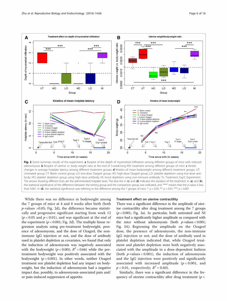
munoreactivity to PR-B, p-p65, COX-2, and TRPV1 inectopic/eutopic endometrium and to OTR in myome-trium among different groups (all p-values <0.01; Figs. 5and 6, and Table 1). In particular, multiple linear regres-sion analyses (all immunoreactivity levels were square-root transformed to improve normality unless statedotherwise) indicated that while adenomyosis inductionwas associated with the increase (decrease for PR-B)while Ozagrel treatment or platelet depletion was associ-ated, in a dose-dependent manner, with a significant re-duction (increase for PR-B) of immunoreactivity to allthese proteins or the extent of platelet aggregation/macrophage infiltration (all p-values <0.01, with R2 ran-ging from 0.31 to 0.76; Table 1 and Fig. 5).In addition to these markers, we also performed im-
munostaining of lesional OTR, a marker of SMM, andcollagen I and IV, markers of fibrosis, in adenomyotic
lesions, as well as OTR in myometrium, which was likelyresponsible for uterine hyperactivity. OTR staining waslocalized in both the cell membrane and the cytoplasmof glandular epithelial and stromal cells as well as myo-metrial smooth muscle cells (Fig. 6). We scored OTRstaining levels in epithelial/stromal cells and myometrialmuscle cells separately. No difference in OTR staininglevels in the epithelial component was found (data notshown), and hence only the data in the stromal compo-nent were demonstrated. Both collagen I and collagen IVstaining was seen nearly uniformly in extracellular matrixof the ectopic endometrial stromal tissues, irrespective ofthe proximity to the glandular epithelial cells or not.We found that for all these markers, the presence
of adenomyosis and, in the case of lesional and myo-metrial OTR staining, the injection of non-immuneantibodies were positively associated with the staininglevels while Ozagrel treatment and platelet depletionwere associated, in a dose-dependent manner, with asignificant reduction of immunoreactivity to all theseproteins (all p-values <0.001; Fig. 5; Table 1).We found that the immunostaining levels of these pro-
teins were all highly correlated, with the positive correlationcoefficients ranged from 0.69 to 0.95 (PR-B excluded; all p-values <0.001) and the negative correlation coefficientsranged from −0.72 to −0.93 (all p-values < 0.001 for PR-Bvs. others).
Fig. 4 Representative photomicrographs of indicated immunostaining in eutopic (for blank control group) or ectopic (all other groups)endometrium among different treatment groups, which are arranged in different columns. CD41: CD41-labeled platelets; F4/80: F4/80-labled macrophages(indicated by yellow arrows); p-p65: phosphorylated form of NF-kB p65 subunit; PR-B: progesterone receptor isoform B; COX-2: Cyclooxygenase 2; TRPV1:transient receptor potential cation channel, subfamily V, member 1. Blank: blank control endometrium; Hi-Oza: high-dose Ozagrel group; Lo-Oza: low-doseOzagrel group; Hi-Dep: platelet depletion with high-dose antibody; Lo-Dep: platelet depletion with low-dose antibody; IgG: non-immune mock antibody.Scale bar = 125 μm
Zhu et al. Reproductive Biology and Endocrinology (2016) 14:66 Page 8 of 16
Fig. 5 Summary of immunohistochemistry results. Boxplot of immunoreactivity against CD41 (a), the number of F4/80+ positive macrophages(b), p-p65 c, PR-B (d), COX-2 (e), TRPV1 (f),OTR (g), myometrial OTR (h), Collagen I (i), and Collagen IV (j) in ectopic/eutopic endometrium. Thegroup labels are the same as used in Fig. 2
Zhu et al. Reproductive Biology and Endocrinology (2016) 14:66 Page 9 of 16

We found that the extent of platelet aggregation andof the macrophage infiltration as well as the immunore-activity to PR-B, p-p65, COX-2 and TRPV1 were allhighly correlated with the depth of myometrial infiltra-tion (all positive except PR-B, which was negative, andthe Spearman’s correlation coefficients ranged from 0.69
to 0.88 (−0.87 for PR-B), all p-values <0.001). The Jonc-kheere trend test indicated that all these immunostain-ing levels were significantly associated with the depth ofmyometrial infiltration (all p-values <0.001; Fig. 7). Amultiple linear regression analysis indicated that theOTR and PR-B staining levels in ectopic endometriumwere the only 2 co-variables that are associated with thedepth of myometrial infiltration (OTR, positive associ-ation, p = 6.6×10⁻¹⁵, PR-B, negative association, and p =0.0020, respectively; R2 = 0.83).
Effect of treatment on the number of GAD65-positiveneurons in the brainstem nucleus raphe magnus (NRM)To see whether Ozagrel treatment and platelet depletionhad any effect on the GABAergic inhibition system in theNRM, we performed an immunofluorescent staining ofGAD65 in the NRM (Fig. 8a) and counted the number ofGAD65-positive and synapsin I-positive neurons in theNRM. This number would be a measure of the number ofGAD65-expressing neurons in the NRM.We found that there is a significant difference in
the number of GAD65-positive neurons in the NRMamong the seven groups (p < 0.001; Fig. 8b). A mul-tiple linear regression analysis (the number of cellswas square-root transformed to improve normality)indicated that while adenomyosis induction was asso-ciated with the reduction in the number of GAD65-positive neurons in the NRM (p < 0.001) as previouslyreported [26] while both Ozagrel treatment and plate-let depletion were associated dose-dependently with asignificant increase of the number of GAD65-positiveneurons (both p-values <0.001; R2 = 0.92).The number of GAD65-positive neurons (log-trans-
formed) in the NRM was found to be positively corre-lated with the hotplate latency after treatment (r = 0.88,p < 0.001; Fig. 8c). It also was found to be negatively cor-related with the plasma CORT levels (r = −0.86, p <0.001; Fig. 8D).
Fig. 6 Representative immunohistochemisty staining of markers of smooth muscle metaplasia and fibrosis in ectopic and eutopic endometrium.Different rows indicate different proteins in different groups (arranged in different columns) with different doses of Ozagrel, different doses of ratanti-mouse GPIbα polyclonal IgG and non-immune rat anti-mouse IgG. For oxytocin receptor (OTR), M stands for staining in the myometrium,stromal and gland epithelium was separately evaluated. Magnification in all figures: ×400. Scale bar = 125 μm
Table 1 Results from early/later platelet depletion experiment.All results were based on multiple regression analyses with theindependent variable square-root transformed and dummy vari-ables indicating the presence or absence of adenomyosis, non-immune IgG antibody injection or not, the dosage of antibodyto deplete platelets and the dosage of Ozagrel as co-variables
Name Induction ofadenomyosis
Ozagreltreatment
Plateletdepletion
R2
Extent of CD41+ plateletaggregation
↑***
↓***
↓***
0.54
Number of infiltrated F4/80+ macrophages
↑***
↓**
↓*
0.30
Phosph-p65 ↑***
↓**
↓**
0.31
PR-B ↓***
↑***
↑***
0.73
COX-2 ↑***
↓***
↓***
0.48
TRPV1 ↑***
↓***
↓***
0.47
OTR ↑***
↓***
↓***
0.58
Collagen I ↑***
↓***
↓*
0.29
Collagen IV ↑***
↓**
↓***
0.33
Myometrial OTR ↑***
↓***
↓***
0.77
↓: Denotes that the immunoreactivity to this protein at hand was significantlydecreased based on multiple linear regression analysis;↑Denotes that theimmunoreactivity to this protein of interest was significantly increased basedon multiple linear regression analysis. The R2 value of the correspondingregression model is shown at the right-most column. Symbols of statistical sig-nificance levels: *: p<0.05; **: p<0.01; ***: p<0.001
Zhu et al. Reproductive Biology and Endocrinology (2016) 14:66 Page 10 of 16

Fig. 7 (See legend on next page.)
Zhu et al. Reproductive Biology and Endocrinology (2016) 14:66 Page 11 of 16

Factors associated with the uterine contractilityBoth contractile amplitude and frequency correlated posi-tively with the myometrial OTR staining levels (r = 0.82, p< 0.001, and r = 0.35, p < 0.01, respectively; Additional file 1:Figure S3A, B of Supplemental Information) and also with
the lesional OTR staining levels in the ectopic endomet-rium (r = 0.94, p < 0.001, and r= 0.50, p < 0.001; Additionalfile 1: Figure S3 c, d of Supplemental Information).For contractile amplitude, the multiple linear regres-
sion incorporating uterine weight/bodyweight ratio,
(See figure on previous page.)Fig. 7 Summary results on immunohistochemistry measures as a function of the depth of myometrial infiltration. Boxplot of the extent of plateletaggregation (a), extent of macrophage infiltration (b), immunoreactivity against p-p65 (c), PR-B (d), COX-2 (e), TRPV1 (f), OTR (g), myometrial OTR (h),collagen I (i), and collagen IV (j) in ectopic endometrium as a function of the depth of myometrial infiltration of endometrial tissues. The p-value shownin each figure is the statistical significance of the Jonckheere trend test
Fig. 8 a Micrographs of immunofluorescent staining of GAD65 in the nucleus raphe magnus (NRM) in different groups of mice. Both GAD65- andSynapsin I-positive neurons were identified, as indicated by white arrows. To see the picture more closely, the area of interested was amplified threetimes. The original amplification:x400. The scale bar represents 125 μm. b Boxplot showing the number of GAD65+ neurons in the NRM among differenttreatment groups. The dashed line represents the median value of all mice. Blank: blank control endometrium; Hi-Oza: high-dose Ozagrel group; Lo-Oza:low-dose Ozagrel group; Hi-Dep: platelet depletion with high-dose antibody; Lo-Dep: platelet depletion with low-dose antibody; IgG: non-immune mockantibody. c Scatter plot of hotplate latency vs. the log-transformed number of GAD65-positive neurons in the NRM for all groups of mice; d Scatter plot ofplasma corticosterone levels vs. the log-transformed number of GAD65-positive neurons in the NRM for all groups of mice. Each alphabet in the figurerepresents one experimental observation, and the alphabets are the abbreviations of different treatment groups. C: Blank control; U: Untreated; o: Low-dose Ozagrel; O: High-dose Ozagrel; d: Platelet depletion using low-dose antibody; D: Platelet depletion using high-dose antibody; N: Non-immune IgG.The correlation coefficient and its statistical significance levels are shown in (c) and (d). ***: p< 0.001
Zhu et al. Reproductive Biology and Endocrinology (2016) 14:66 Page 12 of 16
depth of myometrial infiltration (0 if no adenomyosis),extent of platelet aggregation and macrophage infiltra-tion and all immunostaining measurements in ectopicendometrium identified the depth of myometrial infiltra-tion (p < 0.01), uterine weight/bodyweight ratio (p <0.001), the extent of platelet aggregation (p < 0.001), andthe number of infiltrating macrophages (p < 0.01) as fourcovariates that were associated with the contractile amp-litude (R2 = 0.94). For contractile frequency, we found,through multiple linear regression analysis, that only theuterine weight/bodyweight ratio (p < 0.001), lesional andmyometrial OTR staining levels (both p < 0.05), and PR-B staining levels (p < 0.05) were associated with the con-tractile frequency (R2 = 0.42).
Determinants of thermal response latency after treatmentWe carried out a multiple linear regression analysis toidentify which factors potentially determine the changein thermal response latency before and after drug treat-ment using the pre-treatment latency, bodyweight, depthof myometrial infiltration (grade = 0 if no adenomyosis),uterine weight vs. bodyweight ratio, amplitude and fre-quency of uterine contraction, and presence of adeno-myosis as covariates. We found that the uterine weightvs. bodyweight ratio (p < 0.01), contractile amplitude (p< 0.01), the presence of adenomyosis (p < 0.001) and thedepth of myometrial infiltration (p < 0.001) were allnegatively associate with the change in before-after hot-plate latency (R2 = 0.90).
DiscussionWe have provided evidence that anti-platelet treatment,through either platelet depletion or Ozagrel treatment,resulted in the suppression of myometrial infiltration,improved generalized hyperalgesia, reduced uterineweight vs. bodyweight ratio and stress level, and reducedamplitude and frequency of uterine contraction in micewith induced adenomyosis. The anti-platelet treatmentalso improved the expression of some proteins known tobe involved in adenomyosis and reduced the number ofinfiltrating macrophages. In particular, it reduced thelesional expression of OTR, a SMM marker [29], and ofcollagen I and IV, markers of extracellular matrix de-posits and thus fibrosis. Moreover, it increased the num-ber of GAD65-expressing neurons in the brainstemNRM, thus likely boosting the GABAergic inhibition ofpain due to adenomyosis, which in turn helps pain reliefand reduces the stress level.Our data are consistent with our finding that increased
platelet aggregation and the extent of fibrosis in bothmouse and human adenomyosis [10, 11]. They are alsoconsistent with our previous report that anti-platelettherapy is effective in treating endometriosis in mouse[9, 14, 30] and that the expression of tissue factor in
adenomyosis is elevated [5, 31]. Tissue factor plays acritical role in the initiation of platelet activation and co-agulation [32]. In addition, considerable experimentaldata [33, 34] and limited clinical data [35] support theinvolvement of hyperprolactinemia in adenomyosis, yetprolactin is a potent cofactor for platelet aggregation[36, 37]. These data, taken together, seem to suggest thatpatients with adenomyosis may be in a hypercoagulablestate as those with endometriosis [38]. This may explainthe report of cerebral infarcts associated with adeno-myosis [39] and increased mean platelet volume inwomen with adenomyosis [40].Our data are also consistent with our previous reports
that PR-B expression in adenomyosis is reduced [41] duepossibly to PR-B promoter hypermethylation [42]. Inaddition, they are consistent with reported constitutive acti-vation of NF-κB [5], increased expression of COX-2 [7],TRPV1 [43] and OTR in adenomyosis [29, 43, 44]. The in-creased uterine contractility, and, in particular, its close as-sociation with the OTR expression and with the reducedhotplate latency as reported in this study are consistentwith what we reported in human adenomyosis [44]. Inother words, the mouse model used in this study recapitu-lates several important features of human adenomyosis, i.e.,inflammation and angiogenesis as displayed by the consti-tutive activation of NF-κB and increased COX-2 but de-creased PR-B expression in adenomyotic lesions, increaseduterine weight and presumably enlarged uterus, increasedgeneralized hyperalgesia, and elevated uterine contractility,due possibly to elevated myometrial OTR expression. Re-markably, anti-platelet treatment either reversed or abro-gated these changes.We note that the anti-platelet treatment achieved the
therapeutic effects very similar to EGCG [20, 26, 45] andresveratrol [46, 47] as we reported earlier. However, it isperhaps no coincidence that EGCG is anti-platelet [48]and so is resveratrol [46]. In fact, some compounds thatare reported to be promising in treating adenomyosis inpreclinical and clinical studies, such as andrographolide[2, 5, 49], valproic acid [19, 50, 51], and statins [52, 53], allturn out to be anti-platelet [54–57]. Even danazol, a once-popular, FDA-approved drug for treating endometriosis,has long been reported to have anti-platelet effect [58, 59].That said, we should emphasize that, despite promis-
ing results of anti-platelet treatment by either plateletdepletion or Ozagrel treatment as shown here, we arenot advocating their use in clinical setting per se, eventhough Ozagrel is a prescription drug as of now. Adeno-myosis is a benign disease and certainly not life-threatening. As such, it places higher premium on drugsafety as compared with other life-threatening diseasessuch as cancer. While Ozagrel is generally safe and holdspromises in treating adenomyosis, the hemorrhage riskit entails deserves caution. This study was meant to be a
Zhu et al. Reproductive Biology and Endocrinology (2016) 14:66 Page 13 of 16
proof-of-concept study, demonstrating the therapeuticpotential of anti-platelet therapy for adenomyosis. It isnot intended to advocate Ozagrel per se for the treat-ment of human adenomyosis. More research is neededto determine which anti-platelet compound has the de-sirable benefit-to-risk ratio in treating adenomyosis.While the exact mechanisms of action for anti-
platelet therapy remain to be investigated, it is possiblethat anti-platelet treatment suppresses the activation ofthe TGF-β1/Smad3 signaling pathway and the expres-sion of ER-β, both of which can be induced by activatedplatelets [15, 60]. In addition, the treatment suppressesthe activation of NF-κB, which also can be induced byplatelets (Zhang et al., unpublished data). Moreover, ac-tivated platelets express P-selectin (CD62P/GMP-140)on their cell surface [61–63], which binds to its ligand,P-selectin glycoprotein ligand-1 (PSGL-1), that isexpressed on the cell surface of most leukocytes, suchas neutrophils, monocytes, Th1 lymphocytes, eosino-phils, and basophils, and facilitates inflammation,hemostasis, thrombosis, and the growth and metastasisof cancer [62, 64]. P-selectin interacts with PSGL-1, atransmembraine homodimer, to mediate the rolling ofleukocytes on stimulated endothelial cells and the het-erotypic aggregation of activated platelets and leuko-cytes [65], and activate mitogen-activated proteinkinases (MAPKs) [66] and β2 integrins [67]. Hence,anti-platelet treatment should abrogate or attenuate in-flammation caused by adenomyosis, as seen in reducedp-p65 expression in this study.Anti-platelet therapy can also suppress neurite outgrowth
and thus hyperinnervation in adenomyosis [68, 69] sinceectopic endometrial stromal cells secrete platelet inducerssuch as thrombin and thromboxane A2 (TXA2) [70]. TXA2
has been reported to stimulate neurite outgrowth in cere-bral cortical neurons [71], and we have also found that itcan do so in dorsal ganglia root neurons [72]. Since TXA2,PGH2, and PGI2 have been reported to be potent inducersof uterine contractility [73] and uterine contractility is doc-umented to be correlated with the severity of dysmenor-rhea in adenomyosis [44], the suppression of plateletactivation and the resultant COX-2 down-regulation maysuppress hyperinnervation and uterine hyperactivity, thusresponsible for improved generalized hyperalgesia and re-duced plasma CORT levels.The reduced plasma CORT levels in mice with anti-
platelet treatment is likely to result from the modulationof chronic stress (adenomyosis-induced pain) responsethrough GABA receptors as in a chick model of acutestress [74]. Alternatively, adenomyosis-induced pain orhyperalgesia may result in synaptic dysfunction, for ex-ample, HDAC-mediated impairment of GABA synapticinhibition in the brainstem NRM [75]. However, whethersuppression of platelet activation may restore the GABA
synaptic inhibition in NRM through the reduction ofHDAC activity remains to be clarified.
ConclusionsThis study further provides evidence that platelets playimportant roles in the development of adenomyosis. Inaddition, this study demonstrates that anti-platelet treat-ment is efficacious in suppressing myometrial infiltra-tion, improving generalized hyperalgesia, reducingboth uterine hyperactivity and systemic CORT levels inmice with induced adenomyosis. Collectively, these re-sults demonstrate that anti-platelet therapy holds prom-ises as a non-hormonal treatment for treatingadenomyosis.
Additional file
Additional file 1: Supplemental materials [76–80]. (PDF 358 kb)
AbbreviationsCORT: Corticosterone; COX-2: Cyclooxygenase-2; EGCG: Epigallocatechin-3-gallate; EMT: Epithelial-mesenchymal transition; ER-β: Estrogen receptor β;FDA: FMT: fibroblast-to-myofibroblast transdifferentiation; GABA: γ-aminobutyric acid; GAD65: Glutamic acid decarboxylase 65;H&E: Hematoxylin and eosin; HD: Platelet depletion by high-dose rat anti-mouse GPIbα polyclonal IgG treatment; HDAC: Histone deacetylase;HO: High-dose Ozagrel treatment; LD: Platelet depletion by low-dose ratanti-mouse GPIbα polyclonal IgG treatment; LO: Low-dose Ozagreltreatment; MAPK: Mitogen-activated protein kinase; NI: Non-immune rat anti-mouse IgG isotope-matched with the anti-GPIbα antibody; NRM: Nucleusraphe magnus; OTR: Oxytocin receptor; PGH2: Prostaglandin H2, PGI2prostaglandin I2; p-p65: Phosphorylated p65 subunit; PR-B: Progesteronereceptor isoform B; PSGL-1: P-selectin glycoprotein ligand-1;ReTIAR: Repeated tissue injury and repair; SMM: Smooth muscle metaplasia;TGF-β1: Transforming growth factor β1; TRPV1: Transient receptor potentialcation channel, subfamily V, member 1; TXA2: Thromboxane A2
AcknowledgmentThe authors would like to thank the Administration of Wenzhou People’sHospital for its support and encouragement, and funding agencies for theirfinancial support.This paper has been presented orally at the First Congress of the Society ofEndometriosis and Uterine Disorders (SEUD) held in Paris on May 9, 2015.
FundingThis work was supported in part by grant Y14H040004 (YMC) from theNational Science Foundation of Zhejiang Province, grants 81471434 (SWG),81270676 (SWG), 81530040 (SWG), and 81370695 (XSL) from the NationalScience Foundation of China. None of the funders, however, has any role inthe design of the study, and the collection, analysis, and interpretation ofdata and in writing the manuscript.
Availability of data and materialsThe data used in this study are available upon request.
Authors' contributionsSWG conceived and designed the study, performed data analysis and datainterpretation, and drafted the manuscript. BZ and YMC carried out most ofthe experiment, XLS and XSL provided assistance in immunohistochemistryanalysis. All participated in writing up the manuscript. All authors read andapproved the final manuscript.
Competing interestsThe authors declare that they have no competing interests.
Zhu et al. Reproductive Biology and Endocrinology (2016) 14:66 Page 14 of 16

Consent for publicationNot applicable.
Ethics approvalThis study was approved by the institutional experimental animals reviewboard of Shanghai OB/GYN Hospital, Fudan University.
Acknowledgment of financial supportThis research was supported in part by grant Y14H040004 (YMC) from theScience Foundation of Zhejiang Province, grants 81471434 (SWG), 81270676(SWG), 81530040 (SWG), 81370695 (XSL) and 81671436 (XSL) from theNational Natural Science Foundation of China.
Author details1Department of Obstetrics and Gynecology, The People’s Hospital, Wenzhou,Zhejiang 325800, China. 2Shanghai Key Laboratory of Female ReproductiveEndocrine-Related Diseases, Shanghai 200011, China. 3Shanghai Obstetricsand Gynecology Hospital, Fudan University, 419 Fangxie Road, Shanghai200011, China.
Received: 17 August 2016 Accepted: 23 September 2016
References1. Benagiano G, Habiba M, Brosens I. The pathophysiology of uterine
adenomyosis: an update. Fertil Steril. 2012;98:572–9.2. Mao X, Wang Y, Carter AV, Zhen X, Guo SW. The retardation of myometrial
infiltration, reduction of uterine contractility, and alleviation of generalizedhyperalgesia in mice with induced adenomyosis by levo-tetrahydropalmatine(l-THP) and andrographolide. Reprod Sci. 2011;18:1025–37.
3. Wood C. Adenomyosis: difficult to diagnose, and difficult to treat. DiagnTher Endosc. 2001;7:89–95.
4. Kitawaki J. Adenomyosis: the pathophysiology of an oestrogen-dependentdisease. Best Pract Res Clin Obstet Gynaecol. 2006;20:493–502.
5. Li B, Chen M, Liu X, Guo SW. Constitutive and tumor necrosis factor-alpha-induced activation of nuclear factor-kappaB in adenomyosis and itsinhibition by andrographolide. Fertil Steril. 2013;100:568–77.
6. Khan KN, Kitajima M, Hiraki K, Fujishita A, Sekine I, Ishimaru T, et al. Changesin tissue inflammation, angiogenesis and apoptosis in endometriosis,adenomyosis and uterine myoma after GnRH agonist therapy. Hum Reprod.2010;25:642–53.
7. Ota H, Igarashi S, Sasaki M, Tanaka T. Distribution of cyclooxygenase-2 ineutopic and ectopic endometrium in endometriosis and adenomyosis. HumReprod. 2001;16:561–6.
8. Ulukus EC, Ulukus M, Seval Y, Zheng W, Arici A. Expression of interleukin-8and monocyte chemotactic protein-1 in adenomyosis. Hum Reprod. 2005;20:2958–63.
9. Ding D, Liu X, Duan J, Guo SW. Platelets are an unindicted culprit in thedevelopment of endometriosis: clinical and experimental evidence. HumReprod. 2015;30:812–32.
10. Shen M, Liu X, Zhang H, Guo SW. Transforming Growth Factor β1 SignalingCoincides with -Mediated Epithelial-Mesenchymal Transition and Fibroblast-to-Myofibroblast Transdifferentiation in Drive the Development ofAdenomyosis in Mice. Hum Reprod 2016; In press.
11. Liu X, Shen S, Qi Q, Zhang H, Guo S-W. Corroborating Evidence forPlatelet-Induced Epithelial-Mesenchymal Transition and Fibroblast-to-Myofibroblast Transdifferentiationin the Development of Adenomyosis.Hum Reprod 2016; In press.
12. Guo SW, Ding D, Shen M, Liu X. Dating endometriotic ovarian cysts basedon the content of cyst fluid and its potential clinical implications. ReprodSci. 2015;22:873–83.
13. Zhang Q, Duan J, Olson M, Fazleabas A, Guo SW. Cellular changesconsistent with epithelial-mesenchymal transition and fibroblast-to-myofibroblast transdifferentiation in the progression of experimentalendometriosis in baboons. Reprod Sci 2016; In press.
14. Guo SW, Ding D, Geng JG, Wang L, Liu X. P-selectin as a potentialtherapeutic target for endometriosis. Fertil Steril. 2015;103:990–1000. e8.
15. Zhang Q, Ding D, Liu X, Guo SW. Activated platelets induce estrogenreceptor beta expression in endometriotic stromal cells. Gynecol ObstetInvest. 2015;80(3):187–92.
16. Loo MH, Egan D, Vaughan Jr ED, Marion D, Felsen D, Weisman S. Theeffect of the thromboxane A2 synthesis inhibitor OKY-046 on renalfunction in rabbits following release of unilateral ureteral obstruction. J Urol.1987;137:571–6.
17. Green AR, Styles JA, Parrott EL, Gray D, Edwards RE, Smith AG, et al.Neonatal tamoxifen treatment of mice leads to adenomyosis but notuterine cancer. Exp Toxicol Pathol. 2005;56:255–63.
18. Parrott E, Butterworth M, Green A, White IN, Greaves P. Adenomyosis–aresult of disordered stromal differentiation. Am J Pathol.2001;159:623–30.
19. Liu X, Guo SW. Valproic acid alleviates generalized hyperalgesia in mice withinduced adenomyosis. J Obstet Gynaecol Res. 2011;37:696–708.
20. Chen Y, Zhu B, Zhang H, Liu X, Guo SW. Epigallocatechin-3-gallate reducesmyometrial infiltration, uterine hyperactivity, and stress levels and alleviatesgeneralized hyperalgesia in mice induced with adenomyosis. Reprod Sci.2013;20(12):1478–91.
21. Council NR. Guide for the Care and use of Laboratory Animals. Washington:National Academies Press; 1996.
22. Bird CC, McElin TW, Manalo-Estrella P. The elusive adenomyosis of theuterus–revisited. Am J Obstet Gynecol. 1972;112:583–93.
23. Barrier BF, Malinowski MJ, Dick Jr EJ, Hubbard GB, Bates GW. Adenomyosisin the baboon is associated with primary infertility. Fertil Steril. 2004;82Suppl 3:1091–4.
24. Vercellini P, Ragni G, Trespidi L, Oldani S, Panazza S, Crosignani PG.Adenomyosis: a deja vu? Obstet Gynecol Surv. 1993;48:789–94.
25. Wang-Tilz Y, Tilz C, Wang B, Tilz GP, Stefan H. Influence of lamotrigine andtopiramate on MDR1 expression in difficult-to-treat temporal lobe epilepsy.Epilepsia. 2006;47:233–9.
26. Chen Y, Zhu B, Zhang H, Ding D, Liu X, Guo SW. Possible Loss of GABAergicInhibition in Mice With Induced Adenomyosis and Treatment WithEpigallocatechin-3-Gallate Attenuates the Loss With Improved Hyperalgesia.Reprod Sci. 2014;21(7):869–82.
27. Liu X, Guo SW. Dysmenorrhea: risk factors in women with endometriosis.Womens Health (Lond Engl). 2008;4:399–411.
28. R Core Team. R: A Language and Environment for Statistical Computing.Vienna: R Foundation for Statistical Computing; 2016.
29. Mechsner S, Grum B, Gericke C, Loddenkemper C, Dudenhausen JW, EbertAD. Possible roles of oxytocin receptor and vasopressin-1alpha receptor inthe pathomechanism of dysperistalsis and dysmenorrhea in patients withadenomyosis uteri. Fertil Steril. 2010;94:2541–6.
30. Guo SW, Ding D, Liu X. Anti-platelet therapy is efficacious in treatingendometriosis induced in mouse..Reprod Biomed Online 2016; In press.
31. Liu X, Nie J, Guo SW. Elevated immunoreactivity to tissue factor and itsassociation with dysmenorrhea severity and the amount of menses inadenomyosis. Hum Reprod. 2011;26:337–45.
32. Engelmann B, Luther T, Muller I. Intravascular tissue factor pathway–a modelfor rapid initiation of coagulation within the blood vessel. Thromb Haemost.2003;89:3–8.
33. Mori T, Singtripop T, Kawashima S. Animal model of uterine adenomyosis: isprolactin a potent inducer of adenomyosis in mice? Am J Obstet Gynecol.1991;165:232–4.
34. Ficicioglu C, Tekin HI, Arioglu PF, Okar I. A murine model of adenomyosis:the effects of hyperprolactinemia induced by fluoxetine hydrochloride, aselective serotonin reuptake inhibitor, on adenomyosis induction in Wistaralbino rats. Acta Eur Fertil. 1995;26:75–9.
35. Yu Y, Jing Z, Zhi-Yu H, Xia M, Yan-Li H, Chang-Tao X, et al. Ultrasound-guided percutaneous microwave ablation for adenomyosis: efficacy oftreatment and effect on ovarian function. Sci Rep. 2015;5:10034.
36. Wallaschofski H, Donne M, Eigenthaler M, Hentschel B, Faber R, Stepan H, etal. PRL as a novel potent cofactor for platelet aggregation. J Clin EndocrinolMetab. 2001;86:5912–9.
37. Urban A, Masopust J, Maly R, Hosak L, Kalnicka D. Prolactin as a factor forincreased platelet aggregation. Neuro Endocrinol Lett. 2007;28:518–23.
38. Wu Q, Ding D, Liu X, Guo SW. Evidence for a hypercoagulable state inwomen with ovarian endometriomas. Reprod Sci. 2015;22:1107–14.
39. Yamashiro K, Tanaka R, Nishioka K, Ueno Y, Shimura H, Okuma Y, et al.Cerebral infarcts associated with adenomyosis among middle-aged women.J Stroke Cerebrovasc Dis. 2012;21:910. e1-5.
40. Bodur S, Gun I, Alpaslan Babayigit M. The significance of mean plateletvolume on diagnosis and management of adenomyosis. Med Glas (Zenica).2013;10:59–62.
Zhu et al. Reproductive Biology and Endocrinology (2016) 14:66 Page 15 of 16

41. Nie J, Lu Y, Liu X, Guo SW. Immunoreactivity of progesterone receptorisoform B, nuclear factor kappaB, and IkappaBalpha in adenomyosis. FertilSteril. 2009;92:886–9.
42. Jichan N, Xishi L, Guo SW. Promoter hypermethylation of progesterone receptorisoform B (PR-B) in adenomyosis and its rectification by a histone deacetylaseinhibitor and a demethylation agent. Reprod Sci. 2010;17:995–1005.
43. Nie J, Liu X, Guo SW. Immunoreactivity of oxytocin receptor and transientreceptor potential vanilloid type 1 and its correlation with dysmenorrhea inadenomyosis. Am J Obstet Gynecol. 2010;202:346. e1-8.
44. Guo SW, Mao X, Ma Q, Liu X. Dysmenorrhea and its severity are associatedwith increased uterine contractility and overexpression of oxytocin receptor(OTR) in women with symptomatic adenomyosis. Fertil Steril.2013;99:231–40.
45. Jin YR, Im JH, Park ES, Cho MR, Han XH, Lee JJ, et al. Antiplatelet activity ofepigallocatechin gallate is mediated by the inhibition of PLCgamma2phosphorylation, elevation of PGD2 production, and maintaining calcium-ATPase activity. J Cardiovasc Pharmacol. 2008;51:45–54.
46. Zhu B, Chen Y, Zhang H, Liu X, Guo SW. Resveratrol reduces myometrialinfiltration, uterine hyperactivity, and stress levels and alleviates generalizedhyperalgesia in mice with induced adenomyosis. Reprod Sci.2015;22(11):1336–49.
47. Shen MY, Hsiao G, Liu CL, Fong TH, Lin KH, Chou DS, et al. Inhibitorymechanisms of resveratrol in platelet activation: pivotal roles of p38 MAPKand NO/cyclic GMP. Br J Haematol. 2007;139:475–85.
48. Ok WJ, Cho HJ, Kim HH, Lee DH, Kang HY, Kwon HW, et al.Epigallocatechin-3-gallate has an anti-platelet effect in a cyclic AMP-dependent manner. J Atheroscler Thromb. 2012;19:337–48.
49. Liu X, Yu S, Guo SW. A pilot study on the use of andrographolide to treatsymtomatic adenomyosis. Gynecol Minim Invas Ther. 2014;3:119–26.
50. Liu X, Guo SW. A pilot study on the off-label use of valproic acid to treatadenomyosis. Fertil Steril. 2008;89:246–50.
51. Liu X, Lei Y, Guo SW. Valproic acid as a therapy for adenomyosis: acomparative case series. Reprod Sci. 2010;17:904–12.
52. Piotrowski PC, Kwintkiewicz J, Rzepczynska IJ, Seval Y, Cakmak H, Arici A, etal. Statins inhibit growth of human endometrial stromal cells independentlyof cholesterol availability. Biol Reprod. 2006;75:107–11.
53. Esfandiari N, Khazaei M, Ai J, Bielecki R, Gotlieb L, Ryan E, et al. Effect of astatin on an in vitro model of endometriosis. Fertil Steril.2007;87:257–62.
54. Lien LM, Su CC, Hsu WH, Lu WJ, Chung CL, Yen TL, et al. Mechanisms ofandrographolide-induced platelet apoptosis in human platelets: regulatoryroles of the extrinsic apoptotic pathway. Phytother Res. 2013;27:1671–7.
55. Lu WJ, Lee JJ, Chou DS, Jayakumar T, Fong TH, Hsiao G, et al. A novel roleof andrographolide, an NF-kappa B inhibitor, on inhibition of plateletactivation: the pivotal mechanisms of endothelial nitric oxide synthase/cyclic GMP. J Mol Med (Berl). 2011;89:1261–73.
56. Davidson DC, Hirschman MP, Spinelli SL, Morrell CN, Schifitto G, Phipps RP, et al.Antiplatelet activity of valproic acid contributes to decreased soluble CD40 ligandproduction in HIV type 1-infected individuals. J Immunol. 2011;186:584–91.
57. Cai A, Zhou Y, Li L. Rho-GTPase and atherosclerosis: pleiotropic effects ofstatins. J Am Heart Assoc. 2015;4.
58. Fraser IS, Burridge J. Danazol treatment and platelet function. Med J Aust.1980;1:313–4.
59. Arrowsmith JB, Dreis M. Thrombocytopenia after treatment with danazol. NEngl J Med. 1986;315:585.
60. Zhang Q, Duan J, Liu X, Guo SW. Platelets drive smooth muscle metaplasiaand fibrogenesis in endometriosis through epithelial-mesenchymaltransition and fibroblast-to-myofibroblast transdifferentiation. Mol CellEndocrinol 2016; In press.
61. Vestweber D, Blanks JE. Mechanisms that regulate the function of theselectins and their ligands. Physiol Rev. 1999;79:181–213.
62. Chen M, Geng JG. P-selectin mediates adhesion of leukocytes, platelets, andcancer cells in inflammation, thrombosis, and cancer growth and metastasis.Arch Immunol Ther Exp (Warsz). 2006;54:75–84.
63. Furie B, Furie BC, Flaumenhaft R. A journey with platelet P-selectin: themolecular basis of granule secretion, signalling and cell adhesion. ThrombHaemost. 2001;86:214–21.
64. Kim YJ, Borsig L, Varki NM, Varki A. P-selectin deficiency attenuates tumorgrowth and metastasis. Proc Natl Acad Sci U S A. 1998;95:9325–30.
65. McEver RP, Zhu C. Rolling cell adhesion. Annu Rev Cell Dev Biol. 2010;26:363–96.
66. Hidari KI, Weyrich AS, Zimmerman GA, McEver RP. Engagement of P-selectinglycoprotein ligand-1 enhances tyrosine phosphorylation and activatesmitogen-activated protein kinases in human neutrophils. J Biol Chem.1997;272:28750–6.
67. Ma YQ, Plow EF, Geng JG. P-selectin binding to P-selectin glycoproteinligand-1 induces an intermediate state of alphaMbeta2 activation and actscooperatively with extracellular stimuli to support maximal adhesion ofhuman neutrophils. Blood. 2004;104:2549–56.
68. Zhang X, Lu B, Huang X, Xu H, Zhou C, Lin J. Endometrial nerve fibers inwomen with endometriosis, adenomyosis, and uterine fibroids. Fertil Steril.2009;92:1799–801.
69. Zhang X, Lu B, Huang X, Xu H, Zhou C, Lin J. Innervation of endometriumand myometrium in women with painful adenomyosis and uterine fibroids.Fertil Steril. 2010;94:730–7.
70. Guo S-W, Du Y, Liu X. Endometriosis-Derivedtic Stromal Cells SecreteThrombin and Thromboxane A2, Inducing Platelet Activation..Reprod Sci2016;In press.
71. Sumimoto S, Muramatsu R, Yamashita T. Thromboxane A2 stimulatesneurite outgrowth in cerebral cortical neurons via mitogen activatedprotein kinase signaling. Brain Res. 2015;1594:46–51.
72. Yan D, Liu X, Guo SW. Endometriosis-Derived Thromboxane A2 InducesNeurite Outgrowth. Reprod Sci 2016; In press.
73. Wilhelmsson L, Wikland M, Wiqvist N. PGH2, TxA2 and PGI2 have potentand differentiated actions on human uterine contractility. Prostaglandins.1981;21:277–86.
74. Adachi N, Tomonaga S, Tachibana T, Denbow DM, Furuse M.(−)-Epigallocatechin gallate attenuates acute stress responses throughGABAergic system in the brain. Eur J Pharmacol. 2006;531:171–5.
75. Zhang Z, Cai YQ, Zou F, Bie B, Pan ZZ. Epigenetic suppression of GAD65expression mediates persistent pain. Nat Med. 2011;17:1448–55.
76. Le Bars D, Gozariu M, Cadden SW. Animal models of nociception.Pharmacol Rev. 2001;53:597–652.
77. Bannon AW, Malmberg AB. Models of nociception: hot-plate, tail-flick, andformalin tests in rodents. Curr Protoc Neurosci. 2007;Chapter 8:Unit 8:9. doi:10.1002/0471142301.ns.0809s41.
78. Wang HW, Wu CC. Effects of oxymetazoline on isolated rat’s trachealsmooth muscle. Eur Arch Otorhinolaryngol. 2008;265:695–8.
79. Calixto JB, Yunes RA. Antagonism of kinin-induced contraction of isolatedrat uterus by the crude hydroalcoholic extract from Mandevilla illustris. GenPharmacol. 1991;22:99–101.
80. Hernandez-Magro PM, Villanueva Saenz E, Alvarez-Tostado Fernandez F, LuisRocha Ramirez J, Valdes Ovalle M. Endoanal sonography in the assessmentof perianal endometriosis with external anal sphincter involvement. J ClinUltrasound. 2002;30:245–8.
• We accept pre-submission inquiries
• Our selector tool helps you to find the most relevant journal
• We provide round the clock customer support
• Convenient online submission
• Thorough peer review
• Inclusion in PubMed and all major indexing services
• Maximum visibility for your research
Submit your manuscript atwww.biomedcentral.com/submit
Submit your next manuscript to BioMed Central and we will help you at every step:
Zhu et al. Reproductive Biology and Endocrinology (2016) 14:66 Page 16 of 16


![Promises, Promises [Score]](https://static.fdocuments.in/doc/165x107/55cf922f550346f57b946648/promises-promises-score.jpg)