Renal Physiology CJASN...
10
Renal Physiology Physiology of the Renal Interstitium Michael Zeisberg* and Raghu Kalluri † Abstract Long overlooked as the virtual compartment and then strictly characterized through descriptive morphologic analysis, the renal interstitium has finally been associated with function. With identification of interstitial renin- and erythropoietin-producing cells, the most prominent endocrine functions of the kidney have now been attributed to the renal interstitium. This article reviews the functional role of renal interstitium. Clin J Am Soc Nephrol 10: ccc–ccc, 2015. doi: 10.2215/CJN.00640114 Introduction The physiologic role of the interstitium of the kidney has received comparatively little interest to date. This may partially be attributed to the fact that it was long considered the exclusive domain of descriptive ultra- structural research, which implied that the interstitium was mostly a passive tissue that structurally supported the tubular epithelium (1,2). The renal interstitium received increasing interest in the context of kidney abnormalities, when the role of interstitial fibrosis in progression of CKD became obvious (3–5). Only since transgenic animal studies revealed the physiologic endocrine function of in- terstitial cells as sources of erythropoietin (Epo) and renin has the physiologic role of the renal interstitium received its due attention. Here we review current knowledge of the form and function of the renal interstitium. Definition and Ultrastructure of the Renal Interstitium The renal interstitium is defined as the intertubular, extraglomerular, extravascular space of the kidney. It is bounded on all sides by tubular and vascular basement membranes and is filled with cells, extra- cellular matrix, and interstitial fluid (1). Its distribution varies within the kidney; it accounts for approximately 8% of the total parenchymal volume in the cortex and up to 40% in the inner medulla (6,7). The term “renal interstitium” is often inadequately used to refer to the peritubular interstitium (the space between tubules, glo- meruli, and capillaries); the periarterial connective tissue and the extraglomerular mesangium are considered spe- cialized interstitia (1). It is debated whether microvessels and capillaries, which are located within the peritubular space, are actually part of the renal interstitium or just run through it (1). Furthermore, lymphatics are considered in- terstitial constituents (1). The tubular interstitium in the cortex and medulla differ with regard to their cellular constituents, extracellular matrix composition, relative volume, and endocrine function, justifying the consid- eration of cortical and medullary interstitium as sepa- rate entities. The intertubular interstitium harbors dendritic cells, macrophages, lymphocytes, lymphatic endothelial cells (in the cortical intertubular interstitium), and various types of fibroblasts, the hallmark cell type of connective tissues (8). In addition, it is believed that the intersti- tium plays a role in fluid and electrolyte exchange and insulation (1). Here we focus on fibroblasts and specif- ically on fibroblasts with endocrine function and their biology in health and disease (other articles have exten- sively reviewed cellular constituents of the immune system within the renal interstitium [9,10]) (Figure 1). Renal Fibroblasts In general, fibroblasts are flattened cells with ex- tended cell processes; in profile they display a fusiform or spindle-like shape and a flattened nucleus (11). Fi- broblasts typically are embedded within the fibrillar matrix of connective tissues and are considered proto- typical mesenchymal cells. Renal fibroblasts anastomose with each other, forming a continuous network in cor- tex and medulla (8). Fibroblasts can acquire an activated phenotype with a relatively large oval nucleus with one or two nucleoli, abundant rough endoplasmatic reticu- lum, and several sets of Golgi apparatus; this reflects their capacity to synthesize substantial amounts of extra- cellular matrix constituents (12). Under physiologic con- ditions, however, adult fibroblasts are relatively inactive, the endoplasmatic reticulum is reduced, and the nucleus is flattened and heterochromatic. On the basis of their appearance, it was assumed that the primary function of renal fibroblasts was to provide structural support to nephrons through deposition of extracellular matrix and through direct cell-cell interactions (1). In addition, fibroblasts play an important role in maintaining vascu- lar integrity in close association with vessels (then typi- cally referred to as vascular smooth muscle cells and pericytes) (13,14). Renal fibroblasts are best known for their role in progression of interstitial fibrosis in pro- gressive CKD because they are principal producers of extracellular matrix (4). Finally, fibroblasts have been identified as sources of Epo and renin in the kidney (15,16). It is obvious that such diverse functions are not fulfilled by one cell type but that renal fibroblasts are instead a heterogeneous cell population with dis- tinct functions. *Department of Nephrology and Rheumatology, Go ¨ttingen University Medical Center, Georg August University, Go ¨ttingen, Germany; and † Department of Cancer Biology and the Metastasis Research Center, University of Texas MD Anderson Cancer Center, Houston, Texas Correspondence: Dr. Michael Zeisberg, Department of Nephrology and Rheumatology, Go ¨ttingen University Medical Center, Georg August University, Robert Koch Str. 40, 37075 Go ¨ttingen, Germany. Email: [email protected] goettingen.de. Dr. Raghu Kalluri, Department of Cancer Biology and the Metastasis Research Center, MD Anderson Cancer Center,1901 East Road, Houston, TX 77054. Email: rkalluri@mdanderson. org www.cjasn.org Vol 10 December, 2015 Copyright © 2015 by the American Society of Nephrology 1 . Published on March 26, 2015 as doi: 10.2215/CJN.00640114 CJASN ePress
-
Upload
trinhtuyen -
Category
Documents
-
view
219 -
download
2
Transcript of Renal Physiology CJASN...
Renal Physiology
Physiology of the Renal Interstitium
Michael Zeisberg* and Raghu Kalluri†
AbstractLong overlooked as the virtual compartment and then strictly characterized through descriptive morphologicanalysis, the renal interstitium has finally been associated with function. With identification of interstitial renin-and erythropoietin-producing cells, the most prominent endocrine functions of the kidney have now beenattributed to the renal interstitium. This article reviews the functional role of renal interstitium.
Clin J Am Soc Nephrol 10: ccc–ccc, 2015. doi: 10.2215/CJN.00640114
IntroductionThe physiologic role of the interstitium of the kidneyhas received comparatively little interest to date. Thismay partially be attributed to the fact that it was longconsidered the exclusive domain of descriptive ultra-structural research, which implied that the interstitiumwas mostly a passive tissue that structurally supportedthe tubular epithelium (1,2). The renal interstitium receivedincreasing interest in the context of kidney abnormalities,when the role of interstitial fibrosis in progression ofCKD became obvious (3–5). Only since transgenic animalstudies revealed the physiologic endocrine function of in-terstitial cells as sources of erythropoietin (Epo) and reninhas the physiologic role of the renal interstitium receivedits due attention. Here we review current knowledge ofthe form and function of the renal interstitium.
Definition and Ultrastructure of the RenalInterstitium
The renal interstitium is defined as the intertubular,extraglomerular, extravascular space of the kidney.It is bounded on all sides by tubular and vascularbasement membranes and is filled with cells, extra-cellular matrix, and interstitial fluid (1). Its distributionvaries within the kidney; it accounts for approximately8% of the total parenchymal volume in the cortex andup to 40% in the inner medulla (6,7). The term “renalinterstitium” is often inadequately used to refer to theperitubular interstitium (the space between tubules, glo-meruli, and capillaries); the periarterial connective tissueand the extraglomerular mesangium are considered spe-cialized interstitia (1). It is debated whether microvesselsand capillaries, which are located within the peritubularspace, are actually part of the renal interstitium or just runthrough it (1). Furthermore, lymphatics are considered in-terstitial constituents (1). The tubular interstitium in thecortex and medulla differ with regard to their cellularconstituents, extracellular matrix composition, relativevolume, and endocrine function, justifying the consid-eration of cortical and medullary interstitium as sepa-rate entities.
The intertubular interstitium harbors dendritic cells,macrophages, lymphocytes, lymphatic endothelial cells
(in the cortical intertubular interstitium), and varioustypes of fibroblasts, the hallmark cell type of connectivetissues (8). In addition, it is believed that the intersti-tium plays a role in fluid and electrolyte exchange andinsulation (1). Here we focus on fibroblasts and specif-ically on fibroblasts with endocrine function and theirbiology in health and disease (other articles have exten-sively reviewed cellular constituents of the immunesystem within the renal interstitium [9,10]) (Figure 1).
Renal FibroblastsIn general, fibroblasts are flattened cells with ex-
tended cell processes; in profile they display a fusiformor spindle-like shape and a flattened nucleus (11). Fi-broblasts typically are embedded within the fibrillarmatrix of connective tissues and are considered proto-typical mesenchymal cells. Renal fibroblasts anastomosewith each other, forming a continuous network in cor-tex and medulla (8). Fibroblasts can acquire an activatedphenotype with a relatively large oval nucleus with oneor two nucleoli, abundant rough endoplasmatic reticu-lum, and several sets of Golgi apparatus; this reflectstheir capacity to synthesize substantial amounts of extra-cellular matrix constituents (12). Under physiologic con-ditions, however, adult fibroblasts are relatively inactive,the endoplasmatic reticulum is reduced, and the nucleusis flattened and heterochromatic. On the basis of theirappearance, it was assumed that the primary function ofrenal fibroblasts was to provide structural support tonephrons through deposition of extracellular matrixand through direct cell-cell interactions (1). In addition,fibroblasts play an important role in maintaining vascu-lar integrity in close association with vessels (then typi-cally referred to as vascular smooth muscle cells andpericytes) (13,14). Renal fibroblasts are best known fortheir role in progression of interstitial fibrosis in pro-gressive CKD because they are principal producers ofextracellular matrix (4). Finally, fibroblasts have beenidentified as sources of Epo and renin in the kidney(15,16). It is obvious that such diverse functions arenot fulfilled by one cell type but that renal fibroblastsare instead a heterogeneous cell population with dis-tinct functions.
*Department ofNephrology andRheumatology,Gottingen UniversityMedical Center, GeorgAugust University,Gottingen, Germany;and †Department ofCancer Biology andthe MetastasisResearch Center,University of TexasMD Anderson CancerCenter, Houston,Texas
Correspondence:Dr. Michael Zeisberg,Department ofNephrology andRheumatology,Gottingen UniversityMedical Center, GeorgAugust University,Robert Koch Str. 40,37075 Gottingen,Germany. Email:[email protected]. Raghu Kalluri,Department of CancerBiology and theMetastasis ResearchCenter, MD AndersonCancer Center,1901East Road, Houston,TX 77054. Email:[email protected]
www.cjasn.org Vol 10 December, 2015 Copyright © 2015 by the American Society of Nephrology 1
. Published on March 26, 2015 as doi: 10.2215/CJN.00640114CJASN ePress

Heterogeneity of Renal FibroblastsOur current knowledge of fibroblast heterogeneity evolved
from morphologic descriptions to immunophenotyping, andonly recently the use of transgenic mouse models providedinsights into origins and functions of renal fibroblasts. Ultra-structural analysis first revealed that cortical fibroblasts dif-fer from medullary fibroblasts, as they form a finer networkthrough their radiating cytoplasmatic processes with tubularcells, endothelial cells, and each other. Furthermore, medul-lary fibroblasts often have a stouter appearance and harborcytoplasmatic lipid droplets (8). However, such ultrastruc-tural analysis revealed little about distinct functions of fibro-blast populations (1,2,8,17). Attempts to link ultrastructuralappearance to function were followed by studies that aimedto define renal fibroblast populations through use of fibro-blast markers. Such fibroblast markers included the interme-diate filament-associated protein vimentin (which is notspecific for fibroblasts because it is also present in endothe-lial cells and neurons) (18); desmin (which is present onlyin a subset of fibroblasts and in muscle cells) (19,20); collagenreceptors, such a1b1 integrin (21) and discoidin domain re-ceptor 2 (which are also present on cells of various otherlineages, including endothelial cells) (22); the intracellularcalcium-binding protein fibroblast-specific protein 1 (whichis present on fibroblasts but also in invasive carcinoma cells)(23); the membrane-bound enzyme ecto-59-nucleotidase
(CD73) (which labels cortical renal fibroblasts but also T-lymphocytes) (24,25); the PDGF receptor PDGF receptor-b(which labels fibroblasts but also macrophages) (26,27); anda-smooth muscle actin (which labels a subset of activatedfibroblasts and vascular smooth muscle cells) (28). In addition,the synthesis of collagen types I, III, and V are considered ahallmark feature of fibroblasts, although many other cell typesalso robustly express these collagens (28). Clearly, all pro-posed markers had deficiencies, creating an as-yet unresolveddispute in the field regarding identity of fibroblasts and theirrespective function in kidney health and disease. The confu-sion is further fueled by assumed different origins of fibro-blasts in diseased kidneys (for extensive information, seeelsewhere [4,29]). For practical purposes, we here define fibro-blasts as the nonvascular, nonepithelial, and noninflammatorycell constituents of the kidney (11). In the following para-graphs we discuss the role of fibroblasts as sources of Epoand renin in kidney health and disease.
EpoEpo is an indispensable glycoprotein hormone that is pro-
duced by interstitial renal fibroblasts and controls hema-topoiesis through promotion of survival, proliferation, anddifferentiation of erythroid progenitors (30). Epo is bestknown through its recombinant protein relatives, which
Figure 1. | Content of the virtual compartment. Left. The photomicrograph displays a cortical section of a paraffin-fixed periodic acid-Schiff–stained mouse kidney. The interstitium, located between tubules, is barely visible. Arrows (black) point to interstitial fibroblasts or to microvessels,respectively (green). Right. Glomerular and tubular cross-sections and contents of the interstitium are illustrated in the schematic. The interstitialcompartment contains non–hormone-producing fibroblasts (blue), microvessels (red), perivascular cells (green), renin-producing perivascular cells(orange), juxtaglomerular cells (lilac), and erythropoietin (Epo)-producing fibroblasts (pink). Bottom. Representative drawings are explained.
2 Clinical Journal of the American Society of Nephrology
are widely used to treat anemia in dialysis patients (a $12billion market worldwide at its peak in 2006) and its mis-use as a performance-enhancing substance in cycling andother sports (31). Insights into the physiologic mechanismthat underlies production of endogenous Epo are of inter-est to understand the molecular basis of Epo deficiency inpatients with CKD and potential therapeutic strategies tocircumvent lack of Epo. Here we review the origins of en-dogenous Epo and molecular mechanisms that control Epoproduction in health and disease.
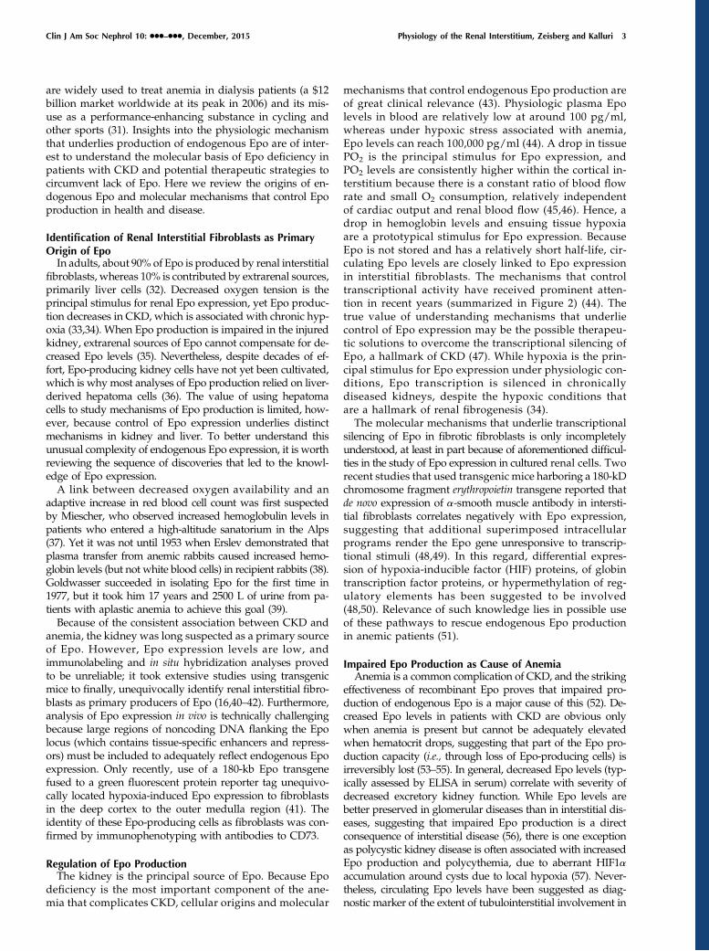
Identification of Renal Interstitial Fibroblasts as PrimaryOrigin of EpoIn adults, about 90% of Epo is produced by renal interstitial
fibroblasts, whereas 10% is contributed by extrarenal sources,primarily liver cells (32). Decreased oxygen tension is theprincipal stimulus for renal Epo expression, yet Epo produc-tion decreases in CKD, which is associated with chronic hyp-oxia (33,34). When Epo production is impaired in the injuredkidney, extrarenal sources of Epo cannot compensate for de-creased Epo levels (35). Nevertheless, despite decades of ef-fort, Epo-producing kidney cells have not yet been cultivated,which is why most analyses of Epo production relied on liver-derived hepatoma cells (36). The value of using hepatomacells to study mechanisms of Epo production is limited, how-ever, because control of Epo expression underlies distinctmechanisms in kidney and liver. To better understand thisunusual complexity of endogenous Epo expression, it is worthreviewing the sequence of discoveries that led to the knowl-edge of Epo expression.A link between decreased oxygen availability and an
adaptive increase in red blood cell count was first suspectedby Miescher, who observed increased hemoglobulin levels inpatients who entered a high-altitude sanatorium in the Alps(37). Yet it was not until 1953 when Erslev demonstrated thatplasma transfer from anemic rabbits caused increased hemo-globin levels (but not white blood cells) in recipient rabbits (38).Goldwasser succeeded in isolating Epo for the first time in1977, but it took him 17 years and 2500 L of urine from pa-tients with aplastic anemia to achieve this goal (39).Because of the consistent association between CKD and
anemia, the kidney was long suspected as a primary sourceof Epo. However, Epo expression levels are low, andimmunolabeling and in situ hybridization analyses provedto be unreliable; it took extensive studies using transgenicmice to finally, unequivocally identify renal interstitial fibro-blasts as primary producers of Epo (16,40–42). Furthermore,analysis of Epo expression in vivo is technically challengingbecause large regions of noncoding DNA flanking the Epolocus (which contains tissue-specific enhancers and repress-ors) must be included to adequately reflect endogenous Epoexpression. Only recently, use of a 180-kb Epo transgenefused to a green fluorescent protein reporter tag unequivo-cally located hypoxia-induced Epo expression to fibroblastsin the deep cortex to the outer medulla region (41). Theidentity of these Epo-producing cells as fibroblasts was con-firmed by immunophenotyping with antibodies to CD73.
Regulation of Epo ProductionThe kidney is the principal source of Epo. Because Epo
deficiency is the most important component of the ane-mia that complicates CKD, cellular origins and molecular
mechanisms that control endogenous Epo production areof great clinical relevance (43). Physiologic plasma Epolevels in blood are relatively low at around 100 pg/ml,whereas under hypoxic stress associated with anemia,Epo levels can reach 100,000 pg/ml (44). A drop in tissuePO2 is the principal stimulus for Epo expression, andPO2 levels are consistently higher within the cortical in-terstitium because there is a constant ratio of blood flowrate and small O2 consumption, relatively independentof cardiac output and renal blood flow (45,46). Hence, adrop in hemoglobin levels and ensuing tissue hypoxiaare a prototypical stimulus for Epo expression. BecauseEpo is not stored and has a relatively short half-life, cir-culating Epo levels are closely linked to Epo expressionin interstitial fibroblasts. The mechanisms that controltranscriptional activity have received prominent atten-tion in recent years (summarized in Figure 2) (44). Thetrue value of understanding mechanisms that underliecontrol of Epo expression may be the possible therapeu-tic solutions to overcome the transcriptional silencing ofEpo, a hallmark of CKD (47). While hypoxia is the prin-cipal stimulus for Epo expression under physiologic con-ditions, Epo transcription is silenced in chronicallydiseased kidneys, despite the hypoxic conditions thatare a hallmark of renal fibrogenesis (34).The molecular mechanisms that underlie transcriptional
silencing of Epo in fibrotic fibroblasts is only incompletelyunderstood, at least in part because of aforementioned difficul-ties in the study of Epo expression in cultured renal cells. Tworecent studies that used transgenic mice harboring a 180-kDchromosome fragment erythropoietin transgene reported thatde novo expression of a-smooth muscle antibody in intersti-tial fibroblasts correlates negatively with Epo expression,suggesting that additional superimposed intracellularprograms render the Epo gene unresponsive to transcrip-tional stimuli (48,49). In this regard, differential expres-sion of hypoxia-inducible factor (HIF) proteins, of globintranscription factor proteins, or hypermethylation of reg-ulatory elements has been suggested to be involved(48,50). Relevance of such knowledge lies in possible useof these pathways to rescue endogenous Epo productionin anemic patients (51).
Impaired Epo Production as Cause of AnemiaAnemia is a common complication of CKD, and the striking
effectiveness of recombinant Epo proves that impaired pro-duction of endogenous Epo is a major cause of this (52). De-creased Epo levels in patients with CKD are obvious onlywhen anemia is present but cannot be adequately elevatedwhen hematocrit drops, suggesting that part of the Epo pro-duction capacity (i.e., through loss of Epo-producing cells) isirreversibly lost (53–55). In general, decreased Epo levels (typ-ically assessed by ELISA in serum) correlate with severity ofdecreased excretory kidney function. While Epo levels arebetter preserved in glomerular diseases than in interstitial dis-eases, suggesting that impaired Epo production is a directconsequence of interstitial disease (56), there is one exceptionas polycystic kidney disease is often associated with increasedEpo production and polycythemia, due to aberrant HIF1aaccumulation around cysts due to local hypoxia (57). Never-theless, circulating Epo levels have been suggested as diag-nostic marker of the extent of tubulointerstitial involvement in
Clin J Am Soc Nephrol 10: ccc–ccc, December, 2015 Physiology of the Renal Interstitium, Zeisberg and Kalluri 3
diabetic nephropathy (58), but due to superimposed regulat-ing factors (including anemia, systemic inflammation, andreduced iron availability), utility of Epo levels as valid diag-nostic marker for interstitial fibrosis could not be validated(53). Impaired Epo production as cause of anemia is not lim-ited to CKD but is also increasingly being recognized as acause of anemia in patients with diabetes mellitus. However,unlike in patients with CKD, Epo production responds ade-quately in diabetic patients to acute hypoxic stress, suggestingthat in principle Epo-producing cells still exist, but that theydon’t sense the anemia appropriately (59). While in CKD in-terstitial cells appear to irreversibly lose capacity to produceEpo, it is conceivable that altered oxygen sensing plays a rolein diabetes mellitus. In this regard, in early diabetes renalblood flow is increased, and it is plausible that resulting in-creased oxygen supply suppresses Epo production. In linewith this, blockade of the renin-angiotensin system in ratsincreased interstitial blood flow (due to decreased intrarenalresistance) and anemia (60), possibly explaining the drop inhematocrit typically observed in the first month of therapywith angiotensin-converting enzyme (ACE) inhibitors or an-giotensin receptor blockers in patients (61).While supplementation with recombinant Epo is highly
effective, it is associated with high costs, and its effectivenesscan be impaired by antibody formation. Hence, stimulationof endogenous Epo production is an attractive therapeu-tic strategy. In this regard, inhibition of prolyl-hydroxylase(PHD; the enzyme that makes HIF accessible for VonHippel–Lindau tumor suppressor protein [VHL] and subsequentproteolytic degradation) has emerged as the primary thera-peutic target as several small molecule inhibitors have beendeveloped. The rationale behind PHD inhibition is that con-ceptually it increases intracellular HIF levels and thus stim-ulates Epo production. In this regard, the PHD inhibitorFG-4592 is undergoing clinical testing in anemic patientswith CKD stages 3 and 4. One possible drawback mightbe that currently available PHD inhibitors do not displayPHD isoform specificity (among the known isoformsPHD1–3, PHD2 is considered the one involved in renal Epo
transcriptional control), and adverse effects of enhanced HIFactivity (such as angiogenesis as the root of diabetic retinop-athy) might be enhanced (62).
AdenosineOne often-underappreciated function of interstitial fibro-
blasts is their involvement in regulation of renal hemody-namics and microvascular function through generation ofextracellular adenosine (63,64). To maintain glomerular fil-tration within a narrow range, the kidney responds to in-creased intratubular NaCl levels (upon increased GFR) byvasoconstriction of afferent arterioles (to decrease GFR) (65).A crucial mediator of this tubuloglomerular feedback loop isextracellular adenosine, which is generated through hydro-lysis of ATP released by macula densa cells into the inter-stitium and which causes vasodilation of efferent arterioles(through activation of A2 adenosine receptors) and vasocon-striction of afferent arterioles through activation of A1 recep-tors, decreasing intraglomerular pressure and GFR (65).Furthermore, adenosine directly inhibits renin release injuxtaglomerular cells. Hence, when GFR and NaCl concen-tration of tubule fluid increase, adenosine is part of the tu-buloglomerular feedback response to lower GFR and NaClsecretion; when GFR and NaCl concentrations decrease,adenosine levels drop under physiologic conditions (66).One enzyme that catalyzes hydrolysis of ATP to adenosineis 59ectonucleotidase (59NT, CD73), which is expressed in thekidney predominantly by resident fibroblasts (a character-istic that has made it a popular marker for renal fibro-blasts) (63). 59NT-deficient mice display substantialreduction of extracellular adenosine levels and reducedkidney weight; however, renal blood flow and GFR areunaltered (67), possibly because of intrinsic activity of in-terstitial ATP on vascular tone (64). In the chronically in-jured kidney, 59NT-positive fibroblasts and extracellularadenosine levels accumulate, possibly contributing to thelowered GFR through afferent arteriole constriction,which is typical of CKD (2).
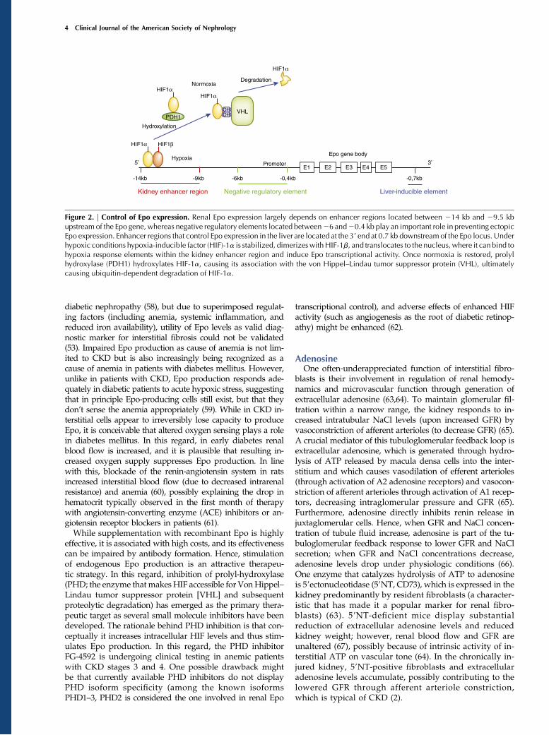
Figure 2. | Control of Epo expression. Renal Epo expression largely depends on enhancer regions located between 214 kb and 29.5 kbupstream of the Epo gene, whereas negative regulatory elements located between26 and20.4 kb play an important role in preventing ectopicEpo expression. Enhancer regions that control Epo expression in the liver are located at the 39 end at 0.7 kb downstream of the Epo locus. Underhypoxic conditions hypoxia-inducible factor (HIF)-1a is stabilized, dimerizeswithHIF-1b, and translocates to the nucleus, where it can bind tohypoxia response elements within the kidney enhancer region and induce Epo transcriptional activity. Once normoxia is restored, prolylhydroxylase (PDH1) hydroxylates HIF-1a, causing its association with the von Hippel–Lindau tumor suppressor protein (VHL), ultimatelycausing ubiquitin-dependent degradation of HIF-1a.
4 Clinical Journal of the American Society of Nephrology

ReninRenin is another protein that is released by renal cells and
indirectly acts systemically (on BP), even though strictlyspeaking it is more an enzyme than a hormone. With morethan 50,000 publications listed in PubMed to date, renin isamong themost-studied proteins in biomedical sciences. Reninis a key regulator of the renin-angiotensin-aldosterone sys-tem, and plasma renin levels reflect the overall activity of thissystem (68). The circulating active form of renin cleaves an-giotensinogen to form angiotensin I (69). Because angiotensi-nogen is highly abundant (its concentration is 1000-foldhigher than that of angiotensin I), it is the plasma renin activ-ity that determines the rate of angiotensin I formation (68).Circulating renin is secretedby juxtaglomerular cells (also called
granular cells or JGA cells), unique round cells of epithelioidappearance that contain myofilaments and abundant peroxi-somes (70). While under physiologic conditions juxtaglomerularcells are located just at the outer edge of the renal interstitium(in the juxtaglomerular interstitium, at the walls of afferentarterioles just at the entrance into glomeruli) additional renin-producing cells are recruited extramurally within the intersti-tium (discussed in more detail below), and hence they arediscussed here as “interstitial cells” for practical purposes(15). Release of renin by juxtaglomerular cells is regulated byhormones such as atrial natriuretic peptide and angiotensin IIand by local factors contributed by adjacent tubular epithelialcells, by vascular smooth muscle cells and endothelial cellsfrom afferent arterioles and by sympathetic nerve endings (71).With regard to local regulatory factors, specialized tubular
cells of the cortical thick ascending limb of the loop of Henleact together with macula densa cells to directly translatechanges in the tubular NaCl concentration into inversechanges in renin secretion (72). In this system, macula densacells sense intratubular NaCl concentrations through activityof the Na1-K–2Cl2 cotransporter NKCC2 (BSC1) and respondto lower NaCl concentrations by release of prostanoids (gen-eration of active prostaglandin E2 and prostacyclin dependson cyclooxygenases) (71).Furthermore, renin release is modulated by vascular
smooth muscle cells and endothelial cells from afferentarterioles (71). In this system, renin stimulatory factors suchas nitric oxide (formed by endothelial nitric oxide synthase)and prostacyclin/prostaglandin E2 are formed in the endo-thelium adjacent to the granular cells, and inhibitory adenosine(which is converted into ATP) is released by vascular smoothmuscle cells (73).The sympathetic input provided by local nerve endings
(in addition to circulating catecholamines) stimulate reninrelease through the b-adrenergic system (73). The magnitudeof sympathetic control of renin release is revealed by double-knockout mice deficient in b1- and b2-receptors, which have85% reduced plasma renin levels compared with wild-typecontrol mice (74). Hence, renal denervation and baroreceptorstimulation conceptually seemed to be sound antihypertensivestrategies. In this regard, failure of renal denervation to signif-icantly lower BP in clinical studies is not yet entirely under-stood (75). Renal denervation also removes the input of thesympathetic system mediated by a-receptors, including hemo-dynamic and tubular effects, and systemic catecholamines re-main fully functional; these findings suggest that baroreceptorstimulation might be the most promising approach to targetthe sympathetic nerve system to lower BP (76,77).
Renin secretion is further controlled by various hormones,autocoids, and other factors, including angiotensin II, as atrialnatriuretic peptide, vasopressin, oxytocin, aldosterone, glu-cocorticoids, thyroid hormones, sex steroids, adipokines, do-pamine, bradykinin, adrenomedullin, and neuropeptide Y(for further review, see extensive review elsewhere [71,73]).While availability of potent ACE inhibitors and angiotensinreceptor blockers (and absence of potent renin blockers) longovershadowed the role of renin in clinical practice, under-standing of direct renin-receptor mediated effects and avail-ability of aliskiren revived interest in renin biology (78).Under physiologic conditions, secretion and activation of
prorenin by juxtaglomerular cells are sufficient for main-tenance of GFR (71). During pathologic conditions, however,additional perivascular fibroblasts (i.e., pericytes, vascularsmooth muscle cells) of afferent arterioles express prorenin,which has led to increased interest in the ontogeny of renin-producing cells (79). Under basal conditions, renin producedby juxtaglomerular cells is sufficient to main BP and intra-vascular volume (71). Upon prolonged threat to volume ho-meostasis (i.e., through exposing mice to a low salt diet andACE inhibitors) additional cells along afferent arterioles ex-press renin, in angiotensinogen-deficient mice renin is evenexpressed by fibroblasts (pericytes) all through the kidneycortex (80,81). Models of CKD are also associated with recruit-ment of additional renin-producing cells. Because increasedrenin expression is often at the root of hypertension, and be-cause higher on-treatment plasma renin activity contributes toprogression of CKD and increased cardiovascular risk, mech-anisms that cause such inopportune increase in renin releaseand particularly recruitment of renin producers are of immi-nent interest (82,83).
Interplay of Renin-Angiotensin and Epo SystemsClinicians know that patients with renovascular hy-
pertension and kidney transplant recipients with trans-plant renal artery stenosis have higher hematocrit valuescompared with patients who have normal renal arteries (84).The underlying pathomechanism of this clinical observationis that ensuing renal hypoperfusion activates the renin-angiotensin system (as described in detail above) and thatangiotensin II stimulates erythropoiesis through stimulatingEpo secretion and through its directs stimulating effect onerythropoiegenesis in the bone marrow (85). This mecha-nisms explains why effective blockage of the renin-angiotensinsystem through ACE inhibitors and/or angiotensin receptorblockers is often associated with a decrease in hematocrit(86,87) and that ACE inhibitors are effective in normalizingpost-transplant erythrocytosis (88). ACE inhibitors even affecthematocrit in hemodialysis patients, who require recombinantEpo substitution because of impaired endogenous Epo pro-duction (as discussed in more detail above). This occurs be-cause treatment with ACE inhibitors is associated withhigher recombinant human Epo (rhEpo) requirements(whereas withdrawal of ACE inhibitors decreases rhEpodoses), reflecting the direct effect of angiotensin II on eryth-ropoiegenesis (89). Exceptions to the link of renin-angiotensinsystem activation and increased hematocrit are often casesin which increment of red blood cells mass are maskedby volume expansion (i.e., in patients with severe heartfailure) (90).
Clin J Am Soc Nephrol 10: ccc–ccc, December, 2015 Physiology of the Renal Interstitium, Zeisberg and Kalluri 5

Hypertension is themost relevant side-effect of rhEpo therapy(91). However, the increase in BP that is often observed indialysis patients receiving rhEpo therapy is independent ofthe renin-angiotensin system; several clinical studies did notdemonstrate renin-angiotensin system activation upon rhEpotherapy (92). Instead, the increased BP observed with rhEpotherapy is most likely a direct systemic vasoconstriction (91).Nevertheless, rhEpo-induced hypertension is correctable withthe typical antihypertensive therapeutic regimen, includingACE/angiotensin receptor blocker inhibition (91).
Origins of Hormone-Producing Interstitial CellsIf we are going to develop specific therapeutic inter-
ventions, we need to understand the origins and developmentof fibroblast subpopulations, including those that produceEpo, those that produce renin, and those that proliferate andlay down the extracellular matrix in fibrosis. Such studies aredone by crossing mice in which cre-recombinase is expressedunder control of promoters specific for a certain lineage (such asmyelin protein zero for neural crest or FoxD1 for mesenchyme)or indicating Epo or renin expression (epo-cre or renin-cre) tomice that harbor a floxed reporter gene, irreversibly taggingthem even when expression of the original marker is lost uponfurther cell differentiation.Published datamostly suggest that renin- and Epo-producing
cells are of two distinct lineages: Renin-producing cells areconsidered to be derivates of FoxD1 mesenchymal cell pro-genitor cells, whereas Epo-producing cells have been sugges-ted to be of neural crest origin, although existing data tosupport this are limited (Figure 3). Evidence for neural crestorigin of Epo-producing cells stems from one study that an-alyzed P0-Cre;R26tdRFP;Epo-GFP triple transgenic mice (49).In this system, Cre-recombinase expression is driven by themyelin protein zero promoter, which is expressed by neuralcrest cells in early embryonic development (93), tagging de-riving cells through recombination of the RFP reporter allele(94). These mice were crossed with reporter mice in whichGFP is fused to a 180-kD chromosomal fragment containingthe mouse Epo coding region and all necessary regulatoryelements and displays specific Epo expression within kidneyinterstitial cells (95). On the basis of the observation that.75% of Epo-GFP–positive cells were also positive for RFP,it was assumed that most Epo-producing cells are of neuralcrest origin (information on percentage of RFP-positive cells
that were also positive for GFP was not provided) (49). Thisstudy also analyzed kidneys of P0-Cre;R26ECFP double trans-genic mice (in these mice, derivate cells of myelin proteinzero-positive cells are tagged through expression of ECFP)and found that almost all interstitial cells in the adult kidneythat expressed PDGF receptor-b and/or CD73 were alsoECFP positive. The researchers concluded that almost all in-terstitial fibroblasts were of neural crest origin. In summary,this study suggested that Epo-producing interstitial cells arederivates of neural crest, just like all interstitial fibroblasts(Figure 3). The authors do not indicate whether Epo-producingcells are a specific subpopulation of fibroblasts or whether allfibroblasts can potentially produce Epo (49). This studysomewhat contradicts a school of thought suggesting thatall resident interstitial fibroblasts in adult kidneys are deri-vates of mesenchymal FoxD1-positive progenitor cells (27).This thinking is based on a study that used FoxD1-GFP-
Cre;R26STOPlacZ double transgenic mice and found thatalmost all interstitial cells in the adult kidney expressingPDGF receptor-b and/or CD73 were also LacZ positive.Those investigators concluded that almost all interstitialfibroblasts were derivates of FoxD1-positive mesenchymalprogenitor cells (albeit only 20% of fibroblasts were LacZpositive when inducible FoxD1-CreER;R26STOPlacZ trans-genic mice were used) (27). Neither study conclusively de-termined whether FoxD1 mesenchymal progenitors are ofextrarenal origin (Figure 3). In this context, there is ampleevidence that juxtaglomerular renin-producing cells areprogeny of renin-positive precursor cells (cells that alsogive rise to perivascular fibroblast-like cells, which can berecruited to produce renin when needed). This concept ismainly based on studies that analyzed kidneys of Ren-1d-Cre;R26STOPlacZ and Ren-1d-Cre;Z/EG double transgenicmice as well as in Ren-1d-Cre;R26STOPlacZ ;ATGF2/2composite mice (in these mice, angiotensinogen deficiencycauses additional recruitment of renin-producing cells [96]),which documented common lineage for perivascular fibro-blasts and renin-producing juxtaglomerular cells (70,79).These aforementioned renin precursor cells are presumablyprogeny of FoxD1-positive progenitor cells, but this informa-tion has been disclosed only as “unpublished data” (Figure 3)(97). A recent study by the Kurtz group demonstrated pro-collagen production by renin precursor cells, hinting at acommon lineage of renin cells and non–renin-producingfibroblasts (98).
Figure 3. | Origins of Epo- and renin-producing renal cells. Epo-producing fibroblasts are presumed to derive from myelin protein zero–positive (P01) neural crest precursor cells.Whether all fibroblasts have the capacity to produce Epo is not yet known. Another school of thoughtsuggests that almost all fibroblasts derive from FoxD1-positive mesenchymal progenitor cells. Renin precursor cells have been suggested tooriginate from FoxD1-positive precursors also. It is not yet known whether renin precursors give rise to generic fibroblasts. One study suggeststhe possibility that renin-producing cells can convert to Epo-producing fibroblasts. VSMC, vascular smooth muscle cell.
6 Clinical Journal of the American Society of Nephrology
To explore the role of hypoxia as possible stimulus forrenin production, Kurtz and coworkers ablated VHL inrenin-producing cells (Renin-1d-Cre;Vhllox/lox) based on theconcept that absence of VHL causes HIF accumulation(due to insufficient proteolytic degradation), mimickinghypoxic signaling. Unexpected findings were that renin-producing cells were decreased (it had been widely pre-sumed that hypoxia stimulates renin production) and thatVHL-deficient juxtaglomerular cells produced Epo insteadof renin (99). Although it is unclear whether such clampingof HIF at abnormally high levels (and resulting Epo over-production and renin suppression) ever happens under innon–genetically modified conditions, this observation rai-ses several conceptual questions. For example, future stud-ies should determine whether renin and Epo productionare interchangeable functions of a specialized interstitialcell type, whether all renal fibroblasts have potential toproduce Epo or renin, or, in a broader view, what renin-and Epo-producing cells truly are.
Pathophysiology of the Renal InterstitiumJust as the renal interstitium plays an integral role in
physiologic kidney function, it is a central determinant ofthe fate of diseased kidneys. Chronic progressive kidneyinjury is unequivocally associated with both quantitative
and qualitative changes of the renal interstitium, which arecommonly referred to as “interstitial fibrosis.” The fibroticinterstitium is no longer invisible because it substantiallyexpands (although in normal kidneys the relative volumetaken up by the interstitium is no higher than 5%–10%, itoften occupies .60% of kidney tissue in a severely dis-eased kidney). Such expansion is predominantly causedby accumulation of extracellular matrix (ECM; fibers),ECM-producing fibroblasts, and mononuclear cells. WhileECM-producing fibroblasts (and often also renin-producingcells) accumulate in the fibrosing interstitium, Epo-producingfibroblasts are lost, raising the question of whether Epo-producing fibroblasts convert into non–Epo-producing fibro-blasts in CKD and whether renin-producing cells could betherapeutically converted into Epo producers (Figure 4). Whilenumerous cell types, including resident non–hormone-producing fibroblasts, endothelial cell, epithelial cells, andbone marrow–derived fibrocytes contribute to accumulationof ECM-producing fibroblasts in kidney fibrosis (29), thecontribution of hormone-producing cells to accumulation ofactivated fibroblasts in CKD is yet unknown. Of note, thereis a glaring disconnect between groups of different researchinterests: Most studies to elucidate origins of fibroblasts infibrotic kidneys, including from our groups, did not analyzepossible Epo or renin expression of fibroblasts. Studies fromgroups with an interest in Epo- and/or renin-expressing cells
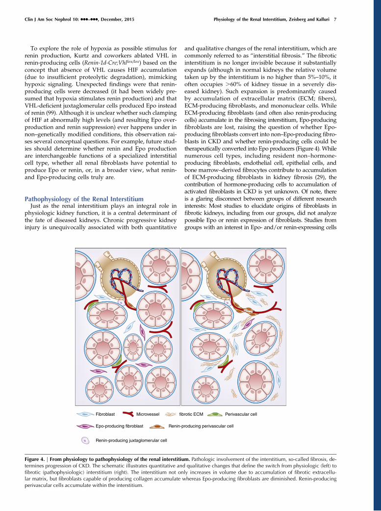
Figure 4. | From physiology to pathophysiology of the renal interstitium. Pathologic involvement of the interstitium, so-called fibrosis, de-termines progression of CKD. The schematic illustrates quantitative and qualitative changes that define the switch from physiologic (left) tofibrotic (pathophysiologic) interstitium (right). The interstitium not only increases in volume due to accumulation of fibrotic extracellu-lar matrix, but fibroblasts capable of producing collagen accumulate whereas Epo-producing fibroblasts are diminished. Renin-producingperivascular cells accumulate within the interstitium.
Clin J Am Soc Nephrol 10: ccc–ccc, December, 2015 Physiology of the Renal Interstitium, Zeisberg and Kalluri 7
reported that these cells could contribute to collagen produc-tion in CKD but did not use transgenic fate mapping systems,which are established in the fibrosis field. Impaired hormoneproduction, however, is not the cardinal feature of the fi-brosed interstitium in progressive CKD, as excretory kidneyfunction is also affected. In fact, the extent of tubulointerstitialfibrosis is the single best determinant for requirement of RRT,even in primary glomerular nephropathies (100–102) becausethe expansion of the interstitium, paired with the overall kid-ney contraction, causes functional nephron loss through en-suing hypoxia (caused by microvascular rarefaction andhindered diffusion of oxygen and nutrients), and stenosis ofglomerulotubular necks and tubular segments through phys-ical pressure (103–105) (for in-depth information regardingthe molecular events which underlie renal fibrogenesis werefer to extensive reviews elsewhere [4,105–108]).
OutlookEpo and renin production are two of the kidney’s most
prominent functions, and these functions are localized tothe fibroblasts of the renal interstitium. Thus, understandingof fibroblast origins and phenotypic plasticity is as relevantas ever. Future work will reveal whether reprogramming offibroblasts may not only circumvent progressive deteriora-tion of excretory kidney function but also limit relative Epodeficiency and renin overproduction in the future.
AcknowledgmentsM.Z. is supported by DFG grant ZE523/2-1. R.K. is supported by
National Institutes of Health grant DK081576 and by the CancerPrevention and Research Institute of Texas and the CancerMetastasisResearch Center.
DisclosuresNone.
References1. Lemley KV, KrizW:Anatomyof the renal interstitium.Kidney Int
39: 370–381, 19912. Kaissling B, Le Hir M: The renal cortical interstitium: Morpho-
logical and functional aspects. Histochem Cell Biol 130: 247–262, 2008
3. Kuncio GS, Neilson EG, Haverty T: Mechanisms oftubulointerstitial fibrosis. Kidney Int 39: 550–556, 1991
4. Zeisberg M, Neilson EG: Mechanisms of tubulointerstitialfibrosis. J Am Soc Nephrol 21: 1819–1834, 2010
5. Eddy AA: Molecular insights into renal interstitial fibrosis[editorial]. J Am Soc Nephrol 7: 2495–2508, 1996
6. Kriz W, Napiwotzky P: Structural and functional aspects of therenal interstitium. Contrib Nephrol 16: 104–108, 1979
7. Knepper MA, Danielson RA, Saidel GM, Post RS: Quantitativeanalysis of renal medullary anatomy in rats and rabbits. KidneyInt 12: 313–323, 1977
8. Takahashi-Iwanaga H: The three-dimensional cytoarchitectureof the interstitial tissue in the rat kidney. Cell Tissue Res 264:269–281, 1991
9. Teteris SA, Engel DR, Kurts C: Homeostatic and pathogenic roleof renal dendritic cells. Kidney Int 80: 139–145, 2011
10. Nelson PJ, Rees AJ, GriffinMD,Hughes J, Kurts C,Duffield J: Therenal mononuclear phagocytic system. J Am Soc Nephrol 23:194–203, 2012
11. Kalluri R, Zeisberg M: Fibroblasts in cancer. Nat Rev Cancer 6:392–401, 2006
12. Zeisberg M, Strutz F, Muller GA: Role of fibroblast activation ininducing interstitial fibrosis. J Nephrol 13[Suppl 3]: S111–S120,2000
13. Enge M, Bjarnegard M, Gerhardt H, Gustafsson E, Kalen M,Asker N, Hammes HP, Shani M, Fassler R, Betsholtz C:Endothelium-specific platelet-derived growth factor-B ablationmimics diabetic retinopathy. EMBO J 21: 4307–4316, 2002
14. Abramsson A, Lindblom P, Betsholtz C: Endothelial and non-endothelial sources of PDGF-B regulate pericyte recruitmentand influence vascular pattern formation in tumors. J Clin Invest112: 1142–1151, 2003
15. Kurtz A: Renin release: Sites, mechanisms, and control. AnnuRev Physiol 73: 377–399, 2011
16. Kurtz A, Eckardt KU, Neumann R, Kaissling B, Le Hir M, BauerC: Site of erythropoietin formation. Contrib Nephrol 76: 14–20,discussion 21–23, 1989
17. Komuro T: Re-evaluation of fibroblasts and fibroblast-like cells.Anat Embryol (Berl) 182: 103–112, 1990
18. Franke WW, Schmid E, Osborn M, Weber K: Differentintermediate-sized filaments distinguished by immunofluores-cence microscopy. Proc Natl Acad Sci U S A 75: 5034–5038,1978
19. Lazarides E, Balzer DR Jr: Specificity of desmin to avian andmammalian muscle cells. Cell 14: 429–438, 1978
20. Tuszynski GP, Frank ED, Damsky CH, Buck CA, Warren L: Thedetection of smooth muscle desmin-like protein in BHK21/C13fibroblasts. J Biol Chem 254: 6138–6143, 1979
21. Gardner H, Kreidberg J, Koteliansky V, Jaenisch R: Deletion ofintegrin alpha 1 by homologous recombination permits normalmurine development but gives rise to a specific deficit in celladhesion. Dev Biol 175: 301–313, 1996
22. Vogel W, Gish GD, Alves F, Pawson T: The discoidin domainreceptor tyrosine kinases are activated by collagen.Mol Cell 1:13–23, 1997
23. Strutz F, Okada H, Lo CW, Danoff T, Carone RL, TomaszewskiJE, Neilson EG: Identification and characterization of afibroblast marker: FSP1. J Cell Biol 130: 393–405, 1995
24. Marxer-Meier A, Hegyi I, Loffing J, Kaissling B: Postnatalmaturation of renal cortical peritubular fibroblasts in the rat.Anat Embryol (Berl) 197: 143–153, 1998
25. Thompson LF, Ruedi JM, Glass A, LowMG, Lucas AH: Antibodiesto 59-nucleotidase (CD73), a glycosyl-phosphatidylinositol-anchored protein, cause human peripheral blood T cells toproliferate. J Immunol 143: 1815–1821, 1989
26. Fellstrom B, Klareskog L, Heldin CH, Larsson E, Ronnstrand L,Terracio L, Tufveson G, Wahlberg J, Rubin K: Platelet-derivedgrowth factor receptors in the kidney—upregulated expressionin inflammation. Kidney Int 36: 1099–1102, 1989
27. Humphreys BD, Lin SL, Kobayashi A, Hudson TE, Nowlin BT,Bonventre JV, Valerius MT, McMahon AP, Duffield JS: Fatetracing reveals the pericyte and not epithelial origin of myofi-broblasts in kidney fibrosis. Am J Pathol 176: 85–97, 2010
28. Gabbiani G, Kapanci Y, Barazzone P, Franke WW: Immuno-chemical identification of intermediate-sized filaments inhuman neoplastic cells. A diagnostic aid for the surgicalpathologist. Am J Pathol 104: 206–216, 1981
29. LeBleu VS, Taduri G, O’Connell J, Teng Y, Cooke VG, Woda C,Sugimoto H, Kalluri R: Origin and function of myofibroblasts inkidney fibrosis. Nat Med 19: 1047–1053, 2013
30. Lacombe C, Mayeux P: Biology of erythropoietin.Haematologica 83: 724–732, 1998
31. Jelkmann W: Physiology and pharmacology of erythropoietin.Transfus Med Hemother 40: 302–309, 2013
32. FriedW, Kilbridge T, Krantz S,McDonald TP, Lange RD: Studieson extrarenal erythropoietin. J Lab Clin Med 73: 244–248,1969
33. Cahan C, Hoekje PL, Goldwasser E, Decker MJ, Strohl KP:Assessing the characteristic between length of hypoxic expo-sure and serum erythropoietin levels.Am J Physiol 258: R1016–R1021, 1990
34. NangakuM, Eckardt KU: Hypoxia and the HIF system in kidneydisease. J Mol Med (Berl) 85: 1325–1330, 2007
35. Weidemann A, Johnson RS: Nonrenal regulation of EPOsynthesis. Kidney Int 75: 682–688, 2009
36. Chiang CK, Nangaku M, Tanaka T, Iwawaki T, Inagi R:Endoplasmic reticulum stress signal impairs erythropoietinproduction: a role for ATF4. Am J Physiol Cell Physiol 304:C342–C353, 2013
8 Clinical Journal of the American Society of Nephrology
37. Miescher F: Uber die Beziehungen zwischen Meereshohe undBeschaffenheit des Blutes. Korrespbl Schweiz Arz 23: 809–830,1893
38. Erslev A: Humoral regulation of red cell production. Blood 8:349–357, 1953
39. Wojchowski D: Eugene Goldwasser (1922-2010). Nature 470:40, 2011
40. Eckardt KU, Koury ST, Tan CC, Schuster SJ, Kaissling B, RatcliffePJ, Kurtz A: Distribution of erythropoietin producing cells inrat kidneys during hypoxic hypoxia. Kidney Int 43: 815–823,1993
41. Pan X, Suzuki N, Hirano I, Yamazaki S, Minegishi N, YamamotoM: Isolation and characterization of renal erythropoietin-producing cells from genetically produced anemia mice.PLoS ONE 6: e25839, 2011
42. Maxwell AP, Lappin TR, Johnston CF, Bridges JM, McGeownMG: Erythropoietin production in kidney tubular cells. Br JHaematol 74: 535–539, 1990
43. Unger EF, Thompson AM, Blank MJ, Temple R: Erythropoiesis-stimulating agents—time for a reevaluation. N Engl J Med 362:189–192, 2010
44. Macdougall IC: Optimizing the use of erythropoietic agents—pharmacokinetic and pharmacodynamic considerations.Nephrol Dial Transplant 17[Suppl 5]: 66–70, 2002
45. Pagel H, Jelkmann W, Weiss C: O2-supply to the kidneys andthe production of erythropoietin. Respir Physiol 77: 111–117,1989
46. JelkmannW: Regulation of erythropoietin production. J Physiol589: 1251–1258, 2011
47. Singh AK: Anemia of chronic kidney disease. Clin J Am SocNephrol 3: 3–6, 2008
48. Dewi FR, Fatchiyah F: Methylation impact analysis of erythro-poietin (EPO) gene to hypoxia inducible factor-1a (HIF-1a)activity. Bioinformation 9: 782–787, 2013
49. AsadaN, TakaseM, Nakamura J, Oguchi A, AsadaM, Suzuki N,Yamamura K, Nagoshi N, Shibata S, Rao TN, Fehling HJ,Fukatsu A, Minegishi N, Kita T, Kimura T, Okano H, YamamotoM, Yanagita M: Dysfunction of fibroblasts of extrarenal originunderlies renal fibrosis and renal anemia in mice. J Clin Invest121: 3981–3990, 2011
50. Souma T, Yamazaki S, Moriguchi T, Suzuki N, Hirano I, Pan X,Minegishi N, AbeM, KiyomotoH, Ito S, YamamotoM: Plasticityof renal erythropoietin-producing cells governs fibrosis. J AmSoc Nephrol 24: 1599–1616, 2013
51. BernhardtWM,WiesenerMS, Scigalla P, Chou J, Schmieder RE,Gunzler V, Eckardt KU: Inhibition of prolyl hydroxylases in-creases erythropoietin production in ESRD. J Am Soc Nephrol21: 2151–2156, 2010
52. Artunc F, Risler T: Serum erythropoietin concentrations andresponses to anaemia in patientswith or without chronic kidneydisease. Nephrol Dial Transplant 22: 2900–2908, 2007
53. Mercadal L, Metzger M, Casadevall N, Haymann JP, Karras A,Boffa JJ, Flamant M, Vrtovsnik F, Stengel B, Froissart M;NephroTest Study Group: Timing and determinants of erythro-poietin deficiency in chronic kidney disease. Clin J Am SocNephrol 7: 35–42, 2012
54. Chandra M, Clemons GK, McVicar MI: Relation of serumerythropoietin levels to renal excretory function: Evidence forlowered set point for erythropoietin production in chronic renalfailure. J Pediatr 113: 1015–1021, 1988
55. Thomas M, Tsalamandris C, MacIsaac R, Jerums G: Anaemia indiabetes: An emerging complication of microvascular disease.Curr Diabetes Rev 1: 107–126, 2005
56. de Klerk G, Wilmink JM, Rosengarten PC, Vet RJ, Goudsmit R:Serum erythropoietin (ESF) titers in anemia of chronic renalfailure. J Lab Clin Med 100: 720–734, 1982
57. Eckardt KU, Mollmann M, Neumann R, Brunkhorst R, BurgerHU, Lonnemann G, Scholz H, Keusch G, Buchholz B, Frei U:Erythropoietin in polycystic kidneys. J Clin Invest 84: 1160–1166, 1989
58. Inomata S, ItohM, ImaiH, Sato T: Serum levels of erythropoietinas a novel marker reflecting the severity of diabetic nephropa-thy. Nephron 75: 426–430, 1997
59. Bosman DR, Osborne CA, Marsden JT, Macdougall IC, GardnerWN, Watkins PJ: Erythropoietin response to hypoxia in patients
with diabetic autonomic neuropathy and non-diabetic chronicrenal failure. Diabet Med 19: 65–69, 2002
60. Naeshiro I, Sato K, Chatani F, Sato S: Possiblemechanism for theanemia induced by candesartan cilexetil (TCV-116), an angio-tensin II receptor antagonist, in rats. Eur J Pharmacol 354: 179–187, 1998
61. Erturk S, Atesx K, Duman N, Karatan O, Erbay B, Ertu�g E:Unresponsiveness to recombinant human erythropoietin inhaemodialysis patients: Possible implications of angiotensin-converting enzyme inhibitors. Nephrol Dial Transplant 11:396–397, 1996
62. Franke K, Gassmann M, Wielockx B: Erythrocytosis: The HIFpathway in control. Blood 122: 1122–1128, 2013
63. Le Hir M, Kaissling B: Distribution and regulation of renalecto-59-nucleotidase: Implications for physiological functionsof adenosine. Am J Physiol 264: F377–F387, 1993
64. Nishiyama A, Rahman M, Inscho EW: Role of interstitial ATPand adenosine in the regulation of renal hemodynamics andmicrovascular function. Hypertens Res 27: 791–804, 2004
65. Li L,Mizel D,Huang Y, Eisner C, HoerlM, Thiel M, SchnermannJ: Tubuloglomerular feedback and renal function in mice withtargeted deletion of the type 1 equilibrative nucleoside trans-porter. Am J Physiol Renal Physiol 304: F382–F389, 2013
66. Vallon V, Muhlbauer B, Osswald H: Adenosine and kidneyfunction. Physiol Rev 86: 901–940, 2006
67. Ozuyaman B, Ding Z, Buchheiser A, Koszalka P, Braun N,Godecke A, Decking UK, Zimmermann H, Schrader J: Adeno-sine produced via the CD73/ecto-59-nucleotidase pathway hasno impact on erythropoietin production but is associated withreduced kidney weight. Pflugers Arch 452: 324–331, 2006
68. KoboriH,NangakuM,Navar LG,NishiyamaA:The intrarenal renin-angiotensin system: from physiology to the pathobiology of hyper-tension and kidney disease. Pharmacol Rev 59: 251–287, 2007
69. Baylis C, Engels K, Hymel A, Navar LG: Plasma renin activityand metabolic clearance rate of angiotensin II in the unstressedaging rat. Mech Ageing Dev 97: 163–172, 1997
70. Sequeira Lopez ML, Pentz ES, Robert B, Abrahamson DR,Gomez RA: Embryonic origin and lineage of juxtaglomerularcells. Am J Physiol Renal Physiol 281: F345–F356, 2001
71. Castrop H, Hocherl K, Kurtz A, Schweda F, Todorov V, WagnerC: Physiology of kidney renin. Physiol Rev 90: 607–673, 2010
72. Skøtt O, Briggs JP: Direct demonstration of macula densa-mediated renin secretion. Science 237: 1618–1620, 1987
73. Hackenthal E, Paul M, Ganten D, Taugner R: Morphology,physiology, and molecular biology of renin secretion. PhysiolRev 70: 1067–1116, 1990
74. Kim SM, Chen L, Faulhaber-Walter R, OppermannM, Huang Y,Mizel D, Briggs JP, Schnermann J: Regulation of renin secretionand expression inmice deficient in beta1- and beta2-adrenergicreceptors. Hypertension 50: 103–109, 2007
75. Bhatt DL, Kandzari DE, O’Neill WW, D’Agostino R, Flack JM,Katzen BT, Leon MB, Liu M, Mauri L, Negoita M, Cohen SA,Oparil S, Rocha-Singh K, Townsend RR, Bakris GL;SYMPLICITY HTN-3 Investigators: A controlled trial of renaldenervation for resistant hypertension.NEngl JMed 370: 1393–1401, 2014
76. Fisher JP, Paton JF: The sympathetic nervous system and bloodpressure in humans: Implications for hypertension. J HumHypertens 26: 463–475, 2012
77. Davidson AC, Bisognano JD: Interventional approaches forresistant hypertension. Curr Opin Nephrol Hypertens 21: 475–480, 2012
78. Zeisberg M, Muller GA: Mechanistic insights into the anti-fibrotic activity of aliskiren in the kidney. Hypertens Res 35:266–268, 2012
79. Sequeira Lopez ML, Pentz ES, Nomasa T, Smithies O, GomezRA: Renin cells are precursors for multiple cell types that switchto the renin phenotype when homeostasis is threatened. DevCell 6: 719–728, 2004
80. Ayan S, Roth JA, Freeman MR, Bride SH, Peters CA: Partialureteral obstruction dysregulates the renal renin-angiotensinsystem in the fetal sheep kidney. Urology 58: 301–306, 2001
81. Mimura I, Nangaku M: The suffocating kidney: Tubulointer-stitial hypoxia in end-stage renal disease. Nat Rev Nephrol 6:667–678, 2010
Clin J Am Soc Nephrol 10: ccc–ccc, December, 2015 Physiology of the Renal Interstitium, Zeisberg and Kalluri 9

82. Sealey JE, Alderman MH, Furberg CD, Laragh JH: Renin-angiotensin system blockers may create more risk than rewardfor sodium-depleted cardiovascular patients with high plasmarenin levels. Am J Hypertens 26: 727–738, 2013
83. Huby AC, Kavvadas P, Alfieri C, Abed A, Toubas J, Rastaldi MP,Dussaule JC, Chatziantoniou C, Chadjichristos CE: The RenTgmice: A powerful tool to study renin-dependent chronic kidneydisease. PLoS ONE 7: e52362, 2012
84. Tarazi RC, Frohlich ED, Dustan HP, Gifford RW Jr, Page IH:Hypertension and high hematocrit. Another clue to renalarterial disease. Am J Cardiol 18: 855–858, 1966
85. Vlahakos DV, Marathias KP, Madias NE: The role of the renin-angiotensin system in the regulation of erythropoiesis. Am JKidney Dis 56: 558–565, 2010
86. Kuriyama S, Tomonari H, TokudomeG, HoriguchiM, Hayashi H,Kobayashi H, Ishikawa M, Hosoya T: Antiproteinuric effectsof combined antihypertensive therapies in patients withovert type 2 diabetic nephropathy.Hypertens Res 25: 849–855,2002
87. Jacobsen P, Andersen S, Jensen BR, Parving HH: Additive effect ofACE inhibition and angiotensin II receptor blockade in type I di-abetic patients with diabetic nephropathy. J Am Soc Nephrol 14:992–999, 2003
88. Vlahakos DV, Marathias KP, Agroyannis B, Madias NE: Post-transplant erythrocytosis. Kidney Int 63: 1187–1194, 2003
89. Erturk S, Nergizo�glu G, Atesx K, Duman N, Erbay B, Karatan O,Ertu�g AE: The impact of withdrawing ACE inhibitors on eryth-ropoietin responsiveness and left ventricular hypertrophy inhaemodialysis patients. Nephrol Dial Transplant 14: 1912–1916, 1999
90. Volpe M, Tritto C, Testa U, Rao MA, Martucci R, Mirante A,Enea I, Russo R, Rubattu S, Condorelli GL, Cangianiello S,Trimarco B, Peschle C, Condorelli M: Blood levels of erythro-poietin in congestive heart failure and correlationwith clinical,hemodynamic, and hormonal profiles. Am J Cardiol 74:468–473, 1994
91. Eckardt KU: Cardiovascular consequences of renal anaemiaand erythropoietin therapy.Nephrol Dial Transplant 14: 1317–1323, 1999
92. Rosario R, Epstein M: Relationship between erythropoietin ad-ministration and alterations of renin-angiotensin-aldosterone.J Renin Angiotensin Aldosterone Syst 7: 135–138, 2006
93. Yamauchi Y, Abe K, Mantani A, Hitoshi Y, Suzuki M, Osuzu F,Kuratani S, Yamamura K: A novel transgenic technique that al-lows specific marking of the neural crest cell lineage in mice.Dev Biol 212: 191–203, 1999
94. Luche H, Weber O, Nageswara Rao T, Blum C, Fehling HJ:Faithful activation of an extra-bright red fluorescent protein in“knock-in” Cre-reporter mice ideally suited for lineage tracingstudies. Eur J Immunol 37: 43–53, 2007
95. Obara N, Suzuki N, Kim K, Nagasawa T, Imagawa S, YamamotoM: Repression via the GATA box is essential for tissue-specificerythropoietin gene expression. Blood 111: 5223–5232, 2008
96. Kim HS, Maeda N, Oh GT, Fernandez LG, Gomez RA, SmithiesO: Homeostasis in mice with genetically decreasedangiotensinogen is primarily by an increased number of renin-producing cells. J Biol Chem 274: 14210–14217, 1999
97. Sequeira Lopez ML, Gomez RA: Development of the renalarterioles. J Am Soc Nephrol 22: 2156–2165, 2011
98. Karger C, Kurtz F, Steppan D, Schwarzensteiner I, Machura K,Angel P, Banas B, Risteli J, Kurtz A: Procollagen I-expressingrenin cell precursors. Am J Physiol Renal Physiol 305:F355–F361, 2013
99. Kurt B, Paliege A, Willam C, Schwarzensteiner I, Schucht K,Neymeyer H, Sequeira-Lopez ML, Bachmann S, Gomez RA,Eckardt KU, Kurtz A: Deletion of von Hippel-Lindau proteinconverts renin-producing cells into erythropoietin-producingcells. J Am Soc Nephrol 24: 433–444, 2013
100. Bohle A, Bader R, Grund KE, Mackensen S, Neunhoeffer J:Serum creatinine concentration and renal interstitial volume.Analysis of correlations in endocapillary (acute) glomerulone-phritis and in moderately severe mesangioproliferativeglomerulonephritis. Virchows Arch A Pathol Anat Histol 375:87–96, 1977
101. Bohle A, Christ H, Grund KE, Mackensen S: The role of the in-terstitium of the renal cortex in renal disease. Contrib Nephrol16: 109–114, 1979
102. Risdon RA, Sloper JC, De Wardener HE: Relationship betweenrenal function and histological changes found in renal-biopsyspecimens from patients with persistent glomerular nephritis.Lancet 2: 363–366, 1968
103. Cohen EP: Fibrosis causes progressive kidney failure. MedHypotheses 45: 459–462, 1995
104. Cohen EP, Regner K, Fish BL, Moulder JE: Stenotic glomer-ulotubular necks in radiation nephropathy. J Pathol 190:484–488, 2000
105. Becker GJ, Hewitson TD: The role of tubulointerstitial injury inchronic renal failure. Curr Opin Nephrol Hypertens 9:133–138, 2000
106. Zeisberg M, Kalluri R: Cellular mechanisms of tissue fibrosis.1. Common and organ-specific mechanisms associated withtissue fibrosis. Am J Physiol Cell Physiol 304: C216–C225,2013
107. Eddy AA: Progression in chronic kidney disease. Adv ChronicKidney Dis 12: 353–365, 2005
108. Remuzzi G, Bertani T: Pathophysiology of progressivenephropathies. N Engl J Med 339: 1448–1456, 1998
Published online ahead of print. Publication date available at www.cjasn.org.
10 Clinical Journal of the American Society of Nephrology