Muscle Cells & Muscle Fiber Contractions Packet # Chapter #49.
32
Muscle Cells & Muscle Fiber Contractions Packet # Chapter #49
-
Upload
phyllis-pitts -
Category
Documents
-
view
220 -
download
0
Transcript of Muscle Cells & Muscle Fiber Contractions Packet # Chapter #49.
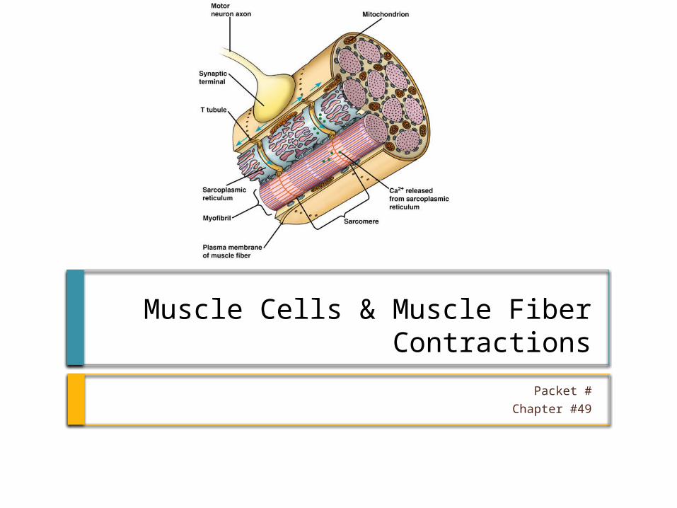
Muscle Cells & Muscle Fiber Contractions
Packet #
Chapter #49

Introduction Skeletal muscle is
attached to bones and is responsible for movement.
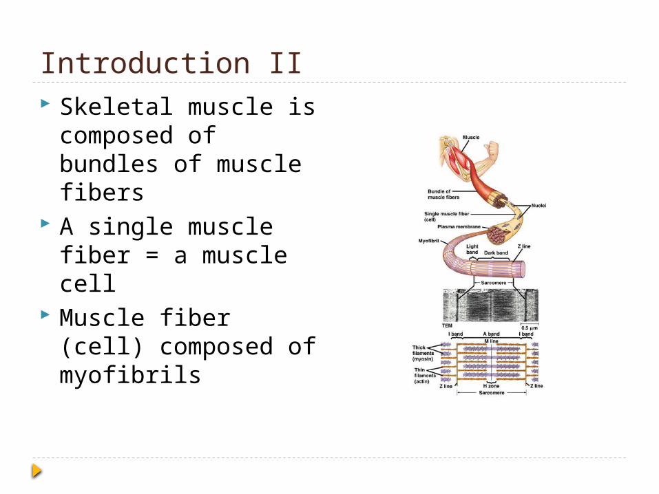
Introduction II Skeletal muscle is
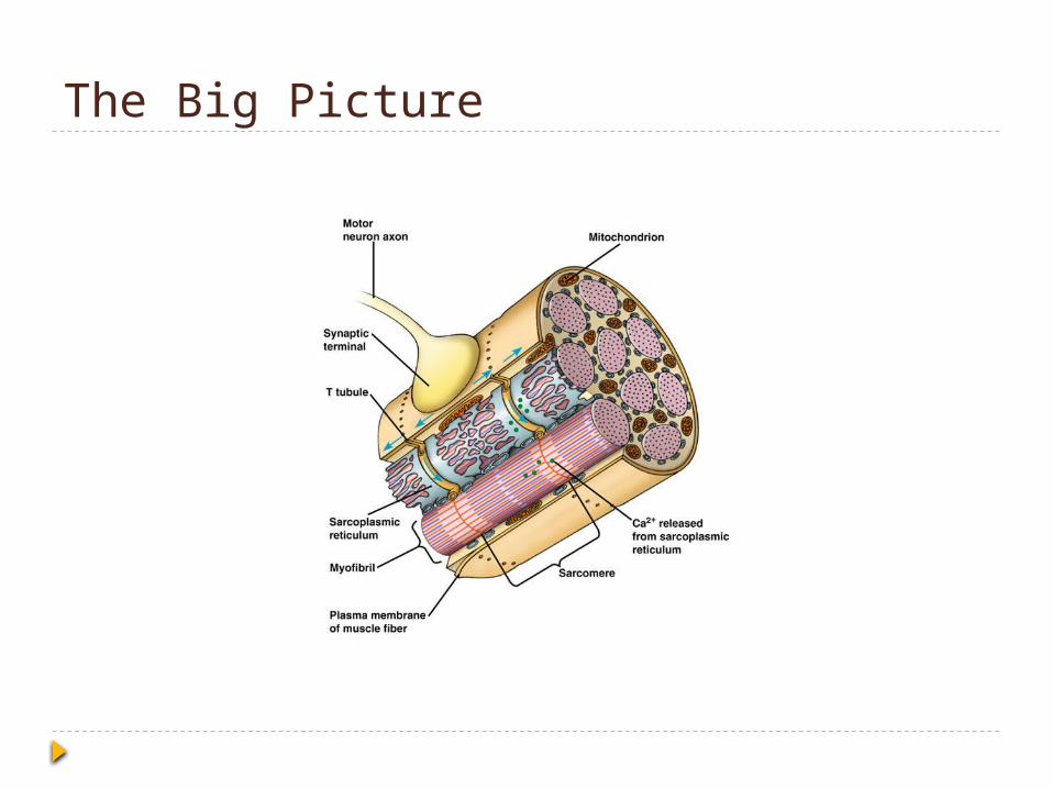
composed of bundles of muscle fibers
A single muscle fiber = a muscle cell
Muscle fiber (cell) composed of myofibrils
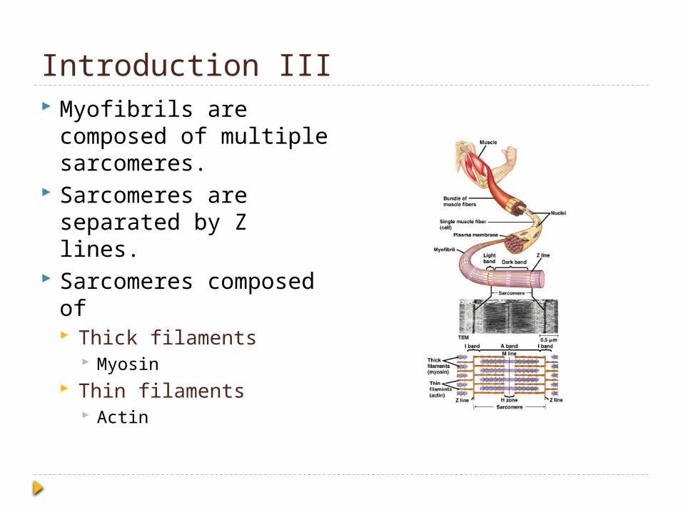
Introduction III Myofibrils are
composed of multiple sarcomeres.
Sarcomeres are separated by Z lines.
Sarcomeres composed of Thick filaments
Myosin Thin filaments
Actin
The Sliding Filament Model and Muscle Contraction
How do muscle cells contract?
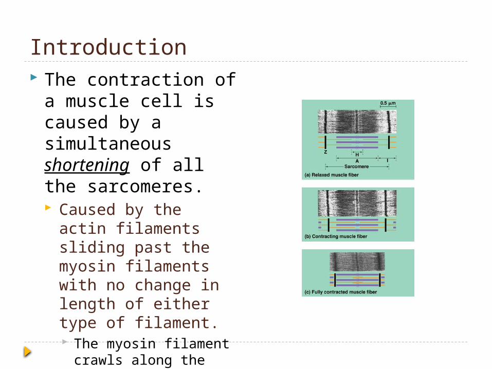
Introduction The contraction of a
muscle cell is caused by a simultaneous shortening of all the sarcomeres. Caused by the actin
filaments sliding past the myosin filaments with no change in length of either type of filament. The myosin filament
crawls along the actin filament.
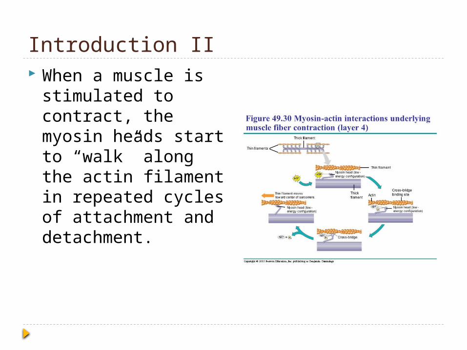
Introduction II When a muscle is
stimulated to contract, the myosin heads start to “walk” along the actin filament in repeated cycles of attachment and detachment.

Sliding Filament Model I Muscles, according to
the sliding filament model, have the actin and myosin filaments that slide past each other during contraction. This produces more
overlap between the two filaments.
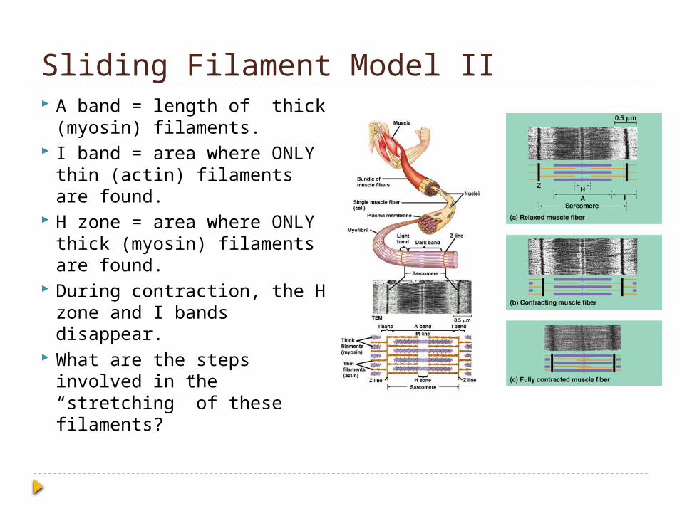
Sliding Filament Model II A band = length of thick
(myosin) filaments. I band = area where ONLY
thin (actin) filaments are found.
H zone = area where ONLY thick (myosin) filaments are found.
During contraction, the H zone and I bands disappear.
What are the steps involved in the “stretching” of these filaments?

Muscle Fiber Contraction I Myosin head is
bound to ATP Low energy state.
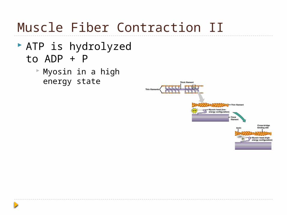
Muscle Fiber Contraction II ATP is hydrolyzed to
ADP + P Myosin in a high
energy state
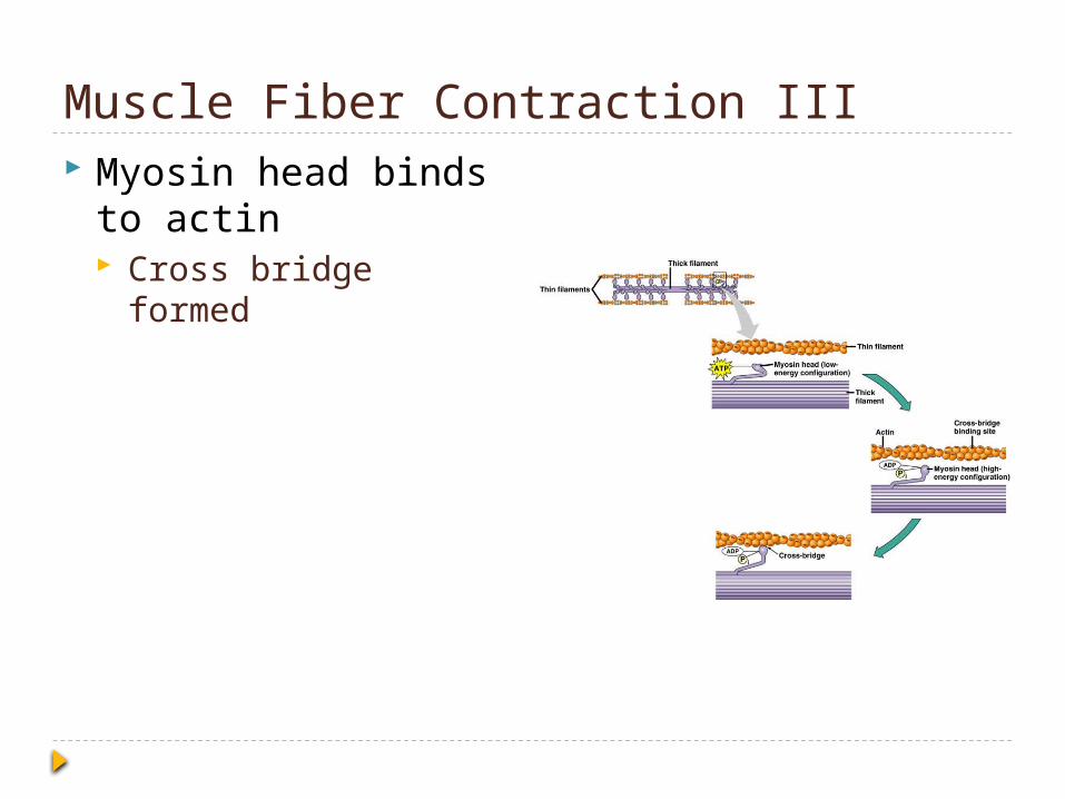
Muscle Fiber Contraction III Myosin head binds to
actin Cross bridge formed
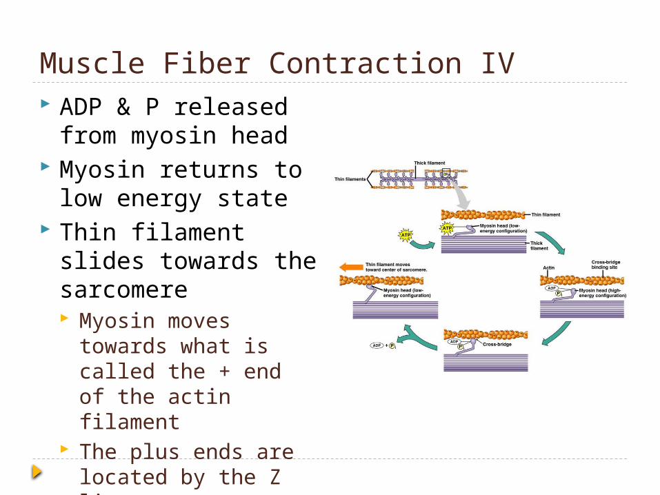
Muscle Fiber Contraction IV ADP & P released
from myosin head Myosin returns to low
energy state Thin filament slides
towards the sarcomere Myosin moves
towards what is called the + end of the actin filament
The plus ends are located by the Z lines
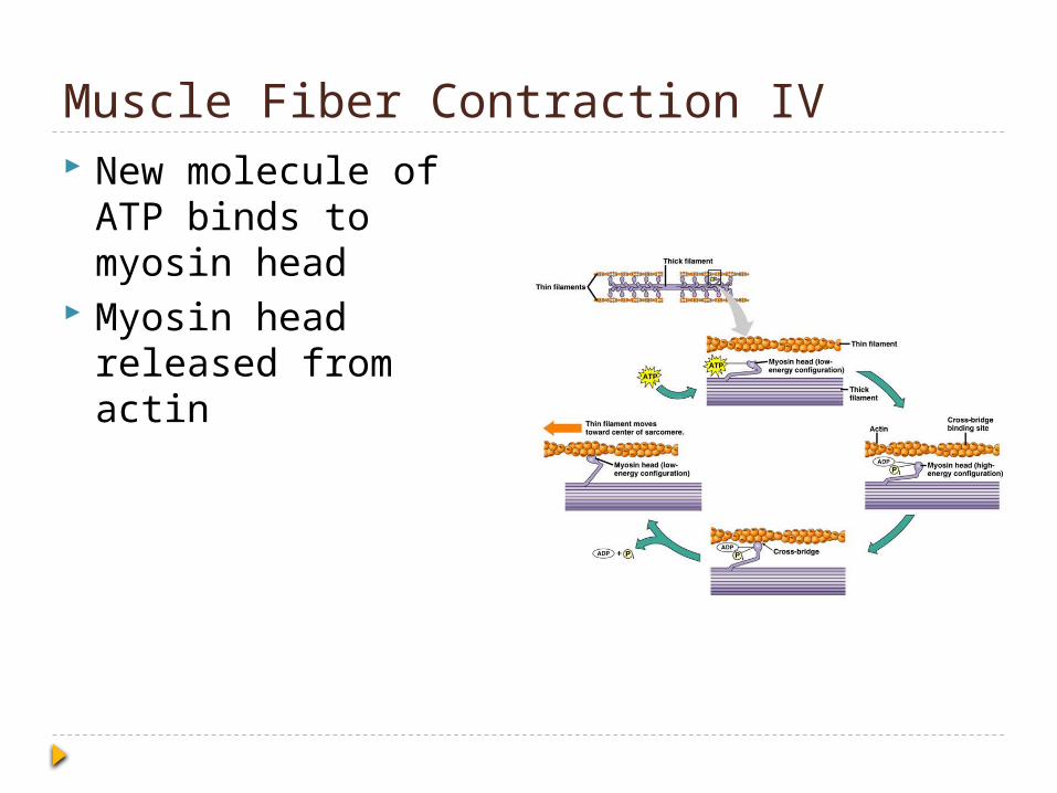
Muscle Fiber Contraction IV New molecule of ATP
binds to myosin head Myosin head
released from actin

Calcium & Regulatory Proteins
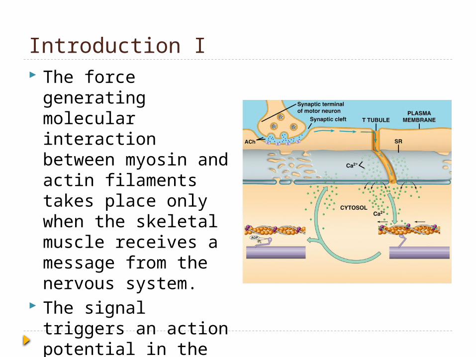
Introduction I The force generating
molecular interaction between myosin and actin filaments takes place only when the skeletal muscle receives a message from the nervous system.
The signal triggers an action potential in the muscle cell.
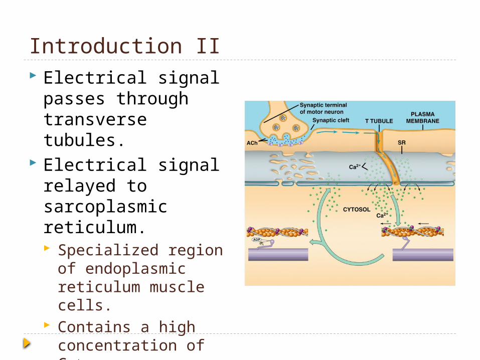
Introduction II Electrical signal
passes through transverse tubules.
Electrical signal relayed to sarcoplasmic reticulum. Specialized region of
endoplasmic reticulum muscle cells.
Contains a high concentration of Ca+

Introduction III Ca+, in response to
electrical excitation, is released into the cytosol through ion channels that open. These ion channels are
located in the membrane of the sarcoplasmic reticulum membrane.
The opening of the voltage channels occur in response to the change in voltage.

The Role of Calcium and Regulatory Proteins
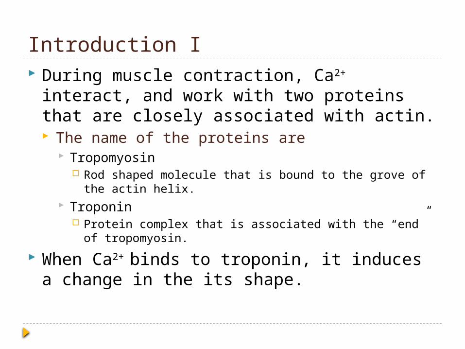
Introduction I During muscle contraction, Ca2+ interact, and
work with two proteins that are closely associated with actin. The name of the proteins are
Tropomyosin Rod shaped molecule that is bound to the grove of the actin
helix. Troponin
Protein complex that is associated with the “end” of tropomyosin.
When Ca2+ binds to troponin, it induces a change in the its shape.
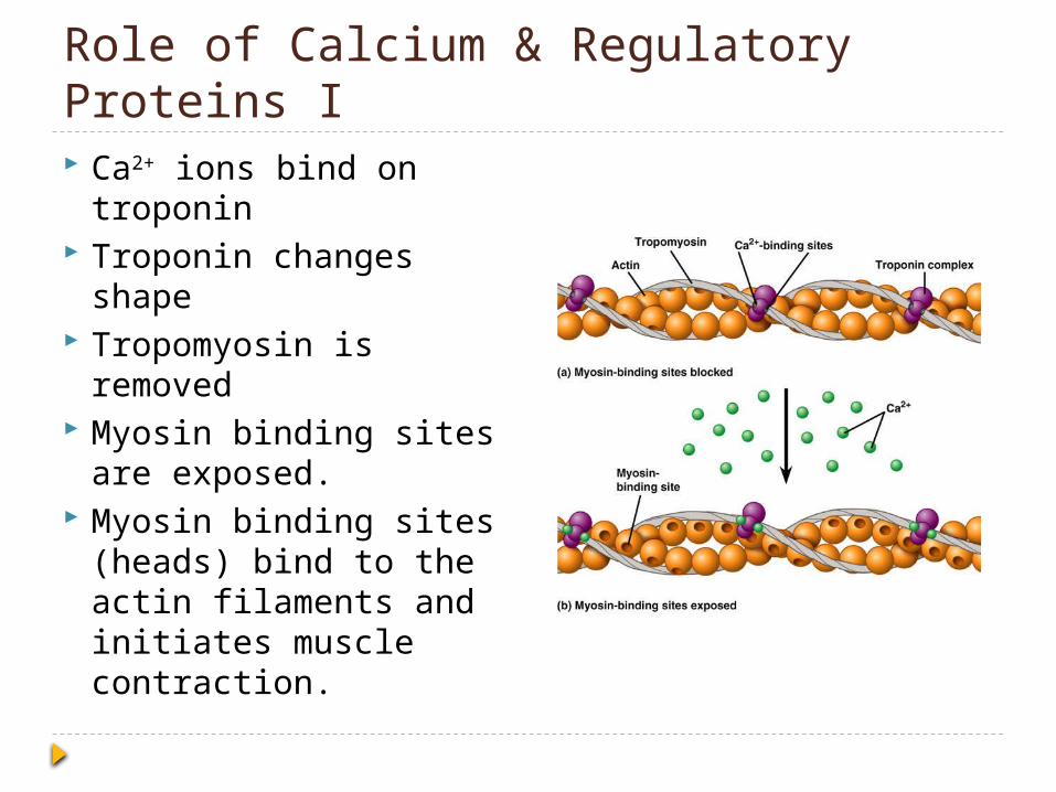
Role of Calcium & Regulatory Proteins I Ca2+ ions bind on
troponin Troponin changes
shape Tropomyosin is
removed Myosin binding sites
are exposed. Myosin binding sites
(heads) bind to the actin filaments and initiates muscle contraction.
Role of Calcium & Regulatory Proteins II The increase in Ca2+
ions, within the cytosol, stops as soon as the nerve signal stops.
Ca2+ is pumped back into the satcoplasmic reticulum.
Putting it TogetherNerve and Muscle Cells
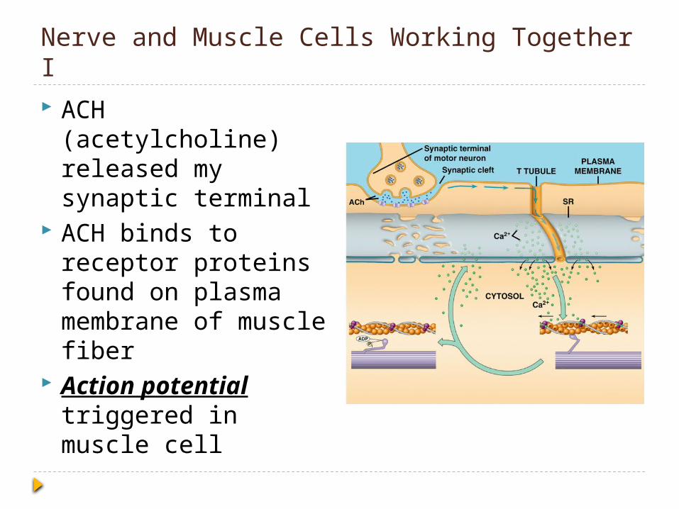
Nerve and Muscle Cells Working Together I ACH (acetylcholine)
released my synaptic terminal
ACH binds to receptor proteins found on plasma membrane of muscle fiber
Action potential triggered in muscle cell

Nerve and Muscle Cells Working Together II Action potential
moves down T tubule and causes the release of Ca+ ions from sarcoplasmic reticulum (SR).
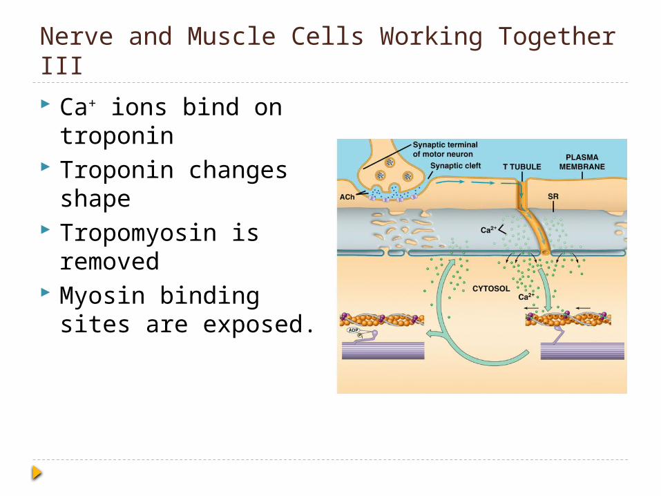
Nerve and Muscle Cells Working Together III Ca+ ions bind on
troponin Troponin changes
shape Tropomyosin is
removed Myosin binding sites
are exposed.
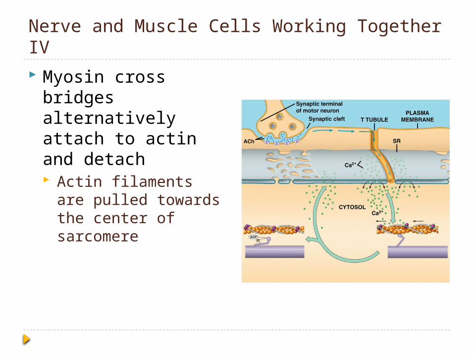
Nerve and Muscle Cells Working Together IV Myosin cross bridges
alternatively attach to actin and detach Actin filaments are
pulled towards the center of sarcomere
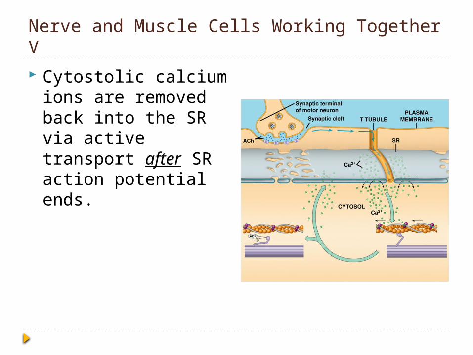
Nerve and Muscle Cells Working Together V Cytostolic calcium
ions are removed back into the SR via active transport after SR action potential ends.
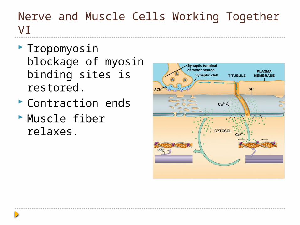
Nerve and Muscle Cells Working Together VI Tropomyosin
blockage of myosin binding sites is restored.
Contraction ends Muscle fiber relaxes.
The Big Picture
Other Important Information

Types of Muscle Skeletal Muscle Fibers
Slow oxidative Fast oxidative Fast glycolytic
Cardiac muscle Heart
Smooth muscle Walls of hollow organs
Blood vessels Arteries
Digestive tract