INTRODUCTION TO GIT · GI Smooth Muscles—Functional Syncytium •Electrical signals initiate...
34
INTRODUCTION TO GIT DR. SUMAIRA IQBAL
Transcript of INTRODUCTION TO GIT · GI Smooth Muscles—Functional Syncytium •Electrical signals initiate...
INTRODUCTION TO GITDR. SUMAIRA IQBAL
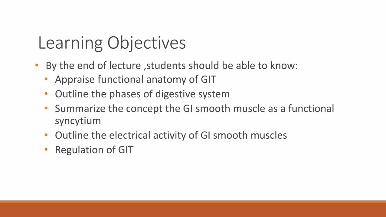
Learning Objectives• By the end of lecture ,students should be able to know:• Appraise functional anatomy of GIT
• Outline the phases of digestive system
• Summarize the concept the GI smooth muscle as a functional syncytium
• Outline the electrical activity of GI smooth muscles
• Regulation of GIT

Introduction• The primary function of the digestive system is to transfer
nutrients, water, and electrolytes from the food we eat into the body’s internal environment.
• Ingested food is essential as an energy source, or fuel, from which the cells can generate ATP to carry out their particular energy-dependent activities, such as active transport, contraction, synthesis, and secretion.
• Food is also a source of building supplies for the renewal and addition of body tissues.

Digestive TractA long muscular tube with many sections and areas.
Parts of the Digestive Tract
1. Mouth
2. Pharynx
3. Esophagus
4. Stomach
5. Small Intestine
6. Large Intestine

Accessory Parts Organs that are not in the digestive tract but helps in the digestion
1. Teeth
2. Tongue
3. Salivary glands
4. Liver
5. Gall bladder
6. Pancreas


Introduction There are four basic digestive processes
1. Motility
2. Secretion
3. Digestion
4. Absorption

Phases of Digestive System
I. Ingestion :occurs when material enters via the mouth
II. Mechanical Processing –Crushing/Shearing–makes material easier to move through the tract
III. Digestion—Chemical breakdown of food into small organic compounds for absorption
IV. Secretion –Release of water acids, buffers, enzymes & salts by epithelium of GI tract and glandular organs
V. Absorption –Movement of organic substrates, electrolytes, vitamins & water across digestive epithelium
VI. Excretion –Removal of waste products from body fluids


Structure Cross section of the intestinal wall, from the outer surface to inward
1. The serosa,
2. A longitudinal smooth muscle layer
3. A circular smooth muscle layer
4. The submucosa
5. The mucosa (additional some mucosal muscle)
Performs motor function of Gut

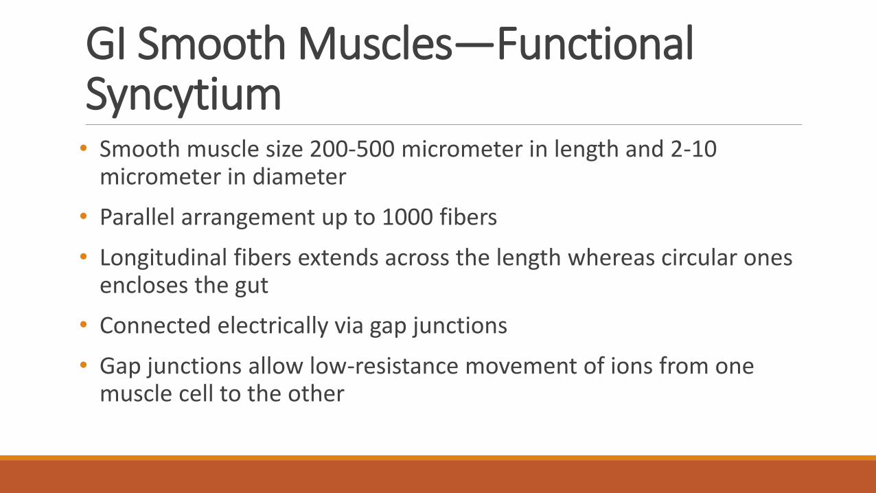
GI Smooth Muscles—Functional Syncytium• Smooth muscle size 200-500 micrometer in length and 2-10
micrometer in diameter
• Parallel arrangement up to 1000 fibers
• Longitudinal fibers extends across the length whereas circular ones encloses the gut
• Connected electrically via gap junctions
• Gap junctions allow low-resistance movement of ions from one muscle cell to the other

GI Smooth Muscles—Functional Syncytium• Electrical signals initiate muscle contractions travel readily from one
fiber to the next within bundle
• Bundles are partly separated by loose connective tissue may fuse at various points
• Represents a branching latticework of smooth muscle bundles
• Each muscle layer functions as a syncytium i.e. When an action potential is elicited anywhere within the muscle, it travels in all directions in the muscle.
• Distance depends on the excitability of the muscle

Interstitial Cells of Cajal• Specialized cells —act as electrical pacemakers for smooth muscle cells
• Slow wave potential is usually caused by complex interactions between smooth muscle cells and interstitial cells of cajal
• These specialized cells form a network with each other and are placed between the smooth muscle layers
• Make synaptic-like contacts to smooth muscle cells.
• Undergo cyclic changes in membrane potential
• Bears unique ion channels that open intermittently and produce inward slow rising currents–generate slow wave activity

Electrical Activity of GIT• Activated by slow, intrinsic electrical activity along sarcolemma
• Two basic types of electrical waves
1. Slow waves
2. Spikes
• RMP of GI smooth muscle is fluctuating that controls motor activity of the gastrointestinal tract.
• Slow waves don’t cause action potential they excite the spike potential
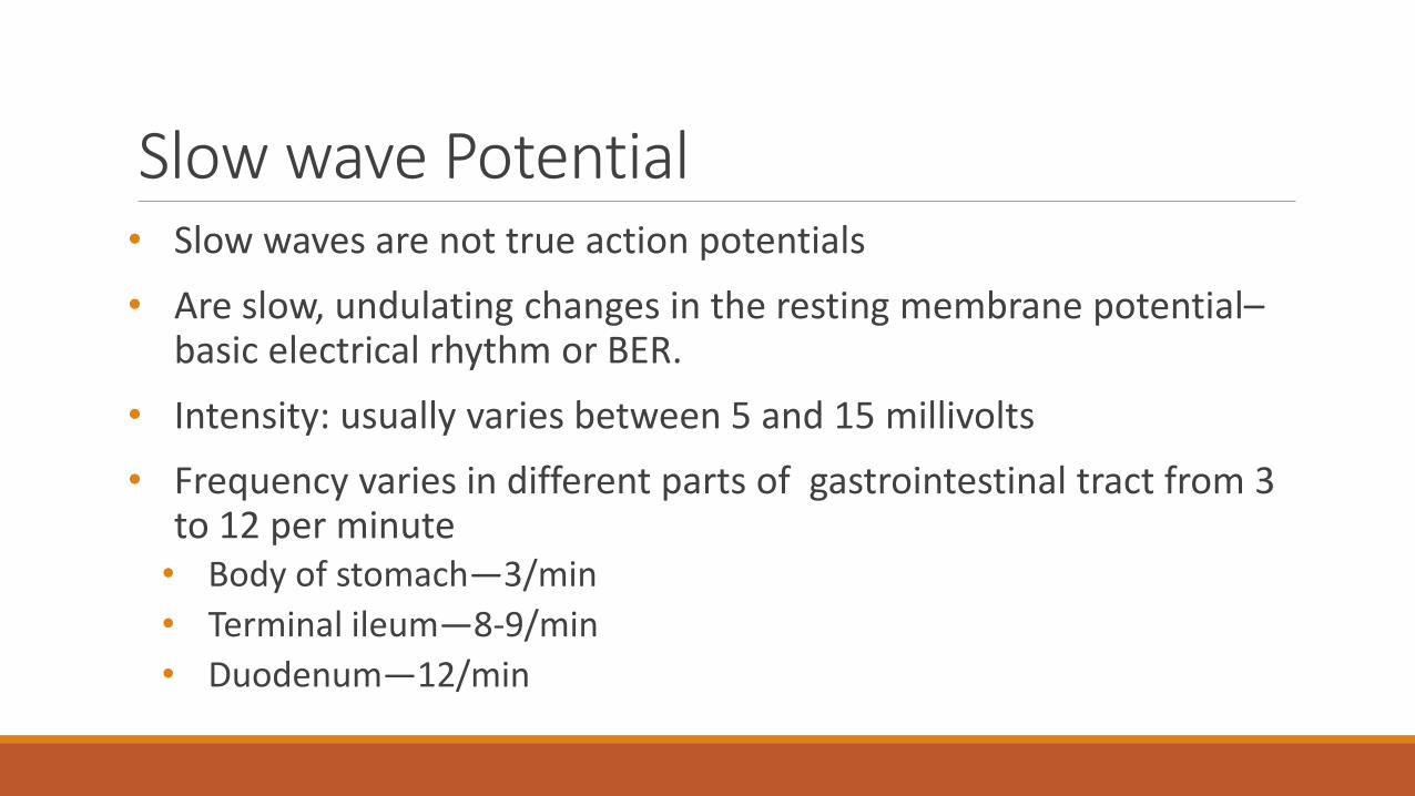
Slow wave Potential • Slow waves are not true action potentials
• Are slow, undulating changes in the resting membrane potential–basic electrical rhythm or BER.
• Intensity: usually varies between 5 and 15 millivolts
• Frequency varies in different parts of gastrointestinal tract from 3 to 12 per minute• Body of stomach—3/min
• Terminal ileum—8-9/min
• Duodenum—12/min

Cause of BER• Slow undulation of pumping activity of Na+/K+ ATPase pump.
• Cyclic variations in Ca++ release from ER & Ca++ uptake by mitochondria of pace setter cells.• Ach → ↑BER
• Ne → ↓BER
• BER does not cause muscle contraction, but coordinates peristaltic activity
Spike Potentials• Are true action potentials.
• Occur automatically when rmp becomes positive than −40mv (normal RMP −50&−60mv)
• Higher the slow wave potential rises, greater the frequency of the spike potentials(ranging 1—10 spikes/sec)
• Each GI spike last 10—20msec(10-40 times longer than action potential in nerve).

Spike Potentials• In nerve fibers, the action potentials caused by rapid entry of Na+
ions through sodium channels to the interior of the fibers.
• In GI muscle fibers, the channels responsible for the action potentials is caused by large numbers of Ca+2 to enter along with smaller numbers of Na+ via Ca+2/Na+ channels.
• These channels are slower to open and close than are the rapid sodium channels of large nerve fibers.
• Slowness of opening and closing of the calcium-sodium channels causes longer duration of action potentials

Factors Affecting Membrane ExcitabilityNormal conditions –RMP about −56mV
• When the potential becomes less negative i.e. Depolarization of the membrane, refers to excitability of nerve fiber
• Factors that depolarize the membrane• Stretching of muscle
• Stimulation by acetylcholine from parasympathetic nerves
• Stimulation by specific gastrointestinal hormones
• When the potential becomes more negative i.e. Hyperpolarization refers to less excitability
• Factors that hyperpolarize the membrane• Effect of norepinephrine or
epinephrine on the fiber membrane and
• Stimulation of the sympathetic nerves that secrete mainly norepinephrine at their endings.
Entry of Calcium Ions Causes Smooth Muscle Contraction• Calcium ions, acting through a calmodulin control mechanism,
• Activate the myosin filaments in the fiber,
• Causing attractive forces to develop between the myosin filaments and the actin filaments,
• Causing the muscle to contract.
• The slow waves do not cause calcium ions to enter the smooth muscle fiber just cause entry of sodium ions
Entry of Calcium Ions Causes Smooth Muscle Contraction• Some smooth muscles exhibit tonic contraction
• Tonic contraction is continuous not associated with BER
• Tonic contraction is sometimes caused by continuous repetitive spike potentials
• Greater the frequency, the greater the degree of contraction.
• Contraction is caused by hormones or other factors but sometimes entry of continuous calcium entry

Regulation of Digestion • Mechanical and chemical stimuli – stretch receptors, osmolarity,
and presence of substrate in the lumen
• Extrinsic control by CNS centers
• Intrinsic control by local centers

Receptors of the GI Tract• Mechano and chemoreceptors respond to: Stretch, osmolarity,
and pH, Presence of substrate, and end products of digestion
• They initiate reflexes that:
• Activate or inhibit digestive glands
• Mix lumen contents and move them along
Nervous control of GIT• Intrinsic controls• Nerve plexuses near the GI tract initiate short reflexes
• Short reflexes are mediated by local enteric plexuses--(gut brain)
• Extrinsic controls• Long reflexes arising within or outside the GI tract
• Involve CNS centers and extrinsic autonomic nerves

Regulation of GITFour factors are involved in regulation:
1. Autonomous smooth muscle function ‘BER’
2. Intrinsic nerve plexuses
i. Myenteric plexus (Auerbach’s plexus)
ii. Sub mucus plexus (Meissner's plexus)
• Enteric Nervous System or Gut Brain’: Contain as many neurons as in spinal cord.
• Has quality of self-regulation.

• Consists of various types of neurons; (linked through interneurons)a. Sensory or input neurons: respond to local stimuli of GIT.b. Output neurons: innervate smooth muscles, exocrine & endocrine
cells → affect motility, secretion of digestive juices & GI hormones.
3. Extrinsic nerves:-Both branches of ANS; ‘sympathetic & parasympathetic’i. Sympathetic fibers; through Myenteric & Sub mucus plexuses.* Reduce peristalsis & secretions* Contraction of sphincter musclesii. Parasympathetic fibers;◦ Through vagus nerve from esophagus to upper part of large intestine.◦ From spinal nerves in sacral region to lower part of large intestine.
stimulate motility & secretions.
4. Gastrointestinal hormones


Gastrointestinal ReflexesSupported by:
1. Enteric nervous system
2. Sympathetic & parasympathetic system.
Three Types:
i. Reflex entirely within enteric nervous system e.g.: Controlling: G.I Secretions, Mixing contractions and local inhibitory effects.
ii. Reflex from gut to paravertebral sympathetic ganglia & back to G.I.T e.g: Gastrocolic, Enterogastric and Colonoileal

Gastrointestinal Reflexesiii. Reflex from gut to the spinal cord or brain stem & then back to
G.I.T
• From stomach & Duodenum to Brain stem & back through Vagus• Pain reflexes
• Defecation reflex

THANK YOU