Modification of the fatty acid composition of cultured human fibroblasts
12
Modification of the fatty acid composition of cultured human fibroblasts Arthur A. Spector, Ruthann E. Kiser, Gerene M. Denning, Shy-Whey M. Koh, and Lawrence E. DeBault Departments of Biochemistry and Pathology and the Arteriosclerosis Specialized Center of Research, University of Iowa, Iowa City, IA 52242 Abstract The fatty acid composition of human skin fibro- blasts grown in 10% dialyzed fetal calf serum can be modi- fied considerably by adding supplemental fatty acids to the culture medium. The degree of modification was dependent on the concentration of added fatty acid over the range tested, 2.5 x M. At the higher concentra- tion, the extent of the modifications was as large as those which can be produced in nonhuman or malignant cell lines. Although the greatest changes were produced in the neutral lipid fraction, the cellular phospholipids also exhibited ap- preciable modifications. The phospholipids isolated from a microsomal fraction prepared from the cell homogenate exhibited similar changes in fatty acyl composition. These findings indicate that the human fibroblast can tolerate con- siderable variability in fatty acid composition, even in mem- brane phospholipids. The triglyceride content of the cells increased when they were grown in the presence of added fatty acids, but the phospholipid and cholesterol content remained unchanged. Growth was not affected by either oleic or linoleic acids, but it was reduced up to 50% when palmitic, linolenic, or arachidonic acid was added in concen- trations of 5 x lo+ M or above. Extensive modifications in phospholipid fatty acid composition also were produced in confluent monolayers of these fibroblasts. This suggests that some membrane lipid turnover occurs even when the cul- tures are not rapidly growing. Fatty acid modifications also were produced in the commercially available IMR-90 strain of human lung fibroblasts, suggesting that the ability to tolerate considerable differences in fatty acid composition is not a special property of the skin fibroblast line that was isolated locally.-Spector, A. A., R. E. Kiser, G. M. Den- ning, S-W. M. Koh, and L. E. DeBault. Modification of the fatty acid composition of cultured human fibroblasts. J. Lipid Res. 1979. 20: 536-547. to 1 x Supplementary key words phospholipids . neutral lipids * membranes . growth confluent monolayers * triglycerides . cholesterol Modifications in fatty acid composition have been produced in a number of mammalian cell lines by al- tering the lipids to which they are exposed. Cells that have been modified in this way include HeLa (l), LM (2,3), L (4), 3T3 (5), Ehrlich ascites (6), L1210 (7), and L6 (8). Such changes in fatty acid composition are reported to have an effect on several membrane prop- erties of animal cells, including the activity of certain membrane-bound enzymes (9), the degree of expo- sure of surface proteins (lo), lateral mobility of recep- tors (5), responses to mitogenic stimuli (1 l), endocyto- sis (12), amino acid transport (13), and cholesterol esterification (14). All of the cells in which extensive fatty acid modifications have been produced are con- tinuous lines that have been maintained for long periods, and except for HeLa (human), all of the above are of murine or rat origin. Although HeLa is a human line, it is heteroploid and was derived from a malig- nant tumor. In order to further explore the factors that regulate cellular and membrane lipid composi- tion, we wished to determine whether fatty acid modifications of a similar degree were possible in normal human diploid cell lines. The present com- munication describes the rather extensive modifica- tions in fatty acid composition that can be produced in cultured human skin and lung fibroblasts, in both rapidly growing cells and confluent monolayers. MATERIALS AND METHODS Cells and media The diploid, finite cell line that was used in this study was derived from a primary culture of human fore- skin fibroblasts by the State Hygienic Laboratory, University of Iowa. A second human diploid cell line, IMR-90, used for comparative experiments, has been characterized as a cell strain by Nichols et al. (15). The IMR-90 cells, isolated from the lung tissue of a human female fetus, were obtained from the American Type Culture Collection (Cell Repository designation CCL 186). Stock cultures of the fibroblasts isolated locally were maintained as monolayers in Eagle’s Basal Medium with Earle’s Salts (Gibco G-11) (16)supplemented with 536 Journal of Lipid Research Volume 20, 1979 by guest, on January 3, 2019 www.jlr.org Downloaded from
Transcript of Modification of the fatty acid composition of cultured human fibroblasts

Modification of the fatty acid composition of cultured human fibroblasts
Arthur A. Spector, Ruthann E. Kiser, Gerene M. Denning, Shy-Whey M. Koh, and Lawrence E. DeBault Departments of Biochemistry and Pathology and the Arteriosclerosis Specialized Center of Research, University of Iowa, Iowa City, IA 52242
Abstract The fatty acid composition of human skin fibro- blasts grown in 10% dialyzed fetal calf serum can be modi- fied considerably by adding supplemental fatty acids to the culture medium. The degree of modification was dependent on the concentration of added fatty acid over the range tested, 2.5 x M. At the higher concentra- tion, the extent of the modifications was as large as those which can be produced in nonhuman or malignant cell lines. Although the greatest changes were produced in the neutral lipid fraction, the cellular phospholipids also exhibited ap- preciable modifications. The phospholipids isolated from a microsomal fraction prepared from the cell homogenate exhibited similar changes in fatty acyl composition. These findings indicate that the human fibroblast can tolerate con- siderable variability in fatty acid composition, even in mem- brane phospholipids. The triglyceride content of the cells increased when they were grown in the presence of added fatty acids, but the phospholipid and cholesterol content remained unchanged. Growth was not affected by either oleic or linoleic acids, but it was reduced up to 50% when palmitic, linolenic, or arachidonic acid was added in concen- trations of 5 x lo+ M or above. Extensive modifications in phospholipid fatty acid composition also were produced in confluent monolayers of these fibroblasts. This suggests that some membrane lipid turnover occurs even when the cul- tures are not rapidly growing. Fatty acid modifications also were produced in the commercially available IMR-90 strain of human lung fibroblasts, suggesting that the ability to tolerate considerable differences in fatty acid composition is not a special property of the skin fibroblast line that was isolated locally.-Spector, A. A., R. E. Kiser, G. M. Den- ning, S-W. M. Koh, and L. E. DeBault. Modification of the fatty acid composition of cultured human fibroblasts. J . Lipid Res. 1979. 20: 536-547.
to 1 x
Supplementary key words phospholipids . neutral lipids *
membranes . growth confluent monolayers * triglycerides . cholesterol
Modifications in fatty acid composition have been produced in a number of mammalian cell lines by al- tering the lipids to which they are exposed. Cells that have been modified in this way include HeLa (l), LM (2,3), L (4), 3T3 ( 5 ) , Ehrlich ascites (6), L1210 (7), and L6 (8). Such changes in fatty acid composition are
reported to have an effect on several membrane prop- erties of animal cells, including the activity of certain membrane-bound enzymes (9), the degree of expo- sure of surface proteins (lo), lateral mobility of recep- tors (5) , responses to mitogenic stimuli (1 l), endocyto- sis (12), amino acid transport (13), and cholesterol esterification (14). All of the cells in which extensive fatty acid modifications have been produced are con- tinuous lines that have been maintained for long periods, and except for HeLa (human), all of the above are of murine or rat origin. Although HeLa is a human line, it is heteroploid and was derived from a malig- nant tumor. In order to further explore the factors that regulate cellular and membrane lipid composi- tion, we wished to determine whether fatty acid modifications of a similar degree were possible in normal human diploid cell lines. The present com- munication describes the rather extensive modifica- tions in fatty acid composition that can be produced in cultured human skin and lung fibroblasts, in both rapidly growing cells and confluent monolayers.
MATERIALS AND METHODS
Cells and media
The diploid, finite cell line that was used in this study was derived from a primary culture of human fore- skin fibroblasts by the State Hygienic Laboratory, University of Iowa. A second human diploid cell line, IMR-90, used for comparative experiments, has been characterized as a cell strain by Nichols et al. (15). The IMR-90 cells, isolated from the lung tissue of a human female fetus, were obtained from the American Type Culture Collection (Cell Repository designation CCL 186).
Stock cultures of the fibroblasts isolated locally were maintained as monolayers in Eagle’s Basal Medium with Earle’s Salts (Gibco G-11) (16) supplemented with
536 Journal of Lipid Research Volume 20, 1979
by guest, on January 3, 2019w
ww
.jlr.orgD
ownloaded from

10% heat-inactivated fetal calf serum, 0.1 mM Mini- mal Essential Medium nonessential amino acids (Gibco 114), 100 unitdm1 of penicillin, and 100 pg/ ml of streptomycin. The medium was buffered at pH 7.4 with 0.015 M NaHC03 and 5% C 0 2 in air. The stock cultures were maintained at 37°C in a C02 in- cubator and subcultured every 3-4 days with 0.25% trypsin (Gibco 505) in 0.01 M phosphate-buffered saline at a subculture ratio of 1 :2. The medium was not renewed between subcultures. The IMR-90 cells were maintained using similar conditions in Eagle’s Mini- mum Essential Medium with Earle’s salts (Gibco F-1 1) (17). All media and supplements were sterilized by filtration through a 0.22 pm Millipore filter.
Fetal calf serum, obtained from Gibco (Grand Island, NY), was heat-inactivated at 56°C for 30 min. Serum from a single lot was used in all experiments. One volume of heat-inactivated serum was dialyzed against 20 volumes of phosphate-buffered saline at 4°C for 36 hr, the buffer being changed twice during the dialysis. After dialysis, the fetal calf serum con- tained 26.8 ? 1.2 mg/dl phospholipids, 28.5 * 0.08 mg/dl cholesterol, 1.8 ? 0.09 mg/dl triglycerides, and 0.20 & 0.05 pmol/ml free fatty acids. The composition of the major components of the free fatty acid frac- tion was 21% 16:0,’ 15% 18:0, 26% 18:1, 7.5% 18:2, and 7.9% 20:4, whereas that of the phospholipid fraction was 23% 16:0, 24% 18:0, 26% 18:1, 2.4% 18:2, 5.3% 20:4,4.1% 22:5, and 3.9% 22:6. The con- centration of dialyzed serum in the culture medium was lo%, so the inherent free fatty acid concentration to which the cells were exposed was about 0.02 @mol/ ml. Based on the percentage composition, the con- centrations of the most abundant individual free fatty acids in the culture medium were 4.2 x M pal- mitate, 5.2 X M oleate, and 1.5 X lov6 M lino- leate when no supplemental fatty acid was added.
Fatty acids were obtained from Applied Science Inc. (State College, PA) or NuChek Prep (Elysian, MN). The sodium salt of the fatty acid was prepared and added dropwise to the dialyzed serum with stir- ring (18). The free fatty acid content of the solutions, determined by the method of Lowry and Tinsley (19), was between 5 x and 2 x M. The serum protein concentration was determined by the biuret method (20). These solutions contained 1-6 pmol of fatty acid/pmol of serum albumin. The fatty acid- containing serum solutions were sterilized by filtra- tion and stored at -20°C.
Fatty acids are abbreviated as chain length: number of double bonds. Thus, the abbreviation 16:O signifies a fatty acid con- taining 16 carbon atoms and no unsaturated bonds.
Experimental cultures Fibroblast cultures were seeded in their respective
maintenance media at 5-5.5 x lo3 cells/cm2 in 60-mm Falcon tissue culture petri dishes (P-60) for growth and toxicity experiments. For experiments where larger numbers of cells were required, cultures were seeded at 7- 11 x lo3 cells/cm2 in 25-cm2 (50 ml) Falcon flasks (T-25) or in 75-cmZ (250 ml) Falcon flasks (T-75). Culture media volumes were 3 ml for P-60 dishes, 3.5 ml for T-25 flasks, and 10 ml for T-75 flasks. The experimental medium was the same as the maintenance medium for the locally isolated fibro- blasts, except that the intact fetal calf serum was re- placed with either dialyzed serum or dialyzed serum containing added fatty acid. All of the media were ad- justed to a constant protein concentration of 4.4 mg/ml.
Each experiment was initiated by seeding the fibro- blasts in their complete maintenance media. For experiments with rapidly growing cells, the cultures were washed with phosphate-buffered saline 24 hr after seeding and then fed with the experimental medium. This was defined as day 1. Cultures were refed on day 4 and harvested on day 7. In experi- ments with confluent cells, the cultures were refed with maintenance Earle’s Basal Medium twice at 3- day intervals. On day 9, when the cultures were con- fluent, they were washed with phosphate-buffered saline and fed with experimental medium. The cells were then maintained in culture for either 3 or 21 days. Those maintained for only 3 days were not refed, whereas those maintained for 21 days were refed a total of 5 times at 3-day intervals and harvested 72 hr after the fifth medium change, on day 27. Confluency is defined as the maximum number of cells per cm2 attainable under these culture conditions and is equivalent to the saturation density (2 1). This value varied between 7.0 and 8.5 X lo4 cells/cm2. The mi- totic index was 1.02 ? 0.23% for log phase cultures and 0.19 & 0.16% for confluent cultures. There was no significant difference in the mitotic index of cells exposed to dialyzed serum as compared with supple- mented media containing either oleic or linoleic acids. In addition, refeeding the confluent cultures did not stimulate proliferation as measured by [3H]thymi- dine incorporation during a l-hr pulse administered 24 hr after refeeding.
Growth curves were generated by enumerating the cells in each culture daily according to the method of Martinez-Lopez and Black (22), using an inverted phase microscope. The data were expressed as num- ber of cells per culture dish. Population doubling times were calculated from these growth curves be-
Spector et al. Fatty acid-modified cells 537
by guest, on January 3, 2019w
ww
.jlr.orgD
ownloaded from

tween 2 and 4 x lo5 cells per dish (21). The popula- tion doubling times for the cells grown in the dialyzed serum, oleic acid, or linoleic acid-supplemented serum media were 30.8 * 1.5,34.5 * 2.0, and 30.2 -C 2.9 hr, respectively. The values for the cells grown in media supplemented with palmitic, linolenic, or arachidonic acid were dependent on the concentration of fatty acid added. They varied from 35.0 & 4.0 hr with 1 x M fatty acid to 54.5 ? 8.2 hr with 1 x M fatty acid. Phase contrast photomicrography of living cells and bright field microscopic observations of stained material were done with a Leitz Diavert in- verted microscope with an Orthomat camera attach- ment.
The cells were harvested after the monolayers were washed four times with phosphate-buffered saline, followed by two washes with 0.25% trypsin at 37°C. The cells, covered by a residual trypsin film, were incubated at 37°C until detachment occurred, and they were then freed from the dish or flask by washing several times with cold phosphate-buffered saline. The fibroblasts were isolated from the suspension by sedimentation at 800g for 5 min, and the pellet was resuspended in fresh buffer by gentle pipetting. Cell counts of the suspension were taken for lipid analysis and measurement of protein content (23). Aliquots also were taken from representative cultures for cell viability determinations. The cells were ex- posed to a phosphate-buffered saline solution con- taining 0.2% trypan blue for 10 min. From 90 to 100% of the cells were judged to be viable by this pro- cedure. In addition, seeding efficiency was compared with cultures grown in dialyzed serum and serum sup- plemented with fatty acid. Seeding efficiency was cal- culated as the percentage of the inoculum that at- tached to the surface of the culture vessel within 3 hr at 37°C (21). Subculturing of cells after 7 days of growth in either 5 x M linoleic-, oleic-, or pal- mitic acid-supplemented medium or in l x M linoleic acid-supplemented medium resulted in seed- ing efficiencies of 85-100%. Although there were variations in the absolute seeding efficiency among the different experiments, the value relative to corre- sponding control cells grown in dialyzed serum within a given experiment always approached 100%.
Preparation of membranes An enriched membrane preparation was isolated
from fibroblasts pooled from 20-30 T-75 flasks by a modification of the procedure of Schimmel et al. (24). The cells were harvested by washing the cultures four times with 4 ml of cold phosphate-buffered saline and then scraping the cells free with a rubber policeman into 1.0 ml of the buffer. This was followed by two
additional washes of the flask. The cells were sedi- mented by centrifugation at 1 100 g for 5 min at 4°C. The pellet was resuspended in 6 ml of 0.25 M su- crose-1 mM triethanolamineeHC1, pH 7.4. The sus- pended cells were then broken in a Dounce homog- enizer with a B pestle. Twenty to twenty-five up-and- down strokes were sufficient to rupture most of the cells as determined by phase contrast microscopy. The homogenate was centrifuged at 1700 g for 10 min at 4°C. The supernatant solution was removed, and the pellet was washed with 2 ml of homogenizing solu- tion and resedimented. The supernatant and wash solutions were combined and centrifuged at 12,000 g for 15 min at 4°C to sediment mitochondria. The resulting pellet was washed with 1 ml of homogenizing solution and resedimented, and the supernatant solu- tion was added to the first postmitochondrial frac- tion. A membrane fraction was sedimented from these combined supernatant solutions by centrifugation at 100,OOOg for 1 hr at 4°C. After the supernatant fluid was removed and the tube and compacted pellet were rinsed three times with cold phosphate buf- fered saline, the pellet was suspended in 2.0 ml of cold buffer. The content of the membrane prepara- tion was assessed by electron microscopy.
Lipid analyses Cellular lipids were extracted with chloroform-
methanol 2:l (v/v) (25). Aliquots of the chloroform phase were taken for colorimetric measurement of phospholipids (26) and measurement of triglycerides by a micromodification of a fluorometric method utilizing sodium periodate and acetyl acetone (27). Unesterified cholesterol was determined isothermally at 290°C using a Hewlett-Packard 5710A gas-liquid chromatograph equipped with a flame ionization detector and a glass column (6 ft x 2 mm i.d.) packed with 3% SP-2250 on 100/120 mesh Supelcoport (Supelco, Inc., Bellefonte, PA). Peak areas were meas- ured with a Hewlett-Packard 3380A integrator. The internal standard was 5-a-cholestane-3-P-01 (Supelco, Inc.). Total cholesterol was analyzed in the same man- ner after saponification of the lipid extract by a modifi- cation of the procedures reported by Driscoll et al. (28) and Lillienberg and Svanborg (29). Each of the dried lipid extract samples, containing internal standard, was mixed with 4.7 ml of 95% ethanol and 0.3 ml of 33% KOH. The samples were saponified for 60 min at 57°C. After cooling, 1.0 ml of heptane was added with mixing, followed by 5 ml of H,O. After removal of the heptane phase, the aqueous portion was re- extracted twice with 1.0 ml of heptane. The amount of cholesteryl ester was determined by difference. The above procedures also were employed for measure-
538 Journal of Lipid Research Volume 20, 1979
by guest, on January 3, 2019w
ww
.jlr.orgD
ownloaded from

ment of the lipids contained in the fetal calf serum. Free fatty acid concentration in the serum was meas- ured by the method of Laurel1 and Tibbling (30).
Gas-liquid chromatography also was employed to analyze the fatty acid composition of isolated phos- pholipid and neutral lipid fractions. Phospholipids and neutral lipids were separated by thin-layer chro- matography on Silica Gel G plates by developing in a solvent system containing hexane-diethyl ether- acetic acid-methanol 170:40:4:4 (31, 32). Nu-Chek Prep 18-5C and Nu-Chek Prep 18-1C (Nu-Chek Prep, Elysian, MN) were used as standards. After elution from the silica gel with chloroform-methanol 1: 1 (v/v) (14), the lipid fractions were saponified for 45 min at 70°C in 2.5 ml of 95% ethanol and 0.05 ml of 33% KOH. The nonsaponifiable components were removed by extraction with n-hexane, the solution was acidified, and the fatty acids were extracted into n- hexane. The fatty acids were methylated with 14% BF3 in methanol for 10 min at 95°C and the methyl esters were extracted into n-hexane (33). The fatty acid methyl esters were separated by gas-liquid chromatography using a glass column (6 ft X 2 mm i.d.) packed with either 10% Apolar 1OC on 100/120 mesh Gas-Chrom Q (Applied Science Laboratories, Inc., State College, PA) or 10% SP-2340 or SP-2330 on 100/120 mesh Chromosorb W-AW (Supelco, Inc.).
Relative mass amounts were determined with the integrator.
RESULTS
Modifications in fatty acid composition produced by linoleic acid
Our initial attempts to modify the fatty acid com- position of the human skin fibroblasts were made by adding increasing amounts of linoleic acid to the cul- ture medium. The medium contained 10% dialyzed fetal calf serum, and cells grown in this medium with- out any supplemental fatty acid served as the control. Linoleic acid was selected for study because, as seen in Table 1, the lipids of the cells grown under control conditions contained only 3-4% of this biologically important fatty acid. Under these control conditions, the cell phospholipids contained 35% saturated, 29% monoenoic, and 32% polyenoic fatty acid. In pre- liminary experiments, fatty acid compositions similar to this were observed when the fibroblasts were grown in a medium containing either lipoprotein-poor fetal calf serum prepared according to Brown, Dana, and Goldstein (34) or lipid-extracted fetal calf serum pre- pared according to Rothblat et al. (35) instead of the dialyzed serum. In both of these cases, however, cell
TABLE 1. Effect of added linoleic acid on cellular fatty acid composition"
Phospholipid Fatty Acids Neutral Lipid Fatty Acids
2.5 x 10-JM 5 x 1 0 - 5 ~ I x io-'M 2.5 x 10⁻⁵ M 5 x 10⁻⁵ M I x IO-' M Fatty Acid 0 (l0)b (4) (4) (4) 0 (10)b (4) (4) (4)
Classc Saturated Monoenoic Polyenoic
16:O 16: 1 18:O 18: 1 18:2 20:2 20:4 22:4 22:5 22:6 Others Unidentified
Individual acidsd
35 29 32
14.2 f 2.3 3.1 f 0.5
18.2 f 0.6 25.7 f 1.1 3.4 f 0.1 0.3 f 0.1
12.4 f 0.5 2.6 f 0.4 4.3 f 0.6 4.9 f 0.4
7.0 3.9
40 24 33
16.0 f 1.3 2.6 f 0.2
19.3 f 2.3 21.2 f 1.5 16.8 f 1.3 2.4 f 0.2 8.6 f 1.0
<0.5 1.4 f 0.2 1.7 f 0.3
7.5 2.5
38 19 41
16.1 f 0.4 2.7 f 0.1
15.0 f 0.5 16.1 f 2.1 22.0 f 0.5 4.4 f 0.1 6.9 f 0.2 3.7 f 1.5 1.4 f 0.1 1.6 f 0.1
7.8 2.3
39 21 39
16.4 f 0.7 2.3 f 0.2
16.6 f 0.6 18.6 f 3.8 23.0 f 3.0 5.7 f 0.9 5.2 f 0.8 0.9 f 0.3 0.9 f 0.1 1.3 f 0.2
6.9 2.2
%
50 25 19
22.0 f 1.2 3.2 f 0.4
22.2 f 1.8 20.9 f 1.3 4.1 f 0.6 1.3 f 0.4 3.8 f 0.8 1.8 f 0.5 2.4 f 0.4 1.4 f 0.2
11.2 5.7
50 19 28
23.0f 1.7 3.2 f 0.5
17.8 f 1.9 15.9 f 2.7. 15.8 f 0.8 0.8 f 0.1 3.4 f 0.3 2.0 f 1.1 0.9 f 0.2 0.5 -c 0.1
13.6 3.1
42 17 39
20.2 f 2.5 2.5 f 0.3
15.7 f 2.2 14.0 f 2.5 25.5 f 3.3 0.4 5 0.1 2.6 f 0.4 2.9 f 0.7 1.5 f 0.4 0.8 f 0.2
10.9 3.0
24 11 63
11.8 f 0.4 1.8 f 0.2 8.4 f 0.3 9.0 f 1.8
43.5 f 0.8 1.0 2 0.1 3.5 f 0.1 3.6 & 0.5 1.4 f 0.1 0.7 f 0.1
12.3 3.0
a About 5 X los celldcm' were seeded in 25-cm' flasks, and 3.5 ml of medium was added. The medium contained either no added fatty acid or the concentration of linoleic acid shown at the top of each column. The medium was changed on days 1 and 4, and the cells were harvested on day 7. At that time, there were approximately 1 X 10⁵ celldcm'.
Concentration of linoleic acid added to the culture medium, followed in parentheses by the number of cultures analyzed. Each of the media contained 10% dialyzed fetal calf serum.
Since between 2.0 and 3.9% of the fatty acids were not identified, the sum of the fatty acid classes does not add up to 100%. The individual fatty acids are abbreviated as number of carbon atoms:number of double bonds. Each value is the mean f SE.
Spector et al. Fatty acid-modified cells 539
by guest, on January 3, 2019w
ww
.jlr.orgD
ownloaded from

growth was reduced considerably. Since these lipid- depleted sera provided no advantage in terms of a more simplified phospholipid fatty acyl composition in cells grown under control conditions and caused a considerable disadvantage in terms of reduced growth, we elected to use serum containing its in- herent lipids in the culture medium.
As the concentration of added linoleic acid in the medium was raised from 2.5 x M, the polyenoic fatty acid content of the cell phospho- lipids increased, and the monoenoic fatty acid con- tent decreased. The maximum changes occurred when the medium contained 5 x M linoleic acid. The relative gain in polyenoic fatty acids was ac- counted for primarily by an increase in 18:2 from 3.7 to 25.4% and adecrease in 18: 1 from 25.7 to 12.4%. In addition, 20:2, the direct elongation product of 18:2, increased from 0.5 to 6.0%. By contrast, 20:4 de- creased from 12.0 to 5.2% as the linoleic acid content of the medium was raised. Likewise, the percentage of the higher polyunsaturates also decreased, sug-
to 1 x
- 50 - ? e
25 palmitic oleic linoleic
05 10 $ 0 2 01 05
251, , ,,j .?j, , ,I p i , , , I palmitic oleic linoleic
01 05 10 01 05 10 $ 0 2 01 05
arachidonic linolenic 25t, 0 01 05 I 10 i\2:u 01 05 10
[Fatty acid] x lo4 M
Fig. 1. Effect of concentration of the added fatty acid on the growth of the cultured fibroblasts. The cells were seeded in 60-mm Falcon petri dishes (P-60) at a concentration of about 5-5.5 x 10³/cm2, and a total of 3 ml of culture medium was present in each flask. After 24 hr, the medium was removed and the cells were washed with phosphate-buffered saline solution. Each experimental medium contained 10% dialyzed fetal calf serum with supplemental fatty acid. The medium was changed on days 1 and 4, and cell counts were made on days 4-7 by the method of Martinez-Lopez and Black (22) using an inverted phase contrast microscope. The data points are the mean of four separate determinations, each of which was made using a different culture. Control cultures were grown in media that contained 10% dialyzed fetal calf serum but no added fatty acid. Although cell counts were determined each day, only the results obtained on day 6 are shown for clarity.
gesting that one or more of the fatty acid desaturases of these fibroblasts may be inhibited when large amounts of linoleic acid are available. Inhibition of the A9-desaturase by linoleic acid also would account for the reduction in monoenoic fatty acid content that was observed.
Similar changes in the fatty acid composition of the fibroblast neutral lipids occurred when linoleic acid was added to the culture medium. Most of the fatty acids present in this fraction were contained in tri- glycerides. The changes in neutral lipid fatty acid com- position were even more marked than those in the phospholipids. For example, the 18:2 content in- creased from 4 to 44% and the 18: 1 content decreased from 22 to 9% as the linoleic acid content of the medium was raised. Unlike the phospholipid frac- tion, the neutral lipids did not exhibit any plateau in either 18:2 to total polyenoic acid content, and the highest values occurred when 1 x lop4 M linoleic acid was added to the culture medium.
Effects of supplemental fatty acids on cell growth The effect of the added linoleic acid on the growth
of the cells was tested during a 7-day period. For comparison, control cultures containing only the 10% dialyzed fetal calf serum with no supplemental fatty acid were included. As a further comparison, cultures containing added oleic acid were tested in a second series of experiments. The concentrations of the added fatty acids were varied from 1 x IOp5 to 1 x M. Neither oleic nor linoleic acid affected the growth rate over this range of concentrations as meas- ured by the microscopic field counts (22). As a further test, the cells were harvested on day 7 for analysis of protein content and counting of the cell suspension using a hemocytometer. Both of these procedures yielded the same results as those obtained by the field counting procedure, namely, that the addition of these fatty acids did not affect growth relative to con-' trol cultures grown in 10% dialyzed fetal calf serum.
These types of growth experiments were carried out with other long-chain fatty acids. The results, presented in Fig. 1, show relative growth plotted as a function of fatty acid concentration. For clarity, only the data points obtained on day 6 are shown, but these values are representative of those obtained on the other days. As opposed to the results obtained with oleic and linoleic acids, there was a reduction in growth when the higher concentrations of palmitic, linolenic (n-3),2 and arachidonic acids were used. The extent of the reduction and the fatty acid concentra-
The linolenic acid utilized is 18:3A9,12,15 and, therefore, is a member of the n-3 class of polyenoic fatty acids.
540 Journal of Lipid Research Volume 20, 1979
by guest, on January 3, 2019w
ww
.jlr.orgD
ownloaded from
TABLE 2. Fatty acid composition of lipids in fibroblasts exposed to different fatty acids during growth"
Neutral Lipid Fatty Acids Phospholipid Fatty Acids
Palmitic Oleic Linolenic Arachidonic Palmitic Oleic Linolenic Arachidonic Fatty Acid Acid (4)* Acid (4) Acide (4) Acid (4) Acid (4) Acid (4) Acid (4) Acid (4)
9% Class
Saturated 50 27 28 37 78 10 33 22 Monoenoic 19 52 2 1 23 11 77 13 17 Polyenoic 30 13 47 36 7 8 50 56
Individual acids 16:O 24.0 f 0.4 10.8 2 0.8 10.1 f 2.5 14.8 2 0.2 36.6 0.5 4.8 2 0.3 1 4 . 0 2 1.3 10.7 f 1.1 16: 1 3.2 f 0.2 1.7 k 0.1 1.7 f 0.4 2.6 2 0.5 2.0 f 0.1 1.4 2 0.1 2 . 0 k 0.3 2.5 f 0.3 18:O 19.9 f 0.1 14.0 2 0.3 17.0 f 0.3 18.2 2 0.6 25 .8k0 .4 3 . 8 f 0 . 2 1 2 . 4 2 1 . 1 8.920.2 18:l 15.3 k 0.3 49.9 2 1.4 19.Of 0.7 19.7 f 1.1 9.3 2 0.6 75.5 & 0.9 10.8 2 0.4 14.6 2 0.4 18:2 3.2 2 0.3 1.6 2 0.3 3.0 2 0.1 2.9 f 0.1 2.9 & 0.1 1.2 f 0.3 3.7 2 0.7 3.3 f 0.2 18:3 0.4 f 0.1 0.5 2 0.1 7.6 f 0.5 2.2 ? 0.9 2.0 f 0.1 0.3 2 0.1 15.4 k 1.5 0.5 f 0.1 20:3 1.4 2 0.1 0.7 f 0.1 1.4 2 0.1 1.7f 0.1 0.3 f 0.1 0.4 k 0.1 2.1 k 0.5 3.9 f 0.3 20:4 13.6 k 0.3 5.2 f 0.2 15.0 k 1.5 12.8 & 1.9 0.8 2 0.2 1.9 f 0.2 7.7 f 0.4 20.5 2 0.7 22:4 1 . 7 2 0 . 1 0 . 8 2 0 . 1 2 .220 .4 7 . 7 2 1 . 4 N.D. 1.0 2 0.1 3.7 k 0.3 19.1 k 1.1 22:5 4.2 2 0.4 1.7 2 0.1 3.8 f 0.6 1.7 f 0.2 0 .8f 0.1 0 .9f 0.1 4.9 2 0.7 2.9 2 0.1 22:6 3.9 f 0.2 2.4 k 0.1 2.6 f 0.5 1.5 k 0.3 0 . 4 f 0 . 1 0 .220 .1 1 . 5 k 0 . 2 1 .6kO. l Others 8.0 2.7 13.0 9.7 15.2 3.2 17.7d 7.0 Unidentified 1.2 8.0 3.6 4.5 3.9 5.4 4.1 4.5
a The cells were exposed to media supplemented with one of the fatty acids for 7 days. About 5 x lo3 cells/cm2 were seeded in 25 cm2 flasks, and 3.5 ml of the media was added. The media were changed on days 1 and 4 and the cells were harvested on day 7, at which time there were about 1 x lo5 cells/cm*. Each value is the mean f SE of the percentage composition. All of the abbreviations are the same as those listed in Table 1.
M; oleic, 1 X lo-' M; linolenic, 5 x
Type of fatty acid added to the culture medium. The fatty acid concentrations were: palmitic, 5 X
18:3A9,12,15, a member of the n-3 class of polyenoic fatty acids. Contains 46% as 20:5. The remainder of the unidentified acids also are polyenoic.
M; arachidonic, 5 x M. The number of separate cell cultures analyzed is given in parentheses.
tion at which a decrease first was observed were dif- ferent for each fatty acid. For example, palmitic acid produced a 50% decrease in growth at 1 x lop4 M, and it began to depress growth at 7.5 x lop5 M. By contrast, linolenic acid reduced growth by only 20% at 1 x M, but it began to exert an inhibitory effect at 5 x lop5 M. Similarly, arachidonic acid began to de- crease growth at 5 x M and reduced growth by 50% at 1 x low4 M. Because of their inhibitory ef- fects on growth at the higher concentrations, we elected to test the effects of palmitic, linolenic, and arachidonic acids on cell fatty acid composition at 5 x M rather than at 1 x M. Since oleic acid did not cause any inhibition of growth, we tested it at 1 x M in an attempt to produce maximal effects on the fatty acid composition of the cells.
Modifications in composition produced by other fatty acids
Table 2 shows the changes in fibroblast fatty acid composition that were produced by adding long-chain fatty acids other than linoleic to the culture medium. As compared with the control cells (Table l) , those exposed to 5 x M palmitic acid contained a higher percentage of saturated and a lower per- centage of monoenoic fatty acids in their phospho-
lipids. This was accounted for primarily by an increase in 16:0, the added fatty acid, and a decrease in 18: 1. By contrast, cells exposed to 1 x M oleic acid con- tained much more monoenoic and less polyenoic fatty acids in their phospholipids, as compared with the
TABLE 3. Lipid content of the fibroblasts"
Amount
Cell Lipid None Oleic Acid Linoleic Acid
pgimg protein
Phospholipids 251 f 34 180 f 21 220 f 34 Cholesterol 37 k 5 34 k 1 36 f 3 Triglycerides 18 f 3 144 f 32b 202 f 38b
(I For each case, two T-75 flasks were seeded with 7 x lo3 cells/ cm2, and 10 ml of media containing 10% dialyzed fetal calf serum was added. The cells were harvested for analysis after 7 days growth in culture, and the contents of the two flasks were pooled. In the cases where fatty acids were added to the culture medium, the sup- plemental fatty acid concentration was 1 X lo-' M. The values are the mean 2 SE of six determinations, each of which was made on a separate culture of cells.
Differences between the cells grown in the presence of added fatty acid and those in the unsupplemented medium are statistically significant; none vs. oleate, 1 = 3.92, P < 0.01; none vs. linoleate, t = 4.83, P < 0.01. The difference between the cells grown in the presence of oleate and linoleate is not significant; t = 1.17, P > 0.1.
Spector et al. Fatty acid-modified cells 541
by guest, on January 3, 2019w
ww
.jlr.orgD
ownloaded from
TABLE 4. Fatty acid composition of phospholipids isolated from fibroblast microsomesa
Composition
Oleic Linoleic Fatty Acid None Acid Acid
Class Saturated Monoenoic Polyenoic
16:O 16: 1 18:O 18:l 18:2 20:2 20:3 20:4 22:4 22:5 22:6 Others Unassigned
Individual acids
%
34 20 33 62 29 18
17.4 7.9 5.3 2.6
16.4 11.3 28.1 54.9
3.9 1.4 <0.5 <0.5
1.6 0.6 10.8 7.6 2.6 1.2 4.0 2.1 5.6 4.8 1.9 5.6 1.5 <0.5
26 14 59
14.0 2.5
10.6 11.8 36.7
9.9 1.3 5.1 1.5 2.0 2.5 1.0 1.2
~~ ~
a Newly seeded cells were grown for 7 days in media containing either 10% dialyzed fetal calf serum or this serum supplemented with either 1 X lo-' M oleic or 1 x lo-' M linoleic acid. After isola- tion and washing, the cells were homogenized and a microsome fraction was isolated by sedimentation between 12,000 and 100,000 g for 1 hr. This particulate fraction was washed once, and its lipid content was extracted with a chloroform-methanol solu- tion. The phospholipids contained in the lipid extract were isolated by thin-layer chromatography, and their fatty acid composition was determined by gas-liquid chromatography following saponifica- tion and methylation. Each value is from a single sample of microsomes obtained by pooling the cells from 30 Falcon flasks (T-75, 250 ml). This provided a total of 2-5 X loB cells for each preparation. Four separate aliquots of each sample of fatty acid methyl esters were chromatographed, and the values listed are the means calculated from the four chromatograms.
control cells. This was due primarily to an increase in the percentage of 18: 1. With 5 x M linolenic acid (n-3 class), there was an increase in the percentage of polyenoic fatty acids and a reduction in both saturated and monoenoic fatty acids in the cell phospholipids. Although the 18:3 content in these cells was high rela- tive to those exposed to the other fatty acids or the con- trol medium containing no added fatty acid, it still comprised only 7.6% of the phospholipid fatty acyl groups. As opposed to the other fatty acids that were tested, 5 x M arachidonic acid produced little change in the overall phospholipid fatty acyl composi- tion. An elongation product, 22:4, accumulated rather than 20:4 itself.
Even more marked changes occurred in the com- position of the neutral lipid fraction. For example, the neutral lipids contained 78% saturated fatty acids when the cells were grown in media containing added palmitic acid and 77% monoenoic fatty acid when the medium contained added oleic acid. Even arachidonic
542 Journal of Lipid Research Volume 20, 1979
acid, which did not modify the phospholipids to any appreciable extent, produced considerable changes in the neutral lipid fatty acid composition as compared with the control cells (Table 1).
Cell lipid content Table 3 shows the effect of 1 x M oleic or
linoleic acids on the lipid content of the fibroblasts. The cells were grown for 7 days in media that con- tained 10% dialyzed fetal calf serum without any added fatty acid or this medium supplemented with one of these fatty acids. No significant differences were observed in either the cellular phospholipid or cholesterol content under any of these conditions. Furthermore, very little of the cholesterol contained in the cells was esterified, with 94-100% of the total cholesterol being recovered in the free form. No ap- preciable differences in the distribution between the free and esterified forms were observed when the fatty acids were added. The triglyceride content of the cells increased considerably when the medium con- tained supplemental fatty acids, but there was no significant difference between the cells grown in the medium containing added oleic as compared with linoleic acid.
The fibroblasts that were grown in media containing added fatty acid developed numerous cytoplasmic in- clusions. For the most part, these were in the peri- nuclear region. The inclusions appeared during the first 24 hr after exposure to media that contained 5 x M or more added fatty acid, and they per- sisted as long as the cells were in contact with the fatty acid-supplemented medium. They were produced by all of the long-chain fatty acids that were tested. Cytoplasmic triglyceride droplets have been observed in several animal cell lines when they were grown in media containing large quantities of lipid (36, 37). Since the fibroblasts accumulated triglycerides when they were grown in the presence of fatty acids, it is likely that the inclusions that we observed also are tri- glyceride droplets. Except for the cytoplasmic inclu- sions, no other morphological abnormalities were de- tected in the cells by either light or phase contrast microscopy when they were grown in the presence of supplemental fatty acids.
Phospholipid fatty acid composition of microsomes Table 4 shows the extent to which the phospholipid
fatty acid composition of a microsomal fraction pre- pared from fibroblast homogenates was modified when the cells were grown in medium containing either oleic or linoleic acid. Examination of these microsomal fractions by electron microscopy revealed that they did not contain any appreciable mito- chondrial contamination. Furthermore, the ap-
by guest, on January 3, 2019w
ww
.jlr.orgD
ownloaded from

TABLE 5. Changes in the fatty acid composition of the phospholipid fraction of confluent fibroblastsa
Phospholipid Fatty Acids
Confluent-3 days Confluent-2 1 days
Oleic Linoleic Oleic Linoleic Fatty Acid None Acid Acid None Acid Acid
%
Class Saturated 37 27 35 35 22 31 Monoenoic 36 47 24 27 53 14 Polyenoic 25 23 36 35 22 51
16:O 20.0 f 0.4 12.7 f 0.4 17.5 f 0.5 13.2 f 0.9 8.1 f 1.6 13.9 f 0.4 16: 1 7.2 f 0.4 4.0 f 0.3 4.1 f 0.2 3.0 f 0.3 3.3 f 0.2 2.1 f 0.1 18:O 14.2 f 0.1 13.3 f 0.6 14.5 2 0.4 19.0 f 0.4 12.0 f 0.2 15.0 f 0.2 18: 1 28.6 f 0.2 41.9 f 1.0 19.5 f 1.7 23.9 f 0.3 47.7 f 0.9 12.1 * 0.2
27.3 ? 0.2 18:2 3.6 f 0.1 3.4 f 0.2 15.9 f 0.6 3.0 f 0.2 20:3 1.1 ? 0.1 0.7 f 0.1 1.3 f 0.2 1.4 f 0.1 0.7 f 0.1 7.9 f 0.1 20:4 11.9 ? 0.2 9.9 f 0.5 10.7 f 0.2 19.3 2 1.3 11.0 f 0.3 9.7 f 0.1 22:4 1.6 2 0.2 1.9 f 0.1 1.8 f 0.2 1.7 f 0.1 1.1 +- 0.1 1.2 f 0.2 22:5 2.4 f 0.1 2.6 f 0.1 2.4 ? 0.3 3.2 f 0.2 1.9 f 0.1 1.9 f 0.1 22:6 4.0 f 0.1 4.3 ? 0.3 3.2 f 0.1 5.5 5 0.3 3.6 f 0.1 2.8 f 0.1 Others 4.0 0.1 3.7 3.8 5.8 2.5 Unidentified 1.4 2.3 1.5 3.0 2.9 3.6
Individual acids
1.9 f 0.1
a About 5 x IO3 cells/cm* were seeded in 25-cm* flasks, and the monolayer was grown to confluence in the standard medium containing 10% fetal calf serum. After this initial growth period requiring 9 days, the medium was changed and the cells were maintained for either 3 or 21 days as a confluent monolayer. There was no additional medium change in the confluent cultures maintained for only 3 days. In those maintained in the confluent state for 21 days, the medium was changed every 3 days, beginning on day 4. In one set of flasks, the same medium as used to grow the cells was present during the 21-day confluent period, except that dialyzed fetal calf serum was used. In the other two sets of flasks, the medium added during the confluent period contained 10% dialyzed fetal calf serum that was supplemented with either 1 x lO-'M oleic acid or 1 x IO-' M linoleic acid. After either the 3- or 21-day maintenance period, the cells were harvested, washed, and their lipid content was extracted and separated into phospholipid and neutral lipid fractions. The fatty acid composition of each of these fractions was determined by gas-liquid chromatography. Only the results for the phospholipid fraction are presented in this table. Each value is the mean f SE of four separate cultures. Since some of the fatty acids were not positively identified, the values for the fatty acid classes do not add up to 100%.
pearance of the membrane preparations was similar by electron microscopy when the cells were grown without supplemental fatty acid or with added oleate or linoleate. The fatty acid modifications produced in these microsomal phospholipids were even larger than those produced in the cell total phospholipids. As compared with the microsomal phospholipids of the control cells, those from the cells grown in the medium supplemented with oleic acid contained almost twice as much monoenoic fatty acid and considerably less saturated and polyenoic fatty acids. Conversely, the microsomal phospholipids of the cells grown in the linoleic acid supplemented medium contained twice as much polyenoic fatty acid and less saturated and monoenoic fatty acids than the microsomal phospho- lipids of the control cells.
Between 2 x lo8 and 5 x lo8 fibroblasts were needed to prepare sufficient microsomes for fatty acid analysis of the phospholipids. Therefore, it was not feasible to analyze more than a single microsomal preparation in each case. Likewise, there was insuf- ficient material to subfractionate any of the micro- somal preparations. The microsomes have been sub-
fractionated previously in studies using Ehrlich ascites tumor cells, a system in which very large numbers of cells can be grown on a routine basis. Those results indicated that, although there are some differences, the fatty acid modifications observed in the intact microsomal fraction by and large reflect those that occur in the separated endoplasmic reticulum and plasma membrane fractions (6, 14).
Confluent monolayers As shown in Table 5, phospholipid fatty acid modi-
fications similar to those obtained in rapidly growing cells also can be produced after the fibroblasts form a confluent monolayer. The cells were grown to con- fluency in maintenance medium, and this medium then was replaced by one supplemented with either oleic or linoleic acid. In the first experiment, the monolayer was maintained in the supplemented medium for 3 days, and the medium was not changed during this 3-day period. The monolayer in the second experiment was maintained in the supplemental medium for 2 1 days, the medium being replaced every third day. In both cases, the phospholipids of the
Spector et al. Fatty acid-modified cells 543
by guest, on January 3, 2019w
ww
.jlr.orgD
ownloaded from
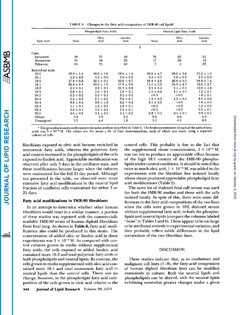
TABLE 6. Changes in the fatty acid composition of IMR-90 cell lipids”
Phospholipid Fatty Acids Neutral Lipid Fatty Acids
Oleic Linoleic Oleic Linoleic Fatty Acid None Acid Acid None Acid Acid
%
Class Saturated 38 37 38 70 63 55 Monoenoic 35 34 20 17 28 19 Polyenoic 24 25 40 7 5 22
Individual acids 16:O 18.0 f 1.4 18.6 f 1.6 18.0 f 1.4 26.2 5 4.7 28.2 3.6 21.2 f 1.3
2.2 f 0.3 16: 1 4.2 t 0.6 4.5 f 0.6 2.0 f 0.2 2.3 f 0.3 2.6 5 0.2 24.8 2 1.4 18:O 17.4 f 0.8 16.4 5 0.4 16.6 f 0.7 34.4 f 2.6 26.9 f 0.5
18: 1 30.4 f 0.4 29.0 f 1.6 17.8 t 0.8 15.1 f 3.2 24.9 f 4.7 16.5 2 3.7 18:2 3.0 2 0.1 2.8 f 0.1 21.3 t 0.4 2.3 2 0.4 2.1 f 0.1 10.0 5 1.4 18:3 0.8 f 0.1 1.0 f 0.1 1.0 f 0.1 1.5 f 0.2 1.1 f 0.1 1.2 f 0.1 20:2 0.3 f 0.2 0.4 f 0.1 3.4 2 0.2 <0.5 <0.5 1.6 f 0.1 20:3 2.4 t 0.2 2.1 f 0.2 3.0 f 0.1 1.9 2 0.3 1.3 t 0.1 4.5 2 0.6 20:4 8.8 f 0.4 9.0 t 1.0 6.2 f 0.4 0.1 t 0.1 <0.5 1.8 t 0.3 22:4 1.1 f 0.1 1.2 f 0.2 1.8 f 0.1 <0.5 <0.5 1.2 t 0.2 22:5 2.4 f 0.1 3.2 f 0.4 1.5 t 0.1 <0.5 <0.5 0.5 f 0.1 2 2 6 4.6 f 0.2 4.4 f 0.5 2.4 2 0.2 0.8 f 0.1 0.4 f 0.1 0.8 f 0.1 Others 3.4 3.0 3.2 9.8 6.6 8.9 Unassigned 3.2 4.4 1.8 5.5 4.4 4.8
(I The proceduresand conditions are the same as those described in Table 2. The final concentration of each of the added fatty M. The values are the mean f SE of four determinations, each of which was made using a separate acids was 5 x
culture of cells
fibroblasts exposed to oleic acid became enriched in monoenoic fatty acids, whereas the polyenoic fatty acid content increased in the phospholipids of the cells exposed to linoleic acid. Appreciable modification was observed after only 3 days in the confluent state, and these modifications became larger when the cultures were maintained for the full 2 1 day period. Although not presented in the table, we observed even more extensive fatty acyl modifications in the neutral lipid fraction of confluent cells maintained for either 3 or 21 days.
Fatty acid modifications in IMR-90 fibroblasts In an attempt to determine whether other human
fibroblasts would react in a similar manner, a portion of these studies was repeated with the commercially available IMR-90 strain of human diploid fibroblasts from fetal lung. As shown in Table 6, fatty acid modi- fications also could be produced in this strain. The concentration of added oleic or linoleic acid in these experiments was 5 x M. As compared with con- trol cultures grown in media without supplemental fatty acids, the cells exposed to added linoleic acid contained more 18:2 and total polyenoic fatty acids in both phospholipids and neutral lipids. By contrast, the cells grown in media supplemented with oleic acid con- tained more 18:l and total monoenoic fatty acid in neutral lipids than the control cells. There was no change, however, in the phospholipid fatty acid com- position of the cells grown in oleic acid relative to the 544 Journal of Lipid Research Volume 20, 1979
control cells. This probably is due to the fact that the supplemental oleate concentration, 5 x M, was too low to produce an appreciable effect because of the high 18:l content of the IMR-90 phospho- lipids under control conditions. It should be noted that twice as much oleic acid, 1 x M, was added in the experiments with the fibroblast line isolated locally where oleate produced appreciable phospholipid fatty acid modifications (Table 2).
The same lot of dialyzed fetal calf serum was used for both the IMR-90 studies and those with the cells isolated locally. In spite of this, there were some dif- ferences in the fatty acid compositions of the two lines when the cells were grown in 10% dialyzed serum without supplemental fatty acid, in both the phospho- lipids and neutral lipids (compare the columns labeled “none” in Tables 1 and 6). These appear to be too large to be attributed entirely to experimental variation, and they probably reflect subtle differences in the lipid metabolism of the two fibroblast lines.
DISCUSSION
These studies indicate that, as in nonhuman and malignant cell lines (1-8), the fatty acid composition of human diploid fibroblast lines can be modified extensively in culture. Both the neutral lipids and phospholipids can be altered, with the neutral lipids exhibiting somewhat greater changes under a given
by guest, on January 3, 2019w
ww
.jlr.orgD
ownloaded from
set of conditions. Almost all of the neutral lipid fatty acids were present in triglycerides, and a net accumu- lation of triglycerides occurred when the cells were grown in high concentrations of fatty acids. These triglycerides were formed primarily from the supple- mental fatty acids added to the medium. By contrast, there was no appreciable increase in phospholipid content when the cells were grown in the presence of high concentrations of fatty acid. Therefore, the changes in composition of the phospholipid fraction cannot be explained simply by accumulation of excess material. One might expect that a cell would exert fairly rigid control over its phospholipid fatty acids because changes in the composition of this fraction have been associated with effects on membrane func- tion (8-13). In fact, we questioned whether the ex- tensive phospholipid fatty acyl replacements that have been observed previously might reflect to some extent a regulatory defect in the malignant and continuously cultured murine and rat lines that so far have been tested (1-8). The present results clearly indicate that this is not the case and that equally extensive changes can be produced in a human diploid cell.
The mechanism whereby exogenous free fatty acids produce alterations in the cellular fatty acid composi- tion can be deduced from the metabolic studies that have been reported with human fibroblasts. These cells can synthesize fatty acids de novo, but this is a regulated pathway that is inhibited by 26-67% when fatty acids are added to the culture medium (38). The reduction in fatty acid biosynthesis is compensated for by an uptake of fatty acid from the extracellular fluid (39). Overcompensation apparently occurs when high concentrations of fatty acid are available in the medium, leading to triglyceride accumulation (Table 3). Since human fibroblasts contain elongation enzymes (40), some of the fatty acid that is taken up from the medium is structurally modified. This accounts for the increase in 20:2 that occurred when linoleate was available (Table l), as well as the increase in 22:4 that occurred when arachidonate was available (Table 2). Since human fibroblasts also are reported to contain A5- and A6-desaturases (40), it is not clear why there was no increase in the desaturation products of linoleate when the medium was supplemented with this fatty acid. Even more puzzling is the 50% decrease in 20:4 in phospholipids when high concentrations of its pre- cursor, linoleate, were available (Table 1). Since similar effects occurred when the IMR-90 cells were exposed to linoleate, this finding is not a peculiar property of the fibroblast line that was isolated locally. It should be noted, however, that the 20:4 content of the neutral lipids did not change appreciably under these conditions (Table 1).
The fact that the phospholipids of the confluent
fibroblasts could be modified just as extensively as those of the rapidly growing cells suggests that some membrane lipid turnover occurs even in fairly dense monolayers. Since a low mitotic index and a small amount of [3H]thymidine incorporation was observed in these confluent cultures, it is possible that some of the observed fatty acid incorporation was due to cell growth rather than turnover of existing membrane lipids in stationary cells. On the other hand, a rapid turnover of phospholipid fatty acyl groups has been reported in stationary Ehrlich ascites cells (41) and in monolayers of BHK-21 cells (42). This, coupled with the fact that extensive phospholipid fatty acid modification occurred after only 3 days in the confluent state under conditions where the cultures were not refed, suggests that at least some of the modification resulted from membrane lipid turnover. As opposed to this picture of continuing phospholipid fatty acid turnover, the phospholipid fatty acids of the mouse L-fibroblast appear to be quite stable (43). Even in the case of the L-cell, however, phospholipid stability is perturbed when fatty acids are added to the culture medium, and less of the original fatty acyl groups are retained under these conditions (44). The ability to produce similar types of modifications in rapidly growing and confluent human fibroblasts provides an approach to the important question of whether the functional consequences of a given lipid modification might depend on the growth status of the culture. Metabolic studies with cells modified by these pro- cedures should provide valuable information concern- ing this fundamental point.
High free fatty acid concentrations were used in much of this work in an attempt to produce maximal fatty acid modifications in the cells. Except for the cytoplasmic inclusions, which probably are triglyceride droplets (36, 37), no morphological abnormalities were detected by light microscopy. Yet, decreased growth occurred when the medium contained high concentrations of palmitate, linolenate, or arachidonate. In the latter two cases, peroxidation products may have been generated in the medium due to the presence of 02, and this could have produced the inhibitory effect. Such an explanation, however, cannot be in- voked in the case of palmitate. Since the molar ratio of free fatty acid to albumin present in the fetal calf serum never exceeded 6, most of the fatty acid added to the medium probably was bound to proteins and, therefore, should not have produced damaging ef- fects (45). Although direct toxicity of the high free fatty acid concentration cannot be ruled out, it is pos- sible that other mechanisms such as the accumulation of phospholipids or triglycerides of a certain fatty acid composition were responsible for the reduction in growth rate. In this regard, recent studies with
Spector et al. Fatty acid-modified cells 545
by guest, on January 3, 2019w
ww
.jlr.orgD
ownloaded from

cultured skin fibroblasts suggest that triglyceride buildup, of itself, can have damaging effect on the cells (46). In addition, reduced growth of mouse LM cells is thought to result from an imbalance in satu- rated fatty acid content of membrane phospholipids (47). Saturated fatty acids usually account for only 35% of the phospholipid fatty acyl groups in the LM cell. This increases when the medium is supplemented with saturated fatty acids, and growth is inhibited severely when it rises to 50%. In LM cell cultures, addition of 5 x to 1 X M palmitate reduces growth and causes the saturated fatty acyl content of the phospholipids to increase, in agreement with the present findings. Therefore, this type of mechanism also may be responsible for the growth inhibition produced by palmitate supplementation in the human fibroblast cultures. As opposed to the present findings with the fibroblasts, however, supplementation with up to 1 x M linolenic or arachidonic acids did not affect the growth of the LM cell cultures (47).
In spite of the problems encountered with several of the fatty acids, this system still provides a means for determining the extent to which fatty acid com- position can influence the properties of a human diploid cell. Cultured human fibroblasts contain a wide range of metabolic pathways, and many mutant strains are available. Therefore, the system lends itself to an examination of fatty acid effects on many cellular functions, such as receptor interactions, transport, and the regulation of metabolic processes that take place on or within membranes. Although this type of information has recently become available for some malignant and animal cell lines in continuous culture (9- 14), it is important to determine whether a human diploid cell will respond in a similar manner.m
This research was supported by an Arteriosclerosis Special- ized Center of Research grant from the National Heart, Lung and Blood Institute, National Institutes of Health No. HL 14,230. Manuscript received 4 August 1978; accepted 14 December 1978
REFERENCES
1. Gerschenson, L. E., J . F. Mead, I. Harary, and D. F. Haggerty, Jr. 1967. Studies on the effects of essential fatty acids on growth rate, fatty acid composition, oxidative phosphorylation and respiratory control of HeLa cells in culture. Biochim. Biophys. Acta. 131: 42-49.
2. Ferguson, K. A., M. Glaser, W. H. Bayer, and P. R. Vagelos. 1975. Alteration of fatty acid composition of LM cells by lipid supplementation and temperature. Biochemistry. 14: 146- 151.
3. Williams, R. E., B. J. Wisnieski, H. G. Rittenhouse, and
C. F. Fox. 1974. Utilization of fatty acid supplements by cultured animal cells. Biochemistv. 13: 1969- 1977.
4. Mulligan, J. J., R. D. Lynch, E. E. Schneeberger, and R. P. Geyer. 1977. Utilization of exogenous linolenic and oleic acids for plasma membrane phosphoglyceride synthesis in L-fibroblasts. Bwchim. Biophys. Acta. 470:
5. Horwitz, A. F., M. E. Hatten, and M. M. Burger. 1974. Membrane fatty acid replacements and their effect on growth and lectin-induced agglutinability. Proc. Natl.
6. Awad, A. B., and A. A. Spector. 1976. Modification of the fatty acid composition of Ehrlich ascites tumor cell plasma membranes. Biochim. Biophys. Acta. 426: 723-73 1.
7. Burns, C. P., D. G. Luttenegger, S-P. L. Wei, and A. A. Spector. 1977. Modification of the fatty acid composition of L1210 murine leukemia cells. Lipids. 12: 747-752.
8. Horwitz, A. F., A. Wright, P. Ludwig, and R. Cornell. 1978. Interrelated lipid alterations and their influence on the proliferation and fusion of cultured myogenic cells. J. Cell Biol. 77: 334-357.
9. Coleman, R. 1973. Membrane-bound enzymes and membrane ultrastructure. Biochim. Biophys. Acta. 300:
10. Shinitzky, M., and B. Rivnay. 1977. Degree of exposure of membrane proteins determined by fluorescence quenching. Biochemistry. 16: 982-986.
11. Maccecchini, M-L., and M. M. Burger. 1977. Stimula- tion of lymphocytes by concanavalin A. Temperature- dependent effect of fatty acid replacements. Biochim. Biophys. Acta. 469: 33-44.
12. Mahoney, E. M., A. L. Hamill, W. A. Scott, and Z. A. Cohn. 1977. Response of endocytosis to altered fatty acyl composition of macrophage phospholipids. Proc. Natl. Acad. Sci. USA. 74: 4895-4899.
13. Kaduce, T. L., A. B. Awad, L. J. Fontenelle, and A. A. Spector. 1977. Effect of fatty acid saturation on a- aminoisobutyric acid transport in Ehrlich ascites cells. J. Biol. Chem. 252: 6624-6630.
14. Brenneman, D. E., T. Kaduce, and A. A. Spector. 1977. Effect of dietary fat saturation on acylcoenzyme A: cholesterol acyltransferase activity of Ehrlich cell microsomes.]. Lipid Res. 18: 582-591.
15. Nichols, W. W., D. G. Murphy, V. J. Cristofalo, L. H. Toji, A. E. Greene, and S. A. Dwight. 1977. Characteri- zation of a new human diploid cell strain, IMR-90. Science. 196: 60-63.
16. Eagle, H. 1955. Propagation in a fluid medium of a human epidermoid carcinoma strain KB. Proc. Soc. Exp. Biol. Med. 89: 362-364.
17. Eagle, H. 1959. Amino acid metabolism in mammalian cell culture. Science. 130: 432-437.
18. Spector, A. A., D. Steinberg, and A. Tanaka. 1964. Uptake of free fatty acids by Ehrlich ascites tumor cells.
J . Biol. Chem. 240: 1032-1041. 19. Lowry, R. R., and I. J. Tinsley. 1975. Rapid colorimetric
determination of free fatty acids.]. Am. Oil Chem. Sac. 53: 470-472.
20. Gornall, A. G., C. J . Bardawill, and M. M. David. 1949. Determination of serum proteins by means of the biuret reaction.]. Biol. Chem. 177: 751-766.
21. Schaeffer, W. I. 1978. Usage of vertebrate cell, tis- sue and organ culture terminology. Tissue Culture Association Manual. 4: 779-782.
92- 103.
Acad. Sci. USA. 71: 3115-3119.
1-30.
546 Journal of Lipid Research Volume 20, 1979
by guest, on January 3, 2019w
ww
.jlr.orgD
ownloaded from

22. Martinez-Lopez, G., and L. M. Black. 1973. A simple and accurate method for the measurement of the growth of cell monolayers.
23. Lees, M. B., and S. Paxman. 1972. Modification of the Lowry procedure for the analysis of proteolipid protein.
24. Schimmel, S. D., C. Kent, R. Bischoff and P. R. Vagelos. 1973. Plasma membranes from cultured muscle cells: Isolation procedure and separation of putative plasma- membrane marker enzymes. Proc. Natl. Acad. Sci.
25. Folch, J., M. Lees, and G. H. Sloane Stanley. 1957. A simple method for the isolation and purification of total lipids from animal tissues. J . Biol. Chem. 226:
26. Raheja, R. K., C. Kaur, A. Singh, and I. S. Bhatia. 1973. New colorimetric method for the quantitative estimation of phospholipids without acid digestion. J . L i p d Res. 14: 695-697.
27. Kessler, G., and H. Lederer. 1966. Fluorometric measurement of triglycerides. In Automation in Analyt- ical Chemistry. Technicon Symposium. L. T. Skeggs, Jr., editor. Mediad, New York. 341-344.
28. Driscoll, J. L., D. Aubuehon, M. Descoteaux, and H. F. Martin. 1971. Semiautomated, specific routine serum cholesterol determination by gas-liquid chromatog- raphy. Anal. Chem. 43: 1196- 1200.
29. Lillienberg, L., and A. Svanborg. 1976. Determination of plasma cholesterol: Comparison of gas-liquid chromatographic, colorimetric and enzymatic analyses. Clin. Chim. Acta. 68: 223-233.
30. Laurell, S., and G. Tibbling. 1967. Colorimetric micro-determination of the free fatty acids in plasma. Clin Chim. Acta. 16: 57-62.
31. Brown, J. L., and J. M. Johnston. 1962. Radioassay of lipid components separated by thin-layer chromatog- raphy. J. L i p d Res. 3: 480-481.
32. Garland, P. B., and P. J. Randle. 1963. Effects of alloxan diabetes and adrenaline on concentrations of free fatty acids in rat heart and diaphragm muscles. Nature. 199: 381-382.
33. Morrison, W. R., and L. M. Smith. 1964. Preparation of fatty acid methyl esters and dimethylacetals from lipids with boron fluoride-methanol. J. L i p d Res. 5:
34. Brown, M. S., S. E. Dana, and J. L. Goldstein. 1974. Regulation of 3-hydroxy-3-methylglutaryl coenzyme A reductase activity in cultured human fibroblasts. Com-
Vitro. 9: 1-7.
A d . B i o c h a . 47: 184- 192.
USA. 70: 3195-3199.
497-509.
600- 608.
parison of cells from a normal subject and from a pa- tient with homozygous familial hypercholesterolemia. J . Biol. Chem. 249: 789-796.
35. Rothblat, G. H., L. Y. Arbogast, L. Ouellette, and B. V. Howard. 1976. Preparation of delipidized serum pro- tein for use in cell culture systems.In Vitro. 12: 554-557.
36. Mackenzie, C. G., J. B. Mackenzie, and 0. K. Riess. 1967. Increase in cell lipid and cytoplasmic particles in mammalian cells cultured at reduced pH. J . L i p d Res. 8: 642-645.
37. Schneeberger, E. E., R. D. Lynch, and R. P. Geyer. 197 1 . Formation and disappearance of triglyceride droplets in strain L fibroblasts. Exp. Cell Res. 69:
38. Jacobs, R. A., and P. W. Majerus. 1973. The regulation of fatty acid synthesis in human skin fibroblasts. In- hibition of fatty acid synthesis by free fatty acids. J . Biol. Chem. 248: 8392-8401.
39. Howard, B. V., and D. Kritchevsky. 1969. The source of cellular lipid in the human diploid cell strain W1-38. Biochim. Biophys. Acta. 187: 293-301.
40. Dunbar, L. M., and J. M. Bailey. 1975. Enzyme deletions and essential fatty acid metabolism in cultured cells. J . Biol. Chem. 250: 1152- 1153.
41. Spector, A. A., and D. Steinberg. 1967. Turnover and utilization of esterified fatty acids in Ehrlich ascites tumor cells. J . Biol. Chem. 242: 3057-3062.
42. Gallaher, W. R., and H. A. Blough. 1975. Synthesis and turnover of lipids in monolayer cultures of BHK-2 1 cells. Arch. Biochem. Biophys. 168: 104- 114.
43. Lynch. R. D., E. E. Schneeberger, and R. P. Geyer. 1976. Complete retention of phospholipid acyl groups by mammalian cells in culture. Biochemistry. 15: 193- 200.
44. Tsai, P-Y., and R. P. Geyer. 1977. Fatty acid synthesis and metabolism of phospholipid acyl groups in strain L mouse fibroblasts. Biochim. Bwphys. Acta. 489:
45. Spector, A. A. 1975. Fatty acid binding to plasma albumin. J . L i p d Res. 16: 165- 179.
46. Rosenthal, M. D., and R. P. Geyer. 1978. Incorporation of moderate and excessive levels of fatty acids by human skin fibroblasts. Federation Proc. 37: 1832 (Abst.).
47. Doi, O., F. Doi, F. Schroeder, A. W. Alberts, and P. R. Vagelos. 1978. Manipulation of fatty acid composition of membrane phospholipid and its effects on cell growth in mouse LM cells. Biochim. Biophys. Acta. 509: 239-250.
193-206.
38 1 - 389.
Spector et al. Fatty acid-modified cells 547
by guest, on January 3, 2019w
ww
.jlr.orgD
ownloaded from


![Fatty acids from fat cell lipolysis do not activate an ......E-ZAmri(CNRS,UniversityofNice-SophiaAntipolis,Nice, France) were cultured and differentiated as previously de-scribed [16].](https://static.fdocuments.in/doc/165x107/60d5d034ddec376fe92041e2/fatty-acids-from-fat-cell-lipolysis-do-not-activate-an-e-zamricnrsuniversityofnice-sophiaantipolisnice.jpg)