Chronic fibroblasts cultured in 3D collagen matrices as an...
40
UPTEC X 08 050 Examensarbete 30 hp December 2008 Chronic fibroblasts cultured in 3D collagen matrices as an in vitro model for isolated healing processes in a chronic wound Emma Stegberg
Transcript of Chronic fibroblasts cultured in 3D collagen matrices as an...

UPTEC X 08 050
Examensarbete 30 hpDecember 2008
Chronic fibroblasts cultured in 3D collagen
matrices as an in vitro model for isolated
healing processes in a chronic wound
Emma Stegberg

Molecular Biotechnology Programme Uppsala University School of Engineering
UPTEC X 08 050 Date of issue 2008-12 Author
Emma Stegberg
Title (English) Chronic fibroblasts cultured in 3D collagen matrices as an in vitro
model for isolated healing processes in a chronic wound
Title (Swedish) Abstract
Wound healing is a complex process which involves numerous cell types, extracellular matrix components and cytokines. Chronic wounds fail to proceed through the normal phases of healing and are instead locked in a state of pathologic inflammation. In order to understand the underlying mechanisms causing the chronicity and to develop new and more effective treatments there is an immense need for improved models for chronic wounds.
In the present project, a three dimensional in vitro model of dermal like tissue in chronic wounds has been developed. The model was utilized for the investigation of the differences in cell behavior between chronic dermal fibroblasts and normal dermal fibroblasts on cellular processes important for dermal repair and regeneration. Furthermore, the effect of a wound treatment substance, enamel matrix derivative (EMD), on these cellular processes has been examined.
Keywords
3D dermal model, wound healing, chronic wounds, enamel matrix derivative (EMD)
Supervisors Maria Werthén
Mölnlycke Health Care AB
Scientific reviewer Peter Thomsen
Gothenburg University
Project name
Sponsors
Language English
Security
ISSN 1401-2138
Classification
Supplementary bibliographical information Pages 38
Biology Education Centre Biomedical Center Husargatan 3 Uppsala Box 592 S-75124 Uppsala Tel +46 (0)18 4710000 Fax +46 (0)18 555217

Chronic fibroblasts cultured in 3D collagen matrices as an in vitro model for isolated healing processes in a chronic
wound
Emma Stegberg
Sammanfattning
Sårläkning är en komplicerad process som involverar åtskilliga celltyper, vävnads-komponenter och signalmolekyler. Kroniska sår karaktäriseras av en oförmåga att fort-skrida genom läkningsprocessen, beroende av flera olika faktorer där infektion, syrebrist och försämrad blodförsörjning bara är några. För att kunna undersöka mekanismerna som orsakar kroniska sår och för att kunna utveckla nya och mer effektiva behandlings-metoder så krävs förbättrade modeller av dessa sår. I detta projekt utvecklades en tredimensionell modell av dermis i kroniska sår. Modellen användes för att analysera skillnaderna i beteende mellan kroniska och normala hud-celler vad gäller de processer som är viktiga vid sårläkningen i huden. Dessutom under-söktes effekten av en sårläkningsprodukt, Xelma®, på dessa processer. Resultaten visar att kroniska hudceller har nedsatt funktionalitet i flera av de undersökta processerna i jämförelse med normala hudceller. Vid behandling med Xelma® stimuleras de kroniska cellerna till ett mer normalt beteende, vilket visar att Xelma® är en produkt med bra potential att läka kroniska sår.
Examensarbete 20p
Civilingenjörsprogrammet Molekylär bioteknik
Uppsala universitet december 2008

CONTENTS
ABBREVIATIONS .......................................................................................................................................... 1
1. INTRODUCTION ........................................................................................................................................ 2
2. AIM ............................................................................................................................................................. 3
3. BACKGROUND.......................................................................................................................................... 4
3.1. The skin ..........................................................................................................................................................................4
3.2. Cutaneous wound healing .............................................................................................................................................5
3.2.1. Inflammation ..........................................................................................................................................................5
3.2.2. Proliferation............................................................................................................................................................6
3.2.3. Remodeling............................................................................................................................................................6
3.3. Chronic wounds..............................................................................................................................................................8
3.3.1. Chronic fibroblasts.................................................................................................................................................8
3.4. Models for wound healing..............................................................................................................................................9
3.4.1. The fibroblast-populated collagen matrix (FPCM)................................................................................................9
3.5. Extracellular matrix proteins ........................................................................................................................................10
3.5.1. Collagen...............................................................................................................................................................10
3.5.2. Enamel matrix derivative (EMD) .........................................................................................................................11
3.6. Cell mediators ..............................................................................................................................................................12
4. MATERIALS AND METHODS ................................................................................................................. 13
4.1. Materials .......................................................................................................................................................................13
4.2. Cell culture....................................................................................................................................................................13
4.3. Fabrication of collagen matrices..................................................................................................................................14
4.4. Proliferation analysis ....................................................................................................................................................14
4.5. Histology analysis ........................................................................................................................................................15
4.6. Morphology analysis in 2D and 3D..............................................................................................................................15
4.7. Cytokine analysis .........................................................................................................................................................16
4.8. Contraction analysis.....................................................................................................................................................17
5. RESULTS ................................................................................................................................................. 18
5.1. Proliferation analysis ....................................................................................................................................................18
5.2. Cellular morphology analysis.......................................................................................................................................20
5.3. Matrix contraction analysis ..........................................................................................................................................21
5.4. Histology analysis ........................................................................................................................................................23
5.5. Cytokine analysis .........................................................................................................................................................25
6. DISCUSSION............................................................................................................................................ 27
6.1. Proliferation analysis ....................................................................................................................................................27
6.2. Cellular morphology analysis.......................................................................................................................................28
6.3. Matrix contraction and histology analyses ..................................................................................................................29
6.4. Cytokine analysis .........................................................................................................................................................31
7. CONCLUSION.......................................................................................................................................... 32
8. ACKNOWLEDGEMENTS......................................................................................................................... 33
9. REFERENCES ......................................................................................................................................... 34

ABBREVIATIONS
1
ABBREVIATIONS
BSA Bovine serum albumin CRL Chronic human dermal fibroblasts DAPI 4',6-diamidino-2-phenylindole DMEM Dulbecco’s modified Eagle’s medium ECM Extracellular matrix EDTA Ethylenediaminetetraacetic acid EMD Enamel matrix derivative FBS Fetal bovine serum FITC Fluorescein isothiocyanate FPCM Fibroblast-populated collagen matrix HAc Acetic acid HBSS Hanks’ balanced salt solution IL Interleukin MCP-1 Monocyte chemoattractant protein 1 MMP Matrix metalloproteinase NaOH Sodium hydroxide NHDF Normal human dermal fibroblasts PBS Phosphate buffered saline PDGF Platelet derived growth factor RPE R-Phycoerythrin TGF-β Transforming growth factor β TNF-α Tumor necrosis factor α VEGF Vascular endothelial growth factor
INTRODUCTION
2
1. INTRODUCTION
Wound healing is a complex process which involves cell proliferation and migration, matrix synthesis and contraction and the regulation of these events by the synthesis and release of numerous growth factors and cytokines.1 Chronic wounds, contrary to acute wounds, fail to proceed through the normal phases of healing and are instead locked in a state of pathologic inflammation. The healing process is consequently delayed and incomplete. Impaired healing may be evoked by numerous of different reasons; diabetes, venous stasis and pressure being the most common.2 To be able to analyze the different aspects of the wound healing process and the effect of wound healing treatments, improved models are needed. Numerous models have been developed, both in vitro and in vivo. However, cells cultured in vitro have demonstrated a different behavior compared to cells in vivo, especially cells cultured in monolayers. When modeling chronic wound healing in vitro it is therefore essential to create an environment with conditions similar to the tissues naturally surrounding the cells.3 Fibroblasts, the most abundant cell type in human dermis, are of utmost importance in cutaneous wound healing.4 The fibroblast-populated collagen matrix has therefore been utilized as an in vitro model for wound healing. It provides a reasonable representation of the dermal granulation tissue in a healing wound.5 Xelma® is a new wound healing treatment for hard to heal wounds (Mölnlycke Health Care AB, Gothenburg, Sweden). The active component in this product is enamel matrix derivative (EMD), a mixture of hydrophobic proteins extracted from developing porcine teeth. The principal component of EMD is amelogenins.6 Under physiological conditions, EMD resembles a stable extra cellular matrix and is believed to serve as a temporary matrix for cell attachment and migration. Consequently, in chronic wounds where the extracellular matrix (ECM) is deficient, these proteins have a potential to promote cutaneous wound healing processes.7,8 EMD has been shown to be advantageous in the treatment of chronic venous leg ulcers 8,9 and to accelerate wound closure in rabbits.6 These proteins have demon-strated an ability to increase cell proliferation and contraction of three dimensional matrices and are also shown to stimulate the production of certain growth factors important for dermal repair and regeneration.6,10

AIM
3
2. AIM
The objectives of this thesis project were to: 1. Improve a three dimensional in vitro model of dermal like tissue in chronic
wounds and investigate the differences in cell behavior between chronic human dermal fibroblasts and normal human dermal fibroblasts on cellular processes important for dermal repair and regeneration.
2. Analyze the effect of a wound treatment substance, EMD, on these cellular
processes.

BACKGROUND
4
3. BACKGROUND
3.1. The skin
The skin is the largest organ of the body, covering an area of about two square meters in adults.11 The primary function of the skin is to serve as a barrier, protecting the body against noxious external conditions and preserving the balanced internal environment. It enables prevention of liquid loss, regulation of body temperature, transduction of cutaneous sensations and vitamin D synthesis.11,12 The skin consists of two different tissue layers: the outermost thin epidermis and the inner thicker dermis. The epidermis is a stratified epithelium, hosting four different cell types. The keratinocytes which are the most abundant, produce the fibrous protein keratin and differentiate from basal cells into dead corneocytes while migrating upwards. The epidermis can be subdivided into four layers based on the maturation of the keratinocytes. Aggregation of the keratin fibrils inside the cells gives the epidermis its strength to withstand damage.11,12 Langerhans cells are immunologically active cells with the ability to catch, process and present exo-genous antigens and are an important part of the immune system. Melanocytes synthesize the pigment melanin which protects the skin from harmful ultraviolet radiation. Merkel cells are located near neurons and act as transducers for the sense of touch.11,12 The epidermis and the dermis are separated by the basal membrane which functions as a semipermeable filter for transfer of nutrients and cells from dermis to epidermis. Furthermore, it provides mechanical support and cell adhesion sites.11,13 The dermis is a connective tissue mainly composed of collagen and elastin in a matrix of glucosaminoglycans. The proteins provide tensile strength and elasticity while the ground substance is responsible for the viscosity and hydration. The different components of the extracellular matrix are synthesized by fibroblasts which are the most abundant cell type in the dermis. Other cell types are lymphocytes, macrophages and mast cells. Contrary to epidermis where cells are closely adherent, the dermis shelters only few cells sparsely located in the extracellular matrix. In addition, the dermis contains blood vessels, nerve fibers, lymphatics, muscles, glands and hair follicles. Beneath, the dermis is attached to the hypodermis, a subcutaneous layer which mainly consists of connective tissue and fat.11,12 The structure of the skin is shown in figure 1.

BACKGROUND
5
Figure 1. Structure of the skin. Modified from wikipedia.14
3.2. Cutaneous wound healing
Wound healing is a complex process which involves cell proliferation and migration, matrix synthesis and contraction and the regulation of these events by the synthesis and release of numerous growth factors and cytokines. The process is traditionally divided into three overlapping phases: Inflammation, proliferation and remodeling.1,15 The main outlines of these phases will be described here.
3.2.1. Inflammation
Tissue injury usually causes the disruption of blood vessels and leakage of blood constituents. The immediate response to reestablish homostasis is the formation of a clot. Capillary permeability increases at first, to allow neutrophils, lymphocytes, monocytes and platelets to migrate into the wound and release chemokines. This results in vasoconstriction and prevention of further blood loss.1,16 Coagulation occurs as platelets aggregate with fibrin fibers, derived from the cleavage of fibrinogen by thrombin. The network formed serves as a shelter for temporary protection of the open wound and as a matrix for cell migration.16

BACKGROUND
6
Platelets release several factors, including transforming growth factor β (TGF- β) and platelet derived growth factor (PDGF) which attract neutrophils and monocytes to the wound site. These cells cleanse the injured area from contaminating bacteria and foreign particles.1,17 When the monocytes enter the wound site they become activated macrophages which phagocytose remaining pathogens, other cells and matrix debris. Furthermore, they secrete growth factors such as PDGF and vascular endothelial growth factor (VEGF), thus stimulating fibroblast recruitment and angiogenesis.1,16,17
3.2.2. Proliferation
During the process of reepithelialization, i.e. formation of a new epidermal layer, the keratinocytes migrate over the provisional wound matrix, leaving a stratified layer of proliferating cells and an underlying basal lamina behind. To be able to move through the dense provisional matrix, the keratinocytes utilize specific enzymes, including matrix metalloproteinases (MMP), which degrade matrix proteins. Once a monolayer of keratinocytes has been produced, the epidermal migration ceases.16,18 Fibroblast migration is stimulated by a vast number of cytokines and growth factors. The cells navigate along the provisional matrix fibers and commence to synthesize components of a permanent extracellular matrix, including collagen, glucosamino-glycans and proteoglycans, but also growth factors.18 The new granulation tissue replaces the provisional matrix and numerous new capillaries are created.17 Furthermore, the healing process comprises contraction of the wound. This involves myofibroblasts, a specific type of differentiated fibroblasts which possess contractile forces and therefore are able to reapproximate wound edges. The contraction process is a complex interaction of cells, the extracellular matrix and cytokines.17,18
3.2.3. Remodeling
The remodeling phase of repair implies a tightly coordinated equilibrium between collagen synthesis and catabolism.1 The degradation of collagen is performed by matrix metalloproteinases (MMP), produced by macrophages, epidermal cells, endo-thelial cells and fibroblasts.17 Over time, the proportion of collagen type I increases with the corresponding decrease in collagen type III, proteoglycans and water. Collagen fibrils become more organized and the number of intermolecular cross-links augments.18 This re-modeling persists for several months after wound closure but nevertheless, tensile strength of the remodeled tissue only reaches a maximum of about 70 – 80 % of that of unwounded tissue.1,18

BACKGROUND
7
At the end of the wound healing process, most of the cells in the wound tissue undergo apoptosis and a relatively acellular scar is formed. The molecular mechan-isms of this regulation are still not fully elucidated.17 A summary of the wound healing process is shown in figure 2.
Figure 2. Summary of the wound healing process. (a) Immediately after cutaneous damage neutrophiles, monocytes and platelets migrate into the wound from disrupted blood vessels. (b) Blood coagulation occurs as platelets aggregate with fibrin, a protein derived from the cleavage of fibrinogen by thrombin. (c) Platelets release several factors, including transforming growth factor β (TGF- β) and platelet-derived growth factor (PDGF) which attract neutrophils and (d) monocytes to the wound site. These cells cleanse the injured area from debris and secrete growth factors. (e) Fibroblasts are recruited and commence to synthesize collagen. (f) The tissue is remodeled through collagen crosslinking and reorganization for months after injury. Modified from Beanes et al. (2003).
BACKGROUND
8
3.3. Chronic wounds
Chronic wounds, contrary to acute wounds, fail to proceed through the normal phases of healing and are instead locked in a state of pathologic inflammation. The healing process is delayed, incomplete and consequently, the outcome is poorly functional.2 Impaired healing may be caused by numerous different reasons; diabetes, venous stasis and pressure being the most common. Ischemia, the shortage of blood supply, is generally the underlying cause although several mechanisms have been proposed to explain the chronicity, including microbial contamination and the cellular and systemic effects of aging.2,19 The principal pathological characteristics of a chronic wound are prolonged inflammation, defective extracellular matrix and failed reepithelialization.20 An excessive number of inflammatory cells are present in the tissue and the level of chemokines and pro-inflammatory cytokines, such as tumor necrosis factor α (TNF-α) and interleukin 1 (IL-1) are persistently elevated.2,21 The proteolytic activity is intensified due to an increased concentration of matrix metalloproteinases (MMP), leading to extracellular matrix degradation.21 Moreover, a reduced concentration of certain growth factors, for example transforming growth factor β (TGF- β) 22,23 de-creases the level of mitogenic activity in the wound.2 The result of these conditions is an uncontrolled inflammatory response with additional tissue damage and deterioration of the chronic wound, which further promotes inflammation and prevents wound healing.2
3.3.1. Chronic fibroblasts
Fibroblasts are of utmost importance in cutaneous wound healing. Apart from synthesizing and remodeling the principal components of the extracellular matrix, fibroblasts also produce the mitogens for keratinocytes and endothelial cells.4 Fibroblasts obtained from chronic wounds show decreased proliferation and reduced mitogenic response to exogenous application of growth factors such as platelet derived growth factor-BB (PDGF-BB) and transforming growth factor β (TGF- β).4,21 Besides, migration capacity of these cells is impaired and some studies have demonstrated that the cellular morphology is altered, making them resemble fibroblasts which have become senescent in vitro.2,21 Furthermore, these features are dependent on the duration of the ulcer.4 It remains unclear whether the deterioration of the wound healing activities, such as decreased proliferation and mobility, demonstrate permanent modifications of the cellular phenotype or reversible behaviors caused by the chronic environment.24

BACKGROUND
9
3.4. Models for wound healing
In order to analyze the different aspects of the wound healing process and the effect of wound healing treatments, improved models are needed. Numerous models have been developed, both in vitro and in vivo. Which model to choose depends upon a variety of factors, such as laboratory facilities, financial resources but also ethical and legal aspects.3 The main advantage of an in vivo model in wound healing research is the similarity to wounds in clinical practice. In the case of cutaneous wounds, studies can even be performed in humans. The disadvantages are the difficulties to examine single tissue components without influencing others, a consequence of the inherent hetero-geneity of in vivo models.3 In vitro models are generally rapid, less costly and involve minimal ethical and legal considerations in comparison with in vivo models. Another advantage is the simplicity which provides the possibility to directly evaluate the effect of a single substance in the tissue. The major drawback is the difficulty of extrapolating the in vitro results to the in vivo situation.3 In vitro models can be divided according to the difference in complexity, ranging from single cell systems in two dimensions to multicellular systems in three dimensions and organ cultures.3 Culturing cells in monolayers is the simplest technique of modeling wound tissue and has until recently been the primary approach applied.3 A significant and inherent disadvantage with this model is the absence of dorsal cell adhesion points. These conditions are therefore an unfair representation of the tissue environment, particularly improper for cells that naturally are completely surrounded by a matrix that can be remodeled, e.g. fibroblasts.25,26 Cells cultured in three dimensional matrices behave very differently when compared to cells in two dimensional environments. Remarkable changes in cell morphology, matrix adhesion, migration strategies, gene and protein expression are reported, among others.26 Although the underlying mechanisms of these differences remain unclear, dimensionality and compliance of the matrix are the plausible key factors.26
3.4.1. The fibroblast-populated collagen matrix (FPCM)
The fibroblast-populated collagen matrix (FPCM) has earlier been utilized as an in vitro model for wound healing. It provides a reasonable representation of the dermal granulation tissue in a healing wound.5

BACKGROUND
10
The major disadvantage as well as the advantage of this model is its simplicity. Granulation tissue is a complex composition of several cell types and numerous proteins including fibrin, fibronectin and vitronectin as well as collagen, hyaluronan and a variety of growth factors and release products. There is a continuous evolution of the matrix and as the concentration of the components change, the granulation tissue matures.5 The FPCM on the other hand, consists of only one cell type, fibroblasts, and initially there is only one protein, collagen. However, incubation of this matrix in the presence of serum provides a source of some extracellular matrix molecules. Besides, fibroblasts cultured in this milieu produce a variety of proteins which also are incorporated into the matrix, creating a model with the ability to mature in a similar way as granulation tissue. The most significant matrix constituent absent in the FPCM is the fibrin network, which initiates the wound healing process in vivo.5 Consequently, the simplicity of the fibroblast-populated collagen matrix in comparison with granulation tissue in a healing wound is advantageous because it enables experimental precision. Nevertheless, it is difficult to extrapolate the in vitro results to the much more complex in vivo situation.5
3.5. Extracellular matrix proteins
The extracellular matrix is a complex, heterogeneous, dynamic and tissue specific composition of different biopolymers and water, including proteins and glucos-aminoglycans.22,26 The two key extracellular matrix proteins of the present project are described below.
3.5.1. Collagen
Collagen is the most abundant protein in the body and exists in at least ten different forms in tissues of vertebrates.26,27 The collagen in skin, bone and tendon is pre-dominantly type I collagen. Type II collagen is the major constituent of cartilage whereas type III collagen is predominant in blood vessel walls.27 The tropocollagen is the essential unit of a collagen fiber and consists of three polypeptide strands twisted into a triple helix. The subunits are synthesized and processed intracellularly, subsequently exported to the extracellular matrix where they self-assemble into the triple helical collagen molecule. Several molecules then aggregate into microfibrils and several microfibrils aggregate to form large collagen fibers.18,27

BACKGROUND
11
Collagen possesses cell-adhesion domain sequences which facilitate cell attachment and allow specific cellular interactions. This feature may be important in preserving the phenotypes and activities of many cell types.26 Collagen is easily purified from animal tissues by acid digestion and forms a gel when returned to neutral pH. By altering the collagen concentration, pH or ionic strength during gelation (polymerization), pore size and fiber diameter can be moderately adjusted. As the gel forms, the microfibrils aggregate laterally into fibers which in turn entangle into a matrix. Reconstituted collagen matrices are mechanically weaker and more highly hydrated than natural tissues.26
3.5.2. Enamel matrix derivative (EMD)
Enamel matrix derivative (EMD) is the active component in Xelma®, a wound healing product for hard to heal wounds (Mölnlycke Health Care AB, Gothenburg, Sweden). In clinical studies, Vowden et al. (2006, 2007) have demonstrated the advantages of EMD in the treatment of chronic venous leg ulcers; the wound area was reduced and the exudate production decreased. EMD are extracted from the enamel matrix of developing porcine teeth.6 It is a mixture of hydrophobic proteins where the principal component is amelogenins. The unique amino acid sequences seem to facilitate aggregation of the proteins into large hydrophobic complexes instead of promoting the formation of the usual secondary structures. Under physiological conditions, these aggregates resemble a stable extra cellular matrix, thus serving as a temporary matrix for cell attachment and migration.7 Topical application of EMD has earlier been utilized in periodontal surgery and in acute periodontal injuries since it demonstrated positive effects on periodontal wound healing and tissue regeneration.28 The molecular mechanisms underlying these results are still not fully elucidated but the presence of amelogenin seems to regulate or induce several essential cell responses, including proliferation, migration and differentiation.7,8 EMD have been shown to enhance cell adhesion of numerous cell types.29 The broad cell-type specificity of these proteins proposed the appliance of EMD on injured skin in order to promote the wound healing processes. Mirastschijski et al. (2003) showed that topical treatment with EMD in full-thickness wounds in rabbits increased formation of granulation tissue and accelerated wound closure. Furthermore, it is demonstrated that incubation of human dermal fibroblasts in vitro with EMD generated a strong biological response including increased cell proliferation and contraction of three-dimensional matrices as well as augmented levels of vascular endothelial growth factor (VEGF) and transforming growth factor β1 (TGF-β1).6,10

BACKGROUND
12
3.6. Cell mediators
Numerous cell mediators are involved in the wound healing processes. Besides several growth factors such as vascular endothelial growth factor (VEGF) and platelet derived growth factor (PDGF), three important cytokines synthesized by fibroblasts are interleukin 6 (IL-6), interleukin 8 (IL-8) and monocyte chemo-attractant protein 1 (MCP-1).30 IL-6 is a proinflammatory cytokine shown to be strongly upregulated during the inflammatory phase of wound healing.30 This protein has demonstrated a mitogenic effect for keratinocytes and a chemoattractive effect on neutrophils and is therefore believed to be essential for initiating the healing process. Complete lack of IL-6 seems to impair wound healing whereas extreme levels of the cytokine are related to scar formation. Thus, a coordinated expression of IL-6 seems to be crucial for normal tissue repair.30 IL-8 is a key mediator of the inflammatory response. This chemotactic cytokine was reported to recruit polymorphonuclear leukocytes to the wound site 30, stimulate keratinocyte proliferation 31 but exhibit an inhibitory effect on wound contraction.30 IL-8 has furthermore demonstrated angiogenetic properties.32 Human chronic wound fluid is shown to contain high levels of this cytokine.30 MCP-1 is another chemotactic cytokine, a major chemoattractant for monocytes and macrophages. Moreover, it is demonstrated to recruit T-cells and mast cells 30, influence endothelial cell migration 33 and stimulate collagen production by fibroblasts.34 Total absence of MCP-1 was suggested to result in reduced collagen synthesis and delayed reepithelialization and angiogenesis.30

MATERIALS AND METHODS
13
4. MATERIALS AND METHODS
4.1. Materials
Normal human dermal fibroblasts (NHDF) were derived from a healthy volunteer and purchased from Karocell (Stockholm, Sweden). Near-senescent human dermal fibroblasts (cell line CRL-7815) were derived from a chronic dermatitis patient and purchased from the American Type Culture Collection (Rockville, MD). The latter cells are here referred to as “chronic” fibroblasts. Dulbecco’s modified Eagles’s medium (DMEM) with phenol red and 1 g/L GlutaMAXTM, Dulbecco’s modified Eagle medium 2x (2x DMEM) without phenol red, Hanks’ balanced salt solution (HBSS) without Ca and Mg, penicillin (100 units/mL), streptomycin (100 µg/mL), trypsin (0.25 %) / EDTA (1 mM), fetal bovine serum (FBS), Trypan Blue Stain and Fluorescein isothiocyanate (FITC)-conjugated phalloidin were purchased from Invitrogen (Paisley, UK). Phosphate buffered saline (PBS) was purchased from Bakteriologiska laboratoriet (Göteborg, Sweden). Rat tail collagen type I, T-25 tissue culture flasks, 24-well plates and Cultureslides were purchased from BD Bioscienses (Erembodegem, Belgium). Collagenase type I clostridium histolyticum and Bovine serum albumin (BSA) were purchased from EMD Biosciences (San Diego, US). Vectashield mounting medium with 4',6-diamidino-2-phenylindole (DAPI) was purchased from Vector Laboratories (Peterborough, UK). Enamel matrix derivative (EMD) was purchased from Biora AB (Malmö, Sweden) and Triton X-100 was purchased from Sigma-Aldrich (Basel, Switzerland).
4.2. Cell culture
Normal human dermal fibroblasts (NHDF) and chronic human dermal fibroblasts (CRL) were maintained and expanded in T-25 tissue culture flasks in Dulbecco’s modified Eagle medium (DMEM) with Glutamax 1 g/L, supplemented with 10 % of fetal bovine serum (FBS) and 1 % of penicillin/streptomycin. Cultures were in-cubated at 37oC with 5 % CO2 and 95 % humidity. Culture medium was changed three times weekly and confluent cultures were washed with Hanks’ balanced salt solution (HBSS, without Ca and Mg) and passaged using trypsin/EDTA (0.25 % trypsin in 1 mM EDTA). Cell viability was determined by means of trypan blue exclusion and cell counts were performed in a counting chamber (Bürker hemo-cytometer). All experiments were performed on cells between passage 4 and 12.

MATERIALS AND METHODS
14
4.3. Fabrication of collagen matrices
Collagen matrices were fabricated according to the manufacturer’s instructions and Grayson et al. (2006) with some modifications. In the optimized method, 10 units of 2x DMEM were mixed with 2 units of FBS, 2 units of 0.1 M NaOH, 4 units of ice cold rat-tail type I collagen solution (stock 10 mg/mL) and 2 units of cells suspended in supplemented DMEM. Acellular matrices were formed by replacing the cell suspension with 2 units of supplemented DMEM. Matrices were cast with 400 µL per well in 24-well plates and with a cell density of 6×10⁴ cells per mL matrix. The plates were incubated for 1 hour at 37oC with 5 % CO2 and 95 % humidity to allow collagen polymerization. Thereafter, 1 mL of supplemented DMEM was added to each well and the plates were incubated for 1, 3 and 7 days at 37oC with 5 % CO2 and 95 % humidity without the change of medium. EMD treatment of matrices was performed by topical addition. Instead of adding 1 mL of supplemented DMEM to each well after polymerization, 1 mL of EMD (stock 20 mg/mL diluted in supplemented DMEM to 0.1 mg/mL and 1 mg/mL) was added to each well. The plates were then incubated for 1, 3 and 7 days as previously described. Moreover, to assure that the mechanical conditions in all matrices would remain the same, wells were pre-coated with BSA (100 µg/mL diluted in PBS) in some of the experiments in order to prevent collagen adsorption to the walls of the wells.
4.4. Proliferation analysis
After 1, 3 and 7 days the supernatants from each well were collected in Eppendorf tubes and centrifuged for 5 min at 1000 rpm. The supernatants were collected in two new tubes and stored in -70oC for future cytokine analysis. The pellets in the original tubes were suspended in 20 µL of supplemented DMEM. To each matrix, 400 µL of pre-warmed (37 °C) collagenase solution (1 mg/mL diluted in PBS) were added and the plates were incubated for 120 min at 37oC with 5 % CO2 and 95 % humidity to allow collagen degradation. The solutions from each well were transferred into the Eppendorf tubes from above. Then, 150 µL of trypsin/ EDTA were added to each well and incubated for 5 min at 37oC with 5 % CO2 and 95 % humidity to release the cells still attached to the bottom of the wells. The trypsin was inactivated using 150 µL of supplemented DMEM. These solutions were also added to the Eppendorf tubes from above and centrifuged for 5 min at 1000 rpm. The supernatants were discarded and the pellets were dissolved in 20 µL of supplemented DMEM.

MATERIALS AND METHODS
15
The cell numbers were then measured using the NucleoCounterTM automatic cell counting system (ChemoMetec, Allerød, Denmark). According to the manu-facturer’s instructions, the cell sample was mixed with an equal volume of reagent A-100 (lysis / disaggregation buffer) and reagent B (stabilizing buffer). Nucleo-CassettesTM were loaded with 50 µL of sample mixture and placed in the NucleoCounterTM instrument for measurement.
4.5. Histology analysis
Collagen matrices were fabricated according to the procedure described above with a cell density of 60 000 cells per mL matrix. When EMD treatment of matrices was performed, the substance was added topically after polymerization. After 1, 3 and 7 days the supernatants from each well were discarded and the matrices were fixed with 1 mL of 4 % formaldehyde in PBS for 2 hours at room temperature. The formaldehyde was removed and the matrices were washed twice with PBS and once with 70 % ethanol. The samples were then kept in 70 % ethanol in room temperature until preparation. Paraffin-embedding, microscope sectioning and van Gieson’s staining were performed by HistoCenter – Skandinaviskt Centrum för Histoteknik AB (Västra Frölunda, Sweden).
4.6. Morphology analysis in 2D and 3D
This experiment was performed according to Jiang et al. (2005) with some modifications. Two dimensional collagen surfaces were obtained by coating cultureslides with collagen solution (100 µg/mL in 2x DMEM) following incubating at 4oC over night. Three dimensional collagen surfaces were obtained by incubating cultureslides with 200 µL of collagen solution (2 mg/mL) for 1 hour at 37oC with 5 % CO2 and 95 % humidity to allow collagen polymerization. The collagen solution for the three dimensional surfaces was prepared as previously described with some modifications. Briefly, 11 units of 2x DMEM were mixed with 2 units of FBS, 2 units of 0.1 M NaOH, 4 units of ice cold rat-tail type I collagen solution (stock 10 mg/mL) and 1 unit of 17 mM HAc. Thereafter, 100 µL of fibroblasts suspended in 2x DMEM with 5 mg/mL BSA and 10 % FBS were seeded on top of the coated surfaces and polymerized matrices (cell concentration 50 cells/mm2). The cultureslides were incubated for 30, 60 and 120 min at 37oC with 5 % CO2 and 95 % humidity.

MATERIALS AND METHODS
16
The supernatants were removed and the cells were fixed with 200 µL of 4 % formaldehyde in PBS for 10 min at room temperature. The cells were washed twice with PBS, permeabilized with 100 µL of 0.5 % Triton X-100 in PBS for 5 min and washed twice with PBS again. Cells were then incubated with 200 µL of 1 % BSA in PBS for 30 min and washed twice with PBS. The cells were treated with 100 µL of FITC-conjugated phalloidin (2 U/mL in PBS containing 1 % BSA) for 30 min in room temperature and washed twice with PBS to remove surplus fluorescence. Samples were mounted with one droplet of DAPI medium and kept in darkness to avoid fading of the fluorophores. The cells were examined under a fluorescence microscope (Zeiss) and images were collected using a Canon Powershot G6 camera. EMD treatment of matrices was performed by adding EMD in the collagen solution prior to polymerization. Two different concentrations of EMD were used, namely 0.1 mg/mL and 1 mg/mL, corresponding to collagen concentrations 1.9 mg/mL and 1 mg/mL in order to keep the total protein concentrations of the matrices at 2 mg/mL. Briefly, 11 units of 2x DMEM were mixed with 2 units of FBS, 2 units of 0.1 M NaOH and 4 units of ice cold rat-tail type I collagen solution (stock 10 mg/mL diluted in 17 mM HAc to 9.5 mg/mL and 5 mg/mL for the matrices with lower collagen concentrations). 1 unit of EMD (stock 20 mg/mL diluted in 17 mM HAc to 2 mg/mL for the matrices with low EMD concentration) or 1 unit of 17 mM HAc (control) was added. Thereafter, the matrices were treated as described above.
4.7. Cytokine analysis
Cell culture supernatants were collected from the first four proliferation experiments and stored at -70 °C prior to analysis. Cytokine concentrations in these samples were then analyzed using the Human Cytokine Twenty-Five-Plex Antibody Bead Kit from BioSourceTM (Invitrogen, UK). This kit enables the measurement of human IL-1β, IL-1RA, IL-2, IL-2R, IL-4, IL-5, IL-6, IL-7, IL-8, IL-10, IL-12p40/p70, IL-13, IL-15, IL-17, TNF-α, IFN-α, IFN-γ, GM-CSF, MIP-1α, MIP-1β, IP-10, MIG, Eotaxin, RANTES and MCP-1. The experiment was performed according to the manufacturer’s instructions. Samples were clarified by centrifugation at 14,000 rpm at 4°C for 10 min. Each protein standard was diluted in 0.5 mL (0.25 mL Assay Diluent and 0.25 mL supplemented DMEM) and after complete reconstitution, further serially diluted according to the protocol. The wells of the filter bottom microplate were washed by adding 0.2 mL of Wash Solution and aspirated using a vacuum manifold. The beads of defined spectral properties conjugated to analyte specific antibodies were vortexed for 30 seconds followed by sonication in a sonicating water bath for 30 seconds. Into each well 25 µL of the antibody beads were added.

MATERIALS AND METHODS
17
The plate was washed twice with Wash solution and 50 µL of Incubation Buffer were added to each well. To the wells selected for the standard curve 100 µL of appropriate standard dilution were added. To the wells selected for the samples 50 µL of Assay Diluent followed by 50 µL of sample were added. The microplate was incubated for two hours at room temperature on an orbital shaker in order to bind the analytes to the antibodies on the beads. The plate was washed twice and 100 µL of analyte-specific Biotinylated Detector Antibody were added to each well. The plate was incubated for one hour at room temperature on an orbital shaker in order to bind these antibodies to the immobilized analytes. Excess biotinylated detector antibodies were removed by washing the plate twice with Wash Solution. After the addition of 100 µL of Streptavidin conjugated to the fluorescent protein R-Phycoerythrin (Streptavidin-RPE) to each well, the plate was incubated for 30 min at room temperature on an orbital shaker in order to bind the streptavidin-RPE to the biotinylated detector antibodies. Unbound streptavidin-RPE were removed by washing the plate three times with Wash Solution. Finally, 100 µL of Wash solution were added to each well and the plate was in-cubated for 2-3 min at room temperature on an orbital shaker in order to resuspend the beads. The plate was then analyzed using a LuminexTM 100 Cytometer (Applied Cytometry, Sheffield, UK). The concentrations of the analytes were determined by monitoring the spectral properties of the beads and the amount of associated RPE fluorescence.
4.8. Contraction analysis
Cellular matrices cast in BSA coated wells were all contracted upon incubation. Contraction measurements were performed on all matrices incubated for more than 24 hours. After 1, 3 and 7 days images of the matrices were collected using a Canon Digital IXUS 750 camera. The reduction in matrix diameter was utilized as a measure of contraction.

RESULTS
18
5. RESULTS
Cellular processes important for dermal repair and regeneration have been analyzed in the three dimensional in vitro model using different methods. The results are described below.
5.1. Proliferation analysis
The proliferation assay was performed by culturing cells in collagen matrices for 1, 3 and 7 days. The cell concentrations were then measured using the NucleoCounterTM automatic cell counting system. This technique is based on the quantification of cell nuclei stained with propidium iodide. The results from the proliferation experiment are shown in figure 3 and 4. The collagen matrices were cast in wells either uncoated or coated with BSA. This coating prevents collagen adsorption to the walls of the wells, thus reducing the mechanical tension in the matrices and facilitating matrix contraction. Figure 3 shows the proliferation of normal fibroblasts. Both for the matrices with and without topical addition of EMD, the proliferation of cells were increased in uncoated, attached matrices in comparison with BSA-coated, released matrices. An exception was the measurement taken on day 1 of the matrices without EMD which showed a higher proliferation in released matrices. The standard deviation was however rather high here which necessitates careful interpretation of these results. Between day 3 and day 7 fibroblasts in the released matrices did not seem to proliferate at all. For chronic fibroblasts, the effect of coating the wells was not evident, as seen in figure 4. Only occasionally, the proliferation of these cells seemed to be higher in attached matrices than in released matrices. The reason for this variation is not clear; still the relatively large standard deviations of some measurements need to be taken into account before conclusions can be drawn. The number of fibroblasts increased in all groups of normal fibroblasts, with the exception of the matrices cast in BSA-coated wells without EMD. As mentioned above, the large differences between the replicates need to be considered here. For the chronic fibroblasts, this development was not apparent; for the matrices without EMD the number of cells seemed to be almost constant, or even decreasing. For the matrices with EMD, cell number first increased but after seven days a reduction was observed.

RESULTS
19
The results further demonstrate that the general ability of chronic fibroblasts to proliferate is reduced compared to normal fibroblasts. The effect of topical addition of EMD in a concentration of 1 mg/mL was however obvious for both cell types, increasing the cell concentration in the matrices approximately threefold.
0
10
20
30
40
50
uncoated coated uncoated,
EMD 1 mg/mL
coated,
EMD 1 mg/mL
Nu
mb
er
of
ce
lls
(*1
03)
Day 1
Day 3
Day 7
Figure 3. Number of normal fibroblasts cultured in collagen matrices for 1, 3 and 7 days with or without topical addition of EMD (1 mg/mL) and cast in wells either uncoated or coated with BSA. (n=4)
0
10
20
30
40
50
uncoated coated uncoated,
EMD 1 mg/mL
coated,
EMD 1 mg/mL
Nu
mb
er
of
ce
lls
(*1
03)
Day 1
Day 3
Day 7
Figure 4. Number of chronic fibroblasts cultured in collagen matrices for 1, 3 and 7 days with or without topical addition of EMD (1 mg/mL) and cast in wells either uncoated or coated with BSA. (n=4)

RESULTS
20
5.2. Cellular morphology analysis
To investigate the cellular morphology of fibroblasts in three dimensions in comparison with two dimensions using fluorescence microscopy, cells were incubated for 30, 60 and 120 minutes on surfaces or on collagen matrices and stained for F-actin. Images of cells with representative morphologies were collected. After one hour of incubation the differences between chronic fibroblasts seeded on collagen coated surfaces (2D) and on collagen matrices (3D) were obvious. Figure 5A shows a chronic fibroblast seeded on a collagen coated surface. It is broadly spread and has a typical flattened unnatural morphology. Fibroblasts seeded on collagen matrices on the other hand, show a spindle shape and a reduced cell size, as seen in figure 5B (chronic fibroblast) and 5C (normal fibroblast).
Figure 5. Chronic fibroblasts seeded for 1 hour on (A) a collagen coated surface and (B) a collagen matrix. (C) Normal fibroblast seeded for 1 hour on a collagen matrix. Original magnification x1000.
Increasing the incubation time from 30 to 60 and 120 minutes led to elongated extensions of the cells, as seen in figure 6. However, with the longer incubation the cells commenced to penetrate the protein matrix, complicating the visualizing of the cells in the microscope.
Figure 6. Chronic fibroblasts seeded on collagen matrices for (A) 30 minutes, (B) 60 minutes and (C) 120 minutes. Original magnification x1000.
(B) (A) (C)
(B) (A) (C)

RESULTS
21
No obvious difference in cellular morphology was seen between normal fibroblasts and chronic fibroblasts neither in 2D nor in 3D, implying that the chronicity of chronic fibroblasts do not affect their ability to attach to and spread on surfaces or matrices (Figure 5B and 5C). The effect of EMD on cell morphology was also analyzed but these results are difficult to interpret. The appearance of the matrices fabricated with a collagen concentration of 1 mg/mL and an EMD concentration of 1 mg/mL aroused suspicions of whether high EMD concentrations impair collagen polymerization. If these suspicions were justified, studying cells on these matrices for the evaluation of the EMD effect on fibroblast morphology would not generate a fair result. In addition, these matrices were difficult to handle which decreased the possibility to get representative images of the cells. No evident effect of EMD on cellular morphology could be observed, as seen in figure 7.
Figure 7. Chronic fibroblasts seeded on collagen matrices (A) without EMD, (B) with an EMD concen-tration of 0.1 mg/mL and (C) with an EMD concentration of 1 mg/mL. Original magnification x1000.
5.3. Matrix contraction analysis
Contraction measurements were performed on all matrices incubated for more than 24 hours. The collagen matrices were cast in wells either uncoated or coated with BSA. This coating prevents the matrices from attaching to the walls of the wells, thus reducing the mechanical tension in the matrix and facilitating matrix contraction. Figure 8 shows collagen matrices incubated for 1, 3 and 7 days. During all experiments collagen matrices cast in wells coated with BSA were detached from the well walls whereas matrices cast in uncoated wells remained attached during the entire time of the study, i.e. 7 days. Contraction of detached matrices containing fibroblasts commenced directly after polymerization while contraction of detached acellular matrices never occurred. Moreover, a faster contraction was observed for the matrices with normal fibroblasts in comparison with the matrices containing chronic fibroblasts.
(B) (A) (C)

RESULTS
22
Figure 8. Collagen matrices containing chronic fibroblasts cast in wells coated with BSA and incubated for (A) 1 day, (B) 3 days and (C) 7 days. (D) Acellular collagen matrix cast in a well coated with BSA and incubated for 7 days. Collagen matrices containing normal fibroblasts cast in wells coated with BSA and incubated for (E) 1 day, (F) 3 days and (G) 7 days. (H) Collagen matrix containing normal fibroblasts cast in an uncoated well and incubated for 7 days. (Well diameter 15.5 mm)
The effect of topical addition of EMD (1 mg/mL) to the collagen matrices is shown in figure 9. An increased contraction was observed after EMD incubation, both for matrices containing chronic fibroblasts and for matrices containing normal fibroblasts. This is seen by the slightly reduced diameters of the matrices in figure 9 B and D compared to A and C. Topical addition of EMD did not initiate contraction of cellular matrices cast in uncoated wells, as seen in figure 9 E-H. Nor did addition of EMD initiate contraction of acellular matrices. (data not shown)
(A) (C) (B) (D)
(E) (F) (G) (H)

RESULTS
23
Figure 9. Collagen matrices containing chronic fibroblasts cast in wells coated with BSA and incubated for 7 days (A) without EMD and (B) with 1 mg/mL EMD. Collagen matrices with normal fibroblasts cast in wells coated with BSA and incubated for 7 days (C) without EMD and (D) with 1 mg/mL EMD. Collagen matrices with chronic fibroblasts cast in uncoated wells and incubated for 7 days (E) without EMD and (F) with 1 mg/mL EMD. Collagen matrices with normal fibroblasts cast in uncoated wells and incubated for 7 days (G) without EMD and (H) with 1 mg/mL EMD. (Well diameter 15.5 mm)
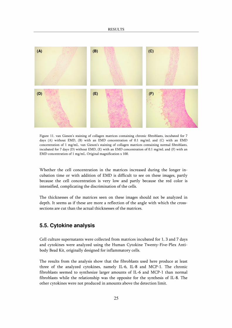
5.4. Histology analysis
Histology analysis was performed by culturing cells in collagen matrices for 1, 3 and 7 days. Cross-sections were then stained with van Gieson’s staining for tissue analysis. Figure 10 shows the van Gieson’s staining of the collagen matrices. Collagen fibers are stained red and cell nuclei are stained dark purple. After 1 day of incubation the matrices containing chronic fibroblasts and normal fibroblasts were comparable; the collagen fibers were straight and oriented in the same direction. After 3 and 7 days of incubation the matrices with chronic fibroblasts still exhibited a rather similar structure although a tendency of less oriented fibers could be observed after 7 days. The structure of the matrices containing normal fibroblasts changed more noticeably with increased incubation time. After 3 days of incubation a darker staining appeared in the close vicinity of the cells, implying an increasing concentration of collagen in these areas. As the incubation time increased further an intensification of the staining could be visualized in the entire matrix and simultaneously the alignment of the collagen fibers became indistinct.
(A) (B) (C) (D)
(E) (F) (G) (H)

RESULTS
24
Figure 10. van Gieson’s staining of collagen matrices containing chronic fibroblasts, incubated for (A) 1 day, (B) 3 days and (C) 7 days. van Gieson’s staining of collagen matrices containing normal fibroblasts, incubated for (D) 1 day, (E) 3 days and (F) 7 days. Original magnification x 100.
The effect on the histological appearance of the collagen matrices upon topical addition of EMD is shown in figure 11. For matrices containing chronic fibroblasts, addition of EMD seemed to increase the remodeling rate. After 7 days, the collagen fibers are still oriented in almost the same direction in the matrices incubated without EMD whereas the matrices incubated with an EMD concentration of 1 mg/mL exhibited many collagen fibers without a particular orientation. The effect is less pronounced in the matrices incubated with an EMD concentration of 0.1 mg/mL. For matrices containing normal fibroblasts the effect of EMD was even more apparent; After 7 days, the staining of the matrices incubated with an EMD concentration of 1 mg/mL was much more intense than the staining of the matrices incubated without EMD. The color intensity and hence the high collagen concentration even complicated the visualization of the collagen fibers. The low EMD concentration elicited intermediate effects. EMD can be visualized as light orange areas on the left side of the matrix containing normal fibroblasts and incubated for 7 days. However, no proteins are detectable inside the matrix according to these images.
(A) (C)
(E) (D) (F)
(B)
RESULTS
25
Figure 11. van Gieson’s staining of collagen matrices containing chronic fibroblasts, incubated for 7 days (A) without EMD, (B) with an EMD concentration of 0.1 mg/mL and (C) with an EMD concentration of 1 mg/mL. van Gieson’s staining of collagen matrices containing normal fibroblasts, incubated for 7 days (D) without EMD, (E) with an EMD concentration of 0.1 mg/mL and (F) with an EMD concentration of 1 mg/mL. Original magnification x 100.
Whether the cell concentration in the matrices increased during the longer in-cubation time or with addition of EMD is difficult to see on these images, partly because the cell concentration is very low and partly because the red color is intensified, complicating the discrimination of the cells. The thicknesses of the matrices seen on these images should not be analyzed in depth. It seems as if these are more a reflection of the angle with which the cross-sections are cut than the actual thicknesses of the matrices.
5.5. Cytokine analysis
Cell culture supernatants were collected from matrices incubated for 1, 3 and 7 days and cytokines were analyzed using the Human Cytokine Twenty-Five-Plex Anti-body Bead Kit, originally designed for inflammatory cells. The results from the analysis show that the fibroblasts used here produce at least three of the analyzed cytokines, namely IL-6, IL-8 and MCP-1. The chronic fibroblasts seemed to synthesize larger amounts of IL-6 and MCP-1 than normal fibroblasts while the relationship was the opposite for the synthesis of IL-8. The other cytokines were not produced in amounts above the detection limit.
(A) (B) (C)
(D) (E) (F)

RESULTS
26
The standard deviations of the measurements were rather high which complicates the interpretation of the results but in general it appears as if the presence of a high EMD concentration (1 mg/mL) reduced the production of all three cytokines, IL-6, IL-8 and MCP-1.

DISCUSSION
27
6. DISCUSSION
6.1. Proliferation analysis
It has been demonstrated that proliferation of cells requires a combination of signals, where mechanical tension within the cells seems to be involved.36 Thus, fibroblasts cultured in attached, mechanically loaded matrices proliferate whereas cells cultured in released or floating matrices tend to become quiescent and apoptotic.36 This phenomenon can clearly be seen for normal fibroblasts, where proliferation was increased in uncoated, attached matrices in comparison with BSA-coated, released matrices. Between day 3 and day 7 fibroblasts in the released matrices did not seem to proliferate at all, indicating the presence of many quiescent cells. For chronic fibroblasts, the effect of coating the wells was not evident. Only occasionally, the proliferation of these cells seemed to be higher in attached matrices than in released matrices. The reason for this variation is not clear; still the relatively large standard deviations of some measurements need to be taken into account before conclusions can be drawn. The reduction in cell number observed after 7 days in EMD treated matrices implies an increased amount of apoptotic cells. The effect of topical addition of EMD in a concentration of 1 mg/mL was obvious for both cell types, increasing the cell concentration in the matrices approximately threefold. Grayson et al. (2006) have earlier demonstrated increased proliferation of normal fibroblasts cultured within collagen matrices with EMD incorporated inside, although these effects were less pronounced. Furthermore, it is shown that EMD increased proliferation of the CRL chronic cell line when cultured in monolayer.37 As described previously by several groups 4,37 these results also demonstrate that the general ability of chronic fibroblasts to proliferate is reduced compared to normal fibroblasts. The fascinating observation was however that topical treatment of chronic fibroblasts with EMD enabled the cells to proliferate even better than untreated normal fibroblasts. This result is of great importance when striving to change the behavior of chronic fibroblasts into a behavior resembling the one of normal fibroblasts. Initially proliferation experiments in the present study were performed on collagen matrices with different EMD concentrations incorporated inside the matrices, a technique earlier used by Grayson et al. (2006). The results from the present experiments were however not consistent and the standard deviations where high (data not shown). Together with the appearance of the matrices, this aroused

DISCUSSION
28
suspicions of whether a high EMD concentration impairs collagen polymerization. In order to make the conditions in all matrices as similar as possible, EMD was instead added on top of the matrix after polymerization. Furthermore, this new approach closer mimics the treatment practice of topical appliance of EMD on a wound. The results are presented in this report. Apparently, EMD does not need to be incorporated in the matrix in order to affect the proliferation of cells. Possibly, small aggregates or monomers released from the aggregated protein enter the collagen matrix in a concentration high enough to influence the fibroblasts in the matrix. The mechanism underlying this stimulatory effect of EMD is not fully elucidated. To avoid large variations in the measurements and assure more statistically reliable results for chronic cells, a suggestion would be to utilize more replicates in this study. This experiment has however been performed several times and the outcome is most often the same; chronic fibroblasts seem to be more sensitive and have more varying ability to proliferate than normal fibroblasts, making these analyses complicated to interpret.
6.2. Cellular morphology analysis
Fibroblasts seeded on collagen coated surfaces demonstrated a typical flattened unnatural morphology. The extreme spreading observed was probably caused by the absence of dorsal adhesion points. The balance between spreading and retraction is altered which stimulates excessive spread out.38 Fibroblasts seeded on collagen matrices on the other hand, show a spindle shape and a reduced cell size. This natural morphology is seen when fibroblasts are cultured in a three dimensional environment with adhesion points in all directions.26 No obvious difference in cellular morphology was seen between normal fibroblasts and chronic fibroblasts neither in 2D nor in 3D, implying that the chronic fibroblasts are not affected in their ability to attach to and spread on surfaces or matrices. These results are consistent with the findings of Cook et al. (2000) who showed that chronic fibroblasts exhibited no difference in morphology compared to patient-matched normal fibroblasts. In contrast, other groups, Ågren et al. (1999) being one example, demonstrate a morphology of chronic fibroblasts that resemble in vitro senescent fibroblasts. Since amelogenin, the principal component of EMD, is an adhesion protein which has been shown to enhance cell adhesion of numerous cell types,29 the expected effect of EMD on cell morphology would be an increase in cell adhesion points. However, these observations could not be made in this experiment.

DISCUSSION
29
The present morphology analysis illustrates the importance of a careful selection of wound model when examining the wound healing process. Apparently, cells express different adhesion patterns and assume different morphologies depending on the dimensionality of the substrate. Culturing cells in two dimensions instead of their natural three dimensional environment therefore increases the risk to misinterpret significant biological behaviors of cells. To extend the morphology study in order to investigate morphology differences over a longer time scale would obviously be of interest. Analyzing morphology of cells cultured within the three dimensional matrices would provide results which more adequately reflect the morphology of cells in tissues. However, these studies require other equipment than an ordinary fluorescence microscope for visualizing cells, confocal laser scanning microscope being one example. Another approach for extension of the morphology study would be to stain cells for other components than F-actin, myosin being a component of interest.
6.3. Matrix contraction and histology analyses
The histology and contraction analysis demonstrated that the remodeling rate and contraction ability is lower in the matrices containing chronic fibroblasts in com-parison with the matrices containing normal fibroblasts. It is earlier shown that fibroblast remodeling of collagen matrices by their migratory activity occurs at two levels. Locally, the density of collagen fibrils increases in the close vicinity of the cells, typically reaching the collagen concentrations of mature tissues. Globally, the total increase in matrix density leads to a decrease in matrix volume, i.e. contraction occurs.25 These remodeling processes can clearly be observed here in the matrices containing normal fibroblasts. Why this phenomenon seems to be deficient in the matrices containing chronic fibroblasts is not clear. However, earlier studies have demonstrated that the ability of cells to reorganize the extracellular matrix in vitro is strongly associated with the wound healing capacity of the corresponding tissue in vivo. Fibroblasts derived from chronic wounds would therefore exhibit an impaired ability to remodel collagen matrices which corresponds to the defective remodeling processes in vivo.20 The exact mechanisms underlying the extracellular matrix remodeling in collagen matrices remain unclear. By comparing cell numbers of the matrices with their ability to contract, it is however demonstrated in the present study that the differences in reorganization of the collagen matrices between chronic and normal fibroblasts was not related only to the higher cell number of the normal fibroblasts in comparison with the chronic cells.

DISCUSSION
30
Since contraction seems to depend on other factors than the ability of cells to proliferate, other explanations have been examined. Cook et al. (2000) for example, have demonstrated that the impaired reorganization by chronic fibroblasts may be associated with alterations in the activity of matrix metalloproteinases, specific enzymes which degrade matrix proteins. According to the present histology results, it appears as if the ability of chronic fibroblasts to remodel the matrices is almost absent whereas the contraction analysis demonstrate that these cells do contract the matrices, although less than normal fibroblasts. (Figure 8 and 10) The reason for this discrepancy could be that the matrices containing chronic fibroblasts fabricated for the histology analysis did not contract as much as the corresponding matrices utilized during the contraction measurements. Probably, the former matrices were attached to the well bottom at some points, which decreased the contraction rate further in comparison with the matrices containing normal fibroblasts. This could explain the larger differences in remodeling ability between the cell types seen on the histology images than on the contraction measurements. It is furthermore important to remember that these contraction estimates only have embraced the horizontal contractions observed, i.e. reduction in diameter have been used as a measure of contraction. Optimally these measurements should also include the vertical contraction and thus be performed using the reduction in matrix volume as a measure of contraction. However, these analyses require other more comp-licated methods, analysis of three dimensional images using confocal microscopy being one example. Remodeling and contraction of collagen matrices by fibroblasts seems to be en-hanced with topical addition of EMD. This effect is observed for both chronic and normal fibroblasts. Grayson et al. (2006) have earlier described similar effects for normal dermal fibroblasts in three dimensional collagen matrices. The mechanisms of EMD and its effects on extracellular reorganization are still not fully elucidated. However, the present results also demonstrate the possibility of adding EMD on top of the matrices and still affect activities of fibroblasts residing inside the matrices. From a clinical point of view this is of utmost importance in the treatment of chronic wounds. At first, the cell concentrations of the collagen matrices seen on the histology images seem to be very low, but in comparison with natural human dermal tissues this is an adequate concentration of fibroblasts. (data not shown) The cross-sections are more-over very thin and calculations of the measured proliferation results confirmed the visualized cell concentrations. An interesting continuation of this remodeling analysis would be to use immuno-histochemistry in order to investigate whether EMD accelerates the differentiation

DISCUSSION
31
of fibroblasts into myofibroblasts, the type of fibroblasts that possess contractile forces. Moreover, it would be useful to examine how EMD is distributed in the matrix after topical appliance.
6.4. Cytokine analysis
The results from the cytokine analysis showed that the presence of an EMD concentration of 1 mg/mL reduced the fibroblast production of all three cytokines that were produced in detectable amounts, i.e. IL-6, IL-8 and MCP-1. Since a chronic wound is a complex composition of different cell types and extracellular proteins, including numerous growth factors and cytokines it is however unclear whether a similar effect of EMD would be seen in clinical practice. To analyze the effect of single tissue components in order to assign each of them functions in this complicated system is not an easy task. Hence, although IL-6 has demonstrated a mitogenic effect for keratinocytes 30 it is not certain that EMD, through its demonstrated inhibitory effect on IL-6 production, would decrease proliferation of keratinocytes. Contrary to the present results, EMD increased the IL-8 secretion by chronic fibroblasts cultured in monolayer in a previous study.37 MCP-1 production was however inhibited by EMD addition 37, which is supported by present data. The variance of these findings could be due to differences in dimensionality. There are probably several reasons for the relatively sparsely amount of cytokines found in these samples. Firstly, the Human Cytokine 25-plex kit utilized in this experiment is developed for the analysis of cytokines primarily involved in inflammation; many of the cytokines included in this assay have never been shown to be produced by fibroblasts. Other cytokines have been shown to be produced by fibroblasts in an inflammatory environment but not during the prevalent conditions of this assay. Therefore, neither of these cytokines should be expected to be found in this analysis. Finally, even if some of the cytokines have been demonstrated to be synthesized by fibroblasts in three dimensional collagen matrices in other studies, small differences in experimental design can lead to remarkable differences in cytokine production. It would be of great interest to investigate the synthesis and release of other cytokines and growth factors important in wound healing processes. Especially analyses of chronic fibroblast expression of vascular endothelial growth factor (VEGF) and transforming growth factor β1 (TGF- β1) would provide important information. Grayson et al. (2006) have earlier demonstrated increased levels of TGF- β1 production by normal fibroblasts cultured in collagen matrices with EMD.

CONCLUSION
32
7. CONCLUSION
Our knowledge of the wound healing process has progressed significantly during the last years and numerous models have been developed for the analysis of the cellular mechanisms regulating these processes. During times when the population ages and the incidence of diabetes increases the prevalence of chronic wounds will inevitably rise as well. In order to understand the underlying mechanisms causing the chronicity and to develop new and more effective treatments there is an immense need of improved models for chronic wounds.3,39 In the present project, a three dimensional model which provides a reasonable rep-resentation of the dermal granulation tissue in a chronic wound has been developed. The model was utilized for the investigation of the differences in cell behavior between chronic human dermal fibroblasts and normal human dermal fibroblasts on cellular processes important for dermal repair and regeneration, i.e. proliferation, cellular morphology, cytokine production, matrix contraction and remodeling. My results demonstrate that the ability of chronic fibroblasts to proliferate and to contract and remodel the matrix is reduced in comparison with normal fibroblasts. No obvious difference in cellular morphology was seen between normal fibroblasts and chronic fibroblasts neither in 2D nor in 3D, implying that the chronicity of chronic fibroblasts do not affect their ability to attach to and spread on surfaces or matrices. Furthermore, the effect of a wound treatment substance, EMD, on these cellular processes has been examined. My results demonstrate that topical addition of EMD affected both cell types by increasing the ability to proliferate and to contract and remodel the matrix. These results are of great importance, especially for chronic fibroblasts as EMD improved their deficient capacities. When combined with basic medical care, EMD and Xelma® therefore have an excellent potential to provide an effective treatment of chronic wounds. In summary, my results show that it is possible to measure the differences in cell behavior between chronic and normal fibroblasts on cellular processes important for dermal repair and regeneration in this three dimensional in vitro model. It was furthermore possible to analyze the effect of a wound treatment substance, EMD, on these cellular processes when topically applied. Moreover, the results are re-producible which gives this model high potential when studying wound healing processes in chronic wounds.
ACKNOWLEDGEMENTS
33
8. ACKNOWLEDGEMENTS
I would like to give a very special thanks to my supervisor Dr Maria Werthén at Mölnlycke Health Care for introducing me into this interesting project, for all the advice and support and for the critical reading of this report. I would also like to express my sincere thanks to Karl Erikson, manager for Research and Concept Development in Wound Care R&D at Mölnlycke Health Care for giving me the opportunity to do my master thesis project in his group. I would like to thank Sofia Almqvist at the Department of Biomaterials at Gothenburg University for important consultation and contribution of cells. I would like to thank Peter Thomsen, Professor at the Department of Biomaterials at Gothenburg University for being my scientific reviewer. I would like to thank the Institute of Clinical Dentistry at Oslo University for letting me perform the cytokine analysis at their laboratory. I would like to thank HistoCenter for the histology slicing and staining. I would like to thank the Institute for Biomaterials and Cell Therapy (IBCT) for the financial contribution. I would like to thank the Biomaterials Research Centre (BRC) for the scholarship which gave me the opportunity to present my project at the 9th New Jersey Symposium on Biomaterials Science and Regenerative Medicine in October 2008. And finally I would like to thank everybody else at Mölnlycke Health Care and the Department of Biomaterials at Gothenburg University for being so helpful and friendly during these six months.

REFERENCES
34
9. REFERENCES
[1] Beanes, S.R., Dang, C., Soo, C. and Ting, K. Skin repair and scar formation: the central role of TGF-β. Expert Rev Mol Med 2003;5:1-22.
[2] Menke, N.B., Ward, K.R., Witten, T.M., Bonchev, D.G. and Diegelmann, R.F. Impaired
wound healing. Clin Dermatol 2007;25:19-25. [3] Gottrup, F., Ågren, M.S. and Karlsmark, T. Models for use in wound healing research: A
survey focusing on in vitro and in vivo adult soft tissue. Wound Rep Reg 2000;8:83-96. [4] Ågren, M.S., Steenfos, H.H., Dabelsteen, S., Hansen, J.B. and Dabelsteen, E. Proliferation
and mitogenic response to PDGF-BB of fibroblasts isolated from chronic venous leg ulcers is ulcer-age dependent. J Invest Dermatol 1999;112:463-469.
[5] Carlson, M.A. and Longaker, M.T. The fibroblast-populated collagen matrix as a model of
wound healing: a review of the evidence. Wound Rep Reg 2004;12:134-147. [6] Mirastschijski, U., Konrad, D., Lundberg, E., Lyngstadaas, S.P., Jorgensen, L.J. and Ågren,
M.S. Effects of a topical enamel matrix derivative on skin wound healing. Wound Rep Reg 2004;12:100-108.
[7] Fincham, A.G., Moradian-Oldak, J. and Simmer, J.P. The structural biology of the
developing dental enamel matrix. J Struct Biol 1999;126:270-299. [8] Vowden, P., Romanelli, M., Peter, R., Boström, Å., Josefsson, A. and Stege, H. The effect of
amelogenins (XelmaTM) on hard-to-heal venous leg ulcers. Wound Rep Reg 2006;14:240-246.
[9] Vowden, P., Romanelli, M. and Price, P. Effect of amelogenin extracellular matrix protein
and compression on hard-to-heal venous leg ulcers. J Wound Care 2007;16:189-195. [10] Grayson, R.E., Yamakoshi, Y., Wood, E.J. and Ågren, M.S. The effect of the amelogenin
fraction of enamel matrix proteins on fibroblast-mediated collagen matrix reorganization. Biomaterials 2006;27:2926-2933.
[11] Hunter, J., Savin, J. and Dahl, M. Clinical dermatology, 3rd Edition, 2002 ISBN 0-632-05916-
8, Blackwell Science. [12] Gawkrodger, D.J. Dermatology – an illustrated colour text, 3rd Edition, 2002 ISBN 0443-
07140-3, Churchill Livingstone. [13] Forslind, B. Hår, hud och naglar – en tillämpad introduktion, 1998 ISBN 91-44-00513-X,
Studentlitteratur. [14] (http://en.wikipedia.org/wiki/Skin, 2007-10-01)

REFERENCES
35
[15] Harding, K.G., Morris, H.L. and Patel, G.K. Science, medicine, and the future: Healing chronic wounds. BMJ 2002;324:160-163.
[16] Martin, P. Wound healing – Aiming for perfect skin regeneration. Science 1997;276:75-81. [17] Singer, A.J. and Clark, R.A.F. Cutaneous wound healing. N Eng J Med 1999;341:738-746. [18] Baum, C.L. and Arpey, C.J. Normal cutaneous wound healing: Clinical correlation with
cellular and molecular events. Dermatol Surg 2005;31:674-686. [19] Telgenhoff, D. and Shroot, B. Cellular senescence mechanisms in chronic wound healing.
Cell Death Differ 2005;12:695-698. [20] Cook, H., Stephens, P., Davies, K.J., Harding, K.G. and Thomas, D.W. Defective extra-
cellular matrix reorganization by chronic wound fibroblasts is associated with alterations in TIMP-1, TIMP-2, and MMP-2 activity. J Invest Dermatol 2000;115:225-233.
[21] Schultz, G.S., Sibbald, R.G., Falanga, V., Ayello, E.A., Dowsett, C., Harding, K., Romanelli,
M., Stacey, M.C., Teot, L. and Vanscheidt, W. Wound bed preparation: a systematic approach to wound management. Wound Rep Reg 2003;11:1-28.
[22] Ågren, M.S. and Werthen, M. The extracellular matrix in wound healing: A closer look at
therapeutics for chronic wounds. Int J Low Extrem Wounds 2007;6:82-97. [23] Cooper, D.M., Yu, E.Z., Hennessey, P., Ko, F. and Robson, M.C. Determination of endo-
genous cytokines in chronic wounds. Ann Surg 1994;219:688-692. [24] Chen, W.Y.J. and Rogers, A.A. Recent insights into the causes of chronic leg ulceration in
venous diseases and implications on other types of chronic wounds. Wound Rep Reg 2007;15:439-449.
[25] Grinnell, F. Fibroblast biology in three-dimensional collagen matrices. Trends Cell Biol
2003;13:264-269. [26] Pedersen, J.A. and Swartz, M.A. Mechanobiology in the third dimension. Ann Biomed Eng
2005;33:1469-1490.
[27] Ratner, B.D., Hoffman, A.S., Schoen, F.J. and Lemons, J.E. Biomaterials science – an introduction to materials in medicine, 2nd Edition, 2004, ISBN 0-12-582463-7, Elsevier Academic Press.
[28] Wennström, J.L. and Lindhe, J. Some effects of enamel matrix proteins on wound healing in
the dento-gingival region. J Clin Periodontol 2002;29:9–14. [29] Hoang, A.M., Klebe, R.J., Steffensen, B., Ryu, O.H., Simmer, J.P. and Cochran D.L.
Amelogenin is a cell adhesion protein. J Dent Res 2002;81:497-500. [30] Werner, S. and Grose, R. Regulation of wound healing by growth factors and cytokines.
Physiol Rev 2003;83:835-870.

REFERENCES
36
[31] Tuschil, A., Lam, C., Haslberger, A. and Lindley, I. Interleukin-8 stimulates calcium
transients and promotes epidermal cell proliferation. J Invest Dermatol 1992;99:294-298.
[32] Koch, A.E., Polverini, P.J., Kunkel, S.L., Harlow, L.A., DiPietro, L.A., Elner, V.M., Elner,
S.G. and Strieter, R.M. Interleukin-8 as a macrophage-derived mediator of angiogenesis.
Science 1992;258:1798-1801.
[33] Weber, K.S., Nelson, P.J., Gröne, H.J. and Weber, C. Expression of CCR2 by endothelial
cells: implications for MCP-1 mediated wound injury repair and in vivo inflammatory
activation of endothelium. Arterioscler Thromb Vasc Biol 1999;19:2085-2093. [34] Gharaee-Kermani, M., Denholm, E.M. and Phan S.H. Costimulation of fibroblast collagen
and transforming growth factor beta1 gene expression by monocyte chemoattractant protein-1 via specific receptors. J Biol Chem 1996;271:17779-17784.
[35] Jiang, H. and Grinnell, F. Cell-matrix entanglement and mechanical anchorage of fibroblasts
in three-dimensional collagen matrices. Mol Biol Cell 2005;16:5070-5076.
[36] Fringer, J. and Grinnell, F. Fibroblast quiescence in floating or released collagen matrices:
contribution of the ERK signaling pathway and actin cytoskeletal organization. J Biol Chem
2001;276:31047-31052. [37] Almqvist, S., Werthén, M., Johansson, A., Törnqvist, J., Ågren, M. and Thomsen, P.
Evaluation of a near-senescent human dermal fibroblast cell line and effect of amelogenin. Brit J Dermatol 2009;accepted for publication.
[38] Birgersdotter, A., Sandberg, R. and Ernberg, I. Gene expression perturbation in vitro – A
growing case for three-dimensional (3D) culture systems. Semin Cancer Biol 2005;15:405-412.
[39] Falanga, V. The chronic wound: impaired healing and solutions in the context of wound
bed preparation. Blood Cells Mol Dis 2004;32:88-94.