MEDICAL RESEARCH FOUNDATION - Sankara Nethralaya Files/insight1085.pdf · Medical Research...
14
VOL.II No.4 OCT.1985 NETHRALAYA INSIGHT HOUSE JOURNAL OF MEDICAL RESEARCH FOUNDATION & VISION RESEARCH FOUNDATION 18, COLLEGE ROAD, MADRAS, INDIA ISSUE HIGHLIGHTS ELITE SCHOOL OF OPTOMETRY EDITORIAL ENDOCRINE OPHTHALMOPATHY – ULTRASONOGRAPHY VS CT SCAN OPHTHALMIC DIATHERMY YAG LASER IRIDECTOMY IN DARKLY PIGMENTED EYES EDITOR : MARY ABRAHAM CONTACT LENSES IN KERATOCONUS ELITE SCHOOL OF OPTOMETRY Medical Research Foundation has taken a further step in the march towards progress this in the field of vision care, correction rehabilitation. The goal was fulfilled when Prof. JayMEnoch, Dean, School of Optometry, University of California, Berkeley, U.S.A. formed an Indo-American Optometric Outreach Society in U S A to provide faculty held until we develop our own care and has gifted equipment and books. This was looked upon with keen interest by Mr.C.K.Shah, one of the Directors of Elite Optical Induatry who made an outstanding donation of Rupees ten lakhs. This enabled the Medical Research Foundation to forge ahead with its plans of expansion and the Elite School of Optometry was inaugurated on 18.9.85 Dr.H.V.Hande. The Baccalaureate Course in Optometry (B.Opt.) organized by the school has a six semester training programme followed by one-year intership at Sankara Nethralaya. We carefully selected syllabus has an inter-disciplinary content with relevant aspects of Physics, Chemistry, Anatomy. Physiology and Microbiology. The core courses will have Basic Sciences and visual, physiological and ophthalmological optics. Besides this practical and clinical training will be provided. Support courses will include communication, accountancy and office administration. The school will keep in touch with similar institutions in India and overseas, particularly the Berkeley School Optometry so that there may be constant curricular updating and methodological improvement. The elite School of Optometry has initially started functioning at the campus of Medical Research Foundation. The Women’s Christian College campus which lies just across the road will offer facilities for the practical sessions. Sankara Nethralaya will be provide the necessary Audio visual facilities and make its library available to the students and staff. Twenty students have joined the course the year.
Transcript of MEDICAL RESEARCH FOUNDATION - Sankara Nethralaya Files/insight1085.pdf · Medical Research...

VOL.II No.4 OCT.1985
NETHRALAYA INSIGHT
HOUSE JOURNAL OF MEDICAL RESEARCH FOUNDATION & VISION RESEARCH FOUNDATION
18, COLLEGE ROAD, MADRAS, INDIA
ISSUE HIGHLIGHTS ELITE SCHOOL OF OPTOMETRY EDITORIAL ENDOCRINE OPHTHALMOPATHY – ULTRASONOGRAPHY VS CT SCAN OPHTHALMIC DIATHERMY YAG LASER IRIDECTOMY IN DARKLY PIGMENTED EYES EDITOR : MARY ABRAHAM CONTACT LENSES IN KERATOCONUS ELITE SCHOOL OF OPTOMETRY Medical Research Foundation has taken a further step in the march towards progress this in the field of vision care, correction rehabilitation. The goal was fulfilled when Prof. JayMEnoch, Dean, School of Optometry, University of California, Berkeley, U.S.A. formed an Indo-American Optometric Outreach Society in U S A to provide faculty held until we develop our own care and has gifted equipment and books. This was looked upon with keen interest by Mr.C.K.Shah, one of the Directors of Elite Optical Induatry who made an outstanding donation of Rupees ten lakhs. This enabled the Medical Research Foundation to forge ahead with its plans of expansion and the Elite School of Optometry was inaugurated on 18.9.85 Dr.H.V.Hande. The Baccalaureate Course in Optometry (B.Opt.) organized by the school has a six semester training programme followed by one-year intership at Sankara Nethralaya. We carefully selected syllabus has an inter-disciplinary content with relevant aspects of Physics, Chemistry, Anatomy. Physiology and Microbiology. The core courses will have Basic Sciences and visual, physiological and ophthalmological optics. Besides this practical and clinical training will be provided. Support courses will include communication, accountancy and office administration. The school will keep in touch with similar institutions in India and overseas, particularly the Berkeley School Optometry so that there may be constant curricular updating and methodological improvement. The elite School of Optometry has initially started functioning at the campus of Medical Research Foundation. The Women’s Christian College campus which lies just across the road will offer facilities for the practical sessions. Sankara Nethralaya will be provide the necessary Audio visual facilities and make its library available to the students and staff. Twenty students have joined the course the year.

In course of time the land acquired by the Medical Research Foundation at St.Thomas Mount will house not only the Elite School of Optometry, but also hostels for men and women students. Amongest the very colourful gathering on 18th September 1985, the inauguration of the School of Optometry took place. The welcome address was delivered by the President Mr. V.Mohan Rao. Prof. S.R.Govindarajan, Hony. Director, School of Optometry gave an insight into Optometry and Dr. S.S.Badrinath introduced the chief guest Prof. Jay M Enoch. The key note address was given by Dr.H.V.Hande, the Hon’ble Minister for Health who also formally inaugurated the school, Mr. Naval Baliwalla, of Baliwalla & Homi Private Limited, spoke about the role of optometry and its usefulness to our country. The vote of thanks was given by Dr.S.Baskaran, Physician, Sankara Nethralaya. With this stride forwards we are sure that the optometrists will provide a vital link in the chain of health care delivery system in India. There role is varied and they would assess and correct lenses, promptly recognize and refer patients with ocular disease. There care will extend to factories, schools and slums. In a country like ours where literacy is low and vision impairment high adequately trained optometrists will prevent the existing malpractise by poorly informed persons and thus prevent serious tragedy. Thus optometrists have a very challenging situation ahead and we are confident that they will in unison with the ophthalmologists help to provide maximum eye care to the people of our country.

At the Inaugural function of Elite School of Optometry seated from L-R are Prof: S.R.Govindarajan, Prof:J.M.Enoch and Hon’ble Minister of Health Mr. H.V.Hande. EDITORIAL
Sankara Nethralaya has completed seven years of service. As we march ahead into the 8th year a long cherished dream materialized when the Elite School of Optometry was formarlly inaugurated on 18th September by Dr.H.V.Hande, Hon’ble Health Minister of Tamil Nadu. The chief guest was prof. Jay M Enoch, Dean, School of Optometry, Berkeley, USA who was gracious enough to be present with us during this auspicious occasion. Mr. C.K.Shah of Elite Optical Industry made a handsome donation of Rs.10 lakhs which has helped to get this project started. Amongst the distinguished guests, well wishers and professional collegues present that day were Ophthalmologists from other states too. Dr. Tony Fernandez from Angamaly. Dr.Natarajan from Bangalore, Dr.S.Kelkar from Pune and Dr. Bakulesh and Mayuri Khammer from Ahmedabad. Prof. Enoch addressed the first batch of 20 students selected for this course earlier in the optometry by tracingitshistory and elaboratingon its present day importance. This was followed by an informat get together at lunch provided by the Mess at the Dharmashala. Dr. M.Ct. Yegappan, Physican who joined us in June 1983, is leaving for U.K. to train in Neurology at London. He has been agreat support to us in providing pre and post operative care, in dealing with any medical emergencies at the out patient and in the medical evaluation of patients with ocular diseases which have a systemic bearing. The staff of Sankara Nethralaya bade him farewull at a small parth on 6th September 1985 at the Dharmashala. We wish him Godspeed, Good health and success in his new endeavour. Dr. B.Sridhar Rao participated in the Glaucoma work shop conducted by Karnataka ophthalmic society and took part in the panel discussion of Glaucoma conducted by the CBM ophthalmic insitute, Angamally, in August. Five of us participated at the 33rd Madras State Ophthalmic Conference at Thanjavur on 14th and 15th September Besides taking part in the panel discussion on Cataract surgery, 4 free papers were presented. As Sankara Nethralaya
is expanding, so are its facilities. The new computer (Pragati AL.2016) was installed on September 3rd to meet the growing demands. This would serve to computerize financial accounting, word processing and the ongoing research projects. A second computer would deal with the day to day appointment and serve as a black up system in any situation when the need arises. Besides this, a photocopier and 3-M lettering system have been acquired which will be of immense value of every one of us. The Medical Research Foundation has updated its library and shifted it to the 4th floor of the Dharmashala complex. The entire place has been modernized. With Mr. Lourduswamy as Librarian, the library has acquired new dimensions and provide very sophisticated audio and video facilities. Dr. Nirmal Subramaniam, Plastic Surgeo, Govt. Stanley Hospital, who is also our oculoplastic surgeon has left for Pittsburg, U.S.A. for further training in Oculplastic surgery and on her return, we intend establishing a full fledged Oculplastic surgery unit. Finanlly readers, we do hope you enjoy reading the ‘Nethralaya Insight’. We always look forward to and welcome your suggestions.
EDITOR
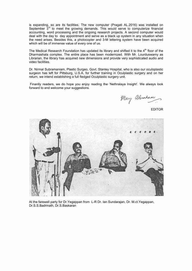
At the farewell party for Dr.Yegappan from L-R Dr. Ian Sundarajan, Dr. M.ct.Yegappan, Dr.S.S.Badrinath, Dr.S.Baskaran

ENDOCRINE OPHTHALMOPATHY – ULTRASONOGRAPHY VS C T SCAN Dr.Gopal L This article serves to highlight the importance of C T Scan in Endocrine Ophthalmopathy. It is probably preferable to use the term Endocrine Ophthalmopathy rather then Thyroid Ophthalmopathy since there is no absolute correlation between. Thyroid disease and the ocular manifestations. In Euthyroid states the clinical diagnosis becomes sometimes difficult and investigations like Ultrasonography and computed tomography can play a very important role the diagnosis. There are inherent advantages and disadvantages of each of case modalities. The present case highlights the occasional necessity to perform C T Scan in cases of suspected endocrine ophthalmopathy. CASE SUMMARY: P.B. a 33 year old male presented to us with history of prominence of the right eye of 1 year duration along with diplopia, which was maximum for distance and in upward gaze. On examination he had a visual acuity of 6/6 in the right eye and 6/5 in the left eye. Ocular movements were apparently full. There was mild proptosis on the right side which was probably apparent because of the lid retraction. Lid retraction was mild and was restricted to the right side. Retrobulbar resistance was normal and no mass was felt. Anterior segments was normal in both the eyes. There were no engorged vessels on the conjunctiva and the pupils were disk in both eyes. Field charting with the Goldmann Perimeter was normal in both the eyes. Hess charting showed apparent underaction of the right lateral rectus.
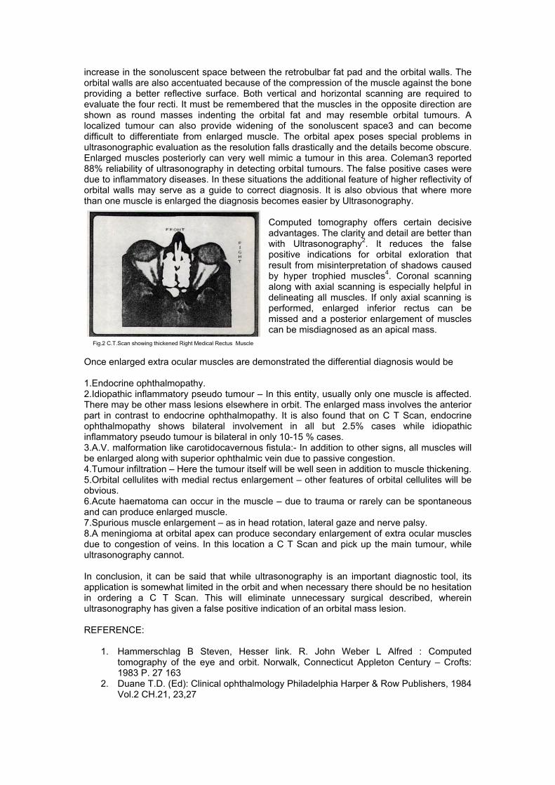
The clinical picture was suggestive of endocrine ophthalmopathy. Haemogram, T3, T4 levels and x-ray skull Caldwell’s view (done elsewhere) were normal. Ultrasonography was performed with the ocuscan 400 (Fig 1). There was a strong suspicion of a mass in the nasal orbit of solid consistency. In view of the suspicion of mass lesion, a C T Scan was done (Fig 2) Only axial scanning was performed. This revealed a thickened medial rectus muscle. Following the confirmation of the diagnosis of endocrine ophthalmopathy, he underwent examination by an endocrinologist who
diagnosed minimal goiter with mild thyrotoxicosis. Repeat hormonal assay showed high normal T4 level and mild elevation of T3 levels. The Thyro globulin antibody titre was weakly positive. He was advised regular follow up since the ocular symptoms were mild and there were no pressure effects on the optic nerve. Fig. 1. Ultrasonogram Showing mass lesion in the nasal orbit. DISCUSSION: Computed tomography is undoubtedly a boon to the ophthalmologist dealing with orbital disease. However one must remember that it is an expensive proposition and the patient is exposed to proposition and the paitent is exposed to radiation which fortunately is very low with the new generation scanners. As a routine, C T Scan is not necessary in the diagnosis of endocrine ophthalmopathy1. Clinical suspicion can be supplemented by Ultrasonography. Ultrasonography shows an
increase in the sonoluscent space between the retrobulbar fat pad and the orbital walls. The orbital walls are also accentuated because of the compression of the muscle against the bone providing a better reflective surface. Both vertical and horizontal scanning are required to evaluate the four recti. It must be remembered that the muscles in the opposite direction are shown as round masses indenting the orbital fat and may resemble orbital tumours. A localized tumour can also provide widening of the sonoluscent space3 and can become difficult to differentiate from enlarged muscle. The orbital apex poses special problems in ultrasonographic evaluation as the resolution falls drastically and the details become obscure. Enlarged muscles posteriorly can very well mimic a tumour in this area. Coleman3 reported 88% reliability of ultrasonography in detecting orbital tumours. The false positive cases were due to inflammatory diseases. In these situations the additional feature of higher reflectivity of orbital walls may serve as a guide to correct diagnosis. It is also obvious that where more than one muscle is enlarged the diagnosis becomes easier by Ultrasonography.
Computed tomography offers certain decisive advantages. The clarity and detail are better than with Ultrasonography2. It reduces the false positive indications for orbital exloration that result from misinterpretation of shadows caused by hyper trophied muscles4. Coronal scanning along with axial scanning is especially helpful in delineating all muscles. If only axial scanning is performed, enlarged inferior rectus can be missed and a posterior enlargement of muscles can be misdiagnosed as an apical mass.
Fig.2 C.T.Scan showing thickened Right Medical Rectus Muscle
Once enlarged extra ocular muscles are demonstrated the differential diagnosis would be 1.Endocrine ophthalmopathy. 2.Idiopathic inflammatory pseudo tumour – In this entity, usually only one muscle is affected. There may be other mass lesions elsewhere in orbit. The enlarged mass involves the anterior part in contrast to endocrine ophthalmopathy. It is also found that on C T Scan, endocrine ophthalmopathy shows bilateral involvement in all but 2.5% cases while idiopathic inflammatory pseudo tumour is bilateral in only 10-15 % cases. 3.A.V. malformation like carotidocavernous fistula:- In addition to other signs, all muscles will be enlarged along with superior ophthalmic vein due to passive congestion. 4.Tumour infiltration – Here the tumour itself will be well seen in addition to muscle thickening. 5.Orbital cellulites with medial rectus enlargement – other features of orbital cellulites will be obvious. 6.Acute haematoma can occur in the muscle – due to trauma or rarely can be spontaneous and can produce enlarged muscle. 7.Spurious muscle enlargement – as in head rotation, lateral gaze and nerve palsy. 8.A meningioma at orbital apex can produce secondary enlargement of extra ocular muscles due to congestion of veins. In this location a C T Scan and pick up the main tumour, while ultrasonography cannot. In conclusion, it can be said that while ultrasonography is an important diagnostic tool, its application is somewhat limited in the orbit and when necessary there should be no hesitation in ordering a C T Scan. This will eliminate unnecessary surgical described, wherein ultrasonography has given a false positive indication of an orbital mass lesion. REFERENCE:
1. Hammerschlag B Steven, Hesser link. R. John Weber L Alfred : Computed tomography of the eye and orbit. Norwalk, Connecticut Appleton Century – Crofts: 1983 P. 27 163
2. Duane T.D. (Ed): Clinical ophthalmology Philadelphia Harper & Row Publishers, 1984 Vol.2 CH.21, 23,27

3. Coleman D Jackson : Ultrasonography of the eye and orbit Philadelphia, Lea & Febiger 1977, Ch.6,P.327,346
4. Hughes F.Wilson (Ed),: Year book of ophthalmology Chicago, Year book medical publishers inc.1981. ch 1, P.10.
“ Little drops of water, Little grains of sand, Make a mighty ocean, And the pleasant land”
Every month we get an unusual
Contribution from three well wishers Who went to help Nethralaya in their
Own way.
Mr K.V. Govinda Rao, managing trustee of Wisdom Trust, Madras 4 contributes Rs.25/- per month. Mrs.Chandra of
Mohan Nagar, Nagpur, sends Rs.10/- per month and Mr. A Rajasekharan of ICF, Madras-38, donates Rs.1/- per month.
We are touched by these three persons; who in their own way contribute what they
can to the war against blindness. Their sincerity and regularity is to be appreciated and we are extremely grateful to them. We are sure that the institution will be
supported by more and more of such donors.

OPHTHALMIC DIATHERMY Santosh Kumar K Bio-Medical Engineer Electrical current is the flow of electrons which are negatively charged particles, When this flow of electrons alternate, between positive and negative peaks it is known as Alternating Current (A.C.). Alternating electrical currents are characterized by the number of cycles (or current alternations) per second. Currents exhibiting a large number of cycles are called high frequency or radio frequency (RF) currents. The frequency is measured in Hertz (cycle/second). The high frequencies are in the range of 1,000,000 Megahertz or MHz. The frequency of the alternating current and the wave length of the current are mathematically related l = 300,000,000 meters/sec = speed of light Where F = frequency of the alternating current = 1,000,000 c/s. Wave length = 300,000,000 = 300 meters 1,000,000 The main effect of RF currents in tissues is heat production lons and Dipoles (Dipoles are molecules with an electrical charge, such as hyaluronic acid) in the tissues oscillate under the influence of the rapidly alternating current and generate heat through a phenomenon analogus to friction. This can also be said as the resistance offered by the tissue to the flow of charged electron particles. Hence, the amount of heat produced in the tissue depends on the electrical resistance offered by the tissue and the power of the high frequency current. Diathermy is defined as the passage of a frequency (Radio frequency) electric current through the tissuewhereby heat is produced. As the frequency of the current increases, the concentration of heat in the tissue is localized or the lateral spread of the current is decreased. To generate high power radio frequency current, very high voltages are required. The first type of medical diathermy was of the step up transformer and tubes version. Here the supply from the mains is stepped up to a few thousands of volts by a transformer. The output from the transformer is made to oscillate between two electron tubes. The alternating (mostly sinusoidal in character) current from the electron tubes is fed to the required spot by an electrode connected by a cable. With the development of storing energy in capacitors, the stepped up voltage is stored in the capacitors and made to oscillate through a spark gap. While the tube and spark gap versions and disadvantages because of their limited capacities, the solid state diathermies are proving to be highly accurate and efficient. Spark gap diathermies produce damped currents which produce intermittent heat in the tissue and the sudden bursts of heat may damage wider sections of the tissue than desired. In the solid state diathermy a crystal which oscillates at 13.56 MHz is used to produce high frequency currents at 13.56 MHz. These currents are amplified and smoothed for all harmonics. The power output is variable from 0.2 to 21 watts over a scale of 0-10 logarithmically. By maintaining a constant current at all levels and increasing the output voltage over a constant frequency of 13.56 MHz, the output power is localized and the desired burn is produced in tissue. This RF current of 13.56 MHz at 0.2 to 21 watts is calculated against a tissue impedance (resistance – capacitance) of 300 to 1200 ohms by consolidating the above points. Ophthalmic diathermy finds a very wide use in retinal detachment and glaucoma procedures and likewise in the destruction of skin and intraocular tumours and epilation for trichiasis. The following factors affect a diathermy burn : -

1.Voltage : The intensity of the burn is proportional to the voltage level for any given frequency. If a faint reaction occurs during scleral diathermy, the voltage (Rf level) must be increased.
2.Time: For a given burn intensity the duration of application is inversely the duration of application is inversely related to the voltage level. Low voltage must be applied for a longer time to obtain a burn equivalent to one obtained by applying high voltage for a shorter time. 3.Hydration: The moistness of the sclera affects the burn in two ways. a)If the sclera is wet, the impedance is low. This decrease the voltage and reduces the burn. b)A wet sclera causes wide dispersion of the current which also results in a less efficient burn. 4.Location: The thickness and impedance of the different layers of the eye are variable in different eyes and in different locations in the same eye. The sclera is thicker and the impedance islower in the posterior segment of the eye. The voltage level or the duration of application must be greater posteriosrly than anteriorly. The most consistent results are obtained when application are made at equal distances from the limbus. 5.Pressure: Pressure does not seems to affect the diameter of a burn although greater pressure will cause a more intense burn as it dries the sclera and decreases choroidal circulation. The effect of a burn also depends on the electrodes. Diathermies using RF currents of the order 13.56 MHz or more use single electrode type. By virtue of high frequency induction these do not require a return path (from the patient to the equipment ground). Hence these are easy to use and by varying the area of their tips the area of burn can be decided by the surgeon. The other type of electrode is the bi-polar type where the tissue is held between two electrodes. By applying the current through one electrode, a complete circute is formed through the tissue to the other electrode. In bi-polar coagulation, the current flow is concentrated in the shortest path between the tips of the forceps. Hence there is little depth or spread of coagulation. However, the best results are obtained using bi-polar electrodes uder saline which minimizes tissue heating,shrinkage, drying and sticking to the forceps. Apart from the above two types of electrodes, a different electrode with insultation around its shaft leaving only the tip exposed is used for optimized under water diathermy. Diathermies available at MRF 1.MIRA TR 3000 operating on 230V 5oHz. Output :ith 13.56 MHz crystal controlled, 0.14 watts adjustable by RF level. Time : Selectable between 0.05 sec to 5.0 sec. Or continuous in manual mode. 2.Wet-Field Coagulator : Operates on 230V50Hz Output:Spark gap type, Power output from 0.1 watts to 21.1 watts adjustable with powersetting from 4 to 70. Bi-polar electrodes. Foot switch controlled. 3.DOTT Solid state Microbipolar: Operates on 230V50Hz Output: Constant current, variable voltage type Adjustable by a power control knob calibrated from 0 to 10. The output is logarithmically controlled. Foot switch controlled. Audio signal when foot switch is activated Bipolar type electrodes.

REFERENCE: 1.Button R.Klein, Introduction to Medical Electronics; Foulsham-Tab Ltd. Slough Burks, England; 1974. pg 190-192. 2.Charles L. Schepens, M.D. Francois Delori, Ph.D, Frank J Rogers, A.S.E.E. and lan J Constable, M.D. Optimized Underwater Diathermy for Vitreous Surgery; Ophthalmic Surgery, Vol. 6; 82-89 3.Ganesan T.R. Electrical Engineering; Aries Printers, Madras 17; 1966; pg 28-29. 4.Operating and Maintainance Instructions for the Ophthalmic Diathermy TR 3000; Medical Instrument Research Associates Inc, Waltham, MASS USA, 1981, pg 2-6 5.Instruction Manual WET-FIELD Coagulator; Mentor Division, Codman & Shurtleff Inc. Randolph, MASS USA;pg 1-5.
HIGHLIGHTS 1985
1. Nethralaya has completed 7 years of service on 18th Sept. 2. His Holiness Sri.Jayendra Saraswati Swamigal, Blessed the Dharmashala on 3rd April 3. Prof. Jay M Enoch (Left) and Mr. V.Mohan Rao at the Inauguration of the Elite School
of Optometry 4. Mr.C.K.Shah (Left) – Elite Opticals with Prof.Jay M Enoch

YAG LASER IRIDECTOMY IN DARKLY PIGMENTED EYES Dr. Sridhar Rao B Dr. Vinoy Nangia N The Argon Laser is capable of creating a peripheral iridectomy in lightly pigmented eyes. But in darkly pigmented eyes, treatment with Argon laser builds up a char that impedes further penetration. These darkly pigmented eyes are usually thicker and difficult to penetrate with the Argon laser. Adequate light absorption to achieve an opening with Argon laser depends on heating the underlying melanotic pigment epitheliu. Since perforation is not achieved rapidly in these eyes before the pigment epithelium is dispersed, attempts at perforation are futile.
Iridectomy at 2nd Meridian created by combined Argon and Yag lasers. Photodisruption using the Nd YAG Laser unlike photocoagulation using the Argon laser is independent of the pigmentation of the target site. This offers the facility of opening the iris by cutting rather than by burning. The advent of Nd YAG Laser helped us renew interest in this noninvasive surgical modality. We soon realized that there was a high incidence of bleeding from the iris when Yag Laser bursts were applied. This is due to the vascular rupture due to short exposure time. In the first cases we treated with Yag Laser, the bleed obscured the area of treatment and further treatment had to be distponed to a later date. To reduce the incidence of bleed from iris and improve the rate of successful iridectomy ina single session we treated the iris with Argon laser first in order to coagulate the iris and decrease the chances of bleed. Following this Argon laser treatment, the Yag laser was used to complete the iridectomy. TECHNIQUE: Pilocarpine 2% was routinely administered 3 times in all patients preoperatively in order to constrict the pupil and stretch the iris. The procedure was done under topical anaesthesia using 4% Xylocine drops. The Abraham Iridectomy lens with a 66 dioptre button was then applied. The site of iridectomy was usually superiorly located so that the opening created would be covered by the upper lid. A basal site was chosen so that the extra margin of safety to the lens is provided since the lens is not in a apposition to the basal iris. A thin area like a crypt was chosen if one was located in a basal site. Spot size 50 Microns Exposure time .05 or .1 sec. Power 200 to 250 mw No. of burns 75 to 100. This confluent treatment with Argon laser achieves a stretch of iris and coagulation of tissue. The patient was then moved to the Yag laser and the same area was treated. The Q-switched yag laser was used in one or 2 burst modes power varying from 5 to 10 mj depending on the thickness and pigmentation of iris. The number of applications required to create an iridectomy varied from 100 to 500. The completion of the procedure is indicated by the release of pigment cloud followed by immediate deepending of the

anterior chamber and widening of the angle. Clear fundal glow on retroillumination with no gauze like strands and clear visualization of anterior lens capsule formed the main criteria for a capsule formed the main criteria for a successful iridectomy. If bleeding during the procedure hampers completion of iridectomy, the area is retreated with Argon laser, before completing iridectomy with further bursts using the Yag laser. Post-operatively these patients were seen routinely after 6 hrs. and subsequently aftera a day and after a week. Transient uveitis and rise of IOP was treated and the patiency of iridectomy checked. INDICATIONS: The main indication in this study was fellow eyes of patients who had acute angle closure. Patients with acite angle closure glaucoma which was controlled with medical treatment, papillary block in aphakia and pseudoaphakia formed a smaller group in this study. RESULTS: Successful iridectomy was achieved in 74 out of the 75 eyes in one or more sittings (Table II). In one patient iridectomy could not be achieved because of patient’s reluctance to have the procedure a third time due to repeated bleed during the first two attempts. After the first few cases, we could achieve an iridectomy in most cases in one session by a judicious combination of thermal iris coagulation using the Argon laser and cold effect or photo-disruption using Q-switched Yag laser. The most common complication was mild to moderate bleed inspite of the thermal coagulation of the Argon. This resulted in transient hyphema which cleared within a few hours. Corneal haze occurred in a few eyes in earlier part of this study when the treatemtn was peripheral and in the area of arcus senilis. This did not leave a permanent opacity. A transient but very high IOP occurred in two eyes. Pigment fill closing the iridectomy occurred in two eyes necessitating retreatment. No lenticular opacity was noted in this study. (Table III) SUMMARY AND CONCLUSION: Combined thermal coagulation of Argon and photodisruptive effect of the yag laser helped us create successful iridectomy in most cases with darkly pigmented eyes in a single session. This combination would be an alternative to tunable dye laser which would soon replace the Yag laser for performing peripheral iridectomy in darkly pigmented eyes. REFERENCE: Belcher CDIII Laser Iridectomy. In Belcher CD III, Thomas JV Simmons RJ (eds) photocoagulation in glaucoma and Anterior segment disease, Baltimore: Willams & WIIkins, 1984 pp 87 – 110.
TABLE I INDICATIONS
TABLE – II YAG LASER IRIDECTOMY
TABLE – III YAG LASER IRIDECTOMY
COMPLICATIONS NO.
OF EYES
NO.OF SITTINGS NO.OF EYES
NO. OF EYES
1. AC CONG. GLAUCOMA 5 ONE SITTING 55 BLEEDING 43 2. FELLOW EYES WITH NARROW ANGLE
66 TWO SITTINGS 19 CORNEAL HAZE 6
3.PUPILLARY BLOCK IN APHAKIA AND PSEUDOPHAKIA
2 INCREASE IN IOP
2
4. IRIS BOMBE DUE TO UVEITIS
2
THREE SITTINGS 1
PIGMENT FILL 3

CONTACT LENSES IN KERATOCONUS Dr. Krishna Kumar R Dr. Surendran T S Patients with keratoconus do not improve with conventional spectacles and hence contact lenses have been advocated. This article presents a simple and effective technique of fitting hard and semisoft contact lenses from a small trial set and compares the improvement in visual acuity in eyes with spectacles and contact lens. MATERIAL AND METHODS 32 eyes of 24 patients with keratoconus were fitted with contact lens between 1980 and August 1985. 19 eyes were fitted with a hard lens and 13 with semisoft lens. One eye had a piggy back. All lenses were fitted in the conventional manner after keratometry study of fluorescein pattern and determination of the power by over refraction. The classical three point touch was observed in most of the cases (Fig. 1.) Of the three eyes which underwent penetrating keratoplasty one had to be fitted with a semi soft lens over the clear graft. A relatively flat lens (47,000) was used in 11 eyes, a moderately steep lens (47.00D – 52.00D) in 18 eyes and a steeper lens (52.00D) in 3 eyes. The highest power of the contact lens used in this series was – 16.50D and the least power was – 0.50D. The overall diameter of the lens varied from 8.4 mm to 9.2 mm. The age of these patients varied from 19-54 years while the majority (80%) were between 19-29 years. 21 (87.5%) were males. The follow up ranged from 6 months to 5 years.
Flourescein Pattern in Keratoconus showing Three-Point Touch. RESULTS: The visual actuity improved in all patients who were fitted with a contact lens in comparison to the visual acuity with spectacles correction. The improvement was substantial in most instances (Table 1). Semi Soft contact lenses were better tolerated than hard lenses. The only patient who was intolerant to semi soft lenses tolerated a Piggy back system 4 patients could not tolerate a hard lens because of its rough edges. Edge smooting and polishing of these lenses led to their tolerance. Flatter lenses with a larger diameter were better tolerated and produced lesser complications. DISCUSSION: Contact lens offer ample scope for improvement of visual acuity in cases of keratoconus where visual acuity cannot be improved by spectacles. Their tolerance is good and there are no complications. Both hard and semi-soft lenses can be Prescribed with equally good tolerance and results.

REFERENCE: 1.Girard, Loi J Soper, Joseph W and Sampson, Whitney G Corneal Contact Lenses. The C V Mosby, St. Louis 1964. 2.Ridley F.Contact Lenses in the treatment of Keratoconus. Brit, J. Ophth. 40:295 – 304, 1956. TABLE 1
Initial best corrected visual
acuity with glasses
Visual acuity with contact lenses 3/60 3/60-6/60 6/36 – 6/18 6/18-6/9 6/6 – 6/4
3/60 5 - 1 3 1 - 3/60-6/60 3 - - 1 - 2 6/36-6/18 8 - - - 4 4 6/18-6/9 14 - - - 5 9 6/6-6/4 2 - - - - 2
MEDICAL RESEARCH FOUNDAITON 18, COLLEGE ROAD, MADRAS 600 006.
EDITOR: Dr. MARY ABRAHAM – FOR PRIVATE CIRCULATION ONLY – DESIGNED & PRINTED BY : ADMIAGE - MADRAS