Journal of Immunological Methods - School of Biomedical...
11
Research paper Role of monocytes in mediating HIV-specific antibody-dependent cellular cytotoxicity M. Kramski a, ⁎, A. Schorcht a , A.P.R. Johnston b , G.F. Lichtfuss c, d , S. Jegaskanda a , R. De Rose a , I. Stratov a , A.D. Kelleher f , M.A. French g , R.J. Center a , A. Jaworowski c, d, e , S.J. Kent a a Department of Microbiology and Immunology, University of Melbourne, Australia b Department of Chemical and Biomolecular Engineering, University of Melbourne, Australia c Centre for Virology, Burnet Institute, Melbourne, Australia d Department of Medicine, Monash University, Melbourne, Australia e Department of Immunology, Monash University, Melbourne, Australia f Kirby Institute, University of NSW, Sydney, Australia g School of Pathology and Laboratory Medicine, University of Western Australia, Perth, Australia article info abstract Article history: Received 26 May 2012 Received in revised form 17 July 2012 Accepted 17 July 2012 Available online 24 June 2012 Antibodies (Abs) that mediate antibody-dependent cellular cytotoxicity (ADCC) activity against HIV-1 are of major interest. A widely used method to measure ADCC Abs is the rapid and fluorometric antibody-dependent cellular cytotoxicity (RFADCC) assay. Antibody-dependent killing of a labelled target cell line by PBMC is assessed by loss of intracellular CFSE but retention of membrane dye PKH26 (CFSE-PKH26+). Cells of this phenotype are assumed to be derived from CFSE+PKH26+ target cells killed by NK cells. We assessed the effector cells that mediate ADCC in this assay. Backgating analysis and phenotyping of CFSE-PKH26 + revealed that the RFADCC assay's readout mainly represents CD3-CD14 + monocytes taking up the PKH26 dye. This was confirmed for 53 HIV + plasma- purified IgG samples when co-cultured with PBMC from three separate healthy donors. Emergence of the CFSE-PKH26 + monocyte population was observed upon co-culture of targets with purified monocytes but not with purified NK cells. Image flow cytometry and microscopy showed a monocyte-specific interaction with target cells without typical morphological changes associated with phagocytosis, suggesting a monocyte-mediated ADCC process. We conclude that the RFADCC assay primarily reflects Ab-mediated monocyte function. Further studies on the immunological importance of HIV-specific monocyte-mediated ADCC are warranted. © 2012 Elsevier B.V. All rights reserved. Keywords: Antibody-dependent cellular cytotoxicity HIV Monocytes NK cells 1. Introduction Virus-specific binding but non-neutralizing antibodies (Abs), in particular Abs that mediate antibody-dependent cellular cytotoxicity (ADCC) activity, have been of major interest in human immunodeficiency virus (HIV) research. Considerable evidence supports a role for ADCC activity in the control of HIV infection with a beneficial impact on disease progression (Ahmad et al., 2001; Huber and Trkola, 2007; Alter and Altfeld, 2009; Chung et al., 2011; Johansson et al., 2011). In the context of vaccination, ADCC-Abs correlate positively with protection in animal models of HIV infection (Gómez-Román et al., 2005; Hidajat et al., 2009; Xiao et al., 2010). Passive transfer experiments in macaques support a role for ADCC in assisting in the control of chimeric SIV-HIV infection (Hezareh et al., 2001; Hessell et al., 2007). The recent partially successful Journal of Immunological Methods 384 (2012) 51–61 Abbreviations: HIV-1, human immunodeficiency virus type 1; ADCC, antibody-dependent cellular cytotoxicity; RFACCC assay, rapid fluorometric antibody-dependent cellular cytotoxicity assay; CFSE, Carboxyfluorescein succinimidyl ester. ⁎ Corresponding author at: Department of Microbiology and Immunology, The University Melbourne, Royal Parade, Parkville , Victoria 3010, Australia. Tel.: +61 3 8344 9938; fax: +61 3 8344 3846. E-mail address: [email protected] (M. Kramski). 0022-1759/$ – see front matter © 2012 Elsevier B.V. All rights reserved. http://dx.doi.org/10.1016/j.jim.2012.07.006 Contents lists available at SciVerse ScienceDirect Journal of Immunological Methods journal homepage: www.elsevier.com/locate/jim
Transcript of Journal of Immunological Methods - School of Biomedical...
Journal of Immunological Methods 384 (2012) 51–61
Contents lists available at SciVerse ScienceDirect
Journal of Immunological Methods
j ourna l homepage: www.e lsev ie r .com/ locate / j im
Research paper
Role of monocytes in mediating HIV-specific antibody-dependentcellular cytotoxicity
M. Kramski a,⁎, A. Schorcht a, A.P.R. Johnston b, G.F. Lichtfuss c,d, S. Jegaskanda a, R. De Rose a,I. Stratov a, A.D. Kelleher f, M.A. French g, R.J. Center a, A. Jaworowski c,d,e, S.J. Kent a
a Department of Microbiology and Immunology, University of Melbourne, Australiab Department of Chemical and Biomolecular Engineering, University of Melbourne, Australiac Centre for Virology, Burnet Institute, Melbourne, Australiad Department of Medicine, Monash University, Melbourne, Australiae Department of Immunology, Monash University, Melbourne, Australiaf Kirby Institute, University of NSW, Sydney, Australiag School of Pathology and Laboratory Medicine, University of Western Australia, Perth, Australia
a r t i c l e i n f o
Abbreviations: HIV-1, human immunodeficiencyantibody-dependent cellular cytotoxicity; RFACCC assantibody-dependent cellular cytotoxicity assay; CFSsuccinimidyl ester.⁎ Corresponding author at: Department of Microbio
The University Melbourne, Royal Parade, Parkville , ViTel.: +61 3 8344 9938; fax: +61 3 8344 3846.
E-mail address: [email protected] (M. Kr
0022-1759/$ – see front matter © 2012 Elsevier B.V. Ahttp://dx.doi.org/10.1016/j.jim.2012.07.006
a b s t r a c t
Article history:Received 26 May 2012Received in revised form 17 July 2012Accepted 17 July 2012Available online 24 June 2012
Antibodies (Abs) that mediate antibody-dependent cellular cytotoxicity (ADCC) activity againstHIV-1 are of major interest. A widely used method to measure ADCC Abs is the rapid andfluorometric antibody-dependent cellular cytotoxicity (RFADCC) assay. Antibody-dependentkilling of a labelled target cell line by PBMC is assessed by loss of intracellular CFSE but retention ofmembrane dye PKH26 (CFSE-PKH26+). Cells of this phenotype are assumed to be derived fromCFSE+PKH26+ target cells killed by NK cells.We assessed the effector cells that mediate ADCC in this assay. Backgating analysis andphenotyping of CFSE-PKH26+ revealed that the RFADCC assay's readout mainly representsCD3-CD14+ monocytes taking up the PKH26 dye. This was confirmed for 53 HIV+plasma-purified IgG samples when co-cultured with PBMC from three separate healthy donors.Emergence of the CFSE-PKH26+monocyte populationwas observed upon co-culture of targetswith purified monocytes but not with purified NK cells. Image flow cytometry and microscopyshowed a monocyte-specific interaction with target cells without typical morphologicalchanges associated with phagocytosis, suggesting a monocyte-mediated ADCC process.We conclude that the RFADCC assay primarily reflects Ab-mediated monocyte function.Further studies on the immunological importance of HIV-specific monocyte-mediated ADCCare warranted.
© 2012 Elsevier B.V. All rights reserved.
Keywords:Antibody-dependent cellular cytotoxicityHIVMonocytesNK cells
1. Introduction
Virus-specific binding but non-neutralizing antibodies(Abs), in particular Abs that mediate antibody-dependent
virus type 1; ADCC,ay, rapid fluorometricE, Carboxyfluorescein
logy and Immunology,ctoria 3010, Australia.
amski).
ll rights reserved.
cellular cytotoxicity (ADCC) activity, have been of majorinterest in human immunodeficiency virus (HIV) research.Considerable evidence supports a role for ADCC activity in thecontrol of HIV infection with a beneficial impact on diseaseprogression (Ahmad et al., 2001; Huber and Trkola, 2007; Alterand Altfeld, 2009; Chung et al., 2011; Johansson et al., 2011). Inthe context of vaccination, ADCC-Abs correlate positively withprotection in animal models of HIV infection (Gómez-Románet al., 2005; Hidajat et al., 2009; Xiao et al., 2010). Passivetransfer experiments in macaques support a role for ADCC inassisting in the control of chimeric SIV-HIV infection (Hezarehet al., 2001; Hessell et al., 2007). The recent partially successful
52 M. Kramski et al. / Journal of Immunological Methods 384 (2012) 51–61
RV144 clinical HIV vaccination trial (Rerks-Ngarm et al., 2009)also suggested a potential role of non-neutralizing and ADCCAbs in protection (Haynes et al., 2012). Given the potentialprotective role for non-neutralizing antibodies robust assaysare needed to measure HIV-specific ADCC responses.
ADCC responses to HIV-1 were first assayed using the51Chromium release assay with envelope (Env) protein coatedtarget cell lines (Baum et al., 1996; Cox, 1999; Cox et al., 1999;Battle-Miller et al., 2002; Nag et al., 2004; Yamada et al., 2004).More recently, several non-radioactive assays measuring ADCCresponses have been developed (Sheehy et al., 2001; Pollara etal., 2011). The rapid and fluorometric antibody-dependentcellular cytotoxicity (RFADCC) assay developed by Robert-Guroff and colleagues (Gómez-Román et al., 2006) has beenwidely used in the context of HIV and SIV vaccine andimmunology research (Gómez-Román et al., 2005; Chunget al., 2009; Vaine et al., 2010; Xiao et al., 2010; Pattersonet al., 2011). The target cells used in the RFADCC assay areCEM.NKr-CCR5 cells, a CD4+ T cell line resistant to NK cellkilling mediated by natural cytotoxicity receptors, that arecoated with HIV-1 or SIV Env protein. To assess anti-HIV ADCCactivity mediated by HIV-specific IgG present in HIV+patientsera the Env-coated CEM.NKr-CCR5 are double labelled withthe cell membrane dye PKH26 and the cytoplasmic dye CFSEand co-cultured with PBMC from a healthy, HIV-uninfectedsubject in the presence of defined amounts of test serum.The killing by PBMC is defined by the loss of CFSE but theretention of PKH26 resulting in the emergence of a "killed"PKH26+CFSE- population within the PKH26+ gate.
While using the RFADCC assay, we noted that the popula-tion of cells that has previously been accepted as “killed” targetcells (PKH26+CFSE-), based on their two-way CFSE-PKH26+dot plot signature, appeared to emerge from the unlabelledPBMC population, rather than from the PKH26+CFSE+CEM.NKr-CCR5 target cells.We investigated the PKH26+CFSE-population further and found that antibody-dependent mono-cyte uptake of PKH26-stained target cell fragments is a morelikely explanation of the biological events measured in thisassay. An analysis of samples from 53 HIV+subjects confirmedthat gating on PKH26+monocytes was a more robust methodto define ADCC in this assay. Further studies analysing the roleof monocyte-mediated ADCC are warranted.
2. Material and methods
2.1. Plasma samples
Plasma was collected from HIV+subjects (n=53) andHIV- subjects (n=2). HIV+subjects were receiving antire-troviral therapy at the time of recruitment. HIV+subjects hadamean CD4 T cell count of 494 cells/μl (range 3–1360 cells/μl)and a mean plasma HIV RNA level of 7.1×10⁴ copies/ml,(range 4×101–7.5×10⁵ copies/ml) to reflect the spectrum ofHIV disease states. All subjects provided written informedconsent and the studies were approved by the relevantinstitutional ethics committees.
2.2. Purification of total IgG from plasma samples
Total IgG was purified from 300 μl plasma using Protein AIgG binding buffer (Thermo Fisher Scientific) and Protein AHP
MultiTrap plates (GE Healthcare) according tomanufacturer'sprotocol with the following modifications: IgG binding wasperformed for 2 h at room temperature on a shaker, columnswere not washed and elution was performed 3 times withelution buffer. Purified IgG was re-buffered in PBS andconcentrated back to 300 μl using 30 kDa cut-off AmiconUltra-4 filter units (Millipore).
2.3. Cells
CEM.NKr-CCR5 cells were obtained from the AIDS Researchand Reference Reagent Program, Division of AIDS, NIAID, NIHand maintained in RPMI; supplemented with 10% FCS and 1xPenicillin/ Streptomycin/ L-Glutamine (RF10) according toprovider's protocol. PBMCs and NK cells were isolated fromheparinized blood from healthy donors and monocytes fromperipheral blood buffy packs provided by the Melbourne RedCross Blood Bank. PBMCs were prepared by Ficoll densitygradient centrifugation. NK cells were purified by negativeselection using the RosetteSep NK cell isolation kit accordingto manufacturer's protocol (Stemcell Technologies). NK cellpurity was above 90% detected by flow cytometry. Monocyteswere purified by counter-current elutriation using a BeckmanCoulter J-6 M/E centrifuge equipped with a JE-5.0 rotor, at2200 rpm, 12 °C. Monocyte-containing fractions were col-lected and pooled. Monocyte purity was above 90% detectedby flow cytometry.
2.4. RFADCC assay
The RFADCC assay was performed as previously described(Gómez-Román et al., 2006). Briefly, 1×10⁶ CEM.NKr-CCR5cells in 100 μl of RF10 medium were coated with 3 μg ofpurified HIVAD8 gp140 (clade B) (production of gp140described elsewhere (Center et al., 2009)) for 1 h at roomtemperature. Uncoated CEM.NKr-CCR5 cells were treatedidentically but without adding gp140 protein. Coated anduncoated CEM.NKr-CCR5 cells were initially labeled with7.5×10−7 M PKH26 (Sigma) solution for 4 min followedby addition of 400 μl FCS and 2 PBS washes. Cells werethen stained with 5×10⁻⁸ M CFSE (Sigma) solution for3 min followed by addition of 400 μl FCS. Cells were washedtwice with PBS and once with RF10. 2×10⁴ PKH26+CFSE+labeled CEM.NKr-CCR5 target cells in 20 μl wereincubated (opsonized) with either 2 μl human plasma orhuman plasma-purified IgG for 30 min at 37 °C. 2×10⁵
PBMCs, NK cells or purified monocytes were added totarget cells at a target to effector cell ratio of 1:10. Cells wereincubated for 4 h at 37 °C and then stained for CD3 (anti-humanCD3-PerCP, clone SK7), CD14 (anti-human CD14-APC-H7, cloneMøP9) and CD56 (anti-human CD56-PECy7, clone NCAM-16.2)(all from BD Biosciences). After 30 min incubation at roomtemperature cells were washed once with PBS, fixed in 1%formaldehyde and analyzed using a FACS-Canto II cytometer andFlowJo analysis software (Version 9.4.9.). To assess CD107amobilization in NK cells in specific experiments, Golgi Stop(5 μg/ml), BD Biosciences) and CD107a (anti-humanCD107a-APC, BD Biosciences, H4A3) were added together withtarget cells.

53M. Kramski et al. / Journal of Immunological Methods 384 (2012) 51–61
2.5. Image stream flow cytometry
For image flow cytometry analysis, the RFADCC assay wasperformed as described above with the following minormodifications. For NK cell analysis CEM.NKr-CCR5 cells weredouble labelled with PKH26 and CFSE as above and PBMCswere stained for CD56 (CD56-Brilliant Violet, Biolegend,clone NCAM-16.2). For monocyte analysis CEM.NKr-CCR5cells were single labelled with PKH26 and PBMCs werestained for CD14 (anti-human CD14-FITC, BD Biosciences,clone M5E2). All cells were co-stained with the nuclear stainDRAQ5 (1:200 final dilution, Invitrogen) before being fixedwith 1% formaldehyde. Samples were acquired using theImageStream 100 and analysed with IDEAS software (bothAmnis). Single cells were identified using a bivariate plot ofcell area vs aspect ratio (from the brightfield channel) andgating cells with an aspect ratio >0.7 and an area between50 and 450 pixels. Due to the large size difference betweenthe CEM cells and PBMC's, round clusters of PBMC's werea similar in size and aspect ratio to the CEM cells, so anadditional gate was used to identify single cells. Using thespot count feature of the IDEAS software, the number ofnuclei in each event was identified using the Draq5 nuclearstain, and events with single nuclei gated.
2.6. Deconvolution microscopy
For time course experiments using deconvolution micros-copy, the RFADCC was performed as described above withthe following modifications. CEM.NKr-CCR5 cells were la-belled with PKH26 and PBMCs were stained for CD56(CD56-Brilliant Violet, Biolegend, clone NCAM-16.2) andCD14 (anti-human CD14-FITC, BD Biosciences, clone M5E2)before target and effector cells were mixed together. At timezero DRAQ5 (Invitrogen) was added at a final dilution of1:200 and cells were incubated at 37 °C/5%CO2 using aPersonal DeltaVision (Applied Precision) live cell imaging,deconvolution microscope. Images were taken after 10, 30,90 and 120 min and deconvoled using SoftWorx Suite 2.0(Applied Precision) and analysed using Imaris 3D software(Bitplane).
3. Results
3.1. Modified analysis and gating strategy using the RFADCCassay
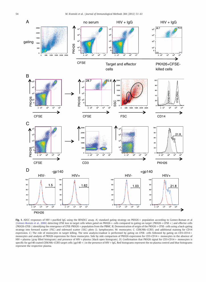
The RFADCC assay has proved a very useful non-radioactiveassay to measure ADCC, although in our hands the standardmethod of gating on PKH26+ targets cells and determiningthe proportion of CSFE-negative cells within this gate resultedin relatively high background levels of “killing” of non-antigencoated targets (Fig. S1) up to 20%. Although no informationof background killing of uncoated target cells of previouslypublished results is available, a mean background “killing” of14.4% for HIV-pooled sera in the presence of gp120 has beenreported (Gómez-Román et al., 2006). Following incubation offresh PBMC and HIV+plasma with HIV-1 Env (gp140) coatedCEM.NKr-CCR5 target cells for 4 h, we gated cells accordingto the published protocol for the RFADCC assay (Fig. 1A), inwhich PKH26+CFSE- cells are considered to derive from
PKH26+CFSE+target cells loosing CFSE during early eventsof IgG-effector cell mediated cytolysis (Gómez-Román et al.,2006). In the example presented in Fig. 1, this resulted inan apparent killing of 28.7% of the target cells (Fig. 1A, righthand panel). Based on population characteristics shown inthe bivariate CFSE/PKH26 dot-plot shown in Fig. 1A, centreright panel, PKH26+CFSE- cells clearly emerge from thePKH26-CFSE- pool of PBMC/effector cells. This prompted usto explore the origin of the CFSE-PKH+cells by back-gatingthe PKH26+CFSE-population onto the whole populationin the forward (FSC) and side scatter (SSC) plot. Althougha few of the PKH26+CFSE- cells were located within thelymphocyte and CEM.NKr-CCR5 gate the majority of PKH26+CFSE- cells fell within the characteristic monocyte area(Fig. 1B, second right). Staining with CD3 and CD14 antibodiesshowed that the PKH26+CFSE- cells were also CD3-CD14+(Fig. 1B right), confirming their monocyte origin. Importantly,none of the PKH26+CFSE+target cells were positive forCD14, showing that monocytes do not phagocytose wholeunkilled target cells within the time frame of the assay.
To determine the role of monocytes in the apparentkilling of target cells we gated on all CFSE- cells (Fig. 1C) andthen on CD3- and CD14+ cells within this population(Fig. 1C, centre panels). The proportion of monocytes positivefor PKH26 was then determined (Fig. 1C, right). The ADCCeffector activity in this gating strategy is defined as thepercent of PKH26+CD3-CD14+ monocytes above the noantibody control. Using this new CD14 gating strategy weshowed that the emergence of PKH26+CD3-CD14+ mono-cytes was dependent on the presence of HIV gp140 Envprotein and plasma IgG from HIV+subjects (Fig. 1D).
3.2. RFADCC assay using purified NK cells and monocytes aseffector cells
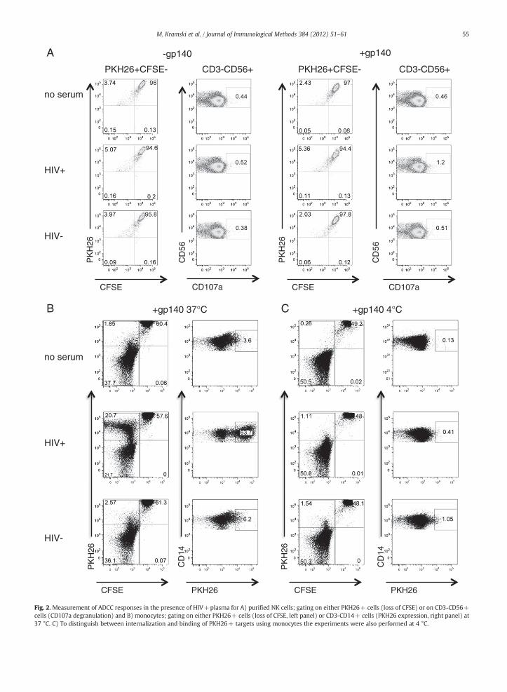
The above results suggested that monocytes play a majorrole in the RFADCC assay, but it is unclear whether theykill target cells directly or phagocytose cells and/or cellulardebris killed by NK cell ADCC activity. To address this wecultured NK cells purified by negative selection (>90% purity)or monocytes purified by countercurrent elutriation (>90%purity) with PKH26+CFSE+CEM.NKr-CCR5 gp140-coatedtarget cells in the presence and absence of HIV+serum.PKH26+CFSE- cells were not detected when gp140-coatedtargets were cultured with purified NK cells in the presence ofHIV+plasma with known ADCC activity (Fig. 2A). Consistentwith a lack of apparent killing, there was no detectabledegranulation, as assessed by CD107a staining, in NK cells inthe presence of HIV+plasma and gp140.
In contrast, PKH26+CFSE-cells were detected in thepresence of gp140-coated targets and HIV+plasma (Fig. 2B,left panel). The CD3-CD14+ monocytes acquired PKH26from gp140-coated target cells in the presence of HIV+plasma but not in the presence of HIV- plasma or in theabsence of any serum (Fig. 2B right panel). Acquisition ofPKH26 staining by monocytes was abolished when the assaywas conducted at 4 °C suggesting that it is due to internal-ization and not attachment of labeled cells to the monocytesurface (Fig. 2C). Overall these results confirm that mono-cytes, rather then NK cells, play a major role in the RFADCCassay.
gating
Target and effectorcells
PKH26+CFSE- killed cells
no serum HIV + IgG
HIV- HIV+ HIV- HIV+
HIV + IgG
CFSE
PK
H26
CFSE
PK
H26
CD3 PKH26
FSC S
SC
CD14 CFSE
PK
H26
L M
C
CFSE
PK
H26
-gp140 +gp140
PKH26
A
B
C
D
Fig. 1. ADCC responses of HIV+purified IgG using the RFADCC assay. A) standard gating strategy on PKH26+ population according to Gomez-Roman et al(Gómez-Román et al., 2006) detecting CFSE loss in target cells when gated on PKH26+ cells compared to gating on target (PKH26+CFSE+) and effector cells(PKH26-CFSE-) identifying the emergence of CFSE-PKH26+population from the PBMC. B) Demonstration of origin of the PKH26+CFSE- cells using a back-gatingstrategy into forward scatter (FSC) and sideward scatter (SSC) plots (L: lymphocytes; M: monocytes; C: CEM.NKr-CCR5) and additional staining for CD14expression. C) The role of monocytes in target killing. The new analysis/readout is performed by gating on CFSE- cells followed by gating on CD3-CD14+monocytes and analysis of PKH26 expression for these monocytes. Side by side comparison of PKH26 expression for CD3-CD14+ monocytes in the absence ofHIV+plasma (gray filled histogram) and presence of HIV+plasma (black open histogram). D) Confirmation that PKH26 signal for CD3-CD14+ monocytes isspecific for gp140 coated CEM.NKr-CCR5 target cells (gp140+) in the presence of HIV+IgG. Red histograms represent the no plasma control and blue histogramsrepresent the respective plasma.
54 M. Kramski et al. / Journal of Immunological Methods 384 (2012) 51–61
+gp140 -gp140
no serum
HIV+
HIV-
PKH26+CFSE- PKH26+CFSE-CD3-CD56+ CD3-CD56+
CFSE
PK
H26
CFSE P
KH
26
CD107a
CD
56
CD107a
CD
56
CFSE
PK
H26
no serum
HIV+
HIV-
CFSE PKH26 PKH26
+gp140 37°C +gp140 4°C
CD
14
PK
H26
CD
14
A
B C
Fig. 2. Measurement of ADCC responses in the presence of HIV+plasma for A) purified NK cells; gating on either PKH26+ cells (loss of CFSE) or on CD3-CD56+cells (CD107a degranulation) and B) monocytes; gating on either PKH26+ cells (loss of CFSE, left panel) or CD3-CD14+ cells (PKH26 expression, right panel) at37 °C. C) To distinguish between internalization and binding of PKH26+ targets using monocytes the experiments were also performed at 4 °C.
55M. Kramski et al. / Journal of Immunological Methods 384 (2012) 51–61

56 M. Kramski et al. / Journal of Immunological Methods 384 (2012) 51–61
3.3. Interaction of monocytes with target cells by Image flowanalysis
Monocytes are largely known for their ability to engulfand phagocytose target cells. From the above experiments,it was unclear if monocytes were simply ingesting whole,opsonized target cells or ingesting fragments of cellsfollowing killing by other mechanisms. To further investigatethe monocyte-target cell interaction we performed theRFADCC assay and imaged monocytes, NK cells andCEM.NKr-CCR5 target cells in the presence of gp140 andHIV+plasma by image flow cytometry. The "brightfieldaspect ratio" (measures the ratio of the cell's minor axis toits major axis which is close to 1 for round cells) was plottedagainst the “brightfield cell area” to identify single cells. Dueto the large size difference between the CEM cells and PBMCs,single events were further distinguished by single nucleiusing the spot count feature in the Draq5 channel (nucleistain) (Fig. 3A1 and B1). Singlets were plotted on bivariateCD14+PKH26+ and singlet CD56+PKH26+ dot plots.Images from CD14+PKH26+ monocytes showed thatPKH26 staining of monocytes is not due to bound targetcells but suggest that monocytes had internalized PKH26+membrane rather than phagocytosed whole target cellsas PKH26 stain appears in endocytic/phagocytic vesicles(Fig. 3A2). In contrast to monocytes, NK cells were notobserved to take up the PKH26 dye (Fig. 3B2).
3.4. Uptake of PKH26+ membrane by monocytes withoutphagocytosis
Imaging flow cytometry showed distinct uptake by normalsized monocytes of the PKH26, but due to the relativelylow resolution the possibility of phagocytosis could not becompletely excluded. To further examine the mechanism of cellkilling and exclude the possibility of phagocytosis of wholetarget cells, we analyzed the fate of target cells over time inthe RFADCC assay using high resolution deconvolution micros-copy. PBMC were cultured with PKH26+CFSE+gp140-coatedCEM.NKr-CCR5 target cells in the presence of HIV+plasma.Within 10 min we observed physical attachment ofCEM.NKr-CCR5 target cells to CD14+monocytes that persistedover time (Fig. 4). From 30 min onwards attachment ofmonocytes to CEM.NKr-CCR5 target cells was associated withformation of dense PKH26-staining bodies. After 60 min weobserved that monocytes associated with target cells showedpunctuate staining with PKH26 similar to that of the targetcells. There was no evidence of phagocytosis of entire targetcells (Fig. 4).
3.5. Analysis of RFADCC assay in 53 HIV+human samples
The above experiments show that in the RFADCC assay,PKH26 dye derived from opsonised target cells becomes
Fig. 3. Image stream flow cytometry analysis after RFADCC assay using healthy PBMaspect ratio (R1) and distinguishing events with single nuclei using the spot countCEM.NKr-CCR5 cells were plotted on CD14/PKH16 dot plots (R3-5). A2) repreCEM.NKr-CCR5 cells (R3) and CD14+PKH26+ double positive monocytes (singleaspect ratio (R1) and distinguishing events with single nuclei using the spot count feCEM.NKr-CCR5 cells were plotted on CD56/PKH16 dot plots (R3-R5). B2) reprCEM.NKr-CCR5 cells (R3) are shown. DRAQ5 was used for co-staining of the nuclei
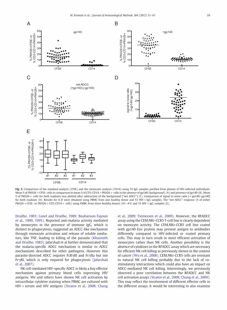
associated with monocytes. This suggested that an alternatemethod to analyze ADCC activity in the RFADCC assay would beto gate on monocytes taking up the PKH26 dye. To validate theuse of a monocyte gating strategy, and to confirm that our initialobservations with plasma were not mediated by complementactivation,we tested purified IgG fromplasma of 53HIV-infectedsubjects and PBMC from three uninfected individuals in theRFADCC assay. We compared the standard gating on thePKH26+ population and the loss of CFSE ("CFSE") with thenew PKH26+CD3-CD14+ readout (“CD14”) (Fig. 5). Wedetected significant background killing of target cells in theabsence of gp140 using the standard approach of analysis(PKH26+CFSE-, mean 17.81%, range 6.3-39.77). However,there was little or no background using our CD14 based gatinganalysis (PKH26+CD3-CD14+, mean 1.25%, range 0.31–7.56)(Fig. 5A). To obtain the net ADCC response (Fig. 5C) wesubtracted the background measured for target cells withoutgp140 (Fig. 5A) from the response measured for gp140 coatedtarget cells (Fig. 5B). The overall ADCC response ("net ADCC")was higher for the standard analysis of PKH26+CFSE- cellscompared to the CD14 gating analysis with mean killing of18.49% (2–35.84) and 12.65% (7.25-17.89), respectively(Fig. 5C). Using the PKH26+CD3-CD14+ readout for theRFADCC assay, a higher signal to noise ratio of gp140-coatedand gp140-uncoated (background) cells was detected com-pared to the standard PKH26+CFSE- analysis (Fig. 5D).
There is typically some variability in the killing mediatedby NK cells from different healthy donors. To ensure that theresults were valid across multiple sources of healthy donoreffector cells, we tested fresh PBMC from three subjectsagainst the entire panel of 53 HIV+IgG samples. Fig. 5Eshows the "net ADCC" response which was different for boththe standard CFSE- and CD14 readout but similar magnitudesof responses were obtained for all three donors.
4. Discussion
ADCC is an immune response of emerging interest with apotential role in controlling HIV infection (Lambotte et al., 2009;Berger and Alter, 2011; Chung et al., 2011). ADCC activity ismostcommonly attributed to NK cells although monocytes andgranulocytes also bear Fcγ receptors (FcγR), including FcγRI,FcγRIIa/b and FcγIII andhavebeen shown to kill targets viaADCC(Barker and Reisfeld, 1993; Horner et al., 2007; Wu et al., 2008).Effector functions of monocytes/macrophages focus mainly onphagocytosis with an increasing interest in antibody-dependentphagocytosis in the context of HIV (Ackerman et al., 2011;Dugast et al., 2011; McAndrew et al., 2011).
The RFADCC assay has become a widely used tool to studyNK cell ADCC. We demonstrate that NK cell activity is notthe major killing mechanism detected in the RFADCC assay ifPBMCs are used as effector cells. We performed the RFADCCwith purified NK cells and were unable to detect significantdegranulation in the presence of HIV+IgG and gp140.
C and HIV+plasma. A1) Single cells were identified by plotting cell area vsfeature in the Draq5 channel (R2). Singlet CD14+ monocytes and PKH26+sentative images for cell controls CD14+ monocytes (R5) and PKH26+ts) (R4) are shown. B1) Single cells were identified by plotting cell area vsature in the Draq5 channel (R2). Singlet CD56+ NK cells and PKH26+CFSE-esentative images for cell controls CD56+ NK cells (R5) and PKH26+.

A1
A2
B1
B2
R5
R4
R3R4
R5
R3
57M. Kramski et al. / Journal of Immunological Methods 384 (2012) 51–61
However this could be due to NK cell activation during thepurification process or because the target:effector ratio isnot ideal. Further evaluation of the RFADCC with purified NK
cells or NK cell lines is warranted to confirm that the RFADCCassay can measure NK cell-mediated ADCC. In fact our dataindicates that the RFADCC assay does not measure killing by

Fig. 4. Deconvolution microscopy assessing interaction of monocytes and NK cells in the RFADCC assay over time. Cem.NKr-CCR5 cells were labelled with PKH26(orange), monocytes with CD14-FITC (green) and NK cells with CD56-Brilliant violet (blue). Nuclei were co-stained with DRAQ5. Arrows indicate PKH26+membrane uptake by monocytes. Scale bars indicate a size of 20 μm.
58 M. Kramski et al. / Journal of Immunological Methods 384 (2012) 51–61
NK cells but rather measures IgG-mediated monocyte func-tion. The PKH26+CFSE- cell population typically reported as“killed” target cells in this assay are actually CD3-CD14+monocytes with internalized PKH26 membrane fragments,rather than CEM.NKr.CCR5 target cells that have lost CFSE.Moreover, our microscopy results clearly show that mono-cytes take up PKH26-stained target cell membrane withoutevidence of characteristic for classical phagocytosis of theintact target cell. Our microscopy results showed that
membranes of monocyte-engaged target cells display distinctmembrane vesicles that could be apoptotic bodies passedon to the monocytes. Further experiments are required toelucidate the precisemechanisms of how andwhen the targetcells are killed during this process.
Previous studies have reported monocyte-mediated non-phagocytosis effector function in the context of malaria andcancer using either antibody-dependent cellular inhibition or51Cr release assays (Shaw et al., 1978b, 1978a; Khusmith and
A B
C D
E
Fig. 5. Comparison of the standard analysis (CFSE) and the monocyte analysis (CD14) using 53 IgG samples purified from plasma of HIV-infected individuals.Mean % of PKH26+CFSE- cells in comparison to mean % of CD3-CD14+PKH26+ cells in the absence of gp140 (background) (A) and presence of gp140 (B). Mean% of PKH26+ cells for both readouts was plotted after subtraction of the background [“net ADCC”] (C). Comparison of signal to noise ratio [+gp140/-gp140]for both readouts (D). Results for A-D were obtained using PBMC from one healthy donor and 53 HIV+IgG samples. The “net ADCC” response (% of eitherPKH26+CFSE- or PKH26+CD3-CD14+ cells) using PMBC from three healthy donors (#1- #3) and 53 HIV+IgG samples (E).
59M. Kramski et al. / Journal of Immunological Methods 384 (2012) 51–61
Druilhe, 1983; Lunel and Druilhe, 1989; Bouharoun-Tayounet al., 1990, 1995). Reported anti-malaria activity mediatedby monocytes in the presence of immune IgG, which isdistinct to phagocytosis, suggested an ADCC-like mechanismthrough monocyte activation and release of soluble media-tors, like TNF, leading to killing of the parasite (Khusmithand Druilhe, 1983). Jafarshad et al further demonstrated thatthe malaria-specific ADCC mechanism is similar to ADCCmechanisms described for other pathogens. However thisparasite-directed ADCC requires FcRγIII and FcγIIa but notFcγRI, which is only required for phagocytosis (Jafarshadet al., 2007).
NK cell-mediated HIV-specific ADCC is likely a key effectormechanism against primary blood cells expressing HIVantigens. We and others have shown NK cell activation byintracellular cytokine staining when PBMC are cultured withHIV+serum and HIV antigens (Stratov et al., 2008; Chung
et al., 2009; Tiemessen et al., 2009). However, the RFADCCassay using the CEM.NKr-CCR5 T-cell line is clearly dependenton monocyte activity. The CEM.NKr-CCR5 cell line coatedwith gp140 Env protein may present antigen to antibodiesdifferently compared to HIV-infected or coated primarycells. This may in turn result in more efficient activation ofmonocytes rather than NK cells. Another possibility is theabsence of cytokines in the RFADCC assaywhich are necessaryfor efficient NK cell killing as previously shown in the contextof cancer (Wu et al., 2008). CEM.NKr-CCR5 cells are resistantto natural NK cell killing probably due to the lack of co-stimulatory interactions which could also have an impact onADCC-mediated NK cell killing. Interestingly, we previouslyobserved a poor correlation between the RFADCC and NKcell activation assays (Stratov et al., 2008; Chung et al., 2009).This may reflect the involvement of different effector cells inthe different assays. It would be interesting to also examine

60 M. Kramski et al. / Journal of Immunological Methods 384 (2012) 51–61
monocyte activation in other ADCC assays to further probemonocyte-mediated ADCC activities. Results obtained fromassays using infected cells (Pollara et al., 2011) suggestthat NK cells play an important role in HIV ADCC. Howeverthe involvement of monocytes in these assays has not beenstudied.
We have studied IgG samples from HIV-infected subjectswith varying stages of disease. It would be of interest toevaluate IgG or serum samples in the context of vaccinationstudies. Others have detected ADCC responses after vacci-nation using the RFADCC assay (Gómez-Román et al., 2005;Chung et al., 2009; Florese et al., 2009; Vaine et al., 2010;Xiao et al., 2010; Patterson et al., 2011); whether theseresponses also reflect monocyte-mediated ADCC remains tobe shown.
5. Conclusion
Our studies clarify the understanding of the cellularevents underlying IgG-mediated HIV-specific cellular effectorfunction detected by the RFADCC assay. We demonstrate thatthis assay primarily reflects Ab-mediated monocyte functionand not NK cell ADCC and therefore has to be treated withcaution in regards to the interpretation for NK cell-mediatedADCC. Presumably, lysis is not mediated by complementactivation because the effect is seen with purified IgG. Ouramended gating on monocytes taking up PKH26 membranereduces the background killing signals remarkably. Thisshould be a useful improvement of the assay when ADCCresponses are expected to be low, as in the case of earlyresponses following vaccination.
Supplementary data to this article can be found online athttp://dx.doi.org/10.1016/j.jim.2012.07.006.
Acknowledgements
This work was supported by Australian NHMRC award510448 and NIH award R21AI081541. We are grateful to allthe subjects who kindly provided blood samples and theircarers. We also thank Damian Purcell for helpful discussions.
References
Ackerman, M.E., Moldt, B., Wyatt, R.T., Dugast, A.S., McAndrew, E., Tsoukas, S.,Jost, S., Berger, C.T., Sciaranghella, G., Liu, Q., Irvine, D.J., Burton, D.R., Alter,G., 2011. A robust, high-throughput assay to determine the phagocyticactivity of clinical antibody samples. J. Immunol. Methods 366, 8.
Ahmad, R., Sindhu, S.T., Toma, E., Morisset, R., Vincelette, J., Menezes, J.,Ahmad, A., 2001. Evidence for a correlation between antibody-dependent cellular cytotoxicity-mediating anti-HIV-1 antibodies andprognostic predictors of HIV infection. J. Clin. Immunol. 21, 227.
Alter, G., Altfeld, M., 2009. NK cells in HIV-1 infection: evidence for their rolein the control of HIV-1 infection. J. Intern. Med. 265, 29.
Barker, E., Reisfeld, R.A., 1993. A mechanism for neutrophil-mediated lysisof human neuroblastoma cells. Cancer Res. 53, 362.
Battle-Miller, K., Eby, C.A., Landay, A.L., Cohen, M.H., Sha, B.E., Baum, L.L.,2002. Antibody-dependent cell-mediated cytotoxicity in cervical lavagefluids of human immunodeficiency virus type 1‐infected women.J. Infect. Dis. 185, 439.
Baum, L.L., Cassutt, K.J., Knigge, K., Khattri, R., Margolick, J., Rinaldo, C.,Kleeberger, C.A., Nishanian, P., Henrard, D.R., Phair, J., 1996. HIV-1 gp120-specific antibody-dependent cell-mediated cytotoxicity correlates withrate of disease progression. J. Immunol. 157, 2168.
Berger, C.T., Alter, G., 2011. Natural killer cells in spontaneous control of HIVinfection. Curr. Opin. HIV AIDS 6, 208.
Bouharoun-Tayoun, H., Attanath, P., Sabchareon, A., Chongsuphajaisiddhi, T.,Druilhe, P., 1990. Antibodies that protect humans against Plasmodiumfalciparum blood stages do not on their own inhibit parasite growth andinvasion in vitro, but act in cooperation with monocytes. J. Exp. Med.172, 1633.
Bouharoun-Tayoun, H., Oeuvray, C., Lunel, F., Druilhe, P., 1995. Mechanismsunderlying the monocyte-mediated antibody-dependent killing ofPlasmodium falciparum asexual blood stages. J. Exp. Med. 182, 409.
Center, R.J., Wheatley, A.K., Campbell, S.M., Gaeguta, A.J., Peut, V., Alcantara, S.,Siebentritt, C., Kent, S.J., Purcell, D.F., 2009. Induction of HIV-1 subtype Band AE-specific neutralizing antibodies in mice and macaques with DNAprime and recombinant gp140 protein boost regimens. Vaccine 27, 6605.
Chung, A.W., Rollman, E., Center, R.J., Kent, S.J., Stratov, I., 2009. Rapiddegranulation of NK cells following activation by HIV-specific antibodies.J. Immunol. 182, 1202.
Chung, A.W., Navis, M., Isitman, G., Wren, L., Silvers, J., Amin, J., Kent, S.J.,Stratov, I., 2011. Activation of NK cells by ADCC antibodies and HIVdisease progression. J. Acquir. Immune Defic. Syndr. 58, 127.
Cox, J.H., 1999. HIV-1-Specific Antibody-Dependent Cellular Cytotoxicity(ADCC). Methods Mol. Med. 17, 373.
Cox, J.H., Garner, R.P., Redfield, R.R., Aronson, N.E., Davis, C., Ruiz, N., Birx, D.L.,1999. Antibody-dependent cellular cytotoxicity in HIV type 1-infectedpatients receiving VaxSyn, a recombinant gp160 envelope vaccine. AIDSRes. Hum. Retroviruses 15, 847.
Dugast, A.S., Tonelli, A., Berger, C.T., Ackerman, M.E., Sciaranghella, G., Liu, Q.,Sips, M., Toth, I., Piechocka-Trocha, A., Ghebremichael, M., Alter, G., 2011.Decreased Fc receptor expression on innate immune cells is associatedwith impaired antibody-mediated cellular phagocytic activity in chroni-cally HIV-1 infected individuals. Virology 415 (2), 160–167 (Jul 5).
Florese, R.H., Demberg, T., Xiao, P., Kuller, L., Larsen, K., Summers, L.E.,Venzon, D., Cafaro, A., Ensoli, B., Robert-Guroff, M., 2009. Contributionof nonneutralizing vaccine-elicited antibody activities to improvedprotective efficacy in rhesus macaques immunized with Tat/Envcompared with multigenic vaccines. J. Immunol. 182, 3718.
Gómez-Román, V.R., Patterson, L.J., Venzon, D., Liewehr, D., Aldrich, K.,Florese, R., Robert-Guroff, M., 2005. Vaccine-elicited antibodies mediateantibody-dependent cellular cytotoxicity correlated with significantlyreduced acute viremia in rhesus macaques challenged with SIVmac251.J. Immunol. 174, 2185.
Gómez-Román, V.R., Florese, R.H., Patterson, L.J., Peng, B., Venzon, D., Aldrich,K., Robert-Guroff, M., 2006. A simplified method for the rapid fluorometricassessment of antibody-dependent cell-mediated cytotoxicity. J. Immunol.Methods 308, 53.
Haynes, B.F., Gilbert, P.B., McElrath, M.J., Zolla-Pazner, S., Tomaras, G.D.,Alam, S.M., Evans, D.T., Montefiori, D.C., Karnasuta, C., Sutthent, R., Liao,H.X., DeVico, A.L., Lewis, G.K., Williams, C., Pinter, A., Fong, Y., Janes, H.,DeCamp, A., Huang, Y., Rao, M., Billings, E., Karasavvas, N., Robb, M.L.,Ngauy, V., de Souza, M.S., Paris, R., Ferrari, G., Bailer, R.T., Soderberg, K.A.,Andrews, C., Berman, P.W., Frahm, N., De Rosa, S.C., Alpert, M.D., Yates,N.L., Shen, X., Koup, R.A., Pitisuttithum, P., Kaewkungwal, J., Nitayaphan,S., Rerks-Ngarm, S., Michael, N.L., Kim, J.H., 2012. Immune-correlatesanalysis of an HIV-1 vaccine efficacy trial. N. Engl. J. Med. 366, 1275.
Hessell, A.J., Hangartner, L., Hunter, M., Havenith, C.E., Beurskens, F.J., Bakker,J.M., Lanigan, C.M., Landucci, G., Forthal, D.N., Parren, P.W., Marx, P.A.,Burton, D.R., 2007. Fc receptor but not complement binding is importantin antibody protection against HIV. Nature 449, 101.
Hezareh, M., Hessell, A.J., Jensen, R.C., van de Winkel, J.G., Parren, P.W., 2001.Effector function activities of a panel of mutants of a broadlyneutralizing antibody against human immunodeficiency virus type 1.J. Virol. 75, 12161.
Hidajat, R., Xiao, P., Zhou, Q., Venzon, D., Summers, L.E., Kalyanaraman, V.S.,Montefiori, D.C., Robert-Guroff, M., 2009. Correlation of vaccine-elicitedsystemic and mucosal nonneutralizing antibody activities with reducedacute viremia following intrarectal simian immunodeficiency virusSIVmac251 challenge of rhesus macaques. J. Virol. 83, 791.
Horner, H., Frank, C., Dechant, C., Repp, R., Glennie, M., Herrmann, M.,Stockmeyer, B., 2007. Intimate cell conjugate formation and exchange ofmembrane lipids precede apoptosis induction in target cells duringantibody-dependent, granulocyte-mediated cytotoxicity. J. Immunol.179, 337.
Huber, M., Trkola, A., 2007. Humoral immunity to HIV-1: neutralization andbeyond. J. Intern. Med. 262, 5.
Jafarshad, A., Dziegiel, M.H., Lundquist, R., Nielsen, L.K., Singh, S., Druilhe, P.L.,2007. A novel antibody-dependent cellular cytotoxicity mechanisminvolved in defense against malaria requires costimulation of monocytesFcgammaRII and FcgammaRIII. J. Immunol. 178, 3099.
Johansson, S.E., Rollman, E., Chung, A.W., Center, R.J., Hejdeman, B., Stratov,I., Hinkula, J., Wahren, B., Kärre, K., Kent, S.J., Berg, L., 2011. NK cellfunction and antibodies mediating ADCC in HIV-1-infected viremic andcontroller patients. Viral Immunol. 24, 359.

61M. Kramski et al. / Journal of Immunological Methods 384 (2012) 51–61
Khusmith, S., Druilhe, P., 1983. Cooperation between antibodies and mono-cytes that inhibit in vitro proliferation of Plasmodium falciparum. Infect.Immun. 41, 219.
Lambotte, O., Ferrari, G., Moog, C., Yates, N.L., Liao, H.X., Parks, R.J., Hicks, C.,Owzar, K., Tomaras, G.D., Montefiori, D.C., Haynes, B.F., Delfraissy, J.F.,2009. Heterogeneous neutralizing antibody and antibody-dependentcell cytotoxicity responses in HIV-1 elite controllers. AIDS 23, 897.
Lunel, F., Druilhe, P., 1989. Effector cells involved in nonspecific and antibody-dependent mechanisms directed against Plasmodium falciparum bloodstages in vitro. Infect. Immun. 57, 2043.
McAndrew, E.G., Dugast, A.S., Licht, A.F., Eusebio, J.R., Alter, G., Ackerman, M.E.,2011. Determining the phagocytic activity of clinical antibody samples.J. Vis. Exp. e3588.
Nag, P., Kim, J., Sapiega, V., Landay, A.L., Bremer, J.W.,Mestecky, J., Reichelderfer,P., Kovacs, A., Cohn, J., Weiser, B., Baum, L.L., 2004. Women withcervicovaginal antibody-dependent cell-mediated cytotoxicity have lowergenital HIV-1 RNA loads. J. Infect. Dis. 190, 1970.
Patterson, L.J., Daltabuit-Test, M., Xiao, P., Zhao, J., Hu, W., Wille-Reece, U.,Brocca-Cofano, E., Kalyanaraman, V.S., Kalisz, I., Whitney, S., Lee, E.M.,Pal, R., Montefiori, D.C., Dandekar, S., Seder, R., Roederer, M., Wiseman,R.W., Hirsch, V., Robert-Guroff, M., 2011. Rapid SIV Env-specific mucosaland serum antibody induction augments cellular immunity in protectingimmunized, elite-controller macaques against high dose heterologousSIV challenge. Virology 411, 87.
Pollara, J., Hart, L., Brewer, F., Pickeral, J., Packard, B.Z., Hoxie, J.A., Komoriya,A., Ochsenbauer, C., Kappes, J.C., Roederer, M., Huang, Y., Weinhold, K.J.,Tomaras, G.D., Haynes, B.F., Montefiori, D.C., Ferrari, G., 2011. High-throughput quantitative analysis of HIV-1 and SIV-specific ADCC-mediating antibody responses. Cytometry A 79, 603.
Rerks-Ngarm, S., Pitisuttithum, P., Nitayaphan, S., Kaewkungwal, J., Chiu, J.,Paris, R., Premsri, N., Namwat, C., de Souza, M., Adams, E., Benenson, M.,Gurunathan, S., Tartaglia, J., McNeil, J.G., Francis, D.P., Stablein, D., Birx, D.L.,Chunsuttiwat, S., Khamboonruang, C., Thongcharoen, P., Robb, M.L.,Michael, N.L., Kunasol, P., Kim, J.H., Investigators, M.-T., 2009. Vaccinationwith ALVAC and AIDSVAX to prevent HIV-1 infection in Thailand. N. Engl.J. Med. 361, 2209.
Shaw, G.M., Levy, P.C., LoBuglio, A.F., 1978a. Human monocyte antibody-dependent cell-mediated cytotoxicity to tumor cells. J. Clin. Invest. 62,1172.
Shaw, G.M., Levy, P.C., LoBuglio, A.F., 1978b. Human monocyte cytotoxicityto tumor cells. I. Antibody-dependent cytotoxicity. J. Immunol. 121,573.
Sheehy, M.E., McDermott, A.B., Furlan, S.N., Klenerman, P., Nixon, D.F., 2001.A novel technique for the fluorometric assessment of T lymphocyteantigen specific lysis. J. Immunol. Methods 249, 99.
Stratov, I., Chung, A., Kent, S.J., 2008. Robust NK cell-mediated humanimmunodeficiency virus (HIV)-specific antibody-dependent responsesin HIV-infected subjects. J. Virol. 82, 5450.
Tiemessen, C.T., Shalekoff, S., Meddows-Taylor, S., Schramm, D.B.,Papathanasopoulos, M.A., Gray, G.E., Sherman, G.G., Coovadia, A.H.,Kuhn, L., 2009. Cutting Edge: Unusual NK cell responses to HIV-1peptides are associated with protection against maternal-infant trans-mission of HIV-1. J. Immunol. 182, 5914.
Vaine, M., Wang, S., Liu, Q., Arthos, J., Montefiori, D., Goepfert, P., McElrath,M.J., Lu, S., 2010. Profiles of human serum antibody responses elicited bythree leading HIV vaccines focusing on the induction of Env-specificantibodies. PLoS One 5, e13916.
Wu, L., Adams, M., Carter, T., Chen, R., Muller, G., Stirling, D., Schafer, P., Bartlett,J.B., 2008. lenalidomide enhances natural killer cell and monocyte-mediated antibody-dependent cellular cytotoxicity of rituximab-treatedCD20+ tumor cells. Clin. Cancer Res. 14, 4650.
Xiao, P., Zhao, J., Patterson, L.J., Brocca-Cofano, E., Venzon, D., Kozlowski, P.A.,Hidajat, R., Demberg, T., Robert-Guroff, M., 2010. Multiple vaccine-elicited nonneutralizing antienvelope antibody activities contribute toprotective efficacy by reducing both acute and chronic viremia followingsimian/human immunodeficiency virus SHIV89.6P challenge in rhesusmacaques. J. Virol. 84, 7161.
Yamada, T., Watanabe, N., Nakamura, T., Iwamoto, A., 2004. Antibody-dependent cellular cytotoxicity via humoral immune epitope of Nefprotein expressed on cell surface. J. Immunol. 172, 2401.