Journal of Immunological Methods - Perelman School of ... et all 2010.pdf · Journal of...
12
Research paper Rapid isolation of high-affinity human antibodies against the tumor vascular marker Endosialin/TEM1, using a paired yeast-display/secretory scFv library platform Aizhi Zhao a , Selene Nunez-Cruz a,1 , Chunsheng Li a,1 , George Coukos a,b , Don L. Siegel b , Nathalie Scholler a, ⁎ a Penn Ovarian Cancer Research Center, Center for Research on Reproduction and Women's Health (CRRWH), Department of Obstetrics and Gynecology, University of Pennsylvania, Philadelphia, PA 19104, United States b Department of Pathology and Laboratory Medicine, University of Pennsylvania, Philadelphia, PA 19104, United States article info abstract Article history: Received 16 March 2010 Received in revised form 12 August 2010 Accepted 1 September 2010 Available online 15 September 2010 Endosialin/TEM1 is predominantly expressed on neovasculature, thus ideally suited for diagnostic, targeted imaging and therapy of cancer. To isolate TEM1-specific affinity reagents, we thought to screen a recombinant antibody (scFv) library derived from the repertoire of a patient with thrombotic thrombocytopenic purpura (TTP), as autoimmune disorders may produce self-reactive specificities. The yeast-display scFv library was constructed by homologous recombination of the TTP patient repertoire originally expressed on M13 bacteriophage in the novel vector pAGA2 for yeast-display expression. The TTP yeast-display library (10 9 members) was screened by magnetic and flow sorting with human TEM1 recombinant protein. A pool of yeast-display scFv able to detect 2 nM of TEM1 was obtained and transformed into yeast-secreted scFv by homologous recombination using the novel p416 BCCP vector for yeast secretion of biotinylated scFv. Anti-TEM1 yeast-secreted scFv were independently validated in vitro by flow cytometry analysis and ELISA assays, then in vivo biotinylated in N-termini to produce biobodies. Biobody-78 bound specifically to Endosialin/TEM1-expressing ovarian tumor in vivo, with functional stability over 48 h. Our results suggest that our novel paired display-secretory yeast libraries can serve as an ideal platform for the rapid isolation of high-affinity reagents, and that anti-TEM1 biobody-78 can be used for in vitro assays including flow cytometry analysis, as well as in vivo for targeted imaging and therapy of cancer. © 2010 Elsevier B.V. All rights reserved. Keywords: Tumor vascular markers Ovarian cancer Yeast-display scFv Biobody Flow cytometry In vivo targeting 1. Introduction The growth of solid tumors beyond a diameter of 1–2 mm critically depends on the formation of a supporting stroma of newly formed blood vessels (Folkman, 1985). Tumor endo- thelial cells, stromal fibroblasts (activated fibroblast or myofibroblasts) and/or vascular pericytes acquire a pheno- type different from that of normal stromal cells (Rettig et al., 1992; Christian et al., 2008) and express tumor vascular markers (TVM). TVM provide attractive targets for antibody- based tumor diagnosis and therapy (St Croix et al., 2000; Marty et al., 2006; Teicher, 2007; Rouleau et al., 2008) due to Journal of Immunological Methods 363 (2011) 221–232 Abbreviations: scFv, Single chain variable fragments; vs., Versus; pM, Picomolar; nM, Nanomolar; rhTEM1-GST, Recombinant human protein Endosialin/TEM1 fused to GST tag; rGST, Recombinant GST tag; RT, Room temperature; ON, Overnight; PBS, Phosphate buffered saline; PBSM, PBS supplemented with 5% dry milk; PBST, PBS supplemented with 0.05% Tween; mAb, Mouse monoclonal antibody; μg/ml, Microgram per ml; IV, Intrave- nous; EPR, Enhanced Permeability and Retention effect. ⁎ Corresponding author. CRRWH, 1309 BRBII/III, 421 Curie Boulevard, University of Pennsylvania, Philadelphia, PA 19104, United States. Tel.: + 1 215 898 0164; fax: +1 215 573 5129. E-mail address: [email protected] (N. Scholler). 1 These authors equally contributed to the work. 0022-1759/$ – see front matter © 2010 Elsevier B.V. All rights reserved. doi:10.1016/j.jim.2010.09.001 Contents lists available at ScienceDirect Journal of Immunological Methods journal homepage: www.elsevier.com/locate/jim
Transcript of Journal of Immunological Methods - Perelman School of ... et all 2010.pdf · Journal of...

Journal of Immunological Methods 363 (2011) 221–232
Contents lists available at ScienceDirect
Journal of Immunological Methods
j ourna l homepage: www.e lsev ie r.com/ locate / j im
Research paper
Rapid isolation of high-affinity human antibodies against the tumor vascularmarker Endosialin/TEM1, using a paired yeast-display/secretory scFvlibrary platform
Aizhi Zhao a, Selene Nunez-Cruz a,1, Chunsheng Li a,1, George Coukos a,b,Don L. Siegel b, Nathalie Scholler a,⁎a Penn Ovarian Cancer Research Center, Center for Research on Reproduction and Women's Health (CRRWH), Department of Obstetrics and Gynecology, University ofPennsylvania, Philadelphia, PA 19104, United Statesb Department of Pathology and Laboratory Medicine, University of Pennsylvania, Philadelphia, PA 19104, United States
a r t i c l e i n f o
Abbreviations: scFv, Single chain variable fragmePicomolar; nM, Nanomolar; rhTEM1-GST, RecombiEndosialin/TEM1 fused to GST tag; rGST, Recombinatemperature; ON, Overnight; PBS, Phosphate buffersupplemented with 5% dry milk; PBST, PBS supplementmAb, Mouse monoclonal antibody; μg/ml, Microgramnous; EPR, Enhanced Permeability and Retention effec⁎ Corresponding author. CRRWH, 1309 BRBII/III,
University of Pennsylvania, Philadelphia, PA 19104, U215 898 0164; fax: +1 215 573 5129.
E-mail address: [email protected] (N. Sc1 These authors equally contributed to the work.
0022-1759/$ – see front matter © 2010 Elsevier B.V.doi:10.1016/j.jim.2010.09.001
a b s t r a c t
Article history:Received 16 March 2010Received in revised form 12 August 2010Accepted 1 September 2010Available online 15 September 2010
Endosialin/TEM1 is predominantly expressed on neovasculature, thus ideally suited fordiagnostic, targeted imaging and therapy of cancer. To isolate TEM1-specific affinity reagents,we thought to screen a recombinant antibody (scFv) library derived from the repertoire of apatient with thrombotic thrombocytopenic purpura (TTP), as autoimmune disorders mayproduce self-reactive specificities. The yeast-display scFv library was constructed by homologousrecombination of the TTP patient repertoire originally expressed on M13 bacteriophage in thenovel vector pAGA2 for yeast-display expression. The TTP yeast-display library (109 members)was screened by magnetic and flow sorting with human TEM1 recombinant protein. A pool ofyeast-display scFv able to detect 2 nM of TEM1was obtained and transformed into yeast-secretedscFv by homologous recombination using the novel p416 BCCP vector for yeast secretion ofbiotinylated scFv. Anti-TEM1 yeast-secreted scFv were independently validated in vitro by flowcytometry analysis and ELISA assays, then in vivo biotinylated in N-termini to produce biobodies.Biobody-78 bound specifically to Endosialin/TEM1-expressing ovarian tumor in vivo, withfunctional stability over 48 h. Our results suggest that our novel paired display-secretory yeastlibraries can serve as an ideal platform for the rapid isolation of high-affinity reagents, and thatanti-TEM1 biobody-78 can be used for in vitro assays including flow cytometry analysis, aswell asin vivo for targeted imaging and therapy of cancer.
© 2010 Elsevier B.V. All rights reserved.
Keywords:Tumor vascular markersOvarian cancerYeast-display scFvBiobodyFlow cytometryIn vivo targeting
nts; vs., Versus; pM,nant human proteinnt GST tag; RT, Roomed saline; PBSM, PBSed with 0.05% Tween;per ml; IV, Intrave-
t.421 Curie Boulevard,nited States. Tel.: +1
holler).
All rights reserved.
1. Introduction
The growth of solid tumors beyond a diameter of 1–2 mmcritically depends on the formation of a supporting stroma ofnewly formed blood vessels (Folkman, 1985). Tumor endo-thelial cells, stromal fibroblasts (activated fibroblast ormyofibroblasts) and/or vascular pericytes acquire a pheno-type different from that of normal stromal cells (Rettig et al.,1992; Christian et al., 2008) and express tumor vascularmarkers (TVM). TVM provide attractive targets for antibody-based tumor diagnosis and therapy (St Croix et al., 2000;Marty et al., 2006; Teicher, 2007; Rouleau et al., 2008) due to
222 A. Zhao et al. / Journal of Immunological Methods 363 (2011) 221–232
i) the relative stability of TVM-expressing cells comparing totumor cells; ii) neovasculature essential function for tumormaintenance, as demonstrated by the widespread necrosis ofsolid tumor after destruction of their blood vessels (Hinnenand Eskens, 2007); iii) neovasculature leaky capillaries thatpermit circulating antibodies and antibody conjugates toeasily access TVM.
Endosialin/Tumor EndothelialMarker 1 (TEM1 or CD248) isa TVM and a type I transmembrane protein which comprisesan 80.9 kDa protein core modified by extensive sialylated O-linked glycosylation that gives rise to an approximately175 kDa mature glycoprotein (Christian et al., 2001). Endosia-lin/TEM1 was originally discovered by an anti-fibroblastmonoclonal antibody (FB5) as a glycoprotein expressed bythe pericytes and myofibroblasts associated with tumorvasculature (MacFadyen et al., 2005; Christian et al., 2008;Rouleau et al., 2008) as well as by tumor-associated vascularendothelial cells in various human cancers (Rettig et al., 1992;Davies et al., 2004; Rmali et al., 2005; Becker et al., 2008),including ovarian cancer (Conejo-Garcia et al., 2005). Endo-sialin/TEM1 plays a unique role in tumor progression as apromoting factor of tumor angiogenesis (Bagley et al., 2008),proliferation, migration and metastasis through interactionwith matrix proteins such as fibronectin, collagen type I and IV(Tomkowicz et al., 2007) and Mac-2 BP/90K (Becker et al.,2008). Importantly, mice without functional Tem1 genepresent a striking reduction in tumor growth, invasiveness,and metastasis after tumor transplantation to abdominal sites(Nanda et al., 2006). Taken together, these results suggest thattargeting Endosialin/TEM1 for diagnostic and/or therapy couldprovide a valuable strategy against cancer.
Isolation of antigen-specific antibodies has been achievedthrough a variety of methods, including screening of phage-and yeast-display recombinant antibody (scFv) libraries(Vaughan et al., 1996; Feldhaus et al., 2003; Paschke, 2006;Scholler et al., 2006, 2008a,b; Bergan et al., 2007). Yeast displayrecently emerged as an efficient alternative strategy due to theadvantages it offers over prokaryotic systems, including fasterandmore controlledflow cytometry-based selection comparedto solid phase panning (Feldhaus et al., 2003; Bergan et al.,2007); a highly efficient sampling of the immune antibodyrepertoire (Bowley et al., 2007); post-translational modifica-tions (glycosylation) due to the eukaryotic expression; andabsence of scFv-induced growth bias because scFv are notdisplayed during the amplification step, when yeast multiply.Yet, transfer of scFv from displayed to secreted forms has oftenresulted in loss of antigen specificity and/or affinity, requiringadditional time-consuming and costly steps, including in vitromaturation of scFv sequence and/or recloning of scFv fused toimmunoglobulin (Ig) constant regions. Mechanisms underly-ing the loss of scFv function include changes in scFvconformation and post-translational modifications, due to theuse of different expression systems for displayed and secretedforms.
We sought to generate a highly efficient system for high-throughput identification of antigen-specific affinity reagents.Because patients with autoimmune disorders produce largevariety of antibodies, we hypothesized that a library derivedfrom an autoimmune patient could contain high-affinityantibodies against various antigens, including tumor vascularmarkers. We also hypothesized that only one expression
system (Saccharomyces cerevisiae) for both scFv display andsecretion could eliminate changes in scFv post-translationalmodifications, while keeping the advantages of a eukaryoticsystem for the expression of high-affinity antibodies, and thatconformational changes would be minimized during the shiftfrom displayed to secreted scFv forms if both displayed andsecreted scFv were modified only at the N-terminus, whichbinds to the yeast surface or to secondary reagents, respec-tively. To test our hypotheses, we generated a 1×10⁹ yeast-display scFv library by homologous recombination of our newvector pAGA2 with scFv amplified from a phage-display scFvlibrary derived from a patient with thrombotic thrombocyto-penic purpura (TTP), an autoimmune system disease related tothe production of autoantibodies against coagulation factors(Siegel, 2008). We screened the TTP yeast-display scFv libraryin two steps using two complementary yeast systems ofexpression that permit to express yeast-display and yeast-secreted scFv with the same post-translational modificationswhile minimizing conformational changes. We identifiedseveral Endosialin/TEM1-specific scFv using human TEM1recombinant protein, including one with affinity in thenanomolar range, that were further transformed in biobodies(Scholler et al., 2006). Antigen-specific binding of anti-TEM1scFv and biobodies was characterized in vitro by flowcytometry analysis and ELISA assay, and in vivo by injectionin an orthotopic mouse model of ovarian cancer. The anti-TEM1 biobody of highest affinity was able to bind specificallyto both murine and human Endosialin/TEM1 and to targetEndosialin/TEM1-expresser tumor cells in vivo, paving the wayto the development of novel anti-angiogenesis targeted-theranostics.
2. Experimental
2.1. Materials and methods
2.1.1. Development of companion vectors for yeast-display andyeast secretion of N-terminal biotinylated scFv
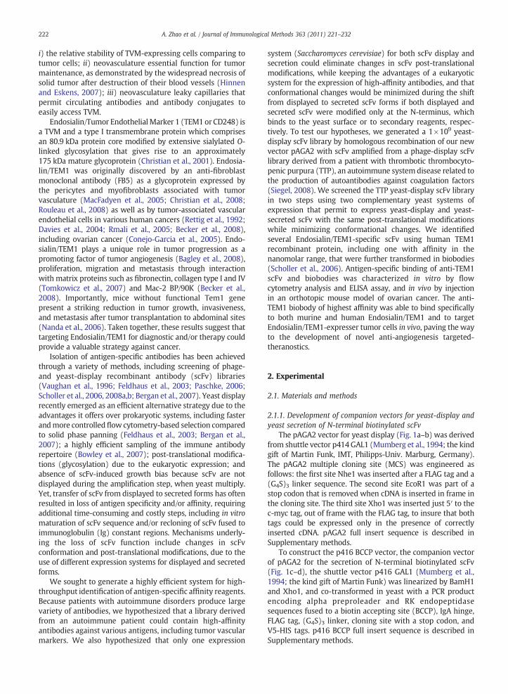
The pAGA2 vector for yeast display (Fig. 1a–b) was derivedfrom shuttle vector p414 GAL1 (Mumberg et al., 1994; the kindgift of Martin Funk, IMT, Philipps-Univ. Marburg, Germany).The pAGA2 multiple cloning site (MCS) was engineered asfollows: the first site Nhe1 was inserted after a FLAG tag and a(G4S)3 linker sequence. The second site EcoR1 was part of astop codon that is removed when cDNA is inserted in frame inthe cloning site. The third site Xho1 was inserted just 5′ to thec-myc tag, out of frame with the FLAG tag, to insure that bothtags could be expressed only in the presence of correctlyinserted cDNA. pAGA2 full insert sequence is described inSupplementary methods.
To construct the p416 BCCP vector, the companion vectorof pAGA2 for the secretion of N-terminal biotinylated scFv(Fig. 1c–d), the shuttle vector p416 GAL1 (Mumberg et al.,1994; the kind gift of Martin Funk) was linearized by BamH1and Xho1, and co-transformed in yeast with a PCR productencoding alpha preproleader and RK endopeptidasesequences fused to a biotin accepting site (BCCP), IgA hinge,FLAG tag, (G4S)3 linker, cloning site with a stop codon, andV5-HIS tags. p416 BCCP full insert sequence is described inSupplementary methods.
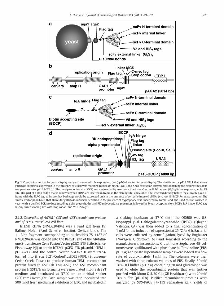
Fig. 1. Companion vectors for yeast-display and yeast-secreted scFv expression. (a–b) pAGA2 vector for yeast display. The shuttle vector p414 GAL1 that allowsgalactose-inducible expression in the presence of uracil was modified to include Nhe1, EcoR1 and Xho1 restriction enzyme sites matching the cloning sites of itscompanion vector p416 BCCP (d). Themultiple cloning site (MCS) was engineered by inserting a Nhe1 site after the FLAG tag and (G4S)3 linker sequence; an EcoR1site, also part of a stop codon that is removed when cDNA are inserted in frame in the cloning site; and a Xho1 site, inserted directly before the c-myc tag, out offrame with the FLAG tag, to insure that both tags would be expressed only in the presence of correctly inserted cDNA. (c–d) p416-BCCP for yeast secretion. Theshuttle vector p416 GAL1 that allows for galactose-inducible secretion in the presence of tryptophane was linearized by BamH1 and Xho1 and co-transformed inyeast with a purified PCR product encoding alpha preproleader and RK endopeptidase sequences followed by biotin accepting site (BCCP), IgA hinge, FLAG tag,(G4S)3 linker, cloning site with stop codon, and V5-HIS tags.
223A. Zhao et al. / Journal of Immunological Methods 363 (2011) 221–232
2.1.2. Generation of rhTEM1-GST and rGST recombinant proteinsand of TEM1-transduced cell lines
hTEM1 cDNA (NM_020404) was a kind gift from Dr.Ballmer-Hofer (Paul Scherrer Institut, Switzerland). The1113 bp fragment corresponding to nucleotides 75–1187 ofNM_020404 was cloned into the BamH1 site of the Glutathi-one S-transferase Gene Fusion Vector pGEX-2TK (Life Science,Piscataway, NJ) to obtain hTEM1-pGEX-2TK plasmid. hTEM1-pGEX-2TK and the control vector pGEX-2TK were trans-formed into E. coli BL21-CodonPlus(DE3)-RIPL (Stratagene,Cedar Creek, Texas) to produce human TEM1 recombinantprotein fused to GST (rhTEM1-GST) and GST recombinantprotein (rGST). Transformants were inoculated into fresh 2YTmedium and incubated at 37 °C on an orbital shaker(200 rpm) overnight. Each sample was then inoculated into500 ml of freshmedium at a dilution of 1/50, and incubated in
a shaking incubator at 37 °C until the OD600 was 0.8.Isopropyl β-d-1-thiogalactopyranoside (IPTG) (Qiagen,Valencia, CA) was then added to a final concentration of1 mM for the induction of expression at 25 °C for 6 h. Bacterialcells were collected by centrifugation, lyzed by Bugbuster(Novagen, Gibbstown, NJ) and sonicated according to themanufacturer's instructions. Glutathione Sepharose 4B col-umns were equilibrated with phosphate buffered saline (PBS,pH 7.4) and lysate supernatant samples were loaded at a flowrate of approximately 1 ml/min. The columns were thenwashed with three column-volumes of PBS. Finally, 50 mMTris–HCl buffer (pH 7.4) containing 20 mM glutathione wasused to elute the recombinant protein that was furtherpurified with Mono Q 5/50 GL (GE Healthcare) with 20 mMTris buffer (pH 6.8). Purified recombinant proteins wereanalyzed by SDS-PAGE (4–15% separation gel). Yields of

0
1.0
1.5
2.0
2.5
3.0
1μg 5μg 10μg 1μg 2.5μg 5μgTra
nsf
orm
ants
(x1
0^6)
Plasmid Homologousrecombination
a
0.5
100
102
104
106
108
0.01μg 0.1μg 1μg 10μg
DNA Input
Tra
nsf
orm
ants
b
224 A. Zhao et al. / Journal of Immunological Methods 363 (2011) 221–232
rhTEM1-GST and rGST proteinswere approximately 1 mg and35 mg per liter of culture, respectively. Recombinant proteinswere biotinylated using the EZ-Link Sulfo-NHS-Biotin-Reagents kit (Pierce, Rockford, IL) according to the manufac-turer's instructions and dialyzed against PBS.
The full length hTEM1 cDNA was cloned into MluI/PacI-digested lentiviral plasmid vector pHRSIN-GFP (Hasegawa etal., 2006) to generate pHRSIN-TEM1 vector that expresseshuman TEM1 but not GFP. The corresponding lentivirus wasgenerated by transient transfection of HEK293T cells withcalcium phosphate. Conditioned medium containing viralvectors was harvested 24 and 48 h after transfection, filtered(0.45 μm) and frozen at −80 °C until use. MS1, a murinepancreatic islet endothelial cell line transformed by SV40(ATCC number CRL-2279™), H5V, a murine heart endothelialcell line transformed by Polyomamiddle-size T antigen (PmT;Garlanda et al., 1994) and SKOV3, a human ovarian carcinomacell line (Chan et al., 1988) (2×10⁴ per 24-well plate) weretransduced using 500 μl viral supernatants, and expression ofhTEM1 was confirmed by RT-PCR (Supplementary Fig. 4 andmethods), Western blot and flow cytometry analysis using arabbit anti-TEM1 polyclonal antibody (kindly provided to GCby Morphotek, Inc., Malvern, PA; data not shown).
Fig. 2. Optimization of yeast transformations for EBY100 and YVH10 yeaststrains. (a) Chemical transformations. Chemical transformation of EBY100(black bars) vs. YVH10 (grey bars) using condition 2 as described in Table 1,with different concentrations of intact DNA plasmid (1 μg, 5 μg and 10 μg, asindicated) or of combinations of linearized plasmid and insert for homologousrecombination (1 μg, 2.5 μg and 5 μg of linearized vector combined with 3-foldmolar excess of scFv fragments, as indicated). The number of transformants×106 is plotted on the Y axis. (b) Electroporations. All experiments wereperformed in 0.2 cm cuvettes, in 50 μl volume, with settings of 1.5 kV, 25 mFand 200Ω. Electroporation of EBY100using condition G as described in Table 2,with different concentrations of linearized vector (white bars) or intact DNAplasmid (grey bars) or combination of linearized plasmid and insert (blackbars) for homologous recombinations (as indicated). The number oftransformants per transformation is plotted on the Y axis.
2.1.3. Yeast transformationTable 1 summarizes the procedure for optimization of the
preparation of chemical competent cell and transformation.Optimization was conducted in two steps, first by calibratingcompetent cell preparation and transformation, and then byoptimizing DNA input. Condition 2 was best for chemicaltransformation of both EBV100 (Table 1) and YVH10 (datanot shown), thus was applied for the transformation of bothstrains with intact plasmid or with combinations of linearizedvector and insert for homologous combination (Fig. 2a).YVH10 chemical transformation yielded 106transformants/μgof DNA by homologous recombination, which was unexpect-edly higher than transformation with intact plasmid (Fig. 2a,grey bars). However, for EBY100 yeast the chemical trans-formation efficiency was low (Fig. 2a, black bars). Thus, wedeveloped an alternate protocol for EBY100 transformationusing electroporation (Table 2). While electroporation ofYVH10 resulted in low yield and was not pursued (data notshown), condition G (Table 2) gave the highest yield oftransformants for EBV100, producing 1×10⁸ transformantsper electroporation with 10 μg of linearized vector and insertcombined (Fig. 2b). Thus, both YVH10 and EBV100 yeaststrains are more efficiently transformed by homologousrecombination than by intact plasmids. YVH10 is besttransformed by chemical transformation with heat shock,
Table 1Calibration of chemical transformations. EBY100 cells in log phase were treated wplasmid.
Conditions Yeastresuspension
Yeast washes w/waterand LIAC/TE
Yeast incvector+
1 Chilled in ice 60 min 4 °C w/o DMS2 Room temperature 4 °C w/o DMS3 Room temperature 4 °C w/ DMSO4 Room temperature Room temperature w/o DMS5 Room temperature Room temperature w/ DMSO
while EBV100 is best transformed by electroporation, and atlevels compatible with generation of scFv libraries.
2.1.4. Shuffling of phage-display scFv library into yeastScFv initially cloned in the M13 phage-display vector
pComb3X (Scripps Research Institute, La Jolla, CA) (Barbas etal., 2001–07) were rescued by PCR from a phagemid DNApreparation. The primers were designed to bind to theoriginal phage library scFv 5′ and 3′ end sequences, as wellas to promote homologous recombination with the yeast-display vector pAGA2. Since the scFv constructs comprisedlight chain (kappa or lambda) variable regions (VL's) followedby a GS-rich linker peptide followed by γ heavy chain variable
ith five different conditions, as indicated, and transformed with 1 μg of DNA
ubation w/insert
Heat Shockat 42 °C
Yeast recoveryin YPED
Transformants perμg of DNA
O 30 min 2 h 3×10⁴
O 30 min 2 h 8.1×10⁴
30 min 2 h 0.5×10⁴
O 30 min 2 h 0.7×10⁴
15 min 2 h 1×10⁴

Table 2Calibration of electroporation. All experiments were performed in 0.2 cm cuvettes, in 50 μl volume, with settings of 1.5 kV, 25 mF and 200 Ω. EBY100 cells in logphase were treated with fifteen different conditions, as indicated, and transformed with 1 μg of DNA plasmid.
Conditions DTT conc. Buffer Sorbitol conc. Shaking time Shaking temperature Transformants per μg of DNA
A 25 mM 15 min 30 °C 0.05×10⁶
B 25 mM 30 min 30 °C 0.05×10⁶
C 25 mM 10 mM Tris 30 min 30 °C 0.23×10⁶
D 25 mM 20 mM Hepes 30 min 30 °C 0.14×10⁵
E 25 mM 1 M 30 min 30 °C 0.01×10⁵
F 25 mM 1 M 60 min 4 °C 0.46×10⁶
G 25 mM 20 mM Hepes 0.6 M 30 min 30 °C 1.15×10⁶
H 25 mM 20 mM Hepes 1 M 30 min 30 °C 0.74×10⁶
I 25 mM LiAc-TE 0.6 M 30 min 30 °C 0.69×10⁶
J 25 mM LiAc-TE 0.6 M 60 min 4 °C 0.44×10⁵
K 25 mM LiAc-TE 1 M 30 min 30 °C 0.1×10⁵
L 25 mM LiAc-TE 1 M 60 min 4 °C 0.14×10⁵
M 25 mM LiAc-TE 30 min 30 °C 0.37×10⁵
N 25 mM LiAc-TE 30 min 4 °C 0.05×10⁶
O 25 mM LiAc-TE 60 min 4 °C 0.17×10⁶
225A. Zhao et al. / Journal of Immunological Methods 363 (2011) 221–232
region sequences (VH's), forward primers annealed to the 5′ends of VL's and reverse primers annealed to the conserved 5′end of the γ1,2,3,4 CH1 domain of the IgG heavy chain. Theprimer sequences are listed in the Supplementary methods.
PCR conditions for scFv amplification were: 94 °C for5 min followed by 25 cycles of 94 °C 1 min, 55 °C 1 min and72 °C 1 min, and a final extension of 7 min at 72 °C. PCRproducts were then purified by electrophoresis using theQiaquick Gel Extraction Kit (Invitrogen, Carlsbad, CA). pAGA2vector for yeast display was linearized with NheI and XhoIand purified using the Qiaquick PCR Purification Kit (Invitro-gen, Carlsbad, CA). EBY100 competent cells were prepared forelectroporation according to the condition G (Table 2), andco-transfected with purified PCR products and pAGA2linearized vector. Transfected yeast cells were finally ex-panded in SD-CAA medium (Scholler et al., 2006) at 30 °C at200 rpm until saturation. Ten-fold serial dilutions of thetransfected yeast were cultured on SD-CAA plates forcalculation of the library's size.
The library's diversity and gap repair efficiency wereevaluated by sequencing and flow cytometry analysis,respectively. In brief, 30 individual clones were randomlyselected, induced in the medium SGR-CAA (Scholler et al.,2006), and assessed for scFv expression using the c-myc tagexpressed only by yeast displaying scFv. Plasmid DNA wasextracted from these clones using MasterPure Yeast DNAPurification Kit (Epicentre Biotechnologies, Madison, WI) andscFv gene fragments were amplified for sequencing withprimers flanking the gap repair sites. PCR amplificationprimers are: pAGA2-scFv-For: 5′-ccgtactctttgtcaacgac-3′ andpAGA2-scFv-Rev: 5′-ttaaagccttcgagcgtccc-3′. Sequencing pri-mers are: pAGA2-seq-For: 5′-gggaaggcaatgcaagga g-3′ andpAGA2-seq-Rev: 5′-tgcgtacacgcgtctgtacag-3′.
2.1.5. AntibodiesYeast-display scFv expression was detected with anti-c-
myc mouse monoclonal antibody (mAb), 9E10 (Santa CruzBiotechnology, Inc., Santa Cruz, CA) and Alexa-488 F(ab′)2fragment of goat anti-mouse IgG (H+L) (488 anti-IgG)(Invitrogen, Carlsbad, CA) or PE-Cy7 goat F(ab′)2 anti-mouseIgG(H+L) (PE-Cu5 anti-IgG) (Cedarlane Laboratories Limit-ed, Burlington, NC). Biotinylated antigen binding to yeast-
display scFv was detected with goat anti-biotin-FITC (Abcam,Cambridge, MA) or streptavidin-PE (BD Pharmingen, SanJose, CA). ScFv binding to cell lines was detected with APC-conjugated anti-V5 mouse mAb (AbD Serotec, Raleigh, NC;APC anti-V5) and scFv binding to plastic-immobilized antigenwas detected by HRP-conjugated mouse anti-V5 mAb (AbDSerotec; HRP-anti-V5). Biobody binding to TEM1-expressercells was detected with APC-labeled streptavidin(eBioscience, San Diego, CA; APC-streptavidin). Confocalmicroscopy was performed with anti-SV40 Tag mAb (Santa-Cruz Biotech, Santa Cruz, CA) detected by goat anti-mouseIgG1k-Alexa-488 mAb (488 anti-IgG1k; Invitrogen, Carlsbad,CA), and rhodamine-labeled streptavidin (Invitrogen; Rho-SA).
2.1.6. Identification of anti-TEM1 scFvThe TTP yeast-display scFv library was first screened by
magnetic and flow sorting for anti-TEM1 scFv using progres-sively decreasing concentrations of human TEM1 recombi-nant protein fused to a GST tag (rhTEM1-GST; SupplementaryFig. 1), as previously described (Scholler et al., 2006; Berganet al., 2007). cDNAs encoding selected yeast-display scFvwere PCR amplified, purified and transformed into yeast-secreted scFv. Briefly, the library was magnetically enrichedonce for scFv that bound to 20 nM of biotinylated rhTEM1-GST, twice for scFv that bound to 6 nM of biotinylatedrhTEM1-GST, and then magnetically depleted for the scFvthat bound to 60 nM of control biotinylated rGST. Selectedyeast-display scFv were flow sorted for c-myc/TEM1 doublepositive clones. Progressively decreasing concentrations ofrhTEM1-GST were further used during the screening (from2 nM to 40 pM). DNA plasmids were extracted from yeast-display scFv that bound to 400 pM of rhTEM1-GST but not to2 nM of rGST (Supplementary Fig. 1b–d), and scFv fragmentswere amplified using primers allowing homologous recom-bination with the yeast secretion vector p416-BCCP. Theprimers used were: Forward shuffling primer: 5′-ggttctggtggtggaggttctggtggtggtggatctg-3′; Reverse shufflingprimer: 5′-gagaccgaggagagggttagggataggcttaccgtcgac-caagtcttcttcagaaataagctt-3′. ScFv fragments and linearizedp416-BCCP were co-transfected into YVH10 by chemicaltransformation. Soluble scFv screening for specific binding to
226 A. Zhao et al. / Journal of Immunological Methods 363 (2011) 221–232
TEM1 was performed by ELISA with yeast supernatants of 470random transformants in Maxisorp ELISA plates (Nunc,Rochester, NY) coated with 0.8 μg/ml of rhTEM1-GST vs. rGST.Recombinant proteins were plastic-immobilized in carbonate–bicarbonate buffer 0.5 M, pH 9.6 (Fisher Scientific, Pittsburgh,PA)ONat 4 °C.Wellswere thenblockedwith 5%drymilk in PBS(PBMS; Biorad, Hercules, CA) for 2 h at RTwith gentle agitationand incubated with yeast supernatants diluted 1/1 with PBSMat RT for 1 h. After three washes with PBS supplemented with0.05% Tween (PBST; Biorad), scFv binding to immobilizedproteins was detected with HRP anti-V5. Colorimetric signalswere developed with TMB substrate solution (KPL, Inc.,Gaithersburg, MD), quenched with sulfuric acid (KPL, Inc.)and read at 450 nm on a Fluoroskan Ascent FL (Thermo FisherScientific, Pittsburgh, PA). Approximately 50% of the colonies(232/470) gave a colorimetric signal higher than the averagebackground plus 3 standard deviations. No cross-reactivitywith rGST control protein was detected. Sequencing of 40 scFvidentified five unique clones that were then produced and Ni-purified as previously described (Scholler et al., 2006).
2.1.7. Measurement of scFv affinity by ELISATo assess scFv-78 affinity, maxisorp ELISA plates were
coated with rhTEM1-GST at two-fold decreasing concentra-tions from 0.4 to 0.05 μg/ml, in carbonate–bicarbonate buffer.After blocking with PBSM, wells were incubated with ten-foldserial dilutions of scFv-78, starting from 1 μM. ScFv binding toimmobilized proteins was detected with HRP anti-V5.Colorimetric signals were developed as previously described.The same procedure was followed for the other scFv, butusing 2 and 1 μg/ml of coated rhTEM1-GST and three-foldserial dilutions of scFv, starting at 2 μM.
2.1.8. Flow cytometry analysisAnalysis of scFv expression by yeast was performed as
previously described (Scholler et al., 2006). Briefly, binding ofanti-TEM1 scFv and biobodies to TEM1-expresser mammali-an cells was evaluated using various human or murine celllines expressing TEM1 endogenously (HEK-293T, MOV1 and2H11) or stably transduced with pHRSIN-TEM1 (hTEM1-MS1, hTEM1-H5V and hTEM1-SKOV3). MS1 (SV40-trans-formed murine endothelial cells), H5V (PmT-transformedmurine endothelial cells) and SKOV3 (human ovariancarcinoma cell) lines were used as negative or weaklypositive control cell lines, in accordance with previouspublished findings reporting Endosialin/TEM1 to beexpressed by mouse immortalized endothelial cells andvarious tumors, including carcinomas and sarcomas (Dolzniget al., 2005; Teicher, 2007). Anti-TEM1 scFv were preincu-bated for 30 min at RT with APC-anti-V5 at a ratio 1/1 andanti-TEM1 biobodies were preincubated with APC-labeledstreptavidin at a ratio of 1/4. A non-relevant scFv was used asa negative control for binding.
2.1.9. Orthotopic mouse model of ovarian cancerMOV1mouse ovarian cancer cell line was derived from an
ovarian cancer that spontaneously arose in female transgenicmice that express the transforming region of SV40 undercontrol of the Mullerian inhibitory substance type II receptorgene promoter (Tg-MISIIR-TAg; Connolly et al., 2003). MOV1cell line expresses SV40 antigen and TEM1. To emulate
ovarian cancer mouse ovarian cancer cells, MOV1 wereorthotopically injected in the ovarian bursa of NOD-Scid-γcnull (NSG) mice (Shultz et al., 2000). Four month-oldmultiparous females were anesthetized according to theprotocol approved by the University of Pennsylvania Institu-tional Animal Care and Use Committee (IACUC). A dorso-lateral incision on left caudal portion of the animal dorsumwas made. The retroperitoneum was dissected to expose theleft ovary using the forceps to grasp, retract, position, andsecure the organ for injection. Five million MOV1 cells wereinjected in the ovarian bursa in a volume of 20 μl of PBS usingan insulin syringe. Retroperitoneal incisions were closed,animals were administered antibiotics and fluids, and tumorgrowth was monitored by in vivo imaging.
2.1.10. Analysis of in vivo distribution of anti-TEM1 biobodies byconfocal microscopy
Anti-TEM1 biobody-78 of high affinity was injectedintravenously (IV) 3 weeks after tumor cell implantation. Ascontrol for the Enhanced Permeability and Retention (EPR)effect (Maeda and Matsumura, 1989; Greish, 2007), we usedthe anti-TEM1 biobody-137 of low affinity in the sameconditions. Spleen, liver, kidney and ovaries were harvested24 or 48 h after biobody injection and preserved in frozentissue matrix OCT compound (Tissue-Tek, Sakura FinetekUSA). Slides of 5 μ thickness were cut from frozen sections, airdried 1 h at RT and fixed by immersion in cold 100% acetone5 min. After 2 washes in PBS, slides were blocked forendogenous biotin by pre-treatment with avidin/biotinblocking solution (avidin-skim milk 0.001% in PBS). Anti-TEM1 biobody binding was detected with rhodamine-conjugated streptavidin. MOV1 tumor cells derived from aTg-MISIIR-TAg tumor express SV40 and thus could bedetected with anti-SV40 Tag antibody (2 μg/ml) for 30 minat RT, followed by 1 μg/ml Alexa-488 goat anti-mouse IgG1κfor 30 min at RT. Slides were incubated with 1/2000 dilutedDAPI (Invitrogen) for 30 min at RT to visualize the nuclei.Fluorescent signals were acquired by confocal analysis (ZeissLSM 510META NLO) at 63× magnification.
3. Results
3.1. Generation of companion vectors for yeast display andyeast secretion of N-terminal biotinylated scFv
To overcome the loss of antigen specificity and/or affinitydue to scFv transfer from cell surface display to secreted, wedeveloped two complementary vectors, pAGA2 and p416BCCP, which permit scFv to be displayed (Fig. 1a–b) or secreted(Fig. 1c–d) by the same expression system (S. cerevisiae) andthrough engineering at the same domain (N-terminus), thuswith similar post-translational modifications and conforma-tion. In pAGA2 vector, scFv are fused at the N-terminus to Aga2,to permit yeast display (Fig. 1a–b). ScFv expressed by p416BCCP are soluble and fused at the N-terminus to anenzymatically biotinylable domain (BCCP) separated fromthe scFv functional site by a flexible IgA hinge (Fig. 1c–d).The BCCP domain is biotinylated in vivo by a biotin ligaseexpressed by yeast mating partners, and the presence of theIgA hinge minimizes scFv conformational changes when thebiotinylated BCCP binds to immobilized or labeled streptavidin
227A. Zhao et al. / Journal of Immunological Methods 363 (2011) 221–232
(Scholler et al., 2006). The vectors pAGA2 (Fig. 1a–b) and p416BCCP (Fig. 1c–d) permit galactose-inducible expression in thepresence of uracil or tryptophane, respectively, which mini-mizes growth bias (Mumberg et al., 1994).
3.2. Construction and validation of TTP yeast-display scFv library
TheM13 phage-display human scFv library derived from apatient with thrombotic thrombocytopenic purpura (TTP), acoagulation system disorder caused by autoantibodies to themetalloprotease ADAMTS13, was previously reported (Siegel,2008). ScFv gene segments from the TTP phage-displaylibrary were rescued by PCR from a phagemid DNApreparation, and co-transformed with pAGA2 linearizedvector by electroporation in EBV100 yeast strain to allowhomologous recombination and cell surface display. Thediversity of the resulting yeast-display scFv library wasvalidated by the sequencing of 20 randomly selected clonesthat demonstrated a gap repair rate of 95% (data not shown).The correct display of scFv at the yeast cell surface wasassessed by flow cytometry through the detection of a c-myctag fused to the scFv C-terminus. Nine out of 24 randomlyselected clones displayed scFv on their surface after induction(Supplementary Fig. 2, clones a,g,i,k,o,p,r,u,w), which wasconsistent with the expression ratio of phage and yeastlibraries previously reported (Vaughan et al., 1996; Blaise etal., 2004).
3.3. Identification of anti-TEM1 scFv
Targeting TEM1 with antibody or antibody-conjugatedreagents such as an immunotoxin, isotope, or nanoparticles, isa promising approach for both diagnosis and therapy. Toidentify recombinant antibodies directed against TEM1, theTTP yeast-display library was enriched for scFv that bind torecombinant human TEM1-GST protein (rhTEM1-GST), firstby magnetic sorting then by flow sorting, and finally depletedby magnetic sortings for the scFv that bound to rGST controlprotein. Magnetic sorting is based on one parameter ofselection only (capture of antigen-binding yeast), therebyproviding a rapid means for robust enrichment of antigen-specific scFv. However, to prevent the selection of yeast thatnon-specifically bind to antigens, a flow sorting step usingtwo parameters of selection was included. Yeast that bothbound to TEM1-1 and expressed c-myc tag due to the displayof scFv at their surface was sorted and their cDNA rescued foramplification with primers allowing homologous recombina-tion with p416 BCCP vector. Resulting transformants secretedscFv enriched for TEM1-specific binders.
Four hundred and seventy yeast-secreting scFv weretested by ELISA detection assays for their ability to detectrhTEM1-GST. Almost 50% (232/470) of the soluble scFvbound to rhTEM1-GST but not to rGST. Anti-TEM1 solublescFv were classified into two affinity categories: “high” whenthe optical density (OD) by detection ELISA was greater than0.5, and “low” when lower than 0.5. Twenty scFv of eachcategory were sequenced. The high OD group contained onlyone sequence (scFv-78) and three clones presented each onepoint mutation in the VL; #6 had a point mutation in FR1 thatchanged a serine in leucin; #8 a point mutation in FR3changing a glycin in valine; #10 a point mutation in CDR2
changing a leucine in methionine. The low OD group includedfour different scFv (scFv-131; scFv-132; scFv-133; scFv-137)(Fig. 3). Alignment of scFv heavy and light chain variableregion sequences to a database of human immunoglobulingermline sequences (V Base Directory of Human V GeneSequences (Tomlinson et al., 1996)) indicated that the heavychain variable region sequences used VH3- and VH4-family-encoded gene products and showed extensive somaticmutation particularly in the CDR regions as though theyevolved during an antigen-driven immune response (Fig. 3a).The light chain sequences used by all 5 anti-TEM1 scFv's werelambda and were minimally mutated from their most likelyVL and JL germline genes (Fig. 3b).
3.4. Characterization and in vitro validation of anti-TEM1 scFvand biobodies by ELISA assays and flow cytometry analysis
ELISA provides a convenient way to evaluate the affinity ofan antibody (Beatty et al., 1987). Because a scFv has only onebinding site for antigen, the affinity can be calculated by theequation of Kd=2[Ab′]t− [Ab]t, derived from the Beatty'sequation Kaff=1/2(2[Ab′]t− [Ab]t), where [Ab′]t refers toscFv concentration at half the maximal OD (OD50) forrhTEM1-GST-coated wells at half concentration, and [Ab]refers to scFv concentration at OD50 for rhTEM1-GST-coatedwells at whole concentration. The calculated Kd's of the fiveanti-TEM1 scFv with distinct sequences were 4.3±1.5 nM forscFv-78 (Fig. 4), 148 nM for scFv-132, 218 nM for scFv-133,682 nM for scFv-131, and 4.4 μM for scFv-137, respectively(Supplementary Fig. 3). R2 of the curve fittings for all ELISAswere above 0.99. ScFvwere then biotinylated on the BCCP sitein N-termini by yeast mating with biotin-ligase-bearing yeastto produce biobodies, as described by Scholler et al. (2006),and expressed at a yield of 10 mg per liter.
Flow cytometry showed that all anti-TEM1 scFv bound tothe human TEM1-transfected mouse endothelial cell linehTEM1-MS1 but not to wild-type MS1 cells, while bindingcould be blocked with rhTEM1 but not with rGST (Fig. 5a–l).The remainder of the study was performed with the high-affinity anti-TEM1 scFv-78 and low affinity scFv-137 aftertargeted biotinylation, resulting in biobody-78 and biobody-137, respectively. Biobody-78 strongly bound to cell linestransduced with human TEM1 (Fig. 5o–q) and cells expres-sing high levels of endogenous human (Fig. 5m) as well asmouse (Fig. 5r,s) TEM1. Biobody-78 also bound to cell linesthat express moderate levels of endogenous mouse or humanTEM1, such as H5V (Fig. 5n) and SKOV3 (Fig. 5p), respec-tively. TEM1 mRNA expression level was verified by qRT-PCRin all cell lines (Supplementary Fig. 4).
3.5. In vivo validation of anti-TEM1 scFv
To test the ability of anti-TEM1 scFv to recognize target invivo, immunodeficient mice were transplanted orthotopicallyin the left ovarian bursawithMOV1 ovarian cancer cells, whichexpress mouse TEM1 (Supplementary Fig. 4b). Groups of micewere injected IV with a bolus of either the high-affinitybiobody-78 or the low affinity biobody-137 (50 μg/mouse) tocontrol for the EPR effect. Animals were euthanized after 24 or48 h, and spleens, livers, kidneys and ovaries were collected tomonitor biobody distribution by confocal microscopy. Anti-

aGermline Ig
GeneAbClone
VH D JH FR 1 CDR 1 FR 2 CDR 2 FR 3 CDR 3 FR 4
#NucleotideDifferencesfrom Germ.
VH gene
scFv-78
3-23
2-21 4 EVQLLESGGTLVQPGGSLRLSCEASGFTFS NYAMG WVRQTPGKGLEWVS AIRKSGTTTYADSLKG RFIISRDNSKNTLYLQMNSLRVEDTATYYCAT HPIAG WGQGTLVTVSS 23
scFv-131
4-39
2-2
3 QLQLQESGPGLVKPSETLSLTCTVSGGSIN SNSYYWG WIRQPPGKGLEWIG SIYYRGNTYYNPPLKS RVTMSVDTSKNHFSLNLSSVTAADTAVYYCAR VRRSPA WGQGTMVTVSS 14
scFv-132
4-39
6-13
4 QLQLQESGPGLVKPSETLSLTCTVSGGSIS SSNYYWG WIRQPPGKGLEWIG TIFYSGRTYYNPSLRS RLTMSVDTSKNQFSLKLSSVTAADTAVYFCAR VLPRWSSVDQ WGHGTLVTVSS 18
scFv-133
4-39
6-13 4 QLQLQESGPRLVKPSETLSLTCTVSGDSIS SNNYYWG WIRQPSGKGLEWIG SIDYSGKTYYNPSLES RVTMSVDTSKNQFSLNLSAVTAADTAVYYCAR VPGSSTWMVNY WGQGTLVTVSS 20
scFv-137
3-66
3-3
6 EVQLVESGGGLVQPGGSLRLSCAASGFTFS SYSMN WVRQAPGKGLEWVS AISGSGGSTYYTGSVKG RFTISRDNSKNTLFLQMNSLRGENTAVYYCAR YGAMNV WGQGTTVTVSS 15
bGermlineIg Gene
AbClone
Vλ J λFR 1 CDR 1 FR 2 CDR 2 FR 3 CDR 3 FR 4
#NucleotideDifferencesfrom Germ.
VL gene
scFv-78 5c 3b QPVLTQPPSLSASPGASASLTC TLRSDINVGTYRIS WYQQKPGSPPQYLLS YKSDSDKQKGS GVPSRFSGSKDASADAGILLISGLQSENEANYYC MIWHNSAG VVFGGGTKLTVL 11
scFv-131 1b 3b LPVLTQSPSVSAAPGQKVTISC SGSSSNIGNNYVS WYQQLPGTAPKLLIY DNNKRPS GIPDRFSGSKSGTSATLGITGLQTGDEADYYC GTWDSSLSA WVFGGGTELTVL 5
scFv-132 1b 3b VSVLTQPPSVSAAPGQKVTISC SGSSSNIGNNYVS WYQQLPGTAPKLLIY DNNKRPS GIPDRFSGSKSGTSATLGITGLQTGDEADYYC GTWDNSMSA WVFGGGTKLTVL 4
scFv-133 1g 3b QSVLTQPPSASGTPGQRVTISC SGSSSNIGSNYVY WYQQLPGTAPKLLIY RNNQRPS GVPDRFSGSKSGTSASLAISGLQSEDEADYYC ATWDDSLNG VSFGGGTKLTVL 5
scFv-137 1g 2 QSVLTQPPSASGTPGQRVTISC SGRSPNIGSNYVY WYQFLPGTAPKLLIY RNNQRPS GVPDRFSGSKSGTSASLAISGLRSEDEADYYC AAWDDSLSG LVFGGGTQLTVL 4
Fig. 3. Germline immunoglobulin gene usage and predicted amino acid sequences of anti-TEM1 scFv. (a) Anti-TEM1 heavy chain variable regions. The numberof nucleotide differences from germline VH is tabulated to the right of each sequence. In general, D segments showed very poor homology with known D genesso mutations were not scored in these regions. FR (framework region) and CDR (complementarily determining region) designations as per Kabat et al. (1991)(b) Anti-TEM1 light chain variable regions. The number of nucleotide differences from germline VL is tabulated to the right of each sequence.
228 A. Zhao et al. / Journal of Immunological Methods 363 (2011) 221–232
TEM1 biobody-78 was detected in kidneys 24 h after IVinjection (Supplementary Fig. 5c upper and lower rightpanels), but was cleared after 48 h (Supplementary Fig. 5fupper and lower right panels). Both anti-TEM1 biobody-78 andbiobody-137 specifically localized to MOV1 cells implanted inthe left ovaries after 48 h (Fig. 6A,C,E, upper and lower rightpanels) but not in the contralateral normal ovaries (Fig. 6B,D,F)or in the other normal mouse organs (spleen, liver and kidney)(Supplementary Fig. 5, as indicated). The signal generated bythe biobody-137 was weaker than the signal from biobody-78(compare Fig. 6C with Fig. 6E) which demonstrated acorrelation between biobody affinity and tumor binding, andsuggested that biobody-78 binding to the tumor was bothindependent of the EPR effect and antigen-specific.
00.5
11.5
22.5
33.5
44.5
1E.+01 1.E+02 1.E+03 1.E+04 1.E+05 1.E+06scFv concentration (pM)
OD
450
Fig. 4. Measurement of scFv-78 affinity by ELISA. Kd was calculated by theequation Kd=2[Ab′]t− [Ab]t, where [Ab′]t refers to the scFv concentration atOD50 for the half concentration rhTEM1-coated wells while [Ab]t refers to thescFv concentration at OD50 for one concentration rhTEM1-GST-coated wells.The calculated Kd for scFv-78 was 5.8 nM using antigen concentrations of0.4 μg/ml (diamonds) and 0.2 μg/ml (squares), 4.5 nM using antigenconcentrations of 0.2 μg/ml and 0.1 μg/ml (open triangles), or 2.8 nM usingantigen concentration of 0.1 μg/ml and 0.05 μg/ml (open circles). ELISAswere performed in duplicate in two independent experiments. Lines arefitted using the antibody–antigen reaction equation.
.
4. Discussion
We isolated high-affinity reagents (scFv and biobodies)that bound to bothmouse and human Endosialin/TEM1, usinga high-throughput yeast-based platform and a two-stepscreening method. Anti-TEM1 scFv and biobodies werevalidated for flow cytometry use, ELISA detection assays andin vivo targeting. The affinity of our novel anti-TEM1 biobody-78 was in the nanomolar range which permitted to generatehigh mean fluorescence intensities (MFI) by flow cytometryanalysis at very low concentration (10 nM, Fig. 5), whichfavorably compared to the anti-TEM1 scFv previouslypublished by Marty et al. (2006). In addition and in contrastto the other available anti-TEM1 antibodies, our novel anti-TEM1 biobody-78 binds to both mouse and human TEM1,which is invaluable for preclinical model systems. Ourscreening strategy was based on cell sorting of yeast-displayscFv, combined to ELISA screening and flow cytometryanalysis of yeast-secreted biotinylated scFv (biobodies),using a comprehensive set of vectors to shuttle scFv withoutloss of function from displayed to secreted, labeled forms.
High-affinity anti-TEM1 human scFv and biobodies wereisolated from a yeast-display scFv library derived from apatient with TTP, an autoantibody-mediated disorder (Siegel,2008). Given that patients with autoimmune disorders havelost some degree of immune tolerance, we thought that animmune library derived from such a patient could containhigh-affinity antibodies against various self-antigens. Thetransfer of the TTP scFv library from phage display to yeastdisplay permitted a highly efficient initial selection by cellsorting (Bowley et al., 2007). Although here we used a phagelibrary as starting material because it was previously shownto comprise high-affinity clones against vascular targets(Siegel, 2008), we do not foresee phage display as a necessaryinitial step in the development of yeast scFv libraries.

qponm
APC anti-V5mAb
g h i
f
020406080
100
MS1
scFv-132
MS1
scFv-133
MS1
scFv-137eb d
scFv-131scFv-78a
020406080
100
020406080
100
APC streptavidin
Control-scFv
100103104105
100103104105 100103104105 100103104105 100103104105 100103104105 100103104105
100103104105 100103104105 100103104105
r
c
s
j k l
100103104105 100103104105 100103104105
Fig. 5. Analysis of anti-TEM1 scFv binding by flow cytometry. Wild-type (a–f) and hTEM1-transfected (g–l) microvascular endothelial cells of murine pancreaticorigin (Mile-Sven, MS1 cells, American Type Culture Collection, Manassas, VA) were incubated with five different anti-TEM1 scFv (as indicated) at 10 nM for scFv-78 and at 1 mM for scFv-132, scFv-133, scFv-131, scFv-137, and an irrelevant control scFv. ScFv binding to cell surface was detected with APC-labeled anti-V5(solid black line). Blocking conditions were performed in the presence of 20 nM of rhTEM1-GST (small dotted line) or 100 nM of rGST control protein (dashed line)for scFv78 and 1 μM of rhTEM1-GST (small dotted line) or 1 μM of rGST control protein (dashed line) for scFv-131, -132, -133 and -137. As a negative control, cellswere incubated with APC anti-V5 mAb only (grey line). (m–s) Targeted biotinylated ScFv-78 (biobody-78) was incubated with TEM1-endogenous expresserembryonic kidney 293 cells (HEK293) (m); wild-type heart endothelial mouse cells (H5V) (n); hTEM1-transfected H5V cells (o); wild-type human ovarian cancercells (SKOV3) (p); hTEM1-transfected SKOV3 cells (q); mouse TEM1-endogenous expresser endothelial cells (2H11) (r), and mouse TEM1-endogenous expresserovarian cancer cells (MOV1) (s). Biobody-78 binding was detected by 30 nM of APC-labeled streptavidin (black line). As a negative control, cells were incubatedwith APC-labeled streptavidin only (grey line).
229A. Zhao et al. / Journal of Immunological Methods 363 (2011) 221–232
Furthermore, although the library derived from a patient withautoimmune disorder exhibited high-affinity antibody clonesagainst TEM1, it is possible that patients with cancer alsoexhibit high-affinity clones against tumor-associated self-antigens (Anderson and LaBaer, 2005; Tan and Zhang, 2008;Qiu and Hanash, 2009), including antigens up-regulated bytumor vasculature.
Our new vector for yeast display, pAGA2, prevents the cellsurface expression of c-myc in the absence of scFv insertion inframe with the cloning site, which permits efficient positiveand negative selections by cell sorting. We also generated acompanion vector for yeast secretion of scFv, p416-BCCP. Thecombined use of pAGA2 and p416-BCCP vectors permitsconsistency both in the expression systems used for scFvsurface display or secretion (yeast), and in the scFvorientation (attachment to the N-termini when displayed atthe yeast cell surface as well as when soluble and bound tostreptavidin), which minimizes post-translational and con-formational changes from displayed to secreted forms. Inaddition, the transfer from display to secreted forms can besimply achieved by yeast homologous recombination with anoptimized protocol permitting high yields of transformation.Finally, yeast-secreting soluble scFv cloned into p416 BCCP
can be mated with biotin-ligase-bearing yeast (Scholler et al.,2006) to produce diploid yeast that secrete N-terminibiotinylated scFv (biobodies) directly in the yeast culturemedium. Biobodies are inexpensive to produce, can beretrieved by any streptavidin-coated surfaced directly fromthe yeast culture medium or purified through their HIS6 tag,and are detectable with labeled streptavidin.
5. Conclusion
The foreseeable clinical and laboratory applications offully human scFv and especially biobodies are quite diverse.We describe here for the first time a biobody (biobody-78)that can detect both human and mouse Endosialin/TEM1 forflow cytometry analysis and ELISA applications, and that isstable in vivo with intact functional binding 48 h after IVinjection (Fig. 6C upper and lower right panels), permittingconfocal analysis of harvested tissues. Therefore, biobody-based targeting of tumor targets in vivo for the purpose ofimaging or to deliver therapeutic payloads is now possible.Antibody-conjugated reagents such as an immunotoxins,immunoisotopes, or immuno-nanoparticles are high promis-ing approaches for diagnosis and/or therapy. Indeed, the
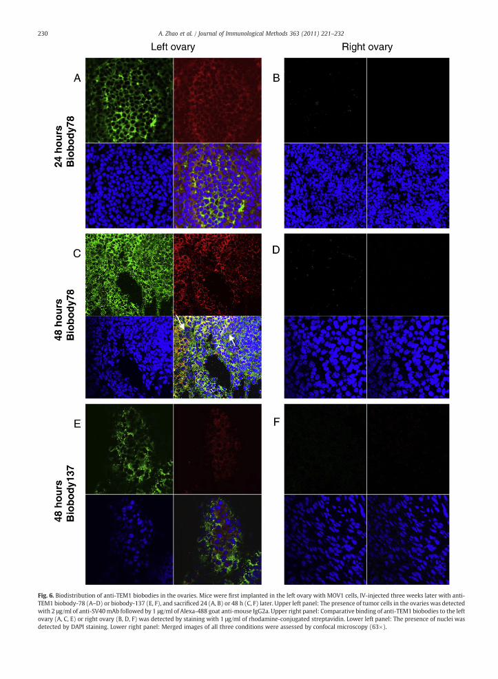
ig. 6. Biodistribution of anti-TEM1 biobodies in the ovaries. Mice were first implanted in the left ovary with MOV1 cells, IV-injected three weeks later with anti-EM1 biobody-78 (A–D) or biobody-137 (E, F), and sacrificed 24 (A, B) or 48 h (C, F) later. Upper left panel: The presence of tumor cells in the ovaries was detectedith 2 μg/ml of anti-SV40mAb followed by 1 μg/ml of Alexa-488 goat anti-mouse IgG2a. Upper right panel: Comparative binding of anti-TEM1 biobodies to the leftvary (A, C, E) or right ovary (B, D, F) was detected by staining with 1 μg/ml of rhodamine-conjugated streptavidin. Lower left panel: The presence of nuclei wasetected by DAPI staining. Lower right panel: Merged images of all three conditions were assessed by confocal microscopy (63×).
230 A. Zhao et al. / Journal of Immunological Methods 363 (2011) 221–232
FTwod
231A. Zhao et al. / Journal of Immunological Methods 363 (2011) 221–232
versatility of biobodies allows for modular design of bothimaging and therapeutic tools, thus paving the way for thedevelopment of personalized paired imaging and therapy, ortheranostic tools. We believe that we resolved severallimitations existing presently in the field of scFv antibodydevelopment and we provide the first evidence of a high-throughput paired yeast platform that can lead to the rapidisolation of functional high-affinity biotinylated scFv for thedevelopment of theranostics. To our knowledge, biobody-78is the only high-affinity antibody of human origin directedagainst both human and murine Endosialin/TEM1, which willgreatly facilitate Endosialin/TEM1-based preclinical targetedimaging and therapeutic studies.
Competing interests statement
The authors declare no competing financial interests.Supplementary materials related to this article can be
found online at doi:10.1016/j.jim.2010.09.001.
Acknowledgments
We acknowledge Yi Cheng and Lindsay Bergan forexcellent technical assistance, John Facciaponte for helpwith the qRT-PCR assays, Carmine Carpenito for the prepa-ration of lentiviruses from the pTurboFP635-C vector, TonySecreto for assistance with orthotopic injections, and Ryan D.Wychowanec for flow sorting. The project described was firstsupported by the Pacific Ovarian Cancer Research Consor-tium, Award Number P50 CA083636 from the National CancerInstitute (Nicole Urban) and the Canary Foundation (NicoleUrban). The content is solely the responsibility of the authorsand does not necessarily represent the official views of theNational Cancer Institute or the National Institute of Health.The project described was also supported by the GrantNumber UL1RR024134 from the National Center For ResearchResources, in part by the Institute for the TranslationalMedicine and Therapeutics (ITMAT) Transdisciplinary Pro-gram in Translational Medicine and Therapeutics, a CancerCenter Support Grant (CA016520) from the National CancerInstitute, NIH (Nathalie Scholler), the Claneil Foundation(George Coukos, Nathalie Scholler), the Alliance for CancerGene Therapy (George Coukos), the Ovarian Cancer ResearchFoundation (George Coukos), and the grant number P50-HL081012 from the National Heart, Lung, and Blood Institute(Don. L. Siegel).
Appendix A
AZ designed and performed optimizations of yeasttransfection, transferred the phage-display scFv library toyeast-display form, screened the yeast-display library, iden-tified anti-TEM1 scFv, validated them in vitro and contributedto manuscript preparation. SNC established MOV1 cell lineand the orthotopic model of ovarian cancer, performed invivo experiments and confocal analysis and contributed tomanuscript preparation. CL generated hTEM1-pGEX-2TK andpHRSIN-TEM1, transduced the cell lines, and produced andpurified rhTEM1-GST and rGST. GC supervised CL and
contributed to project conception and design and manuscriptpreparation. DLS constructed the phage-displayed scFvlibrary and contributed to the design of scFv transfer fromphage to yeast and to manuscript preparation. NS generatedpAGA2 and p416-BCCP vectors, contributed to the design ofscFv transfer from phage to yeast, supervised AZ and SNC, andwrote the manuscript.
References
Anderson, K.S., LaBaer, J., 2005. The sentinel within: exploiting the immunesystem for cancer biomarkers. J. Proteome Res. 4, 1123.
Bagley, R.G., Rouleau, C., St Martin, T., Boutin, P., Weber, W., Ruzek, M.,Honma, N., Nacht, M., Shankara, S., Kataoka, S., Ishida, I., Roberts, B.L.,Teicher, B.A., 2008. Human endothelial precursor cells express tumorendothelial marker 1/endosialin/CD248. Mol. Cancer Ther. 7, 2536.
Barbas III, C.F., Burton, D.R., Scott, J.K., Silverman, G.J., 2001–07. PhageDisplay: A Laboratory Manual. Cold Spring Harbor Laboratory Press.
Beatty, J.D., Beatty, B.G., Vlahos, W.G., 1987. Measurement of monoclonalantibody affinity by non-competitive enzyme immunoassay. J. Immunol.Meth. 100, 173.
Becker, R., Lenter, M.C., Vollkommer, T., Boos, A.M., Pfaff, D., Augustin, H.G.,Christian, S., 2008. Tumor stroma marker endosialin (Tem1) is a bindingpartner of metastasis-related protein Mac-2 BP/90K. FASEB J. 22, 3059.
Bergan, L., Gross, J.A., Nevin, B., Urban, N., Scholler, N., 2007. Developmentand in vitro validation of anti-mesothelin biobodies that prevent CA125/Mesothelin-dependent cell attachment. Cancer Lett. 255, 263.
Blaise, L., Wehnert, A., Steukers, M., van den Beucken, T., Hoogenboom, H.,Hufton, S., 2004. Construction and diversification of yeast cell surfacedisplayed libraries by yeast mating: application to the affinity matura-tion of Fab antibody fragments. Gene 342, 211.
Bowley, D.R., Labrijn, A.F., Zwick, M.B., Burton, D.R., 2007. Antigen selectionfrom an HIV-1 immune antibody library displayed on yeast yields manynovel antibodies compared to selection from the same library displayedon phage. Protein Eng. Des. Sel. 20, 81.
Chan, H.S., Bradley, G., Thorner, P., Haddad, G., Gallie, B.L., Ling, V., 1988. Asensitive method for immunocytochemical detection of P-glycoproteinin multidrug-resistant human ovarian carcinoma cell lines. Lab. Investig.59, 870.
Christian, S., Ahorn, H., Koehler, A., Eisenhaber, F., Rodi, H.P., Garin-Chesa, P.,Park, J.E., Rettig, W.J., Lenter, M.C., 2001. Molecular cloning andcharacterization of endosialin, a C-type lectin-like cell surface receptorof tumor endothelium. J. Biol. Chem. 276, 7408.
Christian, S., Winkler, R., Helfrich, I., Boos, A.M., Besemfelder, E., Schadendorf,D., Augustin, H.G., 2008. Endosialin (Tem1) is a marker of tumor-associatedmyofibroblasts and tumor vessel-associatedmural cells. Am. J.Pathol. 172, 486.
Conejo-Garcia, J.R., Buckanovich, R.J., Benencia, F., Courreges, M.C., Rubin, S.C.,Carroll, R.G., Coukos, G., 2005. Vascular leukocytes contribute to tumorvascularization. Blood 105, 679.
Connolly, D.C., Bao, R., Nikitin, A.Y., Stephens, K.C., Poole, T.W., Hua, X., Harris,S.S., Vanderhyden, B.C., Hamilton, T.C., 2003. Female mice chimeric forexpression of the simian virus 40 TAg under control of the MISIIRpromoter develop epithelial ovarian cancer. Cancer Res. 63, 1389.
Davies, G., Cunnick, G.H., Mansel, R.E., Mason, M.D., Jiang, W.G., 2004. Levelsof expression of endothelial markers specific to tumour-associatedendothelial cells and their correlation with prognosis in patients withbreast cancer. Clin. Exp. Metastasis 21, 31.
Dolznig, H., Schweifer, N., Puri, C., Kraut, N., Rettig, W.J., Kerjaschki, D., Garin-Chesa, P., 2005. Characterization of cancer stroma markers: in silicoanalysis of anmRNA expression database for fibroblast activation proteinand endosialin. Cancer Immun. 5, 10.
Feldhaus, M.J., Siegel, R.W., Opresko, L.K., Coleman, J.R., Feldhaus, J.M., Yeung, Y.A.,Cochran, J.R., Heinzelman, P., Colby, D., Swers, J., Graff, C.,Wiley, H.S.,Wittrup,K.D., 2003. Flow-cytometric isolationofhumanantibodies fromanonimmuneSaccharomyces cerevisiae surface display library. Nat. Biotechnol. 21, 163.
Folkman, J., 1985. Tumor angiogenesis. Adv. Cancer Res. 43, 175.Garlanda, C., Parravicini, C., Sironi, M., De Rossi, M., Wainstok de Calmanovici,
R., Carozzi, F., Bussolino, F., Colotta, F., Mantovani, A., Vecchi, A., 1994.Progressive growth in immunodeficient mice and host cell recruitmentby mouse endothelial cells transformed by polyoma middle-sized Tantigen: implications for the pathogenesis of opportunistic vasculartumors. Proc. Natl. Acad. Sci. USA 91, 7291.
Greish, K., 2007. Enhanced permeability and retention of macromoleculardrugs in solid tumors: a royal gate for targeted anticancer nanomedi-cines. J. Drug Target. 15, 457.

232 A. Zhao et al. / Journal of Immunological Methods 363 (2011) 221–232
Hasegawa, K., Pham, L., O'Connor, M.K., Federspiel, M.J., Russell, S.J., Peng,K.W., 2006. Dual therapy of ovarian cancer using measles virusesexpressing carcinoembryonic antigen and sodium iodide symporter.Clin. Cancer Res. 12, 1868.
Hinnen, P., Eskens, F.A., 2007. Vascular disrupting agents in clinicaldevelopment. Br. J. Cancer 96, 1159.
Kabat, E.A., Wu, T.T., Perry, H.M., K.S., G. and Foeller, C. (1991) Sequences ofProteins of Immunological Interest. In: NIH (Ed) Sequences of Proteins ofImmunological Interest (ed 5). Bethesda, MD.
MacFadyen, J.R., Haworth, O., Roberston, D., Hardie, D., Webster, M.T., Morris,H.R., Panico, M., Sutton-Smith, M., Dell, A., van der Geer, P., Wienke, D.,Buckley, C.D., Isacke, C.M., 2005. Endosialin (TEM1, CD248) is a marker ofstromal fibroblasts and is not selectively expressed on tumourendothelium. FEBS Lett. 579, 2569.
Maeda, H., Matsumura, Y., 1989. Tumoritropic and lymphotropic principlesof macromolecular drugs. Crit. Rev. Ther. Drug Carrier Syst. 6, 193.
Marty, C., Langer-Machova, Z., Sigrist, S., Schott, H., Schwendener, R.A.,Ballmer-Hofer, K., 2006. Isolation and characterization of a scFv antibodyspecific for tumor endothelial marker 1 (TEM1), a new reagent fortargeted tumor therapy. Cancer Lett. 235, 298.
Mumberg, D., Muller, R., Funk, M., 1994. Regulatable promoters ofSaccharomyces cerevisiae: comparison of transcriptional activity andtheir use for heterologous expression. Nucleic Acids Res. 22, 5767.
Nanda, A., Karim, B., Peng, Z., Liu, G., Qiu, W., Gan, C., Vogelstein, B., St Croix,B., Kinzler, K.W., Huso, D.L., 2006. Tumor endothelial marker 1 (Tem1)functions in the growth and progression of abdominal tumors. Proc. Natl.Acad. Sci. USA 103, 3351.
Paschke, M., 2006. Phage display systems and their applications. Appl.Microbiol. Biotechnol. 70, 2.
Qiu, J., Hanash, S., 2009. Autoantibody profiling for cancer detection. Clin. Lab.Med. 29, 31.
Rettig, W.J., Garin-Chesa, P., Healey, J.H., Su, S.L., Jaffe, E.A., Old, L.J., 1992.Identification of endosialin, a cell surface glycoprotein of vascularendothelial cells in human cancer. Proc. Natl. Acad. Sci. USA 89, 10832.
Rmali, K.A., Puntis, M.C., Jiang, W.G., 2005. Prognostic values of tumorendothelial markers in patients with colorectal cancer. World J. Gastro-enterol. 11, 1283.
Rouleau, C., Curiel, M., Weber,W., Smale, R., Kurtzberg, L., Mascarello, J., Berger,C., Wallar, G., Bagley, R., Honma, N., Hasegawa, K., Ishida, I., Kataoka, S.,
Thurberg, B.L., Mehraein, K., Horten, B., Miller, G., Teicher, B.A., 2008.Endosialin protein expression and therapeutic target potential in humansolid tumors: sarcoma versus carcinoma. Clin. Cancer Res. 14, 7223.
Scholler, N., Garvik, B., Quarles, T., Jiang, S., Urban, N., 2006. Method forgeneration of in vivo biotinylated recombinant antibodies by yeastmating. J. Immunol. Meth. 317, 132.
Scholler, N., Gross, J.A., Garvik, B., Wells, L., Liu, Y., Loch, C.M., Ramirez, A.B.,McIntosh, M.M., Lampe, P.D., Urban, N., 2008a. Use of cancer-specificyeast-secreted in vivo biotinylated recombinant antibodies for serumbiomarker discovery. J. Transl. Med. 6, 41.
Scholler, N., Lowe, K.A., Bergan, L.A., Kampani, A.V., Ng, V., Forrest, R.M.,Thorpe, J.D., Gross, J.A., Garvik, B.M., Drapkin, R., Anderson, G.L., Urban,N., 2008b. Use of yeast-secreted in vivo biotinylated recombinantantibodies (biobodies) in bead-based ELISA. Clin. Cancer Res. 14, 2647.
Shultz, L.D., Lang, P.A., Christianson, S.W., Gott, B., Lyons, B., Umeda, S., Leiter,E., Hesselton, R., Wagar, E.J., Leif, J.H., Kollet, O., Lapidot, T., Greiner, D.L.,2000. NOD/LtSz-Rag1null mice: an immunodeficient and radioresistantmodel for engraftment of human hematolymphoid cells, HIV infection,and adoptive transfer of NOD mouse diabetogenic T cells. J. Immunol.164, 2496.
Siegel, D.L., 2008. Translational applications of antibody phage display.Immunol. Res. 42, 118.
St Croix, B., Rago, C., Velculescu, V., Traverso, G., Romans, K.E., Montgomery,E., Lal, A., Riggins, G.J., Lengauer, C., Vogelstein, B., Kinzler, K.W., 2000.Genes expressed in human tumor endothelium. Science 289, 1197.
Tan, E.M., Zhang, J., 2008. Autoantibodies to tumor-associated antigens:reporters from the immune system. Immunol. Rev. 222, 328.
Teicher, B.A., 2007. Newer vascular targets: endosialin (review). Int. J. Oncol.30, 305.
Tomkowicz, B., Rybinski, K., Foley, B., Ebel, W., Kline, B., Routhier, E., Sass, P.,Nicolaides, N.C., Grasso, L., Zhou, Y., 2007. Interaction of endosialin/TEM1with extracellular matrix proteins mediates cell adhesion and migration.Proc. Natl. Acad. Sci. USA 104, 17965.
Tomlinson, I.M., Williams, S.C., Corbett, S.J., Cox, J.P.L., Winter, G., 1996. BaseDirectory of Human V Gene Sequences.
Vaughan, T.J., Williams, A.J., Pritchard, K., Osbourn, J.K., Pope, A.R., Earnshaw,J.C., McCafferty, J., Hodits, R.A., Wilton, J., Johnson, K.S., 1996. Humanantibodies with sub-nanomolar affinities isolated from a large non-immunized phage display library. Nat. Biotechnol. 14, 309.