Innovation for you. Innovation with you.
40
Innovation for you. Innovation with you. MRI of patients with MR Conditional implants Relaxed patients, reduced motion, improved productivity Running a successful MRI business Approaches to integrate MRI into radiation therapy planning Not for distribution in the USA Publication for the Philips MRI Community Issue 53 – 2016/1 MRI Magazine
Transcript of Innovation for you. Innovation with you.

Innovation for you. Innovation with you.
MRI of patients with MR Conditional implants
Relaxed patients, reduced motion, improved productivity
Running a successful MRI business
Approaches to integrate MRI into radiation therapy planning
Not for distribution in the USA
Publication for the Philips MRI Community Issue 53 – 2016/1
MRI Magazine
FieldStrength - Issue 53 - 2016/12
Visit the NetForum online community to download ExamCards and view application tips, case studies, online training and more. Scan the QR code with your smartphone or use www.philips.com/netforum.
NetForumwww.philips.com/netforum
At Philips, we believe that MR can touch far more lives than it does today. We focus our innovations on contributing to delivering better care at a lower cost and put the patient at the center. Our solutions are designed to create value for our customers. We think that collaboration with you, our customers and partners, is critical to drive new value in MRI, to the benefit of all stakeholders.
At ISMRM 2016 you can learn about our range of Open Innovation tools, designed to give you the flexibility you need to realize your ideas and to accelerate your innovation. With our flexible user interface, the Paradise pulse sequence programming environment, our Recon 2.0 image reconstructor, and image processing with the research tools of IntelliSpace Portal Discovery, we provide you with access to our systems to aid you in advancing your MR research.
This issue of FieldStrength provides you with articles on interesting trends in MRI. You can read about the rapidly increasing need to scan patients with MR Conditional implants and expert views on how to respond to that. Discover the relations between possible financial benefits, reducing motion artifacts and improving patient MRI experience. UMC Utrecht shares their approaches for the integration of MRI into radiation therapy planning. And there is much more!
We look forward to exchanging ideas with you at our booth 115 at ISMRM and on the other occasions that we meet.
Enjoy reading!
Marc van CauterenDirector MR Clinical Science Asia Pacific, Philips Healthcare
Dear Friends,
Editorial

Innovation for you. Innovation with you.
www.philips.com/fieldstrength 3
User experiences 26 100 patients per day on one Multiva MRI scanner Beyhekim MOH Hospital needs to have a very high MRI
throughput to serve all patients. Mr. Tuna, Bay-Tuna
Radiology Center, Konya, Turkey
30 Imaging small cerebral aneurysms using non-invasive MR angiography
Dr. Li and his team have high confidence in 3D TOF MRA for
imaging small cerebral aneurysms. Dr. Yuehua Li, Shanghai
Jiao Tong University, China
34 Smart Display Protocols help radiologists speed up their viewing of MRI cases
IntelliSpace Portal saves radiologists precious time in the
daily workflow. Dr. Muriel Viala-Trentini, Beau Soleil Clinic,
Montpellier, France
News 4 Touching more lives with MR Recent innovations aim to make MR more accessible, more
definitive and expand into treatment guidance
38 Philips is the world’s largest patent applicant at the European Patent Office
Trends in MRI 6 Scanning patients with MR Conditional implants Increasing numbers of patients carry implants. How should
MRI departments deal with that? Dr. Kanal, Mr. Brown,
Dr. de Bruin, Dr. Kugel
11 Relaxed patients, reduced motion, improved productivity
Motion artifacts have impact on operational cost. Improving
patient experience may help reduce motion artifacts.
Dr. Andre, University of Washington, and Mrs. Johansson,
Astrid Lindgren Children’s Hospital
16 Approaches for including MRI in radiation therapy
planning Utrecht shares best practices on integrating MRI in the
radiotherapy department. Dr. Philippens, University Medical
Center Utrecht, Netherlands
21 Running a successful MRI service, what does it take? What challenges do radiologists/owners face when running a
private practice? Dr. Gulati, India, and Mrs. Schiffer, Berlin
21
30
16
11
MR Safe 6
Results from case studies are not predictive of results in other cases. Results in other cases may vary. Results obtained by facilities described in this issue may not be typical for all facilities.
In this issue
News
FieldStrength - Issue 53 - 2016/14
Touching more lives with MRRecent innovations aim to make MR more accessible, more definitive and expand into treatment guidance
MRI is universally acknowledged as a diagnostic imaging modality with
excellent capabilities, bringing superb clinical results, helping the radiologist
and referring physician’s diagnostic confidence. Yet it is often considered
expensive, with elaborate setup and lengthy exams, which is why it is often
not used as initial imaging method. Our ambition is to touch more lives
with MR, so that when MR is the best choice for patients, it is also an easy
choice operationally and economically.
High value MR Our vision dovetails nicely with the ISMRM Value Initiative,
which describes its goal as “to increase the robustness of MR
in the context of changing healthcare economics.” With our
innovations focused on expanding the use of MR we believe
we are in the right position to contribute to the development
of high value MR.
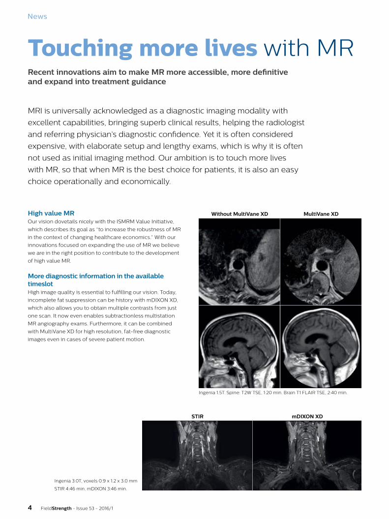
More diagnostic information in the available timeslotHigh image quality is essential to fulfilling our vision. Today,
incomplete fat suppression can be history with mDIXON XD,
which also allows you to obtain multiple contrasts from just
one scan. It now even enables subtractionless multistation
MR angiography exams. Furthermore, it can be combined
with MultiVane XD for high resolution, fat-free diagnostic
images even in cases of severe patient motion.
Ingenia 3.0T, voxels 0.9 x 1.2 x 3.0 mm
STIR 4:46 min. mDIXON 3:46 min.
STIR mDIXON XD
Ingenia 1.5T. Spine: T2W TSE, 1:20 min. Brain T1 FLAIR TSE, 2:40 min.
Without MultiVane XD MultiVane XD

www.philips.com/fieldstrength 5
Enhancing workflow for efficient exams and predictable schedulingWe focus on providing an exceptional patient and user
experience. Our patient in-bore experience solution aims
to help relax patients during scanning to support fast and
robust imaging. It especially benefits patients who may have
avoided MR imaging.
We also designed ScanWise Implant to make it easier to
scan patients with MR Conditional implants, and in the hope
that it will help to reduce the number of patients who are
unnecessarily denied MRI because of these implants.
Increase diagnostic value through quantitative and other advanced techniquesOur advanced and quantitative techniques, such as mDIXON
Quant, ADC maps, MRE and CardiacQuant, can help increase
MR’s diagnostic value. Advanced visualization and analysis
through IntelliSpace Portal can add to the diagnostic
value of MR, by providing tools such as tumor tracking and
multimodality fusion, among many others.
Expand the use of MR into therapy guidance MR-guided procedures, including MR biopsy guidance,
MR brachytherapy, intraoperative MR, and MR-RT, extend
the value of MR to new areas. For example, using MR in
combination with, or instead of CT, adds the benefits of
high soft tissue contrast in radiation therapy planning for
oncology patients.
Innovation for you. Innovation with you.Imagine a future where MR is optimized to help radiologists give a quick definitive answer to a specific clinical question. Together with you, we’re on the path to make MR more informative and more accessible – and expanding its reach beyond diagnosis to MR-guided approaches in therapy.
As we’ve expanded what MR can do, we have also built a community, based on our open
innovation platform, and a belief that colleagues aren’t limited to those who work in the same place, but include all who pursue similar goals.
Do you have a vision about how we can touch more lives with MR? We’d love to hear your ideas. And to learn more about our innovations and to view cases, visit www.philips.com/ISMRM.
Number of interrupted scans in a year in the six scanning roomsCourtesy of Herlev Hospital, Denmark
MRI 1 MRI 3 MRI 5MRI 2 Ingenia with In-bore
solution
Average rescans without Ambient and In-bore experience
70%* reduction
MRI 6
*Compared to the average of the other 5 MR scanners without Ambient and in-bore experience. Results from case studies are not predictive of results in other cases. Results in other cases may vary
Trends in MRI
FieldStrength - Issue 53 - 2016/16
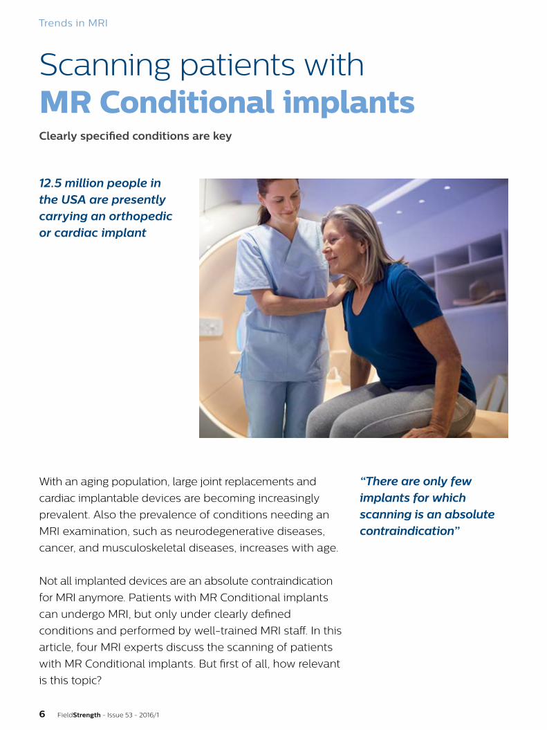
Scanning patients withMR Conditional implants
12.5 million people in the USA are presently carrying an orthopedic or cardiac implant
With an aging population, large joint replacements and
cardiac implantable devices are becoming increasingly
prevalent. Also the prevalence of conditions needing an
MRI examination, such as neurodegenerative diseases,
cancer, and musculoskeletal diseases, increases with age.
Not all implanted devices are an absolute contraindication
for MRI anymore. Patients with MR Conditional implants
can undergo MRI, but only under clearly defined
conditions and performed by well-trained MRI staff. In this
article, four MRI experts discuss the scanning of patients
with MR Conditional implants. But first of all, how relevant
is this topic?
Clearly specified conditions are key
“There are only few implants for which scanning is an absolute contraindication”
www.philips.com/fieldstrength 7
Emanuel Kanal, MD, FACR, FISMRM, AANG, Professor of Radiology and Neuroradiology and Director of Magnetic Resonance Services at the University of Pittsburgh, Pittsburgh, USA. He is Chairman of the American Board of Magnetic Resonance Safety and is lead author of the American College of Radiology’s White Paper on MR Safety and MR Safe Practice Guidelines.
Greg Brown, MRI Consultant and PhD candidate at the Centre for Advanced Imaging, The University of Queensland, St Lucia, Australia. He is an Honorary member of the SMRT, and serves on their MR safety group. He is a non-voting Board Member and SMRT delegate on the American Board of Magnetic Resonance Safety, and holds their MRSO certification. Mr. Brown is an active user of social media platforms on the use of MRI and MRI safety.
Paul W. de Bruin, PhD, Medical Physicist at the Radiology Department, Leiden University Medical Center, Leiden, Netherlands. As a member of the Clinical Physics Group and the C.J. Gorter Center for High-field MRI at Leiden University, he has various research interests in the field of MRI including high-field MR (3T/7T) applications, DCE-MRI, sodium MRI, pharmacokinetics, and musculoskeletal applications.
Harald Kugel, PhD, MR Physicist at the Department of Clinical Radiology of the University of Münster, Münster, Germany. He is the author of numerous articles on MR applications and safety, has been actively involved in the teaching of practical ISMRM courses in MR safety, and is a member of the Technical Committee ‘MR Procedures’ of the German Standards Committee Radiology.
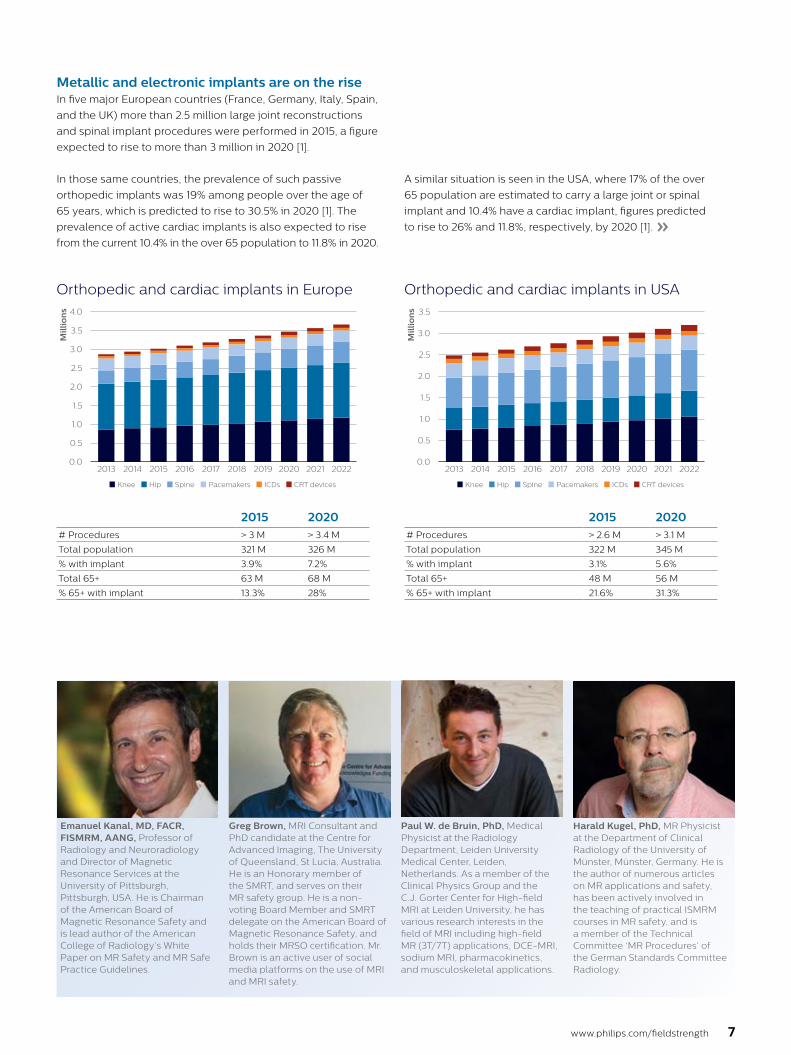
Metallic and electronic implants are on the riseIn five major European countries (France, Germany, Italy, Spain,
and the UK) more than 2.5 million large joint reconstructions
and spinal implant procedures were performed in 2015, a figure
expected to rise to more than 3 million in 2020 [1].
In those same countries, the prevalence of such passive
orthopedic implants was 19% among people over the age of
65 years, which is predicted to rise to 30.5% in 2020 [1]. The
prevalence of active cardiac implants is also expected to rise
from the current 10.4% in the over 65 population to 11.8% in 2020.
A similar situation is seen in the USA, where 17% of the over
65 population are estimated to carry a large joint or spinal
implant and 10.4% have a cardiac implant, figures predicted
to rise to 26% and 11.8%, respectively, by 2020 [1]. »
2015 2020# Procedures > 3 M > 3.4 M
Total population 321 M 326 M
% with implant 3.9% 7.2%
Total 65+ 63 M 68 M
% 65+ with implant 13.3% 28%
2015 2020# Procedures > 2.6 M > 3.1 M
Total population 322 M 345 M
% with implant 3.1% 5.6%
Total 65+ 48 M 56 M
% 65+ with implant 21.6% 31.3%
20130.0
0.5
1.0
1.5
2.0
2.5
3.0
3.5
Mill
ion
s 4.0
2014 2015 2016 2017 2018 2019 2020 2021 2022
Knee Hip Spine Pacemakers ICDs CRT devices
20130.0
0.5
1.0
1.5
2.0
2.5
3.0
3.5
Mill
ion
s
2014 2015 2016 2017 2018 2019 2020 2021 2022
Knee Hip Spine Pacemakers ICDs CRT devices
Orthopedic and cardiac implants in Europe Orthopedic and cardiac implants in USA
FieldStrength - Issue 53 - 2016/18
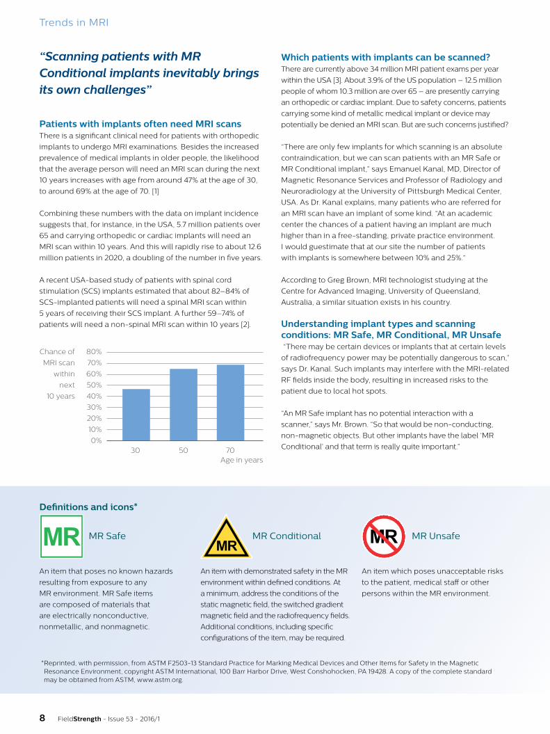
Definitions and icons*
An item that poses no known hazards
resulting from exposure to any
MR environment. MR Safe items
are composed of materials that
are electrically nonconductive,
nonmetallic, and nonmagnetic.
An item with demonstrated safety in the MR
environment within defined conditions. At
a minimum, address the conditions of the
static magnetic field, the switched gradient
magnetic field and the radiofrequency fields.
Additional conditions, including specific
configurations of the item, may be required.
An item which poses unacceptable risks
to the patient, medical staff or other
persons within the MR environment.
* Reprinted, with permission, from ASTM F2503-13 Standard Practice for Marking Medical Devices and Other Items for Safety in the Magnetic Resonance Environment, copyright ASTM International, 100 Barr Harbor Drive, West Conshohocken, PA 19428. A copy of the complete standard may be obtained from ASTM, www.astm.org.
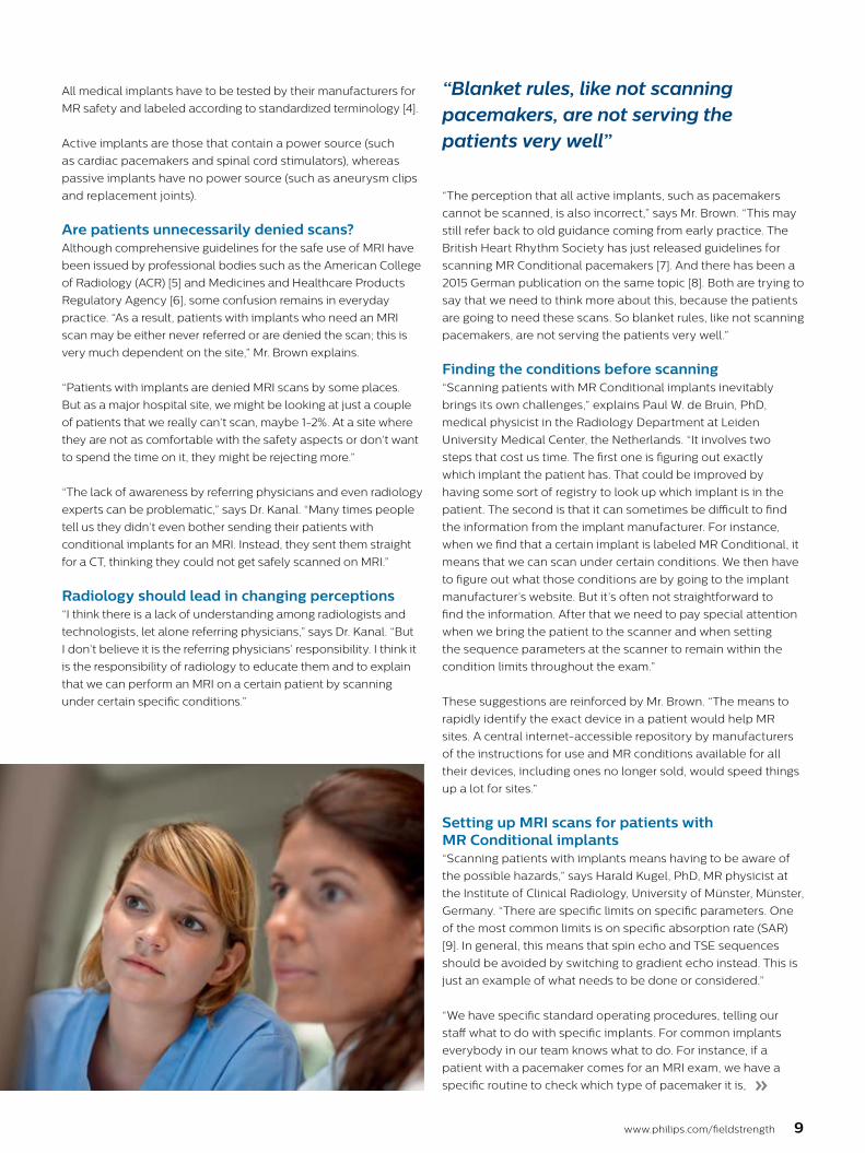
Patients with implants often need MRI scansThere is a significant clinical need for patients with orthopedic
implants to undergo MRI examinations. Besides the increased
prevalence of medical implants in older people, the likelihood
that the average person will need an MRI scan during the next
10 years increases with age from around 47% at the age of 30,
to around 69% at the age of 70. [1]
Combining these numbers with the data on implant incidence
suggests that, for instance, in the USA, 5.7 million patients over
65 and carrying orthopedic or cardiac implants will need an
MRI scan within 10 years. And this will rapidly rise to about 12.6
million patients in 2020, a doubling of the number in five years.
A recent USA-based study of patients with spinal cord
stimulation (SCS) implants estimated that about 82–84% of
SCS-implanted patients will need a spinal MRI scan within
5 years of receiving their SCS implant. A further 59–74% of
patients will need a non-spinal MRI scan within 10 years [2].
Which patients with implants can be scanned?There are currently above 34 million MRI patient exams per year
within the USA [3]. About 3.9% of the US population – 12.5 million
people of whom 10.3 million are over 65 – are presently carrying
an orthopedic or cardiac implant. Due to safety concerns, patients
carrying some kind of metallic medical implant or device may
potentially be denied an MRI scan. But are such concerns justified?
“There are only few implants for which scanning is an absolute
contraindication, but we can scan patients with an MR Safe or
MR Conditional implant,” says Emanuel Kanal, MD, Director of
Magnetic Resonance Services and Professor of Radiology and
Neuroradiology at the University of Pittsburgh Medical Center,
USA. As Dr. Kanal explains, many patients who are referred for
an MRI scan have an implant of some kind. “At an academic
center the chances of a patient having an implant are much
higher than in a free-standing, private practice environment.
I would guestimate that at our site the number of patients
with implants is somewhere between 10% and 25%.”
According to Greg Brown, MRI technologist studying at the
Centre for Advanced Imaging, University of Queensland,
Australia, a similar situation exists in his country.
Understanding implant types and scanning conditions: MR Safe, MR Conditional, MR Unsafe “There may be certain devices or implants that at certain levels
of radiofrequency power may be potentially dangerous to scan,”
says Dr. Kanal. Such implants may interfere with the MRI-related
RF fields inside the body, resulting in increased risks to the
patient due to local hot spots.
“An MR Safe implant has no potential interaction with a
scanner,” says Mr. Brown. “So that would be non-conducting,
non-magnetic objects. But other implants have the label ‘MR
Conditional’ and that term is really quite important.”
80%
70%
60%
50%
40%
30%
20%
10%
0%30 50 70
Chance of
MRI scan
within
next
10 years
Age in years
MR Safe MR Conditional MR Unsafe
“Scanning patients with MR Conditional implants inevitably brings its own challenges”
Trends in MRI
www.philips.com/fieldstrength 9
All medical implants have to be tested by their manufacturers for
MR safety and labeled according to standardized terminology [4].
Active implants are those that contain a power source (such
as cardiac pacemakers and spinal cord stimulators), whereas
passive implants have no power source (such as aneurysm clips
and replacement joints).
Are patients unnecessarily denied scans?Although comprehensive guidelines for the safe use of MRI have
been issued by professional bodies such as the American College
of Radiology (ACR) [5] and Medicines and Healthcare Products
Regulatory Agency [6], some confusion remains in everyday
practice. “As a result, patients with implants who need an MRI
scan may be either never referred or are denied the scan; this is
very much dependent on the site,” Mr. Brown explains.
“Patients with implants are denied MRI scans by some places.
But as a major hospital site, we might be looking at just a couple
of patients that we really can’t scan, maybe 1-2%. At a site where
they are not as comfortable with the safety aspects or don’t want
to spend the time on it, they might be rejecting more.”
“The lack of awareness by referring physicians and even radiology
experts can be problematic,” says Dr. Kanal. “Many times people
tell us they didn’t even bother sending their patients with
conditional implants for an MRI. Instead, they sent them straight
for a CT, thinking they could not get safely scanned on MRI.”
Radiology should lead in changing perceptions“I think there is a lack of understanding among radiologists and
technologists, let alone referring physicians,” says Dr. Kanal. “But
I don’t believe it is the referring physicians’ responsibility. I think it
is the responsibility of radiology to educate them and to explain
that we can perform an MRI on a certain patient by scanning
under certain specific conditions.”
“The perception that all active implants, such as pacemakers
cannot be scanned, is also incorrect,” says Mr. Brown. “This may
still refer back to old guidance coming from early practice. The
British Heart Rhythm Society has just released guidelines for
scanning MR Conditional pacemakers [7]. And there has been a
2015 German publication on the same topic [8]. Both are trying to
say that we need to think more about this, because the patients
are going to need these scans. So blanket rules, like not scanning
pacemakers, are not serving the patients very well.”
Finding the conditions before scanning“Scanning patients with MR Conditional implants inevitably
brings its own challenges,” explains Paul W. de Bruin, PhD,
medical physicist in the Radiology Department at Leiden
University Medical Center, the Netherlands. “It involves two
steps that cost us time. The first one is figuring out exactly
which implant the patient has. That could be improved by
having some sort of registry to look up which implant is in the
patient. The second is that it can sometimes be difficult to find
the information from the implant manufacturer. For instance,
when we find that a certain implant is labeled MR Conditional, it
means that we can scan under certain conditions. We then have
to figure out what those conditions are by going to the implant
manufacturer’s website. But it’s often not straightforward to
find the information. After that we need to pay special attention
when we bring the patient to the scanner and when setting
the sequence parameters at the scanner to remain within the
condition limits throughout the exam.”
These suggestions are reinforced by Mr. Brown. “The means to
rapidly identify the exact device in a patient would help MR
sites. A central internet-accessible repository by manufacturers
of the instructions for use and MR conditions available for all
their devices, including ones no longer sold, would speed things
up a lot for sites.”
Setting up MRI scans for patients with MR Conditional implants“Scanning patients with implants means having to be aware of
the possible hazards,” says Harald Kugel, PhD, MR physicist at
the Institute of Clinical Radiology, University of Münster, Münster,
Germany. “There are specific limits on specific parameters. One
of the most common limits is on specific absorption rate (SAR)
[9]. In general, this means that spin echo and TSE sequences
should be avoided by switching to gradient echo instead. This is
just an example of what needs to be done or considered.”
“We have specific standard operating procedures, telling our
staff what to do with specific implants. For common implants
everybody in our team knows what to do. For instance, if a
patient with a pacemaker comes for an MRI exam, we have a
specific routine to check which type of pacemaker it is, »
“Blanket rules, like not scanning pacemakers, are not serving the patients very well”
FieldStrength - Issue 53 - 2016/110
References
1. Philips, data on file. Based on Millennium research group reports RPUS21LJ09; RPUS21LJ10; RPUS21LJ15; RPEU21LJ08; RPEU21LJ15; RPUS50DI14; RPUS20SP10; RPEU20SP10; RPUS21LJ10; RPEU20SP13; RPUS20SP15; US12CR07; RPGL12CR10; RPGL12CR14 and Barmer GEK Arztreport 2011.
2. Desai MJ, Hargens LM, Breitenfeldt MD, Doth AH, Ryan MP, Gunnarsson C, Safriel Y. The rate of magnetic resonance imaging in patients with spinal cord stimulation. Spine (Phila Pa 1976). 2015;40:E531-7.
3. IMV 2013 MR Market Outlook Report.
4. ASTM F2503-13, Standard Practice for Marking Medical Devices and Other Items for Safety in the Magnetic Resonance Environment, ASTM International, West Conshohocken, PA, 2013.
5. Expert Panel on MR Safety. ACR Guidance Document on MR Safe Practices: 2013. J. Magn. Reson. Imaging 2013;37:501-30.
6. Medicines and Healthcare Products Regulatory Agency. Safety Guidelines for Magnetic Resonance Imaging Equipment in Clinical Use. 2014
7. Lowe MD, Plummer CJ, Manisty CH3, Linker NJ. Safe use of MRI in people with cardiac implantable electronic devices. Heart. 2015;101:1950-3
8. Sommer T, et al. German Roentgen Society Statement on MR Imaging of Patients with Cardiac Pacemakers. Fortschr Röntgenstr 2015; 187: 777-87.
9. Woods, TO. Establishing Safety and Compatibility of Passive Implants in the Magnetic Resonance (MR) Environment, FDA, 2014.
10. For instance: IEC 60601-2-33 – Requirements for the Safety of MR Equipment for Medical Diagnosis; FDA – Guidelines for Premarket Notifications for MR Diagnostic Devices; NEMA MS 1 through 9 – Safety and Performance Standards.
11. American College of Radiology. Accreditation.
to check that a cardiologist is present as an MR Conditional
pacemaker usually needs to be switched in an MR-compatible
mode, etc. So some actions have to be taken and this is all laid
down in our procedure.”
Education is an important step“I think a lot more education of technologists and radiologists is
needed, so that we can develop an efficient and structured way
forward,” Mr. Brown says.
“We know that MRI scanners have to pass certain levels of
safety and show that they are documented to be kept at certain
guidelines and thresholds [10]. MR site accreditation in the USA [11]
documents that the site is appropriately designed,” says Dr. Kanal.
“But who is missing in all this? There was no certification process
to show that the magnetic resonance medical director, radiologist,
technologist, or physicist have a comprehensive understanding
of the safety issues associated with magnetic resonance
environments or how to apply them. We therefore created the
American Board of MR Safety in 2014. Its sole purpose is to certify
and credential MR medical directors, MR safety officers, and
MR safety experts, who represent the radiologists/physicians,
technologists, and physicists who are charged with overseeing
safety in clinical and research magnetic resonance environments.”
Saying ‘no’ is easy, but saying ‘yes’ requires knowledge“Sometimes patients have certain implants and the site is not
sufficiently familiar with what can and can’t be done to decrease
the risk of an MRI scan,” says Dr. Kanal. “They may choose, for
ostensible ‘safety’ objectives, to not scan that patient. I put the
word safety in quotes because not scanning a patient for whom
a diagnostic MRI was requested has its own risks. The patient
may go undiagnosed or may have to be sent for a more invasive
study to make a diagnosis.”
“Saying ‘no’ is easy, but saying ‘yes’ requires knowledge,
confidence in that knowledge, and the willingness to say yes and
to apply that knowledge.”
“Not scanning a patient for whom a diagnostic MRI was requested can also potentially impact patient care”
«
“Scanning patients with implants means having to be aware of the possible hazards”
Trends in MRI
Trends in MRI
www.philips.com/fieldstrength 11
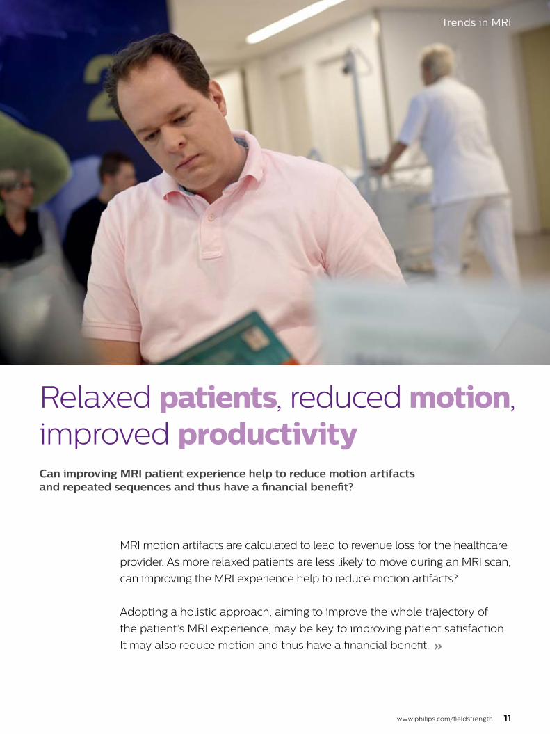
Can improving MRI patient experience help to reduce motion artifacts and repeated sequences and thus have a financial benefit?
Relaxed patients, reduced motion, improved productivity
MRI motion artifacts are calculated to lead to revenue loss for the healthcare
provider. As more relaxed patients are less likely to move during an MRI scan,
can improving the MRI experience help to reduce motion artifacts?
Adopting a holistic approach, aiming to improve the whole trajectory of
the patient’s MRI experience, may be key to improving patient satisfaction.
It may also reduce motion and thus have a financial benefit. »
FieldStrength - Issue 53 - 2016/112
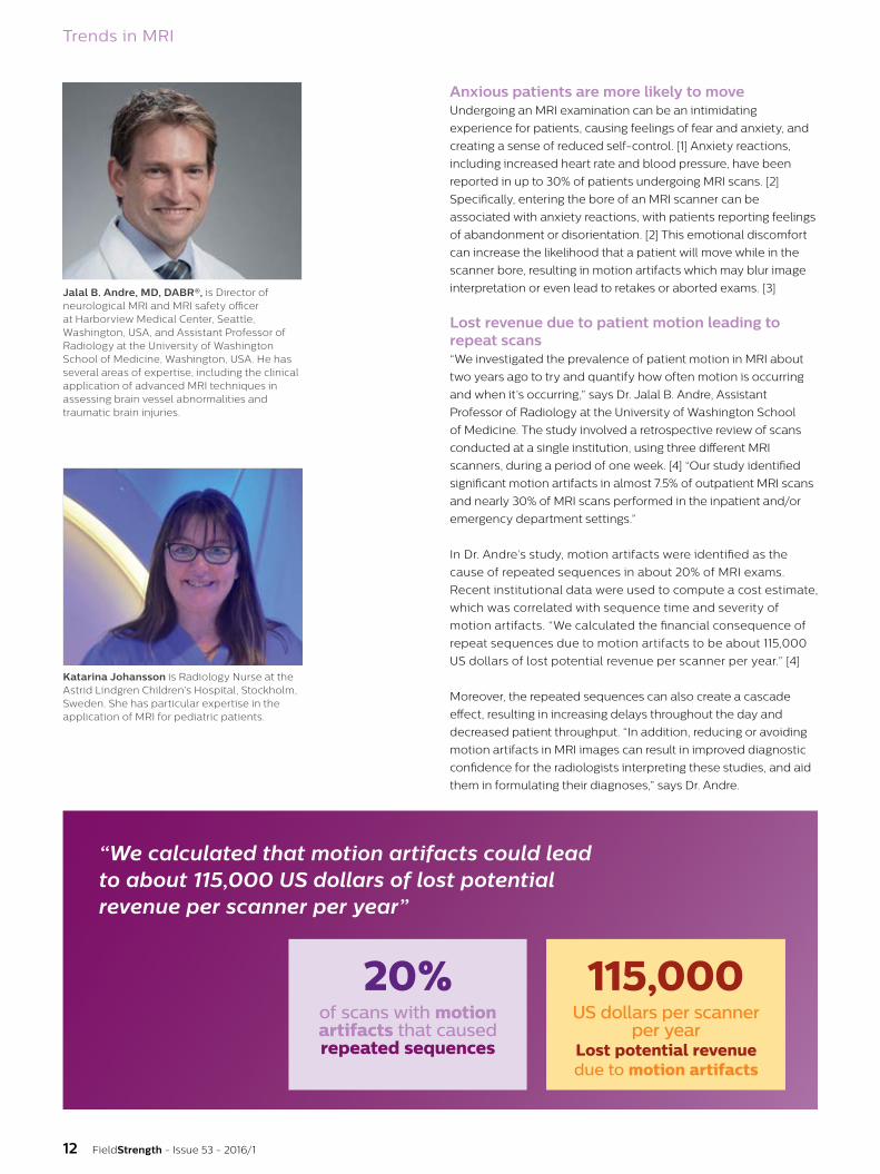
Jalal B. Andre, MD, DABR®, is Director of neurological MRI and MRI safety officer at Harborview Medical Center, Seattle, Washington, USA, and Assistant Professor of Radiology at the University of Washington School of Medicine, Washington, USA. He has several areas of expertise, including the clinical application of advanced MRI techniques in assessing brain vessel abnormalities and traumatic brain injuries.
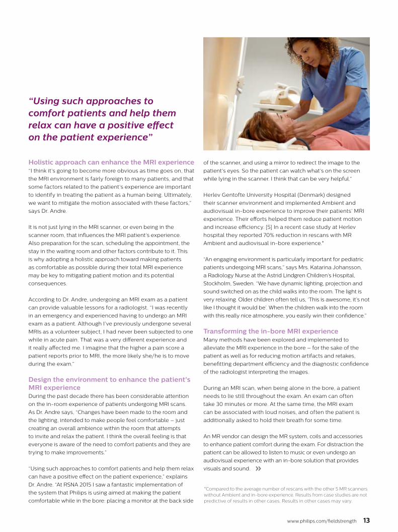
Katarina Johansson is Radiology Nurse at the Astrid Lindgren Children’s Hospital, Stockholm, Sweden. She has particular expertise in the application of MRI for pediatric patients.
Anxious patients are more likely to move Undergoing an MRI examination can be an intimidating
experience for patients, causing feelings of fear and anxiety, and
creating a sense of reduced self-control. [1] Anxiety reactions,
including increased heart rate and blood pressure, have been
reported in up to 30% of patients undergoing MRI scans. [2]
Specifically, entering the bore of an MRI scanner can be
associated with anxiety reactions, with patients reporting feelings
of abandonment or disorientation. [2] This emotional discomfort
can increase the likelihood that a patient will move while in the
scanner bore, resulting in motion artifacts which may blur image
interpretation or even lead to retakes or aborted exams. [3]
Lost revenue due to patient motion leading to repeat scans“We investigated the prevalence of patient motion in MRI about
two years ago to try and quantify how often motion is occurring
and when it’s occurring,” says Dr. Jalal B. Andre, Assistant
Professor of Radiology at the University of Washington School
of Medicine. The study involved a retrospective review of scans
conducted at a single institution, using three different MRI
scanners, during a period of one week. [4] “Our study identified
significant motion artifacts in almost 7.5% of outpatient MRI scans
and nearly 30% of MRI scans performed in the inpatient and/or
emergency department settings.”
In Dr. Andre’s study, motion artifacts were identified as the
cause of repeated sequences in about 20% of MRI exams.
Recent institutional data were used to compute a cost estimate,
which was correlated with sequence time and severity of
motion artifacts. “We calculated the financial consequence of
repeat sequences due to motion artifacts to be about 115,000
US dollars of lost potential revenue per scanner per year.” [4]
Moreover, the repeated sequences can also create a cascade
effect, resulting in increasing delays throughout the day and
decreased patient throughput. “In addition, reducing or avoiding
motion artifacts in MRI images can result in improved diagnostic
confidence for the radiologists interpreting these studies, and aid
them in formulating their diagnoses,” says Dr. Andre.
“We calculated that motion artifacts could lead to about 115,000 US dollars of lost potential revenue per scanner per year”
20%of scans with motion artifacts that caused repeated sequences
US dollars per scanner per year
Lost potential revenue due to motion artifacts
115,000
Trends in MRI
www.philips.com/fieldstrength 13
of the scanner, and using a mirror to redirect the image to the
patient’s eyes. So the patient can watch what’s on the screen
while lying in the scanner. I think that can be very helpful.”
Herlev Gentofte University Hospital (Denmark) designed
their scanner environment and implemented Ambient and
audiovisual in-bore experience to improve their patients’ MRI
experience. Their efforts helped them reduce patient motion
and increase efficiency. [5] In a recent case study at Herlev
hospital they reported 70% reduction in rescans with MR
Ambient and audiovisual in-bore experience.*
“An engaging environment is particularly important for pediatric
patients undergoing MRI scans,” says Mrs. Katarina Johansson,
a Radiology Nurse at the Astrid Lindgren Children’s Hospital,
Stockholm, Sweden. “We have dynamic lighting, projection and
sound switched on as the child walks into the room. The light is
very relaxing. Older children often tell us, ‘This is awesome, it’s not
like I thought it would be’. When the children walk into the room
with this really nice atmosphere, you easily win their confidence.”
Transforming the in-bore MRI experienceMany methods have been explored and implemented to
alleviate the MRI experience in the bore – for the sake of the
patient as well as for reducing motion artifacts and retakes,
benefitting department efficiency and the diagnostic confidence
of the radiologist interpreting the images.
During an MRI scan, when being alone in the bore, a patient
needs to lie still throughout the exam. An exam can often
take 30 minutes or more. At the same time, the MRI exam
can be associated with loud noises, and often the patient is
additionally asked to hold their breath for some time.
An MR vendor can design the MR system, coils and accessories
to enhance patient comfort during the exam. For distraction the
patient can be allowed to listen to music or even undergo an
audiovisual experience with an in-bore solution that provides
visuals and sound.
Holistic approach can enhance the MRI experience“I think it’s going to become more obvious as time goes on, that
the MRI environment is fairly foreign to many patients, and that
some factors related to the patient’s experience are important
to identify in treating the patient as a human being. Ultimately,
we want to mitigate the motion associated with these factors,”
says Dr. Andre.
It is not just lying in the MRI scanner, or even being in the
scanner room, that influences the MRI patient’s experience.
Also preparation for the scan, scheduling the appointment, the
stay in the waiting room and other factors contribute to it. This
is why adopting a holistic approach toward making patients
as comfortable as possible during their total MRI experience
may be key to mitigating patient motion and its potential
consequences.
According to Dr. Andre, undergoing an MRI exam as a patient
can provide valuable lessons for a radiologist. “I was recently
in an emergency and experienced having to undergo an MRI
exam as a patient. Although I’ve previously undergone several
MRIs as a volunteer subject, I had never been subjected to one
while in acute pain. That was a very different experience and
it really affected me. I imagine that the higher a pain score a
patient reports prior to MRI, the more likely she/he is to move
during the exam.”
Design the environment to enhance the patient's MRI experienceDuring the past decade there has been considerable attention
on the in-room experience of patients undergoing MRI scans.
As Dr. Andre says, “Changes have been made to the room and
the lighting, intended to make people feel comfortable – just
creating an overall ambience within the room that attempts
to invite and relax the patient. I think the overall feeling is that
everyone is aware of the need to comfort patients and they are
trying to make improvements.”
“Using such approaches to comfort patients and help them relax
can have a positive effect on the patient experience,” explains
Dr. Andre. “At RSNA 2015 I saw a fantastic implementation of
the system that Philips is using aimed at making the patient
comfortable while in the bore: placing a monitor at the back side
“Using such approaches to comfort patients and help them relax can have a positive effect on the patient experience”
»
*Compared to the average number of rescans with the other 5 MR scanners without Ambient and in-bore experience. Results from case studies are not predictive of results in other cases. Results in other cases may vary.
FieldStrength - Issue 53 - 2016/114
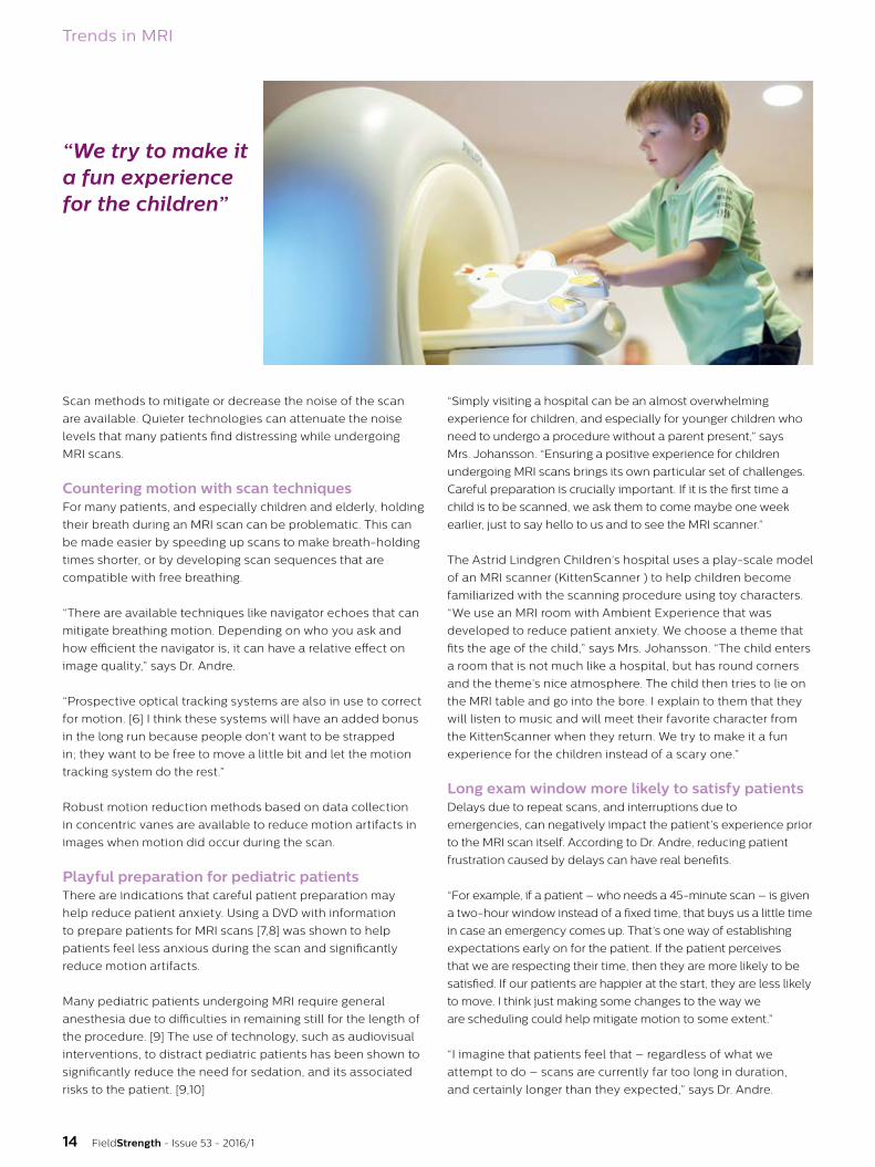
“Simply visiting a hospital can be an almost overwhelming
experience for children, and especially for younger children who
need to undergo a procedure without a parent present,” says
Mrs. Johansson. “Ensuring a positive experience for children
undergoing MRI scans brings its own particular set of challenges.
Careful preparation is crucially important. If it is the first time a
child is to be scanned, we ask them to come maybe one week
earlier, just to say hello to us and to see the MRI scanner.”
The Astrid Lindgren Children’s hospital uses a play-scale model
of an MRI scanner (KittenScanner ) to help children become
familiarized with the scanning procedure using toy characters.
“We use an MRI room with Ambient Experience that was
developed to reduce patient anxiety. We choose a theme that
fits the age of the child,” says Mrs. Johansson. “The child enters
a room that is not much like a hospital, but has round corners
and the theme’s nice atmosphere. The child then tries to lie on
the MRI table and go into the bore. I explain to them that they
will listen to music and will meet their favorite character from
the KittenScanner when they return. We try to make it a fun
experience for the children instead of a scary one.”
Long exam window more likely to satisfy patientsDelays due to repeat scans, and interruptions due to
emergencies, can negatively impact the patient’s experience prior
to the MRI scan itself. According to Dr. Andre, reducing patient
frustration caused by delays can have real benefits.
“For example, if a patient – who needs a 45-minute scan – is given
a two-hour window instead of a fixed time, that buys us a little time
in case an emergency comes up. That’s one way of establishing
expectations early on for the patient. If the patient perceives
that we are respecting their time, then they are more likely to be
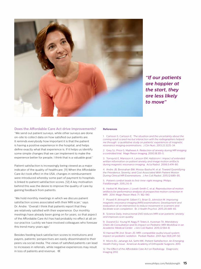
satisfied. If our patients are happier at the start, they are less likely
to move. I think just making some changes to the way we
are scheduling could help mitigate motion to some extent.”
“I imagine that patients feel that – regardless of what we
attempt to do – scans are currently far too long in duration,
and certainly longer than they expected,” says Dr. Andre.
Scan methods to mitigate or decrease the noise of the scan
are available. Quieter technologies can attenuate the noise
levels that many patients find distressing while undergoing
MRI scans.
Countering motion with scan techniquesFor many patients, and especially children and elderly, holding
their breath during an MRI scan can be problematic. This can
be made easier by speeding up scans to make breath-holding
times shorter, or by developing scan sequences that are
compatible with free breathing.
“There are available techniques like navigator echoes that can
mitigate breathing motion. Depending on who you ask and
how efficient the navigator is, it can have a relative effect on
image quality,” says Dr. Andre.
“Prospective optical tracking systems are also in use to correct
for motion. [6] I think these systems will have an added bonus
in the long run because people don’t want to be strapped
in; they want to be free to move a little bit and let the motion
tracking system do the rest.”
Robust motion reduction methods based on data collection
in concentric vanes are available to reduce motion artifacts in
images when motion did occur during the scan.
Playful preparation for pediatric patientsThere are indications that careful patient preparation may
help reduce patient anxiety. Using a DVD with information
to prepare patients for MRI scans [7,8] was shown to help
patients feel less anxious during the scan and significantly
reduce motion artifacts.
Many pediatric patients undergoing MRI require general
anesthesia due to difficulties in remaining still for the length of
the procedure. [9] The use of technology, such as audiovisual
interventions, to distract pediatric patients has been shown to
significantly reduce the need for sedation, and its associated
risks to the patient. [9,10]
“We try to make it a fun experience for the children”
Trends in MRI
www.philips.com/fieldstrength 15
Does the Affordable Care Act drive improvements?“We send out patient surveys, while other surveys are done
on-site to collect data on how satisfied our patients are.
It reminds everybody how important it is that the patient
is having a positive experience in the hospital, and helps
define exactly what that experience is. If it helps us identify
some simple changes that we can implement to make the
experience better for people, I think that is a valuable goal.”
Patient satisfaction is increasingly being viewed as a major
indicator of the quality of healthcare. [11] When the Affordable
Care Act took effect in the USA, changes in reimbursement
were introduced whereby some part of payment to hospitals
is linked to patient satisfaction scores. [12] A key motivation
behind this was the desire to improve the quality of care by
gaining feedback from patients.
“We hold monthly meetings in which we discuss patient
satisfaction scores associated with their MRI scan,” says
Dr. Andre. “Overall I think that patients report that they
are relatively satisfied with their experience. Our monthly
meetings have already been going on for years, so that aspect
of the Affordable Care Act has had probably no effect at all on
our practice. Luckily we have involved colleagues who foresaw
this trend many years ago.”
Besides feeding back satisfaction scores to institutions and
payers, patients’ perspectives are easily disseminated to their
peers via social media. The views of satisfied patients can lead
to increases in referrals, while negative experiences may result
in loss of patients and revenue.
References
1. Carlsson S, Carlsson E. ‘The situation and the uncertainty about the coming result scared me but interaction with the radiographers helped me through’: a qualitative study on patients’ experiences of magnetic resonance imaging examinations. J Clin Nurs. 2013;22:3225-34.
2. Grey SJ, Price G, Mathews A. Reduction of anxiety during MR imaging: a controlled trial. Magn Reson Imaging. 2000;18:351-5.
3. Tornqvist E, Mansson A, Larsson EM, Hallstrom I. Impact of extended written information on patient anxiety and image motion artifacts during magnetic resonance imaging. Acta Radiol. 2006;5:474-80.
4. Andre JB, Bresnahan BW, Mossa-Basha M, et al. Toward Quantifying the Prevalence, Severity, and Cost Associated With Patient Motion During Clinical MR Examinations. J Am Coll Radiol. 2015;12:689-95.
5. Patient comfort leads to first-time-right imaging. Philips FieldStrength. 2015;2:6-9.
6. Herbst M, Maclaren J, Lovell-Smith C, et al. Reproduction of motion artifacts for performance analysis of prospective motion correction in MRI. 2014. Magn Reson Med; 71: 182-190.
7. Powell R, Ahmad M, Gilbert FJ, Brian D, Johnston M. Improving magnetic resonance imaging (MRI) examinations: Development and evaluation of an intervention to reduce movement in scanners and facilitate scan completion. Br J Health Psychol. 2015;20:449-65.
8. Science Daily. Instructional DVD reduces MRI scan patients’ anxiety and improves scan quality.
9. Durand DJ, Young M, Nagy P, Tekes A, Huisman TA. Mandatory Child Life Consultation and Its Impact on Pediatric MRI Workflow in an Academic Medical Center. J Am Coll Radiol. 2015;12:594-8.
10. Harned RK 2nd, Strain JD. MRI-compatible audio/visual system: impact on pediatric sedation. Pediatr Radiol. 2001 Apr;31(4):247-50.
11. Morris BJ, Jahangir AA, Sethi MK. Patient Satisfaction: An Emerging Health Policy Issue. American Academy of Orthopedic Surgeons. 2013.
12. The Effect of the Affordable Care Act on Radiology. Diagnostic Imaging 2012. «
“If our patients are happier at the start, they are less likely to move”
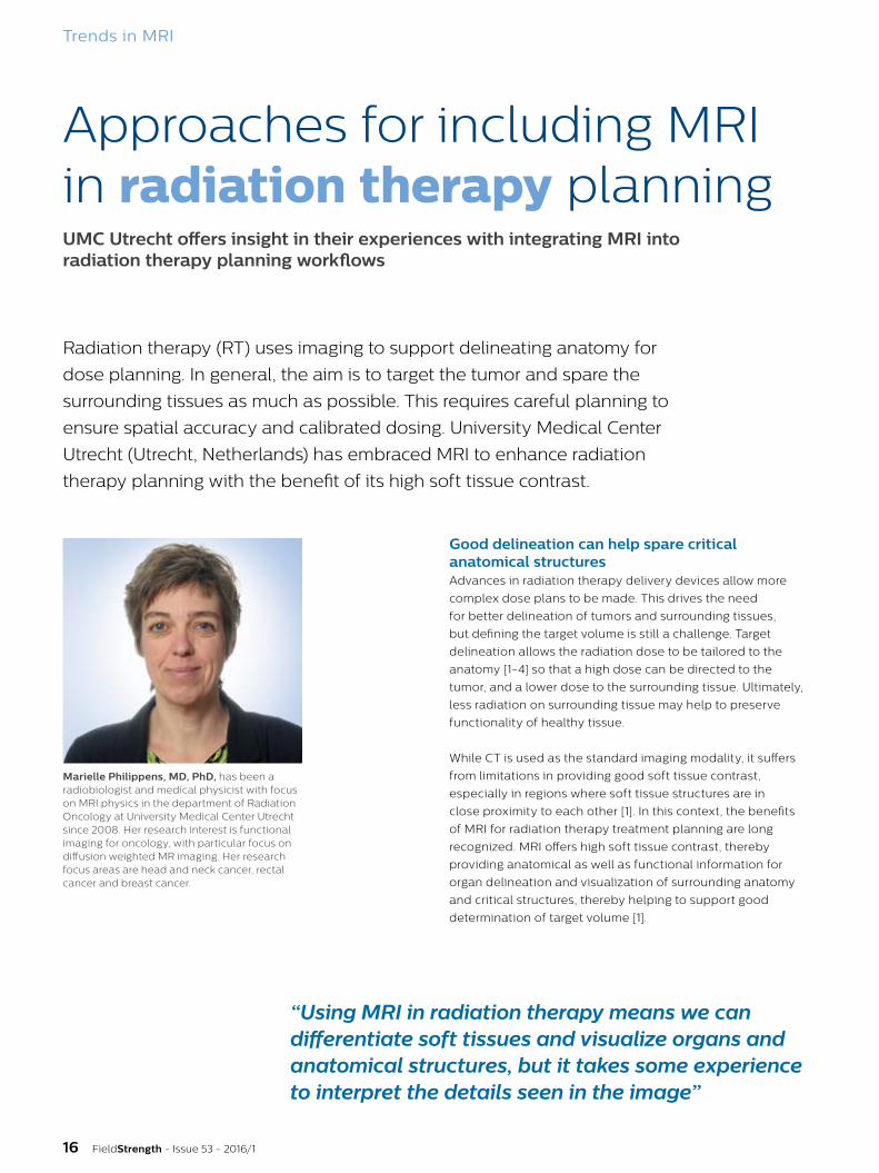
Marielle Philippens, MD, PhD, has been a radiobiologist and medical physicist with focus on MRI physics in the department of Radiation Oncology at University Medical Center Utrecht since 2008. Her research interest is functional imaging for oncology, with particular focus on diffusion weighted MR imaging. Her research focus areas are head and neck cancer, rectal cancer and breast cancer.
FieldStrength - Issue 53 - 2016/116
Radiation therapy (RT) uses imaging to support delineating anatomy for
dose planning. In general, the aim is to target the tumor and spare the
surrounding tissues as much as possible. This requires careful planning to
ensure spatial accuracy and calibrated dosing. University Medical Center
Utrecht (Utrecht, Netherlands) has embraced MRI to enhance radiation
therapy planning with the benefit of its high soft tissue contrast.
Approaches for including MRI in radiation therapy planningUMC Utrecht offers insight in their experiences with integrating MRI into radiation therapy planning workflows
“Using MRI in radiation therapy means we can differentiate soft tissues and visualize organs and anatomical structures, but it takes some experience to interpret the details seen in the image”
Good delineation can help spare critical anatomical structuresAdvances in radiation therapy delivery devices allow more
complex dose plans to be made. This drives the need
for better delineation of tumors and surrounding tissues,
but defining the target volume is still a challenge. Target
delineation allows the radiation dose to be tailored to the
anatomy [1-4] so that a high dose can be directed to the
tumor, and a lower dose to the surrounding tissue. Ultimately,
less radiation on surrounding tissue may help to preserve
functionality of healthy tissue.
While CT is used as the standard imaging modality, it suffers
from limitations in providing good soft tissue contrast,
especially in regions where soft tissue structures are in
close proximity to each other [1]. In this context, the benefits
of MRI for radiation therapy treatment planning are long
recognized. MRI offers high soft tissue contrast, thereby
providing anatomical as well as functional information for
organ delineation and visualization of surrounding anatomy
and critical structures, thereby helping to support good
determination of target volume [1].
Trends in MRI
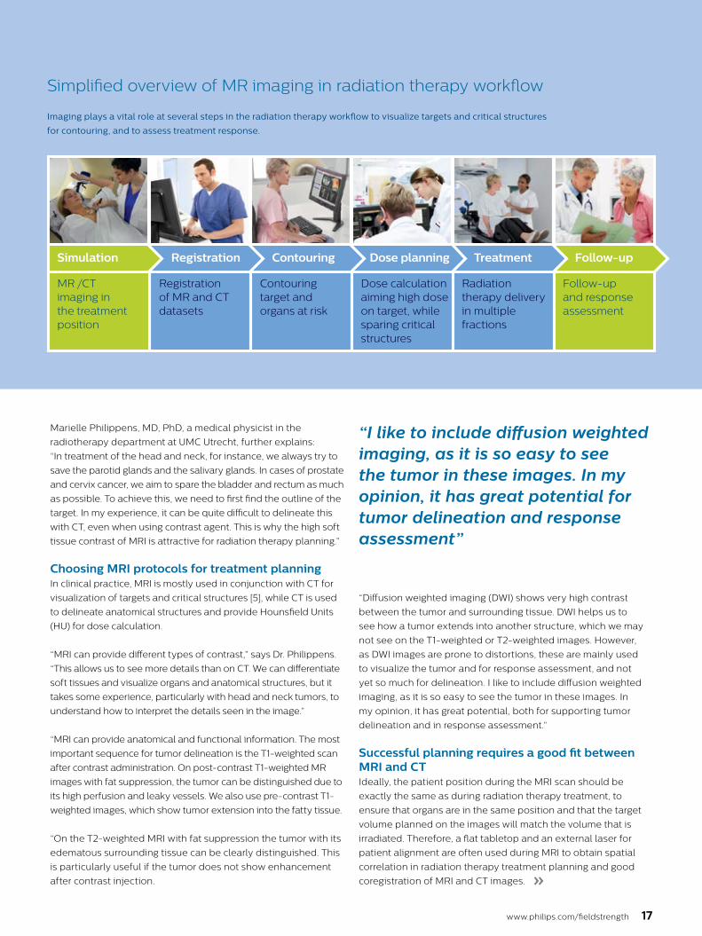
Simplified overview of MR imaging in radiation therapy workflow
Imaging plays a vital role at several steps in the radiation therapy workflow to visualize targets and critical structures
for contouring, and to assess treatment response.
www.philips.com/fieldstrength 17
Marielle Philippens, MD, PhD, a medical physicist in the
radiotherapy department at UMC Utrecht, further explains:
“In treatment of the head and neck, for instance, we always try to
save the parotid glands and the salivary glands. In cases of prostate
and cervix cancer, we aim to spare the bladder and rectum as much
as possible. To achieve this, we need to first find the outline of the
target. In my experience, it can be quite difficult to delineate this
with CT, even when using contrast agent. This is why the high soft
tissue contrast of MRI is attractive for radiation therapy planning.”
Choosing MRI protocols for treatment planningIn clinical practice, MRI is mostly used in conjunction with CT for
visualization of targets and critical structures [5], while CT is used
to delineate anatomical structures and provide Hounsfield Units
(HU) for dose calculation.
“MRI can provide different types of contrast,” says Dr. Philippens.
“This allows us to see more details than on CT. We can differentiate
soft tissues and visualize organs and anatomical structures, but it
takes some experience, particularly with head and neck tumors, to
understand how to interpret the details seen in the image.”
“MRI can provide anatomical and functional information. The most
important sequence for tumor delineation is the T1-weighted scan
after contrast administration. On post-contrast T1-weighted MR
images with fat suppression, the tumor can be distinguished due to
its high perfusion and leaky vessels. We also use pre-contrast T1-
weighted images, which show tumor extension into the fatty tissue.
“On the T2-weighted MRI with fat suppression the tumor with its
edematous surrounding tissue can be clearly distinguished. This
is particularly useful if the tumor does not show enhancement
after contrast injection.
“I like to include diffusion weighted imaging, as it is so easy to see the tumor in these images. In my opinion, it has great potential for tumor delineation and response assessment”
“Diffusion weighted imaging (DWI) shows very high contrast
between the tumor and surrounding tissue. DWI helps us to
see how a tumor extends into another structure, which we may
not see on the T1-weighted or T2-weighted images. However,
as DWI images are prone to distortions, these are mainly used
to visualize the tumor and for response assessment, and not
yet so much for delineation. I like to include diffusion weighted
imaging, as it is so easy to see the tumor in these images. In
my opinion, it has great potential, both for supporting tumor
delineation and in response assessment.”
Successful planning requires a good fit between MRI and CTIdeally, the patient position during the MRI scan should be
exactly the same as during radiation therapy treatment, to
ensure that organs are in the same position and that the target
volume planned on the images will match the volume that is
irradiated. Therefore, a flat tabletop and an external laser for
patient alignment are often used during MRI to obtain spatial
correlation in radiation therapy treatment planning and good
coregistration of MRI and CT images. »
Simulation
MR /CT imaging in the treatment position
Registration
Registration of MR and CT datasets
Contouring
Contouring target and organs at risk
Treatment
Radiation therapy delivery in multiple fractions
Dose planning
Dose calculation aiming high dose on target, while sparing critical structures
Follow-up
Follow-up and response assessment

FieldStrength - Issue 53 - 2016/118
“For coregistration of MRI and CT images, we aim to use an
MRI contrast type with few non-linearities and good tissue
differentiation, preferably acquired as non-angulated axial slices,”
says Dr. Philippens. “Either 3D scan protocols or multislice 2D
protocols with contiguous slices are used to allow target volume
reconstruction in the different orthogonal directions.”
MRI is used for planning in a variety of anatomiesAt UMC Utrecht, Dr. Philippens and her colleagues are using
MRI in planning external beam radiation therapy (EBRT) for
treatment of tumors in a variety of anatomies, such as organs in
the pelvis (including bladder, prostate, rectum and cervix), the
brain, the esophagus, pancreas, the larynx and oropharynx, bone
metastases and sarcomas. In addition, MRI is also used to guide
brachytherapy in the prostate and cervix. [6]
“MRI also helps in visualizing the lesions inside the prostate, which may not be possible in CT”
High dose on the lesion rather than on the whole prostateMRI is capable of visualizing the prostate and the surrounding
organs such as rectum, penile bulb, bladder, the apex and
seminal vesicles, as well as visualizing intra-prostatic
lesions [2,4].
“All our patients undergo an MRI exam – along with CT –
before radiotherapy of the prostate,” says Dr. Philippens.
“For prostate delineation, we are scanning a balanced TFE with
fat suppression. We can also see the gold fiducial markers in
these images, which are used for position verification and are
therefore used for registration to CT. For geometric accuracy of
the image, we choose a 3D sequence, which is corrected for the
gradient non-linearities in all directions.
In 2015 the UMC Utrecht RT department has performed 1300 MR exams in their routine clinical patients.
The University Medical Center Utrecht Department of Radiotherapy is a leading center in radiation therapy, continuously striving to improve methods and provide excellent patient care. UMC Utrecht has two Philips Ingenia MR-RT systems (1.5T and 3.0T) for RT treatment simulation as well as a Philips Ingenia 1.5T system dedicated for brachytherapy. Furthermore, it is an Atlantic MR-Linac consortium member. UMC Utrecht regularly organizes courses on MRI in radiotherapy for physicians, technologists and radiation oncologists, see: http://mri-in-radiotherapy.nl/
Use of MR in RT planning at UMC Utrecht in 2015
29%
25%17%
UrologyBrainGIHead and neckGynecologyOther
15%
7%8%
29%
25%17%
UrologyBrainGIHead and neckGynecologyOther
15%
7%8%
0
RT treatments
MR use
500 1000 1500 2000 2500 3000 3500 4000 4500
Curative Other
About 4200 RT treatments
More than 1300 MR examsin radiation therapy department
3.0T 1.5T Brachytheraphy
Visualizing critical structures with MRI before prostate radiation therapy
A 63-year-old patient with prostate cancer, cT3bNxM, Gleason 7, underwent MRI on Ingenia 3.0T MR-RT before radiation therapy.
Intraprostatic lesions are visible on the bTFE MR image, but not on the CT image. MRI shows excellent soft-tissue contrast for the visualization of critical structures like the rectum and penile bulb.
Fiducial markers (green arrows) are used in registration of MR images to CT, to transfer the MR-based delineations onto the CT image dataset.
bTFE SPAIR zoomed
CT
CT zoomed T2W TSE zoomed
Penilebulb
Rectum
Trends in MRI
www.philips.com/fieldstrength 19
“In addition to helping in delineation of the prostate, MRI also
helps in visualizing the lesions inside the prostate, which may
not be possible in CT.
“When we can visualize intraprostatic lesions, the radiation
therapist can then plan to boost them, giving a higher dose
to those lesions instead of giving a uniform dose to the whole
prostate, in the hope to better treat the patient and have less risk
of recurrent tumors. However, this is not yet clinical routine. For
visualizing the lesions, we not only use anatomical, T2-weighted
imaging, but also diffusion weighted MRI and dynamic contrast-
enhanced MRI."
Visualizing critical structures in the head and neck“In patients with a primary tumor in the head and neck area,
we do use MRI in daily clinical radiation therapy practice to
visualize the tumor and critical structures. This may be used to
help sparing of critical structures, such as the parotid glands,
submandibular glands, esophagus, optic nerves, brain stem and
spinal cord [7]. And postoperatively we scan patients that have
tumor growth along the cranial nerves for target delineation,”
says Dr. Philippens.
“Because of the challenges posed by CT MRI coregistration
in this area with many degrees of freedom for motion, we
image these patients in a radiotherapy mask. However, one
disadvantage of using the mask is that a regular head and neck
coil cannot be used; a dedicated coil solution would be needed
for imaging with a mask. For this we make use of flexible coils
that we position close to the target area. This setup can also
be combined with the anterior coil for a larger coverage and
enhanced SNR.”
“We use pre- and post-contrast T1- and T2-weighted sequences
with the fast and robust mDIXON method for fat suppression,”
says Dr. Philippens. “Dynamic contrast-enhanced imaging
is performed with high temporal resolution and low spatial
resolution, to see the contrast agent uptake in the tumor. Diffusion
weighted imaging is used qualitatively to see how the tumor
extends into another structure, rather than for strict delineation.”
“In postoperative patients who have had tumor growth along the
cranial nerves, we use T2-weighted gradient echo (FFE) on our
3.0T MR-RT scanner to show the nerves for target delineation and
look to see if there is still tumor left.” »
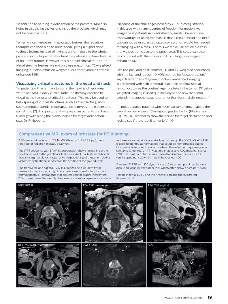
Comprehensive MRI exam of prostate for RT planning A 70-year-old male with cT3bNxM0, Gleason 6, PSA 7.9 µg/L, was referred for radiation therapy treatment.
The bTFE sequence with SPAIR fat suppression shows the outline of the prostate as well as the gold fiducials. It is important that both are defined in the same, high resolution image, since the positioning of the patient during radiotherapy treatment is based on the position of the gold fiducials.
The transverse and sagittal T2W TSE images help us identify the prostate tumor foci, which typically have lower signal intensity than normal prostate. For patients that are referred for brachytherapy the T2W image is used to identify the presence of extracapsular extensions
as these are a contraindication for brachytherapy. The 3D T1 mDIXON FFE is used to identify abnormalities that could be hemorrhages due to biopsies or insertion of fiducial markers. These hemorrhages may look similar to tumor foci on T2-weighted images and ADC map.Transverse DWI with SPAIR and 6 b-values is used to visualize the tumor foci (bright appearance), which mostly have a low ADC.
Dynamic T1 FFE with 120 dynamics and 2.4 sec. temporal resolution is also used visualize the tumor foci, which often show a high perfusion.
Philips Ingenia 3.0T using the Anterior coil and the integrated Posterior coil.
bTFE SPAIR
3D T1 mDIXON FFE water
T2W TSE transverse T2W TSE sagittal
DWI b1000 ADC

FieldStrength - Issue 53 - 2016/120
References
1. Schmidt MA, Payne GS. Radiotherapy planning using MRI. Phys Med Biol 2015, 60: R323–361
2. Doemer A, Chetty IJ, Glide-Hurst C, et al. Evaluating organ delineation, dose calculation and daily localization in an open-MRI simulation workflow for prostate cancer patients. Radiat Oncol. 2015;10:37.
3. Devic S. MRI simulation for radiotherapy treatment planning. Med Phys 2012, 39: 6701-11
4. Paulson ES, Erickson, B, Schultz C, Li XA. Comprehensive MRI simulation methodology using a dedicated MRI scanner in radiation oncology for external beam radiation treatment planning. Med Phys 2015, 42: 28-39
5. Glynne-Jones R, Nilsson PJ, Aschele C, et al. Anal cancer: ESMO-ESSO-ESTRO clinical practice guidelines. Ann Oncol. 2014;25:iii10-20.
6. Metcalfe P, Liney GP, Holloway L, et al. The potential for an enhanced role for MRI in radiation-therapy treatment planning. Technol Cancer Res Treat 2013, 12: 429-46
7. Nuyts S. Defining the target for radiotherapy of head and neck cancer. Cancer Imaging. 2007; 7:S50-5.
MRI of head and neck for radiation therapy planning
A 75-year-old male was referred for radiation therapy treatment of oropharynx squamous cell carcinoma in the left tonsil region with extension into the soft palate, caudal border lower tonsil region, no midline crossing. On the left side in the neck there are also three enlarged lymph nodes on level 2 and 3 with central necrosis and signs of limited extracapsular extension, T2N2b.
The patient undergoes MRI in the radiotherapy (5-point) positioning mask in Ingenia 3.0T using the Flex coils. DWI with SPIR is used to visualize the extension of the tumor and lymph nodes, especially retropharyngeal. Transverse T1 and T2 TSE mDIXON water and in-phase images (2 mm thick slices) help to visualize the tumor size and its extension into fatty tissue. The post-contrast T1W TSE mDIXON also shows this.
Dynamic 3D T1 FFE with 45 dynamics and temporal resolution of 2.5 seconds is performed to follow contrast agent distribution. Contrast agent distribution is modeled after conversion of the signal to T1 relaxation times using the small flip angle method.
Clinical value Using different contrasts (T1, T2, diffusion, post-contrast T1) in MRI allows us to appreciate contrast changes in the tumor and in the vicinity of the tumor. This helps to delineate the tumor. MRI and especially DWI also helps to visualize the retropharyngeal lymph nodes.
T2 TSE mDIXON water
T1 TSE mDIXON in phase
T1 TSE mDIXON water
DWI b0
T2 TSE mDIXON in phase
T1 TSE mDIXON in phase post contrast
Dynamic 3D T1 FFE
T1 TSE mDIXON water post contrast
DWI b800 ADC
Trends in MRI

www.philips.com/fieldstrength 21
Running a successful MRI service, what does it take?
In a highly competitive marketplace, running an MRI service as a successful
business goes beyond the provision of high quality imaging. Two owners of
thriving MRI services in Germany and India share their insights into the key
success factors of running and expanding a successful MRI practice.
Clinical excellence and operational efficiency drive growth Expanding MRI services typically requires investment in
innovative technological solutions that will enhance clinical
excellence while also boosting operational efficiency.
Clinical excellence is augmented by solutions that deliver
high performance imaging, allowing for diagnostic confidence
across the spectrum of clinical procedures. Operational
efficiency is supported by equipment that allows high patient
throughput to accommodate an increasing demand for MRI
scans. Ideally, reliability and high uptime are combined with a
fast and comfortable operator workflow.
Cost considerations are paramount An entrepreneur who is building or expanding MRI services,
needs costs to be predictable and affordable to ensure
financial security over the entire lifetime of the equipment.
It includes maintenance costs, system upgrades, application
support and also staff training.
The patient remains at the center of it all Ensuring patient comfort during scans, improving patient
satisfaction, and reacting to patient feedback are key
objectives for any MRI service. Satisfied patients enhance the
reputation of an MRI practice, attracting new patients and
expanding business opportunities.
Insights from private practices in Germany and India that are planning on expanding their MRI capabilities
“Especially in the private sector, uptime is very, very important”
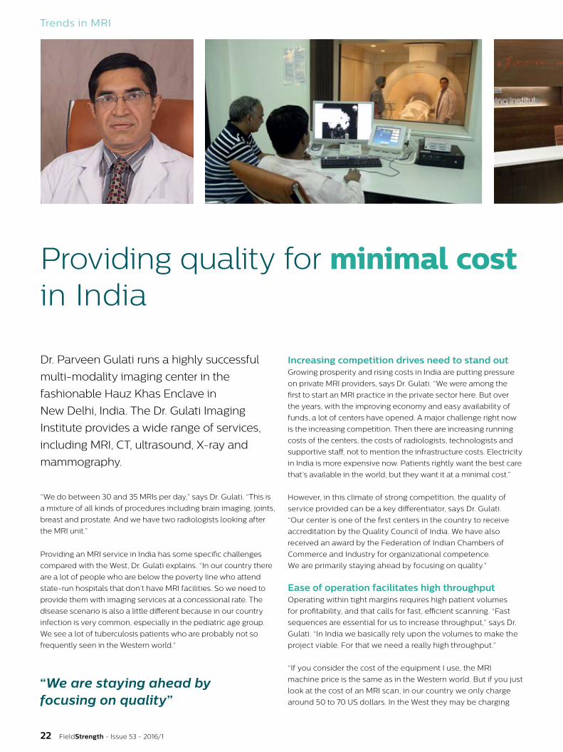
Dr. Parveen Gulati runs the Dr. Gulati Imaging Institute, a privately owned, multi-modality imaging center in New Delhi, India. Dr. Gulati’s institute offers MRI, CT, ultrasound, X-ray and mammography. Dr. Gulati is planning to expand the Institute’s MRI capabilities, adding new equipment in the near future.
Mrs. Silvia Schiffer runs Radiology Schiffer, a privately owned imaging center attached to a major hospital just outside Berlin, Germany. Radiology Schiffer currently offers MRI, CT, and X-ray services, and Mrs. Schiffer will soon establish a new fully equipped center with two new MRI machines, CT, X-ray and ultrasound systems.
»
Trends in MRI
FieldStrength - Issue 53 - 2016/122
“We do between 30 and 35 MRIs per day,” says Dr. Gulati. “This is
a mixture of all kinds of procedures including brain imaging, joints,
breast and prostate. And we have two radiologists looking after
the MRI unit.”
Providing an MRI service in India has some specific challenges
compared with the West, Dr. Gulati explains. “In our country there
are a lot of people who are below the poverty line who attend
state-run hospitals that don’t have MRI facilities. So we need to
provide them with imaging services at a concessional rate. The
disease scenario is also a little different because in our country
infection is very common, especially in the pediatric age group.
We see a lot of tuberculosis patients who are probably not so
frequently seen in the Western world.”
Providing quality for minimal cost in India
Increasing competition drives need to stand outGrowing prosperity and rising costs in India are putting pressure
on private MRI providers, says Dr. Gulati. “We were among the
first to start an MRI practice in the private sector here. But over
the years, with the improving economy and easy availability of
funds, a lot of centers have opened. A major challenge right now
is the increasing competition. Then there are increasing running
costs of the centers, the costs of radiologists, technologists and
supportive staff, not to mention the infrastructure costs. Electricity
in India is more expensive now. Patients rightly want the best care
that’s available in the world, but they want it at a minimal cost.”
However, in this climate of strong competition, the quality of
service provided can be a key differentiator, says Dr. Gulati.
“Our center is one of the first centers in the country to receive
accreditation by the Quality Council of India. We have also
received an award by the Federation of Indian Chambers of
Commerce and Industry for organizational competence.
We are primarily staying ahead by focusing on quality.”
Ease of operation facilitates high throughputOperating within tight margins requires high patient volumes
for profitability, and that calls for fast, efficient scanning. “Fast
sequences are essential for us to increase throughput,” says Dr.
Gulati. “In India we basically rely upon the volumes to make the
project viable. For that we need a really high throughput.”
“If you consider the cost of the equipment I use, the MRI
machine price is the same as in the Western world. But if you just
look at the cost of an MRI scan, in our country we only charge
around 50 to 70 US dollars. In the West they may be charging
Dr. Parveen Gulati runs a highly successful
multi-modality imaging center in the
fashionable Hauz Khas Enclave in
New Delhi, India. The Dr. Gulati Imaging
Institute provides a wide range of services,
including MRI, CT, ultrasound, X-ray and
mammography.
“We are staying ahead by focusing on quality”
Trends in MRI
www.philips.com/fieldstrength 23
something like 1,000 US dollars, maybe more. We can only
compensate on volume, so it is very important for us that our
equipment should be capable of doing scans that are fast. For
that we need proper training, availability of faster sequences,
good technical support and good application support. The
equipment should be user friendly, both for the operations at
the console as well as for managing the patient at the magnet.”
High uptime is crucial for financial viability“Especially in the private sector, uptime is very, very important,”
says Dr. Gulati. “We do not want our equipment to be down for
a single minute. Not even for routine servicing and preventative
maintenance. We always prefer that this should happen either
on a Sunday or at night when the working day is over. For me,
service back-up is very important. What I want is the confidence
that somebody is there to support me if my equipment goes
down in the night. Those types of support are crucial.”
Confidence in image quality and lifetime support“Next to cost, the main factor is the performance of the MR system.
It comes down to confidence: my confidence in the image quality,
my confidence in its user friendliness for my technical staff,
and my confidence that I will get timely upgrades. But most
importantly – which I want to give the maximum emphasis to –
is the confidence that I have good application support and a
service back-up.”
Predictable costs facilitate growthThe Dr. Gulati Imaging Institute has plans to consolidate its
position as one of the foremost imaging centers in India with a
program of expansion in the near future. “Before we can really
think of expansion,” says Dr. Gulati, “as with any other business,
we want to know the total cost of the project’s expected inflows
and outflows. Predictable outflows of money are very important,
and that includes the service contracts for equipment, and the
scope and cost of future upgrades.”
Finding well-trained staff can also be problematic for an
expanding business. “Even getting good technologists can be
difficult. We need expert technologists who can manage the
MR machines. In fact, I have suggested to the vendors that they
should start their own training program for technologists. If they
trained more technologists they would be an asset to established
centers and the up-and-coming centers as well.”
Upgrades needed to stay at forefrontThe ability to remain at the forefront of technological
developments without frequent investment in new hardware
is a key factor for a private MRI practice in India, explains Dr.
Gulati. “Upgradability is a very important factor. In fact, every
company should take that into account with their equipment.
We cannot change equipment very frequently, but vendors are
constantly coming out with newer features for MR, and those
advancements should be made available to the customer at a
reasonable price and with proper application support so they
can exploit the equipment to its maximum.”
Satisfied patients are the best measure of successThere are many possible measures of success, but Dr. Gulati
explains that in his opinion, one measure far outweighs the
others. “Success is a continuous dynamic process,” he says.
“In any organization there are a number of parameters you
can define, but for me the most important parameter is patient
satisfaction. Our practice is totally patient oriented. We are
basically doctors, so our primary goal is to keep our patients
happy. And on the business front, if patients are happy they will
bring you back another patient.”
“If patients are happy they will bring you back another patient”
“I want the confidence that somebody will support me if my equipment goes down in the night”
“Fast sequences are essential for us to increase throughput”
»
FieldStrength - Issue 53 - 2016/124
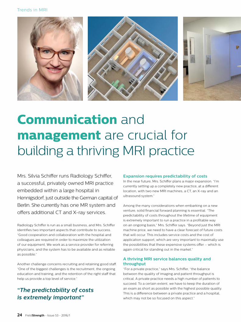
Communication and management are crucial for building a thriving MRI practice
Expansion requires predictability of costsIn the near future, Mrs. Schiffer plans a major expansion. “I’m
currently setting up a completely new practice, at a different
location, with two new MRI machines, a CT, an X-ray and an
ultrasound system.”
Among the many considerations when embarking on a new
venture, solid financial forward planning is essential. “The
predictability of costs throughout the lifetime of equipment
is extremely important to run a practice in a profitable way
on an ongoing basis,” Mrs. Schiffer says. “Beyond just the MRI
machine price, we need to have a clear forecast of future costs
that will occur. This includes service costs and the cost of
application support, which are very important to maximally use
the possibilities that these expensive systems offer – which is
again critical for standing out in the market.”
A thriving MRI service balances quality and throughput“For a private practice,” says Mrs. Schiffer, “the balance
between the quality of imaging and patient throughput is
critical. A private practice needs a high number of patients to
succeed. To a certain extent, we have to keep the duration of
an exam as short as possible with the highest possible quality.
This is a difference between a private practice and a hospital,
which may not be so focused on this aspect.”
Mrs. Silvia Schiffer runs Radiology Schiffer,
a successful, privately owned MRI practice
embedded within a large hospital in
Hennigsdorf, just outside the German capital of
Berlin. She currently has one MR system and
offers additional CT and X-ray services.
Radiology Schiffer is run as a small business, and Mrs. Schiffer
identifies two important aspects that contribute to success.
“Good cooperation and collaboration with the hospital and
colleagues are required in order to maximize the utilization
of our equipment. We work as a service provider for referring
physicians, and the system has to be available and as reliable
as possible.”
Another challenge concerns recruiting and retaining good staff.
“One of the biggest challenges is the recruitment, the ongoing
education and training, and the retention of the right staff that
help us provide a top level of service.”
“The predictability of costs is extremely important”
Trends in MRI
www.philips.com/fieldstrength 25
Maintaining that balance between quality and throughput has
many associated factors, but it is facilitated by the ease of daily
operation of MRI scanners, Mrs. Schiffer explains. “We keep
our staff well trained, but having machines that are intuitive
to use, that are quasi-self-explanatory, with a well-structured
operating interface, is very important to support our fast and
efficient workflow.”
High uptime is essential to successSustaining a high patient throughput requires keeping MRI
scanners in continuous operation. “Minimizing downtime is
essential for us, because we have a fully booked schedule of
patient exams and waiting times.
We feel it would offend referring physicians and patients
when we have downtime and then need to reschedule patient
exams,” says Mrs. Schiffer. “Keeping patients to schedule
demands a high uptime. Maintenance should be limited
to those hours where the practice is not open, in order to
ensure the optimization of uptime. It is important also for the
reputation of the practice, and for maintaining referrals.
High uptime gains trust and confidence in the practice.”
Upgradability helps maintain a competitive positionStaying ahead of the competition means staying ahead in
terms of technological advances, explains Mrs. Schiffer. “The
ability to upgrade equipment from time to time is essential to
provide a state-of-the-art level of service. There are practices
here in Germany that are still using 20-year-old, 0.5 tesla
equipment to generate images and results. And as long as the
patients are not harmed, this is fine. But that’s not the point.
If the images are not of the highest quality, then the results
derived from those images, which are handed back to referring
physicians, are also not of the highest quality. Providing the
highest possible image quality, and therefore optimal results and
diagnoses, is how we want to earn our position in the market.”
Total package, not just MRI machine price, determines best offerHow does a private MRI practice make purchase decisions? “It is
always the total package that determines what the best offer is,”
says Mrs. Schiffer. “It is not only equipment itself, although that is
obviously very important, but also the added value that comes
with it – the future collaboration, service, and support on technical
as well as operational matters, and hopefully, also support for
the marketing aspects. Branding of the practice is an important
consideration, but also cooperation with the right vendor. Both
are important if I am to gain a premium position for my practice
compared with the competition around me.”
To conclude, Mrs. Schiffer summarizes what she considers crucial
for building a thriving practice. “I believe that the success of a
radiology practice is ultimately based on the combination of
being a knowledgeable radiologist and at the same time being
an entrepreneur. Staff management is very important and so
is finding your niche in the market. It requires branding and
evidently a lot of communication, with the manufacturer, but
also particularly with referrers and colleagues. After having
been preparing this for a year, I can clearly say that marketing,
networking and good management of a practice are certainly
different from just taking a physician’s viewpoint.” «
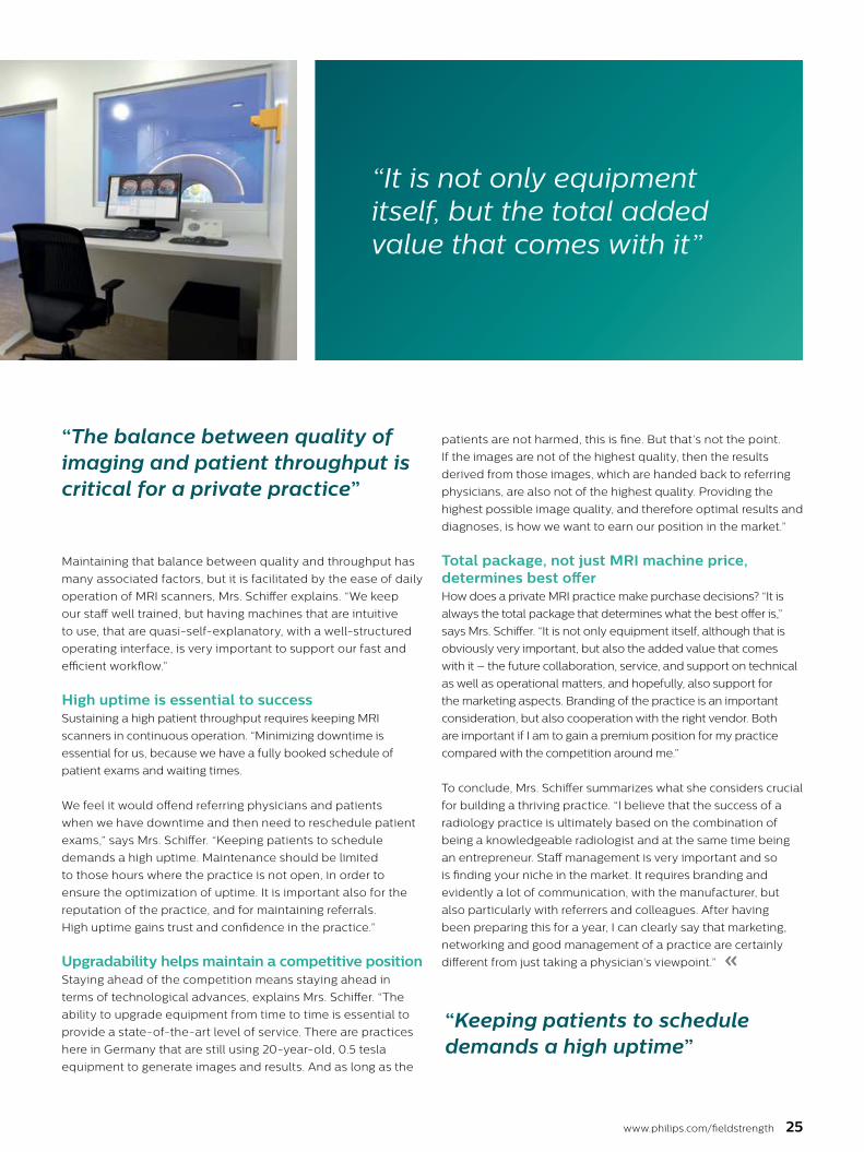
“It is not only equipment itself, but the total added value that comes with it”
“The balance between quality of imaging and patient throughput is critical for a private practice”
“Keeping patients to schedule demands a high uptime”
User experiences
FieldStrength - Issue 53 - 2016/126
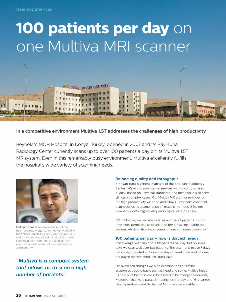
Erdogan Tuna is general manager of the Bay-Tuna Radiology Center. He has worked in the field of radiology since 2004. His goal is to make his company a leader in the field, while keeping patient comfort, correct diagnosis, effective service and employee satisfaction a top priority.
In a competitive environment Multiva 1.5T addresses the challenges of high productivity
Beyhekim MOH Hospital in Konya, Turkey, opened in 2007 and its Bay-Tuna
Radiology Center currently scans up to over 100 patients a day on its Multiva 1.5T
MR system. Even in this remarkably busy environment, Multiva excellently fulfills
the hospital’s wide variety of scanning needs.
Balancing quality and throughputErdogan Tuna is general manager of the Bay-Tuna Radiology
Center. “We aim to provide our services with uncompromised
quality, based on universal standards, and meanwhile also solve
clinically complex cases. Our Multiva MR scanner provides us
the high productivity we need and allows us to make confident
diagnoses using a large range of imaging methods. It fits our
company motto ‘high quality radiological care'," he says.
“With Multiva, we can scan a large number of patients in short
time slots, permitting us to adapt to the prevailing healthcare
system, which limits reimbursement more and more every day.”
100 patients per day – how is that achieved?“On average, we scan about 80 patients per day, but on some
days we scan well over 100 patients. The scanner is in use 7 days
per week, operated 20 hours per day on week days and 8 hours
per day in the weekend,” Mr. Tuna says.
“To avoid coil changes we plan examinations of similar
anatomies back to back, such as head and spine. Multiva helps
us here a lot because coils don’t need to be changed frequently.
Moreover, thanks to parallel imaging technology and 16-channel
HeadSpineTorso and 8-channel MSK coils we are able to
100 patients per day on one Multiva MRI scanner
“Multiva is a compact system that allows us to scan a high number of patients”
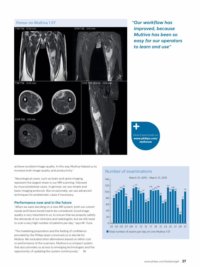
“ Our workflow has improved, because Multiva has been so easy for our operators to learn and use”
www.philips.com/fieldstrength 27
»
01 03 05 07 09 11 13 15 17 19 21 23 25 27 29 310
20
40
60
80
100
120
140
Total number of exams per day on one Multiva 1.5T
March 01, 2015 - March 31, 2015
Number of examinations
1
8186
46
9499
106112
103105
106 106
73
115
105
9597
111
7168
64
89
69
114
101108
103
29
50
77
101
achieve excellent image quality. In this way Multiva helped us to
increase both image quality and productivity.”
“Neurological cases, such as brain and spine imaging,
represent the largest share in our MR scanning, followed
by musculoskeletal cases. In general, we use simple and
basic imaging protocols. But occasionally, we use advanced
techniques for problematic cases if necessary.
Performance now and in the future“When we were deciding on a new MR system, both our current
needs and future trends had to be considered. Good image
quality is very important to us, to ensure that we properly satisfy
the demands of our clinicians and radiologists, but we still need
to scan a very high number of patients per day,” says Mr. Tuna.
“The marketing proposition and the feeling of confidence
provided by the Philips team convinced us to decide for
Multiva. We excluded other alternatives based on either cost
or performance of the scanners. Multiva is a compact system
that also provides us access to emerging technologies and the
opportunity of updating the system continuously.”
Femur on Multiva 1.5T
STIR TSE 1:01 min.
STIR TSE 2:51 min.
STIR TSE SENSE 1:06 min.T1W TSE 0:53 min.
T1W TSE 0:54 min.
View ExamCards on www.philips.com/
netforum

FieldStrength - Issue 53 - 2016/128
Sacroiliac joint on Multiva 1.5T
For the total examination of the sacroiliac joint scan time was 13:23 min. The SENSE Torso coil is used.
T2W TSE SPAIR 1.09 x 1.36 x 4.0 mm STIR TSE 1.0 x 1.35 x 4.0 mmT1W TSE 1.09 x 1.36 x 4.0 mm
T1W TSE SPIR 1.14 x 1.34 x 4.0 mm T1W TSE SPIR post contrast 1.14 x 1.34 x 4.0 mm T1W TSE post contrast 1.09 x 1.36 x 4.0 mm
View ExamCard on www.philips.com/
netforum
Breast MRI on Multiva 1.5T
T2W SPAIR tra 1.25 x 1.52 x 3.0 mm
STIR tra 1.25 x 1.79 x 3.0 mm
T2W TSE tra 1.00 x 1.42 x 3.0 mm
DWI tra 1.49 x 1.50 x 3.0 mm
T1W TSE tra 1.00 x 1.26 x 3.0 mm
Dynamic eTHRIVE 0.99 x 1.03 x 1.0 mm
For the total breast examination the scan time was 24 minutes. The SENSE Breast 7ch coil is used.
User experiences
“The coils are lightweight and don’t need to be changed frequently”
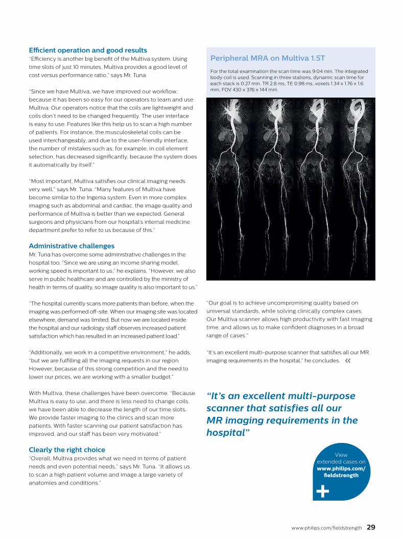
www.philips.com/fieldstrength 29
Efficient operation and good results“Efficiency is another big benefit of the Multiva system. Using
time slots of just 10 minutes, Multiva provides a good level of
cost versus performance ratio,” says Mr. Tuna.
“Since we have Multiva, we have improved our workflow,
because it has been so easy for our operators to learn and use
Multiva. Our operators notice that the coils are lightweight and
coils don’t need to be changed frequently. The user interface
is easy to use. Features like this help us to scan a high number
of patients. For instance, the musculoskeletal coils can be
used interchangeably, and due to the user-friendly interface,
the number of mistakes such as, for example, in coil element
selection, has decreased significantly, because the system does
it automatically by itself.”
“Most important, Multiva satisfies our clinical imaging needs
very well,” says Mr. Tuna. “Many features of Multiva have
become similar to the Ingenia system. Even in more complex
imaging such as abdominal and cardiac, the image quality and
performance of Multiva is better than we expected. General
surgeons and physicians from our hospital’s internal medicine
department prefer to refer to us because of this.”
Administrative challengesMr. Tuna has overcome some administrative challenges in the
hospital too. “Since we are using an income sharing model,
working speed is important to us,” he explains. “However, we also
serve in public healthcare and are controlled by the ministry of
health in terms of quality, so image quality is also important to us.”
“The hospital currently scans more patients than before, when the
imaging was performed off-site. When our imaging site was located
elsewhere, demand was limited. But now we are located inside
the hospital and our radiology staff observes increased patient
satisfaction which has resulted in an increased patient load.”
“Additionally, we work in a competitive environment,” he adds,
“but we are fulfilling all the imaging requests in our region.
However, because of this strong competition and the need to
lower our prices, we are working with a smaller budget.”
With Multiva, these challenges have been overcome. “Because
Multiva is easy to use, and there is less need to change coils,
we have been able to decrease the length of our time slots.
We provide faster imaging to the clinics and scan more
patients. With faster scanning our patient satisfaction has
improved, and our staff has been very motivated.”
Clearly the right choice“Overall, Multiva provides what we need in terms of patient
needs and even potential needs,” says Mr. Tuna. “It allows us
to scan a high patient volume and image a large variety of
anatomies and conditions.”
“Our goal is to achieve uncompromising quality based on
universal standards, while solving clinically complex cases.
Our Multiva scanner allows high productivity with fast imaging
time, and allows us to make confident diagnoses in a broad
range of cases.”
“It’s an excellent multi-purpose scanner that satisfies all our MR
imaging requirements in the hospital,” he concludes. «
Peripheral MRA on Multiva 1.5T
For the total examination the scan time was 9:04 min. The integrated body coil is used. Scanning in three stations, dynamic scan time for each stack is 0:27 min. TR 2.8 ms, TE 0.98 ms, voxels 1.34 x 1.76 x 1.6 mm, FOV 430 x 376 x 144 mm.
View extended cases on www.philips.com/
fieldstrength
“It’s an excellent multi-purpose scanner that satisfies all our MR imaging requirements in the hospital”
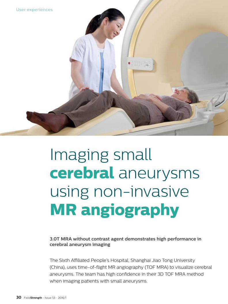
User experiences
Imaging small cerebral aneurysms using non-invasive MR angiography
3.0T MRA without contrast agent demonstrates high performance in cerebral aneurysm imaging
The Sixth Affiliated People’s Hospital, Shanghai Jiao Tong University
(China), uses time-of-flight MR angiography (TOF MRA) to visualize cerebral
aneurysms. The team has high confidence in their 3D TOF MRA method
when imaging patients with small aneurysms.
FieldStrength - Issue 53 - 2016/130
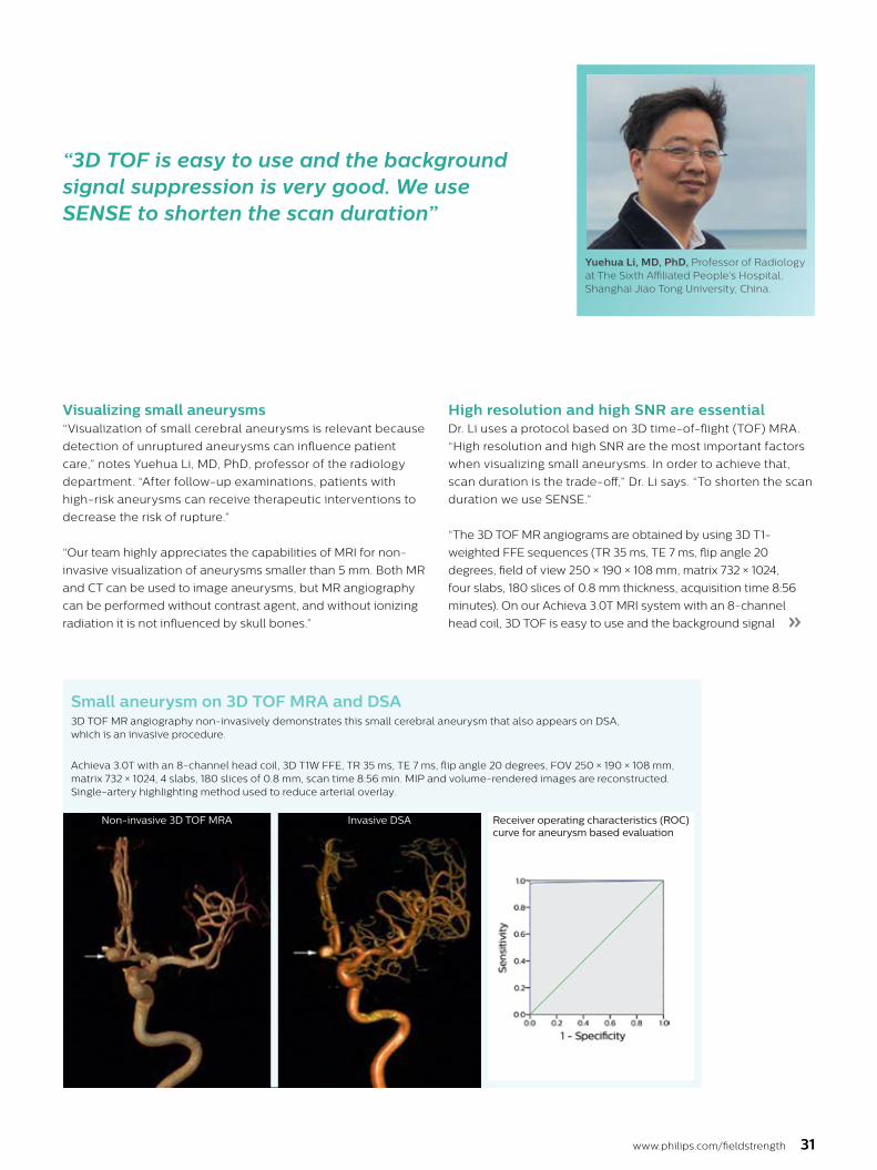
Yuehua Li, MD, PhD, Professor of Radiology at The Sixth Affiliated People’s Hospital, Shanghai Jiao Tong University, China.
www.philips.com/fieldstrength 31
High resolution and high SNR are essential Dr. Li uses a protocol based on 3D time-of-flight (TOF) MRA.
“High resolution and high SNR are the most important factors
when visualizing small aneurysms. In order to achieve that,
scan duration is the trade-off,” Dr. Li says. “To shorten the scan
duration we use SENSE.”
“The 3D TOF MR angiograms are obtained by using 3D T1-
weighted FFE sequences (TR 35 ms, TE 7 ms, flip angle 20
degrees, field of view 250 × 190 × 108 mm, matrix 732 × 1024,
four slabs, 180 slices of 0.8 mm thickness, acquisition time 8:56
minutes). On our Achieva 3.0T MRI system with an 8-channel
head coil, 3D TOF is easy to use and the background signal »
Visualizing small aneurysms“Visualization of small cerebral aneurysms is relevant because
detection of unruptured aneurysms can influence patient
care,” notes Yuehua Li, MD, PhD, professor of the radiology
department. “After follow-up examinations, patients with
high-risk aneurysms can receive therapeutic interventions to
decrease the risk of rupture.”
“Our team highly appreciates the capabilities of MRI for non-
invasive visualization of aneurysms smaller than 5 mm. Both MR
and CT can be used to image aneurysms, but MR angiography
can be performed without contrast agent, and without ionizing
radiation it is not influenced by skull bones.”
“3D TOF is easy to use and the background signal suppression is very good. We use SENSE to shorten the scan duration”
Small aneurysm on 3D TOF MRA and DSA
Non-invasive 3D TOF MRA Invasive DSA
3D TOF MR angiography non-invasively demonstrates this small cerebral aneurysm that also appears on DSA, which is an invasive procedure.
Achieva 3.0T with an 8-channel head coil, 3D T1W FFE, TR 35 ms, TE 7 ms, flip angle 20 degrees, FOV 250 × 190 × 108 mm, matrix 732 × 1024, 4 slabs, 180 slices of 0.8 mm, scan time 8:56 min. MIP and volume-rendered images are reconstructed. Single-artery highlighting method used to reduce arterial overlay.
Receiver operating characteristics (ROC) curve for aneurysm based evaluation

User experiences
“We recommend that 3D TOF MRA would be an additional approach for patients with small cerebral aneurysms, due to its non-invasive nature and high degree of accuracy and sensitivity” [2]
FieldStrength - Issue 53 - 2016/132
MR angiography of multiple cerebral aneurysms
A-P direction P-A direction
3D TOF MRA demonstrates bilateral aneurysms arising from C5 and C6 segments.
Achieva 3.0T with an 8-channel head coil, 3D T1W FFE, TR 35 ms, TE 7 ms, flip angle 20 degrees, FOV 250 × 190 × 108 mm, matrix 732 × 1024, 4 slabs, 180 slices of 0.8 mm, scan time 8:56 min. MIP and volume-rendered images are reconstructed. Single-artery highlighting method used to reduce arterial overlay.
5 years MRA follow-up of unruptured aneurysm
First exam 1 year later 2.5 years later 5 years later
A cerebral aneurysm on the C5 segment in a 57-year-old female was followed over a period of 5 years using 3D TOF MRA. The 5 years follow-up using 3D TOF MRA shows that the aneurysms didn’t change much, which indicates that the aneurysm is stable and intervention therapy was not needed.
Achieva 3.0T with an 8-channel head coil, 3D T1W FFE, TR 35 ms, TE 7 ms, flip angle 20 degrees, FOV 250 × 190 × 108 mm, matrix 732 × 1024, 4 slabs, 180 slices of 0.8 mm, scan time 8:56 min. MIP and volume-rendered images are reconstructed. Single-artery highlighting method used to reduce arterial overlay.

www.philips.com/fieldstrength 33
suppression is very good. Maximum intensity projections (MIPs)
and volume-rendered images are reconstructed from the data.
We use a single-artery highlighting method to reduce arterial
overlay. The method can also be used for carotids or other small
intracranial vessels.”
Study shows high accuracy“Advances in MR imaging, the high SNR and spatial resolution
of 3.0T, increasing observer experience and improved
postprocessing techniques have improved the capabilities
of MRA over the last decade. We found that 3D TOF MRA
provides excellent images for the visualization of small cerebral
aneurysms which enables us to accurately diagnose, and
this accuracy appears to be similar to that obtained with DSA
according to data from a 2014 study," [2] Dr. Li says.
“Aneurysm location can influence how easy it is recognized.
Infundibula can mimic aneurysms, adding to the difficulty of
interpretation. On MIP images, small aneurysms can also be
easily misinterpreted when the aneurysm overlaps with the
vessel. This is why in our method we are viewing the images
from multiple angles.”
“In the hundreds of patients with aneurysms that we scanned, we
saw no significant differences in accuracy, sensitivity or specificity
among the locations. [2] We think this is the result of technical
advances in image acquisition and postprocessing algorithms.”
References
1. Li MH, Cheng YS, Li YD, Fang C, Chen SW, Wang W, Hu DJ, Xu HW. Large-cohort comparison between three-dimensional time-of-flight magnetic resonance and rotational digital subtraction angiographies in intracranial aneurysm detection. Stroke. 2009;40:3127-9.
2. Li Y, et al. Accurate diagnosis of small cerebral aneurysms ≤5 mm in diameter with 3.0-T MR angiography. Radiology. 2014;271:553-60.
3. Li Y, et al. Prevalence of unruptured cerebral aneurysms in Chinese adults aged 35 to 75 years: A cross-sectional study. Ann Intern Med. 2013;159(8):514-521.
3D TOF MRA chosen for prevalence study “Our high confidence in the 3D TOF MRA method was the
reason to select this method for a study on the prevalence of
unruptured cerebral aneurysms in Chinese adults, which
involved finding the location and size of unruptured cerebral
aneurysms. By the way, that study found a prevalence of 7% and
most lesions had a diameter of less than 5 mm," [3] says Dr. Li.
According to Dr. Li, the good performance of MRA makes it a
candidate to replace DSA. “We recommend that 3D TOF MR
angiography would be an additional approach for patients
with small cerebral aneurysms. This is due to its non-invasive
nature, ease of use and high degree of accuracy and
sensitivity,” he concludes. «
MRA of high risk cerebral aneurysm
3D TOF MRA DSA during therapy After therapy
Volume rendered 3D TOF MRA image visualizes an irregularly shaped cerebral aneurysm arising from the C7 segment. The physician qualified the lesion as instable and interventional therapy was performed. 3D DSA after therapy indicates that the aneurysm has been occluded completely.
Achieva 3.0T with an 8-channel head coil, 3D T1W FFE, TR 35 ms, TE 7 ms, flip angle 20 degrees, FOV 250 × 190 × 108 mm, matrix 732 × 1024, 4 slabs, 180 slices of 0.8 mm, scan time 8:56 min. MIP and volume-rendered images are reconstructed. Single-artery highlighting method used to reduce arterial overlay.
User experiences
FieldStrength - Issue 53 - 2016/134
Smart Display Protocols help radiologists speed up their viewing of MRI casesIn a routine radiology practice, IntelliSpace Portal 8.0 saves radiologists precious time in the daily workflow
By automating many steps, Smart Display Protocols (SDP) eliminate many
of the manual steps that previously added to the time needed for reviewing
and processing MRI images on IntelliSpace Portal. This significantly improves
the workflow for radiologists at Beau Soleil Clinic.
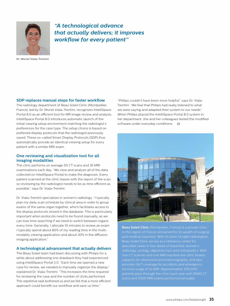
Dr. Muriel Viala-Trentini
www.philips.com/fieldstrength 35
SDP replaces manual steps for faster workflowThe radiology department of Beau Soleil Clinic (Montpellier,
France), led by Dr. Muriel Viala-Trentini, recognizes IntelliSpace
Portal 8.0 as an efficient tool for MR image review and analysis.
IntelliSpace Portal 8.0 introduces automatic launch of the
initial viewing setup environment matching the radiologist’s
preferences for the case type. The setup choice is based on
preferred display protocols that the radiologist previously
saved. These so-called Smart Display Protocols (SDP) thus
automatically provide an identical viewing setup for every
patient with a similar MRI exam.
One reviewing and visualization tool for all imaging modalities The clinic performs on average 50 CT scans and 35 MRI
examinations each day. “We view and analyze all of the data
collected on IntelliSpace Portal to make the diagnosis. Every
patient scanned at the clinic leaves with the report of the scan,
so reviewing by the radiologist needs to be as time efficient as
possible,” says Dr. Viala-Trentini.
Dr. Viala-Trentini specializes in women’s radiology. “I typically
plan my daily scan schedule by clinical area in order to group
exams of the same organ together, which facilitates access to
the display protocols stored in the database. This is particularly
important when protocols need to be found manually, as we
can lose time searching if we need to switch between organs
every time. Generally, I allocate 10 minutes to review an exam.
I typically spend about 80% of my reading time in the multi-
modality viewing application and about 20% in the diffusion
imaging application.”
A technological advancement that actually delivers The Beau Soleil team had been discussing with Philips for a
while about addressing one drawback they had experienced
using IntelliSpace Portal 5.0. “Each time we opened a new
case for review, we needed to manually organize the display,”
explained Dr. Viala-Trentini. “This increases the time required
for reviewing the case and the number of clicks performed.
This repetitive task bothered us and we felt that a more efficient
approach could benefit our workflow and save us time.”
Beau Soleil Clinic (Montpellier, France) is a private clinic,
in this region of France renowned for its wealth of surgical
and medical expertise. With its team of eight radiologists,
Beau Soleil Clinic serves as a reference center for
specialist cases in four areas of expertise: women’s
radiology, urology, digestive tract and orthopedics. With
one CT scanner and one MRI machine the clinic boasts
capacity for ultrasound and mammography, and also
provides 24/7 coverage for accidents and emergency
services usage of its MRI. Approximately 500,000
patients pass through the clinic each year with 9000 CT
scans and 7000 MRI exams performed annually.
“A technological advance that actually delivers; it improves workflow for every patient”
“Philips couldn’t have been more helpful”, says Dr. Viala-
Trentini. “We feel that Philips had really listened to what
we were saying and adapted their system to our needs”.
When Philips placed the IntelliSpace Portal 8.0 system in
her department, she and her colleagues tested the modified
software under everyday conditions. »
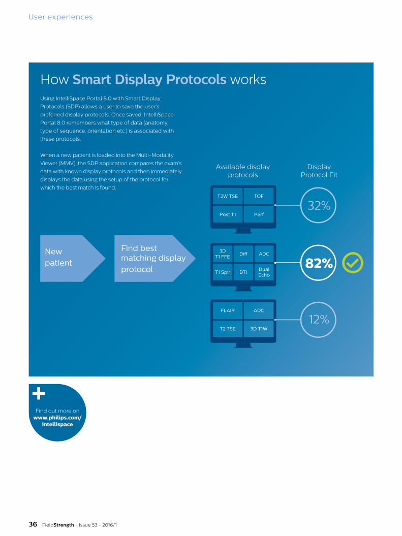
User experiences
FieldStrength - Issue 53 - 2016/136
How Smart Display Protocols worksUsing IntelliSpace Portal 8.0 with Smart Display
Protocols (SDP) allows a user to save the user’s
preferred display protocols. Once saved, IntelliSpace
Portal 8.0 remembers what type of data (anatomy,
type of sequence, orientation etc.) is associated with
these protocols.
When a new patient is loaded into the Multi-Modality
Viewer (MMV), the SDP application compares the exam’s
data with known display protocols and then immediately
displays the data using the setup of the protocol for
which the best match is found.
New patient
Find best matching display protocol
Available display protocols
Display Protocol Fit
32%
82%
12%
3D T1 FFE
Diff ADC
T1 Spir DTIDual Echo
T2W TSE
Post T1
TOF
Perf
FLAIR
T2 TSE
ADC
3D T1W
Find out more on www.philips.com/
intellispace
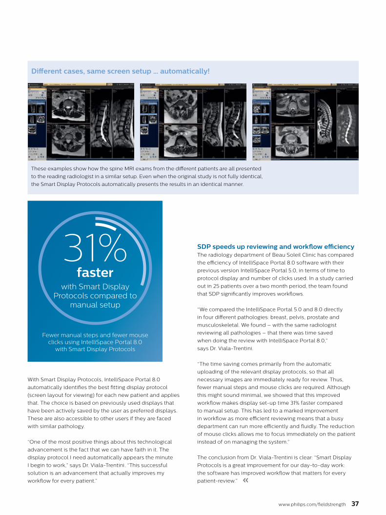
www.philips.com/fieldstrength 37
With Smart Display Protocols, IntelliSpace Portal 8.0
automatically identifies the best fitting display protocol
(screen layout for viewing) for each new patient and applies
that. The choice is based on previously used displays that
have been actively saved by the user as preferred displays.
These are also accessible to other users if they are faced
with similar pathology.
“One of the most positive things about this technological
advancement is the fact that we can have faith in it. The
display protocol I need automatically appears the minute
I begin to work,” says Dr. Viala-Trentini. “This successful
solution is an advancement that actually improves my
workflow for every patient.”
31%faster
with Smart Display Protocols compared to
manual setup
Fewer manual steps and fewer mouse clicks using IntelliSpace Portal 8.0
with Smart Display Protocols
«
SDP speeds up reviewing and workflow efficiency The radiology department of Beau Soleil Clinic has compared
the efficiency of IntelliSpace Portal 8.0 software with their
previous version IntelliSpace Portal 5.0, in terms of time to
protocol display and number of clicks used. In a study carried
out in 25 patients over a two month period, the team found
that SDP significantly improves workflows.
“We compared the IntelliSpace Portal 5.0 and 8.0 directly
in four different pathologies: breast, pelvis, prostate and
musculoskeletal. We found – with the same radiologist
reviewing all pathologies – that there was time saved
when doing the review with IntelliSpace Portal 8.0,”
says Dr. Viala-Trentini.
“The time saving comes primarily from the automatic
uploading of the relevant display protocols, so that all
necessary images are immediately ready for review. Thus,
fewer manual steps and mouse clicks are required. Although
this might sound minimal, we showed that this improved
workflow makes display set-up time 31% faster compared
to manual setup. This has led to a marked improvement
in workflow as more efficient reviewing means that a busy
department can run more efficiently and fluidly. The reduction
of mouse clicks allows me to focus immediately on the patient
instead of on managing the system.”
The conclusion from Dr. Viala-Trentini is clear: “Smart Display
Protocols is a great improvement for our day-to-day work:
the software has improved workflow that matters for every
patient-review.”
Different cases, same screen setup … automatically!
These examples show how the spine MRI exams from the different patients are all presented
to the reading radiologist in a similar setup. Even when the original study is not fully identical,
the Smart Display Protocols automatically presents the results in an identical manner.
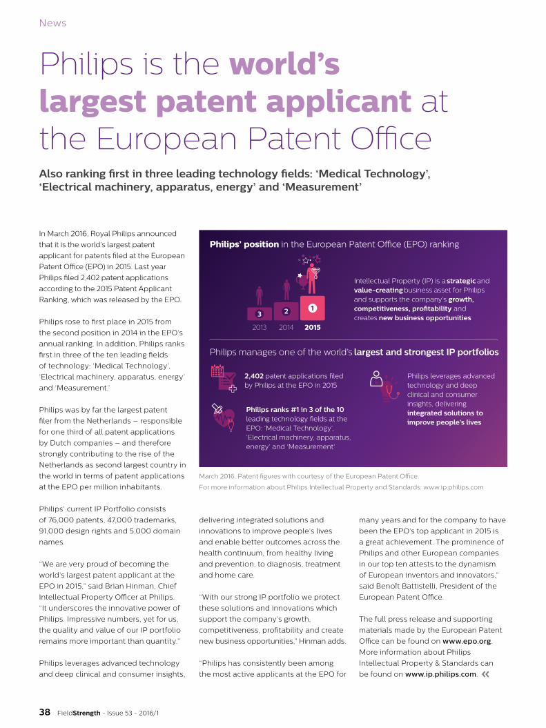
News
Philips manages one of the world’s largest and strongest IP portfolios
Philips’ position
Philips’ total IP portfolio consists of
2,402 by Philips at the EPO in 2015
Philips leverages advanced technology and deep clinical and consumer insights, delivering integrated solutions to improve people’s lives
Intellectual Property (IP) is a strategic and value-creating business asset for Philips and supports the company’s growth,
and creates new business opportunities
Philips ranks #1 in 3 of the 10
EPO: ‘Medical Technology’, ‘Electrical machinery, apparatus, energy’ and ‘Measurement’
Patent rights Trademarks Domain names Design rights
76,000 47,000 91,000 5,000
201520142013
2 13
FieldStrength - Issue 53 - 2016/138
In March 2016, Royal Philips announced
that it is the world’s largest patent
applicant for patents filed at the European
Patent Office (EPO) in 2015. Last year
Philips filed 2,402 patent applications
according to the 2015 Patent Applicant
Ranking, which was released by the EPO.
Philips rose to first place in 2015 from
the second position in 2014 in the EPO’s
annual ranking. In addition, Philips ranks
first in three of the ten leading fields
of technology: ‘Medical Technology’,
‘Electrical machinery, apparatus, energy’
and ‘Measurement.’
Philips was by far the largest patent
filer from the Netherlands – responsible
for one third of all patent applications
by Dutch companies – and therefore
strongly contributing to the rise of the
Netherlands as second largest country in
the world in terms of patent applications
at the EPO per million inhabitants.
Philips’ current IP Portfolio consists
of 76,000 patents, 47,000 trademarks,
91,000 design rights and 5,000 domain
names.
“We are very proud of becoming the
world’s largest patent applicant at the
EPO in 2015,” said Brian Hinman, Chief
Intellectual Property Officer at Philips.
“It underscores the innovative power of
Philips. Impressive numbers, yet for us,
the quality and value of our IP portfolio
remains more important than quantity.”
Philips leverages advanced technology
and deep clinical and consumer insights,
delivering integrated solutions and
innovations to improve people’s lives
and enable better outcomes across the
health continuum, from healthy living
and prevention, to diagnosis, treatment
and home care.
“With our strong IP portfolio we protect
these solutions and innovations which
support the company’s growth,
competitiveness, profitability and create
new business opportunities,” Hinman adds.
“Philips has consistently been among
the most active applicants at the EPO for
many years and for the company to have
been the EPO’s top applicant in 2015 is
a great achievement. The prominence of
Philips and other European companies
in our top ten attests to the dynamism
of European inventors and innovators,”
said Benoît Battistelli, President of the
European Patent Office.
The full press release and supporting
materials made by the European Patent
Office can be found on www.epo.org.
More information about Philips
Intellectual Property & Standards can
be found on www.ip.philips.com.
Philips is the world’s largest patent applicant at the European Patent Office Also ranking first in three leading technology fields: ‘Medical Technology’,‘ Electrical machinery, apparatus, energy’ and ‘Measurement’
«
March 2016. Patent figures with courtesy of the European Patent Office.
For more information about Philips Intellectual Property and Standards: www.ip.philips.com
FieldStrength 39
Stay in touch with Philips MRI
FieldStrength is a professional magazine for users
of Philips MR systems. It provides results of MR
studies performed by Philips users.
Be the first to see new articlesAnd don’t miss an issue.
Register now for your personal email subscription at:
www.philips.com/fieldstrength
Colophon
© Royal Philips N.V. 2016
All rights are reserved. Reproduction in whole or in part
is prohibited without the prior written consent of the
copyright holder.
Philips Healthcare reserves the right to make changes in
specifications and/or to discontinue any product at any
time without notice or obligation and will not be liable
for any consequences resulting from the use of
this publication.
Printed in Belgium.
4522 991 19851
FieldStrength is also available via the Internet:
www.philips.com/fieldstrength
www.philips.com/mri
www.philips.com/netforum
Editor-in-chief
Karen Janssen
Editorial team
Annemarie Blotwijk, Paul Folkers (PhD),
Liesbeth Geerts (PhD), Karen Janssen,
Marc Van Cauteren (PhD).
Contributors
Faiza Admiraal (PhD), Jalal Andre (MD), Greg Brown,
Paul W. de Bruin (PhD), Patrick Crowley (PhD),
Parveen Gulati (MD), Frank Hoogenraad (PhD),
Karen Janssen, Katarina Johansson, Emanuel Kanal (MD),
Ozlem Kandemir, Melahat Kirimli, Harry Klijn, Simon
Krijnen (PhD), Harald Kugel (PhD), Jane MacDougall,
Yuehua Li (MD, PhD), Marielle Philippens (MD, PhD),
Ramesh Radhakrishnan, Silvia Schiffer, Polly Schmidt,
Erdogan Tuna, Marieke van Grootel, Elizabeth van
Vorstenbosch, Muriel Viala-Trentini (MD), Randall Watson,
Stephanie Wasek, Xin Xing.
Subscriptions
Please subcribe on www.philips.com/fieldstrength
Correspondence
FieldStrength, Philips Healthcare, Building QR 0119
P.O. Box 10 000, 5680 DA Best, The Netherlands
Notice
FieldStrength is created for users of Philips MRI systems.
It is a professional magazine for users of Philips medical
equipment. It provides the healthcare community with
results of studies performed by colleagues. Some articles
in this magazine may describe research conducted
outside the USA on equipment not yet available for
commercial distribution in the USA. Some products
referenced may not be licensed for sale in Canada.
Results from case studies are not predictive of results
in other cases. Results in other cases may vary. Results
obtained by facilities described in this issue may not be
typical for all facilities. Images that are not part of User
experiences articles and that are not labeled otherwise
are created by Philips.

FieldStrength MRI articlesare first available online. Subscribe to not miss any articleswww.philips.com/fieldstrength