Imaging the Tricuspid Valve · 2020. 6. 17. · Stankovic, EE 2012. Anatomy & Function P. Iaizzo...
50
Jens-Uwe Voigt Dpt. of Cardiovascular Diseases University Hospital Gasthuisberg Leuven, Belgium European Association of Cardiovascular Imaging Teaching Course Imaging the Tricuspid Valve
Transcript of Imaging the Tricuspid Valve · 2020. 6. 17. · Stankovic, EE 2012. Anatomy & Function P. Iaizzo...

Jens-Uwe VoigtDpt. of Cardiovascular DiseasesUniversity Hospital Gasthuisberg
Leuven, Belgium
European Associationof Cardiovascular Imaging
Teaching Course
Imaging the Tricuspid Valve

Tricuspid Valve
Anatomy & Function

Anatomy & Function
position
„slightly more apical“than the mitral valve
Lancellotti et al., EJE 2010

Anatomy & Function
LL L > 8mm/m²
position
Ebstein Anomaly

Anatomy & Function
3 leaflets“anterior”infundibular
“posterior”inferior, marginal
“septal”medial
C. Lawrence

Anatomy & Function
atrial view
C. Lawrence
APS
Ao
MV
RVOT

Anatomy & Function
ventricular view
A
P SAo
MV
RVOT

Anatomy & Function
leaftlets
P. Iaizzo

Variable Leaflet Morphology
How many leaflets ?8% 90% 2%
Stankovic, EE 2012

Anatomy & Function
P. Iaizzo
papillary muscles and cordae

Anatomy & Function
papillary muscles and cordae
anterior PM andmoderator bandanterior PM andmoderator band
TV

Anatomy & Function
interlinked with MV
Fukuda et al., Circ 2006
S
saddle shaped
annulus

Tricuspid Valve
Imaging theTricuspid Valve

Imaging the TV: Standard Views
parasternal short axis

Imaging the TV: Standard Views
RV Inflow view

Imaging the TV: Standard Views
apical4 chamber
view

Imaging the TV: Standard Views
apical4 chamber
view
RV only

Imaging the TV: Standard Views
subcostal4 chamber
view

Simultaneous Visualization of 3 Leaflets
Impossible ?
Anwar et al., Int J Cardiovasc Imaging 2007

Simultaneous Visualization of 3 Leaflets
equally good with 2D and 3D
Stankovic et al., submitted
study:
155 consecutive patientsfrom clinical routine
all 3 leaflets visible:
58% with 2D subcostal view
56% with 3D reconstruction

Imaging the TV: Standard Views
subcostal4CV und
SAX
AP
S
AoMV
RVO
T

Imaging the TV: Standard Views
subcostalSAX
AP
S
AoMV
RVO
T

Leaflet Identification
relevant to describe pathologyprolaps not visible prolaps visible

Leaflet Identification
... using the subcostal viewleaflet identification regurgitation assessment

Leaflet Identification
... using the subcostal viewprolaps not visible prolaps visible
Ao
A
P
S
Ao
S A
P

- variable valve morphology
- variable scan plane
Imaging the TV
Which leaflet is which ?
RV
RA
LA
LV
SAX RV inflow 4CV subcostal

Leaflet Identification
... using dedicated software

Apical 4 Chamber View
ALALAL11
11
SLSLSL
PLPLPL22
22
ALALAL SLSLSL PLPLPL SLSLSL
11
22
2
11
81%

RV Inflow View
SLSLSL ALALAL
1 2
ALALALSLSLSL
3
ALALALPLPLPL
1133
ALALAL
SLSLSLPLPLPL22
1133 22
1 2 3
100% 77%

Parasternal Short Axis View
11
22
ALALAL
SLSLSLPLPLPL
1 2
ALALALPLPLPL PLPLPL SLSLSL
1
2
1 2
62%

Tricuspid Valve
AssessingTricuspid Valve
Pathology

Primary Tricuspid Regurgitation
pacemaker lead endocarditis

carcinoid
Primary Tricuspid Regurgitation

traumacordarupture
and flail after
chest trauma in childhood
Primary Tricuspid Regurgitation
image courtesy of: D. Muraru, Padua

iatrogenanterior flail
after pulmonary
valve stenting
Primary Tricuspid Regurgitation

Tricuspid Valve Regurgitation
secondary (functional) TR is frequent
mechanisms: annulus dilatation- RV dilatation- RA dilatation
tethering / tenting- RV dilatation- papillary muscle displacement

TV annulus dilatation
Functional Tricuspid Regurgitation
normal dilated
Ton-Nu et al., Circulation 2006

Functional Tricuspid Regurgitation
normal dilated
Ton-Nu et al., Circulation 2006
RV
RA
TV annulus flattening

Functional Tricuspid Regurgitation
annulus dilatation + papillary muscle displacement
Spinner et al., Ann Biomed Engineering 2012

Functional Tricuspid Regurgitation
Spinner et al., Ann Biomed Engineering 2012
annulus dilatation + papillary muscle displacement

tethering(tenting)
+annulus
dilatation
Functional Tricuspid Regurgitation

failed repair: recurrent TR
Functional Tricuspid Regurgitation
image courtesy of: D. Muraru, Padua

Tricuspid Valve
Tricuspid ValveWork-Up

RV Geometry & Function
normal dilated RV + annulus

3D has advantageous
Annulus Sizing
D. Muraru, Padua
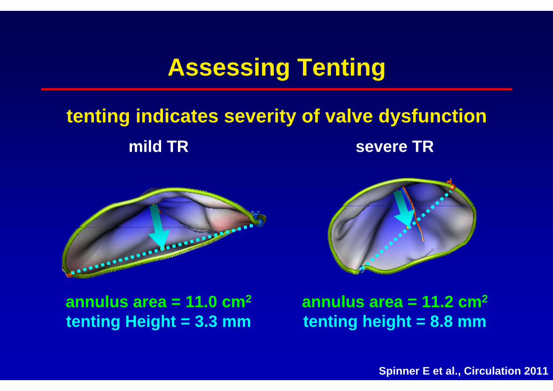
tenting indicates severity of valve dysfunction
Assessing Tenting
annulus area = 11.0 cm2
tenting Height = 3.3 mmannulus area = 11.2 cm2
tenting height = 8.8 mm
mild TR severe TR
Spinner E et al., Circulation 2011

relevant abnormality of TVTV annulus diameter >40mm (21mm/m²)coaptation height (tenting) >8mm
relevant abnormality of RVRVed Area >20cm²excentricity Index >2
relevant RV dysfunctionTAPSE <15mmVpeaksys <11cm/s
Morphology Assessment
Vahanian, EHJ 2012, ESC GuidelinesLancellotti, EJE 2010, EACVI Recommendations

qualitativeTR jet signal density (CW) denshepatic veins syst. flow reversalsystolic inflow dominat E wave
quantitativevena contracta width >7mmReg Vol (PISA) >45ml
Grading TR Severity
Lancellotti, EJE 2010, EACVI Recommendations

qualitativeright atrium severely enlargedIVC dilated
quantitativemean pressure gradient >5mmHgPHT <190msvalve area (cont. equation) < 1cm²
Significant Tricuspid Stenosis
Baumgartner, EJE 2009, EACVI/ASE Recommendations

Tricuspid valve function is complex and depends on size and function of
RV, RA, papillary muscles, leaflets and cordae.
Echocardiography is the method of choice to assess TV function.
Grading of TV dysfunction is difficult due to a lack of reproducible parameters and reliable normal values.
Assessment of TV function must therefore integrate all available (clincal & technical) information.
Summary