HTA REPORT: BEVACIZUMAB FOR AGE-RELATED MACULAR ... · HTA REPORT: BEVACIZUMAB FOR AGE-RELATED...
69
Transcript of HTA REPORT: BEVACIZUMAB FOR AGE-RELATED MACULAR ... · HTA REPORT: BEVACIZUMAB FOR AGE-RELATED...

HTA REPORT: BEVACIZUMAB FOR AGE-RELATED MACULAR DEGENERATION AND DIABETIC RETINOPATHY
i
DISCLAIMERThis Health Technology Assessment has been developed from analysis, interpretation and synthesis of scientific research and/or technology assessment conducted by other organizations. It also incorporates, where available, Malaysian data, and information provided by experts to the Ministry of Health Malaysia. While effort has been made to do so, this document may not fully reflect all scientific research available. Additionally, other relevant scientific findings may have been reported since completion of the review.
Please contact : [email protected], if you like further information.
For further information please contact: Health Technology Assessment Section (MaHTAS),Medical Development Division Ministry of Health MalaysiaLevel 4, Block E1, Precinct 1Government Office Complex62590 PutrajayaTel: 603 88831246Fax: 603 8883 1230
Available at the following website: http://www.moh.gov.my.
Health TechnologyAssessment Report
ENDOBRONCHIALULTRASOUND (EBUS)
MINISTRY OF HEALTH MALAYSIA
BEVACIZUMAB FOR :1. AGE-RELATED MACULAR DEGENERATION 2. DIABETIC RETINOPATHY
HTA REPORT: BEVACIZUMAB FOR AGE-RELATED MACULAR DEGENERATION AND DIABETIC RETINOPATHY
ii
AUTHORS
Dr. Izzuna Mudla Mohamed GhazaliPrincipal Assistant DirectorHealth Technology Assessment Section (MaHTAS)Medical Development DivisionMinistry of Health Malaysia
Madam Noormah Mohd DarusPrincipal Assistant DirectorHealth Technology Assessment Section (MaHTAS)Medical Development DivisionMinistry of Health Malaysia
Mr. Mohd Said MoradSenior Assistant Medical OfficerHealth Technology Assessment Section (MaHTAS)Medical Development DivisionMinistry of Health Malaysia
EXPERT COMMITTEE
Dr. Mariam IsmailSenior Consultant OphthalmologistHospital Selayang
Dr. Tara Mary GeorgeOphthalmologistHospital Selayang
Dr. Norfariza NgahOphthalmologistHospital Selayang
Dr. Lim Kean SengOphthalmologistInternational Specialist Eye CentreLevel 7 & 8, Centrepoint South,The Boulevard, Mid Valley City,Lingkaran Syed Putra,59200 Kuala Lumpur
Madam Rosminah Mohd Din PharmacistPharmaceutical Services DivisionMinistry of Health Malaysia

HTA REPORT: BEVACIZUMAB FOR AGE-RELATED MACULAR DEGENERATION AND DIABETIC RETINOPATHY
iii
REVIEWERS
Dr. Bethel LivingstoneHead, Ophthalmology Service, Ministry of Health Malaysia Senior Consultant Ophthalmologist and Head of DepartmentOphthalmology Department,Hospital Tuanku Jaafar, Seremban
Associate Professor Dr. Fong Choong SianHead, University of Malaya Eye Research Centre (UMERC)Consultant Ophthalmologist and Vitreoretinal SurgeonDepartment of OphthalmologyUniversity of Malaya Medical Centre
Dr. Mae-Lynn Catherine BastionSenior Lecturer and Consultant Ophthalmologist (Vitreo-retinal Service),Department of Ophthalmology Universiti Kebangsaan Malaysia Medical Centre
Datin Dr. Rugayah BakriDeputy DirectorHealth Technology Assessment SectionMedical Development DivisionMinistry of Health Malaysia
ACKNOWLEDGEMENTThe authors of this Health Technology Assessment Report would like to express their gratitude and appreciation to the following for their contribution and assistance:
• HealthTechnologyAssessmentandClinicalPracticeGuidelinesCouncil.
• TechnicalAdvisoryCommitteeforHealthTechnologyAssessment.
DISCLOSUREThe authors of this report have no competing interest in this subject and the preparation of this report is totally funded by the Ministry of Health, Malaysia.

HTA REPORT: BEVACIZUMAB FOR AGE-RELATED MACULAR DEGENERATION AND DIABETIC RETINOPATHY
iv
EXECUTIVE SUMMARY
Background
Age-related macular degeneration (AMD) is a disease associated with aging that gradually
destroys sharp, central vision. It is the leading cause of vision loss for people over the
age of 50 in the western world, affecting approximately 25-30 million people.
Diabetic retinopathy (DR) is the major blinding ocular complication of diabetes mellitus
(DM). The overall prevalence of DR varies in different population. Among Malaysians
diagnosed to have DM before the age of 40 years, the prevalence of DR was 12.3% in
type 1 and 22.3% in type 2 DM, while the prevalence of proliferative DR was 4.0% in
type 1 and 9.3% in type 2 DM.
The prevalence of AMD and DR is expected to increase with the increasing aging
population and prevalence of diabetes in Malaysia.
Technical features
Bevacizumab (Avastin®, Genentech) is a monoclonal antibody that binds and inhibits all
isoforms of Vascular Endothelial Growth Factor (VEGF). It was approved by US Food and
Drug Administration (FDA) in 2004 for metastatic colorectal cancer and later approved for
non-squamous non-small cell lung cancer and advanced HER-2 negative breast cancer.
It is not yet approved for intraocular use. However, as cost is a factor, bevacizumab has
been used off-label for intraocular diseases by many ophthalmologist worldwide.
Objective
To undertake a systematic review on the effectiveness, safety and cost-effectiveness
of bevacizumab in the treatment of age-related macular degeneration and diabetic
retinopathy.
Methods
Electronic databases were searched for published literatures on intravitreal bevacizumab
usage for the treatment of AMD and DR. The following databases were searched including
MEDLINE, PubMed, EBM Reviews – Cochrane Database of Systematic Reviews, Cochrane
Central Register of Controlled Trials, HTA Databases, EBM Reviews - NHS Economic
Evaluation Database and DARE. Additional articles were identified from reviewing the
bibliographies of retrieved articles and handsearching of journals. Further information
was sought from unpublished report. The search was limited to human studies only. The
quality of the papers was assessed using Critical Appraisal Skills Programme (CASP)
checklists and evidence was graded according to US/Canadian Preventive Services Task
Force Levels of Evidence.

HTA REPORT: BEVACIZUMAB FOR AGE-RELATED MACULAR DEGENERATION AND DIABETIC RETINOPATHY
v
Results and conclusion
In conclusion, the evidence suggest that bevacizumab was effective for AMD but
the evidence was only of poor to fair quality and the studies were of short duration.
There was fair evidence to show that bevacizumab was more effective compared to
verteporfin photodynamic therapy for patients with minimally classic or occult choroidal
neovascularization due to AMD. There were two studies that compared bevacizumab
and ranibizumab for AMD but both studies were non-randomised and one of the studies
was retrospective.
There was poor to good quality of evidence retrieved on effectiveness/efficacy of
bevacizumab for diabetic retinopathy. There was good evidence to show that bevacizumab
was more effective in patients with clinically significant diabetic macular oedema compared
to macular photocoagulation or combined therapy with intravitreal triamcinolone. There
was good evidence to show that bevacizumab treatment given after phacoemulsification
and intraocular lens implantation reduced diabetic retinopathy progression. There was
fair evidence to suggest that preoperative treatment with bevacizumab for patients
undergoing pars plana vitrectomy was beneficial.
There was evidence to show that bevacizumab was more cost-effective compared to
other treatment modalities for the management of AMD. There was no evidence on cost-
effectiveness of bevacizumab for DR.
There was evidence to support the safety of bevacizumab for management of AMD and
DR; however caution should be taken in high risk patients.
Recommendation
Based on this review, intraocular bevacizumab can be used selectively in patients
with predominantly classic, minimally classic or occult choroidal neovascularisation
due to age-related macular degeneration and patients with diabetic macular oedema.
However, caution needs to be taken for high risk patients with history of ischaemic heart
disease or thrombo-embolic events. For other indications such as proliferative diabetic
retinopathy, more clinical research is warranted. Effort should be made to register this
drug for intraocular use.

HTA REPORT: BEVACIZUMAB FOR AGE-RELATED MACULAR DEGENERATION AND DIABETIC RETINOPATHY
vi
HTA REPORT: BEVACIZUMAB FOR AGE-RELATED MACULAR DEGENERATION AND DIABETIC RETINOPATHY
vii
TABLE OF CONTENTS
AUTHORS ii
EXPERT COMMITTEE ii
REVIEWERS iii
ACKNOWLEDGEMENT iii
DISCLOSURE iii
EXECUTIVE SUMMARY iv
TABLE OF CONTENTS vii
1 BACKGROUND 1
1.1 Age-related Macular Degeneration 1
1.2 Diabetic Retinopathy 2
2 TECHNICAL FEATURES 4
2.1 Anti Vascular Endothelial Growth Factor (Anti- VEGF) 4
2.1.1 Pegaptanib 4
2.1.2 Ranibizumab 4
2.1.3 VEGF-Trap 5
2.1.4 Sirna-027 5
2.1.5 Ruboxistaurin mesylate 5
2.2 Bevacizumab 5
2.3 Other competing technologies 7
2.3.1 Photodynamic therapy with Verteporfin 7
2.3.2 Laser photocoagulation 7
2.3.3 Triamcinolone 7
3 POLICY QUESTION 7
4 OBJECTIVE 8
5 METHODOLOGY 8
5.1 Literature search strategy 8
5.2 Inclusion and exclusion criteria 8
5.3. Quality assessment strategy 9
5.4. Data extraction strategy 9
5.5. Data analysis 9

HTA REPORT: BEVACIZUMAB FOR AGE-RELATED MACULAR DEGENERATION AND DIABETIC RETINOPATHY
viii

HTA REPORT: BEVACIZUMAB FOR AGE-RELATED MACULAR DEGENERATION AND DIABETIC RETINOPATHY
ix
6 RESULTS AND DISCUSSION 9
6.1 SEARCH RESULTS 9
6.1.1 Quantity and quality of research available 9
6.2 REVIEW OF AVAILABLE SYSTEMATIC REVIEW OR META-ANALYSIS 11
6.3 EFFECTIVENESS 12
6.3.1 Age-related Macular Degeneration 12
6.3.2 Diabetic Retinopathy 15
6.4 COST/ COST-EFECTIVENESS 21
6.5 SAFETY 22
6.5.1 Age-related Macular Degeneration 22
6.5.2 Diabetic Retinopathy 23
7 CONCLUSION 24
8 RECOMMENDATION 25
9 REFERENCES 26
Appendix 1-Levels of evidence and Jadad score 29
Appendix 2- Protocol 30
Appendix 3- Abbreviations 34
Appendix 4- Evidence Table 35
Appendix 5- List of excluded studies 58

HTA REPORT: BEVACIZUMAB FOR AGE-RELATED MACULAR DEGENERATION AND DIABETIC RETINOPATHY
x

HTA REPORT: BEVACIZUMAB FOR AGE-RELATED MACULAR DEGENERATION AND DIABETIC RETINOPATHY
1
BEVACIZUMAB FOR: 1. AGE-RELATED MACULAR DEGENERATION 2. DIABETIC RETINOPATHY
1 BACKGROUND
1.1 Age-related Macular Degeneration
Age-related macular degeneration (AMD) is a disease associated with aging that gradually destroys sharp, central vision. AMD is the leading cause of vision loss for people over the age of 50 in the western world, affecting approximately 25-30 million people. With increasing aging population globally it is estimated that the prevalence of AMD will increase.1
The United Nation (UN) estimates the number of people with AMD at 20-25 million worldwide. According to the World Health Organization (WHO), 8 million people have severe blindness due to AMD, excluding the countries where data are scarce. AMD was found to be second only to cataract as the cause of severe visual loss.1 Prevalence of AMD varies from 1.2% to 29.3%. Three population based studies; the Beaver Dam Eye Study, Blue Mountain Eye Study and the Rotterdam study reported the prevalence rates to be 1.7% in US, 1.9% in Australia and 1.7% in Netherlands respectively.2-4
AMD can be classified into 4 stages based on Age-related Eye Disease Study (AREDS) classification as shown below:5
1. No AMD (AREDS category 1) – none or a few small drusen (<63 microns in diameter).
2. Early AMD (AREDS category 2) – any or all of the following: multiple small drusen, few intermediate drusen (63 to 124 microns in diameter), or retinal pigment epithelium (RPE) abnormalities.
3. Intermediate AMD (AREDS category 3) – any or all of the following: extensive intermediate drusen, and at least one large drusen (≥ 125 microns in diameter), or geographic atrophy (GA) not involving the centre of the fovea.
4. Advanced AMD (AREDS category 4) – GA involving the fovea and/or any of the features of neovascular AMD.
Drusen are tiny yellow or white accumulations of extracellular material that build up in Bruch’s membrane of the eye. The presence of a few small (“hard”) drusen is normal with advancing age, and most people over 40 have some hard drusen. However, the presence of larger and more numerous drusen in the macula is a common early sign of AMD.

HTA REPORT: BEVACIZUMAB FOR AGE-RELATED MACULAR DEGENERATION AND DIABETIC RETINOPATHY
2
Neovascular AMD can be classified based on the temporal and spatial features of the patterns of fluorescence as observed on fluorescein angiography (FA) as given below:5
i) Classic choroidal neovascularisation (CNV) is said to be present when an area of well delineated hyperfluorescence appears in the early phases of the FA usually before 30 seconds have elapsed following injection of the fluorescent dye into a peripheral vein
ii) Occult CNV is the presence of leakage without clear evidence of neovascular profiles in the early angiographic images.
Figure 1. Anatomy of the eye
1.2 Diabetic Retinopathy
Diabetic retinopathy (DR) is not only a common complication of diabetes mellitus (DM) but it leads to disability. It is the main contributor to blindness among the working age group.6
DR is the major blinding ocular complication of DM. DR includes non-
proliferative and proliferative retinopathy and the late sequelae of vitreous haemorrhage and tractional detachment. Visual loss may occur from neovascularization, as in proliferative diabetic retinopathy (PDR), or from increased retinal vascular permeability leading to diffuse leakage from capillary wall, focal leakage from microaneurysms, and accumulation of fluid and macromolecules in the retina, as in diabetic macular oedema (DME).7
Iris
Cornea
Lens
Iris
Optic nerve
Vitreous gel
Macula
Fovea
Retina
Pupil
HTA REPORT: BEVACIZUMAB FOR AGE-RELATED MACULAR DEGENERATION AND DIABETIC RETINOPATHY
3
Early Treatment for Diabetic Retinopathy Study (ETDRS) has classified non-proliferative PDR (NPDR) into mild, moderate, severe and very severe and PDR into early PDR and high-risk PDR.8 This is as follows:
A. Mild NPDR: Presence of at least one microaneurysm, definition not met for B, C, D, E, or F.
B. Moderate NPDR: Haemorrhages and/or microaneurysms more than standard photo 2A, presence of soft exudates, venous beading, intraretinal microvascular abnormalities (IRMA) definitely present, definition not met for C, D, E, or F.
C. Severe NPDR: Haemorrhages and/or microaneurysms more than standard photo 2A in all four quadrants, or venous beading in two or more quadrants, or IRMA > standard photo 8A in at least one quadrant, definition not met for D, E, or F.
D. Very severe NPDR: Any two or more of the changes seen in severe NPDR, definition not met for E, or F.
E. Early PDR: Presence of new vessels, definition not met for F.
F. High-risk PDR: Includes any of the following characteristics - neovascularization of disc (NVD) > 1/3 rd to 1/4 th disc diameter, NVD 1/3 rd to 1/4 th disc diameter with vitreous/pre-retinal haemorrhage, NVE with vitreous/pre-retinal haemorrhage. High-risk characteristics (HRC) were defined by DRS, as the patient, if not treated urgently, is at a high risk of severe visual loss
Diabetic macular oedema (DME) is retinal thickening within two disc diameters of the centre of macula. DME patients were categorized into clinically significant macular oedema (CSME) or non-CSME by ETDRS. CSME includes any one of the following lesions:8
Retinal thickening at or within 500 microns from the center of macula.
Hard exudates at or within 500 microns from the center of macula associated with thickening of the adjacent retina. An area or areas of retinal thickening at least one disc area in size, at least a part of which is within one disc diameter of the center of macula.
The overall prevalence of DR varies in different population (the highest
prevalence is 54%).7 Ocular microvascular complications affect almost all patients with type 1 diabetes and at least 60% of patients with type 2 diabetes.9 Among Malaysians diagnosed to have DM before the age of 40 years, the prevalence of DR was 12.3% in type 1 and 22.3% in type 2 DM, and prevalence of PDR was 4.0% in type 1 and 9.3% in type 2 DM.6

HTA REPORT: BEVACIZUMAB FOR AGE-RELATED MACULAR DEGENERATION AND DIABETIC RETINOPATHY
4
The prevalence of AMD and DR is expected to increase with the increasing
aging population and prevalence of diabetes in Malaysia. The cost of
healthcare will proportionately increase in view of the high cost of the
currently available FDA approved anti Vascular Endothelial Growth Factor
(VEGF). As financial resource is not unlimited, cost constraints will be a
barrier to appropriate care for some patients. The impact would be worse
on young and visually impaired diabetics who would have significantly lost
their earning capacity at presentation. An effort should therefore be made
to improve equity and access to appropriate care for these patients by
exploring into the use of a cheaper alternative to currently available anti
VEGF approved for intraocular use.
This Health Technology Assessment report was conducted following a
request from a Senior Consultant Ophthalmologist who was also the Head
of Ophthalmology Service, Ministry of Health Malaysia then.
2 TECHNICAL FEATURES
2.1 Anti Vascular Endothelial Growth Factor (Anti- VEGF)
VEGF has been found to be one of the key elements in angiogenesis. VEGF
is a member of a family of proteins including VEGF-A, VEGF-B, VEGF-C,
VEGF-D and placental growth factor (PIGF) that exert their function through
tyrosine kinase receptors VEGFR-1, VEGFR-2 and VEGFR-3. VEGF-A and
its isoforms have received the most attention as mediators of pathologic
neovascularisation. Protein Kinase C (PKC) is a family of proteins involved
in signal transduction that increases the synthesis of VEGF.10
There are several anti-VEGF agents in the market as discussed below.
Some of these agents have been approved for intraocular use.
2.1.1 Pegaptanib
Pegaptanib (Macugen®, Eyetech Pharmaceuticals) is a pegylated
ribonucleic acid oligonucleotide ligand (aptamer) that binds to
the VEGF165 isoform that is thought to be primarily responsible for
pathologic neovascularisation. Pegaptanib inhibits VEGF binding to
its receptor resulting in decreased vascular permeability and inhibition
of neovascularisation. The US Food and Drug Administration (FDA)
approved pegaptanib in December 2004 for intravitreal treatment
of subfoveal neovascular AMD.10 It was the first approved drug in
this category. Because of the structural specificity (by only targeting
165 isoform of VEGF), pegaptanib sodium might help in preventing
major systemic vascular accidents.11

HTA REPORT: BEVACIZUMAB FOR AGE-RELATED MACULAR DEGENERATION AND DIABETIC RETINOPATHY
5
2.1.2 Ranibizumab
Ranibizumab (Lucentis®, Genentech) is a humanised monoclonal
antibody fragment designed to bind and inhibit all isoforms of human
VEGF (in contrast to pegaptanib that bind to single isoform).12
Ranibizumab was specifically designed for intraocular use as a
smaller antibody fragment to better penetrate through the retina. The
FDA approved ranibizumab in June 2006 for treatment of subfoveal
AMD and clinical trials are ongoing for the treatment of macular
oedema.10 Ranibizumab has recently been approved to be included
in the Ministry of Health Malaysia Drug Formulary.
Ranibizumab has been shown to have remarkable results following
extensive, stringent clinical trials. MARINA and ANCHOR trial were
both Phase III, multi-center, randomised, double masked trials
which showed visual improvements in patients with wet AMD. In a
significant proportion of patients, unlike pegaptanib, not only was
there a prevention of visual loss but also an improvement in visual
acuity.13
2.1.3 VEGF-Trap
VEGF-Trap (Regeneron Pharmaceuticals) is a decoy receptor fusion
protein with portions of the binding domains of VEGF receptors
VEGFR-1 and VEGFR-2. It effectively blocks the actions of all VEGF-A
isoforms and PIGF and is currently in phase I/II clinical trials for
AMD.10
2.1.4 Sirna-027
Sirna-027 (Sirna Therapeutics) is a short interfering ribonucleic acid
that is directed against the VEGF-1 receptor, inhibiting its production
at the gene level. A phase II clinical trial is underway for patients with
neovascular AMD.10
2.1.5 Ruboxistaurin mesylate
Ruboxistaurin mesylate (Arxxant®, Eli Lilly) is a systemic, selective
inhibitor of the b-isoform of PKC. A randomised controlled trial
showed safety and prevention of vision loss in diabetic patients. The
drug received an approvable letter from the FDA in August 2006 and
the manufacturer (Lilly) is working to provide additional data towards
its full approval.10

HTA REPORT: BEVACIZUMAB FOR AGE-RELATED MACULAR DEGENERATION AND DIABETIC RETINOPATHY
6
2.2 Bevacizumab Bevacizumab (Avastin®, Genentech) is another monoclonal antibody like
ranibizumab that binds and inhibits all isoforms of VEGF, but with lower affinity, and the larger molecule has a longer half life.12
The FDA approved bevacizumab in February 2004 for use in combination with intravenous 5-fluorouracil-based (5-FU-based) chemotherapy as a treatment for first-line metastatic colorectal cancer. In June 2006, the FDA approved bevacizumab in combination with intravenous 5-FU-based chemotherapy for patients with metastatic colorectal cancer who have been previously treated for their cancer (or second-line metastatic colorectal cancer). In October 2006, the FDA approved bevacizumab in combination with carboplatin and paclitaxel for the first-line treatment of patients with unresectable, locally advanced, recurrent or metastatic non-squamous non-small cell lung cancer (NSCLC).14
In February 2008, the FDA granted accelerated approval for bevacizumab in combination with paclitaxel chemotherapy for the first-line treatment of advanced HER2-negative breast cancer.14
The intravitreal use of bevacizumab to treat neovascular AMD and other types of ocular diseases is still under study and is not yet approved by FDA. However, bevacizumab has been officially approved for intravitreal use in the treatment of “exudative maculopathy and neovascular glaucoma” and will be fully reimbursed by the National Health Service in Italy.15
In 2005, prior to US FDA approval of ranibizumab for AMD, Rosenfeld et al. published a case report that demonstrated favourable outcomes in terms of visual acuity and macular appearance under optical coherence tomography after single eye treatment of neovascular AMD with intravitreal bevacizumab.12 Subsequently off-label use of intravitreal bevacizumab became an alternative for patients not eligible for or responding poorly to other approved therapies.12
As cost is a factor, bevacizumab has been used “off-label” for intraocular diseases by many Ophthalmologists worldwide though it was neither developed nor formulated nor studied nor approved for intraocular use. The results have been reported as encouraging. Although, off-label use of drugs is not illegal, it does raise ethical issues and safety concerns.11
Intravenous use of bevacizumab in cancer patients has serious systemic complications, including: increased risk of thromboembolic events, hypertension, haemorrhage, proteinuria, wound healing complications, and gastrointestinal perforation.12 Whether these systemic complications are relevant for patients with ocular diseases receiving low doses of bevacizumab intravitreally is unknown. This is a concern since eye diseases such as AMD is not a life threatening condition compared to cancer.

HTA REPORT: BEVACIZUMAB FOR AGE-RELATED MACULAR DEGENERATION AND DIABETIC RETINOPATHY
7
2.3 Other competing technologies
2.3.1 Photodynamic therapy with Verteporfin
This is a procedure whereby the photosensitizing dye verteporfin (visudyne, Novartis) is given intravenously. Verteporfin is taken up selectively by rapidly proliferating endothelial cells which have greater low density lipoprotein (LDL) receptor expression. This is followed by the delivery of laser light of a wavelength of 689 nm to the CNV lesion as a single spot with a diameter 1000 µm larger than the greatest linear diameter of the lesion. The energy from the laser is taken up by the verteporfin and this leads to damage to vascular endothelial cells and thrombotic occlusion of the blood vessels within the CNV lesion. European Medicines Agency (EMEA) has withdrawn the European license for PDT in occult CNV based on Visudyne in Occult Choroidal Neovascularization (VIO) study which did not reveal any benefit of Verteporfin.5
2.3.2 Laser photocoagulation
Laser photocoagulation was the only available treatment for neovascular AMD in the 1990s. A series of clinical trials of macular photocoagulation showed that treating angiogenesis with laser photocoagulation benefited visual outcome. However, recurrence after treatment is common and is often associated with more severe visual loss. In addition, treating CNV lesions in the subfoveal region is not recommended, because destruction of the overlying retina results in an immediate central scotoma. Therefore, laser photocoagulation remains a treatment option for juxtafoveal and extrafoveal CNV lesions only.12
2.3.3 Triamcinolone
Intravitreal injection of triamcinolone acetonide has been used for the treatment of intraocular oedematous, proliferative and neovascular diseases such as diabetic macular oedema, macular oedema associated with retinal vein occlusion, proliferative diabetic retinopathy, uveitis including sympathetic ophthalmia and exudative AMD.16 Earlier studies showed that intravitreal injection of 4 mg of triamcinolone might be particularly effective for treating neovascular AMD. However, a randomised trial of 139 patients by Gillies reported no benefit in terms of reducing severe visual loss during the first year of the study.12,16
3 POLICY QUESTION Should bevacizumab be made available at selected Ministry of Health Malaysia
hospitals for the treatment of age related macular degeneration and diabetic retinopathy?

HTA REPORT: BEVACIZUMAB FOR AGE-RELATED MACULAR DEGENERATION AND DIABETIC RETINOPATHY
8
4 OBJECTIVE
i. To undertake a systematic review on the effectiveness of bevacizumab in the treatment of age related macular degeneration and diabetic retinopathy.
ii. To assess the cost of bevacizumab in the treatment of age related macular degeneration and diabetic retinopathy.
iii. To assess the safety aspect of bevacizumab, the social and ethical aspects in the treatment of age related macular degeneration and diabetic retinopathy.
5 METHODOLOGY
5.1 Literature search strategy Electronic databases were searched for published literatures on intravitreal
bevacizumab usage for the treatment of age related macular degeneration (AMD) and Diabetic Retinopathy (DR). The following databases were searched including MEDLINE, PubMed, EBM Reviews – Cochrane Database of Systematic Reviews, Cochrane Central Register of Controlled Trials, HTA Databases, EBM Reviews - NHS Economic Evaluation Database, DARE and Science Direct. Additional articles were identified from reviewing the bibliographies of retrieved articles and handsearching of journals. Further information was sought from unpublished reports.
The search was limited to human studies only. The following search terms were used either singly or in combinations: Avastin OR bevacizumab, age related macular degeneration OR AMD OR age related maculopathy OR ARM, diabetic retinopathy OR advanced diabetic maculopathy OR advanced diabetic eye disease OR severe diabetic eye disease OR severe diabetic maculopathy and vitrectomy.
5.2 Inclusion and exclusion criteria Inclusion criteria:
i. Type of studies For systematic review on effectiveness and safety, systematic reviews,
meta-analysis and comparative clinical studies which involved the use of bevacizumab for the treatment of AMD and DR were included.
For assessment of cost-effectiveness, all cost effectiveness studies of satisfactory quality were included.
ii. Types of participants Adults with AMD or DR.

HTA REPORT: BEVACIZUMAB FOR AGE-RELATED MACULAR DEGENERATION AND DIABETIC RETINOPATHY
9
iii. Types of intervention Trials which evaluate intravitreal bevacizumab with placebo or other types
of anti-VEGF or other treatment modalities were included.
iv. Types of outcome measures One or more of the following outcome measures were assessed:
1. Visual improvement
2. Improved quality of life
3. Cost per Quality Adjusted Life Years (QALY) gained
Exclusion criteria
1. Systemic administration of bevacizumab
2. Bevacizumab used for other indications
5.3. Quality assessment strategy The quality of the selected studies was assessed by two reviewers using
Critical Appraisal Skills Programme (CASP) checklists depending on the type of study design. Basic Jadad score was used to assess randomised clinical trial (RCT) and to classify the papers as good or poor RCT. Papers that scored 3-5 were considered as good RCT whereas papers that scored 2-0 were considered as poor RCT.
5.4. Data extraction strategy Data was extracted by a reviewer and checked by a second reviewer using
a pre-tested data extraction form. Disagreements were resolved through discussion. A third person, whose decision is final was consulted when disagreements persisted after discussion.
5.5. Data analysis The studies included in this review were clinically and methodologically
diversed, thus meta-analysis was not performed. The data was analysed qualitatively.
6 RESULTS AND DISCUSSION
6.1 SEARCH RESULTS
6.1.1 Quantity and quality of research available
Sensitive search strategies were used to identify the articles. Many of the articles could be excluded based on titles and abstracts alone. Finally 20 relevant full text articles were selected after applying the inclusion and exclusion criteria. Out of this, one article was a systematic review and meta-analysis that covered AMD and DR, six articles on AMD and 13 articles on DR. Another two articles on economic/cost evaluation were also included.

HTA REPORT: BEVACIZUMAB FOR AGE-RELATED MACULAR DEGENERATION AND DIABETIC RETINOPATHY
10
Three of the articles on AMD were randomised controlled trial (RCT), one non-RCT and 2 retrospective cohort studies. Only one of the RCT can be considered as good quality (Jadad score 3). One of the RCT compared combination of verteporfin photodynamic therapy (PDT) and intravitreal bevacizumab (IVB), with verteporfin PDT alone and IVB alone. One RCT compared IVB with verteporfin PDT and another RCT compared IVB with PDT and intravitreal triamcinolone (IVTA). The non-RCT and one retrospective cohort study compared IVB with intravitreal ranibizumab. The other retrospective cohort study compared IVB with Pegaptanib.
Eight of the articles on DR were RCT, two non-RCT and three retrospective cohort studies. Four of the RCT were of good quality (Jadad score 3 to 5). A good RCT (Soheilian et al., Jadad score 5) compared IVB with IVB plus IVTA and macular photocoagulation. Another RCT (DRCR Network et al., Jadad score 3) compared IVB with photocoagulation. Two RCT (Cheema et al. and Takamura et al.) compared IVB after phacoemulsification and intraocular lens implantation (IOL) with phacoemulsification and IOL alone. One RCT (Tonello et al.) compared panretinal photocoagulation (PRP) plus IVB with PRP alone. One RCT (Cho et al.) and one retrospective cohort study (Mason et al.) compared IVB given before PRP with PRP alone. Four studies compared IVB given before vitrectomy with vitrectomy alone. One RCT (Paccola et al.) compared IVB with IVTA and one retrospective cohort study (Arevalo et al.) compared two doses of IVB.
Figure 1. Flow diagram for identified studies
HTA REPORT: BEVACIZUMAB FOR AGE-RELATED MACULAR DEGENERATION AND DIABETIC RETINOPATHY
11
6.2 REVIEW OF AVAILABLE SYSTEMATIC REVIEW OR META-ANALYSIS Andriolo et al. included 667 eyes from nine randomised trials in their
systemic review and meta analysis. Three studies included patients with diagnoses of diabetic macular oedema, one study included patients with diagnoses of clinically significant macular oedema who had not responded to earlier subsequent photocoagulation therapy, three studies included patients with diagnoses of age-related macular degeneration, one study tested bevacizumab on patients with subfoveal choroidal neovascularisation associated with age and one study included patients with proliferative diabetic retinopathy.17 Level I Results from two studies that compared bevacizumab plus triamcinolone versus bevacizumab alone were pooled. The pooled result showed that bevacizumab alone is better than bevacizumab plus triamcinolone but without statistically significant mean difference (MD 0.02, 95% CI -0.09, 0.14).
Two studies that compared bevacizumab plus triamcinolone versus sham
and bevacizumab isolated versus sham showed statistically significant MD in favour of bevacizumab (MD -0.18, 95% CI -0.28,-0.08) and (MD -0.15, 95% CI -0.26,-0.04) respectively.17 Level I
Three studies evaluated the endpoint best corrected visual acuity. Bevacizumab versus triamcinolone showed statistically non-significant results favouring group treated with bevacizumab (MD 0.01, 95% CI -0.04, 0.06). A study on photocoagulation plus bevacizumab versus photocoagulation showed statistically non-significant results favouring group treated with photocoagulation alone (MD 0.02, 95% CI -0.12, 0.16).
A study on bevacizumab plus triamcinolone versus photocoagulation
showed statistically non-significant results favouring group treated with bevacizumab plus triamcinolone (MD -0.11, 95% CI -0.30 to 0.08).17 Level I
Bevacizumab alone was shown to be better than photodynamic therapy for best-corrected visual acuity (MD -0.09, 95%CI -0.13,-0.06). Bevacizumab in association with photodynamic therapy was shown to be better than both bevacizumab alone and photodynamic therapy alone (MD -0.14, 95% CI -0.18,-0.11) and (MD -0.24, 95% CI -0.27,-0.20). 17 Level I
The review showed that greater proportion of patients whose visual acuity was not reduced by more than three lines in the group treated with bevacizumab compared with the group treated with photodynamic therapy (RR 0.19, 95% CI 0.04, 0.86). The number needed to treat (NNT) was 3 (95% CI 2, 4) which meant that it was necessary to change three patients from photodynamic therapy to bevacizumab to avoid an additional patient presenting any losses in visual acuity.

HTA REPORT: BEVACIZUMAB FOR AGE-RELATED MACULAR DEGENERATION AND DIABETIC RETINOPATHY
12
The review also showed that there were more patients with increased visual acuity in bevacizumab group compared to the group treated with photodynamic therapy alone or combined with triamcinolone (RR 0.49, 95% CI 0.31, 0.78). The NNT was 4 (95% CI 1, 4) which meant that it was necessary to change four patients from photodynamic therapy alone or combine with triamcinolone to bevacizumab to improve visual acuity in one patient.
The most common adverse event reported was moderate anterior chamber reaction (19%), followed by transient anterior chamber reaction (16%), iris neovascularisation (11%), subconjunctival haemorrhage & posterior vitreous detachment (15%) and foreign body sensation.17 Level I
This review only assessed the outcome based on the intervention, the differences in dosage of treatment was not taken into consideration. The dosage of bevacizumab used in the studies included in this review varies from 1.0 mg to 2.5 mg.
6.3 EFFECTIVENESS
6.3.1 Age-related Macular Degeneration
There were six studies that assessed effectiveness/efficacy of bevacizumab for AMD. Three of the RCT have been included in Andriolo et al. systematic review.
Bevacizumab versus verteporfin photodynamic therapy
Lazic et al. compared three groups; combination of verteporfin PDT and intravitreal bevacizumab 1.25 mg (COMB group), intravitreal bevacizumab 1.25 mg alone (IVB) and verteporfin PDT alone (PDT) among 165 subjects with minimally classic or occult CNV due to AMD in one or both eyes where the studied eye had never been treated. 18 Level I They found that at 1 month follow-up, there were significant improvements in best-corrected VA in the three groups. Larger improvement was seen in the COMB group (0.246 logMAR) compared to 0.171 and 0.049 logMAR in the IVB and PDT groups, respectively. At 3 months follow-up significant improvements in comparison with baseline were still observed in the IVB and COMB groups as opposed to the slight worsening noted in the PDT group. In the IVB group, the improvement at 3-month follow up was less than half of the improvement observed at 1 month. The improvement remained the same in the COMB group (0.223 logMAR).
As for central foveal thickness (CFT), at 1 month follow-up, similar and significant reductions in CFT were observed in the three groups. At 3 months follow-up, significant reductions in comparison to baseline were still observed in the 3 groups.18 Level 1

HTA REPORT: BEVACIZUMAB FOR AGE-RELATED MACULAR DEGENERATION AND DIABETIC RETINOPATHY
13
Bashshur et al. compared 2.5 mg bevacizumab with 6 mg verteporfin PDT among 64 patients with predominantly classic CNV AMD.19 Level
I The mean BCVA improved in the bevacizumab group from 20/119 at baseline to 20/89 at 3 months and 20/68 at 6 months. The mean BCVA in the PDT group worsened from 20/108 at baseline to 20/118 at 3 months and 20/143 at 6 months. The difference between the two groups was statistically significant at 6-month (p< 0.01). Five of 30 (16.7%) eyes in the PDT group ended with a BCVA worse than 20/200, while no eyes in the bevacizumab group did. At conclusion of the study all the eyes (32 eyes) in the bevacizumab group and 22 of 30 (73.3%) eyes in the PDT group avoided losing more than 3 lines of BCVA (p=0.002) while 16 eyes (50%) in the bevacizumab group had improvement in the BCVA compared to 5 eyes (16.7%) in the PDT group (p=0.007).
Mean central retinal thickness (CRT) reduced in the bevacizumab group from 354 µm at baseline to 262 at 3-month and 241 µm at 6-month. In the PDT group the mean CRT reduced from 352 µm at baseline to 300 µm at 3-month and 292 µm at 6-month. The difference in the mean CRT between the two groups was statistically significant at 3-month and 6-month.19 Level I
These two studies among patients with predominantly classic CNV and patients with minimally classic or occult CNV due to AMD showed better improvement in BCVA with bevacizumab when compared to verteporfin PDT and more patients avoid losing their vision with bevacizumab.
Bevacizumab versus Photodynamic therapy with Triamcinolone
Weigert et al. compared intravitreal bevacizumab (1 mg) with standard PDT plus 4 mg intravitreal triamcinolone among 28 patients who had neovascular AMD of any lesion type smaller than four disc areas, without any previous treatment for neovascular AMD.20 Level I They found statistically significant difference in visual acuity between both groups in favour of the bevacizumab-treated group as early as day 1 (p=0.03). As for central retinal thickness (CRT), similar changes were seen for both groups up to month-6 follow-up. Compared to baseline, the decrease in CRT became statistically significant as early as week 1 in both groups. There was no statistically significant difference between both groups with regard to CRT. There was significant correlation between the change in VA and a change in CRT for the bevacizumab-treated group at month 1 but not the PDT plus IVTA-treated group. At months 3 and 6 the correlations did not reach significance in both groups.
HTA REPORT: BEVACIZUMAB FOR AGE-RELATED MACULAR DEGENERATION AND DIABETIC RETINOPATHY
14
Bevacizumab versus Ranibizumab
Krishnan et al. assessed the occurrence of submacular haemorrhage among 36 consecutive patients with occult CNV size of greater than or equal to 15 mm2 after intravitreal bevacizumab compared with intravitreal ranibizumab. In the ranibizumab group, no postoperative submacular haemorrhages was observed. Acute postoperative, fresh submacular haemorrhages were seen in four out of 14 patients given intravitreal bevacizumab compared with zero out of 22 patients given intravitreal ranibizumab (p = 0.017, odds ratio 19.29). No retinal pigment epithelium tears were noted in these patients at the time of the haemorrhage or after the haemorrhage had resolved.21 Level II-1
Landa et al. studied 73 consecutive patients who had any evidence of CNV secondary to AMD and were treated with only one type of anti-VEGF agent (either bevacizumab or ranibizumab).22 Level II-2 For visual acuity, at one month, the mean logMAR improved by 0.18 to a Snellen equivalent of 20/107 in the bevacizumab group (p=0.009) and by 0.13 to a Snellen equivalent of 20/117 in the ranibizumab group (p=0.004). The BCVA improved one month after the injection in 78.8% and 80.6% of patients after receiving bevacizumab and ranibizumab respectively. As for CFT, there was a statistically significant reduction in mean CFT in both groups at one month after the injection. The average SD-OCT CFT decreased from 325 ± 72 µm to 300 ± 69 µm in the bevacizumab group (p=0.016) and from 307 ± 57 µm to 289 ± 56 µm in the ranibizumab group (p=0.017). There was no analysis done to compare the two groups.
There are currently two major multicenter randomised clinical trials comparing bevacizumab and ranibizumab for neovascular AMD. The trials are IVAN trial (A Randomised Controlled Trial of Alternative Treatments to Inhibit VEGF in Age-related Choroidal Neovascularisation)23 and CATT trial (Comparison of Age-Related Macular Degeneration Treatments Trials).24 The results of these trials are expected to be released next year.
Bevacizumab versus Pegaptanib
Joeres et al. did a quantitative comparison of optical coherence tomography among 58 cases with subfoveal choroidal neovascularisation due to AMD who received either pegaptanib or bevacizumab injections.25 Level II-2 They revealed that there was greater reduction of total retinal volume after bevacizumab treatment than after pegaptanib treatment (mean decrease of 0.78% from baseline volume, p=0.003). The total retinal volume decreased from 8.23 ± 1.66 mm3 to 7.36 ± 0.93 mm3 (p<0.001) in the bevacizumab group and from 7.59 ± 0.70 mm3 to 7.52 ± 0.73 mm3 in the pegaptanib group. However, the changes in the volume of subretinal fluid (SRF) did not statistically significantly differ between the treatment groups. In the pegaptanib group, the amount of SRF in the foveal central subfield (FCS) decreased from 0.03 ± 0.05 mm3 to 0.01 ± 0.02 mm3, (p=0.007) but the total volume of the fluid in the entire macula did not decrease significantly. The total amount of fluid in the bevacizumab group was significantly lower three months after treatment (p=0.004).
HTA REPORT: BEVACIZUMAB FOR AGE-RELATED MACULAR DEGENERATION AND DIABETIC RETINOPATHY
15
As for subretinal tissue, the changes did not statistically significantly differ between treatment groups. The changes in volume and thickness values for pigment epithelial detachment (PED) also did not significantly differ between pegaptanib and bevacizumab treatments.25 Level II-2
There was significant difference in treatment effect between the two groups (p=0.016) with regards to inner retinal surface height from choroid, with a reduction in the bevacizumab group (mean decrease of 11.96%) and an increase in the pegaptanib group (mean increase of 4.2%).25 Level II-2
When analysed using StratusOCT analysis, both groups showed significant decrease in mean retinal thickness of the FCS at 3 months when compared to baseline but the difference between the two groups was not statistically significant.25 Level II-2
6.3.2 Diabetic Retinopathy
There were 13 studies that assessed effectiveness/efficacy of bevacizumab for diabetic retinopathy. Four of the RCT have been included in Andriolo et al. systematic review.
6.3.2.1 Diabetic Macular Oedema
Bevacizumab versus photocoagulation
Soheilian et al. compared three groups of patients with clinically significant diabetic macular oedema (CSDME) receiving either intravitreal bevacizumab alone (IVB group), intravitreal bevacizumab plus intravitreal triamcinolone (IVB/IVT group) or macular photocoagulation (MPC group).26 Level I The results showed that VA improvement was significant in the IVB groups at all follow-up visits up to 36 weeks (p<0.001) when compared to baseline. In the IVB/IVT group, VA improved significantly only at weeks 6 and 12 (p=0.002 and 0.019 respectively). In the MPC group, VA changes were not significant. Pairwise comparison between groups showed that the VA improvement at 6 weeks in both IVB and IVB/IVT was greater than in the MPC group (p<0.001), with no significant difference between the IVB and IVB/IVT groups. At 24 weeks, difference of VA changes between the IVB and MPC groups was significant in favour of the IVB group (p=0.003). The difference between the IVB/IVT and MPC groups was borderline (p=0.033). There was no significant difference between IVB and IVB/IVT (p=0.373). At 12 and 36 weeks, there was more VA improvement in the IVB group than in the other groups though not to a meaningful level.26 Level I

HTA REPORT: BEVACIZUMAB FOR AGE-RELATED MACULAR DEGENERATION AND DIABETIC RETINOPATHY
16
As for outcome measured using Snellen VA, the percentage of eyes with stable VA was relatively similar among the groups at all follow up. There was greater percentage of cases that gained more than 2 Snellen VA in the IVB and IVB/IVT groups than in the MPC groups. There was greater percentage of eyes which lost more than 2 Snellen lines in the MPC group than in the other groups. These differences were statistically significant among the groups at 6, 12 and 24 weeks.26 Level I
As for central macular thickness (CMT), the reduction of CMT was significantly different among the treatment groups only in the subgroups with the initial CMT ≥ 400 µm at all follow-up visits except at week 24. At week 6, CMT reduction was greater in the IVB group than in the other groups (p = 0.026). At week 12 the difference in CMT reduction was significant among all 3 groups in favour of the IVB/IVT group. At week 36, the difference was significant between IVB and MPC group.26 Level I
Another study conducted by Diabetic Retinopathy Clinical Research Network compared 5 groups of patients with DME.27 Level I The control group received photocoagulation at baseline (Group A). Group B received intravitreal injection of 1.25 mg bevacizumab at baseline and 6 weeks. Group C received intravitreal injection of 2.5 mg bevacizumab at baseline and 6 weeks, Group D received intravitreal injection of 1.25 mg bevacizumab at baseline and sham injection at 6 weeks and Group E received intravitreal injection of 1.25 mg bevacizumab at baseline, focal photocoagulation at 3 weeks, and intravitreal injection of 1.25 mg bevacizumab at 6 weeks. The results showed that when compared with Group A, Group B and C both demonstrated a greater reduction in central subfield thickness at 3 weeks (p=0.006 and <0.001 respectively). As for visual acuity, Groups B and C both had about a median one line improvement at the 3-week visit which was sustained through 12 weeks and was greater than the change in visual acuity in Group A (p=0.01 and 0.003, respectively). As for reduction of central subfield thickness more than 11%, which was considered as clinically significant, was present at 3 weeks in 23 of 60 (38%) in pooled 1.25 mg bevacizumab-treated eyes, 13 of 24 (54%) in 2.5 mg bevacizumab-treated eyes, and in 5 of 18 (28%) eyes treated with laser alone. At 6 weeks, 22 of 61 (36%) in pooled 1.25 mg bevacizumab-treated eyes, 9 of 23 (39%) in 2.5 mg bevacizumab-treated eyes, and in 9 of 18 (50%) eyes treated with laser alone. Both the 1.25 mg and 2.5 mg bevacizumab-treated eyes had a greater reduction in central retinal thickness at 3 weeks. Eyes in the photocoagulation group demonstrated improvement in this parameter with longer follow up. Six weeks may be too long for an optimal injection interval.27 Level I

HTA REPORT: BEVACIZUMAB FOR AGE-RELATED MACULAR DEGENERATION AND DIABETIC RETINOPATHY
17
Phacoemulsification and intraocular lens implantation with and without bevacizumab posttreatment
Cheema et al. studied 68 eyes of 68 diabetic patients with sight limiting cataract with presence of clinically significant macular oedema (CSME), mild, moderate, severe or very severe non-proliferative diabetic retinopathy or PDR or a combination of both.28 Level 1 Diabetic retinopathy progressed in 45.45% of eyes in the control group who did not received intravitreal bevacizumab and 11.42% of eyes in the intervention group who received intravitreal bevacizumab at the end of surgery. The difference between groups was statistically significant (p=0.002). Progression of diabetic maculopathy occurred in 51.51% of eyes in the control group and 5.71% of eyes in the intervention group; the difference between groups was statistically significant (p<0.001). There was no statistically significant difference in postoperative visual acuity and changes in central macular thickness and average macular thickness over 6-month follow up at any time point.
Another similar study conducted by Takamura et al. among 42 patients with Type 2 Diabetes and DME.29 Level I The results showed that the mean CRT increased from 351.6 µm at the baseline to 389.0 µm at month 1 and 379.9 µm at month 3 (p=0.0013 and p=0.0068, respectively) in the control group. Whereas in the bevacizumab group the CRT decreased from 355.0 µm at baseline to 327.0 µm at month 1 and 330.0 µm at month 3 (p=0.0087 and p=0.0217, respectively). As for BCVA, in the control group, VA improved from 0.84 at baseline to 0.61 at month 1 and 0.51 at month 3, (p=0.0057). In the bevacizumab group, VA improved from 0.90 at baseline to 0.43 at month 1 and 0.38 at month 3. Comparison of both groups showed that VA in the bevacizumab group was significantly better at month 3 (p=0.034).29 Level I
Bevacizumab versus triamcinolone
Paccola et al. compared the morphological and visual acuity outcomes associated with a single intravitreal injection of triamcinolone acetonide versus bevacizumab among 28 patients with refractory diabetic macular oedema (DME).30 Level I There was significant reduction (p<0.01) in central macular thickness (CMT) in the intravitreal triamcinolone (IVT) group at weeks 4 (p=0.008), 8 (p=0.006), 12 (p<0.0001), and 24 (p=0.024) compared with the IVB group. Separate within group analysis showed significant changes (reductions) in CMT values from baseline at weeks 4, 8 and 12 (p<0.0001) in the IVT group, and at weeks 4 (p<0.0001) and 8 (p=0.0004) in the IVB group.

HTA REPORT: BEVACIZUMAB FOR AGE-RELATED MACULAR DEGENERATION AND DIABETIC RETINOPATHY
18
There was significant improvement in BCVA in the IVT group compared with the IVB group at weeks 8 (p=0.026) and 12 (p=0.039). Separate within group analysis showed significant improvement in BCVA from baseline at weeks 4, 8 and 12 (p<0.001) in the IVT group, and at week 4 (p=0.011) in the IVB group.30 Level I
Bevacizumab 1.25 mg versus bevacizumab 2.5 mg
Arevalo et al. compared two doses of bevacizumab among 115 patients with diffuse diabetic macular oedema.31 Level II-2 Within 1 month after the initial bevacizumab injection, improvements in BCVA and CMT measurements were observed, and these significant changes continued throughout the 24-month follow up. Subgroup analysis demonstrated that 62 (44.6%) eyes remained stable, 72 (51.8%) eyes improved 2 or more Early Treatment Diabetic Retinopathy Study (ETDRS) lines of BCVA and 5 (3.6%) eyes decreased 2 or more ETDRS lines of BCVA. There was no statistical significant difference in changes of BCVA between doses of 1.25 and 2.5 mg of IVB observed. There was no statistical significant difference in macular thickness with optical coherence tomography (OCT) observed between doses of 1.25 and 2.5 mg of IVB.
6.3.2.2 Proliferative Diabetic Retinopathy
Panretinal photocoagulation with and without bevacizumab
Tonello et al. studied 30 eyes with high risk PDR. All the patients underwent PRP performed at two time-points (at week 1 and week 3).32 Level I The intervention group received one intravitreal injection of 1.5 mg bevacizumab after the completion of the second PRP session (PRP-Plus group). The results showed that there was no difference in the logMAR BCVA between the two groups at different time-points during the study period. There was no significant change in BCVA from baseline at any study period in either group as well.
There was significant reduction in the total area of leakage from active NVs in the PRP-plus group at weeks 4, 9 and 16 compared with the PRP group. There was significant reduction in the total area of leakage from active NVs in the PRP-plus group at weeks 4, 9 and 16 compared with baseline. In the PRP group, changes from baseline in the total area of leakage from active NVs were not significant at any time-point.32 Level I
Cho et al. and Mason et al. also compared PRP with and without intravitreal bevacizumab but the intravitreal bevacizumab was given approximately one week before the initial PRP. 33 Level II-1, 34 Level II-2
HTA REPORT: BEVACIZUMAB FOR AGE-RELATED MACULAR DEGENERATION AND DIABETIC RETINOPATHY
19
In Cho et al. study, the mean BCVA (logMAR) was significantly worse from baseline 0.23 ± 0.18 to 0.29 ± 0.21 at 3 months (p=0.041) in the PRP group. In the PRP-plus group, there was no statistical significant change in the mean BCVA. The central macular thickness decreased statistically significantly from baseline 294.09 ± 135.14 µm to 239.09 ± 71.19 µm at 1 month (p=0.012) and 236.81 ± 61.05 at 3 months (p=0.008) only in the PRP-plus group. In eyes with CSME, there was no significant change in BCVA in both groups. CMT decreased statistically significantly from baseline 359.43 ± 127.11 µm to 267.71 ± 65.28 µm at 1 month (p=0.003) and 252.14 ± 48.92 µm at 3 months (p=0.001) in the PRP-plus group. In eyes without CSME, BCVA (logMAR) was significantly worse from baseline 0.18 ± 0.17 to 0.26 ± 0.19 at 1 month (p=0.047) and 0.30 ± 0.21 at 3 months (p=0.011) in the PRP group. CMT increased statistically significantly from baseline 203.14 ± 32.92 µm to 242.29 ± 76.79 µm at 1 month (p=0.004) and 248.36 ± 87.16 µm at 3 months (p=0.016) in the PRP group. There was no significant difference in BCVA and CMT in the PRP-plus group.33 Level II-1
The proportion of eyes with visual loss ≥ 0.1 logMAR at 1 month was significantly higher in the PRP group (p=0.003). In eyes with CSME, the proportion of eyes with visual loss ≥ 0.1 logMAR at 1 month and 3 months was not statistically different in both groups. In eyes without CSME, the proportion of eyes with an increase in CMT at 1 month and 3 months was significantly higher in the PRP group (p=0.020, 0.035)
There were 4 patients with increased proliferation and development of vitreous haemorrhage during follow up in the PRP group. None in the PLUS group (p=0.023).33 Level II-1
In Mason et al. study, 4 weeks after the first PRP, FT decreased in the PRP-plus group from 278.8 ± 29.5 µm to 252.7 ± 31.8 µm whereas in the PRP group FT increased from 273.5 ± 27.7 µm to a mean of 321.2 ± 57.7 µm. At 12 weeks after PRP, FT in the PRP-plus group was 257.2 ± 31.8 µm and in the PRP group was 307.3 ± 50.5 µm. At 24 weeks after PRP, FT in the PRP-plus group was 264.3 ± 30.1 µm and in the PRP group the FT was 298.2 ± 44.9 µm. The mean BCVA improved in the PRP-plus from 0.073 ± 0.071 at baseline to 0.039 ± 0.054 at 24 weeks after PRP. In the PRP group the mean BCVA decreased to 0.165 ± 0.116 at 12 weeks and 0.149 ± 0.114 at 24 weeks (p≤0.0001). Seven eyes in the PRP group had worse vision by ≥ 2 lines at 24 weeks, whereas none of the eyes in the PRP-plus group had worse vision (p=0.011). In addition, seven eyes had increased FT by ≥ 50 µm at 24 weeks whereas none of the PRP-plus group eyes developed a significant increase in FT (p=0.011).34 Level II-2

HTA REPORT: BEVACIZUMAB FOR AGE-RELATED MACULAR DEGENERATION AND DIABETIC RETINOPATHY
20
Vitrectomy with and without bevacizumab pretreatment
There were 4 studies that assessed the effect of bevacizumab given pre-operatively.
Lucena et al. in a RCT assessed the intraoperative bleeding during vitrectomy in 20 patients with diabetes with macula-involving tractional retinal detachment (TRD) undergoing pars plana vitrectomy with (IVB/PPV group) and without (PPV group) preoperative intravitreal bevacizumab injection.35 Level I The mean erythrocyte count retrieved from the vitrectomy cassette was 14865 x 103 (SD 19332 x 103) cells for patients in the IVB/PPV group, and 176240 x 103(SD 108375 x 103) cells for patients in the PPV group. The mean erythrocyte count was significantly lower in the IVB/PPV group than in the PPV group (p<0.0001).35 Level I
Yeh et al. evaluated the effects of intravitreal bevacizumab pretreatment in vitrectomy with silicone oil infusion for severe diabetic retinopathy.36 Level II-2 One week after intravitreal bevacizumab injection, all cases in IVB/PPV group showed decreased visible neovascularisation with fibrovascular tissue showing a fibrous tissue-like appearance. Intraoperative bleeding was more severe in PPV group compared to IVB/PPV group (p < 0.01) where in IVB/PPV group 90% of the eyes had grade 1 intraoperative bleeding, 10% had Grade 2 and 0% Grade 3. In PPV group , 23.8% had Grade 1 intraoperative bleeding, 71.4% had Grade 2 bleeding and 4.8% had Grade 3 bleeding. Subretinal haemorrhage was identified during surgery in 9 cases in IVB/PPV Group and in 1 case in PPV Group (p=0.004). Early recurrent vitreous haemorrhage occurred in 5 eyes in IVB/PPV Group and in 4 eyes in PPV Group (p=0.72). Postoperative preretinal bleeding and the final blood reabsorption time (blood completely reabsorped within the posterior pole and around the disk area) were significantly different between IVB/PPV and PPV group (p=0.007 and < 0.001, respectively). A transient (< 1 week) increase in intraocular pressure (>25 mmHg) occurred in 12 patients in each group. Multiple linear regression analysis indicated that the likelihood of intraoperative bleeding was decreased by bevacizumab pretreatment (p< 0.01) but increased by hypertension (p=0.03). Intravitreal injection of bevacizumab decreased the grade of postoperative preretinal bleeding (p=0.04).36 Level II-2
Arimura et al. evaluated vitreous mediators after intravitreal bevacizumab or triamcinolone acetonide (TA) in 47 eyes with PDR divided into three groups (IVB/PPV group received intravitreal bevacizumab preoperatively, PPV group received vitrectomy without preoperative therapy and IVTA/PPV group received IVTA preoperatively).37 Level II-2 Significant differences among the 3 groups were observed for the levels of VEGF and stromal derived factor (SDF-1a) (p<0.001 and p=0.010, respectively). The median of VEGF levels in the bevacizumab group was 0 (0-79.2), TA group 343.5 (0-1683.3) and control 1202.5 (76-4213.9). The median of SDF-1a in the bevacizumab group was 149.2 (0-519.4), TA group 87.5 (0-252.5) and control 245.7 (0-856.8).37 Level II-2

HTA REPORT: BEVACIZUMAB FOR AGE-RELATED MACULAR DEGENERATION AND DIABETIC RETINOPATHY
21
Lo et al. evaluated the safety and effect of bevacizumab pretreatment
on the incidence of recurrent vitreous haemorrhage and visual
acuity after vitrectomy for PDR in 137 eyes.38 Level II-2 After 4-6 weeks
of treatment, there was no significant difference in the incidence
or severity of VH between the two groups. Five patients in the
bevacizumab group had predominantly VH where one required repeat
surgery. In the untreated group 13 patients had repeat VH and four
patients required repeat surgery. Three patients in the bevacizumab
group and two patients in untreated group had post-operative retinal
detachment. No additional VH occurred in patients in either group
between 1 month to 3 months. Only one out of 19 eyes with sufficient
follow-up in the bevacizumab group experienced a rebleed (at 5.3
months) and only five eyes in the untreated group experienced late
postoperative VH (3.1-7.7 range, average 5.0 months). There was no
difference between groups at any time point in visual acuity.38 Level II-2
6.4 COST/ COST-EFECTIVENESS
There were three studies that discussed cost implications of AMD. Azad
et al. reviewed the economic implications of the use of anti-vascular
endothelial growth factor drugs in age-related macular degeneration in
India and revealed that, for treatment with ranibizumab the yearly financial
burden will be more than 1.2 billion.39 Level III They also compared the
treatment of AMD with different anti-VEGF treatment as shown in Table 1.
Table 1. Comparison of total estimated cost for different anti-vascular
endothelial growth factor drugs. (Adapted from Azad R, Chandra P, Gupta
R. The economic implications of the use of anti-vascular endothelial
growth factor drugs in age-related macular degeneration. Indian J
Ophthalmol.2007;55:441-443.)
Cost per dose Doses expected Frequency Total cost (Rs)
Photodynamic therapy 65000 3 3-monthly 195,000
Pegaptanib 45000 20 4-6 weekly 900,000
Ranibizumab 65000 20 4-6 weekly 1,300,000
Bevacizumab 2000 20 4-6 weekly 40,000

HTA REPORT: BEVACIZUMAB FOR AGE-RELATED MACULAR DEGENERATION AND DIABETIC RETINOPATHY
22
Raftery et al. developed a cost effectiveness model to assess the cost per quality adjusted life years (QALY) over 10 years for ranibizumab and bevacizumab. The data for ranibizumab was based on ANCHOR trial, MARINA trial and two other trials. As for bevacizumab, since there was lacking of clinical data, the investigators employed a range for its efficacy relative to ranibizumab. A Markov model with six health states, five defined by visual acuity plus a death state was developed. Patients entered the model at 75 years of age with follow-up to 85 or death. The price ratio of ranibizumab to bevacizumab was 39:1, based on the US price of US$1950 (1025) per injection for ranibizumab and (a high) US$50 (26) for bevacizumab. The results indicated that the efficacy of bevacizumab relative to ranibizumab in predominantly classic AMD would have to be low for the latter to achieve an acceptable level of cost-effectiveness. Only when relative efficacy was reduced to 0.4 did the cost per QALY fall to 31,092. At 0.8 the cost per QALY was well over 100,000. Similar results applied to minimally classic or occult classic AMD, which differs only in being less favourable to ranibizumab, due mainly to 2 years of treatment being required. It means that ranibizumab is not cost-effective compared to bevacizumab at current prices unless it is at least 2.5 times more efficacious.40 Level I
In a study conducted by Bashshur et al. comparing IVB and verteporfin PDT, the mean cost of verteporfin for a period of 6 months was US$3450 compared with US$160 for IVB for the same duration.19 Level I
There was no study retrieved on cost-effectiveness of bevacizumab for management of diabetic retinopathy.
6.5 SAFETY
6.5.1 Age-related Macular Degeneration
Lazic et al. in their study reported three pigment epithelial tears (all in the IVB group), 12 posterior vitreous detachments (four in the COMB group and eight in the IVB group) and seven cataract progressions (three in the COMB group and four in the IVB group) observed during the study. There was no cases with inflammation, infection, thromboembolic events, or ocular toxicity reported.18 Level I
Bashshur et al. reported no systemic or ocular complications occurred in any of the patients.19 Level I
Weigert et al. reported no severe ocular (traumatic cataract, retinal detachment, endophthalmitis, severe ocular inflammation) or systemic adverse event reported in both groups. 20 Level I
Krishnan et al. reported no systemic adverse events in both groups.21 Level II-1

HTA REPORT: BEVACIZUMAB FOR AGE-RELATED MACULAR DEGENERATION AND DIABETIC RETINOPATHY
23
Landa et al. reported one patient (0.54%) had lower extremity pain (a thromboembolic event was ruled out) and one event of increased arterial blood pressure (0.54%) in patients treated with bevacizumab. In the ranibizumab group, two patients had a transient elevation of intraocular pressure (IOP) (1.1%), and there was one case (0.53%) with intraocular inflammation following injection reported.22 Level II-2
6.5.2 Diabetic Retinopathy
Soheilian et al. reported transient anterior chamber reaction which resolved spontaneously observed in 10 (20%) and nine (18%) eyes in the IVB and IVB/IVT groups respectively. Ocular hypertension was detected in eight eyes (16%) of the IVB/IVT group and was controlled in all by medical therapy except in one eye that progressed to neovascular glaucoma. Severe lens opacity developed in 5 eyes (4 in IVB/IVT group and one in MPC group). Retinal neovascularisation which resolved in all except one eye in the MPC group was observed in four, two and three eyes in the IVB, IVB/IVT, and MPC groups respectively. Eight eyes developed early PDR but remained stable. Ten eyes progressed to PDR and were excluded from the study.26 Level I
In a phase 2 clinical trial conducted by Diabetic Retinopathy Clinical Research Network, one subject reported endophthalmitis after intravitreal bevacizumab injection.27 Level I Two patients who received at least one bevacizumab injection had myocardial infarction, where one case was fatal. Both had a history of prior coronary artery bypass surgery. The fatal case occurred in a 78 year old man 73 days following the second injection of 1.25 mg bevacizumab and the nonfatal case occurred in a 69 year old man 5 days following an initial injection of 2.5 mg bevacizumab. One episode of congestive heart failure occurred in a 56 year old woman who had a history of 3 prior similar episodes, 40 days following the second injection of 1.25 mg bevacizumab. Three bevacizumab treated subjects experienced elevation of blood pressure (1 had history of hypertension). However, there were no significant differences in mean blood pressure comparing the focal photocoagulation group with the bevacizumab groups at 3, 6, 9 or 12 weeks visit.27 Level I
Takamura et al. reported that no eyes in both groups showed severe ocular or systemic adverse event during the follow-up periods.29 Level I
There were no serious adverse events observed in the 15 eyes treated with bevacizumab in Tonello et al. study. Minor local adverse events related to treatment procedure such as conjunctival haemorrhage and foreign body sensation were reported in seven patients in the PRP-plus group and two patients in the PRP group. These events were transient and resolved by one week after injection.32 Level I

HTA REPORT: BEVACIZUMAB FOR AGE-RELATED MACULAR DEGENERATION AND DIABETIC RETINOPATHY
24
Lucena et al., Cho et al. and Mason et al. reported no significant local or systemic adverse events in their studies. 35 Level I, 33 Level II-1,
34 Level II-2
There was a significant increase in IOP in the IVTA group compared with the IVB group at weeks 1 (p<0.0001), 4 (p<0.0001) and 12 (p=0.043) in Paccola et al. study.30 Level I Separate within group analysis revealed a significant increase in IOP from baseline in the IVTA group at week 4 (p<0.0001); in the IVB group, no significant change in IOP was observed at any study visit. During the 24 week study period, no cataract progression was observed in either the IVTA or IVB group. No systemic or severe adverse event was observed throughout the study.
Arevalo et al. reported several adverse events which included; transient high blood pressure in one patient (0.9%), cerebrovascular accident in one patient (0.9%), heart attack in one patient (0.9%), transient increased intraocular pressure in seven patients (5%), cataract in five patients (3.6%), and tractional retinal detachment in one eye (0.7%). However, this study only compared different doses of bevacizumab. There was no actual control group in this study.31 Level II-2
Majority of the studies reported that no serious local or systemic adverse events occurred among the subjects in their studies.19,21,29,33-35 However, there were some serious systemic adverse events reported such as myocardial infarction, congestive cardiac failure27, cerebrovascular accident41 and elevation of blood pressure.27 The local adverse events reported were transient anterior chamber reaction, ocular hypertension, severe lens opacity, retinal neovascularisation,26 posterior vitreous detachment, pigment epithelial tears and cataract progression.18 The results of these studies is supported by a large internet survey regarding the safety of intravitreal bevacizumab where from 7113 injections given to 5228 patients, the systemic and local adverse event rates did not exceed 0.21%.42
7 CONCLUSION
7.1. Effectiveness
7.1.1 Age-related Macular Degeneration
L There was evidence to suggest that bevacizumab was effective for age-related macular degeneration but the evidence was only of poor to fair quality and the studies were of short duration.
L There was fair evidence to show that bevacizumab was more effective compared to verteporfin photodynamic therapy for patients with minimally classic or occult CNV due to AMD.
L There were two studies that compared bevacizumab and ranibizumab for age-related macular degeneration but both studies were non-randomised and one of the studies was retrospective.

HTA REPORT: BEVACIZUMAB FOR AGE-RELATED MACULAR DEGENERATION AND DIABETIC RETINOPATHY
25
7.1.2 Diabetic Retinopathy
L There was poor to good quality evidence retrieved on effectiveness/efficacy of bevacizumab for diabetic retinopathy
L There was good evidence to show that bevacizumab was more effective in patients with clinically significant diabetic macular oedema compared to macular photocoagulation or combined therapy with intravitreal triamcinolone.
L There was good evidence to show that bevacizumab treatment given after phacoemulsification and intraocular lens implantation reduced diabetic retinopathy progression.
L There was fair evidence to suggest that preoperative treatment with bevacizumab for patients undergoing pars plana vitrectomy is beneficial.
7.2. Cost /Cost-effectiveness There was evidence to show that bevacizumab is more cost-effective
compared to other treatment modalities for the management of age-related macular degeneration. However, there was no evidence on cost-effectiveness of bevacizumab for diabetic retinopathy.
7.3. Safety There were evidence to support the safety of bevacizumab for management
of age-related macular degeneration and diabetic retinopathy. However caution should be taken in patients who are prone to thromboembolic phenomenon such as ischaemic heart disease since there were reported cases of myocardial infarction and cerebrovascular disease.
8 RECOMMENDATION
Based on this review;
L Intraocular bevacizumab can be used selectively in patients with predominantly classic, minimally classic or occult choroidal neovascularisation due to age-related macular degeneration and patients with diabetic macular oedema.
L However, caution needs to be taken for high risk patients with history of ischaemic heart disease or thromboembolic events.
L For other indications, such as proliferative diabetic retinopathy more clinical research is warranted.
L Effort should be made to register this drug for intraocular use.

HTA REPORT: BEVACIZUMAB FOR AGE-RELATED MACULAR DEGENERATION AND DIABETIC RETINOPATHY
26
9 REFERENCES
1. Chopdar A, Chakravathy U, Verma D. Age related macular degeneration. BMJ 2003;326:485-488.
2. Klein R, Klein BE, Linton KL. Prevalence of age-related maculopathy. The Beaver Dam Eye Study.
Ophthalmology 1992;99:933-943.
3. Mitchell P, Smith W, Attebo K, et.al. Prevalence of age-related maculopathy in Australia. The Blue
Mountains Eye Study. Ophthalmology 1995;102:1450-1460.
4. Vingerling JR, Dielemans I, Hofman A, et.al. The prevalence of age-related maculopathy in the Rotterdam
study. Ophthalmology 1995;102:205-210.
5. Chakravarthy U, McAvoy C, Amoaku W, et al. Age-Related Macular Degeneration Guidelines for
Management London: The Royal College of Ophthalmologists, 2009.
6. Goh PP. Status of Diabetic Retinopathy Among Diabetics registered to the Diabetic Eye Registry, National
Eye Database, 2007. Med J Malaysia 2008;63:24-28.
7. Stanga PE, Boyd SR, Hamilton AMP. Ocular Manifestations of Diabetes Mellitus. Current Opinion in
Ophthalmology 1999;10:483-489.
8. Singh R, Ramasamy K, Abraham C, et al. Diabetic retinopathy: an update. Indian Journal of Ophthalmology
2008;56(3):179-188.
9. Ciulla TA, Rosenfeld PJ. Anti-vascular endothelial growth factor therapy for neovascular ocular diseases
other than age-related macular degeneration. Current Opinion in Ophthalmology 2009;20:166-174.
10. Andreoli CM, Miller JW. Anti-vascular endothelial growth factor therapy for ocular neovascular disease.
Current Opinion in Ophthalmology 2007;18:502-508.
11. Nagpal M, Nagpal K, Nagpal PN. A comparative debate on the various antivascular endothelial growth
factor drugs: Pegaptanib sodium (Macugen), ranibizumab (Lucentis) and bevacizumab (Avastin). Indian
J Ophthalmol 2007;55:437-439.
12. Iu LPL, Kwok AKH. An update of treatment options for neovascular age-related macular degeneration.
Hong Kong Med J 2007;13:460-470.
13. Nagpal M, Nagpal K, Nagpal PN. A comparative debate on the various anti-vascular endothelial growth
factor drugs: Pegaptinib sodium (Macugen), ranibizumab (Lucentis) and bevacizumab (Avastin). Indian
J Ophthalmol 2007;55:437-439.
14. Genentech. Avastin: Bevacizumab Prescribing Information. 2009.
15. Cimberle M. Italy approves bevacizumab for reimbursement, sparking controversy. OSN SuperSite,
2009.
16. Jonas JB. The Role of Intravitreal Triamcinolone Acetonide in AMD Management. AMD update.
Ophthalmology Management, c2009.
17. Andriolo RB, Puga ME, Junior RB, et al. Bevacizumab for ocular neovascular diseases: a systematic
review. Sao Paulo Med J, 2009;127(2)(2):84-91.
18. Lazic R, Gabric N. Verteporfin therapy and intravitreal bevacizumab combined and alone in choroidal
neovascularization due to age-related macular degeneration Ophthalmology 2007;114:1179-1185.

HTA REPORT: BEVACIZUMAB FOR AGE-RELATED MACULAR DEGENERATION AND DIABETIC RETINOPATHY
27
19. Bashshur ZF, Schakal A, Hamam RN, et al. Intravitreal Bevacizumab vs Verteporfin Photodynamic Therapy
for neovascular Age-related Macular Degeneration. Arch Ophthalmol 2007;125(10):1357-1361.
20. Weigert G, Michels S, Sacu S, et al. Intravitreal bevacizumab (Avastin) therapy versus photodynamic
therapy plus intravitreal triamcinolone for neovascular age-related macular degeneration: 6-month
results of a prospective, randomised, controlled clinical study. Br J Ophthalmol. 2008;92:356-360.
21. Krishnan R, Goverdhan S, Lochhead J. Submacular haemorrhage after intravitreal bevacizumab
compared with intravitreal ranibizumab in large occult choroidal neovascularization. Clinical and
Experimental Ophthalmology 2009;37:384-388.
22. Landa G, Amde W, Doshi V, et al. Comparative study of intravitreal Bevacizumab(Avastin) versus
Ranibizumab (Lucentis) in the Treatment of neovascular Age-Related Macular Degeneration.
Ophthalmologica 2009;223:370-375.
23. CTEU Bristol. The IVAN trial: A randomised controlled trial of alternative treatments to Inhibit VEGF in
Age-related choroidal Neovascularisation. 2009.
24. Centre for Preventive Ophthalmology and Statistics. Comparison of Age-Related Macular Degeneration
Treatments Trials (CATT). 2008.
25. Joeres S, Kaplowitz K, Brubaker JW, Updike PG, Collins AT, Walsh AC, Romano PW, Sadda SR. Quantitative
Comparison of Optical Coherence Tomography after Pegaptanib or Bevacizumab in Neovascular Age-
Related Macular Degeneration. Ophthalmology 2008;115(2):347-354.e2.
26. Soheilian M, Ramezani A, Obudi A, Bijanzadeh B, Salehipour M, Yaseri M, Ahmadieh H, Dehghan MH,
Azarmina M, Moradian S, Peyman GA. Randomized Trial of Intravitreal Bevacizumab Alone or Combined
with Triamcinolone versus Macular Photocoagulation in Diabetic Macular Oedema. Ophthalmology
2009;116(6):1142-1150.
27. Diabetic Retinopathy Clinical Research Network, Scott IU, Edwards AR, et al. A Phase 2 Randomized Clinical
Trial of Intravitreal Bevacizumab for Diabetic Macular Oedema. Ophthalmology 2007;114(10):1860-
1867.
28. Cheema RA, Al-Mubarak MM, Amin YM, Cheema MA. Role of combined cataract surgery and intravitreal
bevacizumab injection in preventing progression of diabetic retinopathy: Prospective randomized study.
Journal of Cataract & Refractive Surgery 2009;35(1):18-25.
29. Takamura Y, Kubo E, Akagi Y. Analysis of the Effect of Intravitreal Bevacizumab Injection on Diabetic Macular
Oedema after Cataract Surgery. Ophthalmology 2009;116(6):1151-1157.
30. Paccola L, Costa RA, Folgosa MS, et al. Intravireal triamcinolone versus bevacizumab for treatment of
refractory diabetic macular oedema (IBEME study). Br J Ophthalmol 2008;92:76-80.
31. Arevalo JF, Sanchez JG, Wu L, et al. Primary Intravitreal Bevacizumab for Diffuse Diabetic Macular
Oedema: The Pan-American Collaborative Retina Study Group at 24 Months. Ophthalmology
2009;116(8):1488-1497.
32. Tonello M, Costa RA, Almeida FPP, et al. Panretinal photocoagulation versus PRP plus intravitreal
bevacizumab for high-risk proliferative diabetic retinopathy. Acta Ophthalmologica 2008;86:385-389.
HTA REPORT: BEVACIZUMAB FOR AGE-RELATED MACULAR DEGENERATION AND DIABETIC RETINOPATHY
28
33. Cho WB, Oh SB, Moon JW, et al. Panretinal Photocoagulation Combined with Intravitreal Bevacizumab
in High-risk Proliferative Diabetic Retinopathy. Retina 2009;29(4):516-522.
34. Mason III JO, Yunker JJ, Vail R, et.al. Intravitreal Bevacizumab (Avastin) Prevention of Panretinal
Photocoagulation-induced Complications in Patients with Severe Proliferative Diabetic Retinopathy.
Retina 2008;28:1319-1324.
35. Lucena D da R, Ribeiro JAS, Costa RA, et al. Intraoperative bleeding during vitrectomy for diabetic
tractional retinal detachment with versus without preoperative intravitreal bevacizumab (IBeTra study).
Br J Ophthalmol 2009;93:688-691.
36. Yeh PT, Yang CM, Lin YC, et al. Bevacizumab Pretreatment in vitrectomy with silicone oil for severe
diabetic retinopathy. Retina 2009;29:768-774.
37. Arimura N, Otsuka H, Yamakiri K, Sonoda Y, Nakao S, Noda Y, Hashiguchi T, Maruyama I, Sakamoto T.
Vitreous Mediators after Intravitreal Bevacizumab or Triamcinolone Acetonide in Eyes with Proliferative
Diabetic Retinopathy. Ophthalmology 2009;116(5):921-926.
38. Lo WR, Kim SJ, Aaberg TM, et al. Visual outcomes and incidence of Recurrent Vitreous hemorrhage
after vitrectomy in diabetic eyes pretreated with bevacizumab. Retina 2009;29:926-931.
39. Azad R, Chandra P, Gupta R. The economic implications of the use of anti-vascular endothelial growth
factor drugs in age-related macular degeneration. Indian J Ophthalmol 2007;55:441-443.
40. Raftery J, Clegg A, Jones J, et al. Ranibizumab (Lucentis) versus bevacizumab (Avastin): modelling cost
effectiveness. Br J Ophthalmol 2007;91:1244-1246.
41. Arevalo JF, Fromow-Guerra J, Quiroz-Mercado H, Sanchez JG, Wu L, Maia M, Berrocal MH, Solis-
Vivanco A, Farah ME. Primary Intravitreal Bevacizumab (Avastin) for Diabetic Macular Oedema: Results
from the Pan-American Collaborative Retina Study Group at 6-Month Follow-up. Ophthalmology
2007;114(4):743-750.
42. Fung AE, Rosenfeld PJ, Reichel E. The International Intravitreal Bevacizumab safety Survey:using the
internet to assess drug safety worldwide. Br J Ophthalmol 2006;90:1344-1349.

HTA REPORT: BEVACIZUMAB FOR AGE-RELATED MACULAR DEGENERATION AND DIABETIC RETINOPATHY
29
APPENDIX 1
DESIGNATION OF LEVELS OF EVIDENCE
I Evidence obtained from at least one properly designed randomized controlled trial.
II-I Evidence obtained from well-designed controlled trials without randomization.
II-2 Evidence obtained from well-designed cohort or case-control analytic studies, preferably from more than one centre or research group.
II-3 Evidence obtained from multiple time series with or without the intervention. Dramatic results in uncontrolled experiments (such as the results of the introduction of penicillin treatment in the 1940s) could also be regarded as this type of evidence.
III Opinions or respected authorities, based on clinical experience; descriptive studies and case reports; or reports of expert committees.
SOURCE: US/CANADIAN PREVENTIVE SERVICES TASK FORCE (Harris 2001)
JADAD SCORE
Question Yes No
1. Was the study described as random? 1 0
2. Was the randomization scheme described and appropriate? 1 0
3. Was the study described as double-blind? 1 0
4. Was the method of double blinding appropriate? (Were both the patient and the assessor appropriately blinded?)
1 0
5. Was there a description of dropouts and withdrawals? 1 0

HTA REPORT: BEVACIZUMAB FOR AGE-RELATED MACULAR DEGENERATION AND DIABETIC RETINOPATHY
30
APPENDIX 2
HEALTH TECHNOLOGY ASSESSMENT (HTA) PROTOCOL TITLE: BEVACIZUMAB FOR AGE-RELATED MACULAR DEGENERATION AND DIABETIC RETINOPATHY
1. BACKGROUND INFORMATION:
Age-related macular degeneration (AMD) is a disease associated with aging that gradually destroys sharp, central vision. AMD is the leading cause of vision loss for people over the age of 50 in the Western world, affecting approximately 25-30 million people. With increasing aging population globally it is estimated that the prevalence of AMD will increase.
The UN estimates the number of people with age related macular degeneration at 20-25 million worldwide. According to the World Health Organization (WHO), 8 million people have severe blindness due to age related macular degeneration, excluding the countries where data are scarce. AMD was found to be second only to cataract as the cause of severe visual loss. Prevalence of AMD varies from 1.2% to 29.3%. 3 population based studies; the Beaver Dam Eye Study, Blue Mountain Eye Study and the Rotterdam study reported the prevalence rates to be 1.7% in US, 1.4% in Australia and 1.2% in Netherlands respectively.
The overall prevalence of diabetic retinopathy varies in different population (the
highest prevalence is 54%). Ocular microvascular complications affect almost all patients with type 1 diabetes and at least 60% of patients with type 2 diabetes. Diabetic retinopathy is the major blinding ocular complication of diabetes. Diabetic retinopathy includes non-proliferative and proliferative retinopathy and the late sequelae of vitreous hemorrhage and tractional detachment. Visual loss may occur from neovascularization, as in proliferative diabetic retinopathy (PDR), or from increased retinal vascular permeability leading to diffuse leakage from capillary wall, focal leakage from microaneurysms, and accumulation of fluid and macromolecules in the retina, as in diabetic macular oedema (DME).
Vascular endothelial growth factor is a potent pro-angiogenic and permeability
factor that causes new vessel growth and vascular leakage. It is elevated in background diabetic retinopathy, is associated with increased vascular permeability, increases with disease severity, and is soluble in the vitreous and aqueous once neovascularization has developed.
Anti VEGF has been used over recent years as an injection into the eye retard the
growth of choroidal new vessels (CNV) in cases of AMD. Besides its use in the tereatment of other retinal vascular diseases, there is also increasing evidence that intravitreal injection of anti VEGF prior to vitrectomy facilitates surgery and improves outcome in patients with Advanced Diabetic Eye Disease. Ranibizumab (Lucentis) and pegaptanib sodium (Macugen) are the only two FDA approved intravitreal anti VEGF for use in the treatment of neovascular AMD only.

HTA REPORT: BEVACIZUMAB FOR AGE-RELATED MACULAR DEGENERATION AND DIABETIC RETINOPATHY
31
The prevalence of AMD and Advance Diabetic Eye Disease is expected to increase with the increasing aging population and prevalence of diabetes in Malaysia. The cost of healthcare will proportionately increase in view of the high cost of currently available FDA approved anti VEGF. As financial resource is not unlimited, cost constraints will be a barrier to appropriate care for some patients. The impact would be worse on young and visually impaired diabetics who would have significantly lost their earning capacity at presentation. An effort should therefore be made to improve equity and access to appropriate care for these patients by exploring into the use of a cheaper alternative to currently available anti VEGF approved for intraocular use.
Bevacizumab is a full-length humanized monoclonal antibody directed against all isoforms of VEGF-A. This molecule was developed and received FDA approval for intravenous treatment for metastatic colon cancer. Bevacizumab has not been approved for use in the eye. As cost is a factor, Bevacizumab has been used “off-label” for intraocular diseases by many Ophthalmologists worldwide though it was neither developed nor formulated nor studies nor approved for intraocular use. The results have been reported as encouraging. Although, off-label use of drugs is not illegal, it does raise ethical issues and safety concerns. This assessment is requested to evaluate the intraocular use of bevacizumab with regards to its efficacy and safety.
2. POLICY QUESTION Should Anti-VEGF such as Bevacizumab be made available at selected Ministry
of Health Malaysia hospitals for the treatment of age related macular degeneration and Diabetic Retinopathy?
3. OBJECTIVE
i. To undertake a systematic review on the effectiveness of bevacizumab in the treatment of age related macular degeneration and Diabetic Retinopathy.
ii. To undertake a systematic review on the cost-effectiveness of bevacizumab in the treatment of age related macular degeneration and Diabetic Retinopathy.
iii. To assess the safety aspect of bevacizumab, the social and ethical aspects in the treatment of age related macular degeneration and Diabetic Retinopathy.

HTA REPORT: BEVACIZUMAB FOR AGE-RELATED MACULAR DEGENERATION AND DIABETIC RETINOPATHY
32
4. METHODOLOGY
4.1 Search strategy Electronic database will be searched for published literatures on intravitreal
bevacizumab usage for the treatment of age related macular degeneration and Diabetic Retinopathy.
i. Databases as follows; MEDLINE, PubMed, Cochrane database, Cochrane central registers for controlled trials, HTA databases, NHS Economic Evaluation Databases.
ii. Additional information will be searched through other website such as FDA website, ARIF, Clinical Practice Guidelines websites
iii. Additional articles will be identified from reviewing the bibiliographies of retrieved articles.
iv. Expert in the area will be contacted when necessary to get further information.
The search will be limited to human study only. The following search terms will be used either singly or in combinations: Avastin OR Bevacizumab, age related macular degeneration OR AMD OR age related maculopathy OR ARM, advanced diabetic eye disease OR advanced diabetic maculopathy OR severe diabetic eye disease OR severe diabetic maculopathy.
4.2 Inclusion and exclusion criteria
Inclusion criteria:
I. Type of studies
For systematic review on effectiveness, systematic reviews, meta-analysis and clinical studies which involved the use of Bevacizumab for the treatment of AMD and Diabetic Retinopathy will be included.
ii. Types of participants
Adults with age related macular degeneration or Diabetic Retinopathy.
iii. Types of intervention
Trials which evaluate intravitreal Bevacizumab with placebo or other types of anti-VEGF or other treatment modalities will be included.
iv. Types of outcome measures
One or more of the following outcome measures will be assessed:
1. Visual improvement
2. Improve quality of life
3. Cost per Quality Adjusted Life Years (QALY) gained

HTA REPORT: BEVACIZUMAB FOR AGE-RELATED MACULAR DEGENERATION AND DIABETIC RETINOPATHY
33
Exclusion criteria
1. Systemic administration of Bevacizumab
2. Bevacizumab used for other indications
4.3 Data extraction strategy Data will be extracted by a reviewer and checked by a second reviewer
using a pre-tested data extraction form. Disagreements will be resolved through discussion. A third person, whose decision is final will be consulted when disagreements persists after discussion.
4.4 Quality assessment strategy The quality of the selected studies will be assessed by two reviewers using
Critical Appraisal Skills Programme (CASP) checklists depending on the type of study design. Jadad score will also be used to assess randomized clinical trial.
4.5 Methods of analysis/synthesis Data on clinical effectiveness, safety and cost effectiveness will be
summarized in evidence table. A decision on whether to pool the data on effectiveness will be taken after assessing the heterogeneity of the articles retrieved.
5. REPORT WRITING

HTA REPORT: BEVACIZUMAB FOR AGE-RELATED MACULAR DEGENERATION AND DIABETIC RETINOPATHY
34
APPENDIX 3
ABBREVIATIONS
AMD Age-related macular degeneration
AREDS Age-related Eye Disease Study
BCVA Best corrected visual acuity
CFT Central foveal thickness
CMT Central macular thickness
CNV Choroidal neovascularization
CRT Central retinal thickness
CSDME Clinically significant diabetic macular oedema
CSME Clinically significant macular oedema
CST Central subfield thickness
DM Diabetes mellitus
DME Diabetic macular oedema
DR Diabetic retinopathy
DRS Diabetic retinopathy study
ETDRS Early Treatment Diabetic Retinopathy Study
FA Fluorescein angiography
FCS Foveal central subfield
FT Foveal thickness
HRC High risk characteristics
IHC Inner retinal surface height from the choroid
IRMA Intraretinal microvascular abnormalities
IVA Intravitreal Avastin
IVB Intravitreal bevacizumab
IVT Intravitreal Triamcinolone
LogMAR Logarithm of the minimum angle of resolution
MD Mean difference
NPDR Non-proliferative diabetic retinopathy
NVD Neovascularisation of disc
NVE Neovascularisation elsewhere
OCT Optical coherence tomography
PDR Proliferative diabetic retinopathy
PDT Photodynamic therapy
PED Pigment epithelial detachment
PRP Panretinal photocoagulation
UN United Nation
VA Visual acuity
VEGF Vascular endothelial growth factor
VH Vitreous haemorrhage
WHO World Health Organisation

HTA REPORT: BEVACIZUMAB FOR AGE-RELATED MACULAR DEGENERATION AND DIABETIC RETINOPATHY
35
APPENDIX 4
Evidence Table : Bevacizumab for Age-related Macular Degeneration and Diabetic Retinopathy
Question : Is Bevacizumab effective/efficacious and safe for Age-Related Macular Degeneration and Diabetic Retinopathy
Bibliographic CitationAndriolo RB, Puga ME, Junior RB, et al. Bevacizumab for ocular neovascular diseases: a systematic review. Sao Paulo Med J. 2009;127(2):84-91.
Study Type / Methodology Systematic review and meta-analysis of randomized controlled trial.
LE 1
Number of patients andpatient characteristics
667 eyes in nine randomised trials were included.
3 studies included patients with diagnoses of diabetic macular edema
1 study included patients with diagnoses of clinically significant macular edema who had not
responded to earlier subsequent photocoagulation therapy
3 studies included patients with diagnoses of age-related macular degeneration.
One study tested bevacizumab on patients with subfoveal choroidal neovascularisation
associated with age
One study included patients with proliferative diabetic retinopathy.
Studies included:
Ahmadieh 2007
Soheilian 2007
Paccola 2007
Tonello 2007
Lazic 2007
Weigert 2007
Bashshur 2007
DRCN 2007
Hahn 2007
Intervention Bevacizumab
Comparison Other treatment such as Triamcinolone, photocoagulation, photodynamic therapy or sham
Length of follow up

HTA REPORT: BEVACIZUMAB FOR AGE-RELATED MACULAR DEGENERATION AND DIABETIC RETINOPATHY
36
Outcome measures / Effect size
Best corrected visual acuity
Only results from two studies that compared Bevacizumab+triamcinolone versus bevacizumab
alone were pooled. The pooled result showed that bevacizumab alone is better than
bevacizumab+triamcinolone but without statistically significant mean difference (MD 0.02,
95%CI -0.09,0.14).
Two studies that compared bevacizumab+triamcinolone versus sham and bevacizumab isolated
versus sham showed statistically significant MD in favour of bevacizumab (MD -0.18, 95%CI
-0.28,-0.08) and (MD -0.15, 95%CI -0.26,-0.04) respectively.
Three studies evaluated the endpoint best corrected visual acuity. Bevacizumab versus
triamcinolone showed statistically non-significant results favouring group treated with
bevacizumab (MD 0.01, 95%CI -0.04,0.06). Photocoagulation+bevacizumab versus
photocoagulation showed statistically non-significant results favouring group treated with
photocoagulation alone (MD 0.02, 95% CI -0.12,0.16). Bevacizumab + triamcinolone versus
photocoagulation showed statistically non-significant results favouring group treated with
Bevacizumab+triamcinolone (MD -0.11, 95% CI -0.30 to 0.08)
Bevacizumab alone was shown to be better than photodynamic therapy for best-corrected
visual acuity (MD -0.09, 95%CI -0.13,-0.06). Bevacizumab in association with photodynamic
therapy was shown to be better than both bevacizumab alone and photodynamic therapy alone
(MD -0.14, 95% CI -0.18,-0.11) and (MD -0.24, 95% CI -0.27,-0.20)
Patients whose best-corrected visual acuity decreased
Greater proportion of patients whose visual acuity was not reduced by more than three lines
in the group treated with bevacizumab compared with the group treated with photodynamic
therapy (RR 0.19, 95% CI 0.04,0.86). NNT=3(95% CI 2,7). It was necessary to change 3
patients from photodynamic therapy to bevacizumab to avoid an additional patient presenting
any losses in visual acuity.
Improvement from baseline in best-corrected visual acuity
More patients with increased visual acuity in bevacizumab group compared to the group treated
with photodynamic therapy alone or combined with triamcinolone (RR 0.49, 95% CI 0.31,0.78)
NNT 4, 95% CI 1,4)
Adverse events
Most common – moderate anterior chamber reaction (19%), transient anterior chamber
reaction (16%), iris neovascularisation (11%), subconjunctival hemorhage & posterior vitreous
detachment (15%) and foreign body sensation.
General Comments

HTA REPORT: BEVACIZUMAB FOR AGE-RELATED MACULAR DEGENERATION AND DIABETIC RETINOPATHY
37
Evidence Table : Bevacizumab for Age-related Macular Degeneration and Diabetic Retinopathy
Question : Is Bevacizumab effective/efficacious and safe for Age-Related Macular Degeneration
Bibliographic CitationLazic R, Gabric N. Verteporfin therapy and intravitreal bevacizumab combined and alone in choroidal neovascularization due to age-related macular degeneration. Ophthalmology. 2007;114:1179-1185.
Study Type / Methodology Randomized, controlled trial, open label
LE 1
Number of patients andpatient characteristics
165 subjects (mean age 75.7, range 60-87 years) with minimally classic or occult CNV due to AMD in 1 or both eyes.
The studied eye had never been treated
Intervention
COMB group
Combination of Verteporfin PDT and intravitreal bevacizumab
In this group intravitreal bevacizumab was given immediately (within 1 hour) after verteporfin PDT.
Comparison
PDT groupVerteporfin PDT given according to recommended standard procedures
BEV groupBevacizumab 1.25 mg (0.05 ml solution prepared from Avastin 100mg/ 4 ml vial)
Length of follow up 3 months
Outcome measures / Effect size
156 subjects completed study
Baseline best-corrected VA – no significant difference
1 month follow-up – significant improvements in best-corrected VA were observed in the 3 groups. Larger improvement in the COMB group (0.246 logMAR) compared to (0.171 and 0.049 logMAR in the BEV and PDT groups, respectively).
At 3 months follow-up significant improvements in comparison with baseline were still observed in the BEV and COMB groups as opposed to the slight worsening noted in the PDT group. In BEV group, the improvement at 3-month follow up was less than half of the improvement observed at 1 month. The improvement remained the same in the COMB group (0.223 logMAR).
CFT at baseline – no significant differences between the 3 groups.
1 month follow-up – similar and significant reductions in CFT were observed in the 3 groups.
At 3 months follow-up, significant reductions in comparison to baseline were still observed in the 3 groups.
Safety – 3 pigment epithelial tears (all in the BEV group), 12 posterior vitreous detachments ( 4 in the COMB group and 8 in the BEV group) and 7 cataract progressions (3 in the COMB group and 4 in the BEV group) were observed during the study. No inflammation, infection, thromboembolic events, or ocular toxicity was reported.
General CommentsJadad score = 3
Assessor blinded

HTA REPORT: BEVACIZUMAB FOR AGE-RELATED MACULAR DEGENERATION AND DIABETIC RETINOPATHY
38
Evidence Table : Bevacizumab for Age-related Macular Degeneration and Diabetic Retinopathy
Question : Is Bevacizumab aeffective/efficacious for Age-Related Macular Degeneration`
Bibliographic CitationBashshur ZF, Schakal A, Hamam RN, et al.Intravitreal Bevacizumab vs Verteporfin Photodynamic Therapy for neovascular Age-related Macular Degeneration. Arch Ophthalmol. 2007; 125(10):1357-1361.
Study Type / Methodology Randomized Controlled Trial
LE 1
Number of patients andpatient characteristics
64 patients with predominantly classic CNV in AMD, presence of subretinal fluid, cystic maculopathy, or central retinal thickness > 250 µm on OCT, BCVA between 20/50 and 20/200.
32 in each group. Mean age 75.4 and 74.6 in the bevacizumab and PDT group respectively.
Exclusion – prior treatment for CNV, diabetic retinopathy, other ocular conditions that affects vision
Intervention
Bevacizumab2.5 mg bevacizumab was injected intravitreally through pars plana 3.5 mm from limbus.
The need for retreatment after the first injection was based on the presence of subretinal fluid and cystic macuolopathy as determined by OCT, increase in CRT by more than 100 µm, new area of classic CNV, new haemorrhages, decrease of 2 or more lines in highest recorded BCVA associated with increased leakage on fluorescein angiography or OCT.
Patients were examined at 1 week after an injection and then every month for 6 months.
At each visit BCVA, slit lamp examination of the anterior segment and dilated fundus examination were performed. OCT and fluorescein angiography were repeated every month and every 3 months.
Comparison
PDT using verteporfin.
30 ml solution containing 6 mg of verteporfin per square meter of body surface area was infused for 10 minutes followed by activation with 689 nm diode laser. BCVA, slit lamp examination of anterior segment, dilated fundus examination, fluorescein angiography and OCT were performed at 3- and 6-month follow up visits.
Length of follow up 6 months
Outcome measures / Effect size
Mean BCVA improved in the bevacizumab group from 20/119 at baseline to 20/89 at 3 months and 20/68 at 6 months. Mean BCVA in the PDT group worsened from 20/108 at baseline to 20/118 at 3 months and 20/143 at 6 months. The difference between the two groups is statistically significance at 6- month P < 0.01. Five of 30 (16.7%) eyes in the PDT group ended with a BCVA worse than 20/200, while no eyes in the bevacizumab group did.
At conclusion of the study all the eyes (32 eyes) in the bevacizumab group and 22 of 30 (73.3%) eyes in the PDT group avoided losing more than 3 lines of BCVA (p=0.002) while 16 eyes (50%) in the bevacizumab group had improvement in the BVCA vs 5 (16.7%) in the PDT group (p=0.007) .
Mean CRT reduced in the Bevacizumab group from 354µm at baseline to 262 at 3 month and 241 at 6 month. In the PDT group the mean CRT reduced from 352µm at baseline to 300 at 3 month and 292 at 6 month. The difference in the mean CRT between the two groups was statistically significant at 3 month and 6 month.
Safety – Patients in both groups had no systemic or ocular complications.
Cost- the mean cost of verteporfin for a period of 6 months was $3450 vs $160 for bevacizumab.
General CommentsJadad score = 2Small sample sizeNo blinding, randomization method not clear.

HTA REPORT: BEVACIZUMAB FOR AGE-RELATED MACULAR DEGENERATION AND DIABETIC RETINOPATHY
39
Evidence Table : Bevacizumab for Age-related Macular Degeneration and Diabetic Retinopathy
Question : Is Bevacizumab effective/efficacious and safe for Age-Related Macular Degeneration
Bibliographic Citation
Weigert G, Michels S, Sacu S, et al. Intravitreal bevacizumab (Avastin) therapy versus photodynamic therapy plus intravitreal triamcinolone for neovascular age-related macular degeneration: 6-month results of a prospective, randomised, controlled clinical study. Br J Ophthalmol. 2008;92:356-360.
Study Type / Methodology Open label, Single centre, Randomised Controlled Trial
LE 1
Number of patients andpatient characteristics
28 patients (19 women and 9 men) who had neovascular AMD of any lesion type smaller than four disc areas, without any previous treatment for neovascular AMD; and a VA of 20/40 to 20/800.
14 in each arm.
Patients with history of thromboembolic events within the past 3 months and a predictable need for ocular surgery were excluded from the study.
Intervention
Intravitreal bevacizumab 1 mg (0.04ml)
3 initial injections of 1 mg bevacizumab were given at monthly intervals up to month 2, thereafter retreatment was based on findings of OCT only. Indication for retreatment after the third injection was evidence of persistent or recurrent intra or subretinal fluid by OCT.
Comparison
Standard PDT plus 4 mg IVTA
Patients in this group were retreated at 3-month intervals if there was evidence of leakage by fluorescein angiography
Length of follow up 6 months
Outcome measures / Effect size
Visual acuityStatistically significant difference between both groups in favour of the bevacizumab-treated group as early as day 1 (p=0.03)
Mean CRTSimilar changes for both groups up to month 6 follow-up. Compared to baseline, the decrease in CRT became statistically significant as early as week 1 in both groups. There was no statistically significant difference between both groups with regard to CRT.
Significant correlation between the change in VA and a change in CRT for the bevacizumab-treated group at month 1 but not the PDT plus IVTA-treated group. At months 3 and 6 the correlations did not reach significance in both groups.
SafetyNo severe ocular (traumatic cataract, retinal detachment, endophthalmitis, severe ocular inflammation) or systemic adverse event reported in both groups.Two patients in the PDT plus IVTA group experienced slightly elevated intraocular pressure during one week after initial treatment and received topical IOP lowering medication.
General Comments
Jadad score = 2
Small sample sizeNo blindingRandomization method not explained

HTA REPORT: BEVACIZUMAB FOR AGE-RELATED MACULAR DEGENERATION AND DIABETIC RETINOPATHY
40
Evidence Table : Bevacizumab for Age-related Macular Degeneration and Diabetic Retinopathy
Question : Is Bevacizumab safe for Age-Related Macular Degeneration patients with large occult choroidal neovascularization
Bibliographic Citation
Krishnan R, Goverdhan S, Lochhead J. submacular haemorrhage after intravitreal bevacizumab
compared with intravitreal ranibizumab in large occult choroidal neovascularization. Clinical and
Experimental Ophthalmology. 2009;37:384-388.
Study Type / Methodology Non-randomised clinical trial
LE II-1
Number of patients andpatient characteristics
36 consecutive patients with occult CNV size of greater than or equal to 15 mm2 (≥ 15 mm2)
were identified over a 2 year period (June 2006 to June 2008). Patients starting treatment
with their first intravitreal bevacizumab or intravitreal ranibizumab injection were included in
this study.
Excluded- patients with pre-operative haemorrhage
Intervention Bevacizumab 1.25 mg (in 0.05 ml) – 14 patients
Comparison Ranibizumab 0.5 mg (in 0.05 ml) – 22 patients
Length of follow up
Outcome measures / Effect size
All the occult CNVs identified had a pigment epithelial detachment (PED) component.
In the ranibizumab group, no postoperative submacular haemorrhages were observed. Acute
postoperative, fresh submacular haemorrhages were seen in 4 out 14 patients with occult CNV
≥ 15 mm2 given intravitreal bevacizumab compared with 0 out of 22 in occult CNV ≥ 15 mm2
given intravitreal ranibizumab (p = 0.017, odds ratio 19.29). No retinal pigment epithelium
tears were noted in these patients at the time of the haemorrhage or after the haemorrhage
had resolved.
No systemic adverse events were recorded in both groups.
General Comments Jadad score = 0

HTA REPORT: BEVACIZUMAB FOR AGE-RELATED MACULAR DEGENERATION AND DIABETIC RETINOPATHY
41
Evidence Table : Bevacizumab for Age-related Macular Degeneration and Diabetic Retinopathy
Question : Is Bevacizumab effective/efficacious and safe for Age-related Macular Degeneration
Bibliographic CitationLanda G, Amde W, Doshi V, et al. Comparative study of intravitreal Bevacizumab(Avastin) versus Ranibizumab (Lucentis) in the Treatment of neovascular Age-Related Macular Degeneration. Ophthalmologica. 2009;223:370-375.
Study Type / Methodology Retrospective cohort study
LE II-2
Number of patients andpatient characteristics
73 patients consecutive patients who were treated with intravitreal anti-VEGF agents for neovascular AMD.
Inclusion – had any evidence of CNV secondary to AMD and were treated with only one type of anti-VEGF agent (either bevacizumab or ranibizumab)
Exclusion- Patients who had been treated using a combination therapy of more than one anti-VEGF agent or who had received photodynamic therapy (PDT) within 6 months of the first injection were excluded. There were no limitations based on visual acuity.
Intervention
39 eyes received 1.25 mg bevacizumab
Total number of injections – 184 (average 5.0 per eye)
Treatment and retreatment protocols same for both groups of patients.
Intravitreal injections were performed as an office-based procedure
Comparison
34 eyes received 0.05 ml of 10 mg/ml ranibizumab
Total number of injections – 187 (average 5.5 per eye)
Length of follow up Range 5-13 months
Median 8 months in bevacizumab group and 9 months in ranibizumab group.
Outcome measures / Effect size
Visual acuityAt 1 month - The mean logMAR improved by 0.18 to a Snellen equivalent of 20/107 in the bevacizumab group (p=0.009) and by 0.13 to a Snellen equivalent of 20/117 in the ranibizumab group (p=0.004).
BCVA improved 1 month after the injection in 78.8 and 80.6% of patients after receiving bevacizumab abd ranibizumab respectively.
Change in CFTIn both groups, there was a statistically significant reduction in mean CFT at 1 month after the injection. The average SD-OCT CFT decreased from 325 ± 72 to 300 ± 69 µm in the bevacizumab group (p=0.016) and from 307 ± 57 to 289 ± 56 in the ranibizumab group (p=0.017).
SafetyIn bevacizumab group, 1 patient (0.54%) had lower extremity pain (a thromboembolic event was ruled out) and 1 event of increased arterial blood pressure (0.54%).
In the ranibizumab group, 2 patients had a transient elevation of IOP (1.1%), and there was 1 case (0.53%) of intraocular inflammation following injection.
General CommentsOnly one month follow up results given.
No statistical analysis presented to compare the two groups.

HTA REPORT: BEVACIZUMAB FOR AGE-RELATED MACULAR DEGENERATION AND DIABETIC RETINOPATHY
42
Evidence Table : Bevacizumab for Age-related Macular Degeneration and Diabetic Retinopathy
Question : Is Bevacizumab effective/efficacious for Age-related Macular Degeneration
Bibliographic CitationJoeres S, Kaplowitz K, Brubaker JW, et al. Quantitative Comparison of Optical Coherence Tomography after Pegaptanib or Bevacizumab in Neovascular Age-Related Macular Degeneration. Ophthalmology. 2008;115:347-354.
Study Type / Methodology Retrospective cohort study
LE II-2
Number of patients andpatient characteristics
58 cases with subfoveal choroidal neovascularisation due to AMD who received either pegaptanib or bevacizumab injections
Intervention
35 cases treated with intravitreal 1.25 mg (0.05 ml) bevacizumab
15 eyes had been previously treated for AMD with therapies including photodynamic therapy, pegaptanib or thermal laser, but not within 6 weeks of the first bevacizumab injection.
20 patients received reinjections of bevacizumab between baseline and the 3-month follow up
Comparison
18 cases treated with 0.3mg (0.09 ml) pegaptanib
3 eyes had been treated for AMD with photodynamic therapy and/or intravitreal triamcinolone.
17 patients received reinjections of pegaptanib within the 3 month follow up.
Length of follow up 3 months
Outcome measures / Effect size
Baseline morphological data using OCTOR analysis
Total volume of Inner retinal surface height from the choroid (IHC) on the OCT scans at baseline measured 9.93 ± 3.41 mm3 in the bevacizumab group and 8.95 ± 1.68 mm3 in the pegaptanib group.
Morphological Outcome Using OCTOREffect on the Retina –Greater reduction of total retinal volume after bevacizumab treatment than after pegaptanib treatment (mean decrease of 0.78% from baseline volume, p=0.003). The total retinal volume decreased from 8.23 ± 1.66 mm3 to 7.36 ± 0.93 mm3 (p<0.001) in the bevacizumab group and from 7.59 ± 0.70 mm3 to 7.52 ± 0.73 mm3 in the pegaptanib group.
Effect on Subretinal Fluid- Changes in the volume of SRF did not stastistically significantly differ between treatment groups. However after pegaptanib the amount of SRF in the FCS decreased from 0.03 ± 0.05 mm3 to 0.01 ± 0.02 mm3, (p=0.007) but the total volume of the fluid in the entire macula did not decrease significantly. The total amount of fluid in the bevacizumab group was significantly lower 3 months after treatment (p=0.004).
Effect on Subretinal Tissue-The changes in subretinal tissue did not statistically significantly differ between treatment groups.
Effect on Pigment Epithelial Detachment-the changes in volume and thickness values for PEDs did not significantly differ between pegaptanib and bevacizumab treatments.
Effect on Inner Retinal Surface Height from Choroid-significant difference in treatment effect between the 2 groups. (p=0.016) with a reduction in the bevacizumab group (mean decrease of 11.96%) and an increase in the pegaptanib group (mean increase of 4.2%)
Morphological Outcome Using StratusOCT AnalysisBoth groups showed significant decrease in mean retinal thickness of the FCS at 3 months when compared to baseline but the difference between the two groups was not statistically significant.
General Comments
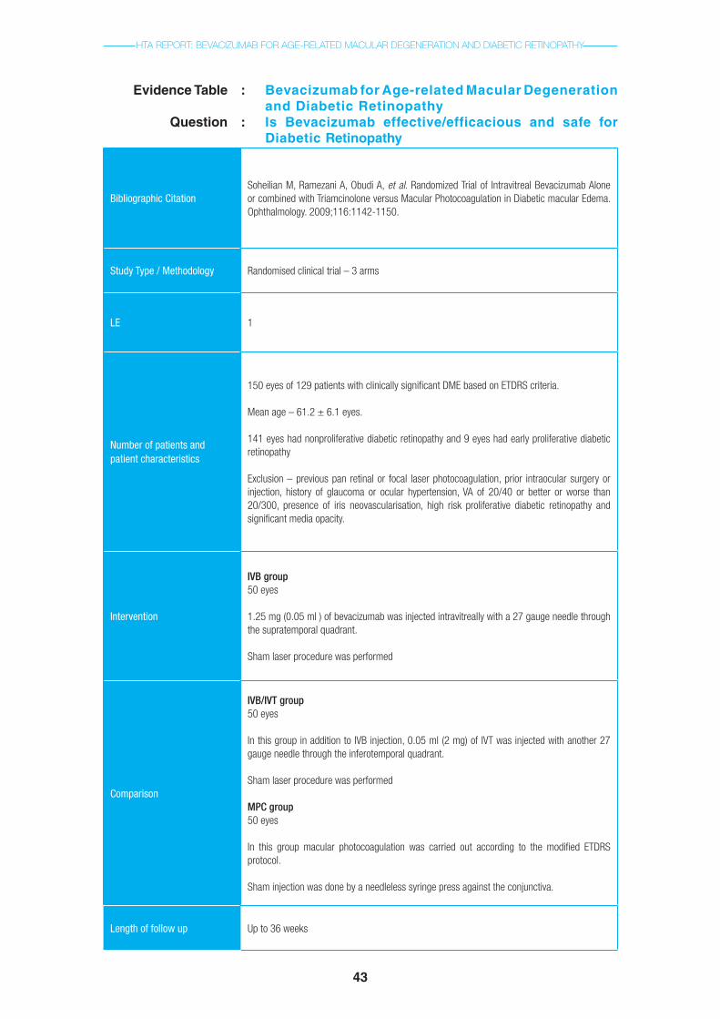
HTA REPORT: BEVACIZUMAB FOR AGE-RELATED MACULAR DEGENERATION AND DIABETIC RETINOPATHY
43
Evidence Table : Bevacizumab for Age-related Macular Degeneration and Diabetic Retinopathy
Question : Is Bevacizumab effective/efficacious and safe for Diabetic Retinopathy
Bibliographic CitationSoheilian M, Ramezani A, Obudi A, et al. Randomized Trial of Intravitreal Bevacizumab Alone or combined with Triamcinolone versus Macular Photocoagulation in Diabetic macular Edema. Ophthalmology. 2009;116:1142-1150.
Study Type / Methodology Randomised clinical trial – 3 arms
LE 1
Number of patients andpatient characteristics
150 eyes of 129 patients with clinically significant DME based on ETDRS criteria.
Mean age – 61.2 ± 6.1 eyes.
141 eyes had nonproliferative diabetic retinopathy and 9 eyes had early proliferative diabetic retinopathy
Exclusion – previous pan retinal or focal laser photocoagulation, prior intraocular surgery or injection, history of glaucoma or ocular hypertension, VA of 20/40 or better or worse than 20/300, presence of iris neovascularisation, high risk proliferative diabetic retinopathy and significant media opacity.
Intervention
IVB group50 eyes
1.25 mg (0.05 ml ) of bevacizumab was injected intravitreally with a 27 gauge needle through the supratemporal quadrant.
Sham laser procedure was performed
Comparison
IVB/IVT group50 eyes
In this group in addition to IVB injection, 0.05 ml (2 mg) of IVT was injected with another 27 gauge needle through the inferotemporal quadrant.
Sham laser procedure was performed
MPC group50 eyes
In this group macular photocoagulation was carried out according to the modified ETDRS protocol.
Sham injection was done by a needleless syringe press against the conjunctiva.
Length of follow up Up to 36 weeks
HTA REPORT: BEVACIZUMAB FOR AGE-RELATED MACULAR DEGENERATION AND DIABETIC RETINOPATHY
44
Outcome measures / Effect size
Retreatment was required for 27 eyes up to 36 weeks (14 eyes in the IVB group, 10 eyes in the
IVB/IVT group and 3 eyes in the MPC group)
Visual acuity
Compared to baseline, VA improvement was significant in the IVB groups at all follow-up visits
up to 36 weeks (p<0.001). In the IVB/IVT group, VA improved significantly only at weeks 6 and
12 (p=0.002 and 0.019 respectively). In the MPC group, VA changes was not significant.
Pairwise comparison between groups showed that the VA improvement at 6 weeks in both IVB
and IVB/IVT was greater than in the MPC group (p<0.001), no significance different between
the IVB and IVB/IVT groups. At 24 weeks, difference of VA changes between the IVB and MPC
groups was significant in favour of the IVB group (p=0.003). This difference between the IVB /
IVT and MPC groups was borderline (p=0.033). No significance different between IVB and IVB/
IVT (p=0.373). At 12 and 36 weeks, more VA improvement in the IVB group than in the other
groups though not to a meaningful level.
Snellen VA
Percentage of eyes with stable VA was relatively similar among the groups at all follow up.
Greater percentage of cases gained more than 2 Snellen VA in the IVB and IVB/IVT groups than
in the MPC groups. Greater percentage of eyes lost more than 2 Snellen lines in the MPC group
than in the other groups. These differences were statistically significant among the groups at
6, 12 and 24 weeks.
CMT
The reduction of CMT was significantly different among the treatment groups only in the
subgroups with the initial CMT ≥ 400 µm at all follow-up visits except week 24. At week 6,
CMT reduction was greater in the IVB group than in the other groups (p=0.026). At week 12 the
difference in CMT reduction was significant among all 3 groups in favour of the IVB/IVT group.
At week 36, the difference was significant between IVB and MPC group.
Safety
Transient anterior chamber reaction which resolved spontaneously observed in 10 (20%) and 9
(18%) eyes in the IVB and IVB/IVT groups respectively.
Ocular hypertension was detected in 8 eyes (16%) of the IVB/IVT group and was controlled in
all by medical therapy except in 1 eye that progressed to neovascular glaucoma.
Severe lens opacity developed in 5 eyes ( 4 in IVB/IVT group and 1 in MPC group) .
Retinal neovascularisation which resolved in all except 1 eye in the MPC group was observed
in 4,2 and 3 eyes in the IVB, IVB/IVT, and MPC groups respectively. 8 eyes developed early PDR
but remained stable.
10 eyes progressed to PDR and were excluded from the study.
General Comments
Jadad score = 5
Allocation concealment done.
Double blind
p-value < 0.029 = significant

HTA REPORT: BEVACIZUMAB FOR AGE-RELATED MACULAR DEGENERATION AND DIABETIC RETINOPATHY
45
Evidence Table : Bevacizumab for Age-related Macular Degeneration and Diabetic Retinopathy
Question : Is Bevacizumab effective/efficacious and safe for Diabetic Retinopathy
Bibliographic CitationDiabetic Retinopathy Clinical Research Network, Scott IU, Edwards AR, et al. A phase II randomized clinical trial of intravitreal bevacizumab for diabetic macular edema. Ophthalmology. 2007;114(10):1860-1867.
Study Type / MethodologyRandomized Phase 2 clinical trial(5 groups) – 19-24 subjects per groupMulticentre
LE 1
Number of patients andpatient characteristics
109 eyes of 109 subjects with DME and Snellen acuity equivalent ranging from 20/32-20/320.Median age – 65 years, 39% women
Intervention
Group BIntravitreal injection of 1.25 mg bevacizumab at baseline and 6 weeks
Group CIntravitreal injection of 2.5 mg bevacizumab at baseline and 6 weeks
Group DIntravitreal injection of 1.25 mg bevacizumab at baseline and sham injection at 6 weeks
Group EIntravitreal injection of 1.25 mg bevacizumab at baseline, focal photocoagulation at 3 weeks, and intravitreal injection of 1.25 mg bevacizumab at 6 weeks
ComparisonGroup AFocal photocoagulation at baseline
Length of follow up 24 weeks
Outcome measures / Effect size
Central Subfield ThicknessCompared with Group A, Group B and C both demonstrated a greater reduction in central subfield thickness at 3 weeks (p=0.006 and <0.001 respectively)
Visual AcuityGroups B and C both had about a median one line improvement at the 3-week visit which was sustained through 12 weeks and was greater than the change in visual acuity in Group A (p=0.01 and 0.003, respectively)
Reduction of central subfield thickness > 11%3 weeks – 23 of 60 (38%) in pooled 1.25 mg bevacizumab-treated eyes, 13 of 24 (54%) in 2.5 mg bevacizumab-treated eyes, and in 5 of 18 (28%) eyes treated with laser alone.6 weeks – 22 of 61 (36%) - in pooled 1.25 mg bevacizumab-treated eyes, 9 of 23 (39%) in 2.5 mg bevacizumab-treated eyes, and in 9 of 18 (50%) eyes treated with laser alone.Both the 1.25 mg and 2.5 mg bevacizumab-treated eyes had a greater reduction in central retinal thickness at 3 weeks.
- Eyes in the photocoagulation group demonstrated improvement in these parameter with longer follow up.
- 6 weeks may be too long for an optimal injection intervalSafetyEndophthalmitis – one subject after intravitreal injection2 patients who received at least one bevacizumab injection had myocardial infarction, one fatal. Both had a history of prior coronary artery by pass surgery. One episode of congestive heart failure occurred in a 56 year old woman who had a history of 3 prior similar episodes. 3 bevacizumab treated subjects experienced elevation of blood pressure (1 had history of hypertension).
General Comments
Jadad score = 3Phase 2 study to generate hypothesesSmall number of subjects in each group– No meaningful differences in central subfield thickness observed for bevacizumab relative to photocoagulation after the 3-week time point.

HTA REPORT: BEVACIZUMAB FOR AGE-RELATED MACULAR DEGENERATION AND DIABETIC RETINOPATHY
46
Evidence Table : Bevacizumab for Age-related Macular Degeneration and Diabetic Retinopathy
Question : Is Bevacizumab effective/efficacious and safe for Diabetic Retinopathy
Bibliographic Citation
Cheema RA, Al-Mubarak MM, Amin YA, Cheema MA. Role of combined cataract surgery and
intravitreal bevacizumab injection in preventing progression of diabetic retinopathy: prospective
randomized study. J Cataract Refract Surg. 2009;35:18-25.
Study Type / Methodology Randomised Controlled Trial
LE 1
Number of patients andpatient characteristics
68 eyes of 68 diabetic patients with sight limiting cataract. Presence of CSME, mild, moderate,
severe or very severe non-proliferative diabetic retinopathy or PDR or a combination of both
InterventionReceived intravitreal 1.25 mg in 0.05 ml bevacizumab through the pars plana into the vitreos
cavity at the end of surgery (standardized phacoemulsification with IOL implantation alone)
ComparisonDid not received intravitreal bevacizumab (only standardized phacoemulsification with IOL
implantation alone)
Length of follow up 6 months
Outcome measures / Effect size
Diabetic retinopathy progressed in 45.45% of eyes in the control group and 11.42% of eyes in
the intervention group. The difference between groups was statistically significant (p=0.002).
Progression of diabetic maculopathy occurred in 51.51% of eyes in the control group and
5.71% of eyes in the intervention group; The difference between groups was statistically
significant (p< 0.001).
There was no statistically significant difference in postoperative visual acuity and changes in
central macular thickness and average macular thickness over 6-month follow up at any time
point.
General Comments Jadad score = 1
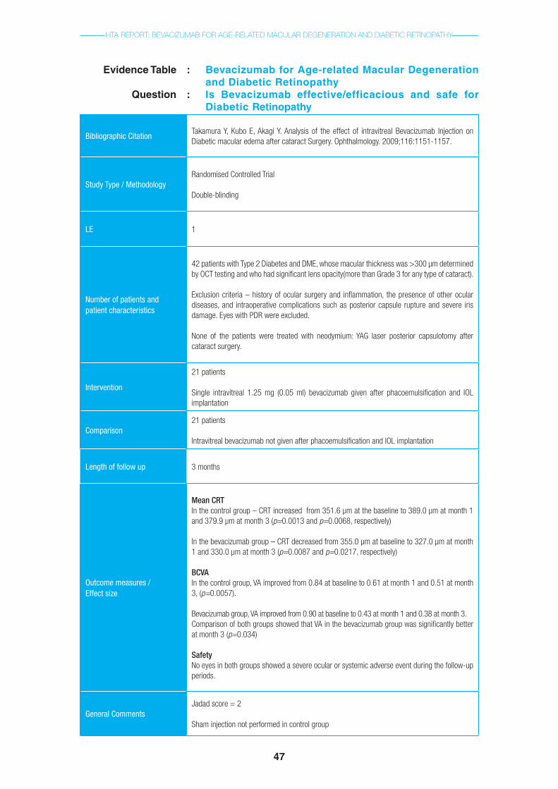
HTA REPORT: BEVACIZUMAB FOR AGE-RELATED MACULAR DEGENERATION AND DIABETIC RETINOPATHY
47
Evidence Table : Bevacizumab for Age-related Macular Degeneration and Diabetic Retinopathy
Question : Is Bevacizumab effective/efficacious and safe for Diabetic Retinopathy
Bibliographic CitationTakamura Y, Kubo E, Akagi Y. Analysis of the effect of intravitreal Bevacizumab Injection on Diabetic macular edema after cataract Surgery. Ophthalmology. 2009;116:1151-1157.
Study Type / MethodologyRandomised Controlled Trial
Double-blinding
LE 1
Number of patients andpatient characteristics
42 patients with Type 2 Diabetes and DME, whose macular thickness was >300 µm determined by OCT testing and who had significant lens opacity(more than Grade 3 for any type of cataract).
Exclusion criteria – history of ocular surgery and inflammation, the presence of other ocular diseases, and intraoperative complications such as posterior capsule rupture and severe iris damage. Eyes with PDR were excluded.
None of the patients were treated with neodymium: YAG laser posterior capsulotomy after cataract surgery.
Intervention
21 patients
Single intravitreal 1.25 mg (0.05 ml) bevacizumab given after phacoemulsification and IOL implantation
Comparison21 patients
Intravitreal bevacizumab not given after phacoemulsification and IOL implantation
Length of follow up 3 months
Outcome measures / Effect size
Mean CRTIn the control group – CRT increased from 351.6 µm at the baseline to 389.0 µm at month 1 and 379.9 µm at month 3 (p=0.0013 and p=0.0068, respectively)
In the bevacizumab group – CRT decreased from 355.0 µm at baseline to 327.0 µm at month 1 and 330.0 µm at month 3 (p=0.0087 and p=0.0217, respectively)
BCVAIn the control group, VA improved from 0.84 at baseline to 0.61 at month 1 and 0.51 at month 3, (p=0.0057).
Bevacizumab group, VA improved from 0.90 at baseline to 0.43 at month 1 and 0.38 at month 3.Comparison of both groups showed that VA in the bevacizumab group was significantly better at month 3 (p=0.034)
SafetyNo eyes in both groups showed a severe ocular or systemic adverse event during the follow-up periods.
General CommentsJadad score = 2
Sham injection not performed in control group

HTA REPORT: BEVACIZUMAB FOR AGE-RELATED MACULAR DEGENERATION AND DIABETIC RETINOPATHY
48
Evidence Table : Bevacizumab for Age-related Macular Degeneration and Diabetic Retinopathy
Question : Is Bevacizumab effective/efficacious and safe for Diabetic Retinopathy
Bibliographic CitationTonello M, Costa RA, Almeida FPP, et al. Panretinal photocoagulation versus PRP plus intravitreal bevacizumab for high-risk proliferative diabetic retinopathy. Acta Ophthalmologica. 2008;86:385-389.
Study Type / Methodology Randomised controlled trial
LE 1
Number of patients andpatient characteristics
30 eyes (22 patients)- 14 patients with unilateral high risk PDR
Were randomly assigned to received PRP or PRP plus intravitreal bevacizumab- 8 patients with bilateral high risk PDR
The eye with worse BCVA was included in the PRP-plus group and the fellow eyes in the PRP group.
Intervention
PRP plus group
15 eyes
All patients underwent PRP performed at two time-points (at week 1 and week 3)
One intravitreal injection of 1.5 mg (0.06 ml) bevacizumab was administered ~ 60 mins after the completion of the second PRP session (week 3) by a single retinal specialist.
Comparison
PRP group
15 eyes
All patients underwent PRP performed at two time-points (at week 1 and week 3)
Length of follow up 16 weeks
Outcome measures / Effect size
logMAR BCVA
There was no difference in BCVA between the two groups at different time-points during the study period.There was no significant change in BCVA from baseline at any study period in either group.
Total area of fluorescein leakage from active NVs
Significant reduction in the total area of leakage from active NVs in the PRP-plus group at weeks 4,9 and 16 compared with the PRP group.
Significant reduction in the total area of leakage from active NVs inthe PRP-plus group at weeks 4, 9 and 16 compared with baseline. In the PRP group, changes from baseline in the total area of leakage from active NVs were not significant at any time-point.
SafetyNo serious adverse events were observed in the 15 eyes treated with bevacizumab in this study.Minor local adverse events related to treatment procedure such as conjunctival haemorrhage and foreign body sensation were reported in seven and two patients respectively. These evevts were transient and resolved by one week after injection.
General Comments
Jadad score = 2
No blindingNot all patients randomized.
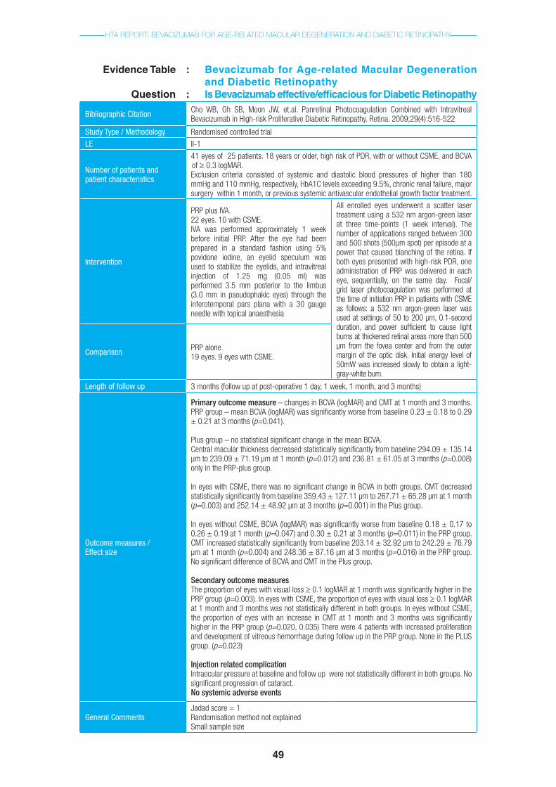
HTA REPORT: BEVACIZUMAB FOR AGE-RELATED MACULAR DEGENERATION AND DIABETIC RETINOPATHY
49
Evidence Table : Bevacizumab for Age-related Macular Degeneration and Diabetic Retinopathy
Question : Is Bevacizumab effective/efficacious for Diabetic Retinopathy
Bibliographic Citation Cho WB, Oh SB, Moon JW, et.al. Panretinal Photocoagulation Combined with Intravitreal Bevacizumab in High-risk Proliferative Diabetic Retinopathy. Retina. 2009;29(4):516-522
Study Type / Methodology Randomised controlled trial
LE II-1
Number of patients andpatient characteristics
41 eyes of 25 patients. 18 years or older, high risk of PDR, with or without CSME, and BCVA of ≥ 0.3 logMAR.Exclusion criteria consisted of systemic and diastolic blood pressures of higher than 180 mmHg and 110 mmHg, respectively, HbA1C levels exceeding 9.5%, chronic renal failure, major surgery within 1 month, or previous systemic antivascular endothelial growth factor treatment.
Intervention
PRP plus IVA. 22 eyes. 10 with CSME.IVA was performed approximately 1 week before initial PRP. After the eye had been prepared in a standard fashion using 5% povidone iodine, an eyelid speculum was used to stabilize the eyelids, and intravitreal injection of 1.25 mg (0.05 ml) was performed 3.5 mm posterior to the limbus (3.0 mm in pseudophakic eyes) through the inferotemporal pars plana with a 30 gauge needle with topical anaesthesia
All enrolled eyes underwent a scatter laser treatment using a 532 nm argon-green laser at three time-points (1 week interval). The number of applications ranged between 300 and 500 shots (500µm spot) per episode at a power that caused blanching of the retina. If both eyes presented with high-risk PDR, one administration of PRP was delivered in each eye, sequentially, on the same day. Focal/grid laser photocoagulation was performed at the time of initiation PRP in patients with CSME as follows: a 532 nm argon-green laser was used at settings of 50 to 200 µm, 0.1-second duration, and power sufficient to cause light burns at thickened retinal areas more than 500 µm from the fovea center and from the outer margin of the optic disk. Initial energy level of 50mW was increased slowly to obtain a light-gray-white burn.
Comparison PRP alone. 19 eyes. 9 eyes with CSME.
Length of follow up 3 months (follow up at post-operative 1 day, 1 week, 1 month, and 3 months)
Outcome measures / Effect size
Primary outcome measure – changes in BCVA (logMAR) and CMT at 1 month and 3 months.PRP group – mean BCVA (logMAR) was significantly worse from baseline 0.23 ± 0.18 to 0.29 ± 0.21 at 3 months (p=0.041).
Plus group – no statistical significant change in the mean BCVA.Central macular thickness decreased statistically significantly from baseline 294.09 ± 135.14 µm to 239.09 ± 71.19 µm at 1 month (p=0.012) and 236.81 ± 61.05 at 3 months (p=0.008) only in the PRP-plus group.
In eyes with CSME, there was no significant change in BCVA in both groups. CMT decreased statistically significantly from baseline 359.43 ± 127.11 µm to 267.71 ± 65.28 µm at 1 month (p=0.003) and 252.14 ± 48.92 µm at 3 months (p=0.001) in the Plus group.
In eyes without CSME, BCVA (logMAR) was significantly worse from baseline 0.18 ± 0.17 to 0.26 ± 0.19 at 1 month (p=0.047) and 0.30 ± 0.21 at 3 months (p=0.011) in the PRP group. CMT increased statistically significantly from baseline 203.14 ± 32.92 µm to 242.29 ± 76.79 µm at 1 month (p=0.004) and 248.36 ± 87.16 µm at 3 months (p=0.016) in the PRP group. No significant difference of BCVA and CMT in the Plus group.
Secondary outcome measuresThe proportion of eyes with visual loss ≥ 0.1 logMAR at 1 month was significantly higher in the PRP group (p=0.003). In eyes with CSME, the proportion of eyes with visual loss ≥ 0.1 logMAR at 1 month and 3 months was not statistically different in both groups. In eyes without CSME, the proportion of eyes with an increase in CMT at 1 month and 3 months was significantly higher in the PRP group (p=0.020, 0.035) There were 4 patients with increased proliferation and development of vitreous hemorrhage during follow up in the PRP group. None in the PLUS group. (p=0.023)
Injection related complicationIntraocular pressure at baseline and follow up were not statistically different in both groups. No significant progression of cataract.No systemic adverse events
General CommentsJadad score = 1 Randomisation method not explainedSmall sample size

HTA REPORT: BEVACIZUMAB FOR AGE-RELATED MACULAR DEGENERATION AND DIABETIC RETINOPATHY
50
Evidence Table : Bevacizumab for Age-related Macular Degeneration and Diabetic Retinopathy
Question : Is Bevacizumab effective/efficacious for Diabetic Retinopathy
Bibliographic CitationMason JO, Yunker JJ, Vail R, et.al. Intravitreal Bevacizumab (Avastin) Prevention of Panretinal Photocoagulation-induced Complications in Patients with Severe Proliferative Diabetic Retinopathy. Retina. 2008;28:1319-1324.
Study Type / Methodology Retrospective cohort study
LE II-2
Number of patients andpatient characteristics
60 consecutive eyes of 30 diabetic patients with severe PDR whose visual acuity was 20/30 or better and whose FT was 280 µm or less. No eyes had CSDME and all had PDR with high risk characteristics. No eyes had a history of prior PRP treatment.Ages ranged from 26 to 63 years . with a mean of 47.7 ± 12.5 years.8 eyes in each group had undergone cataract surgery with intraocular lens implantation, whereas the remaining 44 eyes were phakic.
Intervention
30 eyes (15 patients).1 week before initiation of PRP treatment, 1.25 mg of bevacizumab (Avastin) in 0.05 mL was injected intravitreally to all eyes. IVB was administered under topical anaesthesia using a 30 gauge needle to the inferior pars plana 4 mm from the corneal limbus.
Povidone-iodine solution (5%) was used before injection to cleanse the ocular conjunctival surface and fornices, and each eye was treated with topical gatifloxacin 0.3% ophthalmic drops every 2 hours for two days after the injection. PRP was then performed at 1 week and at 5 weeks after injection. Best corrected visual acuity and Ft were measured before injection and PRP, and during all follow-up examinations.
In all eyes , the PRP treatment was designed to be completed in two sessions. The superior nasal and inferior nasal periphery was treated in the first session and the superior-temporal and inferior-temporal peripheral areas were treated in the second session.The laser spot size on the retina was between 200 µm with duration of 0.7 seconds and power ranging from 200 mJ to 400 mJ. The number of spots in each session was approximately 900 (range 650-1,000). The total number of laser spots after both sessions was approximately 1,800 (range 1,600-2,100) and did not differ among groups. An Iris 532 nm yellow-green diode laser was used to perform all PRP treatments under topical anaesthesia in an office setting.
Comparison
30 eyes (15 patients) were treated in the standard fashion with PRP in two divided sessions.
Length of follow up 24 weeks
Outcome measures / Effect size
FT changes 4 weeks after the first PRPIVB group – decreased from 278.8 ± 29.5 µm to 252.7 ± 31.8 µm.Control group – increased from 273.5 ± 27.7 µm to a mean of 321.2 ± 57.7 µm.
12 weeks after PRPIVB group – 257.2 ± 31.8 µmControl group – 307.3 ± 50.5 µm.
24 weeks after PRPIVB group – 264.3 ± 30.1 µmControl group – 298.2 ± 44.9 µm.
Mean BCVAIVB group – improved from 0.073 ± 0.071 at baseline to 0.039 ± 0.054 at 24 weeks after PRP.Control group – decreased to 0.165 ± 0.116 at 12 weeks and 0.149 ± 0.114 at 24 weeks.(p≤0.0001)
Seven eyes in the control group had worse vision by ≥ 2 lines at 24 weeks, whereas none of the eyes in the IVB group had worse vision (p=0.011). In addition, seven eyes had increased FT by ≥ 50 µm at 24 weeks whereas none of the IVB group eyes developed a significant increase in FT. (p=0.011)
SafetyNo ocular or systemic complications were seen in any patients in the IVB group. None of the eyes had a change in intraocular pressure or lens status. None received focal laser or additional PRP during the follow-up period. No eyes developed endophthalmitis, retinal detachment, preretinal hemorrhage, neovascular glaucoma or vitreous hemorrhage.
General Comments

HTA REPORT: BEVACIZUMAB FOR AGE-RELATED MACULAR DEGENERATION AND DIABETIC RETINOPATHY
51
Evidence Table : Bevacizumab for Age-related Macular Degeneration and Diabetic Retinopathy
Question : Is Bevacizumab effective/efficacious for Diabetic Retinopathy
Bibliographic CitationLucena D da R, Ribeiro JAS, Costa RA, et al. Intraoperative bleeding during vitrectomy for diabetic tractional retinal detachment with versus without preoperative intravitreal bevacizumab (IBeTra study). Br J Ophthalmol. 2009;93:688-691.
Study Type / Methodology Randomised Controlled Trial
LE 1
Number of patients andpatient characteristics
20 Patients with PDR, macula involving TRD and absence of clinically evident vitreous or preretinal haemorrhage
Exclusion criteria – previous history of thromboembolic event, haemodialysis, known coagulation disorders or receiving anticoagulants other than aspirin, history of glaucoma without clinical treatment, previous intraocular surgery other than cataract surgery, clinically evident vitreous or preretinal haemorrhage and any other clinical condition that impeded the examination of the ocular fundus or patient follow up.
Intervention
Pars plana vitrectomy with preoperative intravitreal bevacizumab injection
10 patients
Patients received an intravitreal bevacizumab 1.5 mg (0.06 ml) one week after baseline and were schedules for a 23 gauge pars plana vitrectomy 3 weeks after baseline.
Comparison
Pars plana vitrectomy without preoperative intravitreal bevacizumab injection
10 patients
Patients were scheduled for a 23 gauge pars plana vitrectomy 3 weeks after baseline but no intravitreal injection procedure performed
Length of follow up Until vitrectomy performed
Outcome measures / Effect size
The mean erythrocyte count retrieved from the vitrectomy cassette was 14865 x 103 (SD 19332 x 103) cells for patients in the BEV/PPV group, and 176240 x 103(SD 108375 x 103) cells for patients in the PPV group. The mean erythrocyte count was significantly lower in the BEV/PPV group than in the PPV group.(p<0.0001)
Safety – no local or systemic adverse events were reported
General Comments
Jadad score = 2
Single blinded
Small sample size

HTA REPORT: BEVACIZUMAB FOR AGE-RELATED MACULAR DEGENERATION AND DIABETIC RETINOPATHY
52
Evidence Table : Bevacizumab for Age-related Macular Degeneration and Diabetic Retinopathy
Question : Is Bevacizumab effective/efficacious for Diabetic Retinopathy
Bibliographic CitationYeh PT, Yang CM, Lin YC, et.al. Bevacizumab Pretreatment in vitrectomy with silicone oil for severe diabetic retinopathy. Retina. 2009;29:768-774
Study Type / Methodology Non-randomized clinical trial
LE II-2
Number of patients andpatient characteristics
20 eyes of 19 patients in Group 1 and 21 eyes of 20 patients in Group 2.Consecutive patients undergoing primary pars plana vitrectomy with silicone oil infusion for complications of active PDR were recruited. Cases were alternately divided into Group 1 and Group 2. Mean age 49.4 ± 7.9 in Group 1 and 51.0 ± 7.7 in Group 2.
Exclusion – Patients with pre or postoperative poor diabetes control, antiplatelet or anticoagulant therapy or blood diseases associated with abnormal coagulation
Intervention
Received an intravitreal injection of 1.25 mg bevacizumab (0.05 ml 7 days to 9 days before vitrectomy
Anteroposterior traction release was attempted first, followed by fibrovascular tissue removal with delamination as the principle technique. Hemostasis was achieved by raising the infusion bottle, applying mechanical compression with a soft-tipped canula, endodiathermy, or in combination of the above techniques. Blood clots that formed during tissue dissection were removed carefully except close to bleeding sites where they were trimmed to small islands. Supplementary panretinal photocoagulation extending beyond the equator and peripheral cryotherapy were performed. After fluid-air exchange, internal drainage of subretinal fluid was performed through pre-existing or iatrogenic breaks followed by silicone oil (5,000 cs) infusion. In certain cases, a 360˚ encircling buckle was placed to counter possible residual peripheral vitreous traction.
ComparisonDid not received bevacizumab pretreatment
Length of follow up At least 6 months after surgery
Outcome measures / Effect size
One week after intravitreal bevacizumab injection, all cases in group 1 showed decreased visible neovascularisation with fibrovascular tissue showing a fibrous tissue-like appearance.
Intraoperative bleeding was more severe in group 2 compared to group 1 (p < 0.01) where in group 1 90% of the eyes had grade 1 intraoperative bleeding, 10% had Grade 2 and 0% Grade 3.
In Group 2, 23.8% had Grade 1 intraop bleeding, 71.4% had Grade 2 bleeding and 4.8% had Grade 3 bleeding. Subretinal hemorrhage was identified during surgery in 9 cases in Group 1 and in 1 case in Group 2 (p=0.004).
Early recurrent vitreous hemorrhage occurred in 5 eyes in Group 1 and in 4 eyes in Group 2 (p=0.72). Postoperative preretinal bleeding and the final blood reabsorption time (blood completely reabsorped within the posterior pole and around the disk area were significantly different between Groups 1 and 2 (p=0.007 and < 0.001, respectively)
A transient (< 1 week) increase in intraocular pressure (>25 mmHg) occurred in 12 patients in each group.
Multiple linear regression analysis indicated that the likelihood of intraopearative bleeding was decreased by bevacizumab pretreatment (p< 0.01) but increased by hypertension (p=0.03). Intravitreal injection of bevacizumab decreased the grade of postoperative preretinal bleeding (p=0.04).
General Comments Small sample size

HTA REPORT: BEVACIZUMAB FOR AGE-RELATED MACULAR DEGENERATION AND DIABETIC RETINOPATHY
53
Evidence Table : Bevacizumab for Age-related Macular Degeneration and Diabetic Retinopathy
Question : Is Bevacizumab effective/efficacious for Diabetic Retinopathy
Bibliographic Citation
Arimura N, Otsuka H, Yamakiri K, et al. Vitreous mediators after Intravitreal Bevacizumab or
Triamcinolone Acetonide in Eyes with proliferative Diabetic Retinopathy. Ophthalmology.
2009;116:921-926.
Study Type / Methodology Non-randomised clinical trial
LE II-2
Number of patients andpatient characteristics
47 eyes of 47 patients with type 2 diabetes who were affected by PDR were enrolled in this
study.
Exclusion- eyes with massive vitreous hemorrhage (in which the optic disc was invisible) or
apparent tractional retinal detachment
Intervention
19 consecutive patients receiving preoperative intravitreal bevacizumab. Bevacizumab 1.25 mg
(0.05 ml) was injected into the vitreous cavity as preoperative adjunctive therapy 7 days before
vitrectomy.
Comparison
1. An historical control of 18 consecutive patients received vitrectomy without preoperative
therapy
2. 10 consecutive patients receiving preoperative intravitreal TA. TA 4mg (0.1 ml) was injected
into the vitreous cavity as preoperative adjunctive therapy 7 days before vitrectomy.
Length of follow up Until post –operative
Outcome measures / Effect size
Significance differences among the 3 groups were observed for the levels of VEGF and SDf-1a
(p<0.001 and p=0.010, respectively)
The median of VEGF levels in the Bevacizumab group was 0 (0-79.2), TA group 343.5 (0-
1683.3) and control 1202.5 (76-4213.9).
The median of SDf-1a in the Bevacizumab group was 149.2 (0-519.4), TA group 87.5 (0-
252.5) and control 245.7 (0-856.8)
General Comments Jadad score = 0

HTA REPORT: BEVACIZUMAB FOR AGE-RELATED MACULAR DEGENERATION AND DIABETIC RETINOPATHY
54
Evidence Table : Bevacizumab for Age-related Macular Degeneration and Diabetic Retinopathy
Question : Is Bevacizumab effective/efficacious for Diabetic Retinopathy
Bibliographic CitationLo WR, Kim SJ, Aaberg TM, et al. Visual outcomes and incidence of Recurrent Vitreous hemorrhage after vitrectomy in diabetic eyes pretreated with bevacizumab. Retina. 2009;29:926-931.
Study Type / Methodology Retrospective cohort study
LE II-2
Number of patients andpatient characteristics
137 diabetic eyes undergoing surgery for non clearing vitreous hemorrhage (VH) or tractional retinal detachment with or without rhegmatogenous component were included for analysis.
Eyes undergoing PPV for reasons unrelated to complications from PDR were excluded.
Exclusion criteria – intraocular surgery of any type within the prior 3 months, treatment with bevacizumab or other anti-VEGF agents > 1 month before surgery, use of silicone oil (SO), reoperations on eyes with prior diabetic vitrectomy, fellow eyes of the same patient, history of filtering surgery for neovascular glaucoma and < 6 weeks of follow up.
Intervention
Bevacizumab group. Received an injection of 1.25 mg of bevacizumab< 1 month before surgery. 33 eyes (24% of total) received preoperative bevacizumab; 5 eyes had predominantly VH and 28 eyes had tractional
Comparison
Control104 eyes underwent vitrectomy without pretreatment.
50 eyes had predominately VH and 54 eyes had tractional retinal detachment.
Length of follow up 6 months
Outcome measures / Effect size
Early postoperative Outcome (4-6 weeks) by treatment group- Non significance difference in the incidence or severity of VH between the two
groups- 5 patients in the bevacizumab group ( 1 required repeat surgery) and 13 patients in
the untreated group (4 required repeat surgery)- 3 patients in the bevacizumab group and two patients in untreated group had post-
operative retinal detachment
Late post-operative haemorrhage - No additional VH occurred i patients in either group between 1month to 3 months- Only 1 eye of 19 with sufficient follow-up in the bevacizumab group experienced a
rebleed (at 5.3 months)- Only 5 eyes in the untreated group experienced late postoperative VH (3.1-7.7
range, average 5.0 months)
There was no difference between groups at any time point in visual acuity.
General Comments.

HTA REPORT: BEVACIZUMAB FOR AGE-RELATED MACULAR DEGENERATION AND DIABETIC RETINOPATHY
55
Evidence Table : Bevacizumab for Age-related Macular Degeneration and Diabetic Retinopathy
Question : Is Bevacizumab effective/efficacious for Diabetic Retinopathy
Bibliographic Citation
Paccola L, Costa RA, Folgosa MS, et.al. Intravireal triamcinolone versus bevacizumab for
treatment of refractory diabetic macular oedema (IBEME study). Br J Ophthalmol. 2008;92:
76-80
Study Type / Methodology Randomised controlled trial
LE 1
Number of patients andpatient characteristics
28 patients with refractory DMO in at least one eye, log MAR ETDS BCVA of 0.3 or worse and
CMT greater than 300 µm on OCT.
Exclusion criteria were aphakic or pseudophakic eyes, glycosylated haemoglobin level above
10%, history of glaucoma or ocular hypertension, an ocular condition other than diabetic,
systemic corticosteroid therapy, history of thromboembolic event, major surgery, uncontrolled
hypertension, known coagulation abnormalities and any condition affecting follow up or
documentation.
InterventionIBe group
Receive intravitreal 1.5mg /0.06 ml bevacizumab within 1 week of baseline
Comparison
ITA group
Receive intravitreal 4 mg/0.1 ml triamcinolone acetonide within 1 week of baseline
Length of follow up 24 weeks
Outcome measures / Effect size
26 patients completed the 24-week study period.
Significant reduction (p<0.01) in CMT in the ITA group at weeks 4 (p=0.008), 8 (p=0.006), 12
(p<0.0001), and 24 (p=0.024) compared with the IBe group. Separate within group analysis
showed significant changes (reductions) in CMT values from baseline at weeks 4, 8 and 12
(p<0.0001) in the ITA group, and at weeks 4 (p<0.0001) and 8 (p=0.0004) in the IBe group.
Significant improvement in BCVA in the ITA group compared with the IBe group at weeks 8
(p=0.026) and 12 (p=0.039). Separate within group analysis showed significant improvement
in BCVA from baseline at weeks 4, 8 and 12 (p<0.001) in the ITA group, and at week 4
(p=0.011) in the IBe group.
There was a significant increase in IOP in the ITA group compared with the IBe group at weeks
1 (p<0.0001), 4 (p<0.0001) and 12 (p=0.043). Separate within group analysis revealed a
significant increase in IOP from baseline in the ITA group at week 4 (p<0.0001); in the IBe
group no significant change in IOP was observed at any study visit. During the 24 week study
period, no cataract progression was observed in either the ITa or IBe group. No systemic or
severe adverse event was observed throughout the study.
General Comments
Jadad score = 1
ITT analysis not done.
Randomisation method not explained.

HTA REPORT: BEVACIZUMAB FOR AGE-RELATED MACULAR DEGENERATION AND DIABETIC RETINOPATHY
56
Evidence Table : Bevacizumab for Age-related Macular Degeneration and Diabetic Retinopathy
Question : Is Bevacizumab effective/efficacious for Diabetic Retinopathy
Bibliographic Citation
Arevalo JF, Sanchez JG, Wu L, et al. Primary intravitreal bevacizumab for diffuse diabetic macular
edema: the Pan-American collaborative Retina Study Group at 24 months. Ophthalmology.
2009;116(8):1488-1497.
Study Type / Methodology Multicenter retrospective cohort study
LE II-2
Number of patients andpatient characteristics
115 consecutive patients with diffuse diabetic macular edema
InterventionIntravitreal bevacizumab 1.25mg (0.05 ml) given through the inferotemporal pars plana with a
30-gauge needle under topical anaesthesia or subconjunctival lidocaine.
ComparisonIntravitreal bevacizumab 2.5 mg (0.1 ml) given through the inferotemporal pars plana with a
30-gauge needle under topical anaesthesia or subconjunctival lidocaine
Length of follow up 24 months
Outcome measures / Effect size
Within 1 month after the initial bevacizumab injection, improvements in BCVA and CMT
measurements were observed, and these significant changes continued throughout the
24-month follow up.
Subgroup analysis demonstrated that 62 (44.6%) eyes remained stable, 72 (51.8%) eyes
improved 2 or more ETDRS lines of BCVA and 5 (3.6%) eyes decreased 2 or more ETDRS lines
of BCVA.
No statistical significance differences in changes of BCVA between doses of 1.25 and 2.5 mg of
IVB were observed. There were no statistical significance differences in macular thickness with
OCT were observed between doses of 1.25 and 2.5 mg of IVB.
Safety – Adverse events included transient high blood pressure in 1 patient (0.9%),
cerebrovascular accident in 1 patient (0.9%), heart attack in in 1 patient (0.9%), transient
increased intraocular pressure in 7 patients (5%), cataract in 5 patients(3.6%), and tractional
retinal detachment in 1 eye (0.7%).
General Comments

HTA REPORT: BEVACIZUMAB FOR AGE-RELATED MACULAR DEGENERATION AND DIABETIC RETINOPATHY
57
Evidence Table : Bevacizumab for Age-related Macular Degeneration and Diabetic Retinopathy
Question : Is Bevacizumab cost-effective for Age-related Macular Degeneration
Bibliographic citation
Raftery J, Clegg A, Jones J, et al. Ranibizumab (Lucentis) versus bevacizumab (Avastin):
modelling cost effectiveness. Br J Ophthalmol 2007;91:1244-1246.
Study Type / Methodology Cost-effectiveness study
LE I
Methodology
A Markov model was developed. Data for ranibizumab was based on ANCHOR trial, MARINA
trial and also trials by Brown DM et al. and Bressler NM et al. For bevacizumab, a range for its
efficacy relative to ranibizumab was employed.
The model had six health states, five defined by visual acuity plus a death state. Transition
probabilities for vision loss and vision gain were based on the relevant ranibizumab trial. Utilities
for each health state were based on a study linking visual acuity and utilities.
Patients entered the model at 75 years of age with follow-up to 85 or death. They starte in the
second least severe state to allow improvement. Two groups of patients were modelled, those
gaining and those losing visual acuity, based on the licensing trials. Treatment was continued
monthly as in licensing trials for 1 year in predominantly classic and 2 years in minimally classic/
occult classic. After treatment, disease progression for untreated patients was applied. The
most severe states had an annual cost based on the cost of near-blindness. Patient mortality
reflected UK averages for the relevant ages with a 50% increased mortality risk assumed for
the more severe health states. The model was simulated for 1000 patients in 3-month cycles.
The price ratio of ranibizumab to bevacizumab was 39:1, based on the US price of US$1950
(1025) per injection for ranibizumab and (a high) US$50 (26) for bevacizumab. NHS costs were
estimated for drug administration and monitoring of patients as well as treatment of side effects
using NHS Reference Costs. The eeficacy of bevacizumab relative to ranibizumab was varied
from 0.1 to 0.9. Both cost and utilities were discounted at an annual rate of 3.5%.
Length of follow up
Outcome measures/ Effect size
Efficacy of bevacizumab relative to ranibizumab in predominantly classic AMd would have to
be low for the latter to achieve an acceptable level of cost effectiveness. Only when relative
efficacy was reduced to 0.4 did the cost per QALY fall to 31092. At 0.8 the cost per QALY was
well over 100,000.
Doubling event rates for serious ocukar adverse effects for bevacizumab had minimal (<1%)
effect on the cost per QALY. Reducing the price ratio improved the cost per QALY.
Similar results applied to minimally classic and occult AMD, which differs only in being less
favourable to ranibizumab due mainly to 2 years of treatment being required.
General comments

HTA REPORT: BEVACIZUMAB FOR AGE-RELATED MACULAR DEGENERATION AND DIABETIC RETINOPATHY
58
APPENDIX 5
LIST OF EXCLUDED STUDIES
1. Kaiser PK. Verteporfin Photodynamic Therapy Combined with intravitreal bevacizumab for neovascular age-related macular degeneration. Ophthalmology. 2009;116:747-755.
2. Kook D, Wolf A, Kreutzer T, et al. Long term effect of intravitreal Bevacizumab (Avastin) in patients with chronic diffuse diabetic macular edema. Retina. 2008;28:1053-1060.
3. Haritoglou C, Kook D, Neubauer A, et al. Intravitreal Bevacizumab (Avastin) Therapy for Persistent Diffuse Diabetic Macular Edema. Retina. 2006;26:999-1005.
4. Shah CP, Hsu J, Garg SJ, et al. Retinal Pigment Epithelial Tear After Intravitreal Bevacizumab Injection. Am J Ophthalmology. 2006;142:1070-1071.
5. Spandau UHM, Jonas JB. Retinal Pigment Epithelium Tear after Intravitreal Bevacizumab for Exudative Age-related Macular Degeneration. Am J Ophthalmology. 2006;142:1068-1070.
6. Stifter E, Michels S, Prager F, et al. Intravitreal Bevacizumab Therapy for neovascular Age-related macular Degeneration with large submacular hemorrhage. Am J Ophthalmology. 2007;144:886-892.
7. Wickremasinghe SS, Michalova K, Gilhotra J, et al. Acute Intraocular Inflammation Injections of Bevacizumab for Treatment of neovascular age-related macular degeneration. Ophthalmology. 2008;115:1911-1915.
8. Bashshur ZF, Haddad ZA, Schakal AR, et al. Intravitreal Bevacizumab for Treatment of Neovascular Age-related macular Degeneration: A one-year Prospective Study. Am J Ophthalmology. 2008;145:249-256.
9. Chen CY, Wong TY, Heriot WJ. Intravitreal Bevacizumab (Avastin) for Neovascular Age-related macular Degeneration: A short-term study. Am J Ophthalmol. 2007;143:510-512.
10. Andreoli CM, Miller JW. Anti-vascular endothelial growth factor therapy for ocular neovascular disease. Current Opinion in Ophthalmology. 2007;18:502-508.
11. Forooghian F, Cukras C, Meyerle CB, et al. Tachyphylaxis after intravitreal bevacizumab for exudative age-related macular degeneration. Retina. 2009;29: 723-731.
12. Costa RA, Jorge R, Calucci D et al. Intravitreal Bevacizumab (Avastin) for Central and Hemicentral Retinal Vein Occlusions. Retina. 2007;27:141-149.
13. Atul K, subijay S. Intravitreal bevacizumab (Avastin) treatment of diffuse diabetic macular edema in an Indian population. Indian J Ophthalmol. 2007;55:451-455.
14. Torres-Soriano ME, Reyna-Castelan E, Hernandez-Rojas M, et al. Tractional Retinal Detachment After Intravitreal Injection of bevacizumab in Proliferative Diabetic Retinopathy. Retinal Cases & Brief Repots. 2009;3(1):70-73.
15. Bonini-Filho M, Costa RA, Calucci D, et al. Intravitreal Bevacizumab for Diabetic Macular Edema Associated with Severe Capillary Loss: One-Year Results of a Pilot study. American Journal of Ophthalmology. 2009;147:1022-1030.
16. Avery RL, Pieramici DJ, Rabena MD, et al. Intravitreal bevacizumab (Avastin) for neovascular age-related macular oedema. Ophthalmology. 2006:113;363-372.
17. Kaiser PK. Verteporfin Photodynamic Therapy Combined with intravitreal bevacizumab for neovascular age related macular degeneration. Ophthalmology. 2009;116:747-755.
18. Kook D, Wolf A, Kreutzer T, et al. Long term effect of intravitreal Bevacizumab (Avastin) in patients with chronic diffuse diabetic macular edema. Retina. 2008;28:1053-1060.
19. Haritoglou C, Kook D, Neubauer A, et al. Intravitreal Bevacizumab (Avastin) Therapy for Persistent Diffuse Diabetic Macular Edema. Retina. 2006;26:999-1005.
20. Shah CP, Hsu J, Garg SJ, et al. Retinal Pigment Epithelial Tear After Intravitreal Bevacizumab Injection. Am J Ophthalmology. 2006;142:1070-1071.
21. Spandau UHM, Jonas JB. Retinal Pigment Epithelium Tear after Intravitreal Bevacizumab for Exudative Age-related Macular Degeneration. Am J Ophthalmology. 2006;142:1068-1070.
22. Stifter E, Michels S, Prager F, et al. Intravitreal Bevacizumab Therapy for neovascular Age-related macular Degeneration with large submacular hemorrhage. Am J Ophthalmology. 2007;144:886-892.
23. Wickremasinghe SS, Michalova K, Gilhotra J, et al. Acute Intraocular Inflammation Injections of Bevacizumab for Treatment of neovascular age-related macular degeneration. Ophthalmology. 2008;115:1911-1915.
24. Bashshur ZF, Haddad ZA, Schakal AR, et al. Intravitreal Bevacizumab for Treatment of Neovascular Age-related macular Degeneration: A one-year Prospective Study. Am J Ophthalmology. 2008;145:249-256.
25. Chen CY, Wong TY, Heriot WJ. Intravitreal Bevacizumab (Avastin) for Neovascular Age-related macular Degeneration: A short-term study. Am J Ophthalmol. 2007;143:510-512.
26. Andreoli CM, Miller JW. Anti-vascular endothelial growth factor therapy for ocular neovascular disease. Current Opinion in Ophthalmology. 2007;18:502-508.
27. Forooghian F, Cukras C, Meyerle CB, et al. Tachyphylaxis after intravitreal bevacizumab for exudative age-related macular degeneration. Retina. 2009;29: 723-731.