l Journal of Clinical & Experimental Ophthalmology and Gregorii J Clinic Experiment Ophthalmol 2011...
4
Volume 2 • Issue 12 • 1000195 J Clinic Experiment Ophthalmol ISSN:2155-9570 JCEO an open access journal Open Access Case Report Silva and Gregori J Clinic Experiment Ophthalmol 2011, 2:12 DOI: 10.4172/2155-9570.1000195 Keywords: Juxtafoveal telangiectasis; Ranibizumab; Macular edema; Cystoid macular edema; VEGF; Anti-VEGF Introduction Retinal telangiectases are defined as non-familial, developmental, retinal vascular anomalies characterized by irregular dilation and incompetence of retinal vessels [1]. us, they closely mimic retinal aneurysms, specifically the saccular dilatations of retinal capillaries impelled by ischemia and termed microaneurysms to distinguish them from the fusiform dilations associated with hypertension and termed macroaneurysms. Not unlike aneurysms, telangiectasias in the parafoveal retina (termed idiopathic juxtafoveolar telangiectasis or JXT) have numerous classification schemata having undergone numerous revisions: most notably in 1982, 1993 and finally again in 1996 [2-4]. Type I juxtafoveolar telangiectasis (JXT), also termed Type 1 idiopathic macular telangiectasis (IMT) 4 or aneurysmal telangiectasia, is considered to be a form of Coats’ disease. 2,4 It is reported to occur in approximately 28% of patients [2] with idiopathic juxtafoveolar telangiectasis, though smaller case series have reported a higher prevalence [4-6]. Biomicroscopic findings, as the name suggests, comprise unilateral vascular telangiectases with associated exudates and edema generally within 1-2 disc diameters of the fovea though extra-macular involvement of mid-peripheral or even more anterior fundus has been reported [2,4,5]. e stereotypical patient complaint of blurred vision, with median visual acuity reportedly 20/40 in the affected eye [2], results from the associated cystoid macular edema (CME) and lipid deposition. Data endorsing selected treatment modalities from large clinical trials is notable for its absence, though focal retinal laser photocoagulation [2,3,7] and intravitreal triamcinolone [8] have gained favor amidst their successful utilization in several case reports and small case series. Successful reduction of cystoid macular edema and visual improvement aſter bevacizumab injection has also been reported [9]. We here broaden the range of therapeutic possibilities in reporting the outcome of treatment with 0.5 mg intravitreal ranibizumab *Corresponding author: Ninel Gregori, MD, Assistant Professor of Clinical Ophthalmology Bascom Palmer Eye Institute, University of Miami Miller School of Medicine Chief of Ophthalmology Section, Miami VA Medical Center, Miami, USA, Tel: 305-326-6000, 305-575-7000; Ext: 4592; E-mail: [email protected] Received November 07, 2011; Accepted December 06, 2011; Published December 10, 2011 Citation: Silva RA, Gregori NZ (2011) Intravitreal Ranibizumab for Macular Edema Secondary to Juxtafoveal Retinal Telangiectasia Type 1A. J Clinic Experiment Ophthalmol 2:195. doi:10.4172/2155-9570.1000195 Copyright: © 2011 Silva RA, et al. This is an open-access article distributed under the terms of the Creative Commons Attribution License, which permits unrestricted use, distribution, and reproduction in any medium, provided the original author and source are credited. Intravitreal Ranibizumab for Macular Edema Secondary to Juxtafoveal Retinal Telangiectasia Type 1A Ruwan A. Silva 1 and Ninel Z. Gregori 1,2 * 1 Department of Ophthalmology, Bascom Palmer Eye Institute, University of Miami Miller School of Medicine, Miami, FL, USA 2 Clinical Ophthalmology Bascom Palmer Eye Institute, University of Miami Miller School of Medicine Chief of Ophthalmology Section, Miami VA Medical Center, Miami, USA (Lucentis; Genentech Inc., San Francisco, CA) in a patient with cystoid macular edema (CME) due to idiopathic juxtafoveolar retinal telangiectasis Type 1A. Case A 47 year-old male without significant past medical history presented to his primary ophthalmologist in March 1992 with a several day history of black spots obscuring vision in the leſt eye. e patient was diagnosed with a vitreous hemorrhage as well as four macroaneurysms, one of which was bleeding, and all four were laser photocoagulated. Follow-up several weeks later demonstrated regression of these lesions. Seven months later, the patient was symptom free, but during a routine examination at the Bascom Palmer Eye Institute was found to have new retinal vascular lesions in the leſt eye. His examination was remarkable for uncorrected visual acuity 20/20 in both eyes, intraocular pressure (IOP) of 17 mmHg in the right eye and 16 mmHg in the leſt eye and a normal anterior segment exam. Dilated fundus exam of the right eye demonstrated a physiologic optic disc and normal vessels, macula, and periphery. e leſt eye demonstrated a physiologic appearing optic nerve however, the major vessels appeared somewhat dilated and tortuous (Figure 1A). e previously laser photocoagulated areas of retina were seen along the superotemporal arcade and inferotemporal arcade with exudate extending out from these areas (Figure 1A). No Abstract Purpose: To describe the clinical, angiographic, and optical coherence tomography (OCT) findings of a patient with cystoid macular edema (CME) in juxtafoveal retinal telangiectasis (JXT) treated with intravitreal ranibizumab injections. Methods: In the setting of a tertiary referral center, a patient with a long history of Leber’s miliary aneurysms was later diagnosed with ipsilateral juxtafoveal retinal telangiectasis (JXT) type 1A with associated CME. The patient was treated with eight intravitreal injections of 0.5 mg of ranibizumab and followed with examination and Spectral-Domain Optical Coherence Tomography (OCT) for 14 months. Results: At baseline, fluorescein angiography demonstrated macular telangiecatasis and aneurysms with late leakage. The OCT showed a large area of intraretinal fluid in the area of telangiectasis. CME resolved after the first intravitreal ranibizumab injection and the vision gradually improved from 20/50 to 20/20−1 over 5 months. Repeat ranibizumab injections were required to keep the macula dry over 14 months. No adverse events were noted. Conclusions: Intravitreal ranibizumab injections resulted in restoration of macular architecture and vision improvement in a patient with JXT type 1A. Ranibizumab should be considered as a treatment option in this condition. Journal of Clinical & Experimental Ophthalmology J o u r n a l o f C l i n i c a l & E x p e ri m e n t a l O p h t h a l m o l o g y ISSN: 2155-9570
Transcript of l Journal of Clinical & Experimental Ophthalmology and Gregorii J Clinic Experiment Ophthalmol 2011...

Research Article Open Access
Volume 2 • Issue 12 • 1000195J Clinic Experiment OphthalmolISSN:2155-9570 JCEO an open access journal
Open AccessCase Report
Silva and Gregori J Clinic Experiment Ophthalmol 2011, 2:12 DOI: 10.4172/2155-9570.1000195
Keywords: Juxtafoveal telangiectasis; Ranibizumab; Macular edema;Cystoid macular edema; VEGF; Anti-VEGF
IntroductionRetinal telangiectases are defined as non-familial, developmental,
retinal vascular anomalies characterized by irregular dilation and incompetence of retinal vessels [1]. Thus, they closely mimic retinal aneurysms, specifically the saccular dilatations of retinal capillaries impelled by ischemia and termed microaneurysms to distinguish them from the fusiform dilations associated with hypertension and termed macroaneurysms.
Not unlike aneurysms, telangiectasias in the parafoveal retina (termed idiopathic juxtafoveolar telangiectasis or JXT) have numerous classification schemata having undergone numerous revisions: most notably in 1982, 1993 and finally again in 1996 [2-4]. Type I juxtafoveolar telangiectasis (JXT), also termed Type 1 idiopathic macular telangiectasis (IMT)4 or aneurysmal telangiectasia, is considered to be a form of Coats’ disease.2,4 It is reported to occur in approximately 28% of patients [2] with idiopathic juxtafoveolar telangiectasis, though smaller case series have reported a higher prevalence [4-6]. Biomicroscopic findings, as the name suggests, comprise unilateral vascular telangiectases with associated exudates and edema generally within 1-2 disc diameters of the fovea though extra-macular involvement ofmid-peripheral or even more anterior fundus has been reported [2,4,5].The stereotypical patient complaint of blurred vision, with medianvisual acuity reportedly 20/40 in the affected eye [2], results from theassociated cystoid macular edema (CME) and lipid deposition. Dataendorsing selected treatment modalities from large clinical trials isnotable for its absence, though focal retinal laser photocoagulation[2,3,7] and intravitreal triamcinolone [8] have gained favor amidsttheir successful utilization in several case reports and small case series.Successful reduction of cystoid macular edema and visual improvementafter bevacizumab injection has also been reported [9].
We here broaden the range of therapeutic possibilities in reporting the outcome of treatment with 0.5 mg intravitreal ranibizumab
*Corresponding author: Ninel Gregori, MD, Assistant Professor of Clinical Ophthalmology Bascom Palmer Eye Institute, University of Miami Miller School of Medicine Chief of Ophthalmology Section, Miami VA Medical Center, Miami, USA, Tel: 305-326-6000, 305-575-7000; Ext: 4592; E-mail: [email protected]
Received November 07, 2011; Accepted December 06, 2011; Published December 10, 2011
Citation: Silva RA, Gregori NZ (2011) Intravitreal Ranibizumab for Macular Edema Secondary to Juxtafoveal Retinal Telangiectasia Type 1A. J Clinic Experiment Ophthalmol 2:195. doi:10.4172/2155-9570.1000195
Copyright: © 2011 Silva RA, et al. This is an open-access article distributed under the terms of the Creative Commons Attribution License, which permits unrestricted use, distribution, and reproduction in any medium, provided the original author and source are credited.
Intravitreal Ranibizumab for Macular Edema Secondary to Juxtafoveal Retinal Telangiectasia Type 1ARuwan A. Silva1 and Ninel Z. Gregori1,2*1Department of Ophthalmology, Bascom Palmer Eye Institute, University of Miami Miller School of Medicine, Miami, FL, USA2Clinical Ophthalmology Bascom Palmer Eye Institute, University of Miami Miller School of Medicine Chief of Ophthalmology Section, Miami VA Medical Center, Miami, USA
(Lucentis; Genentech Inc., San Francisco, CA) in a patient with cystoid macular edema (CME) due to idiopathic juxtafoveolar retinal telangiectasis Type 1A.
CaseA 47 year-old male without significant past medical history
presented to his primary ophthalmologist in March 1992 with a several day history of black spots obscuring vision in the left eye. The patient was diagnosed with a vitreous hemorrhage as well as four macroaneurysms, one of which was bleeding, and all four were laser photocoagulated. Follow-up several weeks later demonstrated regression of these lesions. Seven months later, the patient was symptom free, but during a routine examination at the Bascom Palmer Eye Institute was found to have new retinal vascular lesions in the left eye. His examination was remarkable for uncorrected visual acuity 20/20 in both eyes, intraocular pressure (IOP) of 17 mmHg in the right eye and 16 mmHg in the left eye and a normal anterior segment exam. Dilated fundus exam of the right eye demonstrated a physiologic optic disc and normal vessels, macula, and periphery. The left eye demonstrated a physiologic appearing optic nerve however, the major vessels appeared somewhat dilated and tortuous (Figure 1A). The previously laser photocoagulated areas of retina were seen along the superotemporal arcade and inferotemporal arcade with exudate extending out from these areas (Figure 1A). No
AbstractPurpose: To describe the clinical, angiographic, and optical coherence tomography (OCT) findings of a patient with
cystoid macular edema (CME) in juxtafoveal retinal telangiectasis (JXT) treated with intravitreal ranibizumab injections.
Methods: In the setting of a tertiary referral center, a patient with a long history of Leber’s miliary aneurysms was later diagnosed with ipsilateral juxtafoveal retinal telangiectasis (JXT) type 1A with associated CME. The patient was treated with eight intravitreal injections of 0.5 mg of ranibizumab and followed with examination and Spectral-Domain Optical Coherence Tomography (OCT) for 14 months.
Results: At baseline, fluorescein angiography demonstrated macular telangiecatasis and aneurysms with late leakage. The OCT showed a large area of intraretinal fluid in the area of telangiectasis. CME resolved after the first intravitreal ranibizumab injection and the vision gradually improved from 20/50 to 20/20−1 over 5 months. Repeat ranibizumab injections were required to keep the macula dry over 14 months. No adverse events were noted.
Conclusions: Intravitreal ranibizumab injections resulted in restoration of macular architecture and vision improvement in a patient with JXT type 1A. Ranibizumab should be considered as a treatment option in this condition.
Journal of Clinical & Experimental OphthalmologyJo
urna
l of C
linica
l & Experimental Ophthalmology
ISSN: 2155-9570

Citation: Silva RA, Gregori NZ (2011) Intravitreal Ranibizumab for Macular Edema Secondary to Juxtafoveal Retinal Telangiectasia Type 1A. J Clinic Experiment Ophthalmol 2:195. doi:10.4172/2155-9570.1000195
Page 2 of 4
Volume 2 • Issue 12 • 1000195J Clinic Experiment OphthalmolISSN:2155-9570 JCEO an open access journal
fluid or exudation was seen in the macula. A fluorescein angiogram (FA) showed a normal right eye while the left eye demonstrated previously lasered retinal macroaneurysm with exudation and no frank telangiectasias (Figure 1B). The patient was diagnosed with Leber’s miliary aneurysms and given an amsler grid to use at home.
Approximately 17 years later, the patient presented to the Miami Veteran’s Affairs Hospital, Department of Ophthalmology with a chief complaint of painless visual loss in his left eye over several weeks. He presented with an uncorrected visual acuity of 20/20 in his right eye and 20/50 in his left eye with no improvement on pinhole examination. The patient had IOPs of 21 and 23 mmHg in his right and left eye, respectively. His anterior segment exam remained unremarkable for the exception of mild nuclear sclerosis in each eye. Dilated fundus exam of the right eye was unremarkable. The left eye was significant for a normal appearing optic nerve, slightly tortuous vessels and a slightly sclerotic artery in the superior macula (Figure 1C). There was a circinate collection of macular exudates with associated edema surrounding telangiectatic retinal vessels inferotemporal to the fovea. A macroaneurysm with associated exudates was seen superior to the optic nerve. Areas of chorioretinal atrophy with pigmented changes were seen along the inferior arcade and temporally, likely related to previous laser treatment.
Fluorescein angiography of the left eye demonstrated telangiectatic retinal capillaries with aneurysms resulting in late leakage temporal to the fovea as well as window defect and staining of the aforementioned atrophic scars inferotemporally and temporally (Figure 2A-C). Fluorescein angiography of the right eye was unremarkable. Spectral-domain Cirrus Optical Coherence Tomography (Cirrus OCT, Carl Zeiss Meditec Inc., Dublin, CA) showed normal foveal contour in the right eye, but in the left eye demonstrated a large amount of cystic intraretinal fluid involving the fovea with a cube average thickness
(CAT) of 319 microns (µm) (Figure 3A). The patient’s fundus findings were deemed most consistent with a diagnosis of idiopathic juxtafoveal retinal telangiectasis (JXT) type 1A, which presents with unilateral macular telangiectasis and peripheral macroaneurysms [2,4]. Given the subfoveal location of the macular edema and symptomatic visual decline in the left eye, the patient was consented for an off-label trial of intravitreal injection of ranibizumab (IVR), 0.5 mg (March 2009).
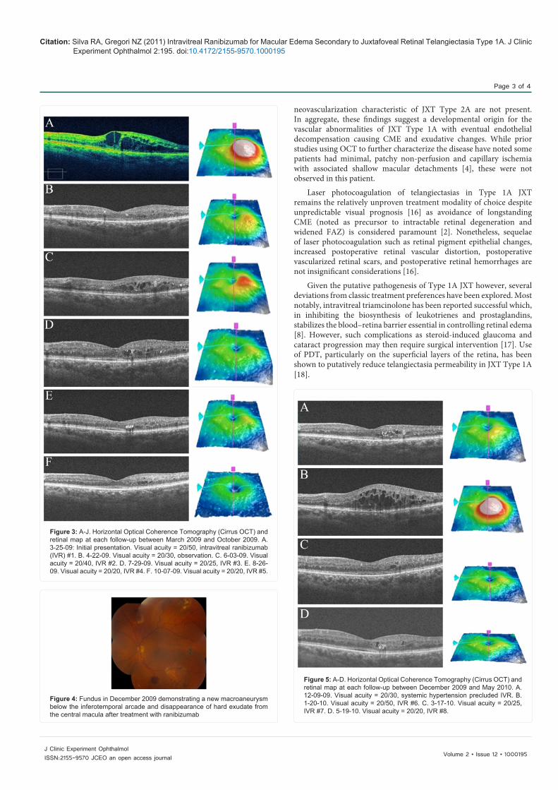
One month later (April 2009), visual acuity improved to 20/30 and the OCT showed significant reduction in the amount of cystoid macular edema with CAT of 281 microns (Figure 3B). The patient though initially observed required another ranibizumab injection 6 weeks later, when his vision declined to 20/40 due to mild interval increase in CME (Figure 3C). Six weeks later (July 2009), his vision was 20/25 and the OCT showed very few cysts in the left eye (Figure 3D). At this time, a third IVR was administered. One month later (August 2009) the patient’s vision in the left eye was 20/20 with very mild cystic changes on the OCT (Figure 3E). Patient was treated with the fourth IVR and extended to follow-up in 6 weeks. At that point (October 2009), vision remained 20/20 and the OCT was fluid-free (Figure 3F). A fifth IVR was given and an 8-week follow-up was scheduled. At that visit (December 2009) a new retinal macroaneurysm was detected (Figure 4) and the OCT showed a few intraretinal cysts, however the patient was not able to receive an injection due to elevated blood pressure (Figure 5A). He returned 1 month later (January 2010), with visual acuity of 20/50 and significantly increased intraretinal cystoid edema (Figure 5B), similar to his initial presentation. A sixth IVR was therefore given and 8 weeks later (March 2010) his vision was 20/25 with scant cysts on the OCT (Figure 5C). A seventh IVR was given at that visit and the patient was asked to return in 8 weeks. At the next follow-up exam (May 2010), the vision was 20/20 with a fluid-free OCT (Figure 5D). The patient was treated with an eighth IVR and extended to a 10-week follow-up interval.
DiscussionThe case we here report is notable for several reasons. Firstly, the
near two decade progression of the patient’s initial peripheral retinal vascular disease (Leber’s miliary aneurysms) to parafoveal disease (JXT Type 1A) iterates a common etiology to both diseases. This case further speaks to the nature of that etiology in reporting a relatively novel and successful use of ranibizumab therapy.
Though its discovery is generally credited to Theodor von Leber in 1912 [10], Leber’s miliary aneurysms were likely described even earlier [11] as peripheral regions of “globular vascular dilatations” occurring in the superficial layers of the retina. As acknowledged by Leber himself [12] and later elegantly illustrated by Reese [13], Leber’s miliary aneurysms in fact represent mild variants of Coats’ disease-Leber himself initially recounting them as hard white and yellowish exudates occurring usually unilaterally and in young males [10]. JXT Type 1A is also considered to be a mild variant of Coats’ disease (Stage 2 Coats’ Disease) [2,4] with the above case demonstrative of long-term extension of Leber’s miliary aneurysms diagnosed nearly 20 years prior to a central manifestation: JXT 1A or Stage 2 Coats’ Disease [15].
Type 1A JXT consists of dilation of capillaries, multiple venular, capillary, and arteriolar aneurysms, leakage, lipid deposition, and minimal nonperfusion seen predominantly unilaterally in males [2,4]. Extra-macular involvement with macroaneurysms has also been noted by Gass and Yanuzzi [2,4]. Macular edema and associated lipid exudate are generally the primary means of visual loss as was described in this case while the right angle venules, pigmented plaques and subretinal
Figure 1: (A) Left fundus in 1992 showing a previously lasered macroaneurysm with surrounding retinal hemorrhages and hard exudates below the inferotemporal arcade. (B) Fluorescein angiography (FA) in 1992 shows lack of macular telangiectasia and presence of exudation from the macroaneurysm inferotemporally. Fundus in February 2009 showing new macroaneurysm superior to optic nerve, intraretinal hemorrhages and circinate hard exudate around telangiectasias in the central macula, and chorioretinal scars due to previous laser.
Figure 2: (A) FA in February 2009 demonstrates telangiectasis and aneurysms in the temporal macula during the early phase. (B) Late leakage is seen on the FA done in February 2009. Magnified view of telangiectasia and aneurysms seen in the macula of panel B.
Citation: Silva RA, Gregori NZ (2011) Intravitreal Ranibizumab for Macular Edema Secondary to Juxtafoveal Retinal Telangiectasia Type 1A. J Clinic Experiment Ophthalmol 2:195. doi:10.4172/2155-9570.1000195
Page 3 of 4
Volume 2 • Issue 12 • 1000195J Clinic Experiment OphthalmolISSN:2155-9570 JCEO an open access journal
neovascularization characteristic of JXT Type 2A are not present. In aggregate, these findings suggest a developmental origin for the vascular abnormalities of JXT Type 1A with eventual endothelial decompensation causing CME and exudative changes. While prior studies using OCT to further characterize the disease have noted some patients had minimal, patchy non-perfusion and capillary ischemia with associated shallow macular detachments [4], these were not observed in this patient.
Laser photocoagulation of telangiectasias in Type 1A JXT remains the relatively unproven treatment modality of choice despite unpredictable visual prognosis [16] as avoidance of longstanding CME (noted as precursor to intractable retinal degeneration and widened FAZ) is considered paramount [2]. Nonetheless, sequelae of laser photocoagulation such as retinal pigment epithelial changes, increased postoperative retinal vascular distortion, postoperative vascularized retinal scars, and postoperative retinal hemorrhages are not insignificant considerations [16].
Given the putative pathogenesis of Type 1A JXT however, several deviations from classic treatment preferences have been explored. Most notably, intravitreal triamcinolone has been reported successful which, in inhibiting the biosynthesis of leukotrienes and prostaglandins, stabilizes the blood–retina barrier essential in controlling retinal edema [8]. However, such complications as steroid-induced glaucoma and cataract progression may then require surgical intervention [17]. Use of PDT, particularly on the superficial layers of the retina, has been shown to putatively reduce telangiectasia permeability in JXT Type 1A [18].
Figure 3: A-J. Horizontal Optical Coherence Tomography (Cirrus OCT) and retinal map at each follow-up between March 2009 and October 2009. A. 3-25-09: Initial presentation. Visual acuity = 20/50, intravitreal ranibizumab (IVR) #1. B. 4-22-09. Visual acuity = 20/30, observation. C. 6-03-09. Visual acuity = 20/40, IVR #2. D. 7-29-09. Visual acuity = 20/25, IVR #3. E. 8-26-09. Visual acuity = 20/20, IVR #4. F. 10-07-09. Visual acuity = 20/20, IVR #5.
Figure 4: Fundus in December 2009 demonstrating a new macroaneurysm below the inferotemporal arcade and disappearance of hard exudate from the central macula after treatment with ranibizumab
Figure 5: A-D. Horizontal Optical Coherence Tomography (Cirrus OCT) and retinal map at each follow-up between December 2009 and May 2010. A. 12-09-09. Visual acuity = 20/30, systemic hypertension precluded IVR. B. 1-20-10. Visual acuity = 20/50, IVR #6. C. 3-17-10. Visual acuity = 20/25, IVR #7. D. 5-19-10. Visual acuity = 20/20, IVR #8.

Citation: Silva RA, Gregori NZ (2011) Intravitreal Ranibizumab for Macular Edema Secondary to Juxtafoveal Retinal Telangiectasia Type 1A. J Clinic Experiment Ophthalmol 2:195. doi:10.4172/2155-9570.1000195
Page 4 of 4
Volume 2 • Issue 12 • 1000195J Clinic Experiment OphthalmolISSN:2155-9570 JCEO an open access journal
We here demonstrate the effectiveness of ranibizumab in the treatment of cystoid macular edema associated with JXT Type 1A. Ranibizumab is a humanized anti-VEGF antibody fragment inhibiting vascular endothelial growth factor Type A protein (VEGF-A) which has been shown to play an important role in angiogenesis and vascular permeability. While seemingly non-toxic to the retina [19] and effective in numerous diseases such as age-related macular degeneration and macular edema from various etiolo gies, with rare exception [9], its use in JXT has generally been limited to treatment of neovascularization of JXT Type 2A [17,20-24]. Though pathogenesis of JXT Type 2A differs from Type 1A, a critical late step to disease progression in both may be an impaired retinal inner nuclear layer. Associated Muller glia dysfunction and loss of vascular endothelial pericytes likely then precipitates the blood-retinal barrier breakdown seen on electron microscopy [23,25]. Resultant VEGF secretion likely then contributes to the characteristic FA finding of intraretinal hyperfluorescent staining- generally interpreted as retinal vascular leakage [23-26]. Inhibition of this step in disease progression by anti-VEGF therapy thus seems quite logical though larger clinical trials are needed to better assess the efficacy and safety of this currently off-label use.
References
1. Gass JDM (1998) Stereoscopic Atlas of Macular Disease In. Mosby-Year Book. 494.
2. Gass JD, Blodi BA (1993) Idiopathic juxtafoveolar retinal telangiectasis. Update of classification and follow-up study. Ophthalmology 100: 1536-1546.
3. Gass JD, Oyakawa RT (1982) Idiopathic juxtafoveolar retinal telangiectasis. Arch ophthalmol 100: 769-780.
4. Yannuzzi LA, Bardal AM, Freund KB, Chen KJ, Eandi CM, et al. (2006) Idiopathic macular telangiectasia. Arch ophthalmol 124: 450-460.
5. Abujamra S, Bonanomi MT, Cresta FB, Machado CG, Pimentel SL, et al. (2000) Idiopathic juxtafoveolar retinal telangiectasis: clinical pattern in 19 cases. Ophthalmologica 214: 406-411.
6. Casswell AG, Chaine G, Rush P, Bird AC (1986) Paramacular telangiectasis. Trans ophthalmol Soc U K 105: 683-692.
7. Chopdar A (1978) Retinal telangiectasis in adults: fluorescein angiographic findings and treatment by argon laser. Br J ophthalmol 62: 243-250.
8. Li KK, Goh TY, Parsons H, Chan WM, Lam DS (2005) Use of intravitreal triamcinolone acetonide injection in unilateral idiopathic juxtafoveal telangiectasis. Clin Experiment Ophthalmol 33: 542-544.
9. Gamulescu MA, Walter A, Sachs H, Helbig H (2008) Bevacizumab in the treatment of idiopathic macular telangiectasia. Graefe’s Arch Clin Exp Ophthalmol 246: 1189-1193.
10. Leber T (1912) Ueber eine durch Vorkommen multipler Mi liaraneurysmen charakterisierten Form der Retinade generation. Arch Ophthalmol 81: 1-14.
11. Story JB (1883) Aneurysms on retinal vessels in a peculiar case of retinitis. Trans Ophthalmol Soc UK 3: 108-110.
12. Leber T (1915) Die Aneurysmen der Zentralarterie und ihrer Verzweigungen. Retinaldegeneration bei multiplen Miliaraneurysmen. In Handbuch der gesamten Augenheilkunde (Graefe und Saemisch, Hrsg.). Engelmann, Leipzig.
13. Reese AB (1956) Telangiectasis of the retina and Coats’ disease. Am J Ophthalmol 42: 1-8.
14. Cahill M, O’Keefe M, Acheson R, Mulvihill A, Wallace D, et al. (2001) Classification of the spectrum of Coats’ disease as subtypes of idiopathic retinal telangiectasis with exudation. Acta ophthalmol Scand 79: 596-602.
15. Shields JA, Shields CL, Honavar SG, Demirci H, Cater J (2001) Classification and management of Coats disease: the 2000 Proctor Lecture. Am J Ophthalmol 131: 572-583.
16. Park DW, Schatz H, McDonald HR, Johnson RN (1997) Grid laser photocoagulation for macular edema in bilateral juxtafoveal telangiectasis. Ophthalmology 104: 1838-1846.
17. Moon SJ, Berger AS, Tolentino MJ, Misch DM (2007) Intravitreal bevacizumab for macular edema from idiopathic juxtafoveal retinal telangiectasis. Ophthalmic Surg Lasers Imaging 38: 164-166.
18. Kotoula MG, Chatzoulis DZ, Karabatsas CH, Tsiloulis A, Tsironi EE (2009) Resolution of macular edema in idiopathic juxtafoveal telangiectasis using PDT. Ophthalmic Surg Lasers Imaging 40: 65-67.
19. Maturi RK, Bleau LA, Wilson DL (2006) Electrophysiologic findings after intravitreal bevacizumab (Avastin) treatment. Retina 26: 270-274.
20. Jorge R, Costa RA, Calucci D, Scott IU (2007) Intravitreal bevacizumab (Avastin) associated with the regression of subretinal neovascularization in idiopathic juxtafoveolar retinal telangiectasis. Graefe’s Arch Clin Exp Ophthalmol 245: 1045-1048.
21. Karagiannis D, Georgalas I, Ladas I, Eustratios P, Mitropoulos P (2009) A case of subretinal neovascularization treated with intravitreal ranibizumab in a patient with idiopathic juxtafoveal retinal telangiectasis. Clin Interv Aging 4: 63-65.
22. Konstantinidis L, Mantel I, Zografos L, Ambresin A (2009) Intravitreal ranibizumab as primary treatment for neovascular membrane associated with idiopathic juxtafoveal retinal telangiectasia. Graefe’s Arch Clin Exp Ophthalmol 247: 1567-1569.
23. Kovach JL, Rosenfeld PJ (2009) Bevacizumab (avastin) therapy for idiopathic macular telangiectasia type II. Retina 29: 27-32.
24. Mandal S, Venkatesh P, Abbas Z, Vohra R, Garg S (2007) Intravitreal bevacizumab (Avastin) for subretinal neovascularization secondary to type 2A idiopathic juxtafoveal telangiectasia. Graefe’s Arch Clin Exp Ophthalmol 245: 1825-1829.
25. Peyman GA (1980) Vascular anomalies of the retina. In Principals and Practices of Ophthalmology S. D. Peyman GA, Goldberg MP, editor. WB Saunders, Philadelphia.
26. Wu L, Evans T, Arévalo JF, Berrocal MH, Rodríguez FJ, et al. (2008) Long-term effect of intravitreal triamcinolone in the nonproliferative stage of type II idiopathic parafoveal telangiectasia. Retina 28: 314-319.