
Henry Ford Hospital Medical Journal
11
Henry Ford Hospital Medical Journal Volume 14 | Number 1 Article 18 3-1966 Idiopathic Retroperitoneal Fibrosis: Ormond's Syndrome John K . Ormond Follow this and additional works at: hps://scholarlycommons.henryford.com/hmedjournal Part of the Life Sciences Commons , Medical Specialties Commons , and the Public Health Commons is Part II is brought to you for free and open access by Henry Ford Health System Scholarly Commons. It has been accepted for inclusion in Henry Ford Hospital Medical Journal by an authorized editor of Henry Ford Health System Scholarly Commons. Recommended Citation Ormond, John K. (1966) "Idiopathic Retroperitoneal Fibrosis: Ormond's Syndrome," Henry Ford Hospital Medical Bulletin : Vol. 14 : No. 1 , 21-29. Available at: hps://scholarlycommons.henryford.com/hmedjournal/vol14/iss1/18
Transcript of Henry Ford Hospital Medical Journal

Henry Ford Hospital Medical Journal
Volume 14 | Number 1 Article 18
3-1966
Idiopathic Retroperitoneal Fibrosis: Ormond'sSyndromeJohn K. Ormond
Follow this and additional works at: https://scholarlycommons.henryford.com/hfhmedjournal
Part of the Life Sciences Commons, Medical Specialties Commons, and the Public HealthCommons
This Part II is brought to you for free and open access by Henry Ford Health System Scholarly Commons. It has been accepted for inclusion in HenryFord Hospital Medical Journal by an authorized editor of Henry Ford Health System Scholarly Commons.
Recommended CitationOrmond, John K. (1966) "Idiopathic Retroperitoneal Fibrosis: Ormond's Syndrome," Henry Ford Hospital Medical Bulletin : Vol. 14 :No. 1 , 21-29.Available at: https://scholarlycommons.henryford.com/hfhmedjournal/vol14/iss1/18
Reprinted f r o m Henry Ford Hosp. Med. Bul l .
Vo l . 10, March, pt. 1, 1962
IDIOPATHIC RETROPERITONEAL FIBROSIS: ORMOND'S SYNDROME*
J O H N K . O R M O N D , M . D .
Chief, Division of Urology, 1920 to 1952. Chief of Urology, Miraj Medical Center, Miraj, India, 1952 to 1955. Currently in private practice of Urology. Present address: 909 Woodward Avenue, Pontiac, Michigan.
I n 1948 I reported two cases of ureteral obstruction due to envelopment and
compression by an inflammatory retroperitoneal process of undetermined etiology.
Since that time many similar cases have been reported, and it is now a recognized
clinical entity, to which many names have been given: Gerota's fascitis, perirenal
fascitis, sclerosing retroperitonitis, sclerosing lipogranuloma, nonspecific retroperitoneal
inflammation, but usually idiopathic retroperitoneal fibrosis or Ormond's syndrome.
As introduction to a discussion of this disease I am going to read to you the
descriptions of the two cases I first reported.
CASE 1 — This 45 year old man was first seen in June, 1942, complaining of substernal pain; low backache of four months duration, radiating down both thighs; and loss of 10 lbs. (4.5 kg.) in weight in the past five months. History and physical examination showed nothing unusual. Urine showed no albumin, pus or red blood cells. The hemoglobin level was 10.8 Gm., with 5,800 leukocytes. Results of a Wassermann test were negative. Complete gastrointestinal examination and orthopedic examination showed no abnormalities.
A few days later he was admitted with complete sudden, unheralded anuria of ten days duration; vomiting, and with a nonprotein nitrogen (NPN) level of 110 per cent. Cystoscopy showed a normal bladder. There was no difficulty in catheterizing the ureters, and clear urine was found in both pelves. Pyelograms were interpreted as showing slight dilation of the left pelvis only. As long as the catheters were left in place there was free flow of urine, but on removal there was again anuria. In the next two weeks, successive nephrostomies were performed. The wounds closed and the patient was sent home in October, 1942, voiding normally but with urinary infection and persistent anemia. During the next three years he was in and out of the hospital. At first the nephrostomy wounds opened occasionally, but after March, 1943, they remained closed. His ureters were frequentiy dilated with bulbs 8 and 10; the urine remained infected; the renal function diminished and the anemia persisted.
In the summer of 1945 he was given penicillin treatment and transfusions, but his down-hifl course continued and in February 1946, he died in the hospital.
Autopsy showed a fibrous retroperitoneal mass covering the promontory of the sacrum and extending up to the kidney. The ureters, which were structurally normal and showed no strictures, ran through the lateral margins of this mass and were enveloped and compressed by it, but not invaded. The kidneys were completely destroyed by infection and back pressure. I quote from the autopsy report:
The retroperitoneal tissues surrounding the aorta and extending from the kidneys to below the sacral promontory show almost complete replacement with a dense, grayish-white fibrous connective tissue mat. This tissue is 2 cm. in thickness at its thickest point and
*Given before the Henry Ford Hospital Alumni Association, Nov. 3-4, 1961.
21
HISTORICAL ISSUE — ORMOND
extends out to and including the ureters. The ureters, aorta and vena cava all traverse this dense mass of fibrous tissue which is sufficient to nearly occlude the lamina of the ureters.
Sections of this dense fibrous mass reveal that it is composed entirely of extremely dense bundles of collagen. This tissue is poorly nucleated, but the cells, where seen, are of typical fibrous connective tissue type. This fibrous connective tissue surrounds without invading the large vessels, lymph nodes, nerves and ureters.
Sections at the edge of this area of fibrosis reveal a rather sharp demarcation, but absolutely no evidence of encapsulation. The cells composing this area of fibrosis are all extremely small and show no evidence of malignancy. In one portion the fibrous tissue is invading the surrounding skeletal muscle, which shows, as previously surrounded muscle is examined, a gradual atrophy and disappearance. This fibrous tissue has no sign of malignant characteristics and has rather the appearance of a keloid.
CASE 2 — This 43 year old man was first seen in the Department of Urology in January. 1946. He had previously been treated by physicians in various divisions of the hospital for 22 years. He seemed susceptible to upper respiratory infections and some years earlier had an appendectomy with wound infection, which was slow in clearing. He had had an attack of bacillary dysentery at one time and was continuously slightly anemic. At no time had the urine shown any abnormality.
He had had low backache for a few months which radiated to his testes. When first seen in urology, the urine was clear; the backache very severe; hemoglobin 11 Gm.; kidney, ureter and bladder, barium enema and chest x-ray normal.
The backache was severe enough that an iophendylate (Pantopaque) myelogram was made, followed by laminectomy. The backache was somewhat relieved, but anemia and asthenia persisted and there was no gain in weight. On March 25, 1946, there was complete, sudden and unheralded anuria. Examination revealed no renal masses or tenderness. Cystoscopy showed a normal bladder; the ureters were easily catheterized, with clear urine in both pelves. Pyelogram showed only slight dilatation of the left pelvis.
As long as the catheters were in place, there was a constant drip, but on their removal, the anuria recurred and it was necessary to reintroduce them.
Because of the patient's anemia and the puzzling character of his course in the past, it was suspected that the ureters might be involved in some malignant process, and the decision was made to explore the abdomen.
On April 4, 1946, a right paramedian, lower abdominal incision was made. The peritoneal surfaces were smooth, the kidneys felt normal, the liver was not enlarged, and no nodules were felt on its surface. The gallbladder had been previously removed and there were numerous adhesions in this region. A retroperitoneal, flat mass whh indistinct lateral margins was felt covering the promontory of the sacrum. A bit of this was removed for biopsy and frozen section showed it to be inflammatory in nature. I quote from the pathological report:
Sections of the retroperitoneal tissue reveal alternating areas of dense, fibrous, connective tissue and fat. Throughout the entire sections examined is a diffuse infiltration of small, round and wandering cefls, occasional eosinophils and polymorphonuclear leukocytes. In sonie instances the lymphatic cells are collected into small follicles. Considerable fibroblastic activity is seen but no abscesses are present and there is no evidence of a specific etiology. There is no microscopic evidence of neoplastic process.
The ureters were located below this mass, traced up, and found to pass through this tissue near the lateral margins. Both ureters were dissected entirely free f rom enveloping tissue and seemed entirely normal. They were left free in the abdominal cavity, no attempt being made to cover them with peritoneum. The ureteral catheters were removed in the next two days. Wound healing and convalescence were uneventful, and there has been free and normal urination ever since. The patient was given deep x-ray therapy directed at the inflammatory mass described above. His course was followed carefully and in the succeeding years he had no impairment of renal function and no symptoms or findings referrable to the urinary tract. He was able to care for his wife through some years of invalidism and, as of this writing, is in reasonably good health. I t seems highly probable that, had he received the same treatment when he had his first attack of anuria, patient no. I would have had a different result.
These two cases between them show practically all the characteristic symptoms,
findings and diagnostic features that have been brought out by the cases since reported,
22
ORMOND SYNDROME
with one very important exception: namely, the x-ray demonstration of medial deviation and obliteration of the mid-ureter, with the consequent dilatation of the upper ureter and pelvis. These were probably present but we did not realize their importance.
Since my first report in 1948, there have appeared in the world literature at least a hundred instances of ureteral obstruction due to involvement in retroperitoneal inflammatory tissue, and many more have been brought to my attention in personal communications. In a minority of these, the origin of the inflammatory process could be traced (either certainly or probably) to pre-existing inflammation in the urinary or gastrointestinal tracts, or to tumor. The classification of "idiopathic" has been reserved for those (by far the majority) in which no such connection can be found or suspected, and in which the etiology, up to the present, is completely in doubt.
DISCUSSION
Enough of these cases have been recorded and followed that it is now possible to conceive a picture of this condition; its pathology and progress, fls diagnosis and treatment; and to conjecture about its etiology. Moreover, it has become clear that while every case so far reported has come to the attention of the clinician because of its urological aspect, the primary condition does not belong in the field of urology but in that of medicine.
Involvement of the urinary tract endangers life and urgently demands surgical treatment. The presence of the retroperitoneal fibrosis, unsuspected beforehand excepting in some of the later cases, was brought to light either at autopsy, or at operation designed to remedy the urinary involvement. In most of these, biopsies were made of the fibrous mass and, in addition, there have been twelve autopsies recorded in which more extensive observation of the fibrosis was possible. In every case, a fibrous mass was found in the retroperitoneal region, well-demarcated, but not encapsulated, through which the ureters passed. Histopathologically, the lesion consists of moderate to dense fibrous tissue with some fat and some cellular infiltration. In general, it can be said that the density varied with the duration of the fibrosis and that the amount of fat and of the cellular infiltration varied inversely with the duration of the fibrosis. Lymph glands were rarely described and the evidence is all for their incidental involvement in the fibrous tissue, rather than for being an active factor in its production.
Usually, the process seemed to start in the vicinity of the great vessels, spreading laterally and seeming to be limited by the bounds of Gerota's fascia. This has been so much the case that Hutch et al., in a recent paper, presented evidence for their belief that the condition is a fascitis of unknown origin, and they speak of "Gerota's fascitis". On the other hand, in the past few years these have been reported instances of a similar process, not confined to Gerota's fascia, (1) in the pelvis affecting the bladder and lower ureters; (2) above the kidney; and (3) in the mediastinum
23
HISTORICAL ISSUE — ORMOND
involving and constricting the large veins. Two cases are on record in which there was coincidental fibrosis in both the mediastinum and the retroperitoneal region.
The fibrous process does not usually invade or intrinsically involve, the structures surrounded, but envelopes them and as contraction occurs, compresses them. Arteries and nerves are sturdy, but veins and ureters are soft and compressible. Collateral circulation can compensate for gradual obstruction of a vein, but there is no collateral organ to take over the function of the ureter, and its obstruction inevitably causes dflatation above, producing symptoms and causing damage to the kidney.
Occasionally, along with the ureteral obstruction, there has been found coincidental venous, and even arterial, compression, and in one in.stance, the condition was brought to light by an arteriogram showing partial blocking of the iliac artery.
The course of the disease can be divided into three periods: (1) the period of the incidence and development of the underiying process, of whose nature and etiology we are wholly ignorant; (2) the period of the activity and spread of the fibrosis and the envelopment of the retroperitoneal structures; and, (3) the period of contracture of the fibrous mass, like that of scar, with compression of the enveloped structures. This period seem.s to be a period of healing or recedence of a self-Umited process. This conclusion receives some confirmation from the fact that after the ureters have been freed and the urinary tract restored as much as possible, usually nothing further is heard from the fibrosis, no matter what kind of treatment to it may be used; — x-ray, cortisone, antibiotics, or no treatment at all.
We have no information about the ultimate fate of the fibrosis. Probably, like scar — which it most likely is — it remains in situ, possibly becoming more and more dense. Only if and when in the normal course of events, death followed by autopsy occurs in some of these patients, after several years will that question be answered.
The impression I gain is that the disease is self-limited, somewhat disabling temporarily, but not in itself serious, and usually dangerous only in its incidental effect on the urinary tract. This is very dangerous indeed and urgentiy demands treatment as eariy as it is recognized.
The disease occurs twice as often in men as in women; usually between the ages of forty and sixty, though it has been reported as eariy as eight and as late as seventy-five.
The picture of a typical case might be given as follows:—A man in the middle years of life begins to feel much under par. to have loss of appetite, to lose weight, may have bloating and fits of vomiting and complain of troublesome backache in the sacral region which may radiate to the lateral abdomen and inguinal regions down the medial surfaces of the thighs and even to the testicles. This backache may be quite severe, even to the point of indicating myelography.
24
ORMOND SYNDROME
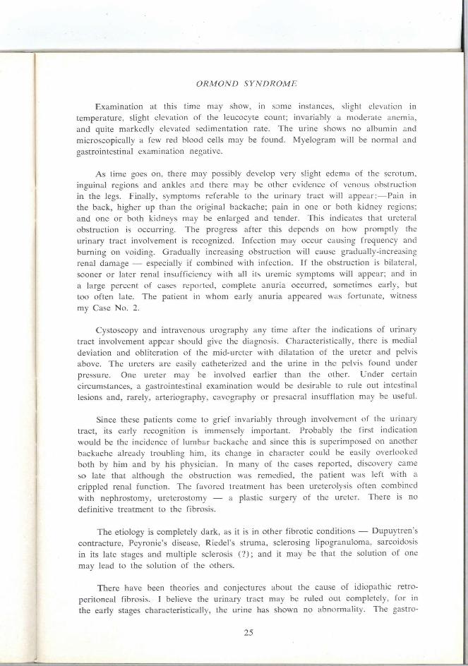
Examination at this time may show, in some instances, slight elevation in temperature, slight elevation of the leucocyte count; invariably a moderate anemia, and quite markedly elevated sedimentation rate. The urine shows no albumin and microscopically a few red blood cells may be found. Myelogram will be normal and gastrointestinal examination negative.
As time goes on, there may possibly develop very slight edema of the scrotum, inguinal regions and ankles and there may be other evidence of venous obstruction in the legs. Finally, symptoms referable to the urinary tract will appear:—Pain in the back, higher up than the original backache; pain in one or both kidney regions; and one or both kidneys may be enlarged and tender. This indicates that ureteral obstruction is occurring. The progress after this depends on how promptly the urinary tract involvement is recognized. Infection may occur causing frequency and burning on voiding. Gradually increasing obstruction wifl cause gradually-increasing renal damage — especially if combined with infection. If the obstruction is bilateral, sooner or later renal insufficiency whh afl its uremic symptoms wfll appear; and in a large percent of cases reported, complete anuria occurred, sometimes early, but too often late. The patient in whom early anuria appeared was fortunate, witness my Case No. 2.
Cystoscopy and intravenous urography any time after the indications of urinary tract involvement appear should give the diagnosis. Characteristically, there is medial deviation and obliteration of the mid-ureter with dilatation of the ureter and pelvis above. The ureters are easily catheterized and the urine in the pelvis found under pressure. One ureter may be involved earlier than the other. Under certain circumstances, a gastrointestinal examination would be desirable to rule out intestinal lesions and, rarely, arteriography, cavography or presacral insufflation may be useful.
Since these patients come to grief invariably through involvement of the urinary tract, its early recognition is immensely important. Probably the first indication would be the incidence of lumbar backache and since this is superimposed on another backache already troubling him, its change in character could be easily overlooked both by him and by his physician. In many of the cases reported, discovery came so late that although the obstruction was remedied, the patient was left with a crippled renal function. The favored treatment has been ureterolysis often combined with nephrostomy, ureterostomy — a plastic surgery of the ureter. There is no definitive treatment to the fibrosis.
The etiology is completely dark, as it is in other fibrotic condflions — Dupuytren's contracture, Peyronie's disease, Riedel's stmma, sclerosing lipogranuloma, sarcoidosis in its late stages and multiple sclerosis (?); and it may be that the solution of one may lead to the solution of the others.
There have been theories and conjectures about the cause of idiopathic retroperitoneal fibrosis. I believe the urinary tract may be ruled out completely, for in the early stages characteristically, the urine has shown no abnormality. The gastro-
25
HISTORICAL ISSUE — ORMOND
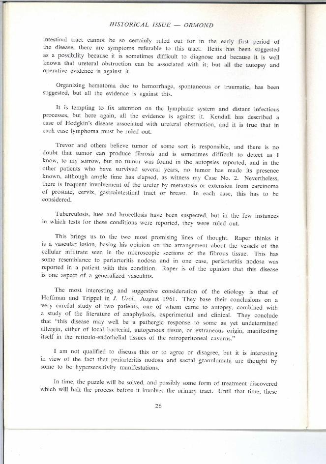
intestinal tract cannot be so certainly ruled out for in the early first period of the disease, there are symptoms referable to this tract. Ileitis has been suggested as a possibility because it is sometimes difficult to diagnose and because it is weU known that ureteral obstruction can be associated with it; but all the autopsy and operative evidence is against it.
Organizing hematoma due to hemorrhage, spontaneous or traumatic, has been suggested, but afl the evidence is against this.
It is tempting to fix attention on the lymphatic system and distant infectious processes, but here again, all the evidence is against it. Kendall has described a case of Hodgkin's disease associated with ureteral obstruction, and it is true that in each case lymphoma must be ruled out.
Trevor and others believe tumor of some sort is responsible, and there is no doubt that tumor can produce fibrosis and is sometimes difficult to detect as 1 know, to my sorrow, but no tumor was found in the autopsies reported, and in the other patients who have survived several years, no tumor has made its presence known, althoiigh ample time has elapsed, as witness my Case No. 2. Nevertheless, there is frequent involvement of the ureter by metastasis or extension from carcinoma of prostate, cervix, gastrointestinal tract or breast. In each case, this has to be considered.
Tuberculosis, lues and brucellosis have been suspected, but in the few instances in which tests for these condflions were reported, they were ruled out.
This brings us to the two most promising lines of thought. Raper thinks it is a vascular lesion, basing his opinion cn the arrangement about the vessels of the cellular infiltrate seen in the microscopic sections of the fibrous tissue. This has some resemblance to periarteritis nodosa and in one case, periarteritis nodosa was reported in a patient with this condition. Raper is of the opinion that this disease is one aspect of a generalized vascuhtis.
The most interesting and suggestive consideration of the etiology is that of Hoffman and Trippel in J. Urol , August 1961. They base their conclusions on a very careful study of two patients, one of whom came to autopsy, combined with a study of the literature of anaphylaxis, experimental and clinical. They conclude that "this disease may well be a pathergic response to some as yet undetermined allergin, either of local bacterial, autogenous tissue, or extraneous origin, manifesting itself in the reticulo-endothelial tissues of the retroperitoneal caverns."
I am not qualified to discuss this or to agree or disagree, but it is interesting in view of the fact that periarteritis nodosa and sacral granulomata are thought by some to be hypersensitivity manifestations.
In time, the puzzle will be solved, and possibly some form of treatment discovered which will halt the process before it involves the urinary tract. Until that time, these
26
ORMOND SYNDROME
cases wifl probably continue to be a problem for the urologist. The important thing is to keep in mind the possibility that this condition, syndrome, or disease may be present in cases of puzzhng backache combined with moderate anemia, weight loss and malaise. Such should be watched carefufly for early signs of ureteral compression and subjected to intravenous urography before the urine shows any abnormality — albumin, pus, or blood. I f this leads to a diagnosis, the urologist will stifl be called on to relieve the situation but he wifl be able to do so before irreparable damage has been done.
In closing, I like to quote from an unsigned editorial in the Lancet, October 19, 1957:
"Many a clinical reputation lies behind the peritoneum. In this hinterland of straggling mesenchyme with its vascular and nervous plexuses; its weird embryonic rests, its shadowy fascial boundaries, the clinician is often left with only his plan and his diagnostic first principles to aid him. Nonspecific periureteric fibrosis is likely to tax them both.
At an early stage the symptoms usually point to a growing retroperitoneal lesion; but this limited diagnosis is rarely made. Retroperitoneal pain can be severe and persistent enough to drive the most phlegmatic patient to distraction, but when there are no abdominal findings, his complaint is likely to be received with incredulity."
SELECTED REFERENCES
Anselm, A.: Pseudosclerosis renal, Med. Esp. 23:230, 1950.
Bates, B. C : Periureteritis obliterans, J. Urol. 82:58, 1959.
Barrett, M. R.: Idiopathic mediastinal fibrosis, Brit. J. Surg. 46:207, 1958.
Bradfield, E. O.: Bilateral ureteral obstruction due to compression by chronic retroperitoneal inflammation, J. Urol. 69:769, 1953.
Bulkley, C. J.: Retroperitoneal fibrosis. Quart. Bull. Northwest Univ. Med. School 34:113, 1960.
Catalono, D.: Retroperitoneal fibrosis, Rass. Int. Clin. Ter. 40:1265, 1960.
Charnock, D., Riddel, H. I . , and Lombardo, H. L., Jr.: Retroperitoneal fibrosis producing ureteral obstruction, J. UroL 85:251, 1961.
Chinn, J., Horton, R., and Rusche, C : Unilateral ureteral obstruction as sole manifestation of endometriosis, J. Urol. 77:144, 1957.
Chisholm, E. R., Hutch, J. A., and Bolomey, A. A.: Bilateral ureteral obstruction due to chronic inflammation of the fasciae around the ureters, J. Urol. 72:812, 1954.
Cibert, J., Durand, L., and Riviere, C : Les compressiones ureterales par sclerose du tissue cellulo-adipeaux periureteral; periureteritis primitives, J. Urol. med. 62:705, 1957.
Daseler, E. H., and Anson, B. J.: Anatomic relations of the ectopic iliolumbar kidney, bilateral in adult; unilateral in fetus, J. Urol. 49:789, 1943.
Dineen, J. P., Thane, A., Pearce, J. M., and Marshall, V. F.: Retroperitoneal fibrosis (Exhibit at the American Urological Association Meeting, Chicago, May 1960).
Dineen, J., Asch, T., and Pearce, J. M. : Retroperitoneal fibrosis; anatomic and radiological review. Four cases, pathogenesis. Radiology 75:380, 1960.
Dix, V. W.: Hydronephrosis, Brit. J. Urol. 31:366, 1959.
27

HISTORICAL ISSUE — ORMOND
Editorial; Lancet 2:780, 1957.
Ewell, G. H., and Bruskewitz, H. W.: Bilateral ureteral obstruction due to envelopment and compression by flammatory process, Urol, and Cutan. Rev. 56:3, 1952.
Francke, C, and Wiggim, K.: Bilateral idiopathic peri-ureteric fibrosis, Arch. chir. neerl. 10:165, 1958.
Hackett, E.: Idiopathic retroperitoneal fibrosis, Brit. J. Surg. 46:3, 1958.
Hamburger, J., Richer, G., Ducrot, H.: Une maladie nouvelle; la liposclerose periureterale, Asquistones Med. Recentes (Paris) 34-36, 1957.
Harlin, H. C, and Hamm, F.: Urologic disease resulting from nonspecific inflammatory condition of the bowel, J. Urol. 68:383, 1952.
Hawk, W. A., and Hazard, J. B.: Sclerosing retroperitonitis and sclerosing mediastinitis. Am. J. Clin. Path. 32:321, 1959.
Hejtmancik, J. H., and Majid, M. A.: Bilateral periureteritis plastica, J. Urol. 76:57, 1956.
Hewett, A. L., and Headstream, J. W.: Pericytitis plastica, J. Urol. 80:103, 1960.
Hosford, J. P.; Some factors in the cause of hydronephrosis, Lancet 1:435, 1932.
Houston, W.: Periureteritis plastica, Brit. J. Urol. 29:38, 1957.
Hughes, J.: Obstruction, secondary to extra-ureteral inflammation, South. M. J. 45:1044, 1952.
Hutch, J. A., Atkinson, R. C, and Lozuvam, G. S.: Perirenal fascitis, J. Urol. 81:76, 1959.
Hyan, J. A., Weinberg, S. R., and Allen, J. L.: Chronic ileitis with concomitant ureteritis. Am. J. Surg. 61:117, 1943.
loszi, L., and Murphy, J. J.: Bilateral ureteral obstruction by retroperitoneal inflammation, J. Urol. 77:402, 1957.
Kickham, J. E., and Colpoys, F. L.: Periureteral fascitis, J.A.M.A. 171:2202, 1959.
Knowlan, D., Corrado, M., Schreiner, G. I . , and Baker, R.: Periureteral fibrosis with a diabetes insipidu.s-like syndrome, Am. J. Med. 29:22, 1960.
Kolischer, G : Nephrolysis and ureterolysis, J. Urol. 8:149, 1922.
Landes. R., and Ransom, C. L.: Presacral retroperitoneal pneumography utilizing carbon dioxide, J. Urol. 82:670, 1959.
Lund, F., and Pederson, J.: Periureteral fibrosis, Ugesk. Laeger 121:1853, 1959.
Lund, F., and Pederson, .1.: Periureteritis fibrosa (Gerota's fascitis) Acta med. scand. 167:105, 1960.
MacDonald, S. A.: Periureteral fibrosis, Canad. J. Surg. 1:162, 1958.
MacLean, J. T.: Unusual conditions of ureter and kidney, Brit. J. Urol. 26:127, 1954.
Margoles, J. S., and McQueeney, A. J.: Ormond's syndrome; discussion of problem; three cases, A.M.A. Arch. Surg. 81:660, I960.
Middleton, W. S.: The riddle of sarcoidosis, Ann. Int. Med. 41:465, 1954.
Millard, D. G., and Wyman, S. M. : Periureteritis fibrosis; radiographic diagnosis, Radiology 72:191, 1959.
Miller, J. M., Lipin, R. J., Meisel, H., and Long, P.: Bilateral ureteral obstruction due to compression of chronic retroperitoneal inflammation, J. Urol. 68:447, 1952.
Mirablie, C. S., and Spillane, R. J.: Bilateral ureteral compression with obstruction from a nonspecific retroperitoneal inflammatory process, J. Urol. 73:783, 1955.
Mulvaney, W. P.: Periureteritis obliterans; a retroperitoneal disease, J. Urol. 79:410, 1958.
Ormond, J, K.: Bilateral ureteral obstruction due to envelopment and compression by inflammatory retroperitoneal process, J. Urol. 59:1072, 1948.
Ormond, J. K.: Idiopathic retroperitoneal fibrosis; an established clinical entity, J.A.M.A. 174:1561, 1960.
2S
ORMOND SYNDROME
Park, H., and Jones, I . : Periureteric fibrosis. Lancet 1:196, 1958.
Partington, P. F.: Diffuse idiopathic fibrosis. Am. J. Surg. 101:239, 1961.
Popham, B. F., and Stevenson, T. D.: Idiopathic retroperitoneal fibrosis with coagulation defect, Ann. Int. Med. 52:894, 1960.
Pugh, R. C: Pathology of fibrotic lesions, Proc. Roy. Soc. Med. 53:685, 1960.
Raper, F. P.: Bilateral symmetrical periureteral fibrosis, Proc. Roy. Soc. Med. 48:736, 1955.
Raper, F. P.: Idiopathic retroperitoneal fibrosis, involving the ureters, J. Urol. 28:436, 1956; also, Proc. Roy. Soc. Med. 53:690, 1960.
Redish, M. H.: Ureteral complications and alteration of ureteral tonus in regional enteritis, New England J. Med. 246:993, 1952.
Radiffe, R. K., and Crenshaw, W. B.: Ureteral obstruction from endrometriosis, Surg. Gynec. and Obst. 100:414, 1955.
Ross, J. A.: Periuretertis fibrosa, J. Fac. Radiol. 9:142, 1958.
Schifrin, A., Oppenheimer, G. O., and Krawlett, D. R.: Idiopathic, non-specific fibrosing retroperitonitis causing bilateral ureteral compression, J. Mt. Sinai Hosp. (New York) 24:1186, 1957.
Shaheen, D. J. and Jonston, A.: Bilateral ureteral obstruction due to an inflammatory retroperitoneal process, J. Urol. 82:51, 1959.
Simeoni, S.: Fibrosis idiopatica retroperitoneale, Policlin. sex prat. 65:1563, 1958.
Simond, H. B., and Nygaard, K. K.: Ureteral obstruction due to retroperitoneal fibrosis, J.A.M.A. 174:1589,1960.
Sleuber, P. J., Jr.: Primary retroperitoneal inflammatory process with ureteral obstruction, J. Urol. 82:41, 1959.
Talbot, H. S., and Mahoney, E.: Obstruction of both ureters by retroperitoneal inflammation, J, UroL 78:738, 1959.
Thompson, R. J., Carter, R., Gibson, D., Reiswig, O. K., and Hinshaw, D. B.: Acute retroperitoneal fibrosis, Ann. Surg. 153:399, 1961.
Trevor, R. W.: Reticulum cell sarcoma producing retroperitoneal and periureteric fibrosis. New England J. Med. 258:268, 1958.
Tullach, W. S.: Idiopathic retroperitoneal fibrosis, J. Roy Coll. Surg. Edinburgh, 5:142, 1960.
Twigg, H. F., Jr.: Periureteral fibrosis, Am. J. Roetgen. 84:876, 1960.
Vest, S. A., and Barclare, B.: Periureteritis plastica, J. Urol. 70:38, 1953.
ADDENDUM September 1965
During the past two years important observaticns have been made in connection with the etiology and pathology of this disease. Reports by Suby of Boston, Utz of the Mayo Clinic, and others, make it clear that some cases of this disease are due to the use of derivatives of ergot, and the inference is that it is a vasculitis caused by drugs or chemicals, and may be on a hypersensitization basis.
1. Ormond, John K. Idiopathic retroperitoneal fibrosis: a discussion of etiology. J. Urology 94:385, 1965.
2. Suby, Howard 1; Kerr, Walter S. Jr.; Graham, John R.; et, al. Retroperitoneal fibrosis: a missing link in the chain. J. Urol. 93:144-52, 1965.
3. Utz, David C; Rooke, E. Douglas; Spittell, John A. Jr.; and Bartholomew, Lloyd G : Retroperitoneal fibrosis in patients taking methysergide. JAMA 191:983, 1965.
4. Mulvaney, W. P.: Personal communication.
29