HEAD AND NECK HEAD AND NECK BLOCK 2 EXAM AND … · head and neck and neuro simultaneously atlas...
52
HEAD AND NECK BLOCK 2 EXAM 1 - WRITTEN EXAM 18 Questions 4.90% of grade 2 - PRACTICAL EXAM 15 Questions Gross Anatomy 2.72% of grade 1) Prosections – already in lab; some correspond to photos; some structures not labeled 2) X-rays, Angiograms NO SKULL SESSIONS: no questions on skulls HEAD AND NECK AND NEURO SIMULTANEOUSLY ATLAS HOLDING UP WORLD
Transcript of HEAD AND NECK HEAD AND NECK BLOCK 2 EXAM AND … · head and neck and neuro simultaneously atlas...
HEAD AND NECK BLOCK 2 EXAM
1 - WRITTEN EXAM18 Questions 4.90% of grade
2 - PRACTICAL EXAM15 Questions Gross Anatomy
2.72% of grade
1) Prosections – already in lab; some correspond to photos; some structures not labeled
2) X-rays, Angiograms
NO SKULL SESSIONS: no questions on skulls
HEAD AND NECK AND NEURO SIMULTANEOUSLY
ATLAS HOLDING UP WORLD
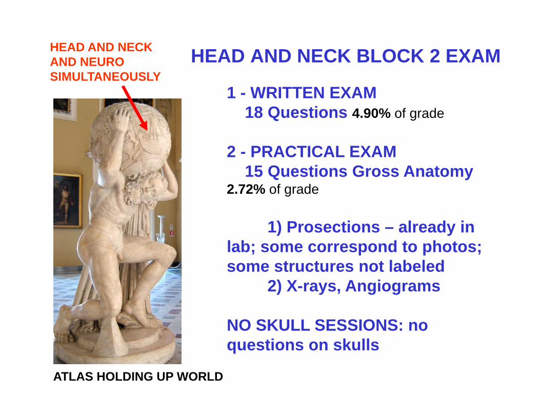
ADVICE ON HOW TO TAKE THE HEAD AND NECK EXAM
DRAW:1. CRANIAL NERVE FUNCTIONAL COMPONENTS
CHART3. SVE MUSCLES - 'incantation'4. BRANCHIAL ARCHES, POUCHES5. GVE PARASYMPATHETICS - HITCHHIKING
PATHWAYS - BRANCHES IX (VIDEO REVIEW)[6. BRANCHES OF MAXILLARY ARTERY]

1. LARYNX2. NECK - Compartments, Carotid
Artery3. PHARYNX - TONSILS, POPCORN4. ORAL CAVITY
REVIEW
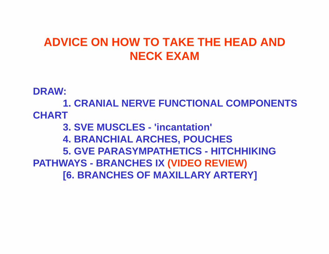
LARYNX REVIEW: CARTILAGES
THYROID CARTILAGE- shield shaped - has Sup. and Inf. Horns- Laryngeal prominence
CRICOID = signet ring - complete ring of cartilage- narrow Arch ant., broad Lamina post.
ARYTENOID - 2 pyramidal shaped cartilages above lamina of cricoid
Sup. Horn
Laryngeal prominence= Adam'sapple
Inf. Horn
Arytenoid
Cricoid
lamina
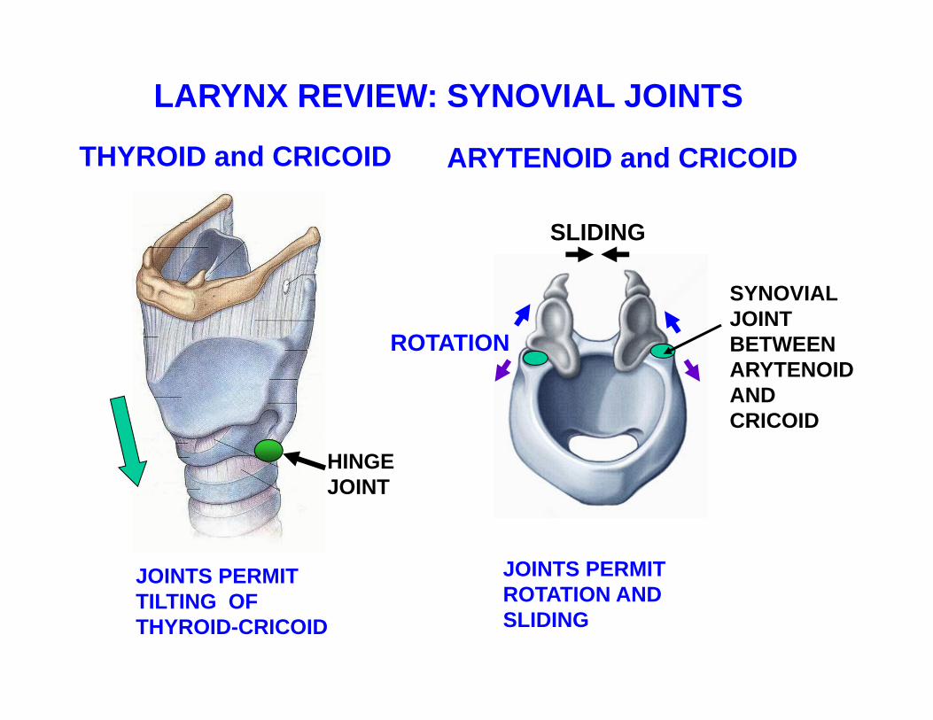
HINGEJOINT
JOINTS PERMIT ROTATION AND SLIDING
SYNOVIALJOINT BETWEEN ARYTENOID AND CRICOID
LARYNX REVIEW: SYNOVIAL JOINTS
THYROID and CRICOID ARYTENOID and CRICOID
JOINTS PERMIT TILTING OF THYROID-CRICOID
SLIDING
ROTATION
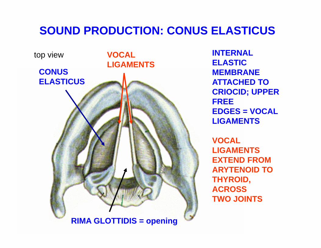
SOUND PRODUCTION: CONUS ELASTICUS
INTERNAL ELASTIC MEMBRANE ATTACHED TO CRIOCID; UPPER FREEEDGES = VOCALLIGAMENTS
VOCAL LIGAMENTS EXTEND FROM ARYTENOID TO THYROID, ACROSSTWO JOINTS
CONUS ELASTICUS
VOCALLIGAMENTS
RIMA GLOTTIDIS = opening
top view

CHANGE PITCH BY TILTING AT HINGE JOINT –Thyroid cartilage tilts down; cricoid tilts up
HINGEJOINT
HINGEJOINT
Tilting - STRETCHESvocal ligaments
STRETCH vocal ligamentINCREASE PITCH -CRICOTHYROID
RELAX vocal ligamentDECREASE PITCH -THYROARYTENOID
THYROID
CRICOID
ARYTENOID

CRICOTHYROID M. -Tenses Vocal LigamentIncreasing Pitch
HINGEJOINT
Tilting - STRETCHESvocal ligaments
STRETCH vocal ligamentINCREASE PITCH -CRICOTHYROID
MUSCLES OF LARYNX
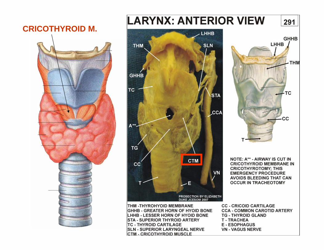
CRICOTHYROID M.

THYROARYTENOIDMUSCLES - adjacent to vocal ligament -RelaxesVocal LigamentsDecreases pitch
HINGEJOINT
RELAX vocal ligamentDECREASE PITCH -THYROARYTENOID
THYROID
CRICOID
MUSCLES OF LARYNX NOTSEE
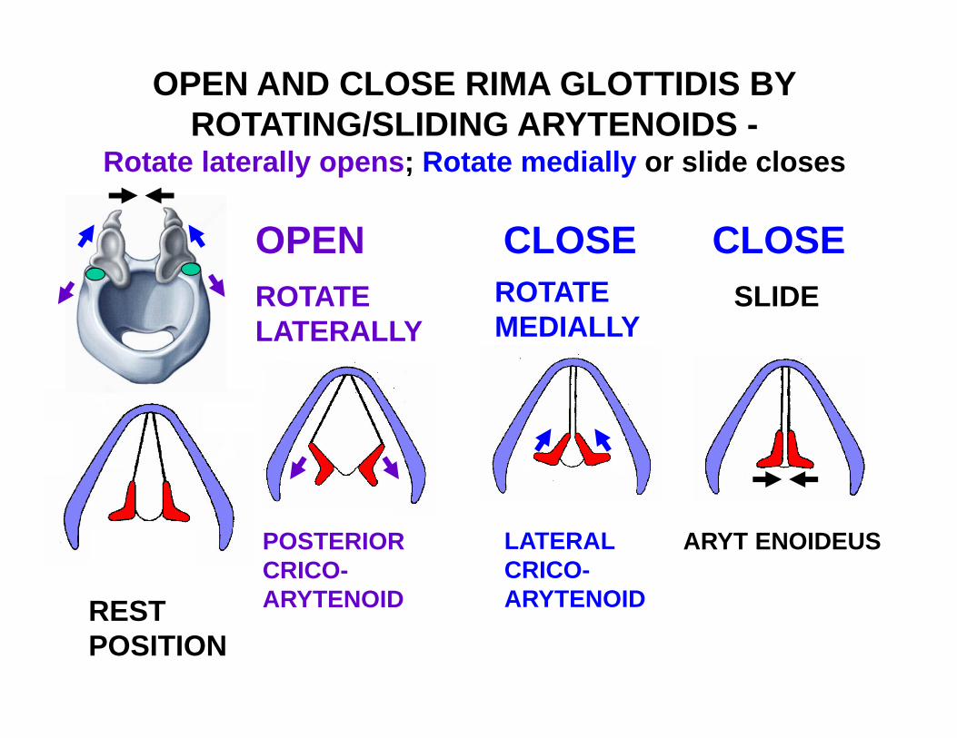
OPEN AND CLOSE RIMA GLOTTIDIS BYROTATING/SLIDING ARYTENOIDS -
Rotate laterally opens; Rotate medially or slide closes
POSTERIORCRICO-ARYTENOID
LATERALCRICO-ARYTENOID
ARYT ENOIDEUS
ROTATELATERALLY
ROTATEMEDIALLY
SLIDE
RESTPOSITION
OPEN CLOSE CLOSE
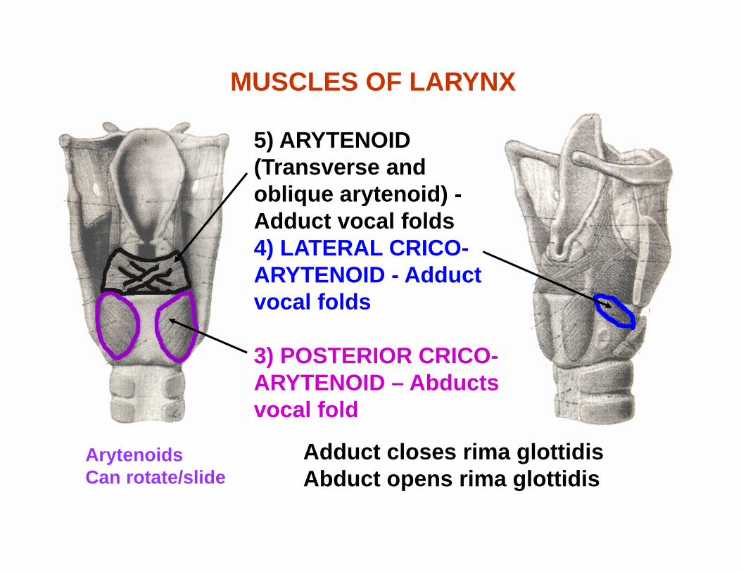
Arytenoids Can rotate/slide
5) ARYTENOID (Transverse and oblique arytenoid) -Adduct vocal folds4) LATERAL CRICO-ARYTENOID - Adduct vocal folds
3) POSTERIOR CRICO-ARYTENOID – Abducts vocal fold
MUSCLES OF LARYNX
Adduct closes rima glottidisAbduct opens rima glottidis
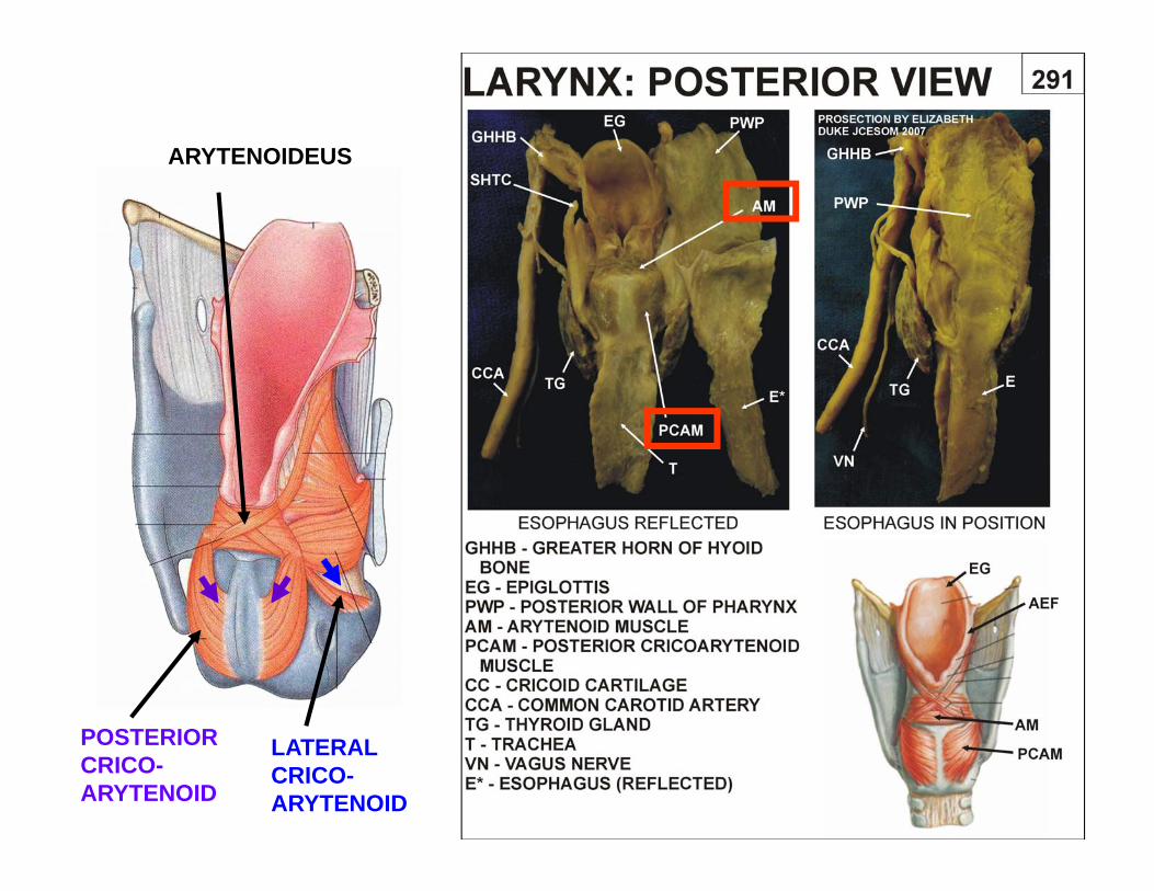
POSTERIORCRICO-ARYTENOID
LATERALCRICO-ARYTENOID
ARYTENOIDEUS
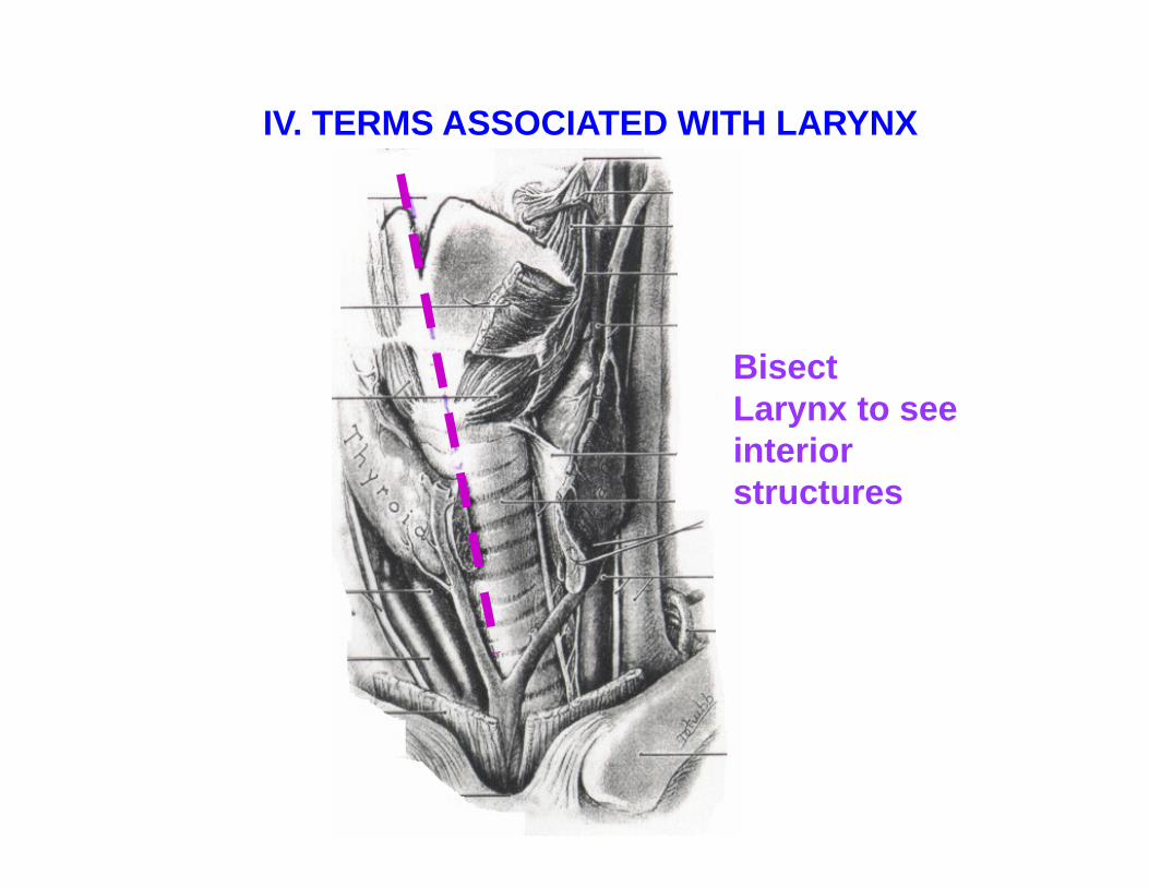
BisectLarynx to seeinteriorstructures
IV. TERMS ASSOCIATED WITH LARYNX
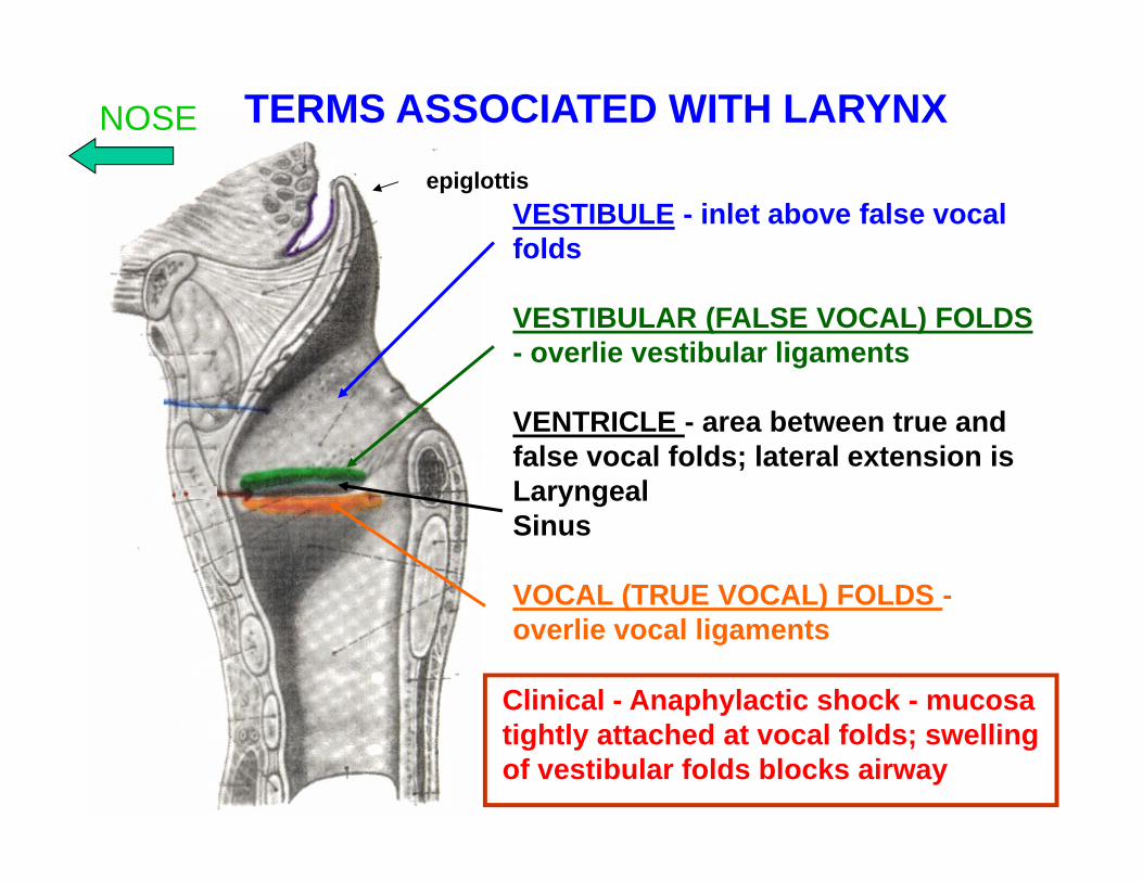
VESTIBULE - inlet above false vocal folds
VESTIBULAR (FALSE VOCAL) FOLDS- overlie vestibular ligaments
VENTRICLE - area between true and false vocal folds; lateral extension is LaryngealSinus
VOCAL (TRUE VOCAL) FOLDS -overlie vocal ligaments
TERMS ASSOCIATED WITH LARYNXepiglottis
NOSE
Clinical - Anaphylactic shock - mucosa tightly attached at vocal folds; swellingof vestibular folds blocks airway
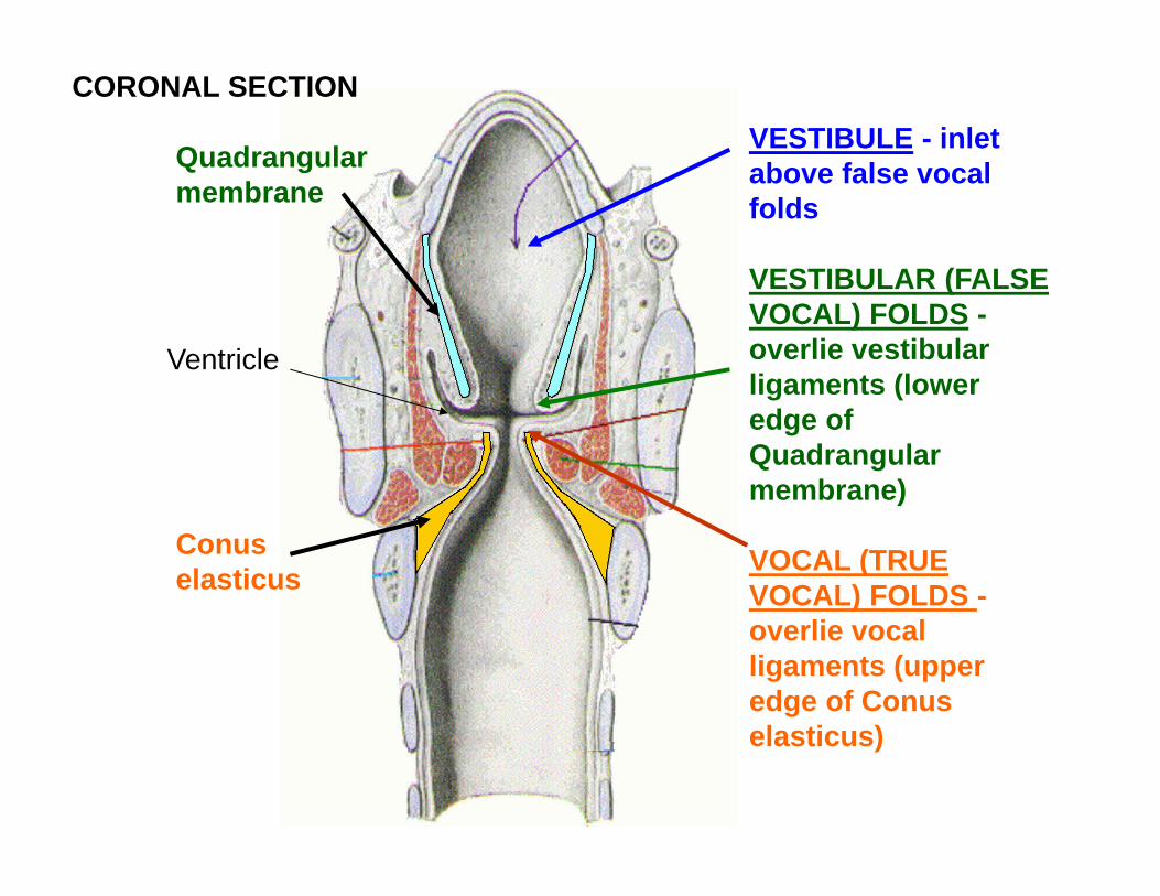
VESTIBULE - inlet above false vocal folds
VESTIBULAR (FALSE VOCAL) FOLDS -overlie vestibular ligaments (lower edge of Quadrangular membrane)
VOCAL (TRUE VOCAL) FOLDS -overlie vocal ligaments (upper edge of Conus elasticus)
Conus elasticus
Quadrangular membrane
Ventricle
CORONAL SECTION
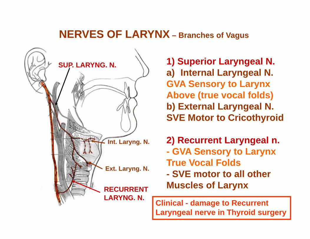
1) Superior Laryngeal N.a) Internal Laryngeal N.GVA Sensory to LarynxAbove (true vocal folds)b) External Laryngeal N.SVE Motor to Cricothyroid
2) Recurrent Laryngeal n.- GVA Sensory to Larynx True Vocal Folds- SVE motor to all other Muscles of Larynx
SUP. LARYNG. N.
RECURRENTLARYNG. N.
Int. Laryng. N.
Ext. Laryng. N.
NERVES OF LARYNX – Branches of Vagus
Clinical - damage to RecurrentLaryngeal nerve in Thyroid surgery

NERVES OF LARYNX –Branches of Vagus
X X
SUPERIORLARYNGEALNERVE - piercesthyrohyoidmembrane
RIGHTRECURRENTLARYNGEALNERVE - passesunderSubclavianArtery
LEFTRECURRENTLARYNGEALNERVE - passesunderArch ofAorta
SUPERIORLARYNGEALNERVE
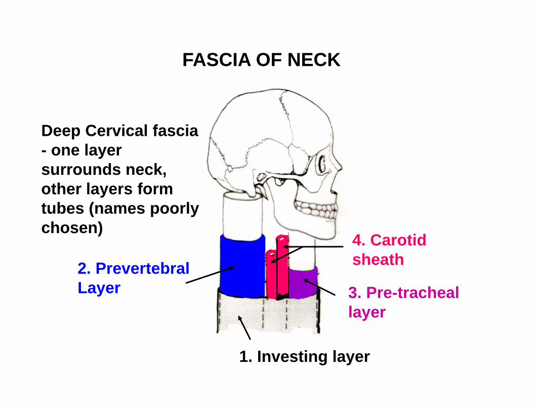
Deep Cervical fascia - one layer surrounds neck, other layers form tubes (names poorly chosen)
2. Prevertebral Layer
1. Investing layer
4. Carotid sheath
3. Pre-tracheal layer
FASCIA OF NECK
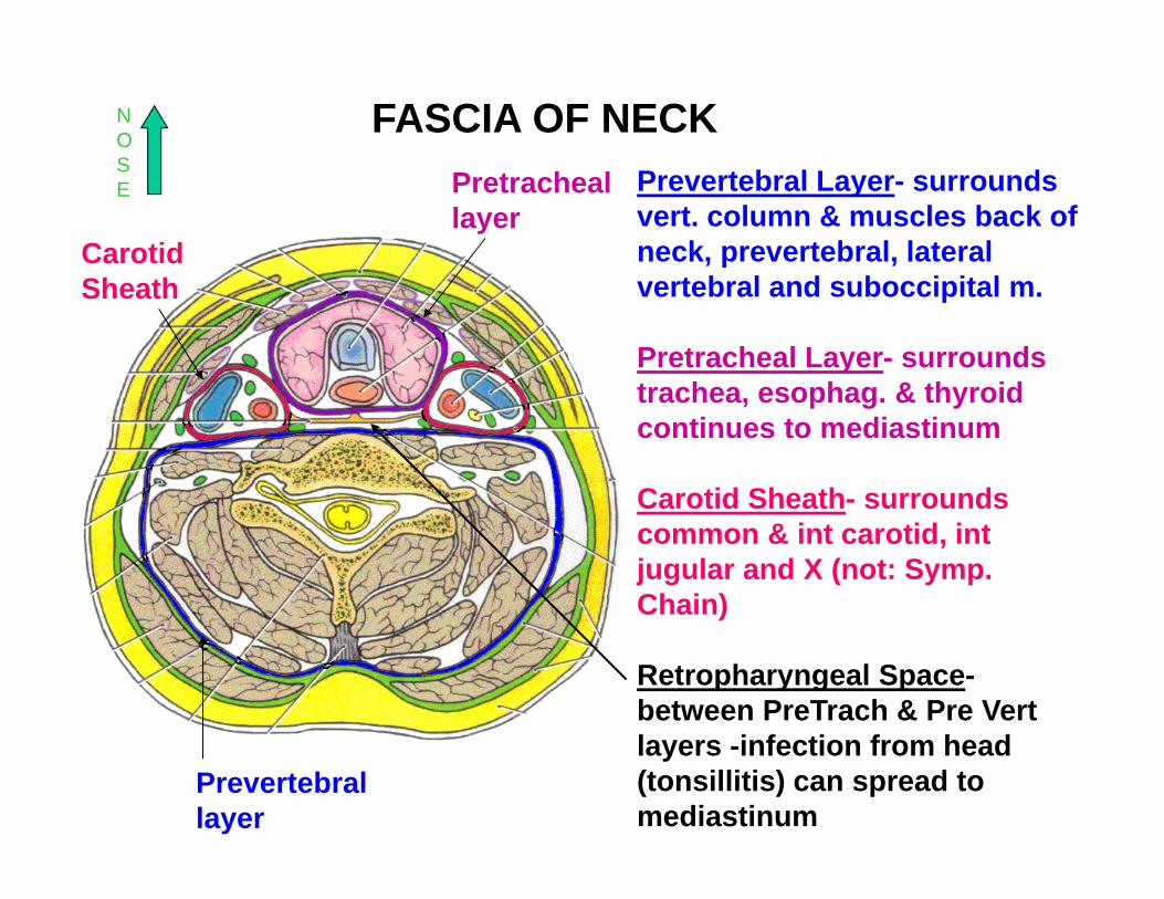
Prevertebral Layer- surrounds vert. column & muscles back of neck, prevertebral, lateral vertebral and suboccipital m.
Pretracheal Layer- surrounds trachea, esophag. & thyroid continues to mediastinum
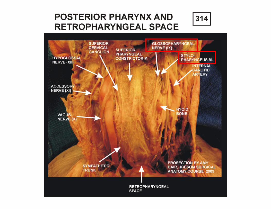
Carotid Sheath- surrounds common & int carotid, int jugular and X (not: Symp. Chain)
Retropharyngeal Space-between PreTrach & Pre Vert layers -infection from head (tonsillitis) can spread to mediastinum
Pretracheal layer
Prevertebral layer
Carotid Sheath
NOSE
FASCIA OF NECK
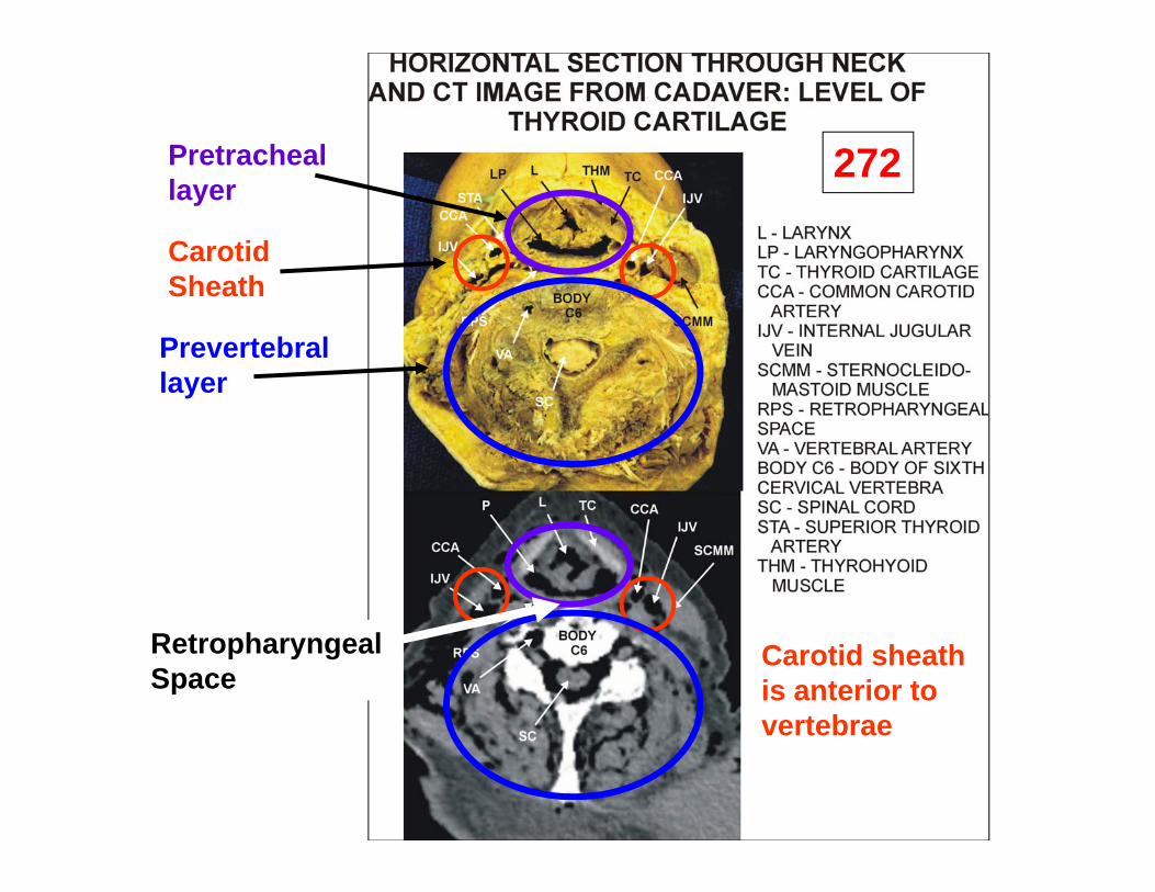
272Pretracheal layer
Prevertebral layer
Carotid Sheath
Retropharyngeal Space
Carotid sheathis anterior tovertebrae
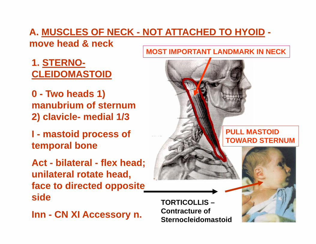
A. MUSCLES OF NECK - NOT ATTACHED TO HYOID -move head & neck
1. STERNO-CLEIDOMASTOID
0 - Two heads 1) manubrium of sternum 2) clavicle- medial 1/3
I - mastoid process of temporal bone
Act - bilateral - flex head; unilateral rotate head, face to directed opposite side
Inn - CN XI Accessory n.TORTICOLLIS –Contracture ofSternocleidomastoid
MOST IMPORTANT LANDMARK IN NECK
PULL MASTOIDTOWARD STERNUM
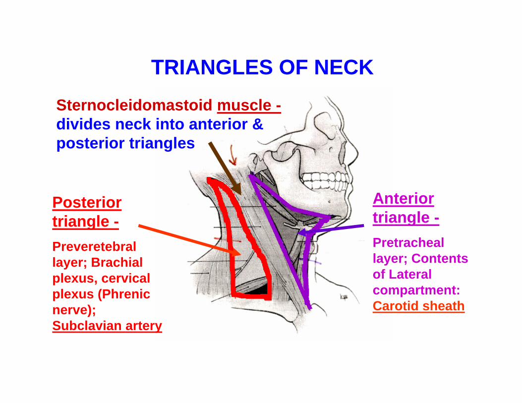
Sternocleidomastoid muscle -divides neck into anterior & posterior triangles
Posterior triangle -Preveretebral layer; Brachial plexus, cervical plexus (Phrenic nerve); Subclavian artery
Anterior triangle -Pretracheal layer; Contents of Lateral compartment: Carotid sheath
TRIANGLES OF NECK

2. SCALENUS ANTERIOR AND SCALENUS MEDIUS
O - vertebrae-trans processes upper cervical
I - rib 1
A - flex neck & elevate rib 1
Inn - ventral rami of cervical spinal nerves
MUSCLES OF NECK - NOT ATTACHED TO HYOID
SECOND MOST IMPORTANT LANDMARKS IN NECK
FLEXION
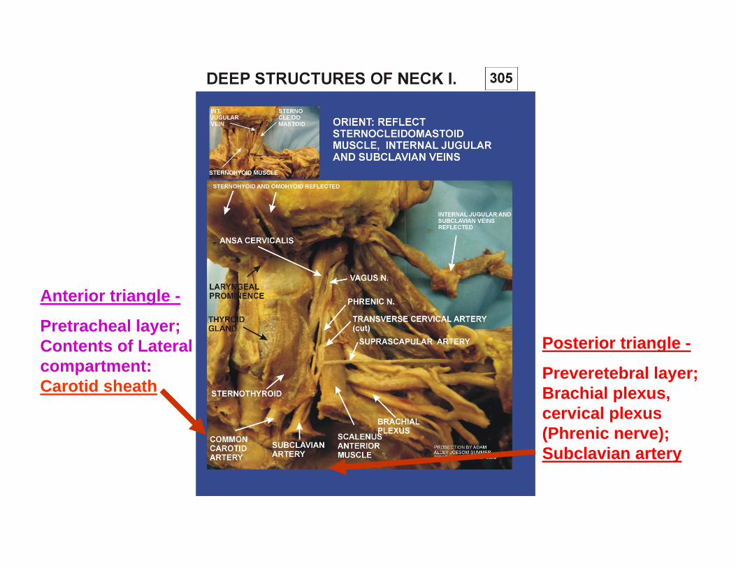
Posterior triangle -
Preveretebral layer; Brachial plexus, cervical plexus (Phrenic nerve); Subclavian artery
Anterior triangle -
Pretracheal layer; Contents of Lateral compartment: Carotid sheath
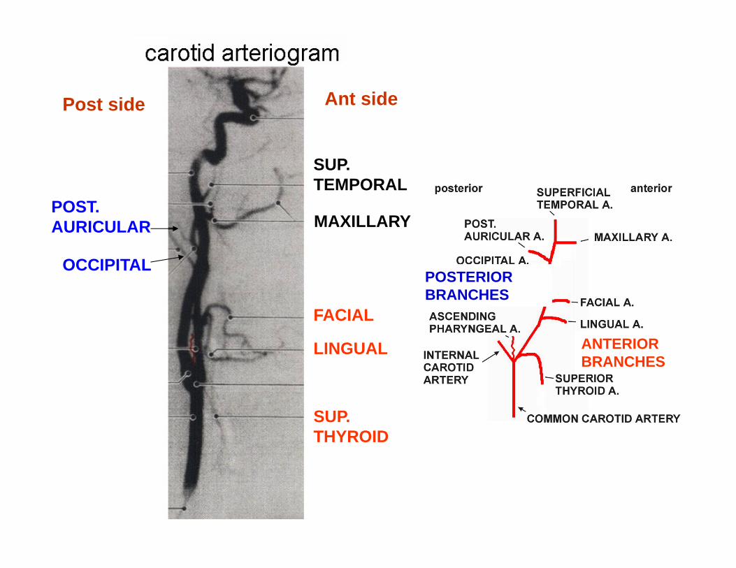
Post side Ant side
SUP. THYROID
LINGUAL
FACIAL
OCCIPITAL
POST.AURICULAR
SUP. TEMPORAL
MAXILLARY
ANTERIOR BRANCHES
POSTERIOR BRANCHES
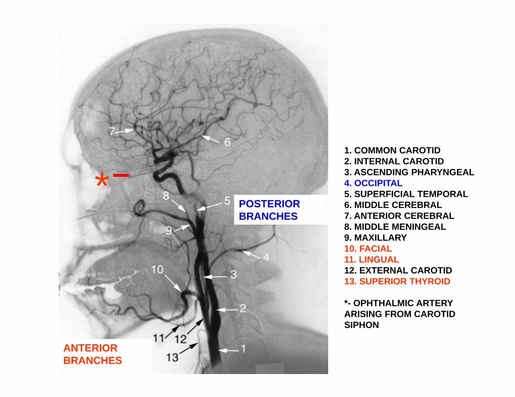
1. COMMON CAROTID2. INTERNAL CAROTID3. ASCENDING PHARYNGEAL4. OCCIPITAL5. SUPERFICIAL TEMPORAL6. MIDDLE CEREBRAL7. ANTERIOR CEREBRAL8. MIDDLE MENINGEAL9. MAXILLARY10. FACIAL11. LINGUAL12. EXTERNAL CAROTID13. SUPERIOR THYROID
*- OPHTHALMIC ARTERYARISING FROM CAROTIDSIPHON
*
ANTERIOR BRANCHES
POSTERIOR BRANCHES

ANTERIOR BRANCHES
POSTERIOR BRANCHES

PHARYNX - is continuouswith esophagus, opens to larynx trachea
ESOPHAGUS
PHARYNX
LARYNX
HYOID BONE
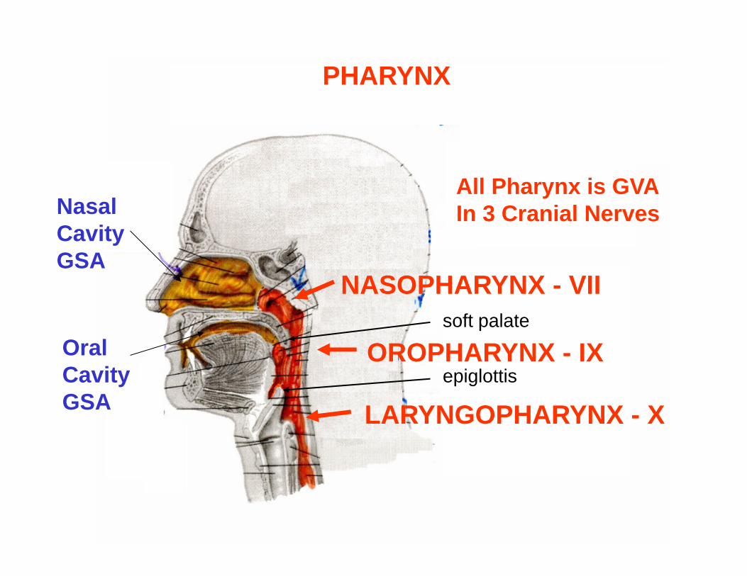
PHARYNX
Nasal CavityGSA
Oral CavityGSA
All Pharynx is GVAIn 3 Cranial Nerves
NASOPHARYNX - VII
OROPHARYNX - IX
LARYNGOPHARYNX - X
soft palate
epiglottis
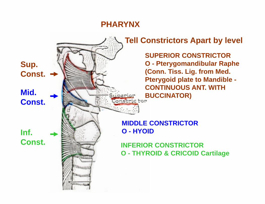
Tell Constrictors Apart by level
Sup. Const.
Mid. Const.
Inf. Const.
SUPERIOR CONSTRICTOR O - Pterygomandibular Raphe (Conn. Tiss. Lig. from Med. Pterygoid plate to Mandible -CONTINUOUS ANT. WITHBUCCINATOR)
MIDDLE CONSTRICTORO - HYOID
INFERIOR CONSTRICTORO - THYROID & CRICOID Cartilage
PHARYNX
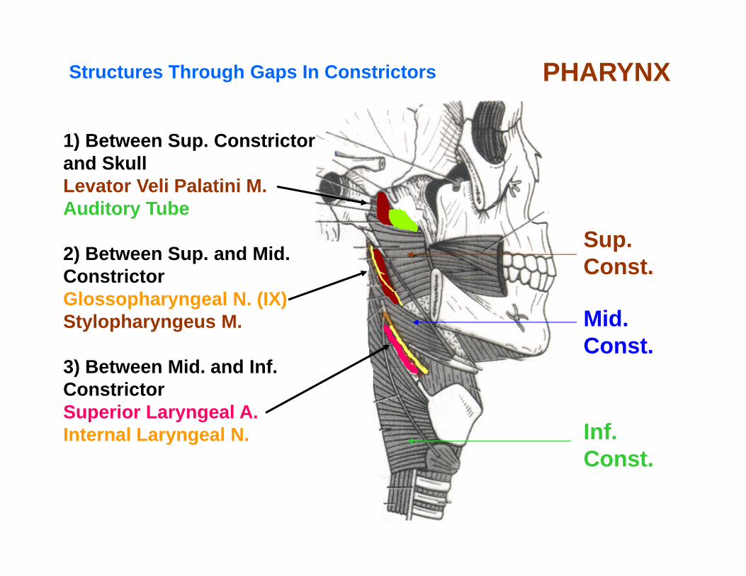
Structures Through Gaps In Constrictors PHARYNX
1) Between Sup. Constrictorand SkullLevator Veli Palatini M.Auditory Tube
2) Between Sup. and Mid.ConstrictorGlossopharyngeal N. (IX)Stylopharyngeus M.
3) Between Mid. and Inf.ConstrictorSuperior Laryngeal A.Internal Laryngeal N.
Sup. Const.
Mid. Const.
Inf. Const.
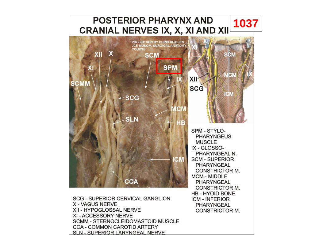
1037
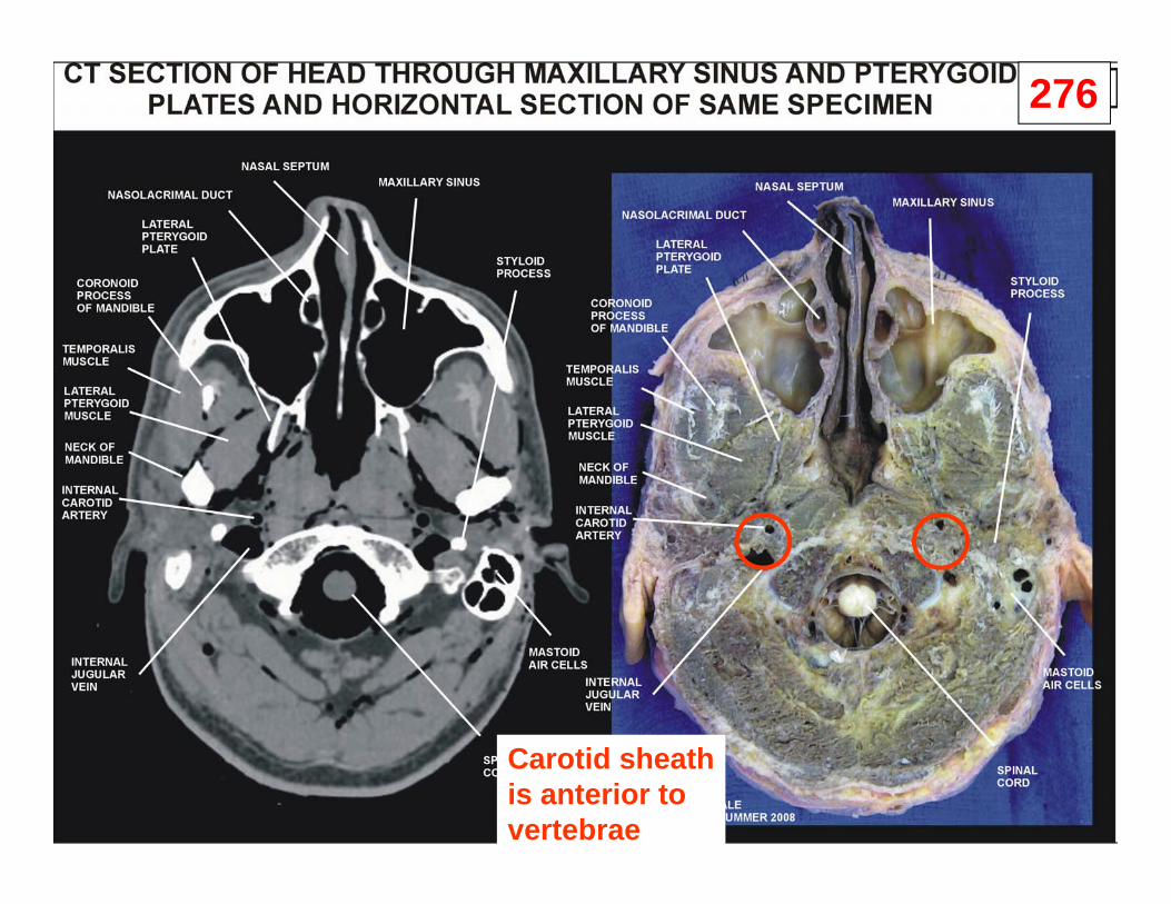
276
Carotid sheathis anterior tovertebrae

CONTENTS OF PHARYNX
in Nasopharynx- Pharyngeal Tonsil (Adenoids)- opening of Auditory Tube
in Oropharynx- Palatine Tonsils (Tonsillitis)posterior to Palatoglossal Arch
Soft Palate
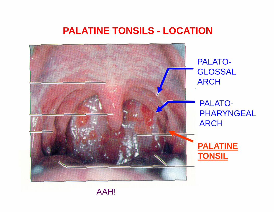
AAH!
PALATO-GLOSSALARCH
PALATO-PHARYNGEALARCH
PALATINETONSIL
PALATINE TONSILS - LOCATION
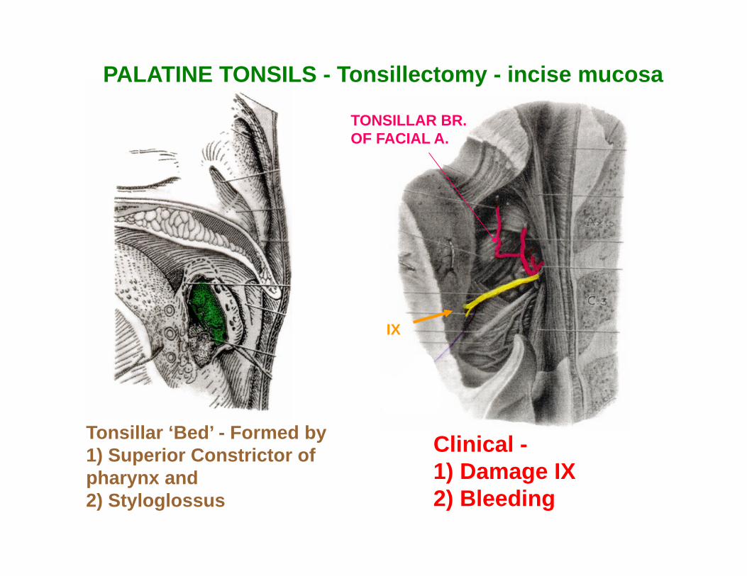
Tonsillar ‘Bed’ - Formed by 1) Superior Constrictor of pharynx and2) Styloglossus
PALATINE TONSILS - Tonsillectomy - incise mucosa
TONSILLAR BR.OF FACIAL A.
IX
Clinical -1) Damage IX 2) Bleeding
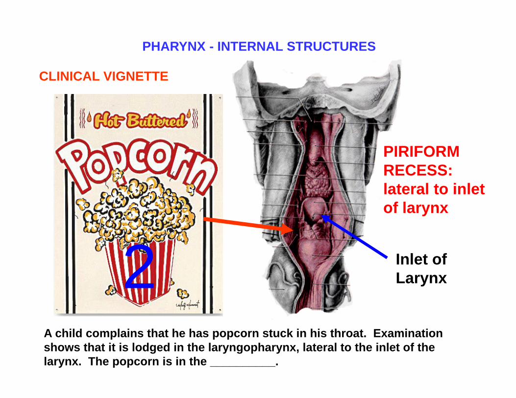
Inlet of Larynx
A child complains that he has popcorn stuck in his throat. Examination shows that it is lodged in the laryngopharynx, lateral to the inlet of the larynx. The popcorn is in the __________.
CLINICAL VIGNETTE
PIRIFORM RECESS: lateral to inlet of larynx
PHARYNX - INTERNAL STRUCTURES
2

PHARYNX -INTERNAL STRUCTURES
VALLECULAE -in Oropharynx
1

A. SUPERFICIAL STRUCTURES
1. SULCUS TERMINALIS - V-SHAPE GROOVE DIVIDES TONGUE INTO: ANT. 2/3- ORAL PART - GSA; POST 1/3 -PHARYNGEAL PART - GVA
2. FORAMEN CAECUM - PIT IN MIDDLE OF SULCUS TERMINALIS- SITE OF
INVAGINATION OF THYROID GLAND
FORAMEN CECUM
ORAL CAVITY - TONGUE
SULCUS TERMINALIS CLINICAL QUESTION:
MASS IN POSTERIOR TONGUE;CHECK IF IT IS THYROID TISSUE BEFORE SURGICAL EXCISION
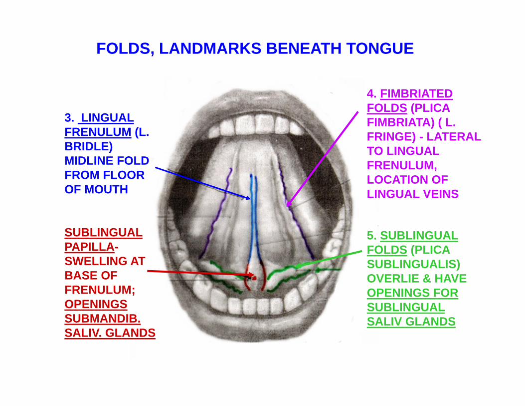
FOLDS, LANDMARKS BENEATH TONGUE
3. LINGUAL FRENULUM (L. BRIDLE) MIDLINE FOLD FROM FLOOR OF MOUTH
SUBLINGUAL PAPILLA-SWELLING AT BASE OF FRENULUM; OPENINGS SUBMANDIB. SALIV. GLANDS
4. FIMBRIATED FOLDS (PLICA FIMBRIATA) ( L. FRINGE) - LATERAL TO LINGUAL FRENULUM, LOCATION OF LINGUAL VEINS
5. SUBLINGUAL FOLDS (PLICA SUBLINGUALIS) OVERLIE & HAVE OPENINGS FOR SUBLINGUAL SALIV GLANDS
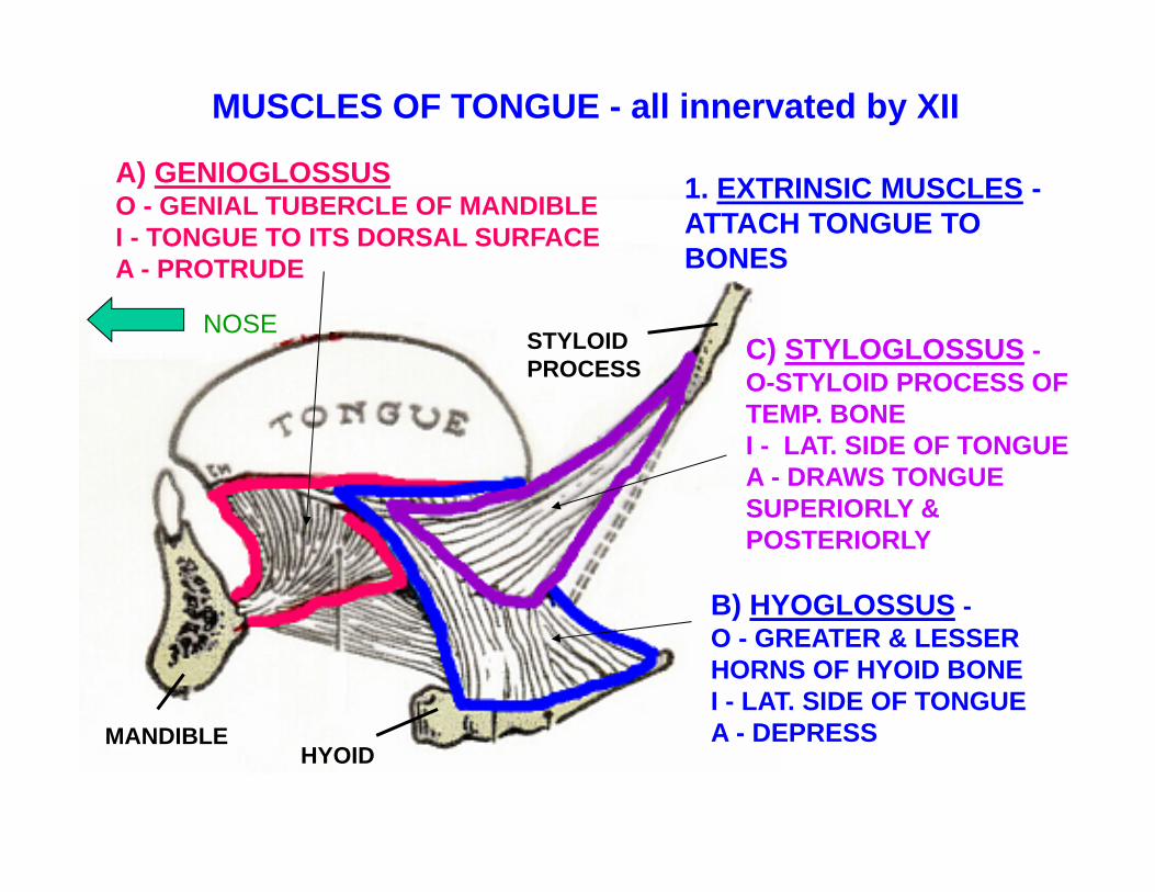
1. EXTRINSIC MUSCLES -ATTACH TONGUE TO BONES
A) GENIOGLOSSUSO - GENIAL TUBERCLE OF MANDIBLE I - TONGUE TO ITS DORSAL SURFACE A - PROTRUDE
C) STYLOGLOSSUS -O-STYLOID PROCESS OF TEMP. BONE I - LAT. SIDE OF TONGUEA - DRAWS TONGUE SUPERIORLY & POSTERIORLY
B) HYOGLOSSUS -O - GREATER & LESSER HORNS OF HYOID BONE I - LAT. SIDE OF TONGUE A - DEPRESS
NOSE STYLOIDPROCESS
MANDIBLEHYOID
MUSCLES OF TONGUE - all innervated by XII

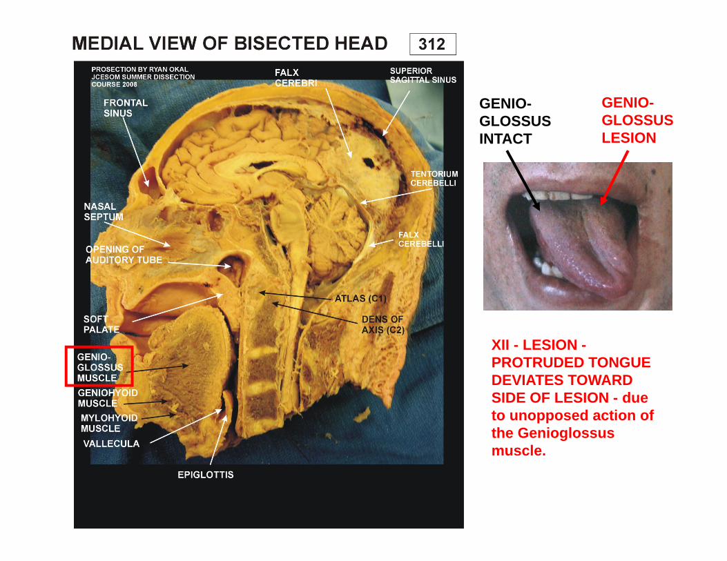
XII - LESION -PROTRUDED TONGUE DEVIATES TOWARD SIDE OF LESION - due to unopposed action of the Genioglossus muscle.
GENIO-GLOSSUSINTACT
GENIO-GLOSSUSLESION
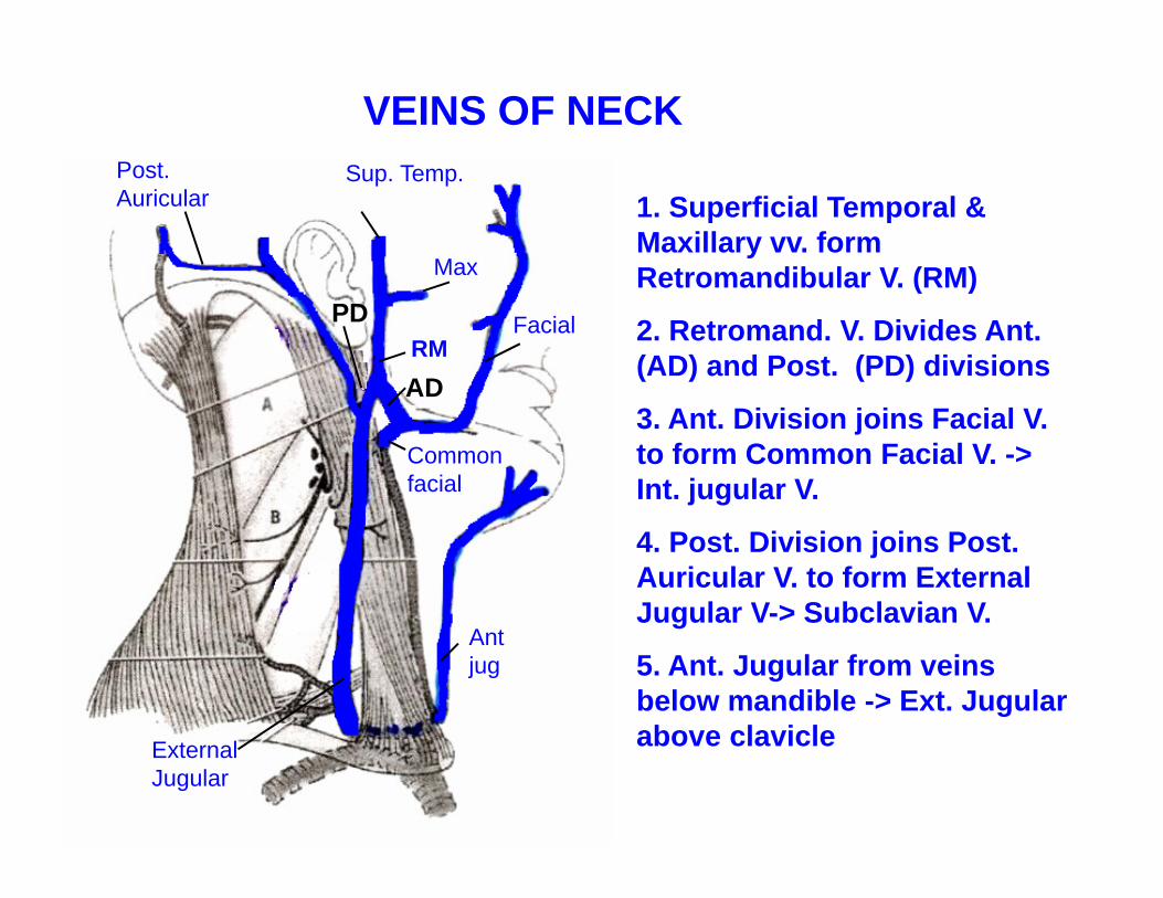
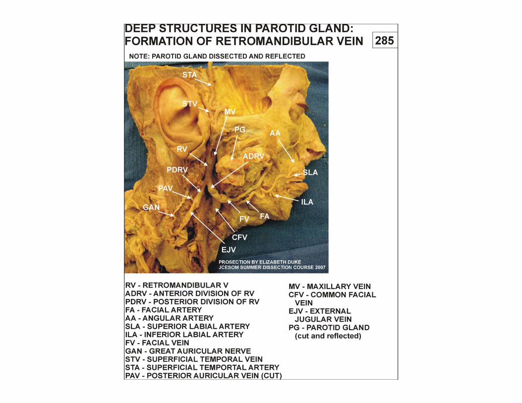
1. Superficial Temporal & Maxillary vv. form Retromandibular V. (RM)
2. Retromand. V. Divides Ant. (AD) and Post. (PD) divisions
3. Ant. Division joins Facial V. to form Common Facial V. -> Int. jugular V.
4. Post. Division joins Post. Auricular V. to form External Jugular V-> Subclavian V.
5. Ant. Jugular from veins below mandible -> Ext. Jugular above clavicle
Sup. Temp.
Max
Post. Auricular
External Jugular
Common facial
Facial
Ant jug
VEINS OF NECK
RMAD
PD

VEINS OF NECK
Pattern of Venous Drainage

Good luck!
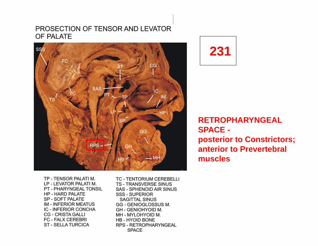
231
RETROPHARYNGEALSPACE -posterior to Constrictors;anterior to Prevertebralmuscles
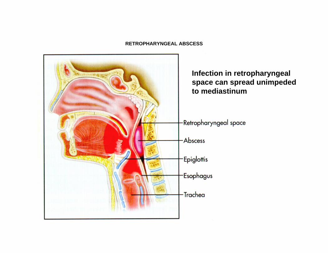
RETROPHARYNGEAL ABSCESS
Infection in retropharyngealspace can spread unimpededto mediastinum