Head and Neck Exam
41
Transcript of Head and Neck Exam

Observation and Palpation
• Inspection face & neck: – Does anything appear out
of ordinary in Head & Neck?
– Bumps/lumps, asymmetry, swelling, discoloration, bruising/trauma?
– anything hidden by hair?
• Inspection & palpation of scalp, hair
Note right sided neck/jaw area swelling
and R v L asymmetry
Hammer & Nails icon indicates A Slide
Describing Skills You Should Perform In Lab
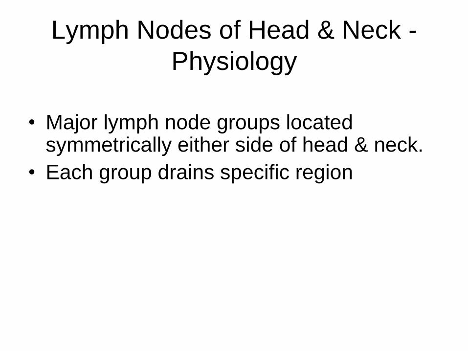
Lymph Nodes of Head & Neck -
Physiology
• Major lymph node groups located symmetrically either side of head & neck.
• Each group drains specific region

Lymph Node Enlargement – Major
Causes • Enlarged if inflammation (most commonly
infection) or malignancy Infection: Acute, tender, warm
– Primary region drained also involved (e.g neck nodes w/strep throat)
– Sometimes diffuse enlargement w/generalized infection or systemic inflammatory process (e.g. TB, HIV, Mono)
Malignancy:
– Slowly progressive, firm, multiple nodes, stuck together & to underlying structures.
– Primary site malignancy could be nodes (e.g. lymphoma) or adjacent region (e.g. intra-oral squamous cell ca)

Lymph Node Anatomy &
Drainage
Ant CervThroat, tonsils, post pharynx, thyroid
Post CervBack of skull
TonsillarTonsils, posterior pharynx
Sub-MandibularFloor of mouth
Sub-MentalTeeth
Supra-ClavicularThorax
Pre-AuricularEar

Lymph Node Exam
• Gently walk fingers along general regions
– comparing R to L

Function CN 7 – Facial Nerve
Facial Symmetry & Expression -
Precise Pattern of Inervation
L UMN R UMN
R LMN -
Forehead
R LMN – Face
L LMN -
Forehead
L LMN -Face

CN 7 – Exam
• Observe facial symmetry
• Wrinkle Forehead
• Keep eyes closed against resistance
• Smile, puff out cheeks
Cute.. and symmetric!

Comparison of a patient with (A) a facial nerve (Bell’s Type - LMN) lesion
and (B) a supra-nuclear (UMN) lesion w/forehead sparing Tiemstra J et al. Bell’s Palsy: Diagnosis and Management, Amer J Fam Practice, 2007;76(7):997-1002.
http://www.aafp.org/afp/2007/1001/p997.pdf
Note forehead
and lower face are affected on the
right, which is same side of the LMN lesion
Note forehead sparing on right side,
opposite the UMN lesion
Upper
Motor
Neuron
(UMN)
Lower
Motor
Neuron
(LMN)
A B

Pathology: Peripheral CN 7 (Bell’s)
Palsy
Central (i.e. UMN) CN 7 dysfunction (e.g. stroke) - not shown: Can
wrinkle forehead bilaterally; will demonstrate loss of lower facial
movement on side opposite stroke.
Patient can’t close L eye, wrinkle L forehead or
raise L corner mouthL CN 7 Peripheral (i.e. LMN)
Dysfunction

Function CN 5 - Trigeminal
• Sensation:
– 3 regions of face: Ophthalmic, Maxillary &
Mandibular
• Motor:
– Temporalis & Masseter muscles

Function CN 5 – Trigeminal
(cont)
Ophthalmic(V1)
Maxillary (V2)
Mandibular (V3)
Temporalis
(clench teeth)
Masseter (move
jaw side-side)
Sensory Motor
Corneal Reflex: Blink when cornea touched - Sensory CN 5, Motor CN 7

Temporalis & Masseter Muscles
Courtesy Oregon Health Sciences University:
http://home.teleport.com/~bobh/

Testing CN 5 - Trigeminal
• Sensory: – Ask pt to close eyes
– Touch ea of 3 areas (ophthalmic, maxillary, & mandibular) lightly, noting whether patient detects stimulus.
• Motor: – Palpate temporalis & mandibular areas as patient
clenches & grinds teeth
• Corneal Reflex: – Tease out bit of cotton from q-tip - Sensory CN 5,
Motor CN 7
– Blink when touch cornea with cotton wisp

The Ear – Functional Anatomy and
Testing
(CN 8 – Acoustic) • Crude tests hearing –
rub fingers next to
either ear; whisper &
ask pt repeat words
• If sig hearing loss,
determine Conductive
(external canal up to
but not including CN
8) v Sensorineural
(CN 8) Image Courtesy: Online Otoscopy Tutorial
http://www.uwcm.ac.uk:9080/otoscopy/index.htm
Vestibular
CN8
Auditory
CN8
Conduction Sensorineural

Great Moments In The History of
Hearing
Uncle Bill Hears Aunt Ruth!

CN 8 - Defining Cause of
Hearing Loss - Weber Test • 512 Hz tuning fork - this
(& not 128Hz) is well w/in range normal hearing & used for testing – Get turning fork vibrate
striking ends against heel of hand or
Squeeze tips between thumb & 1st finger
• Place vibrating fork mid line skull
• Sound should be heard
=ly R & L bone conducts to both sides.
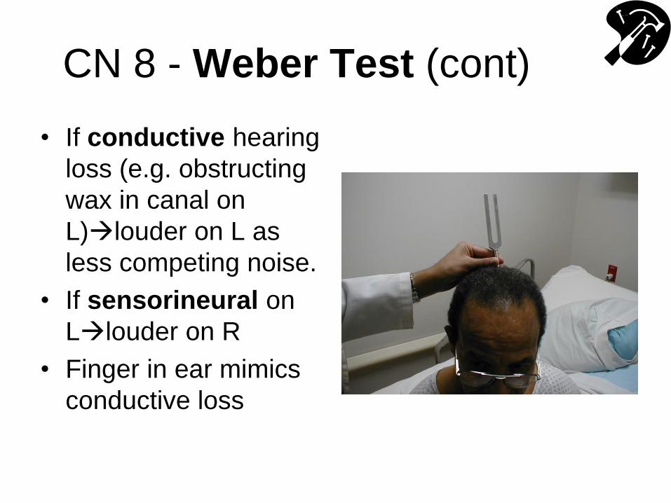
CN 8 - Weber Test (cont)
• If conductive hearing
loss (e.g. obstructing
wax in canal on
L)louder on L as
less competing noise.
• If sensorineural on
Llouder on R
• Finger in ear mimics
conductive loss

CN 8 - Defining Cause of
Hearing Loss - Rinne Test • Place vibrating 512 hz
tuning fork on mastoid bone (behind ear).
• Patient states when can’t hear sound.
• Place tines of fork next to ear should hear it again – as air conducts better then bone.
• If BC better then AC, suggests conductive hearing loss.
• If sensorineural loss, then AC still > BC
Note: Weber & Rinne difficult to perform in Anatomy lab due to competing
noise – repeat @ home in quiet room!

Examining the External Structures of
The Ear - Observation
Tragus
External
Canal
Helix
Anti-Helix
Lobe
Mastoid
Note: Picture on L normal external ear; picture on
R swollen external canal, narrowed by inflammation

Internal Ear Anatomy
Image Courtesy: Online Otoscopy Tutorial
http://www.uwcm.ac.uk:9080/otoscopy/index.htm

Normal Tympanic Membrane
Images courtesy American Academy of Pediatrics
http://www.aap.org/otitismedia/www/
Cone of
Light
Umbo
Short Process
Malleus
Long Process
Malleus Incus
NOSE
Left Ear –
Malleus points
down and back
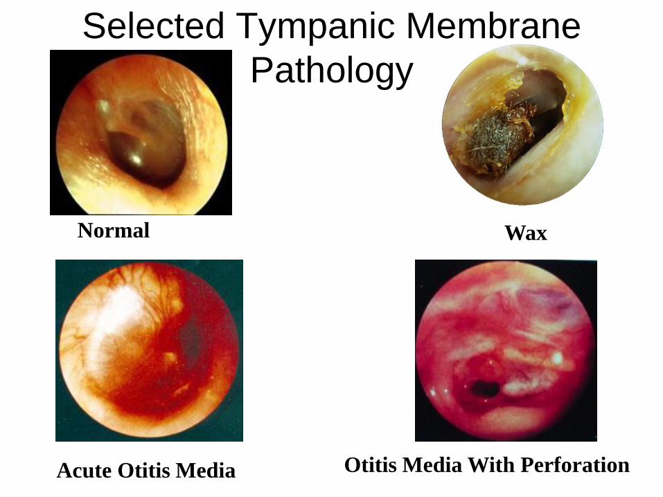
Selected Tympanic Membrane
Pathology
Acute Otitis Media Otitis Media With Perforation
Normal
Wax Normal

Using Your Otoscope
• Make sure battery’s
charged!
• Gently twist Otoscopic
Head (clockwise) onto
handle
• Twist on disposable,
medium sized speculum
• Hold in R hand R ear,
L hand L ear

Otoscope W/Magnified Viewing
Head • Advantage magnified
view, larger field
• Speculum twists on;
viewing same as for
conventional head
• Rotate wheel w/finger
while viewing tympanic
membrane to enhance
focus (default setting is
green line)
•
Focus
Wheel
Speculum
Viewing Window
Welchallyn.com

Otosocopy Basics
• Make sure patient seated
comfortably & ask them not
to move
• Place tip speculum in
external canal under direct
vision
• Gently pull back on top of
ear
• Advance scope slowly as
look thru window – extend
pinky to brace hand
• Avoid fast, excessive
movement – Stop if painful!

Look Dad - Otoscopy Sure is Easy!
NEJM Video - Diagnosing Otitis Media: http://www.nejm.org/doi/full/10.1056/NEJMvcm0904397#figure=preview.jpg

The Nose • Observe external
structure for symmetry
• Check air movement thru ea nostril separately.
• Smell (CN 1 – Olfactory) not usually assessed (unless sx) – use coffee grounds or other
w/distinctive odor
(e.g. mint, wintergreen, etc)
- detect odor when presented @ 10cm.
• Look into each nostril using otoscope w/speculum – note color, septum (medial), turbinates (lateral)
Hmmm..
Coffee!

Sinuses
• Normally Air filled (cuts down weight of skull), lined w/upper respiratory epitheliumkeeps antigens/infection from lung
• Maxillary & frontal accessible to exam (others not)
• Exam only done if concern re sinus infection/pathology
Anatomy
Image: Williams, J. JAMA 270 (10);
1993: 1242-46

Sinuses (cont) If there is concern for acute sinusitis (purulent nasal d/c, facial pain/fullness, nasal congestion, post nasal drip, cough, sometimes fever):
• Palpate (or percuss) sinus elicits pain if inflamed/infected
• Trans-illuminate normally, light passes across sinus visible thru roof of mouth.. Infection swelling & fluid prevents transmission
• Room must be dark
• Placed otoscope on infra-orbital rim while look in mouth for light
Note: Not possible to see transmitted light if room brightly lit (e.g. the anatomy lab) – try this @ home in dark room!
Transillumination
Palpation
Image: Williams, J. JAMA 270 (10); 1993: 1242-46

Oropharynx
• Inspect posterior pharynx (back of throat), tonsils, mucosa, teeth, gums, tongue – use tongue depressor & light – otoscope works as flashlight (on newer Welch Allyn, head twists off)
• Can grasp tongue w/a gauze pad & move it side to side for better visualization
• Palpate abnormalities (gloved hand)

Oropharynx: Anatomy & Function CNs 9
(glosopharyngeal), 10 (vagus) & 12
(hypoglossal)
• Uvula midline - CN 9
• Stick out tongue, say “Ahh” – use tongue depressor if can’t see – palate/uvula rise -CN 9, 10
• Gag Reflex – provoked w/tongue blade or q tip - CN 9, 10
• Tongue midline when
patient sticks it outCN 12 – check strength by directing
patient push tip into inside of either cheek while you push from outside

Selected Pathology of
Oropharynx
L CN 9 palsy – uvula
pulled to R L peri-tonsilar abscess – uvula
pushed to R
L CN 12 palsy –
tongue deviates L

Parotid and other Salivary Glands
• Contribute saliva to food
• Drain into mouth via
discrete ducts
– Parotid next to
upper molars
– Submandibular floor
of mouth
• Glands not easily
palpable
• Painful &/or swollen if:
obstruction, inflammation,
infection or cancer
Images from LSU School of Medicine:
www.medschool.lsuhsc.edu/.../docs/parotitis.pptx
Wharton’s Ducts Stensens’s Duct
(sub-mandibular) (parotid)

What about the Teeth?
• Dental health has big implications:
– Nutrition (ability to eat)
– Appearance • Self esteem
• Employability
• Social acceptance
– Systemic diseaseendocarditis, ? other
– Local problems: • Pain, infection
• Profound lack of access to careMDs primary Rx

Dental Anatomy & Exam
• 16 top, 16 bottom
• Examine all
– Observation teeth, gums
– Gloved hands, gauze, tongue
depressor & lighting if
abnormal
• Look for:
– General appearance
• ? All present
• Broken, Caries, etc?
– Areas pain, swelling ?
infection
• Localize: ? Tooth, gum, extent
http://www.nytimes.com
http://www.nlm.nih.gov/medlineplus

Common Dental Pathology
Caries: Breakdown in Enamel
American Family Physician: Common Dental Emergencies
http://www.aafp.org/afp/20030201/511.html
Facial Swelling (left) Secondary to Tooth Abscess

Thyroid Anatomy
Image: Strome, T. NEJM 344;
2001: 1676-79

Thyroid Exam
• Observe (obvious
abnormalities, trachea)
• From front or behind
Identify landmarks
(touch and vision)
• Palpate as patient
swallows (drinking water
helps)
• ? Focal or symmetric
enlargement, nodules.

Neck Movement
(CN 11 – Spinal Accessory)
• Turn head to L into R
hand function of R
Sternocleidomastoid
(SCM)
• Turn head to R into L
hand (L SCM)
• Shrug shoulders into
your hands

Summary Of Skills □ Wash hands
□ Observation head & scalp; palpation lymph node, parotid and salivary gland regions
□ Facial symmetry, expression (CN 7)
□ Facial sensation, muscles mastication (CN 5)
□ Auditory acuity; Weber & Rinne Tests (CN 8)
□ Ear: external and internal (otoscope)
□ Nose: observation, nares/mucosa (otoscope), smell (CN 1)
□ Sinuses: palpation, trans-illumination (*bag of tricks*)
□ Oropharynx: Inspection w/light & tongue depressor uvula, tonsils, tongue (12); Inspect Teeth, Salivary gland ducts; Tongue movement (CN 12); “Ahh” & Gag reflex (CNs 9, 10);
□ Thyroid: Observation, palpation
□ Neck/Shoulders: Observation, range motion, shrug (CN 11)
□ Wash hands
Time Target: < 10 min