Head, Facial, & Neck Trauma. Sections Introduction to Head, Facial, & Neck Injuries Anatomy and...
119
Head, Facial, & Head, Facial, & Neck Trauma Neck Trauma
-
Upload
aron-simes -
Category
Documents
-
view
258 -
download
2
Transcript of Head, Facial, & Neck Trauma. Sections Introduction to Head, Facial, & Neck Injuries Anatomy and...
Head, Facial, & Head, Facial, & Neck TraumaNeck Trauma
SectionsSections Introduction to Head, Facial, & Neck
Injuries Anatomy and Physiology of the Head,
Face, & Neck Pathophysiology of Head, Facial, &
Neck Injury Assessment and Management of Head,
Facial, & Neck Injuries Head, Facial, & Neck Injury Management
Introduction to Head, Facial, & Neck Injuries
Anatomy and Physiology of the Head, Face, & Neck
Pathophysiology of Head, Facial, & Neck Injury
Assessment and Management of Head, Facial, & Neck Injuries
Head, Facial, & Neck Injury Management
Common major trauma 4 million people experience head
trauma annually Severe head injury is most frequent cause of trauma
death GSW to cranium: 75-80% mortality
At Risk population Males 15-24 Infants Young Children Elderly
Common major trauma 4 million people experience head
trauma annually Severe head injury is most frequent cause of trauma
death GSW to cranium: 75-80% mortality
At Risk population Males 15-24 Infants Young Children Elderly
Introduction to Head, Introduction to Head, Facial, Facial,
& Neck Injuries& Neck Injuries
Injury Prevention Programs Motorcycle Safety Bicycle Safety Helmet & Head Injury Awareness Programs Other Sports
Football Rollerblading Contact Sports
Injury Prevention Programs Motorcycle Safety Bicycle Safety Helmet & Head Injury Awareness Programs Other Sports
Football Rollerblading Contact Sports
Introduction to Head, Introduction to Head, Facial, Facial,
& Neck Injuries& Neck Injuries
TIME IS CRITICAL Intracranial Hemorrhage Progressing Edema
Increased ICP Cerebral Hypoxia Permanent Damage
Severity is difficult to recognize Subtle signs Improve differential diagnosis
Improves survivability
TIME IS CRITICAL Intracranial Hemorrhage Progressing Edema
Increased ICP Cerebral Hypoxia Permanent Damage
Severity is difficult to recognize Subtle signs Improve differential diagnosis
Improves survivability
Introduction to Head, Introduction to Head, Facial, Facial,
& Neck Injuries& Neck Injuries
Anatomy & Physiology of the Head Scalp Cranium Meninges Cerebrospinal Fluid Brain CNS Circulation Blood-Brain Barrier Cerebral Perfusion Pressure Cranial Nerves Ascending Reticular Activating System
Anatomy & Physiology of the Head Scalp Cranium Meninges Cerebrospinal Fluid Brain CNS Circulation Blood-Brain Barrier Cerebral Perfusion Pressure Cranial Nerves Ascending Reticular Activating System
Anatomy & PhysiologyAnatomy & PhysiologyHead, Face & NeckHead, Face & Neck
Scalp Strong Flexible mass of
Skin Fascia Muscular Tissue
Highly Vascular Hair provides Insulation Structures Beneath
Galea Aponeurotica• Between scalp and skull• Fibrous connective sheath
Subaponeurotica (Areolar) Tissue• Permits venous blood flow from the dural sinuses to the venous
vessels of scalp Emissary Veins: Potential route for Infection
Anatomy & Anatomy & Physiology Physiology of the Headof the Head
Recalling Structures of the ScalpS - skinC - connective tissueA - aponeuroticaL - layer of areolar tissueP - periosteum of skull
Recalling Structures of the ScalpS - skinC - connective tissueA - aponeuroticaL - layer of areolar tissueP - periosteum of skull
Anatomy & Anatomy & Physiology Physiology of the Headof the Head
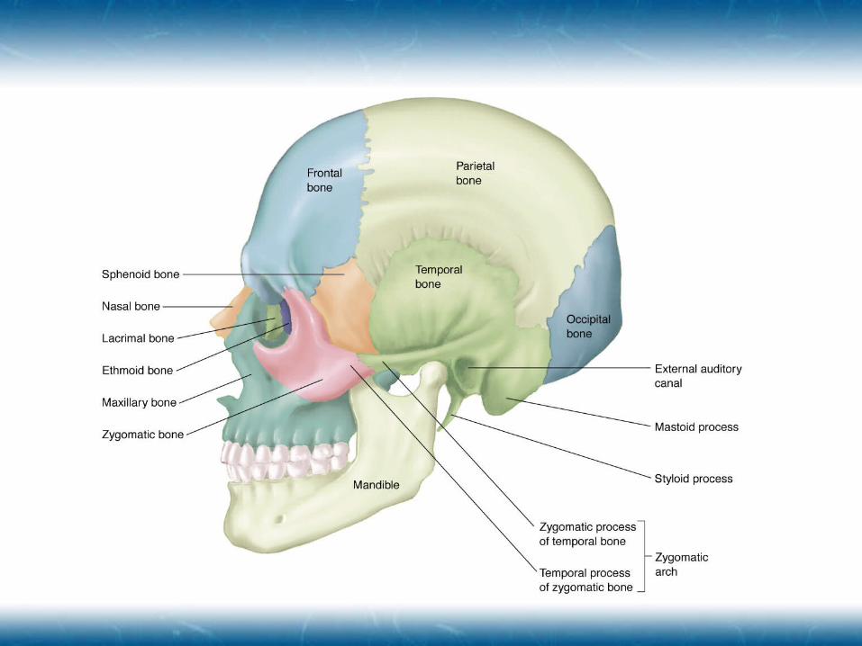
Skull comprised of Facial bones Cranium
Vault for the brain Strong, light, rigid, spherical bone Unyielding to increased intracranial pressure (ICP) Bones
• Frontal• Parietal• Occipital• Temporal• Ethmoid• Sphenoid
Skull comprised of Facial bones Cranium
Vault for the brain Strong, light, rigid, spherical bone Unyielding to increased intracranial pressure (ICP) Bones
• Frontal• Parietal• Occipital• Temporal• Ethmoid• Sphenoid
Anatomy & Anatomy & Physiology Physiology of the Headof the Head
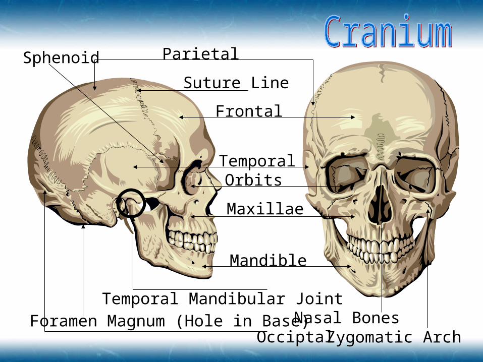
Parietal
Suture Line
Frontal
TemporalOrbits
Maxillae
Mandible
Temporal Mandibular Joint
OcciptalNasal Bones
Zygomatic Arch
Sphenoid
Foramen Magnum (Hole in Base)
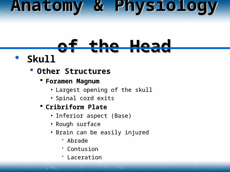
Skull Other Structures
Foramen Magnum• Largest opening of the skull
• Spinal cord exits
Cribriform Plate• Inferior aspect (Base)
• Rough surface
• Brain can be easily injured Abrade Contusion Laceration
Skull Other Structures
Foramen Magnum• Largest opening of the skull
• Spinal cord exits
Cribriform Plate• Inferior aspect (Base)
• Rough surface
• Brain can be easily injured Abrade Contusion Laceration
Anatomy & Anatomy & Physiology Physiology of the Headof the Head
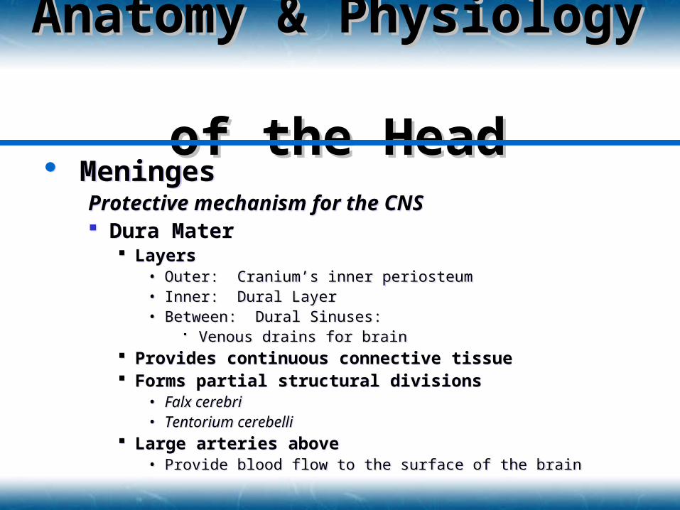
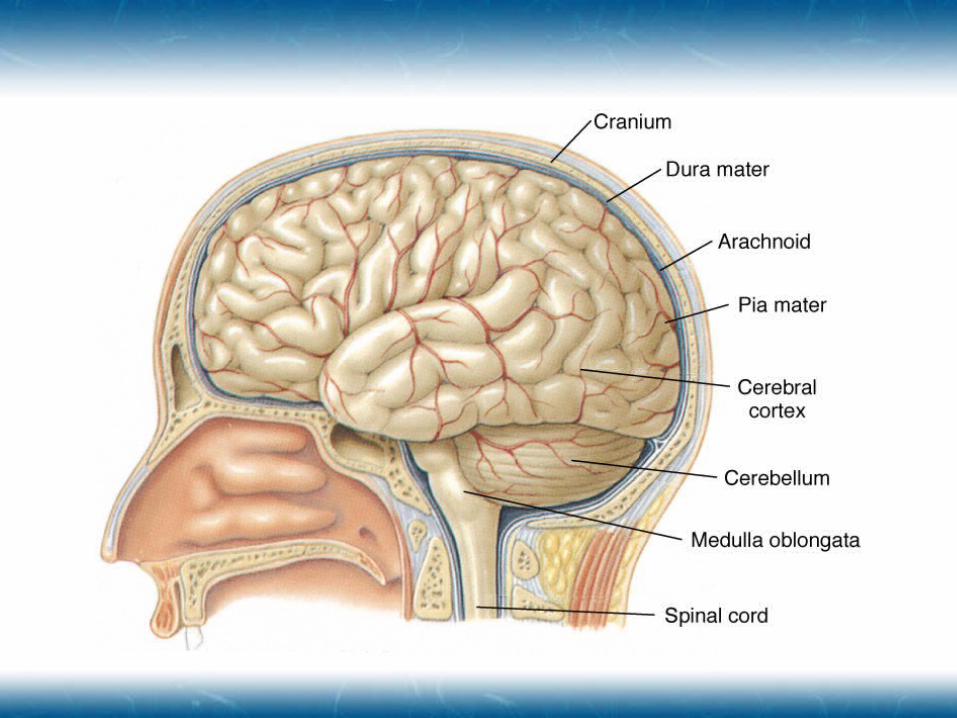
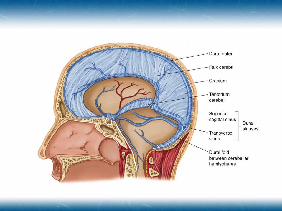
MeningesProtective mechanism for the CNS Dura Mater
Layers• Outer: Cranium’s inner periosteum• Inner: Dural Layer• Between: Dural Sinuses:
Venous drains for brain Provides continuous connective tissue Forms partial structural divisions
• Falx cerebri• Tentorium cerebelli
Large arteries above• Provide blood flow to the surface of the brain
Anatomy & Anatomy & Physiology Physiology of the Headof the Head
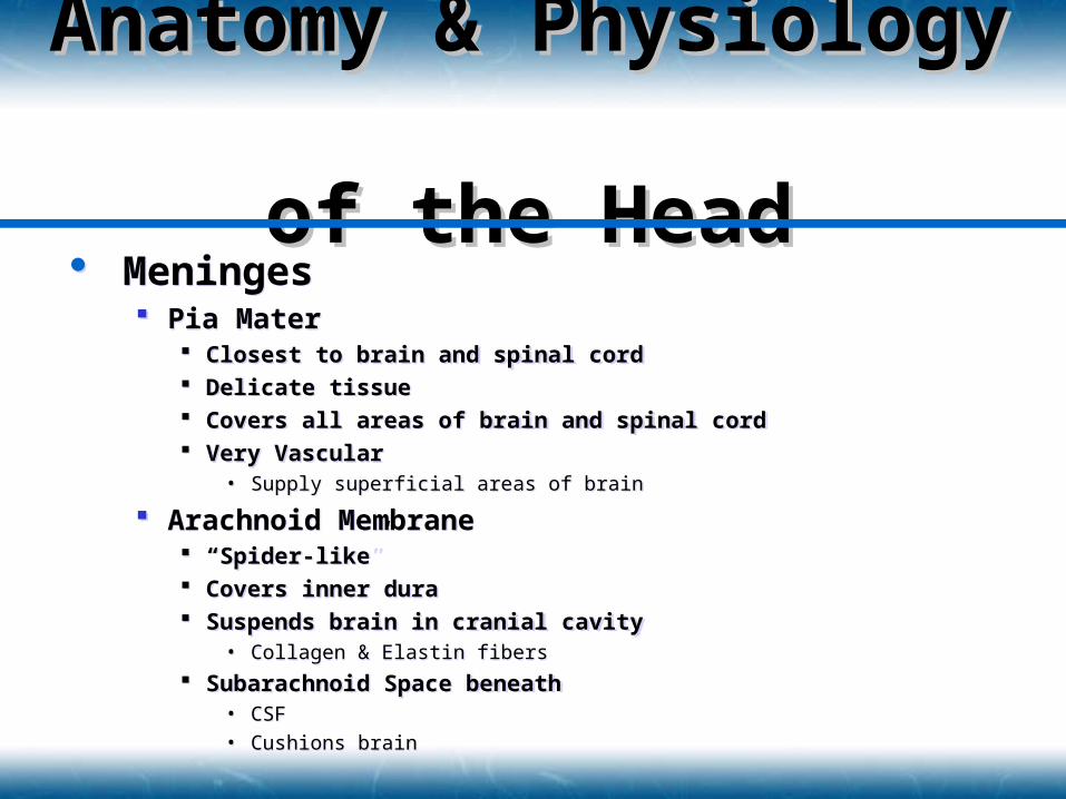
Meninges Pia Mater
Closest to brain and spinal cord Delicate tissue Covers all areas of brain and spinal cord Very Vascular
• Supply superficial areas of brain
Arachnoid Membrane “Spider-like” Covers inner dura Suspends brain in cranial cavity
• Collagen & Elastin fibers
Subarachnoid Space beneath• CSF
• Cushions brain
Anatomy & Anatomy & Physiology Physiology of the Headof the Head
Cerebrospinal Fluid Clear, colorless fluid Comprised of
Water Protein Salts
Cushions CNS Made in largest two ventricles of brain Medium for nutrients and waste products to
diffuse into and out of brain
Cerebrospinal Fluid Clear, colorless fluid Comprised of
Water Protein Salts
Cushions CNS Made in largest two ventricles of brain Medium for nutrients and waste products to
diffuse into and out of brain
Anatomy & Anatomy & Physiology Physiology of the Headof the Head
Brain Occupies 80% of cranium Comprised of 3 Major Structures
Cerebrum Cerebellum Brainstem
High metabolic rate Receives 15% of cardiac output Consumes 20% of body’s oxygen Requires constant circulation
IF Blood supply stops Unconscious within 10 seconds Death in 4-6 minutes
Brain Occupies 80% of cranium Comprised of 3 Major Structures
Cerebrum Cerebellum Brainstem
High metabolic rate Receives 15% of cardiac output Consumes 20% of body’s oxygen Requires constant circulation
IF Blood supply stops Unconscious within 10 seconds Death in 4-6 minutes
Anatomy & Anatomy & Physiology Physiology of the Headof the Head
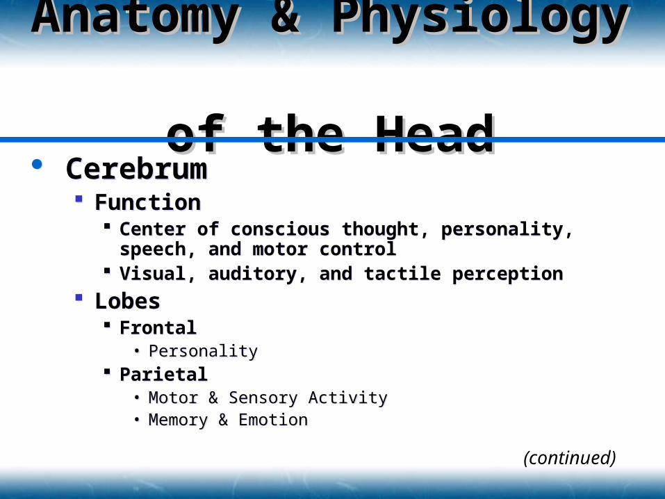
Cerebrum Function
Center of conscious thought, personality, speech, and motor control
Visual, auditory, and tactile perception Lobes
Frontal• Personality
Parietal• Motor & Sensory Activity• Memory & Emotion
Cerebrum Function
Center of conscious thought, personality, speech, and motor control
Visual, auditory, and tactile perception Lobes
Frontal• Personality
Parietal• Motor & Sensory Activity• Memory & Emotion
Anatomy & Anatomy & Physiology Physiology of the Headof the Head
(continued)
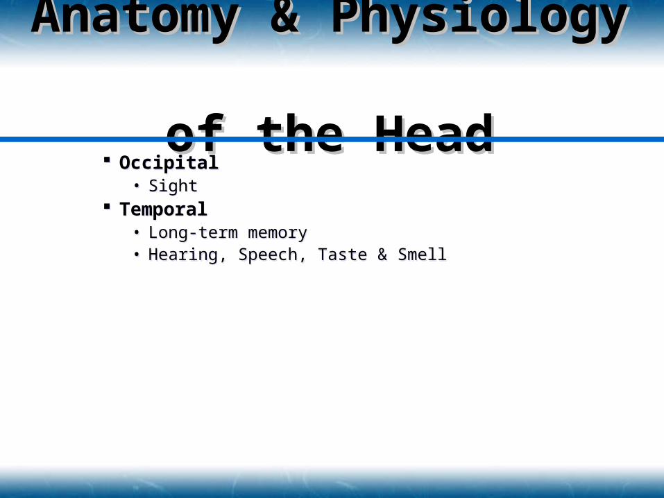
Occipital• Sight
Temporal• Long-term memory• Hearing, Speech, Taste & Smell
Occipital• Sight
Temporal• Long-term memory• Hearing, Speech, Taste & Smell
Anatomy & Anatomy & Physiology Physiology of the Headof the Head
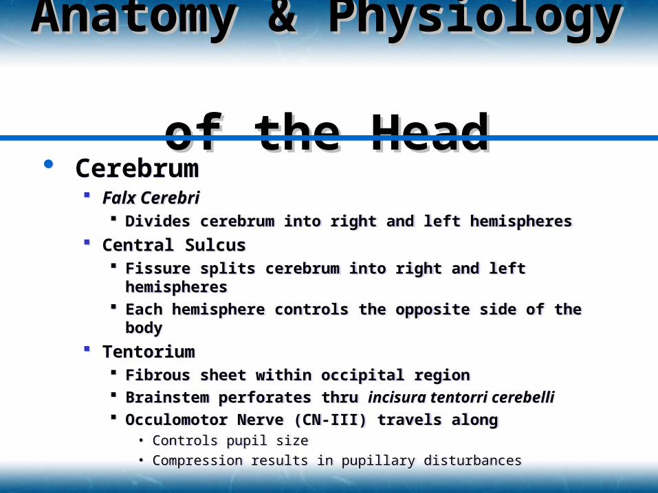
Cerebrum Falx Cerebri
Divides cerebrum into right and left hemispheres
Central Sulcus Fissure splits cerebrum into right and left hemispheres Each hemisphere controls the opposite side of the body
Tentorium Fibrous sheet within occipital region Brainstem perforates thru incisura tentorri cerebelli Occulomotor Nerve (CN-III) travels along
• Controls pupil size• Compression results in pupillary disturbances
Anatomy & Anatomy & Physiology Physiology of the Headof the Head
Cerebrum Hemisphere Functions
Left: DOMINANT• Mathematical computations: Occipital
• Writing: Parietal
• Language interpretation: Occipital
• Speech: Frontal
Right: NON-DOMINANT• Non-verbal imagery
Cerebrum Hemisphere Functions
Left: DOMINANT• Mathematical computations: Occipital
• Writing: Parietal
• Language interpretation: Occipital
• Speech: Frontal
Right: NON-DOMINANT• Non-verbal imagery
Anatomy & Anatomy & Physiology Physiology of the Headof the Head
Cerebellum Located under tentorium Function
“Fine tunes” motor control Allows smooth movement Balance Maintenance of muscle tone
Cerebellum Located under tentorium Function
“Fine tunes” motor control Allows smooth movement Balance Maintenance of muscle tone
Anatomy & Anatomy & Physiology Physiology of the Headof the Head
Brainstem Central processing center Communication junction among
Cerebrum Spinal cord Cranial nerves Cerebellum
Structures Midbrain Pons Medulla Oblongata
Brainstem Central processing center Communication junction among
Cerebrum Spinal cord Cranial nerves Cerebellum
Structures Midbrain Pons Medulla Oblongata
Anatomy & Anatomy & Physiology Physiology of the Headof the Head

Midbrain Upper portion of brainstem Structures
Hypothalamus• Endocrine function, vomiting reflex, hunger, thirst• Kidney function, body temperature, emotion
Thalamus• Switching center between pons & cerebrum• Critical Element in Ascending Reticular Activating System (A-RAS)
ESTABLISHES CONSCIOUSNESS• Major pathways for optic & olfactory nerves
Associated Structures
Anatomy & Anatomy & Physiology Physiology of the Headof the Head
Pons Communication interchange between
cerebellum, cerebrum, midbrain, and spinal cord
Bulb shaped structure above medulla Sleeping phase of the RAS
Pons Communication interchange between
cerebellum, cerebrum, midbrain, and spinal cord
Bulb shaped structure above medulla Sleeping phase of the RAS
Anatomy & Anatomy & Physiology Physiology of the Headof the Head
Medulla Oblongata Bulge in the top of the spinal cord Centers
Respiratory Center• Controls depth, rate and rhythm
Cardiac Center• Regulates rate and strength of cardiac contractions
Vasomotor Center• Distribution of blood
• Maintains blood pressure
Medulla Oblongata Bulge in the top of the spinal cord Centers
Respiratory Center• Controls depth, rate and rhythm
Cardiac Center• Regulates rate and strength of cardiac contractions
Vasomotor Center• Distribution of blood
• Maintains blood pressure
Anatomy & Anatomy & Physiology Physiology of the Headof the Head
CNS Circulation Arterial
Four Major Arteries• 2 Internal Carotid Arteries
From the common carotid• 2 Vertebral Arteries
Circle of Willis• Internal Carotids and Vertebral Arteries• Encircle the base of the brain
Venous Venous drainage occurs through bridging veins Bridge Dural Sinuses Drain into internal jugular veins
Anatomy & Anatomy & Physiology Physiology of the Headof the Head
Blood-Brain Barrier Less permeable than elsewhere in body DO NOT allow flow of interstitial proteins Reduced lymphatic flow Very protected environment Blood acts as irritant resulting in cerebral
edema
Blood-Brain Barrier Less permeable than elsewhere in body DO NOT allow flow of interstitial proteins Reduced lymphatic flow Very protected environment Blood acts as irritant resulting in cerebral
edema
Anatomy & Anatomy & Physiology Physiology of the Headof the Head
Cerebral Perfusion Pressure Pressure within cranium (ICP) resists blood
flow and good perfusion to the CNS Pressure usually less than 10 mmHg
Mean Arterial Pressure (MAP) Must be at least 50 mmHg to ensure adequate
perfusion MAP = DBP + 1/3 Pulse Pressure
Cerebral Perfusion Pressure (CPP) Pressure moving blood through the cranium CPP = MAP - ICP
Anatomy & Anatomy & Physiology Physiology of the Headof the Head
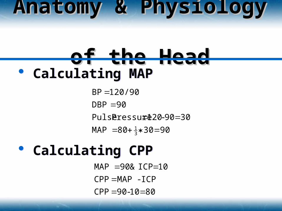
Calculating MAP
Calculating CPP
Calculating MAP
Calculating CPP
Anatomy & Anatomy & Physiology Physiology of the Headof the Head
9030 80 MAP
30 90-120 Pressure Pulse
90DBP
120/90 BP
31
80 10-90 CPP
ICP - MAP CPP
10 ICP & 90 MAP
Cerebral Perfusion Pressure Autoregulation
Changes in ICP result in compensation Increased ICP = Increased BP
• This causes ICP to rise higher and BP to rise Brain injury and death become imminent
Expanding mass inside cranial vault Displaces CSF If pressure increases, brain tissue is displaced
Cerebral Perfusion Pressure Autoregulation
Changes in ICP result in compensation Increased ICP = Increased BP
• This causes ICP to rise higher and BP to rise Brain injury and death become imminent
Expanding mass inside cranial vault Displaces CSF If pressure increases, brain tissue is displaced
Anatomy & Anatomy & Physiology Physiology of the Headof the Head
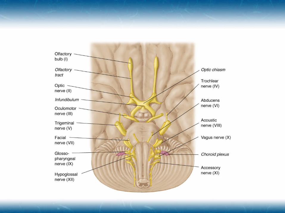
Cranial Nerves 12 pair with distinct pathways Senses, facial innervation, & body function control
Ascending Reticular Activation System Tract of neurons in upper brainstem, pons, and
midbrain Responsible for sleep-wake cycle Monitors input stimulation Regulates body functions
Respiration Heart Rate Peripheral Vascular Resistance
Injury may result in prolonged waking state
Anatomy & Anatomy & Physiology Physiology of the Headof the Head
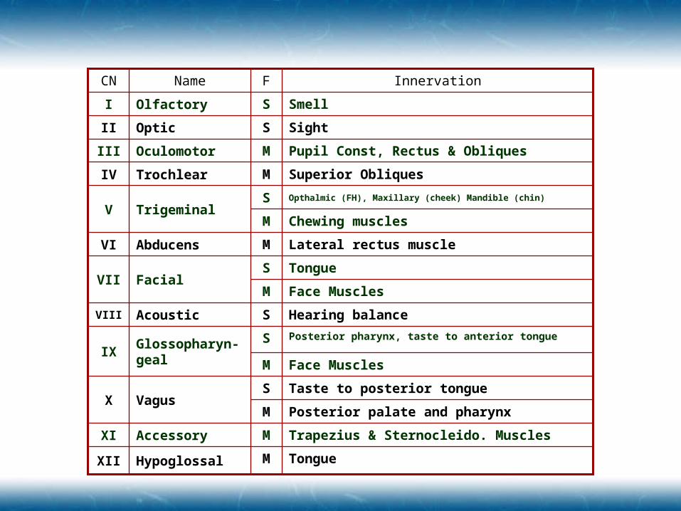
Face MusclesM
Chewing musclesM
Posterior palate and pharynxM
Face MusclesM
SightSOpticII
Pupil Const, Rectus & ObliquesMOculomotorIII
Opthalmic (FH), Maxillary (cheek) Mandible (chin)STrigeminalV
Lateral rectus muscleMAbducensVI
Taste to posterior tongueSVagusX
TongueMHypoglossalXII
Trapezius & Sternocleido. MusclesMAccessoryXI
Hearing balanceSAcousticVIII
Superior ObliquesMTrochlearIV
TongueSFacialVII
Posterior pharynx, taste to anterior tongueSGlossopharyn-geal
IX
SmellSOlfactoryI
InnervationFNameCN
Anatomy & Physiology of the Face Structure Ear Eye
Anatomy & Physiology of the Face Structure Ear Eye
Anatomy & Anatomy & PhysiologyPhysiology
Head, Face & NeckHead, Face & Neck
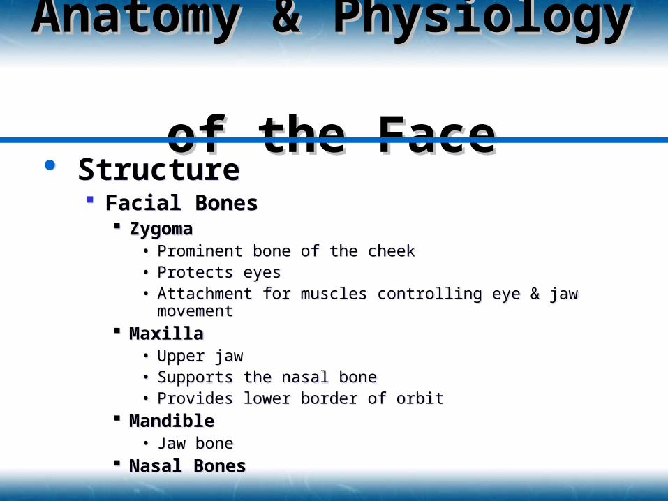
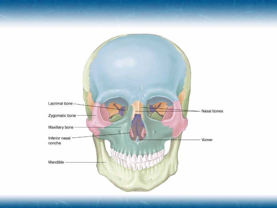
Structure Facial Bones
Zygoma• Prominent bone of the cheek• Protects eyes• Attachment for muscles controlling eye & jaw movement
Maxilla• Upper jaw• Supports the nasal bone• Provides lower border of orbit
Mandible• Jaw bone
Nasal Bones
Structure Facial Bones
Zygoma• Prominent bone of the cheek• Protects eyes• Attachment for muscles controlling eye & jaw movement
Maxilla• Upper jaw• Supports the nasal bone• Provides lower border of orbit
Mandible• Jaw bone
Nasal Bones
Anatomy & Anatomy & Physiology Physiology of the Faceof the Face
Structure Covered with skin
Flexible and thin Highly vascular
Minimal layer of subcutaneous tissue
Circulation External carotid artery
Supplies facial area Branches
• Facial, Temporal & Maxillary Arteries
Structure Covered with skin
Flexible and thin Highly vascular
Minimal layer of subcutaneous tissue
Circulation External carotid artery
Supplies facial area Branches
• Facial, Temporal & Maxillary Arteries
Anatomy & Anatomy & Physiology Physiology of the Faceof the Face
Nerves Trigeminal (CN-V)
Facial Sensation Some eye motor control Enables chewing process
Facial (CN-VII) Motor control for facial muscles Sensation of taste
Nerves Trigeminal (CN-V)
Facial Sensation Some eye motor control Enables chewing process
Facial (CN-VII) Motor control for facial muscles Sensation of taste
Anatomy & Anatomy & Physiology Physiology of the Faceof the Face
Nasal Cavity Upper Border
Bones• Junction of Ethmoid, Nasal, & Maxillary Bones
Bony Septum• Right & Left Chamber
Turbinates• Vascular mucosa support• Warm, Humidify, and Filter incoming air
Lower Border Bony Hard Palate Soft Palate
• Moves upward during swallowing Nasal Cartilage
Forms Nares
Nasal Cavity Upper Border
Bones• Junction of Ethmoid, Nasal, & Maxillary Bones
Bony Septum• Right & Left Chamber
Turbinates• Vascular mucosa support• Warm, Humidify, and Filter incoming air
Lower Border Bony Hard Palate Soft Palate
• Moves upward during swallowing Nasal Cartilage
Forms Nares
Anatomy & Anatomy & Physiology Physiology of the Faceof the Face
Oral Cavity Formed Structures
Maxillary bone Palate Upper teeth meeting the mandible and lower teeth
Floor Tongue
• Connects to hyoid bone Free-floating U-shaped bone inferior & posterior of the
mandible
Mandible Articulates with the TMJ joint
Oral Cavity Formed Structures
Maxillary bone Palate Upper teeth meeting the mandible and lower teeth
Floor Tongue
• Connects to hyoid bone Free-floating U-shaped bone inferior & posterior of the
mandible
Mandible Articulates with the TMJ joint
Anatomy & Anatomy & Physiology Physiology of the Faceof the Face
Special Structures Salivary Glands
First stage in digestion Location
• Anterior and inferior to the ear• Under tongue• Inside the inferior mandible
Tonsils Posterior wall of the pharynx
Special Structures Salivary Glands
First stage in digestion Location
• Anterior and inferior to the ear• Under tongue• Inside the inferior mandible
Tonsils Posterior wall of the pharynx
Anatomy & Anatomy & Physiology Physiology of the Faceof the Face
(continued)
Sinuses Hollow spaces in cranium and facial bones Function
• Lighten head• Protect eyes and nasal cavity• Produce resonant tones of voice• Strengthen area against trauma
Sinuses Hollow spaces in cranium and facial bones Function
• Lighten head• Protect eyes and nasal cavity• Produce resonant tones of voice• Strengthen area against trauma
Anatomy & Anatomy & Physiology Physiology of the Faceof the Face
Cranial Nerves CN-XII (Hypoglossal)
Swallowing & tongue movement
CN-IX (Glossopharyngeal) Saliva production & taste
CN-V (Trigeminal) Sensations from facial region & aids in chewing
CN-VII (Facial) Muscles of facial expression & taste
Cranial Nerves CN-XII (Hypoglossal)
Swallowing & tongue movement
CN-IX (Glossopharyngeal) Saliva production & taste
CN-V (Trigeminal) Sensations from facial region & aids in chewing
CN-VII (Facial) Muscles of facial expression & taste
Anatomy & Anatomy & Physiology Physiology of the Faceof the Face
Pharynx Posterior & Inferior to the oral cavity Aids in swallowing
Bolus of food propelled back & down by tongue Epiglottis moves downward Larynx moves up
• Combined effect seals airway
Peristaltic wave moves food down esophagus
Pharynx Posterior & Inferior to the oral cavity Aids in swallowing
Bolus of food propelled back & down by tongue Epiglottis moves downward Larynx moves up
• Combined effect seals airway
Peristaltic wave moves food down esophagus
Anatomy & Anatomy & Physiology Physiology of the Faceof the Face
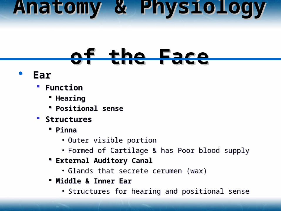
Ear Function
Hearing Positional sense
Structures Pinna
• Outer visible portion
• Formed of Cartilage & has Poor blood supply External Auditory Canal
• Glands that secrete cerumen (wax) Middle & Inner Ear
• Structures for hearing and positional sense
Ear Function
Hearing Positional sense
Structures Pinna
• Outer visible portion
• Formed of Cartilage & has Poor blood supply External Auditory Canal
• Glands that secrete cerumen (wax) Middle & Inner Ear
• Structures for hearing and positional sense
Anatomy & Anatomy & Physiology Physiology of the Faceof the Face
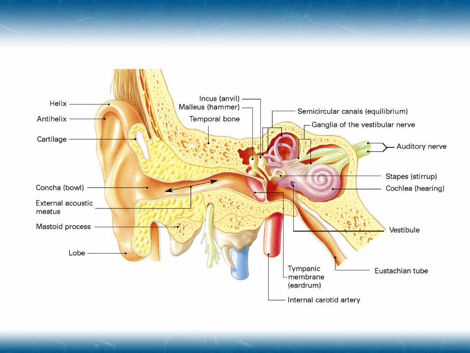
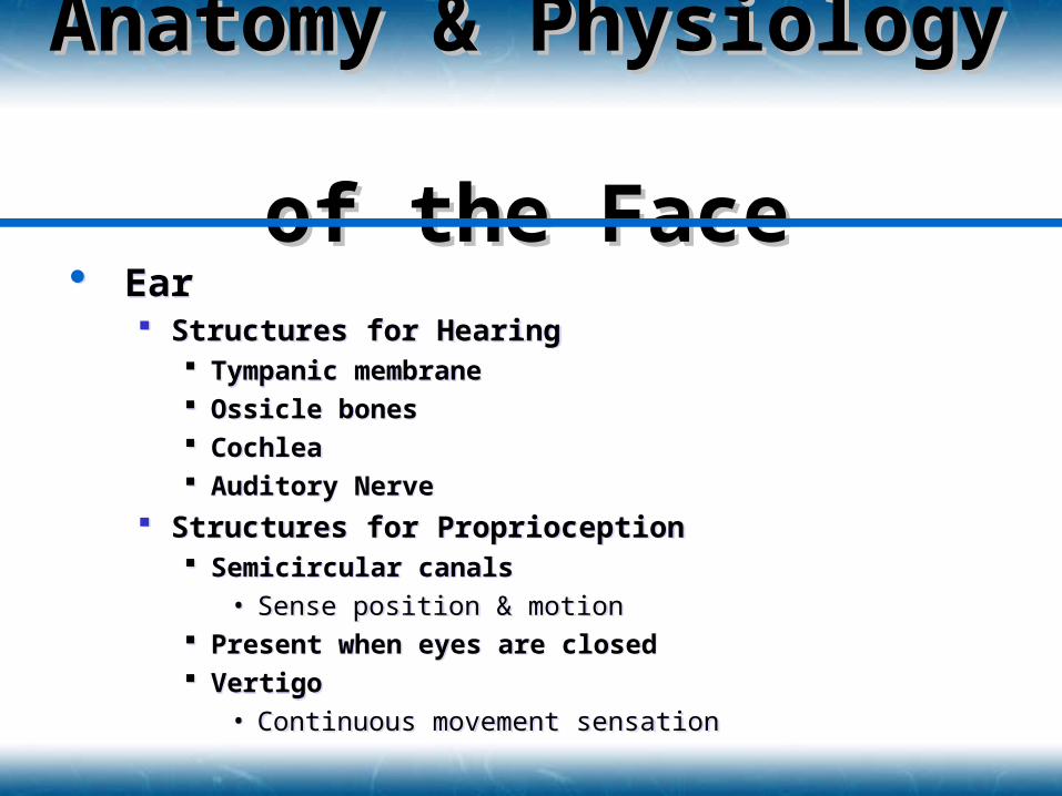
Ear Structures for Hearing
Tympanic membrane Ossicle bones Cochlea Auditory Nerve
Structures for Proprioception Semicircular canals
• Sense position & motion Present when eyes are closed Vertigo
• Continuous movement sensation
Ear Structures for Hearing
Tympanic membrane Ossicle bones Cochlea Auditory Nerve
Structures for Proprioception Semicircular canals
• Sense position & motion Present when eyes are closed Vertigo
• Continuous movement sensation
Anatomy & Anatomy & Physiology Physiology of the Faceof the Face
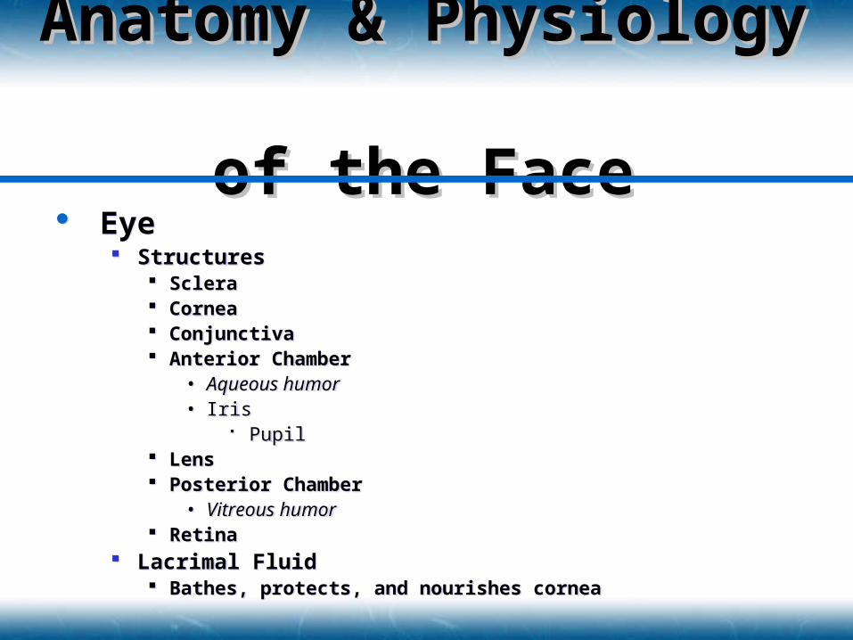
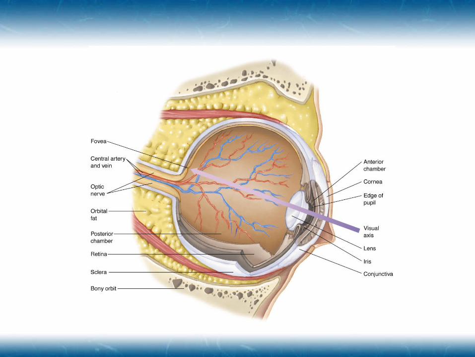
Eye Structures
Sclera Cornea Conjunctiva Anterior Chamber
• Aqueous humor• Iris
Pupil Lens Posterior Chamber
• Vitreous humor Retina
Lacrimal Fluid Bathes, protects, and nourishes cornea
Eye Structures
Sclera Cornea Conjunctiva Anterior Chamber
• Aqueous humor• Iris
Pupil Lens Posterior Chamber
• Vitreous humor Retina
Lacrimal Fluid Bathes, protects, and nourishes cornea
Anatomy & Anatomy & Physiology Physiology of the Faceof the Face
Eye Innervation
CN-III (Oculomotor)• Pupil dilation
• Conjugate movement Movement of eyes together
• Normal range of motion
CN-IV (Trochlear)• Downward & inward movement
CN-VI (Abducens)• Abduction (outward) gaze
Eye Innervation
CN-III (Oculomotor)• Pupil dilation
• Conjugate movement Movement of eyes together
• Normal range of motion
CN-IV (Trochlear)• Downward & inward movement
CN-VI (Abducens)• Abduction (outward) gaze
Anatomy & Anatomy & Physiology Physiology of the Faceof the Face
Vasculature of the Neck Carotid Arteries
Arise from• RIGHT: Brachiocephalic Artery• LEFT: Aorta Artery
Split• Internal & External Carotid Arteries• Upper border of the Larynx• Carotid Bodies & Sinuses located
Bodies: Monitor CO2 and O2 levels Sinuses: Monitor Blood Pressure
Vasculature of the Neck Carotid Arteries
Arise from• RIGHT: Brachiocephalic Artery• LEFT: Aorta Artery
Split• Internal & External Carotid Arteries• Upper border of the Larynx• Carotid Bodies & Sinuses located
Bodies: Monitor CO2 and O2 levels Sinuses: Monitor Blood Pressure
Anatomy & Anatomy & Physiology Physiology of the Neckof the Neck
(continued)
Jugular Veins External
• Superficial, lateral to the trachea Internal
• Sheath with the carotid artery and vagus nerve
Jugular Veins External
• Superficial, lateral to the trachea Internal
• Sheath with the carotid artery and vagus nerve
Anatomy & Anatomy & Physiology Physiology of the Neckof the Neck
Airway Structures Larynx
Epiglottis Thyroid & Cricoid Cartilage
Trachea Posterior border is anterior border of esophagus
Airway Structures Larynx
Epiglottis Thyroid & Cricoid Cartilage
Trachea Posterior border is anterior border of esophagus
Anatomy & Anatomy & Physiology Physiology of the Neckof the Neck
Other Structures Cervical Spine
Musculoskeletal Function• External Skeletal support of the head and neck
• Attachment point for spinal column ligaments
• Attachment point for tendons to move head and shoulders
Nervous Function• Spinal Cord contained within
• Peripheral Nerve Exit between vertebrae
Other Structures Cervical Spine
Musculoskeletal Function• External Skeletal support of the head and neck
• Attachment point for spinal column ligaments
• Attachment point for tendons to move head and shoulders
Nervous Function• Spinal Cord contained within
• Peripheral Nerve Exit between vertebrae
Anatomy & Anatomy & Physiology Physiology of the Neckof the Neck
Other Structures Esophagus Cranial Nerves
CN-IX (Glossopharyngeal)• Carotid Bodies & Carotid Sinuses
CN-X• Speech, swallowing, cardiac, respiratory & visceral function
Thoracic Duct Delivers lymph to the venous system
Other Structures Esophagus Cranial Nerves
CN-IX (Glossopharyngeal)• Carotid Bodies & Carotid Sinuses
CN-X• Speech, swallowing, cardiac, respiratory & visceral function
Thoracic Duct Delivers lymph to the venous system
Anatomy & Anatomy & Physiology Physiology of the Neckof the Neck
(continued)
Glands Thyroid
• Rate of cellular metabolism• Systemic levels of calcium
Brachial Plexus Network of nerves in lower neck and should that control
arm and hand function
Glands Thyroid
• Rate of cellular metabolism• Systemic levels of calcium
Brachial Plexus Network of nerves in lower neck and should that control
arm and hand function
Anatomy & Anatomy & Physiology Physiology of the Neckof the Neck
Mechanism of Injury Blunt Injury
Motor vehicle collisions Assaults Falls
Penetrating Injury Gunshot wounds Stabbing Explosions “Clothesline”
Mechanism of Injury Blunt Injury
Motor vehicle collisions Assaults Falls
Penetrating Injury Gunshot wounds Stabbing Explosions “Clothesline”
Pathophysiology ofPathophysiology ofHead, Facial, & Neck Head, Facial, & Neck
InjuryInjury
Scalp InjuryScalp Injury
Contusions Lacerations Avulsions Significant Hemorrhage
ALWAYS Reconsider MOI for severe underlying problems
Contusions Lacerations Avulsions Significant Hemorrhage
ALWAYS Reconsider MOI for severe underlying problems
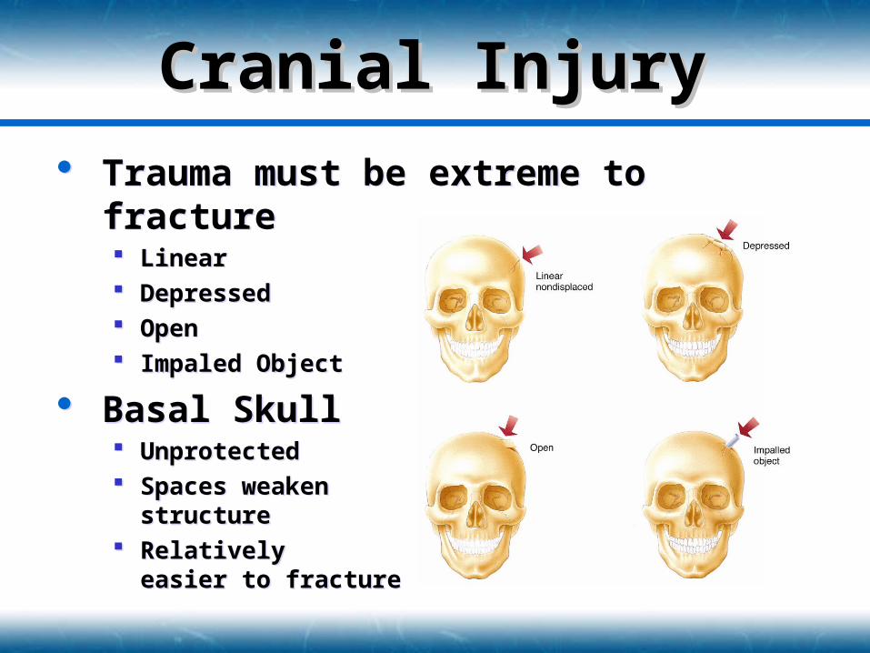
Cranial InjuryCranial Injury Trauma must be extreme to fracture
Linear Depressed Open Impaled Object
Basal Skull Unprotected Spaces weaken
structure Relatively
easier to fracture
Trauma must be extreme to fracture Linear Depressed Open Impaled Object
Basal Skull Unprotected Spaces weaken
structure Relatively
easier to fracture
Cranial InjuryCranial Injury
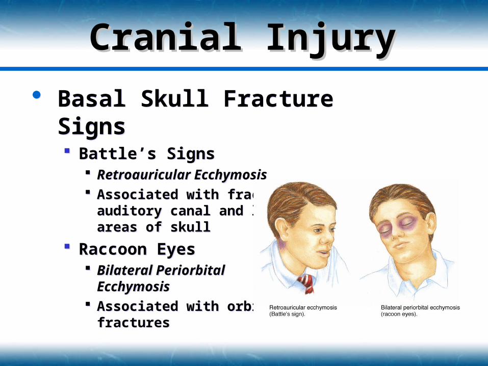
Basal Skull Fracture Signs Battle’s Signs
Retroauricular Ecchymosis Associated with fracture of
auditory canal and lower areas of skull
Raccoon Eyes Bilateral Periorbital
Ecchymosis Associated with orbital
fractures
Basal Skull Fracture Signs Battle’s Signs
Retroauricular Ecchymosis Associated with fracture of
auditory canal and lower areas of skull
Raccoon Eyes Bilateral Periorbital
Ecchymosis Associated with orbital
fractures
Cranial InjuryCranial Injury
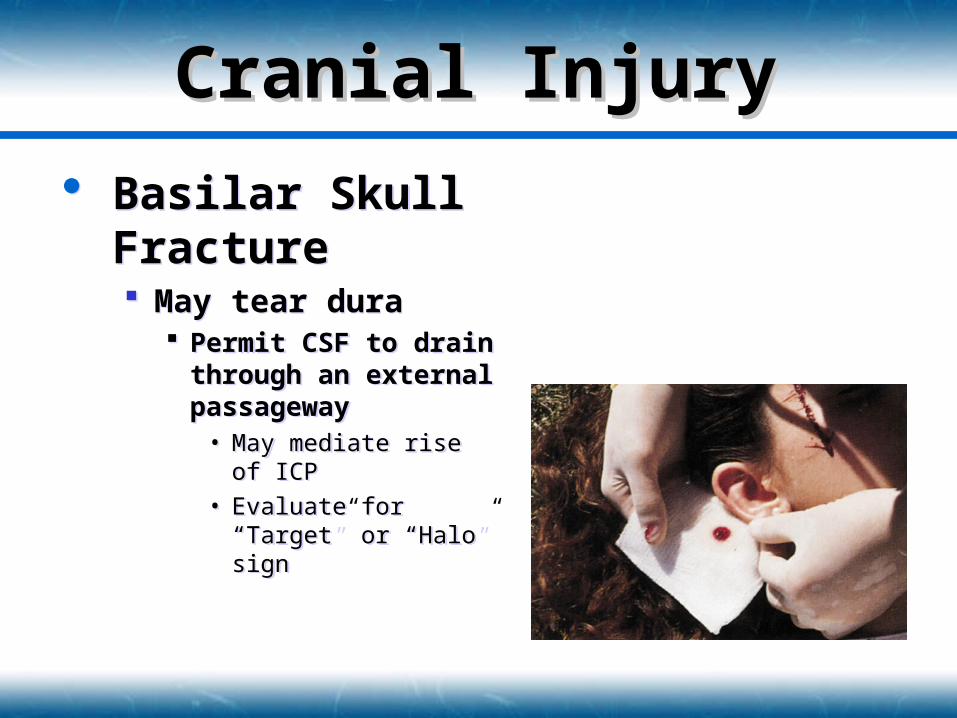
Basilar Skull Fracture May tear dura
Permit CSF to drain through an external passageway
• May mediate rise of ICP
• Evaluate for “Target” or “Halo” sign
Basilar Skull Fracture May tear dura
Permit CSF to drain through an external passageway
• May mediate rise of ICP
• Evaluate for “Target” or “Halo” sign
Brain InjuryBrain Injury
As defined by the National Head Injury Foundation “a traumatic insult to the brain capable of
producing physical, intellectual, emotional, social and vocational changes.”
Classification Direct
• Primary injury caused by forces of trauma
Indirect• Secondary injury caused by factors resulting from the
primary injury
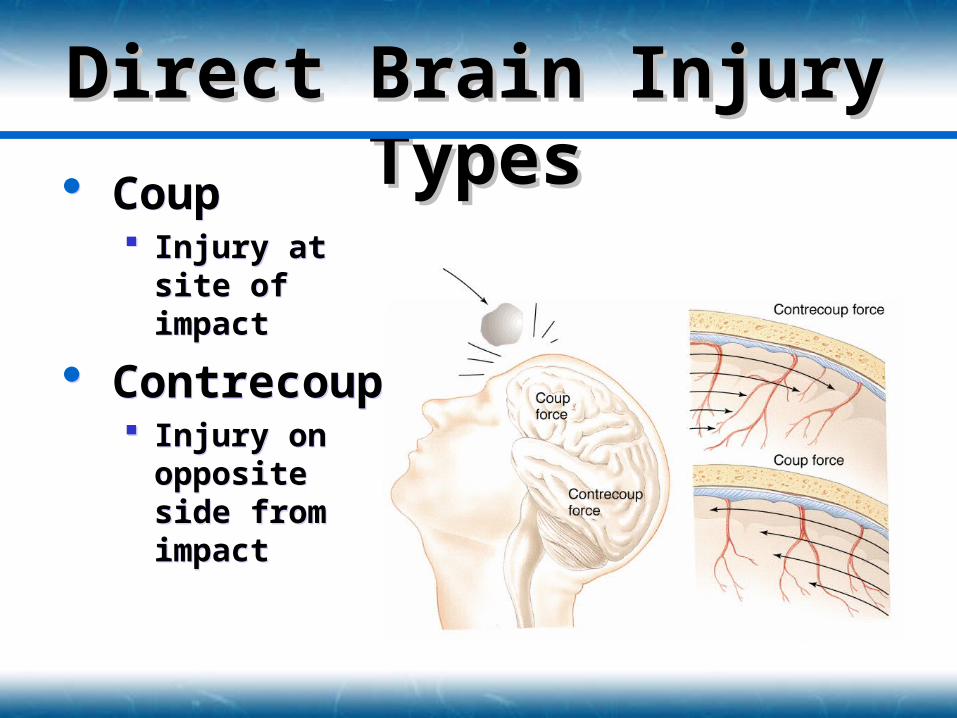
Direct Brain Injury Direct Brain Injury TypesTypes Coup
Injury at site of impact
Contrecoup Injury on
opposite side from impact
Coup Injury at site of
impact
Contrecoup Injury on
opposite side from impact
Direct Brain Injury Direct Brain Injury CategoriesCategories
Focal Occur at a specific location in brain Differentials
Cerebral Contusion Intracranial Hemorrhage
• Epidural hematoma• Subdural hematoma
Intracerebral Hemorrhage
Diffuse Concussion Moderate Diffuse Axonal Injury Severe Diffuse Axonal Injury
Focal Occur at a specific location in brain Differentials
Cerebral Contusion Intracranial Hemorrhage
• Epidural hematoma• Subdural hematoma
Intracerebral Hemorrhage
Diffuse Concussion Moderate Diffuse Axonal Injury Severe Diffuse Axonal Injury
Focal Brain InjuryFocal Brain Injury
Cerebral Contusion Blunt trauma to local brain tissue Capillary bleeding into brain tissue Common with blunt head trauma
Confusion Neurologic deficit
• Personality changes
• Vision changes
• Speech changes
Results from Coup-contrecoup injury
Cerebral Contusion Blunt trauma to local brain tissue Capillary bleeding into brain tissue Common with blunt head trauma
Confusion Neurologic deficit
• Personality changes
• Vision changes
• Speech changes
Results from Coup-contrecoup injury

Epidural Hematoma Bleeding between dura
mater and skull Involves arteries
Middle meningeal artery most common
Rapid bleeding & reduction of oxygen to tissues
Herniates brain toward foramen magnum
Epidural Hematoma Bleeding between dura
mater and skull Involves arteries
Middle meningeal artery most common
Rapid bleeding & reduction of oxygen to tissues
Herniates brain toward foramen magnum
Focal Brain InjuryFocal Brain InjuryIntracranial HemorrhageIntracranial Hemorrhage
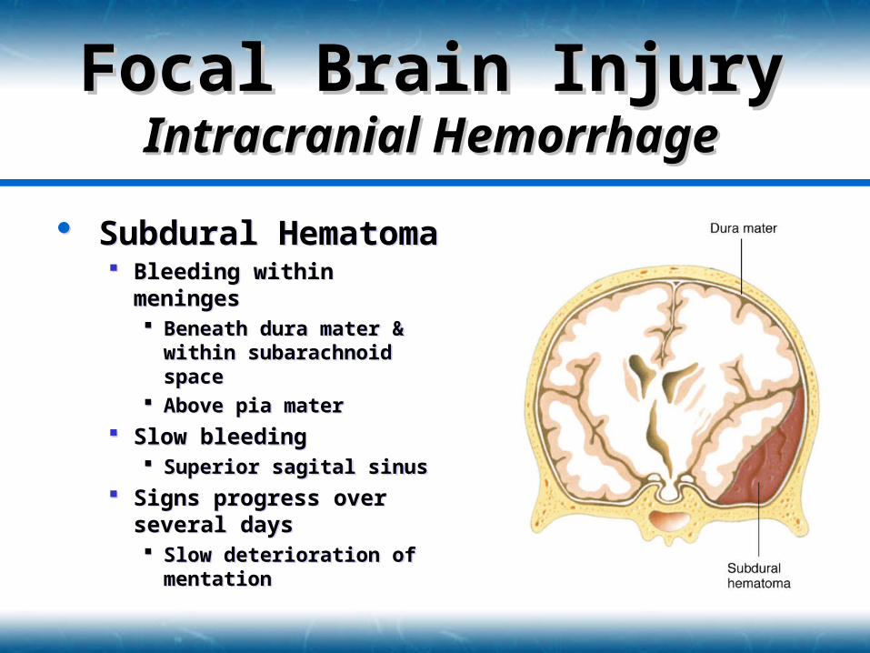
Subdural Hematoma Bleeding within meninges
Beneath dura mater & within subarachnoid space
Above pia mater
Slow bleeding Superior sagital sinus
Signs progress over several days Slow deterioration of
mentation
Subdural Hematoma Bleeding within meninges
Beneath dura mater & within subarachnoid space
Above pia mater
Slow bleeding Superior sagital sinus
Signs progress over several days Slow deterioration of
mentation
Focal Brain InjuryFocal Brain InjuryIntracranial HemorrhageIntracranial Hemorrhage
Intracerebral Hemorrhage Rupture blood vessel within the brain Presentation similar to stroke symptoms Signs and symptoms worsen over time
Intracerebral Hemorrhage Rupture blood vessel within the brain Presentation similar to stroke symptoms Signs and symptoms worsen over time
Focal Brain InjuryFocal Brain InjuryIntracranial HemorrhageIntracranial Hemorrhage
Diffuse Brain InjuryDiffuse Brain Injury
Due to stretching forces placed on axons
Pathology distributed throughout brain
Types Concussion Moderate Diffuse Axonal Injury Severe Diffuse Axonal Injury
Due to stretching forces placed on axons
Pathology distributed throughout brain
Types Concussion Moderate Diffuse Axonal Injury Severe Diffuse Axonal Injury
Mild to moderate form of Diffuse Axonal Injury (DAI) Nerve dysfunction without anatomic damage
Transient episode of Confusion, Disorientation, Event amnesia
Suspect if patient has a momentary loss of consciousness
Management Frequent reassessment of mentation ABC’s
Mild to moderate form of Diffuse Axonal Injury (DAI) Nerve dysfunction without anatomic damage
Transient episode of Confusion, Disorientation, Event amnesia
Suspect if patient has a momentary loss of consciousness
Management Frequent reassessment of mentation ABC’s
Diffuse Brain InjuryDiffuse Brain InjuryConcussionConcussion
“Classic Concussion” Same mechanism as concussion
Additional: Minute bruising of brain tissue
Unconsciousness If cerebral cortex and RAS involved
May exist with a basilar skull fracture Signs & Symptoms
Unconsciousness or Persistent confusion Loss of concentration, disorientation Retrograde & Antegrade amnesia Visual and sensory disturbances Mood or Personality changes
Diffuse Brain InjuryDiffuse Brain InjuryModerate Diffuse Axonal Moderate Diffuse Axonal
InjuryInjury
Brainstem Injury Significant mechanical disruption of
axons Cerebral hemispheres and brainstem
High mortality rate Signs & Symptoms
Prolonged unconsciousness Cushing’s reflex Decorticate or Decerebrate posturing
Brainstem Injury Significant mechanical disruption of
axons Cerebral hemispheres and brainstem
High mortality rate Signs & Symptoms
Prolonged unconsciousness Cushing’s reflex Decorticate or Decerebrate posturing
Diffuse Brain InjuryDiffuse Brain InjurySevere Diffuse Axonal Severe Diffuse Axonal
InjuryInjury
Intracranial Intracranial PerfusionPerfusion Review
Cranial volume fixed 80% = Cerebrum, cerebellum & brainstem 12% = Blood vessels & blood 8% = CSF
Increase in size of one component diminishes size of another Inability to adjust = increased ICP
Review Cranial volume fixed
80% = Cerebrum, cerebellum & brainstem 12% = Blood vessels & blood 8% = CSF
Increase in size of one component diminishes size of another Inability to adjust = increased ICP
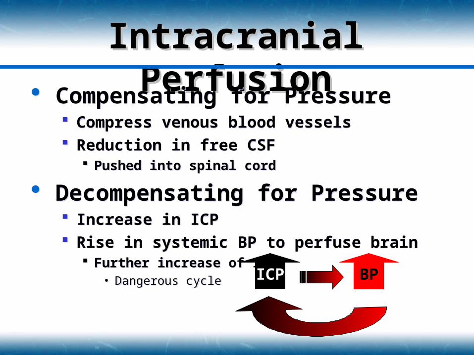
Intracranial Intracranial PerfusionPerfusion Compensating for Pressure
Compress venous blood vessels Reduction in free CSF
Pushed into spinal cord
Decompensating for Pressure Increase in ICP Rise in systemic BP to perfuse brain
Further increase of ICP• Dangerous cycle
Compensating for Pressure Compress venous blood vessels Reduction in free CSF
Pushed into spinal cord
Decompensating for Pressure Increase in ICP Rise in systemic BP to perfuse brain
Further increase of ICP• Dangerous cycle ICP BP
Intracranial PressureIntracranial Pressure
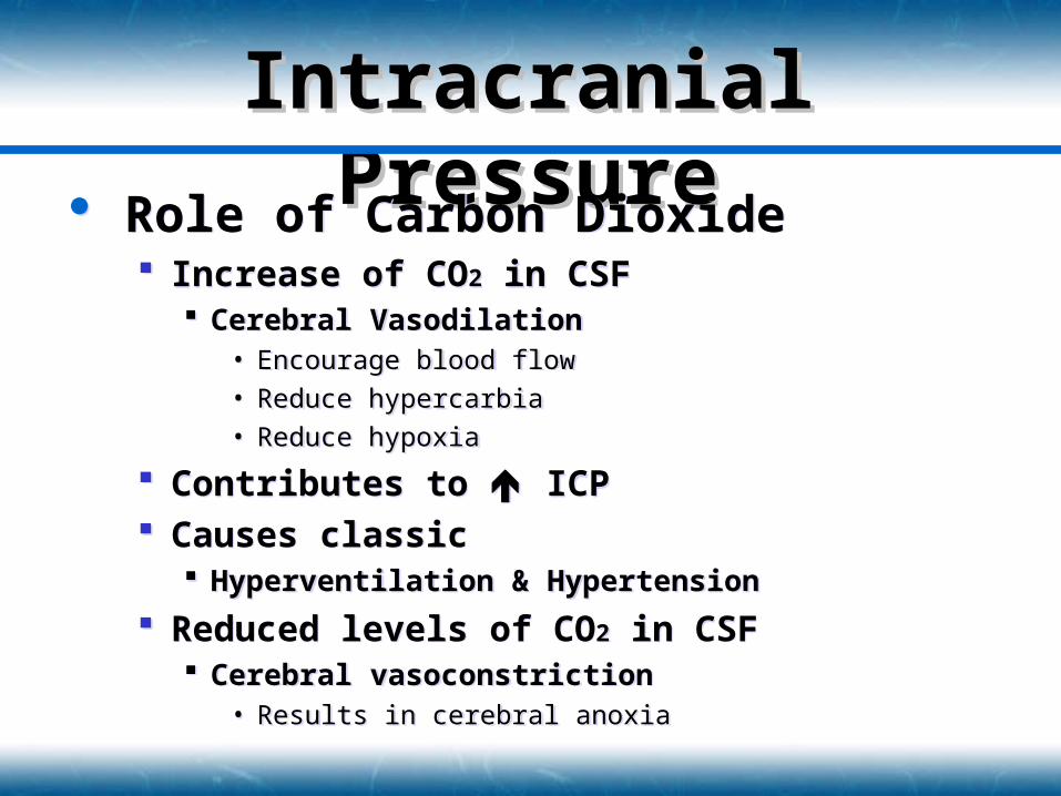
Role of Carbon Dioxide Increase of CO2 in CSF
Cerebral Vasodilation• Encourage blood flow
• Reduce hypercarbia
• Reduce hypoxia
Contributes to ICP Causes classic
Hyperventilation & Hypertension
Reduced levels of CO2 in CSF Cerebral vasoconstriction
• Results in cerebral anoxia
Role of Carbon Dioxide Increase of CO2 in CSF
Cerebral Vasodilation• Encourage blood flow
• Reduce hypercarbia
• Reduce hypoxia
Contributes to ICP Causes classic
Hyperventilation & Hypertension
Reduced levels of CO2 in CSF Cerebral vasoconstriction
• Results in cerebral anoxia
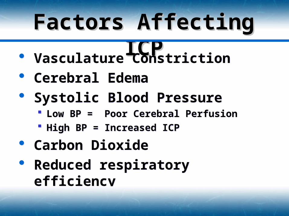
Factors Affecting ICPFactors Affecting ICP
Vasculature Constriction Cerebral Edema Systolic Blood Pressure
Low BP = Poor Cerebral Perfusion High BP = Increased ICP
Carbon Dioxide Reduced respiratory efficiency
Vasculature Constriction Cerebral Edema Systolic Blood Pressure
Low BP = Poor Cerebral Perfusion High BP = Increased ICP
Carbon Dioxide Reduced respiratory efficiency
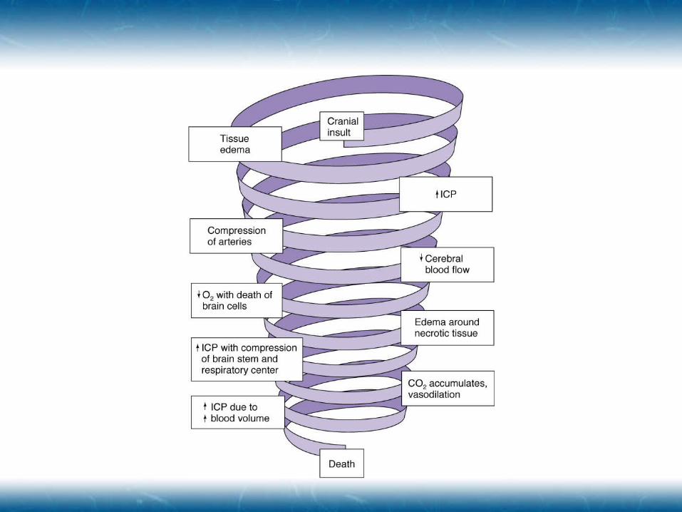
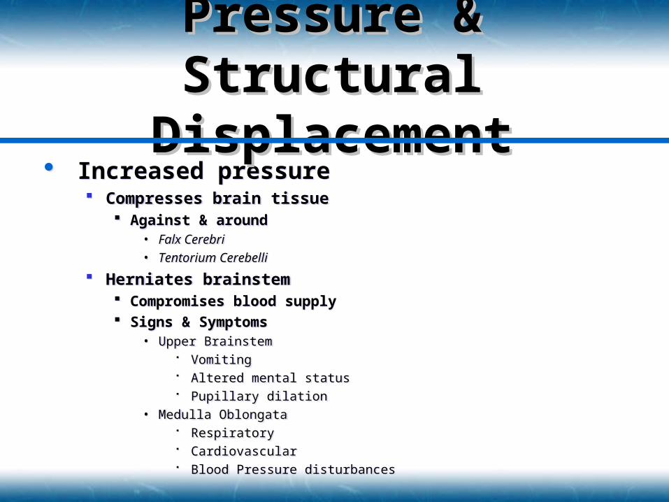
Increased pressure Compresses brain tissue
Against & around• Falx Cerebri
• Tentorium Cerebelli
Herniates brainstem Compromises blood supply Signs & Symptoms
• Upper Brainstem Vomiting Altered mental status Pupillary dilation
• Medulla Oblongata Respiratory Cardiovascular Blood Pressure disturbances
Pressure & Pressure & Structural Structural
DisplacementDisplacement
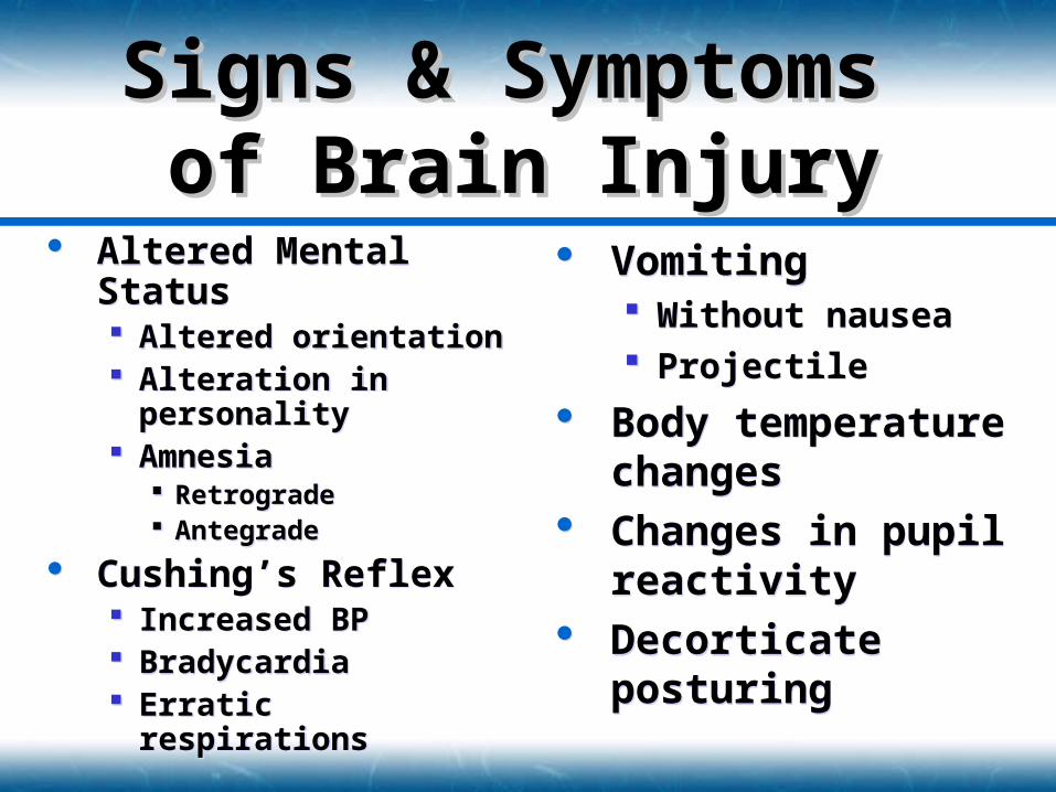
Altered Mental Status Altered orientation Alteration in
personality Amnesia
Retrograde Antegrade
Cushing’s Reflex Increased BP Bradycardia Erratic respirations
Altered Mental Status Altered orientation Alteration in
personality Amnesia
Retrograde Antegrade
Cushing’s Reflex Increased BP Bradycardia Erratic respirations
Signs & Symptoms Signs & Symptoms of Brain Injuryof Brain Injury
Vomiting Without nausea Projectile
Body temperature changes
Changes in pupil reactivity
Decorticate posturing
Vomiting Without nausea Projectile
Body temperature changes
Changes in pupil reactivity
Decorticate posturing
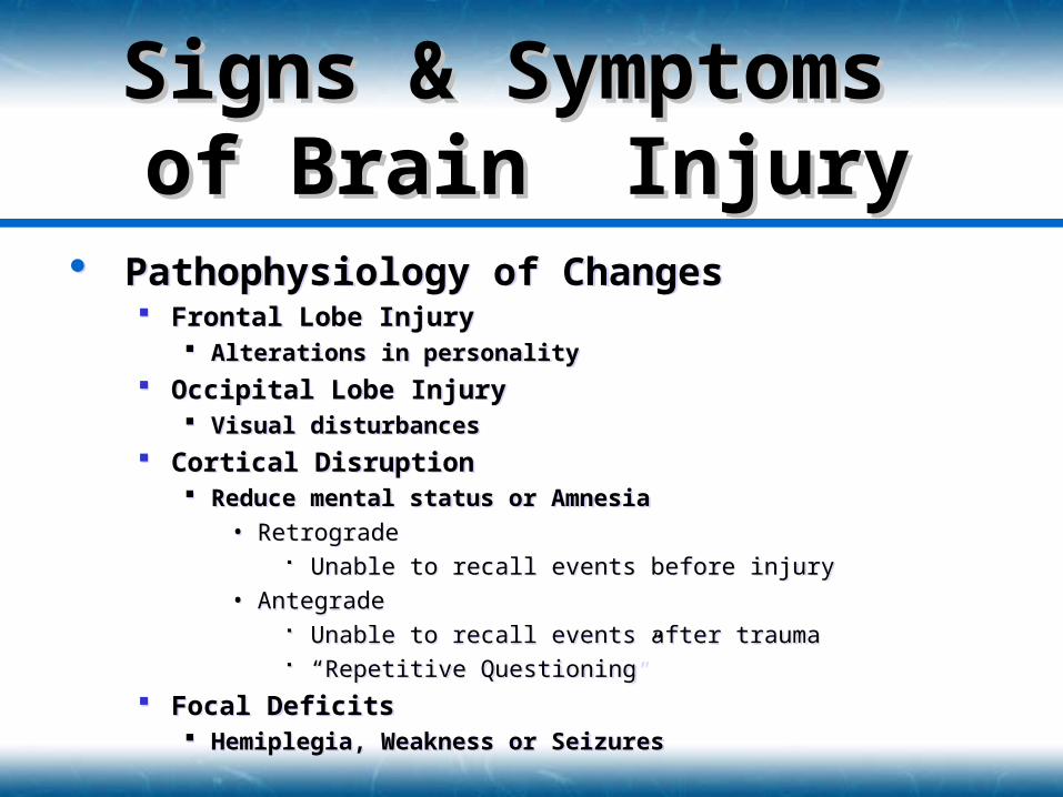
Pathophysiology of Changes Frontal Lobe Injury
Alterations in personality
Occipital Lobe Injury Visual disturbances
Cortical Disruption Reduce mental status or Amnesia
• Retrograde Unable to recall events before injury
• Antegrade Unable to recall events after trauma “Repetitive Questioning”
Focal Deficits Hemiplegia, Weakness or Seizures
Signs & Symptoms Signs & Symptoms of Brain Injuryof Brain Injury
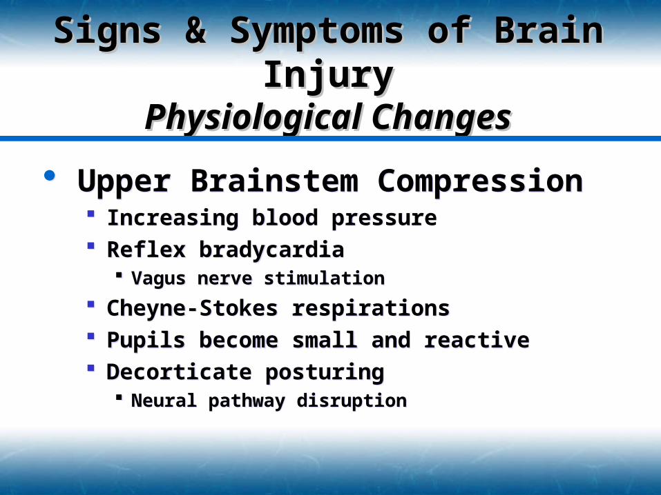
Upper Brainstem Compression Increasing blood pressure Reflex bradycardia
Vagus nerve stimulation
Cheyne-Stokes respirations Pupils become small and reactive Decorticate posturing
Neural pathway disruption
Upper Brainstem Compression Increasing blood pressure Reflex bradycardia
Vagus nerve stimulation
Cheyne-Stokes respirations Pupils become small and reactive Decorticate posturing
Neural pathway disruption
Signs & Symptoms of Brain Signs & Symptoms of Brain InjuryInjury
Physiological ChangesPhysiological Changes
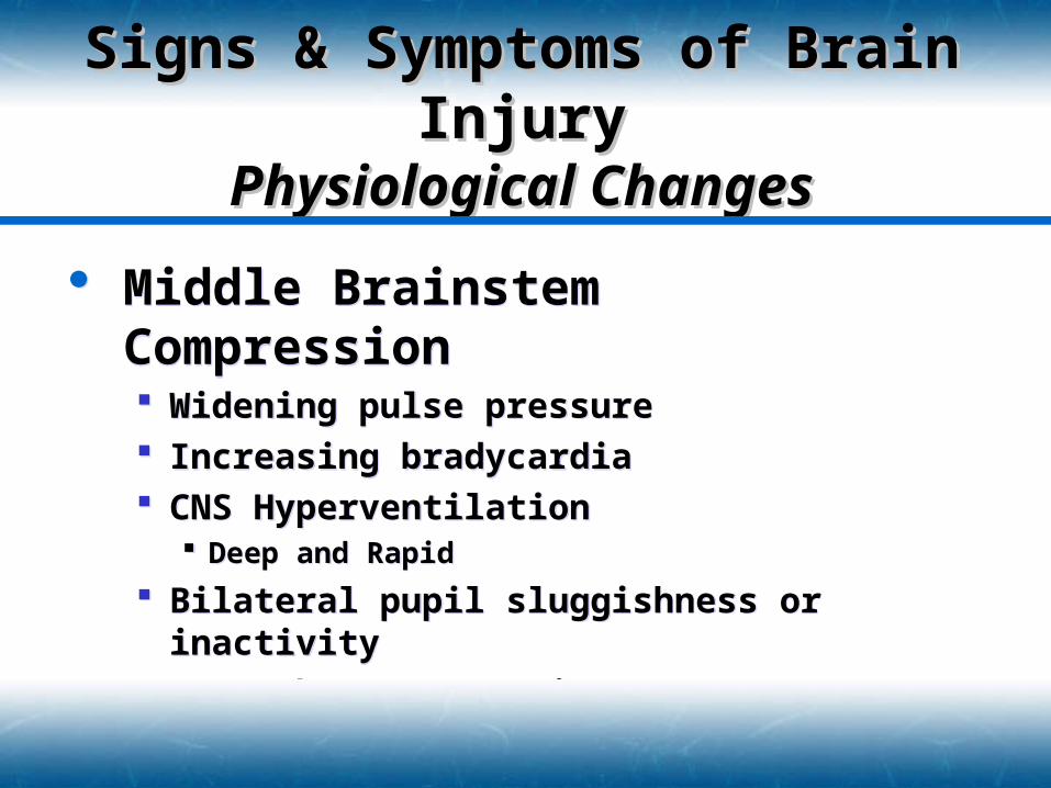
Middle Brainstem Compression Widening pulse pressure Increasing bradycardia CNS Hyperventilation
Deep and Rapid
Bilateral pupil sluggishness or inactivity Decerebrate posturing
Middle Brainstem Compression Widening pulse pressure Increasing bradycardia CNS Hyperventilation
Deep and Rapid
Bilateral pupil sluggishness or inactivity Decerebrate posturing
Signs & Symptoms of Brain Signs & Symptoms of Brain InjuryInjury
Physiological ChangesPhysiological Changes
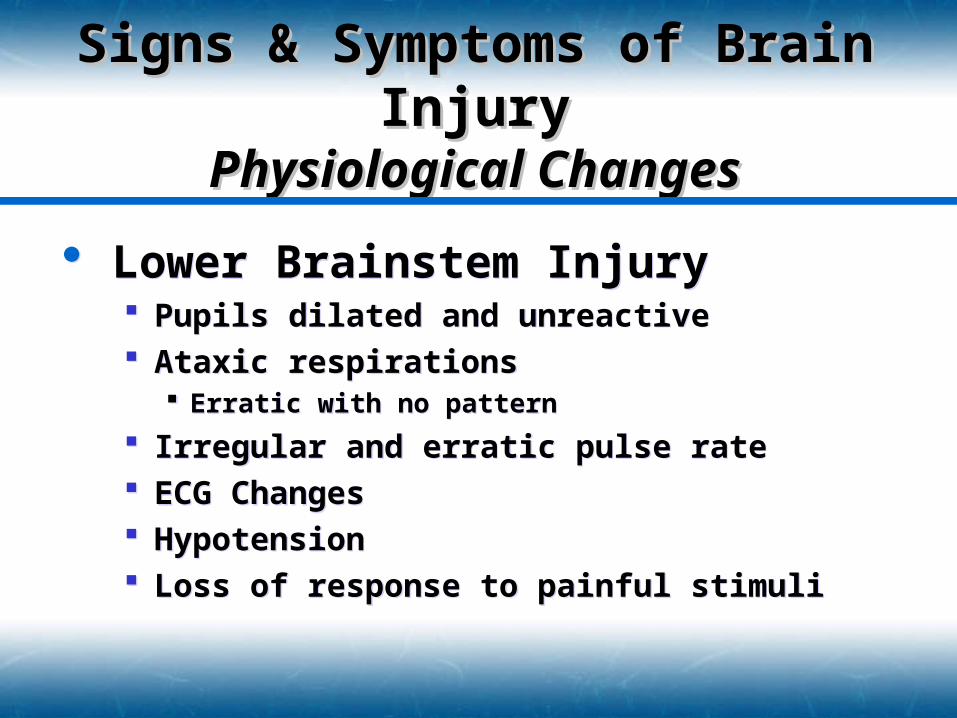
Lower Brainstem Injury Pupils dilated and unreactive Ataxic respirations
Erratic with no pattern
Irregular and erratic pulse rate ECG Changes Hypotension Loss of response to painful stimuli
Lower Brainstem Injury Pupils dilated and unreactive Ataxic respirations
Erratic with no pattern
Irregular and erratic pulse rate ECG Changes Hypotension Loss of response to painful stimuli
Signs & Symptoms of Brain Signs & Symptoms of Brain InjuryInjury
Physiological ChangesPhysiological Changes
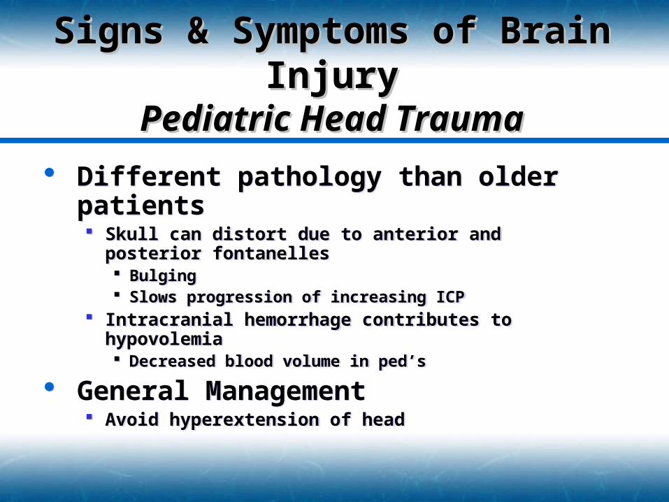
Different pathology than older patients Skull can distort due to anterior and posterior
fontanelles Bulging Slows progression of increasing ICP
Intracranial hemorrhage contributes to hypovolemia Decreased blood volume in ped’s
General Management Avoid hyperextension of head
Tongue pushes soft pallet closed Ventilate through mouth and nose
Signs & Symptoms of Brain Signs & Symptoms of Brain InjuryInjury
Pediatric Head TraumaPediatric Head Trauma
Signs & Symptoms of Brain Signs & Symptoms of Brain InjuryInjury
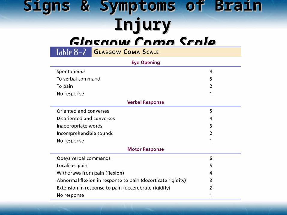
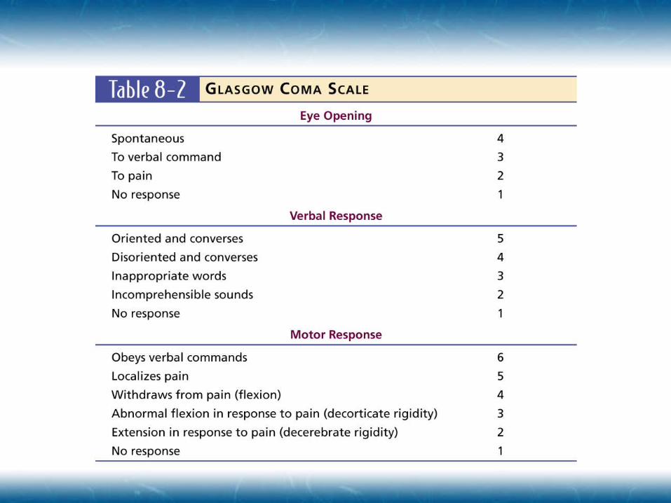
Glasgow Coma ScaleGlasgow Coma Scale
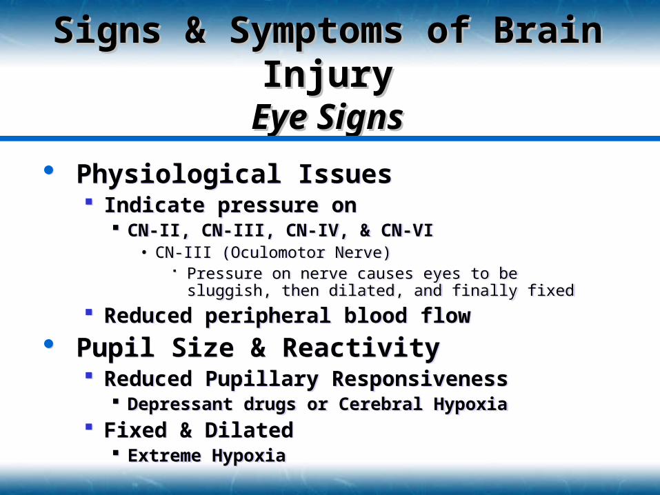
Physiological Issues Indicate pressure on
CN-II, CN-III, CN-IV, & CN-VI• CN-III (Oculomotor Nerve)
Pressure on nerve causes eyes to be sluggish, then dilated, and finally fixed
Reduced peripheral blood flow
Pupil Size & Reactivity Reduced Pupillary Responsiveness
Depressant drugs or Cerebral Hypoxia Fixed & Dilated
Extreme Hypoxia
Signs & Symptoms of Brain Signs & Symptoms of Brain InjuryInjury
Eye SignsEye Signs
Facial InjuryFacial Injury
Facial Soft Tissue Injury Highly vascular tissue
Contribute to hypovolemia
Superficial injuries rarely life threatening and rarely involve the airway
Deep Injuries can result in blood being swallowed and endanger the airway
Soft tissue swelling reduces airflow Consider likelihood of basilar skull fracture or
spinal injury
Facial InjuryFacial Injury Facial Dislocations & Fractures
Common Fractures Mandibular
• Deformity along jaw & loss of teeth• Possible airway compromise if patient placed supine• Evaluate for multiple fracture sites
Maxillary & Nasal• Le Fort I, II and III Criteria
Orbit• Involve Zygoma, Maxilla, and/or interior shelf• Reduction of eye movement
Possible Diplopia• Limitation of jaw movement
Facial Dislocations & Fractures Common Fractures
Mandibular• Deformity along jaw & loss of teeth• Possible airway compromise if patient placed supine• Evaluate for multiple fracture sites
Maxillary & Nasal• Le Fort I, II and III Criteria
Orbit• Involve Zygoma, Maxilla, and/or interior shelf• Reduction of eye movement
Possible Diplopia• Limitation of jaw movement

Facial InjuryFacial Injury
Nasal Injury Rarely life threatening Swelling & Hemorrhage interfere with
breathing Epistaxis
Most common problem
AVOID NASOTRACHEAL INTUBATION Passage of ET tube into the cerebral cavity
Nasal Injury Rarely life threatening Swelling & Hemorrhage interfere with
breathing Epistaxis
Most common problem
AVOID NASOTRACHEAL INTUBATION Passage of ET tube into the cerebral cavity
Facial InjuryFacial Injury Ear Injury
External Ear Pinna is frequently injured due to trauma Poor blood supply Poor healing
Internal Ear Well protected from trauma My be injured due to rapid pressure changes
• Diving, Blast, or Explosions• Temporary or permanent hearing loss• Tinnitus may occur
Ear Injury External Ear
Pinna is frequently injured due to trauma Poor blood supply Poor healing
Internal Ear Well protected from trauma My be injured due to rapid pressure changes
• Diving, Blast, or Explosions• Temporary or permanent hearing loss• Tinnitus may occur
Facial InjuryFacial Injury Eye Injury
Penetrating trauma can result in long term damage Suspect small foreign body if patient complains of sudden
eye pain and sensation of something on the eye DO NOT REMOVE ANY FOREIGN OBJECT
Corneal Abrasions & Lacerations Common & usually superficial
Hyphema Blunt trauma to the anterior chamber of the eye Blood in front of iris or pupil
Sub-conjunctival Hemorrhage Less serious condition May occur after strong sneeze, severe vomiting or direct
trauma
Eye Injury Penetrating trauma
can result in long term damage Suspect small foreign body if patient complains of sudden
eye pain and sensation of something on the eye DO NOT REMOVE ANY FOREIGN OBJECT
Corneal Abrasions & Lacerations Common & usually superficial
Hyphema Blunt trauma to the anterior chamber of the eye Blood in front of iris or pupil
Sub-conjunctival Hemorrhage Less serious condition May occur after strong sneeze, severe vomiting or direct
trauma
Facial InjuryFacial Injury Eye Injury
Acute Retinal Artery Occlusion Non-traumatic origin Painless loss of vision in one eye Occlusion of retinal artery
Retinal Detachment Traumatic origin Complaint of dark curtain/obstruction in the field of
view Possibly painful depending on type of trauma
Soft Tissue Lacerations
Eye Injury Acute Retinal Artery Occlusion
Non-traumatic origin Painless loss of vision in one eye Occlusion of retinal artery
Retinal Detachment Traumatic origin Complaint of dark curtain/obstruction in the field of
view Possibly painful depending on type of trauma
Soft Tissue Lacerations
Neck InjuryNeck Injury Blood Vessel Trauma
Blunt trauma Serious hematoma
Laceration Serious exsanguination Entraining of air embolism
• Cover with occlusive dressing
Airway Trauma Tracheal rupture or dissection from larynx Airway swelling & compromise
Blood Vessel Trauma Blunt trauma
Serious hematoma Laceration
Serious exsanguination Entraining of air embolism
• Cover with occlusive dressing
Airway Trauma Tracheal rupture or dissection from larynx Airway swelling & compromise
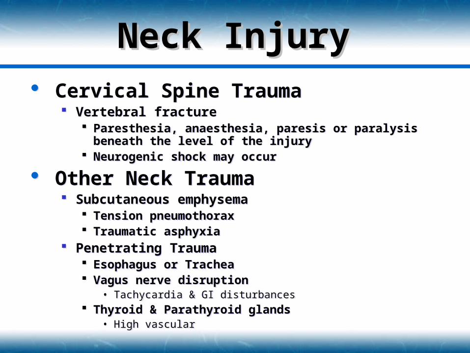
Neck InjuryNeck Injury Cervical Spine Trauma
Vertebral fracture Paresthesia, anaesthesia, paresis or paralysis beneath the
level of the injury Neurogenic shock may occur
Other Neck Trauma Subcutaneous emphysema
Tension pneumothorax Traumatic asphyxia
Penetrating Trauma Esophagus or Trachea Vagus nerve disruption
• Tachycardia & GI disturbances Thyroid & Parathyroid glands
• High vascular
Cervical Spine Trauma Vertebral fracture
Paresthesia, anaesthesia, paresis or paralysis beneath the level of the injury
Neurogenic shock may occur
Other Neck Trauma Subcutaneous emphysema
Tension pneumothorax Traumatic asphyxia
Penetrating Trauma Esophagus or Trachea Vagus nerve disruption
• Tachycardia & GI disturbances Thyroid & Parathyroid glands
• High vascular
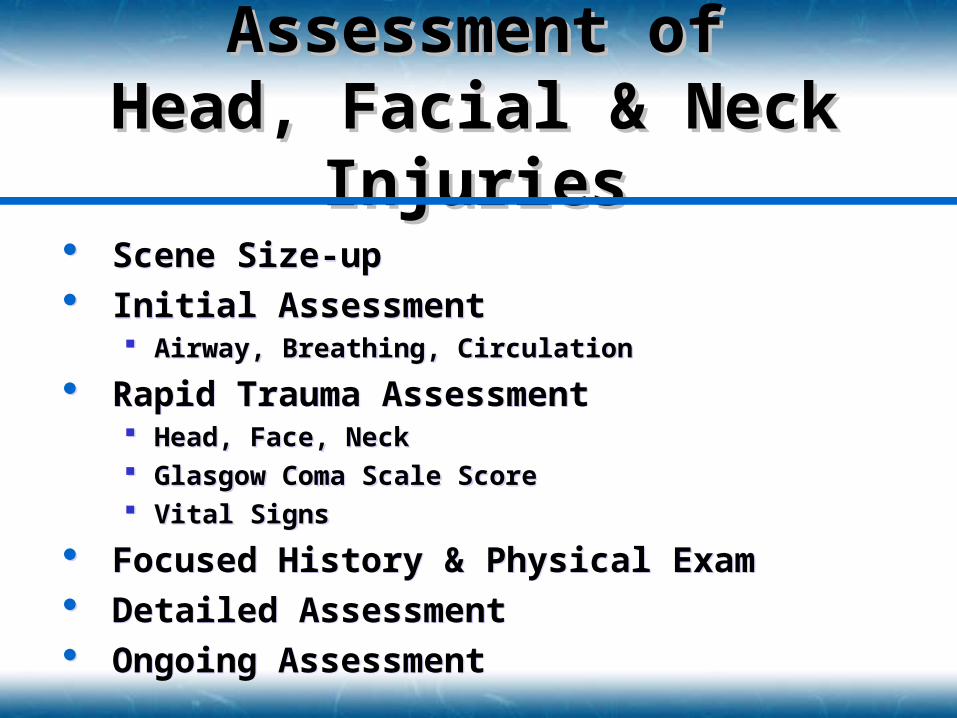
Scene Size-up Initial Assessment
Airway, Breathing, Circulation
Rapid Trauma Assessment Head, Face, Neck Glasgow Coma Scale Score Vital Signs
Focused History & Physical Exam Detailed Assessment Ongoing Assessment
Scene Size-up Initial Assessment
Airway, Breathing, Circulation
Rapid Trauma Assessment Head, Face, Neck Glasgow Coma Scale Score Vital Signs
Focused History & Physical Exam Detailed Assessment Ongoing Assessment
Assessment ofAssessment ofHead, Facial & Neck Head, Facial & Neck
InjuriesInjuries
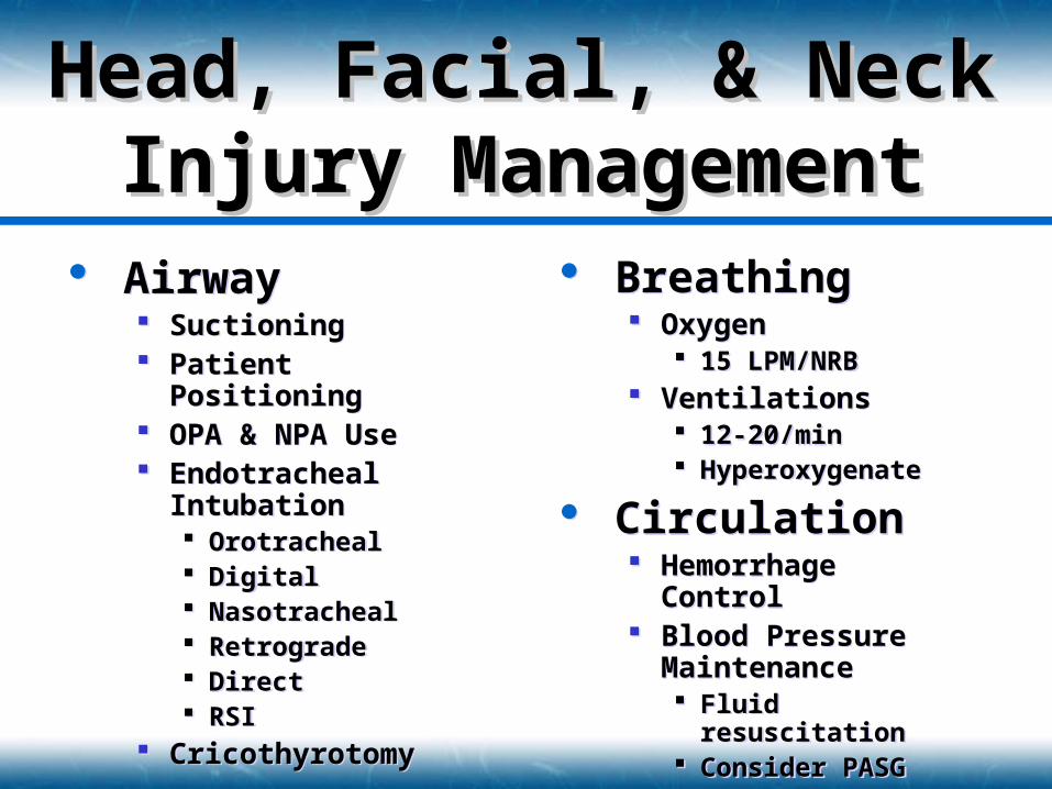
Airway Suctioning Patient Positioning OPA & NPA Use Endotracheal
Intubation Orotracheal Digital Nasotracheal Retrograde Direct RSI
Cricothyrotomy
Airway Suctioning Patient Positioning OPA & NPA Use Endotracheal
Intubation Orotracheal Digital Nasotracheal Retrograde Direct RSI
Cricothyrotomy
Head, Facial, & NeckHead, Facial, & NeckInjury ManagementInjury Management
Breathing Oxygen
15 LPM/NRB Ventilations
12-20/min Hyperoxygenate
Circulation Hemorrhage Control Blood Pressure
Maintenance Fluid resuscitation Consider PASG
Breathing Oxygen
15 LPM/NRB Ventilations
12-20/min Hyperoxygenate
Circulation Hemorrhage Control Blood Pressure
Maintenance Fluid resuscitation Consider PASG
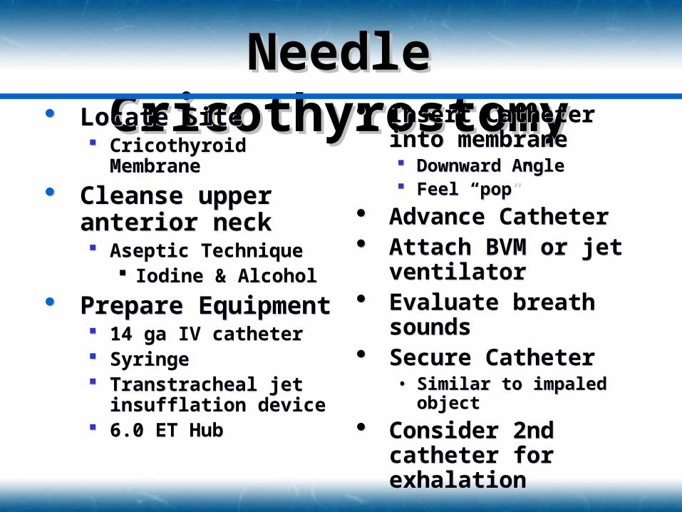
Needle Needle CricothyrostomyCricothyrostomy Locate Site
Cricothyroid Membrane
Cleanse upper anterior neck Aseptic Technique
Iodine & Alcohol
Prepare Equipment 14 ga IV catheter Syringe Transtracheal jet
insufflation device 6.0 ET Hub
Locate Site Cricothyroid Membrane
Cleanse upper anterior neck Aseptic Technique
Iodine & Alcohol
Prepare Equipment 14 ga IV catheter Syringe Transtracheal jet
insufflation device 6.0 ET Hub
Insert Catheter into membrane Downward Angle Feel “pop”
Advance Catheter Attach BVM or jet
ventilator Evaluate breath
sounds Secure Catheter
• Similar to impaled object
Consider 2nd catheter for exhalation
Insert Catheter into membrane Downward Angle Feel “pop”
Advance Catheter Attach BVM or jet
ventilator Evaluate breath
sounds Secure Catheter
• Similar to impaled object
Consider 2nd catheter for exhalation
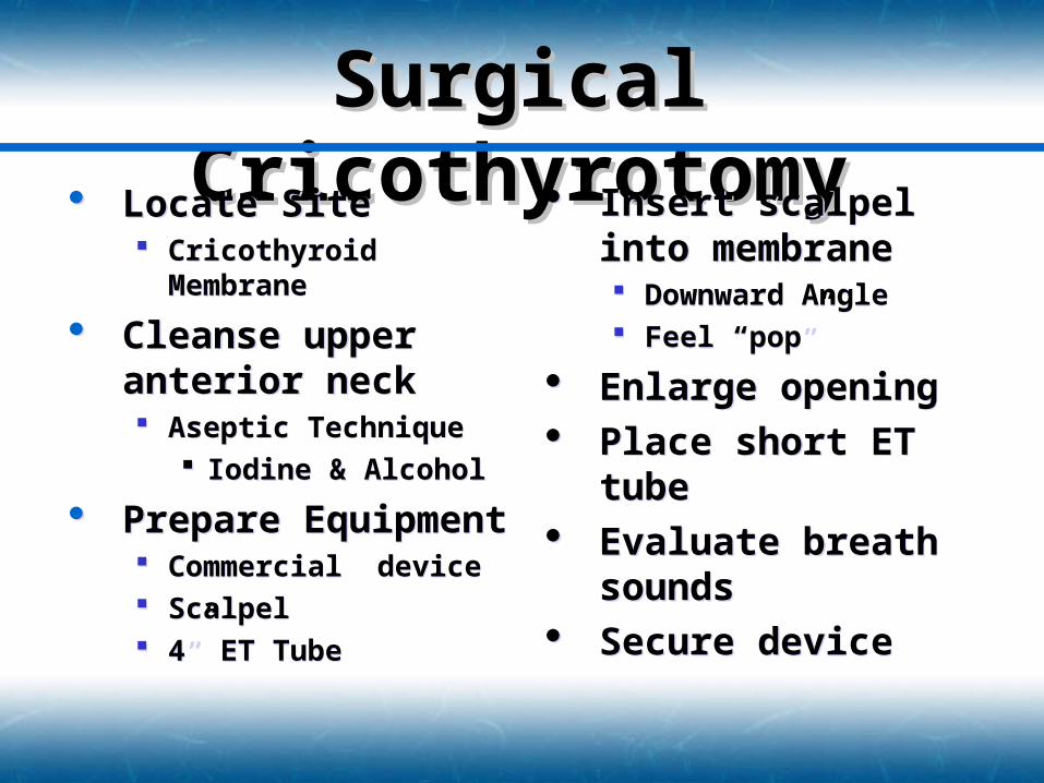
Surgical Surgical CricothyrotomyCricothyrotomy Locate Site
Cricothyroid Membrane
Cleanse upper anterior neck Aseptic Technique
Iodine & Alcohol
Prepare Equipment Commercial device Scalpel 4” ET Tube
Locate Site Cricothyroid Membrane
Cleanse upper anterior neck Aseptic Technique
Iodine & Alcohol
Prepare Equipment Commercial device Scalpel 4” ET Tube
Insert scalpel into membrane Downward Angle Feel “pop”
Enlarge opening Place short ET tube Evaluate breath
sounds Secure device
Insert scalpel into membrane Downward Angle Feel “pop”
Enlarge opening Place short ET tube Evaluate breath
sounds Secure device
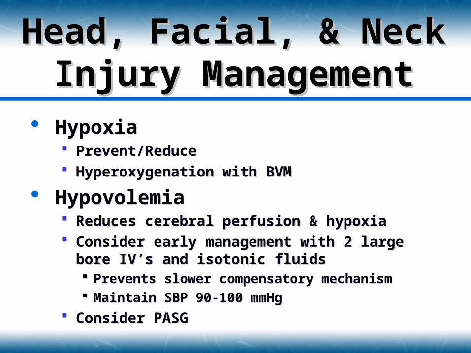
Hypoxia Prevent/Reduce Hyperoxygenation with BVM
Hypovolemia Reduces cerebral perfusion & hypoxia Consider early management with 2 large bore IV’s
and isotonic fluids Prevents slower compensatory mechanism Maintain SBP 90-100 mmHg
Consider PASG
Hypoxia Prevent/Reduce Hyperoxygenation with BVM
Hypovolemia Reduces cerebral perfusion & hypoxia Consider early management with 2 large bore IV’s
and isotonic fluids Prevents slower compensatory mechanism Maintain SBP 90-100 mmHg
Consider PASG
Head, Facial, & NeckHead, Facial, & NeckInjury ManagementInjury Management
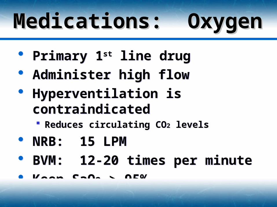
Medications: Medications: OxygenOxygen Primary 1st line drug
Administer high flow Hyperventilation is contraindicated
Reduces circulating CO2 levels
NRB: 15 LPM BVM: 12-20 times per minute Keep SaO2 > 95%
Primary 1st line drug Administer high flow Hyperventilation is contraindicated
Reduces circulating CO2 levels
NRB: 15 LPM BVM: 12-20 times per minute Keep SaO2 > 95%
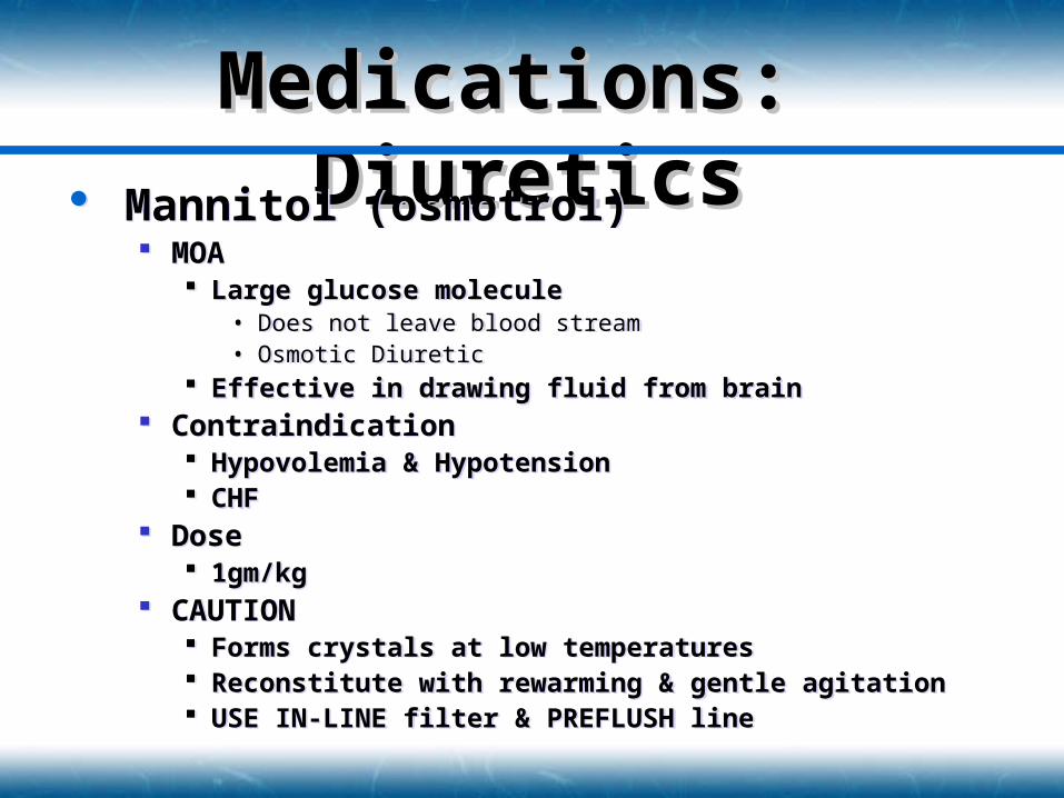
Medications: Medications: DiureticsDiuretics Mannitol (osmotrol)
MOA Large glucose molecule
• Does not leave blood stream• Osmotic Diuretic
Effective in drawing fluid from brain Contraindication
Hypovolemia & Hypotension CHF
Dose 1gm/kg
CAUTION Forms crystals at low temperatures Reconstitute with rewarming & gentle agitation USE IN-LINE filter & PREFLUSH line
Mannitol (osmotrol) MOA
Large glucose molecule• Does not leave blood stream• Osmotic Diuretic
Effective in drawing fluid from brain Contraindication
Hypovolemia & Hypotension CHF
Dose 1gm/kg
CAUTION Forms crystals at low temperatures Reconstitute with rewarming & gentle agitation USE IN-LINE filter & PREFLUSH line
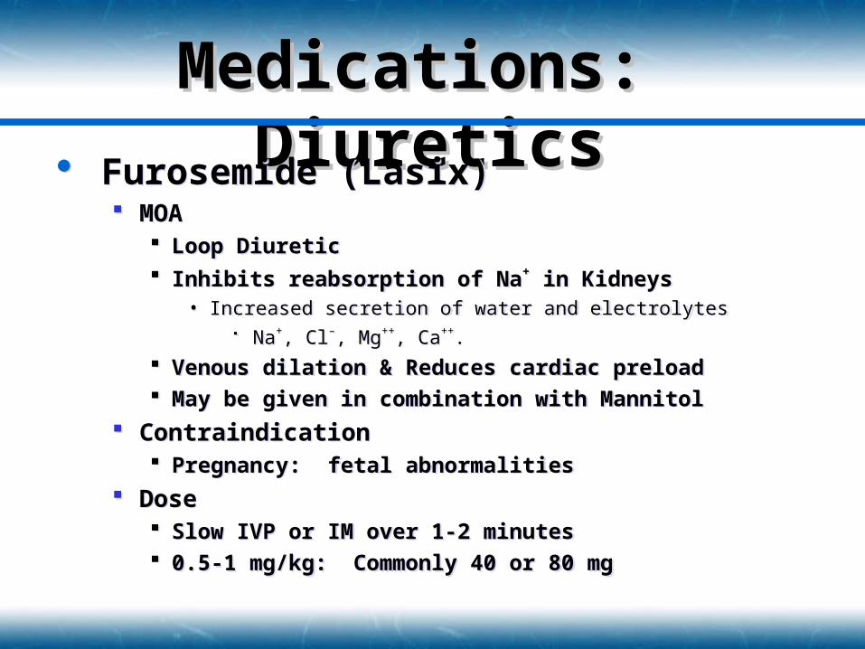
Medications: Medications: DiureticsDiuretics Furosemide (Lasix)
MOA Loop Diuretic Inhibits reabsorption of Na+ in Kidneys
• Increased secretion of water and electrolytes Na+, Cl–, Mg++, Ca++.
Venous dilation & Reduces cardiac preload May be given in combination with Mannitol
Contraindication Pregnancy: fetal abnormalities
Dose Slow IVP or IM over 1-2 minutes 0.5-1 mg/kg: Commonly 40 or 80 mg
Furosemide (Lasix) MOA
Loop Diuretic Inhibits reabsorption of Na+ in Kidneys
• Increased secretion of water and electrolytes Na+, Cl–, Mg++, Ca++.
Venous dilation & Reduces cardiac preload May be given in combination with Mannitol
Contraindication Pregnancy: fetal abnormalities
Dose Slow IVP or IM over 1-2 minutes 0.5-1 mg/kg: Commonly 40 or 80 mg
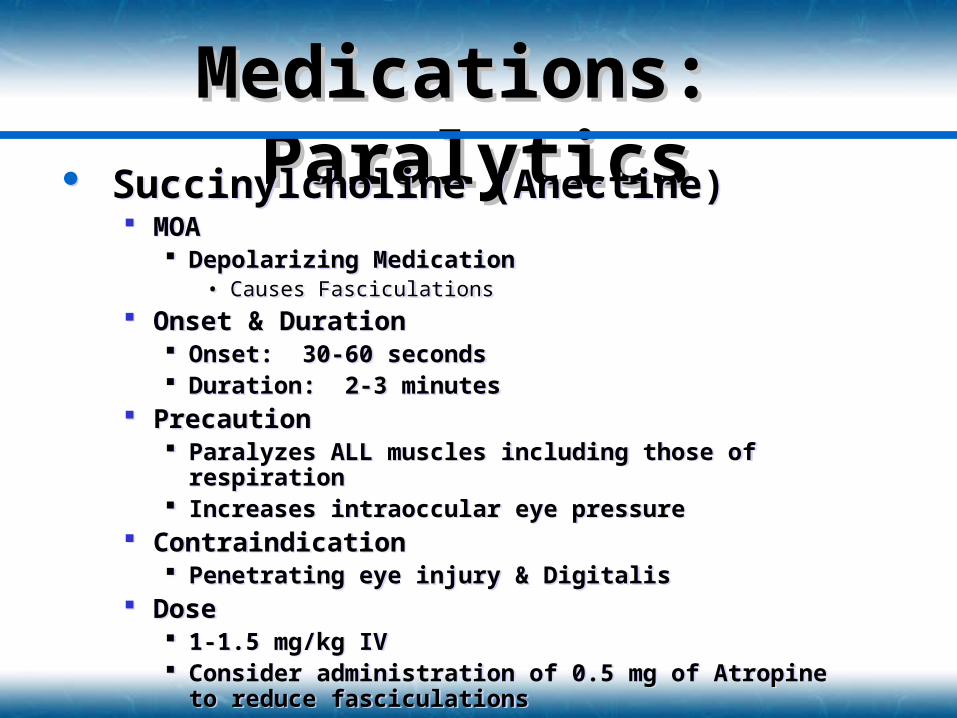
Medications: Medications: ParalyticsParalytics Succinylcholine (Anectine)
MOA Depolarizing Medication
• Causes Fasciculations
Onset & Duration Onset: 30-60 seconds Duration: 2-3 minutes
Precaution Paralyzes ALL muscles including those of respiration Increases intraoccular eye pressure
Contraindication Penetrating eye injury & Digitalis
Dose 1-1.5 mg/kg IV Consider administration of 0.5 mg of Atropine to reduce
fasciculations
Succinylcholine (Anectine) MOA
Depolarizing Medication• Causes Fasciculations
Onset & Duration Onset: 30-60 seconds Duration: 2-3 minutes
Precaution Paralyzes ALL muscles including those of respiration Increases intraoccular eye pressure
Contraindication Penetrating eye injury & Digitalis
Dose 1-1.5 mg/kg IV Consider administration of 0.5 mg of Atropine to reduce
fasciculations
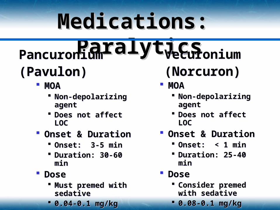
Medications: Medications: ParalyticsParalyticsPancuronium
(Pavulon) MOA
Non-depolarizing agent
Does not affect LOC Onset & Duration
Onset: 3-5 min Duration: 30-60 min
Dose Must premed with
sedative 0.04-0.1 mg/kg
Pancuronium (Pavulon)
MOA Non-depolarizing
agent Does not affect LOC
Onset & Duration Onset: 3-5 min Duration: 30-60 min
Dose Must premed with
sedative 0.04-0.1 mg/kg
Vecuronium(Norcuron)
MOA Non-depolarizing
agent Does not affect LOC
Onset & Duration Onset: < 1 min Duration: 25-40 min
Dose Consider premed with
sedative 0.08-0.1 mg/kg
Vecuronium(Norcuron)
MOA Non-depolarizing
agent Does not affect LOC
Onset & Duration Onset: < 1 min Duration: 25-40 min
Dose Consider premed with
sedative 0.08-0.1 mg/kg
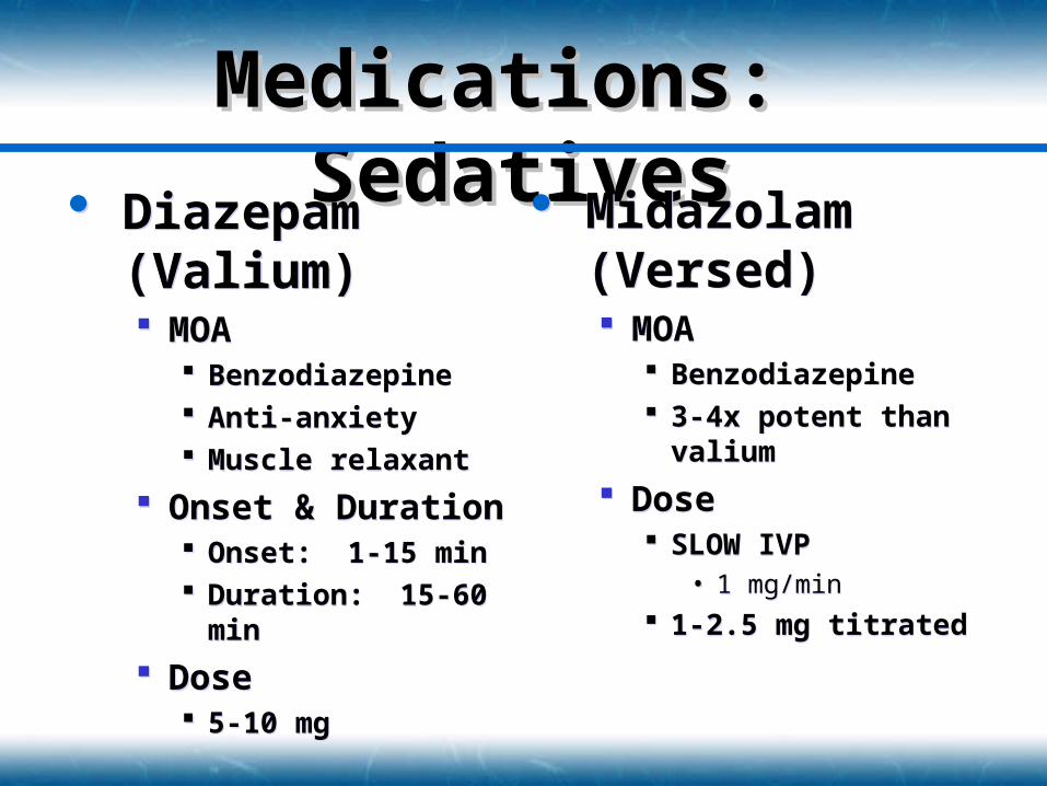
Medications: Medications: SedativesSedatives Diazepam
(Valium) MOA
Benzodiazepine Anti-anxiety Muscle relaxant
Onset & Duration Onset: 1-15 min Duration: 15-60 min
Dose 5-10 mg
Diazepam (Valium) MOA
Benzodiazepine Anti-anxiety Muscle relaxant
Onset & Duration Onset: 1-15 min Duration: 15-60 min
Dose 5-10 mg
Midazolam (Versed) MOA
Benzodiazepine 3-4x potent than
valium
Dose SLOW IVP
• 1 mg/min
1-2.5 mg titrated
Midazolam (Versed) MOA
Benzodiazepine 3-4x potent than
valium
Dose SLOW IVP
• 1 mg/min
1-2.5 mg titrated
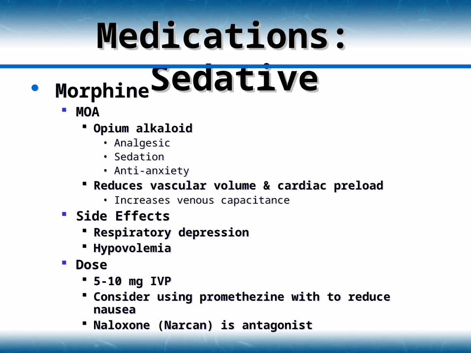
Medications: Medications: SedativeSedative Morphine
MOA Opium alkaloid
• Analgesic• Sedation• Anti-anxiety
Reduces vascular volume & cardiac preload• Increases venous capacitance
Side Effects Respiratory depression Hypovolemia
Dose 5-10 mg IVP Consider using promethezine with to reduce nausea Naloxone (Narcan) is antagonist
Morphine MOA
Opium alkaloid• Analgesic• Sedation• Anti-anxiety
Reduces vascular volume & cardiac preload• Increases venous capacitance
Side Effects Respiratory depression Hypovolemia
Dose 5-10 mg IVP Consider using promethezine with to reduce nausea Naloxone (Narcan) is antagonist
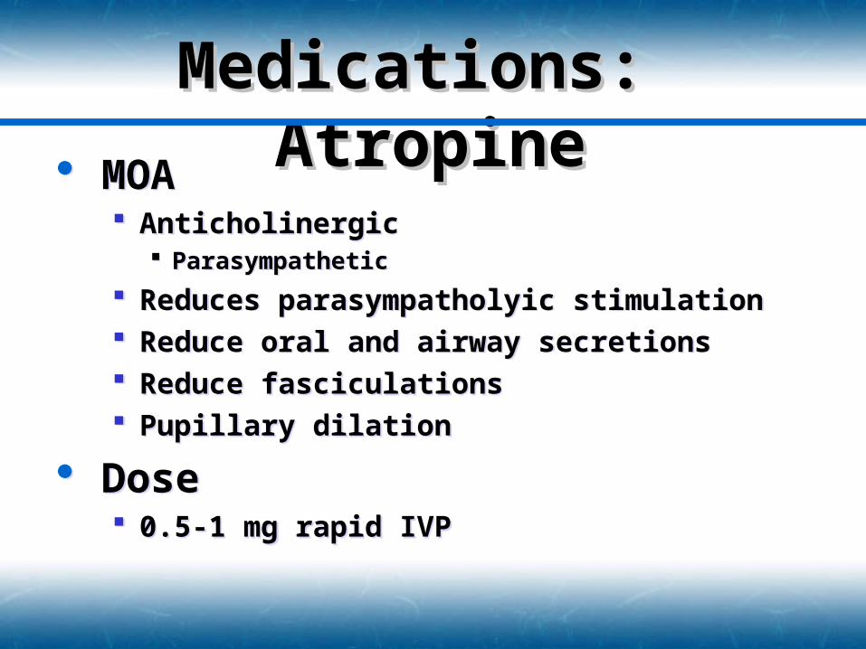
Medications: Medications: AtropineAtropine MOA
Anticholinergic Parasympathetic
Reduces parasympatholyic stimulation Reduce oral and airway secretions Reduce fasciculations Pupillary dilation
Dose 0.5-1 mg rapid IVP
MOA Anticholinergic
Parasympathetic
Reduces parasympatholyic stimulation Reduce oral and airway secretions Reduce fasciculations Pupillary dilation
Dose 0.5-1 mg rapid IVP
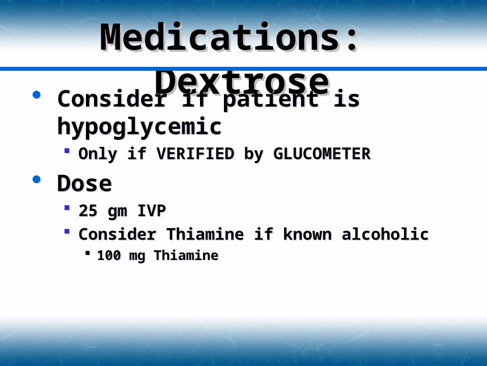
Medications: Medications: DextroseDextrose Consider if patient is hypoglycemic
Only if VERIFIED by GLUCOMETER
Dose 25 gm IVP Consider Thiamine if known alcoholic
100 mg Thiamine
Consider if patient is hypoglycemic Only if VERIFIED by GLUCOMETER
Dose 25 gm IVP Consider Thiamine if known alcoholic
100 mg Thiamine
Medications: Medications: ThiamineThiamine Vitamin B1
Essential for the processing of glucose through Kreb’s cycle
Chronic alcoholics can have B1 depletion
Dose 100 mg IV or IM
Vitamin B1 Essential for the processing of
glucose through Kreb’s cycle Chronic alcoholics can have B1
depletion Dose
100 mg IV or IM
Medications Xylocaine or Benzocaine
Anesthetize oral and pharyngeal mucosa• Reduces gag reflex
• Reduces likelihood of ICP associated with vomiting
Inhibits nerve sensation Onset & Duration
• Onset: 15 seconds
• Duration: 15 minutes
PRECAUTION• Patient has reduced ability to remove oral fluids
• ASPIRATION can occur
Medications: Topical Medications: Topical Anesthetic SprayAnesthetic Spray
Transport Transport ConsiderationsConsiderations Limit external stimulation
Can increase ICP Can induce seizures
Cautious about Air Transport Seizures
Limit external stimulation Can increase ICP Can induce seizures
Cautious about Air Transport Seizures
Emotional SupportEmotional Support
Have friend or family provide constant reassurance
Provided constant reorientation to environment if required Keeps patient calm Reduces anxiety
Have friend or family provide constant reassurance
Provided constant reorientation to environment if required Keeps patient calm Reduces anxiety
Special Injury CareSpecial Injury Care
Scalp Avulsion Cover the open wound with bulky dressing Pad under the fold of the scalp Irrigate with NS to remove gross contamination
Pinna Injury Place in close anatomic position as possible Dress and cover with sterile dressing
Scalp Avulsion Cover the open wound with bulky dressing Pad under the fold of the scalp Irrigate with NS to remove gross contamination
Pinna Injury Place in close anatomic position as possible Dress and cover with sterile dressing
Special Injury CareSpecial Injury Care Eye Injury
General Injury Cover injured and uninjured eye
• Prevents sympathetic motion
Consider sterile dressing soaked in NS
Corneal Abrasion Invert eyelid and examine eye for foreign body Remove with NS moistened gauze or Morgan’s Lens
Avulsed or Impaled Eye Cover and Protect from injury
General Care Calm & reassure patient
Eye Injury General Injury
Cover injured and uninjured eye• Prevents sympathetic motion
Consider sterile dressing soaked in NS
Corneal Abrasion Invert eyelid and examine eye for foreign body Remove with NS moistened gauze or Morgan’s Lens
Avulsed or Impaled Eye Cover and Protect from injury
General Care Calm & reassure patient
Special Injury CareSpecial Injury Care
Dislodged Teeth Rinse in NS Wrap in NS soaked gauze
Impaled Objects Secure with bulky dressing Stabilize object to prevent movement Indirect pressure around wound
Dislodged Teeth Rinse in NS Wrap in NS soaked gauze
Impaled Objects Secure with bulky dressing Stabilize object to prevent movement Indirect pressure around wound