Have Community-Acquired Pneumonia?clinicalevidence.pbworks.com/w/file/fetch/51305718/ESTE...
6
Does This Patient Have Community-Acquired Pneumonia? Diagnosing Pneumonia by History and Physical Examination Joshua P. Metlay, MD, PhD; Wishwa N. Kapoor, MD, MPH; Michael J. Fine, MD, MSc Community-acquired pneumonia is an important cause of acute respiratory symptoms (eg, cough) in the ambulatory care setting. Distinguishing pneumo- nia from other causes of respiratory illnesses, such as acute bronchitis and up- per respiratory tract infections, has important therapeutic and prognostic impli- cations. The reference standard for diagnosing pneumonia is chest radiography, but it is likely that many physicians rely on the patient's history and their physi- cal examination to diagnose or exclude this disease. A review of published studies of patients suspected of having pneumonia reveals that there are no in- dividual clinical findings, or combinations of findings, that can rule in the diag- nosis of pneumonia for a patient suspected of having this illness. However, some studies have shown that the absence of any vital sign abnormalities or any abnormalities on chest auscultation substantially reduces the likelihood of pneumonia to a point where further diagnostic evaluation may be unnecessary. This article reviews the literature on the appropriate use of the history and physical examination in diagnosing community-acquired pneumonia. JAMA. 1997;278:1440-1445 CLINICAL SCENARIO—DOES THIS PATIENT HAVE PNEUMONIA? A 53-year-old woman comes to your office with a cough of more than 1 week's duration. She was in excellent health un¬ til 7 days ago when she developed a non¬ productive cough, mild sore throat, and myalgia. She recalls no past history of asthma or chronic obstructive pulmo¬ nary disease, and she does not smoke. Despite staying home from work for the last 2 days, she has noted increasing spu¬ tum production with her cough and worsening fatigue. She has felt warm but has not documented any fever or night sweats. On physical examination, her oral temperature is 38.3°C (101°F), her heart rate is 110 beats per minute, and auscultation of her chest reveals inspi- ratory crackles on the left side. WHY IS THIS AN IMPORTANT QUESTION TO ANSWER WITH A CLINICAL EXAMINATION? Physicians commonly encounter pa¬ tients with respiratory complaints simi¬ lar to those in the clinical scenario. In 1994, there were over 10 million visits to primary care physicians by adults with a chief complaint of cough, representing over 4% of all visits to physicians that year. Pneumonia represented only 5% of all causes for these visits and was the fifth leading diagnosis, after bron- chitis, upper respiratory tract infection, asthma, and sinusitis.1 Though pneumo¬ nia may represent a small proportion of all acute respiratory illnesses, the accu¬ rate identification of this subgroup is im¬ portant because of the very distinct therapeutic and prognostic features of this illness. In the preantibiotic era, mortality from pneumococcal pneumonia was con¬ sistently higher than 20% for all cases, rising to more than 60% for bacteremic cases.2 Since the introduction of antibi¬ otics no one has reported results from large-scale studies comparing antibiotic therapy to nonantibiotic therapy for pa¬ tients with pneumonia. As a result, such therapy is universally recommended and has become a standard of care for all patients with pneumonia. No such stan¬ dard exists for alternative respiratory infections such as bronchitis3 or the com¬ mon cold.4 Moreover, inappropriate use of antibiotics for these alternative res¬ piratory infections may be an important determinant of the rise in antibiotic re¬ sistance among common respiratory pathogens.5·6 In terms of prognosis, patients with pneumonia continue to have an overall high mortality from this illness, ranging from as low 5% in studies of hospitalized and ambulatory patients to as high as 37% in studies of patients requiring ad¬ mission to intensive care units.7 This per¬ sistently high mortality underscores the From the General Internal Medicine Unit, Department of Medicine, Massachusetts General Hospital and Har- vard Medical School, Boston (Dr Metlay), and the Divi- sion of General Internal Medicine, Department of Medi- cine, Center for Research on Health Care, University of Pittsburgh, Pittsburgh, Pa (Drs Kapoor and Fine). Dr Metlay is now with the Division of General Internal Medicine, Department of Medicine, University of Penn- sylvania Medical Center, Philadelphia. Reprints: Michael J. Fine, MD, MSc, Montefiore Uni- versity Hospital, 8 East Room 824, 200 Lothrop St, Pittsburgh, PA 15213 (e-mail: [email protected]). The Rational Clinical Examination section editors: David L. Simel, MD, MHS, Durham Veterans Affairs Medical Center and Duke University Medical Cen- ter, Durham, NC; Drummond Rennie, MD, Deputy Editor (West), JAMA. at HINARI on August 27, 2011 jama.ama-assn.org Downloaded from
Transcript of Have Community-Acquired Pneumonia?clinicalevidence.pbworks.com/w/file/fetch/51305718/ESTE...

Does This Patient HaveCommunity-Acquired Pneumonia?Diagnosing Pneumonia by History and Physical ExaminationJoshua P. Metlay, MD, PhD; Wishwa N. Kapoor, MD, MPH; Michael J. Fine, MD, MSc
Community-acquired pneumonia is an important cause of acute respiratorysymptoms (eg, cough) in the ambulatory care setting. Distinguishing pneumo-nia from other causes of respiratory illnesses, such as acute bronchitis and up-per respiratory tract infections, has important therapeutic and prognostic impli-cations. The reference standard for diagnosing pneumonia is chest radiography,but it is likely that many physicians rely on the patient's history and their physi-cal examination to diagnose or exclude this disease. A review of publishedstudies of patients suspected of having pneumonia reveals that there are no in-dividual clinical findings, or combinations of findings, that can rule in the diag-nosis of pneumonia for a patient suspected of having this illness. However,some studies have shown that the absence of any vital sign abnormalities or anyabnormalities on chest auscultation substantially reduces the likelihood ofpneumonia to a point where further diagnostic evaluation may be unnecessary.This article reviews the literature on the appropriate use of the history andphysical examination in diagnosing community-acquired pneumonia.
JAMA. 1997;278:1440-1445
CLINICAL SCENARIO—DOESTHIS PATIENT HAVE PNEUMONIA?
A 53-year-old woman comes to youroffice with a cough ofmore than 1 week'sduration. She was in excellent health un¬til 7 days ago when she developed a non¬
productive cough, mild sore throat, andmyalgia. She recalls no past history ofasthma or chronic obstructive pulmo¬nary disease, and she does not smoke.Despite stayinghome from work for the
last 2 days, she has noted increasing spu¬tum production with her cough andworsening fatigue. She has felt warm buthas not documented any fever or nightsweats. On physical examination, heroral temperature is 38.3°C (101°F), herheart rate is 110 beats per minute, andauscultation of her chest reveals inspi-ratory crackles on the left side.
WHY IS THIS AN IMPORTANTQUESTION TO ANSWER WITHA CLINICAL EXAMINATION?
Physicians commonly encounter pa¬tients with respiratory complaints simi¬lar to those in the clinical scenario. In1994, there were over 10 million visits toprimary care physicians by adults with achief complaint of cough, representingover 4% of all visits to physicians thatyear. Pneumonia represented only 5%of all causes for these visits and wasthe fifth leading diagnosis, after bron-
chitis, upper respiratory tract infection,asthma, and sinusitis.1 Though pneumo¬nia may represent a small proportion ofall acute respiratory illnesses, the accu¬rate identification of this subgroup is im¬portant because of the very distincttherapeutic and prognostic features ofthis illness.
In the preantibiotic era, mortalityfrom pneumococcal pneumonia was con¬
sistently higher than 20% for all cases,rising to more than 60% for bacteremiccases.2 Since the introduction of antibi¬otics no one has reported results fromlarge-scale studies comparing antibiotictherapy to nonantibiotic therapy for pa¬tients with pneumonia. As a result, suchtherapy is universally recommendedand has become a standard ofcare for allpatients with pneumonia. No such stan¬dard exists for alternative respiratoryinfections such as bronchitis3 or the com¬mon cold.4 Moreover, inappropriate useof antibiotics for these alternative res¬
piratory infections may be an importantdeterminant of the rise in antibiotic re¬sistance among common respiratorypathogens.5·6
In terms of prognosis, patients withpneumonia continue to have an overallhigh mortality from this illness, rangingfrom as low 5% in studies ofhospitalizedand ambulatory patients to as high as37% in studies of patients requiring ad¬mission to intensive care units.7 This per¬sistently high mortality underscores the
From the General Internal Medicine Unit, Departmentof Medicine, Massachusetts General Hospital and Har-vard Medical School, Boston (Dr Metlay), and the Divi-sion of General Internal Medicine, Department of Medi-cine, Center for Research on Health Care, University ofPittsburgh, Pittsburgh, Pa (Drs Kapoor and Fine). DrMetlay is now with the Division of General InternalMedicine, Department of Medicine, University of Penn-sylvania Medical Center, Philadelphia.
Reprints: Michael J. Fine, MD, MSc, Montefiore Uni-versity Hospital, 8 East Room 824, 200 Lothrop St,Pittsburgh, PA 15213 (e-mail: [email protected]).
The Rational Clinical Examination section editors:David L. Simel, MD, MHS, Durham Veterans AffairsMedical Center and Duke University Medical Cen-ter, Durham, NC; Drummond Rennie, MD, DeputyEditor (West), JAMA.
at HINARI on August 27, 2011jama.ama-assn.orgDownloaded from
need for physicians to choose carefullybetween home or hospital therapy for allpatients with pneumonia.8 For these rea¬
sons, physicians need to know how opti¬mally to use their clinical examination toidentify patients at suitable risk forpneumonia to require further, definitivediagnostic testing.
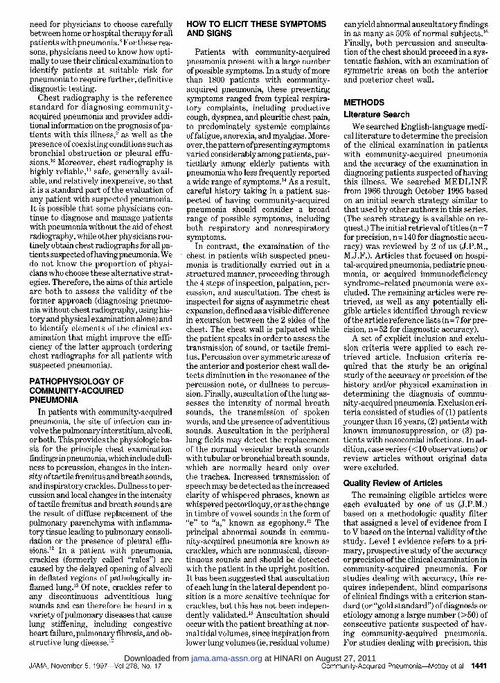
Chest radiography is the referencestandard for diagnosing community-acquired pneumonia and provides addi¬tional information on the prognosis ofpa¬tients with this illness,9 as well as thepresence ofcoexisting conditions such asbronchial obstruction or pleural effu¬sions.10 Moreover, chest radiography ishighly reliable,11 safe, generally avail¬able, and relatively inexpensive, so thatit is a standard part of the evaluation ofany patient with suspected pneumonia.It is possible that some physicians con¬tinue to diagnose and manage patientswith pneumonia without the aid of chestradiography, while otherphysicians rou¬
tinely obtain chest radiographs for all pa¬tients suspected ofhaving pneumonia. Wedo not know the proportion of physi¬cians who choose these alternative strat¬egies. Therefore, the aims of this articleare both to assess the validity of theformer approach (diagnosing pneumo¬nia without chest radiography, using his¬tory and physical examination alone) andto identify elements of the clinical ex¬amination that might improve the effi¬ciency of the latter approach (orderingchest radiographs for all patients withsuspected pneumonia).PATHOPHYSIOLOGY OFCOMMUNITY-ACQUIREDPNEUMONIA
In patients with community-acquiredpneumonia, the site of infection can in¬volve the pulmonary interstitium, alveoli,or both. This provides the physiologic ba¬sis for the principle chest examinationfindings in pneumonia, which include dull¬ness to percussion, changes in the inten¬sity oftactile fremitus and breath sounds,and inspiratory crackles. Dullness to per¬cussion and local changes in the intensityof tactile fremitus and breath sounds arethe result of diffuse replacement of thepulmonary parenchyma with inflamma¬tory tissue leading to pulmonary consoli¬dation or the presence of pleural effu¬sions.12 In a patient with pneumonia,crackles (formerly called "rales") arecaused by the delayed opening of alveoliin deflated regions of pathologically in¬flamed lung.13 Of note, crackles refer toany discontinuous adventitious lungsounds and can therefore be heard in a
variety ofpulmonary diseases that cause
lung stiffening, including congestiveheart failure, pulmonary fibrosis, and ob¬structive lung disease.12
HOW TO ELICIT THESE SYMPTOMSAND SIGNS
Patients with community-acquiredpneumonia present with a large numberofpossible symptoms. In a study ofmorethan 1800 patients with community-acquired pneumonia, these presentingsymptoms ranged from typical respira¬tory complaints, including productivecough, dyspnea, and pleuritic chest pain,to predominately systemic complaintsof fatigue, anorexia, and myalgias. More¬over, the pattern ofpresentingsymptomsvaried considerably among patients, par¬ticularly among elderly patients withpneumonia who less frequently reporteda wide range of symptoms.14 As a result,careful history taking in a patient sus¬
pected of having community-acquiredpneumonia should consider a broadrange of possible symptoms, includingboth respiratory and nonrespiratorysymptoms.
In contrast, the examination of thechest in patients with suspected pneu¬monia is traditionally carried out in astructured manner, proceeding throughthe 4 steps of inspection, palpation, per¬cussion, and auscultation. The chest isinspected for signs of asymmetric chestexpansion, defined as a visible differencein excursion between the 2 sides of thechest. The chest wall is palpated whilethe patient speaks in order to assess thetransmission of sound, or tactile fremi¬tus. Percussion over symmetric areas ofthe anterior and posterior chest wall de¬tects diminution in the resonance of thepercussion note, or dullness to percus¬sion. Finally, auscultation of the lung as¬sesses the intensity of normal breathsounds, the transmission of spokenwords, and the presence of adventitioussounds. Auscultation in the peripherallung fields may detect the replacementof the normal vesicular breath soundswith tubular or bronchial breath sounds,which are normally heard only overthe trachea. Increased transmission ofspeech may be detected as the increasedclarity of whispered phrases, known as
whispered pectoriloquy, or as the changein timbre of vowel sounds in the form of"e" to "a," known as egophony.12 Theprincipal abnormal sounds in commu¬
nity-acquired pneumonia are known as
crackles, which are nonmusical, discon¬tinuous sounds and should be detectedwith the patient in the upright position.It has been suggested that auscultationof each lung in the lateral dependent po¬sition is a more sensitive technique forcrackles, but this has not been indepen¬dently validated.15 Auscultation shouldoccur with the patient breathing at nor¬mal tidal volumes, since inspiration fromlower lung volumes (ie, residual volume)
can yield abnormal auscultatory findingsin as many as 50% of normal subjects.16Finally, both percussion and ausculta¬tion of the chest should proceed in a sys¬tematic fashion, with an examination ofsymmetric areas on both the anteriorand posterior chest wall.
METHODSLiterature Search
We searched English-language medi¬cal literature to determine the precisionof the clinical examination in patientswith community-acquired pneumoniaand the accuracy of the examination indiagnosing patients suspected of havingthis illness. We searched MEDLINEfrom 1966 through October 1995 basedon an initial search strategy similar tothat used by other authors in this series.(The search strategy is available on re¬
quest.) The initial retrieval of titles (n=7for precision, n=140 for diagnostic accu¬
racy) was reviewed by 2 of us (J.P.M.,M.J.F.). Articles that focused on hospi¬tal-acquired pneumonia, pediatrie pneu¬monia, or acquired immunodeficiencysyndrome-related pneumonia were ex¬cluded. The remaining articles were re¬
trieved, as well as any potentially eli¬gible articles identified through reviewofthe article reference lists (n=7 for pre¬cision, =52 for diagnostic accuracy).
A set of explicit inclusion and exclu¬sion criteria were applied to each re¬trieved article. Inclusion criteria re¬
quired that the study be an originalstudy of the accuracy or precision of thehistory and/or physical examination indetermining the diagnosis of commu¬
nity-acquired pneumonia. Exclusion cri¬teria consisted of studies of (1) patientsyounger than 16 years, (2) patients withknown immunosuppression, or (3) pa¬tients with nosocomial infections. In ad¬dition, case series (<10 observations) orreview articles without original datawere excluded.
Quality Review of ArticlesThe remaining eligible articles were
each evaluated by one of us (J.P.M.)based on a méthodologie quality filterthat assigned a level of evidence from Ito V based on the internal validity of thestudy. Level I evidence refers to a pri¬mary, prospective study of the accuracyor precision ofthe clinical examination incommunity-acquired pneumonia. Forstudies dealing with accuracy, this re¬
quires independent, blind comparisonsof clinical findings with a criterion stan¬dard (or "gold standard") ofdiagnosis or
etiology among a large number (>50) ofconsecutive patients suspected of hav¬ing community-acquired pneumonia.For studies dealing with precision, this
at HINARI on August 27, 2011jama.ama-assn.orgDownloaded from
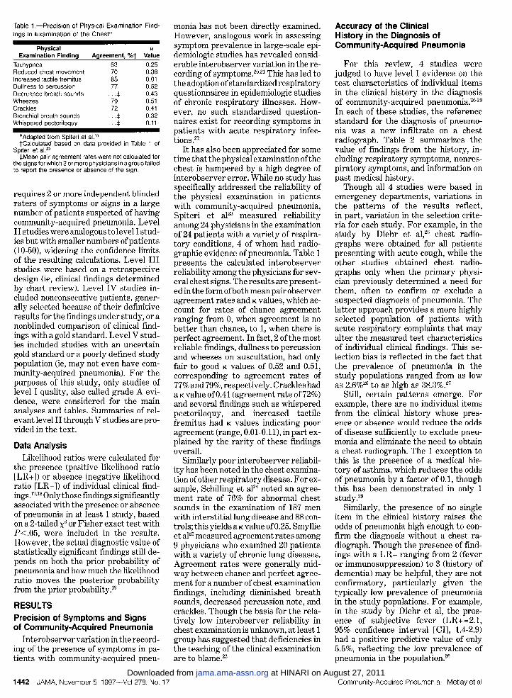
Table 1.—Precision of Physical Examination Find¬ings in Examination of the Chest*
Physical
Examination Finding Agreement, %t Value
Tachypnea 63 0.25Reduced chest movement 70 0.38Increased tactile fremitus 85 0.01Dullness to percussion 77 0.52Decreased breath sounds
..
4 0.43Wheezes 79 0.51Crackles 72 0.41Bronchial breath sounds
..
4 0.32Whispered pectoriloquy
. .
4 0.11
*Adapted from Spiteri et al.23fCalculated based on data provided in Table 1 of
Spiteri et al.23ÍMean pair agreement rates were not calculated for
the signs for which 2 or more physicians in a group failedto report the presence or absence of the sign.
requires 2 or more independent blindedraters of symptoms or signs in a largenumber of patients suspected of havingcommunity-acquired pneumonia. LevelII studies were analogous to level I stud¬ies but with smaller numbers ofpatients(10-50), widening the confidence limitsof the resulting calculations. Level IIIstudies were based on a retrospectivedesign (ie, clinical findings determinedby chart review). Level IV studies in¬cluded nonconsecutive patients, gener¬ally selected because of their definitiveresults for the findings under study, or anonblinded comparison of clinical find¬ings with a gold standard. Level V stud¬ies included studies with an uncertaingold standard or a poorly defined studypopulation (ie, may not even have com¬
munity-acquired pneumonia). For thepurposes of this study, only studies oflevel I quality, also called grade A evi¬dence, were considered for the mainanalyses and tables. Summaries of rel¬evant level II through V studies are pro¬vided in the text.
Data AnalysisLikelihood ratios were calculated for
the presence (positive likelihood ratio[LR+]) or absence (negative likelihoodratio [LR-]) of individual clinical find¬ings.1718 Only those findings significantlyassociated with the presence or absenceof pneumonia in at least 1 study, basedon a 2-tailed 2 or Fisher exact test withP<.05, were included in the results.However, the actual diagnostic value ofstatistically significant findings still de¬pends on both the prior probability ofpneumonia and how much the likelihoodratio moves the posterior probabilityfrom the prior probability.19RESULTSPrecision of Symptoms and Signsof Community-Acquired Pneumonia
Interobserver variation in the record¬ing of the presence of symptoms in pa¬tients with community-acquired pneu-
monia has not been directly examined.However, analogous work in assessingsymptom prevalence in large-scale epi¬demiologie studies has revealed consid¬erable interobserver variation in the re¬
cording of symptoms.20,21 This has led tothe adoption ofstandardized respiratoryquestionnaires in epidemiologie studiesof chronic respiratory illnesses. How¬ever, no such standardized question¬naires exist for recording symptoms inpatients with acute respiratory infec¬tions.22
It has also been appreciated for sometime that the physical examination ofthechest is hampered by a high degree ofinterobserver error. While no study hasspecifically addressed the reliability ofthe physical examination in patientswith community-acquired pneumonia,Spiteri et al23 measured reliabilityamong 24 physicians in the examinationof 24 patients with a variety of respira¬tory conditions, 4 of whom had radio-graphic evidence of pneumonia. Table 1presents the calculated interobserverreliability among the physicians for sev¬eral chest signs. The results are present¬ed in the form ofboth mean pair observeragreement rates and values, which ac¬count for rates of chance agreementranging from 0, when agreement is nobetter than chance, to 1, when there isperfect agreement. In fact, 2 of the mostreliable findings, dullness to percussionand wheezes on auscultation, had onlyfair to good values of 0.52 and 0.51,corresponding to agreement rates of77% and 79%, respectively. Crackles hada value of 0.41 (agreement rate of 72%)and several findings such as whisperedpectoriloquy, and increased tactilefremitus had values indicating pooragreement (range, 0.01-0.11), in part ex¬
plained by the rarity of these findingsoverall.
Similarly poor interobserver reliabil¬ity has been noted in the chest examina¬tion ofother respiratory disease. For ex¬
ample, Schilling et al24 noted an agree¬ment rate of 76% for abnormal chestsounds in the examination of 187 menwith interstitial lung disease and 88 con¬
trols; this yields a value of0.25. Smyllieet al25 measured agreement rates among9 physicians who examined 20 patientswith a variety of chronic lung diseases.Agreement rates were generally mid¬way between chance and perfect agree¬ment for a number of chest examinationfindings, including diminished breathsounds, decreased percussion note, andcrackles. Though the basis for the rela¬tively low interobserver reliability inchest examination is unknown, at least 1group has suggested that deficiencies inthe teaching of the clinical examinationare to blame.23
Accuracy of the ClinicalHistory in the Diagnosis ofCommunity-Acquired Pneumonia
For this review, 4 studies were
judged to have level I evidence on thetest characteristics of individual itemsin the clinical history in the diagnosisof community-acquired pneumonia.26'29In each of these studies, the referencestandard for the diagnosis of pneumo¬nia was a new infiltrate on a chestradiograph. Table 2 summarizes thevalue of findings from the history, in¬cluding respiratory symptoms, nonres-
piratory symptoms, and information on
past medical history.Though all 4 studies were based in
emergency departments, variations inthe patterns of the results reflect,in part, variation in the selection crite¬ria for each study. For example, in thestudy by Diehr et al,26 chest radio¬graphs were obtained for all patientspresenting with acute cough, while theother studies obtained chest radio¬graphs only when the primary physi¬cian previously determined a need forthem, often to confirm or exclude a
suspected diagnosis of pneumonia. Thelatter approach provides a more highlyselected population of patients withacute respiratory complaints that mayalter the measured test characteristicsof individual clinical findings. This se¬lection bias is reflected in the fact thatthe prevalence of pneumonia in thestudy populations ranged from as lowas 2.6%26 to as high as 38.3%.27
Still, certain patterns emerge. Forexample, there are no individual itemsfrom the clinical history whose pres¬ence or absence would reduce the oddsof disease sufficiently to exclude pneu¬monia and eliminate the need to obtaina chest radiograph. The 1 exception tothis is the presence of a medical his¬tory of asthma, which reduces the oddsof pneumonia by a factor of 0.1, thoughthis has been demonstrated in only 1study.29
Similarly, the presence of no singleitem in the clinical history raises theodds of pneumonia high enough to con¬firm the diagnosis without a chest ra¬
diograph. Though the presence of find¬ings with a LR+ ranging from 2 (feveror immunosuppression) to 3 (history ofdementia) may be helpful, they are notconfirmatory, particularly given thetypically low prevalence of pneumoniain the study populations. For example,in the study by Diehr et al, the pres¬ence of subjective fever (LR+=2.1,95% confidence interval [CI], 1.4-2.9)had a positive predictive value of only5.5%, reflecting the low prevalence ofpneumonia in the population.26
at HINARI on August 27, 2011jama.ama-assn.orgDownloaded from
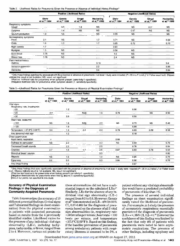
Table 2.—Likelihood Ratios for Pneumonia Given the Presence or Absence of Individual History Findings*Positive Likelihood Ratiof Negative Likelihood Ration
Diehret al,26 1984
Genniset al,271988
Singalet al,281989
Heckerlinget al,291990
Diehret al,261984
Genniset al,271988
Singalet al,281989
Respiratory symptomsCough NS NS NS 0.31 NSDyspnea NS NS 0.67 NS NS
Sputum production 1.3 NS NS 0.55 NS NSNonrespiratory symptoms
Fever 2.1 NS 1.7 0.71 NS 0.59Chills 1.6 1.3 1.7 0.85 0.72 0.70
Night sweats 1.7 0.83
Myalgias 1.3 NS 0.58 NSSore throat 0.78 NS 1.6 NSRhinorrhea 0.78 NS 2.4 NS
Past medical historyAsthma 0.10 3.8
Immunosuppression 2.2 0.85Dementia 3.4 0.94
*Only those findings significantly associated with the presence or absence of pneumonia in at least 1 study were included (P<.05 in a 2-tailed 2 or Fisher exact test). Ellipsesindicate the result is not available; NS, result not significant.
tPositive likelihood ratio for pneumonia when symptom present (sensitivity/1-specificity).^Negative likelihood ratio for pneumonia when symptom absent (1-sensitivity/specificity).
Table 3.—Likelihood Ratios for Pneumonia Given the Presence or Absence of Physical Examination Findings*Positive Likelihood Ratiof Negative Likelihood Ration
I I I IDiehr Gennis Singal Heckerling Diehr Gennis Singal Heckerling
et al,261984 et al,271988 et al,281989 et al,291990 et al,261984 et al,271988 et al,281989 et al,291990Vital signs
Respiratory rate, breaths/min>20 1.2 0.66>25 3.4 NS§ 0.78 NS 0.82>30 2.6 0.80
Heart rate, beats/min>100 NS 1.6 NS§ 2.3 NS 0.73 NS 0.49>120 1.9
Temperature >37.8°C (100°F) 4.4 1.4 2.4 2.4 0.63 0.68 0.58
Any abnormal vital sign 1.2 0.1EChest examination
Asymmetric respiration 0.96Dullness to percussion NS 2.2 NS 0.93 0.79Decreased breath sounds 2.3 2.5 0.78 0.64Crackles 2.7 1.6 2.6 0.87 0.83 0.78 0.62Bronchial breath sounds 3.5 0.90Rhonchi NS 1.5 NS 0.85 0.76
Egophony 8.6 2.0 5.3 0.96 0.96 0.76
Any chest finding 1.3 0.57
*Only those findings that were significantly associated with the presence or absence of pneumonia in at least 1 study were included (P<.05 In a 2-tailed 2 or Fisher exacttest). Ellipses indicate result is not available; NS, result not significant.
tPositive likelihood ratio for pneumonia when finding present (sensitivity/1 —specificity).^Negative likelihood ratio for pneumonia when finding absent (1-sensitivity/specificity).§Actual cut points not specified in this study.
Accuracy of Physical ExaminationFindings in the Diagnosis ofCommunity-Acquired Pneumonia
Table 3 summarizes the accuracy of 10different potential findings (3 vital signsand 7 abnormal findings on chest exami¬nation) from the physical examinationin patients with suspected pneumoniabased on results from the 4 previouslyidentified studies. Likelihood ratios forthe presence ofany individual vital signabnormality (LR+), including tachy-pnea, tachycardia, or fever, ranged from2 to 4. Moreover, various cut points for
these abnormalities did not have a sub¬stantial impact on the calculated LRs.27Similarly, LRs for the absence ofany in¬dividual vital sign abnormality (LR-)ranged from 0.5 to 0.8. However, Genniset al27 demonstrated an LR- of0.18 (95%CI, 0.07-0.46) for the diagnosis of pneu¬monia based on the absence of all 3 vitalsign abnormalities (ie, respiratory rate<30 breaths per minute, heart rate < 100beats per minute, and temperature<37.8°C [100°F]). Based on this finding,if the baseline prevalence of pneumoniaamong ambulatory patients with respi¬ratory illnesses is assumed to be 5%, a
patient without any vital sign abnormali¬ties would have a predicted probabilityof pneumonia of less than 1%.
The presence of several individualfindings on chest examination signifi¬cantly raised the likelihood of pneumo¬nia. For example, in 1 study the presenceof asymmetric respirations essentiallyguaranteed the diagnosis of pneumonia(LR+=oc,95%CI,3.2-^).26Howevertheusefulness of this finding was limited bythe fact that only 4% of patients withpneumonia were noted to have asym¬metric respirations. The presence ofother findings, including egophony and
at HINARI on August 27, 2011jama.ama-assn.orgDownloaded from

Table 4.—Predictive Rules for Pneumonia Diag¬nosed by Chest Radiography*Diehr et al26
Add points when presentsRhinorrheaSore throatNight sweatsMyalgiasSputum all dayRespiratory rate >25 breaths/minTemperature 237.8°C (100°F)
Singal et al28Probability=1/(1+e-Y)4
Y=-3.095+1.214(cough)+1.007 (fever)+0.823 (crackles)
Each variablen if present
Heckerling et al29Determine the number of findings
present§:Absence of asthmaTemperature >37.8°C (100°F)Heart rate >100 beats/minDecreased breath soundsCrackles
*Adapted from Emerman et al.33tFor example, a threshold score of -1 (ie, all patients
with scores a-1 are considered to have pneumonia),yields a positive likelihood ratio (LR+)=1.5 and negativelikelihood ratio (LR-)=0.22, a threshold score of +1yields a LR+=5.0 and LR-=0.47, and a threshold scoreof +3 yields a LR+=14.0 and LR-=0.82, based on theoriginal study data.26
JFIrst calculate Y and then calculate the predictedprobability of pneumonia.
§For example, based on a prevalence of pneumoniaof 5%, the presence of 0,1, 2, 3, 4, or 5 findings yieldsprobabilities of pneumonia of <1%, 1%,3%, 10%, 25%,and 50%, respectively, based on a nomogram providedby Heckerling et al.29
dullness to percussion, significantly in¬creased the likelihood of pneumonia.However, given the low prevalence ofpneumonia in the overall study popula¬tions, the impact ofobserving these find¬ings on estimating the probability ofpneumonia was only modest. For ex¬
ample, the presence of egophony had a
positive predictive value ranging fromas low as 20%26 to no higher than 56%.27
Finally, all 4 studies support the con¬clusion that the presence or absence ofcrackles on examination would not besufficient to rule in or rule out the diag¬nosis. For example, with a prevalence ofpneumonia of 5%, the absence of crack¬les reduces the probability to 3%, at thelowest, and the presence of cracklesraises the probability to 10%, at the high¬est. Moreover, the absence ofany abnor¬mality on chest examination yielded anLR- of 0.57 (95% CI, 0.39-0.83),27 whichis too close to the indeterminate LRvalue of 1.0 to substantially reduce theprobability of pneumonia.
The low accuracy of individual find¬ings on chest examination for detectingpneumonia has also been supported bystudies that relied on retrospective datagathering30·31 or incomplete applicationof chest radiography to all study pa¬tients.32 In 1 study, the absence of crack¬les yielded an LR- of only 0.71 (95% CI,0.47-0.90) and the absence of any abnor-
mal auscultatory findingyielded an LR-of only 0.68 (95% CI, 0.44-0.89), both ofwhich would translate into very smalleffects on the probability ofpneumonia.32In contrast, another study found thatthe absence ofany abnormality on chestauscultation resulted in an LR- of 0.13(95% CI, 0.07-0.24),31 which might sub¬stantially reduce the probability ofpneu¬monia. However, this result has notbeen replicated in prospective studies,which would be subject to less bias inthe recording of physical examinationfindings.Evaluating Algorithmsto Predict Pneumonia
Because the accuracy of individualsymptoms or signs for predicting pneu¬monia is low, several studies have at¬tempted to build prediction rules thatincorporate the presence or absence ofseveral history or physical examinationfindings. Table4summarizesthefeaturesof 3 such rules. Though initially designedas aids in the ordering of chest radio¬graphs for patients with suspected pneu¬monia, they are reasonably considered as
prediction rules for the diagnosis ofpneu¬monia in these patients and yield prob¬abilities ofpneumonia after completion ofthe clinical examination. For the rule ofDiehr et al, points are assigned for eachclinical finding and summed to yield a dis¬criminant score. For example, a thresh¬old score of -1 (ie, all patients with scores>-l are considered to have pneumonia)yields an LR+ of 1.5 and an LR- of 0.22, athreshold score of+1 yields an LR+ of 5.0and an LR- of0.47, and a threshold scoreof+3 yields an LR+ of 14.0 and an LR- of0.82, based on the original study data.26The rule of Singal et al28 is a logistic func¬tion that can yield probabilities of pneu¬monia ranging from 4% (no findings pres¬ent) to 49% (all 3 findings present).28
The final prediction rule, by Hecker¬ling et al,29 is based on the presence orabsence of 5 clinical findings. The perfor¬mance of this prediction rule depends onthe pretest probability of pneumonia inthe population. In most ambulatory care
settings, this probability will be rela¬tively low. For example, as noted earlier,in a national survey, only 5% of all pa¬tients visitingprimary care physicians forcough were diagnosed as having pneumo¬nia.1 In this setting, the presence of2,3, or4 predictors would result in predictedprobabilities ofpneumonia of3%, 10%, or
25%, respectively, based on a nomogramprovided by Heckerling et al.29 The rulewould yield a maximum probability ofpneumonia of 50% if all 5 of its clinicalpredictors were present. These findingsemphasize the inaccuracy in diagnosingpneumonia clinically, in the absence ofconfirmatory chest radiography.
The 3 scores summarized in Table 4,along with the decision rule suggestedby Gennis et al (ie, only obtaining chestradiographs for patients suspected ofhaving pneumonia with at least 1 vitalsign abnormality),27 were compared fortheir ability to predict correctly the re¬sults ofchest radiography in an indepen¬dent study by Emerman et al.33 Patientspresenting to an emergency departmentor outpatient medical clinic with a com¬
plaint of cough were enrolled prospec-tively, and chest radiographs were ob¬tained for all patients regardless of theprimary physician's clinical impression.
Overall, the prevalence of pneumoniaamong the study patients was 7%. In theabsence of an explicit guideline, physi¬cian judgment that the patient did notneed chest radiography reduced theprobability of pneumonia to just lessthan 2% (LR-=0.25, 95% CI, 0.09-0.61),which exceeded all 4 prediction rules.In contrast, physician judgment thatthe patient needed chest radiographyto diagnose pneumonia only increasedthe probability of pneumonia to 13%(LR+=2.0,95% CI, 1.5-2.4), which meantthat reliance on implicit physician judg¬ment alone would have led to many un¬
necessary chest radiographs.In comparison, the simple decision
rule of Gennis et al—ordering chest ra¬
diographs only for patients with abnor¬mal vital signs—yielded the highestoverall LR+ for predicting pneumonia,but the LR+ was a modest 2.6 (95% CI,1.6-3.7). Using this rule, 40% fewer ra¬
diographs would have been orderedcompared with unaided physician judg¬ment. However, excluding pneumoniaon the basis of the absence of any vitalsign abnormalities would have missed38% of patients subsequently shown tohave pneumonia on chest radiography(LR-=0.50 [95% CI, 0.27-0.78], com¬
pared with LR-=0.18 in the originalstudy of Gennis et al27). The clinical sig¬nificance of this finding remains un¬known.
It should be emphasized that an algo¬rithm that is less than perfect, ie, not allordered chest radiographs demonstratea new infiltrate, will still be acceptablegiven the relatively low cost and risk as¬sociated with this test. Ultimately, opti¬mum yields for chest radiography in theevaluation of patients with suspectedpneumonia will need to be determined,balancing the costs of the test with thecosts of missed diagnoses. Additionalfactors, such as illness severity and pa¬tient preferences, will also play a role indetermining the appropriate thresholdfor ordering chest radiographs in pa¬tients with acute respiratory illnesses.For example, thresholds may be lowerfor patients who appear severely ill or
at HINARI on August 27, 2011jama.ama-assn.orgDownloaded from

who express strong desires to have a de¬finitive diagnosis. We suggest that an
algorithm that yields less than a 100%negative predictive value may still beacceptable assuming that the missedcases of pneumonia continue to havegood clinical outcomes. However, thishypothesis will need to be tested.
RETURN TO THE CLINICALSCENARIO
The patient presents with typicalsymptoms ofcommunity-acquired pneu¬monia, including a productive cough andfever. Physical examination reveals fe¬ver and crackles on chest auscultation.In particular, the patient has 4 of5 of theclinical pneumonia predictors identifiedby Heckerling et al (absence of asthma,presence of fever, tachycardia, andcrackles). Using the nomogram of Heck¬erling et al, a 5% prevalence of pneumo¬nia among outpatients yields a 25% prob¬ability ofpneumonia.29 Similarly, the pa¬tient is at the threshold score of+3 pointson the prediction rule of Diehr et al26(presence of sore throat, sputum, myal¬gias, and fever), yielding an LR for pneu¬monia of 14.0 (based on the original study
data) and a calculated probability ofpneumonia of 42%. Finally, the patienthas 3 of 3 of the criteria of Singal et al,yielding a probability of pneumonia of49%, based on their logistic formula.28We conclude that none of these combi¬nations offindings can be said to "rule in"the diagnosis, yet the possibility ofpneu¬monia remains high enough that furtherdiagnostic testing, in particular chest ra¬
diography, is warranted.
THE BOTTOM LINE1. Physicians frequently disagree
about the presence or absence of indi¬vidual findings on chest examinations ofpatients with respiratory illnesses, in¬cluding community-acquired pneumonia.
2. Individual symptoms and signs haveinadequate test characteristics to rule inor rule out the diagnosis of pneumonia.Decision rules that use the presence orabsence of several symptoms and signsto modify the probability of pneumoniaare available, the simplest of which re¬
quires the absence of any vital sign ab¬normalities to exclude the diagnosis.There are no combinations ofhistory andphysical examination findings that con-
firm the diagnosis ofpneumonia. Ifdiag¬nostic certainty is required in themanagement of a patient with sus¬
pected pneumonia, then chest radiogra¬phy should be performed.
3. Future research should examineways to improve the precision of theclinical examination in patients withsuspected pneumonia, as well as to de¬termine the accuracy of the clinical ex¬amination in these patients in settingsoutside the emergency department. Inaddition, studies should address appro¬priate thresholds for obtaining chest ra¬
diographs and treating accordingly vs
empirical antimicrobial therapy vs clini¬cal observation in the management ofpatients with suspected community-acquired pneumonia.
Dr Metlay was supported by a General MedicineResearch Fellowship, National Research ServiceAward 5 T32 PE11001-08. Dr Fine was supported asa Robert Wood Johnson Foundation GeneralistPhysician Faculty Scholar.
The authors gratefully acknowledge the contri¬bution of Melanie E. Smith, MPIA, to the develop¬ment of the MEDLINE search strategy and initialretrieval ofarticles. We also thank Daniel E. Singer,MD, for editorial suggestions on an earlier versionof the manuscript.
References1. Metlay JP, Stafford RS, Singer DE. Nationaltrends in the management of acute cough by pri-mary care physicians. J Gen Intern Med. 1997;12(suppl):77.2. Bullowa JGM,Wilcox C. Incidence ofbacteremiain the pneumonias and its relation to mortality. ArchIntern Med. 1935;55:558-573.3. Orr PH, Scherer K, Macdonald A, Moffatt MEK.Randomized placebo-controlled trials of antibioticsfor acute bronchitis: a critical review of the litera-ture. J Fam Pract. 1993;36:507-512.4. Mainous AG, Hueston WJ, Clark JR. Antibioticsand upper respiratory infection: do some folks thinkthere is a cure for the common cold? J Fam Pract.1996;42:357-361.5. Jernigan DB, Cetron MS, Breiman RF. Minimiz-ing the impact ofdrug-resistant Streptococcus pneu-moniae (DRSP): a strategy from the DRSP Work-ing Group. JAMA. 1996;275:206-209.6. Gonzales R, SteinerJF, Sande M. Antibiotic pre-scribing for adults with colds, upper respiratorytract infections, and bronchitis by ambulatory care
physicians. JAMA. 1997;278:901-904.7. Fine MJ, Smith MA, Carson CA, et al. Prognosisand outcomes of patients with community-acquiredpneumonia: a meta-analysis. JAMA. 1996;275:134-141.8. Fine MJ, Auble TE, Yealy DM, et al. A predictionrule to identify low-risk patients with community-acquired pneumonia. N Engl J Med. 1997;336:243\x=req-\250.9. Hasley PB, Albaum MN, Li YH, et al. Do pulmo-nary radiographic findings at presentation predictmortality in patients with community-acquiredpneumonia? Arch Intern Med. 1996;156:2206-2212.10. American Thoracic Society. Guidelines for theinitial management of adults with community-ac-quired pneumonia: diagnosis, assessment of sever-
ity, and initial antimicrobial therapy. Am Rev RespirDis 1993;148:1418-1426.
11. Albaum MN, Hill LC, Murphy M, et al. Interob-server reliability ofthe chest radiograph in commu-
nity-acquired pneumonia. Chest. 1996;110:343-350.12. Sapira JD. The Art and Science ofBedside Di-agnosis. Baltimore, Md: Williams & Wilkins; 1990.13. Forgacs P. Lung sounds. Br J Dis Chest. 1969;63:1-12.14. Metlay JP, Schulz R, Li YH, et al. Influence ofage on symptoms at presentation in community-acquired pneumonia. Arch Intern Med. 1997;157:1453-1459.15. Gilbert VE. Detection of pneumonia by auscul-tation of the lungs in the lateral decubitus positions.Am Rev Respir Dis. 1989;140:1012-1016.16. Thacker RE, Kraman SS. The prevalence ofaus-
cultatory crackles in subjects without lung disease.Chest. 1982;81:672-674.17. Koopman PAR. Confidence intervals for the ra-tio of two binomial proportions. Biometrics. 1984;40:513-517.18. Centor RM. Estimating confidence intervals oflikelihood ratios. Med Decis Making. 1992;12:229\x=req-\233.19. Sackett DL. A primer on the precision and ac-
curacy ofthe clinical examination. JAMA. 1992;267:2638-2644.20. Cochrane AL, Chapman PJ, Oldham PD. Ob-servers' errors in taking medical histories. Lancet.1951;1:1007-1008.21. Fletcher CM. The problem of observer varia-tion in medical diagnosis with special reference tochest diseases. Method Inform Med. 1964;3:98-103.22. Graham NMH. The epidemiology of acute res-
piratory infections in children and adults: a globalperspective. Epidemiol Rev. 1990;12:149-178.23. Spiteri MA, Cook DG, Clarke SW. Reliability ofeliciting physical signs in examination of the chest.Lancet. 1988;1:873-875.24. SchillingRSF, HughesJPW,Dingwall-Fordyce
I. Disagreement between observers in an epidemio-logical study of respiratory disease. BMJ. 1955;1:65-68.25. Smyllie HC, Blendis LM, Armitage P. Observerdisagreement in physical signs of the respiratorysystem. Lancet. 1966;2:412-413.26. Diehr P, Wood RW, Bushyhead J, Krueger L,Wolcott B, Tompkins RK. Prediction of pneumoniain outpatients with acute cough: a statistical ap-proach. J Chronic Dis. 1984;37:215-225.27. Gennis P, Gallagher J, Falvo C, Baker S, ThanW. Clinical criteria for the detection ofpneumonia inadults: guidelines for ordering chest roentgeno-grams in the emergency department. J EmergMed.1989;7:263-268.28. Singal BM, Hedges JR, Radack KL. Decisionrules and clinical prediction of pneumonia: evalua-tion of low-yield criteria. Ann Emerg Med. 1989;18:13-20.29. Heckerling PS, Tape TG, Wigton RS, et al. Clini-cal prediction rule for pulmonary infiltrates. AnnIntern Med. 1990;113:664-670.30. Osmer JC, Cole BK. The stethoscope and roent-genogram in acute pneumonia. South Med J. 1966;59:75-77.31. Heckerling PS. The need for chest roentgeno-grams in adults with acute respiratory illness: clini-cal predictors. Arch Intern Med. 1986;146:1321\x=req-\1324.32. Melbye H, Straume B, Aasebo U, Dale K. Diag-nosis ofpneumonia in adults in general practice: rela-tive importance of typical symptoms and abnormalchest signs evaluated against a radiographic refer-ence standard. Scand J Prim Health Care. 1992;10:226-233.33. Emerman CL, Dawson N, SperoffT, et al. Com-parison of physician judgment and decision aids forordering chest radiographs for pneumonia in outpa-tients. Ann Emerg Med. 1991;20:1215-1219.
at HINARI on August 27, 2011jama.ama-assn.orgDownloaded from