Francisco J. Florencio Instituto de Bioquímica
43
Copper homeostasis in cyanobacteria. To whom all correspondence should be sent: Francisco J. Florencio Instituto de Bioquímica Vegetal y Fotosíntesis, Universidad de Sevilla-CSIC. Av Americo Vespucio 49. E 41092 Seville, Spain. Telephone: +34 954489509 FAX: +34 954460065 E-Mail: [email protected] Research area: Environmental Stress and Adaptation. Plant Physiology Preview. Published on June 21, 2012, as DOI:10.1104/pp.112.200659 Copyright 2012 by the American Society of Plant Biologists www.plantphysiol.org on March 27, 2018 - Published by Downloaded from Copyright © 2012 American Society of Plant Biologists. All rights reserved.
Transcript of Francisco J. Florencio Instituto de Bioquímica
1
Copper homeostasis in cyanobacteria.
To whom all correspondence should be sent:
Francisco J. Florencio
Instituto de Bioquímica Vegetal y Fotosíntesis, Universidad de Sevilla-CSIC.
Av Americo Vespucio 49.
E 41092 Seville, Spain.
Telephone: +34 954489509
FAX: +34 954460065
E-Mail: [email protected]
Research area: Environmental Stress and Adaptation.
Plant Physiology Preview. Published on June 21, 2012, as DOI:10.1104/pp.112.200659
Copyright 2012 by the American Society of Plant Biologists
www.plantphysiol.orgon March 27, 2018 - Published by Downloaded from Copyright © 2012 American Society of Plant Biologists. All rights reserved.

2
The CopRS two-component system is responsible for resistance to copper in the
cyanobacterium Synechocystis sp. PCC 6803.
Joaquín Giner-Lamia, Luis López-Maury, José C. Reyes and Francisco J.
Florencio.
Instituto de Bioquímica Vegetal y Fotosíntesis, Universidad de Sevilla-CSIC,
Américo Vespucio 49, E-41092 Sevilla, Spain.
www.plantphysiol.orgon March 27, 2018 - Published by Downloaded from Copyright © 2012 American Society of Plant Biologists. All rights reserved.
3
Footnotes: This work was supported by Ministerio de Ciencia e Innovación and the
European Regional Fund Grants BFU2007-60300, BFU2010-15708 and Plan E-
CLPN09-001 and by Junta de Andalucía grants CVI-0129 and BIO-284. J.G-L and
L. L-M were recipients of a fellowship from Junta de Andalucía and a JAE-Doc
contract from CSIC, respectively.
Jose C. Reyes present address: Centro Andaluz de Biología Molecular y Medicina
Regenerativa Consejo Superior de Investigaciones Científicas, Avenida Américo
Vespucio, 41092 Seville, Spain.
Corresponding author: Francisco J. Florencio, Instituto de Bioquímica Vegetal y
Fotosíntesis, Universidad de Sevilla-CSIC, Av Americo Vespucio 49, E 41092
Seville, Spain; Telephone: +34 954489509; FAX: +34 954460065; E-Mail:
www.plantphysiol.orgon March 27, 2018 - Published by Downloaded from Copyright © 2012 American Society of Plant Biologists. All rights reserved.
4
ABSTRACT
Photosynthetic organisms need copper for cytochrome oxidase and for
plastocyanin in the fundamental processes of respiration and photosynthesis.
However, excess of free copper is detrimental inside the cells and therefore
organisms have developed homeostatic mechanisms to tightly regulate its
acquisition, sequestration and efflux. Herein we show that the CopRS two-
component system (also known as Hik31-Rre34) is essential for copper resistance
in Synechocystis sp. PCC 6803. It regulates expression of a putative HME-RND
type copper efflux system (encoded by copBAC) as well as its own expression (in
the copMRS operon) in response to the presence of copper in the media. Mutants
in this two-component system or the efflux system render cells more sensitive to
the presence of copper in the media and accumulate more intracellular copper than
the WT. Furthermore, CopS periplasmic domain is able to bind copper suggesting
that CopS could be able to detect copper directly. Both operons (copMRS and
copBAC) are also induced by the photosynthetic inhibitor DBMIB but this induction
requires the presence of copper in the media. The reduced response of two mutant
strains to copper, one lacking plastocyanin and a second one impaired in copper
transport to the thylakoid, due to the absence of the PI-type ATPases PacS and
CtaA, suggests that CopS can detect intracellular copper. In addition, a tagged
version of CopS with a triple HA epitope localizes to both the plasma and the
thylakoid membranes, suggesting that CopS could involved in copper detection in
both the periplasm and the thylakoid lumen.
www.plantphysiol.orgon March 27, 2018 - Published by Downloaded from Copyright © 2012 American Society of Plant Biologists. All rights reserved.
5
INTRODUCTION
Copper is an element required for essential biological processes such as
respiration, through the cytochrome oxidase, or in photosynthesis through the
electron transfer protein plastocyanin in plants, some algae and cyanobacteria. It is
also used as a metal cofactor of different enzymes including oxidases,
monooxygenases, dioxygenases and superoxide dismutases. The ability of copper
to alternate between its cuprous Cu(I) and cupric Cu(II) oxidation states makes it
an excellent biological cofactor. However, when unbound within a cell redox cycling
means copper is toxic, largely due to its ability to catalyze Fenton-like reaction,
causing the production of highly reactive hydroxyl radicals that damage
biomolecules such as DNA, proteins, and lipids (Imlay, 2003). An alternative
copper toxicity mechanism has been also demonstrated in some bacteria in which
copper interferes with the formation of catalytic Fe-S clusters, damaging essential
enzymatic activities and also leading to the generation of reactive oxygen species
(Macomber and Imlay, 2009; Chillappagari et al., 2010; Tottey et al., 2012). As a
result, microorganisms have developed diverse mechanisms for the control of
copper homeostasis.
Copper homeostasis is a complex process involving acquisition, sequestration, and
efflux of the metal ion. In bacteria, active efflux is one of the key mechanisms for
copper resistance and three non-related families of export system have been
implicated in copper resistance and homeostasis: PI-type ATPases, such as
Escherichia coli CopA (Rensing et al., 2000; Grass and Rensing, 2001; Rensing
and Grass, 2003), Heavy-metal Eflux-Resistance Nodulation and Division (HME-
RND) efflux system, such as CusBAC (Grass and Rensing, 2001), and membrane
proteins such as CopB and CopD from Pseudomonas syringae (Mills et al., 1993;
Osman and Cavet, 2008). Periplasmic copper metabolism has also an important
role in copper homeostasis, since most copper containing proteins are periplasmic
or plasma membrane proteins. In fact, copper homeostasis systems usually
contains periplasmic copper binding proteins, and in some cases, copper oxidases,
that oxidizes Cu(I) to the less toxic Cu(II) (Osman and Cavet, 2008; Kim et al.,
www.plantphysiol.orgon March 27, 2018 - Published by Downloaded from Copyright © 2012 American Society of Plant Biologists. All rights reserved.
6
2010). In addition some bacteria contain intracellular copper chaperones, which
deliver intracellular copper to target proteins (Robinson and Winge, 2010). These
copper resistance systems are in general regulated by metalloregulatory proteins
able to bind the metal. Two unrelated families of copper responsive repressors
have been described: CopY, a winged helix DNA binding protein, and CsoR, that
belongs to new family of transcriptional repressors (Solioz et al., 2010). Whereas,
two other regulatory systems that work as activators have been also described:
CueR, a MerR family copper dependent activator (Outten et al., 2000), and
CopRS, a two component copper responsive system (Osman and Cavet, 2008).
CueR, CopY and CsoR detect cytoplasmic copper levels while CopRS is thought to
detect periplasmic copper.
Photosynthetic organisms have high intracellular copper requirements mainly for
the photosynthetic electron transfer protein plastocyanin, and they have adapted to
accommodate to variable copper concentrations in the environment. In plants,
copper import requires the action of several transporters at different locations in the
plant. The import of copper in the roots is mediated by CTR and ZIP family of
transporters while the PI-type ATPases PAA1 and PAA2 are involved in copper
transport into the chloroplast (Pilon et al., 2006; Pilon et al., 2009; Puig and
Penarrubia, 2009). Copper transport system from roots to shoots is much less
characterized (Puig and Penarrubia, 2009). As in other organisms copper
chaperones assist the trafficking and loading of copper to proteins in the cytosol
(ATX1, CCH1, CCS1), the mitochondria (COX17) or the chloroplast (CCS1) (Puig
et al., 2007). Most of these genes are regulated at the transcriptional level after
copper excess. Thus, transporters such as COPT1-2 and COPT4; ZIP2 and 4;
PAA1, PAA2 and HMA1 are down-regulated, while copper chaperones are induced
(del Pozo et al., 2010). Under copper deficiency conditions, photosynthetic
organisms express alternative isoenzymes that use different metal cofactors to
copper and also induce copper import proteins (Yamasaki et al., 2009; Castruita et
al., 2011; Bernal et al., 2012), in order to save copper for plastocyanin which is
strictly required for photosynthesis in plants (Puig et al., 2007). Some algae and
cyanobacteria can also express an alternative electron transfer protein, a heme
www.plantphysiol.orgon March 27, 2018 - Published by Downloaded from Copyright © 2012 American Society of Plant Biologists. All rights reserved.
7
containing cytochrome c6 (Merchant and Bogorad, 1986; Zhang et al., 1992;
Merchant et al., 2006). This response is regulated by homologous transcriptional
factors in eukaryotic photosynthetic organisms: CRR1 in Chlamydomonas
reinhardtii (Kropat et al., 2005) and SPL7 in Arabidopsis thaliana (Yamasaki et al.,
2009; Bernal et al., 2012). In contrast, very little is known about copper gene
regulation in cyanobacteria despite the early discovery of the switch in gene
expression between plastocyanin (encoded by petE) and cytochrome c6 (encoded
by petJ) depending on copper availability (Zhang et al., 1992). In cyanobacteria
copper metabolism has been analyzed mainly in Synechocystis sp. PCC 6803
(hereafter Synechocystis). Copper import is mediated by two PI-type ATPases,
CtaA and PacS, a small soluble copper metallochaperone Atx1 (SynAtx1) (Tottey
et al., 2002) and a periplasmic iron containing protein FutA2 (Waldron et al., 2007).
These proteins are required for normal photosynthetic electron transfer via
plastocyanin and for the activity of a second thylakoid-located copper protein, a
caa3 type cytochrome oxidase (Tottey et al., 2001; Tottey et al., 2002; Waldron et
al., 2007; Tottey et al., 2012), although the exact role of the periplasmic protein
FutA2 is not completely clear (Waldron et al., 2007). Copper is imported inside the
cell by CtaA which delivers it to SynAtx1 which is then thought to transfer it to PacS
that transports copper to the thylakoid lumen. Recently, glutathione has been
shown to cooperate with SynAtx1 to buffer cytoplasmic copper levels preventing
deleterious side reactions (Tottey et al., 2012).
Here we present evidence that the Hik31/Rre34 two-component system
(designated CopRS here) is involved in copper resistance in Synechocystis by
directly regulating a HME-RND export system (CopBAC; encoded by ORFs
slr6042, slr6043 and slr6044) and a protein of unknown function CopM (encoded
by ORFs sll0788 and slr6039). Although responding to copper, CopRS is neither
involved in the regulation of copper import system nor in the switch between petE
and petJ. Furthermore, using a combination of different genetic and molecular
biology approaches we show that CopS is able to bind copper and partially
www.plantphysiol.orgon March 27, 2018 - Published by Downloaded from Copyright © 2012 American Society of Plant Biologists. All rights reserved.

8
localizes to thylakoid membranes. copMRS is also induced by conditions that alter
the electron transport rate around PSI, which indicates that these genes are under
redox control. Under these conditions plastocyanin protein levels decrease and this
mirrors copMRS induction. This induction strictly requires the presence of copper in
the media and CopRS. Furthermore, induction of copMRS after low copper
addition is diminished in mutants with reduced levels of plastocyanin, suggesting
that part of the signal detected by CopS needs copper to be incorporated into
plastocyanin.
RESULTS
CopRS is involved in copper resistance.
A gene cluster involved in metal resistance in Synechocystis was previously
characterized (Thelwell et al., 1998; Rutherford et al., 1999; Garcia-Dominguez et
al., 2000). The two component system hik31-rre34 (sll0789 and sll0790) is located
next to the metal resistance cluster, downstream to ziaA (Fig. 1A), and code for the
closest homolog to the NrsRS two-component system in Synechocystis (46 %
identity; 64% similarity). Upstream of these two genes there is an additional ORF
(sll0788) that contains two DUF305 domains of unknown function and likely forms
an operon with them. These three genes are repeated in one of the Synechocystis
endogenous plasmids (Kaneko et al., 2003), pSYSX (slr6039, slr6040 and slr6041
with a 93% identity at the nucleotide level, including 71 pb before the starting GTG
for sll0788 and slr6039, and 95% at the amino acid sequence level). We have
named these genes copMRS and pcopMRS, respectively. Their location and
homology led us to study its putative role in metal resistance. As a first step we
have analyzed their expression in response to different metals in the media. We
analyzed expression of both copMRS and pcopMRS since their high sequence
homology did not allow us to distinguish between them (therefore we will refer to
both copies simply as copMRS when analyzing gene expression). As shown is Fig
1B copM expression was induced in the presence of an excess of copper (3 µM
CuSO4), but induction by other metals was negligible (Fig 1B). Furthermore,
northern and RT-PCR analysis confirmed that copM was co-transcribed with copR
www.plantphysiol.orgon March 27, 2018 - Published by Downloaded from Copyright © 2012 American Society of Plant Biologists. All rights reserved.

9
and copS and therefore the three genes form an operon (Fig S1; and Summerfield
et al., 2011). To further study their role in metal homeostasis we analyzed growth
of mutant strains lacking one or both copies of these genes (Table S1) in the
presence of different metals in the media. Mutants lacking a functional copy of
copMRS (GCOP strain) or pcopMRS (PCOP strain; Fig. 1C) are indistinguishable
from WT. In contrast, double mutants lacking functional copies of both copR and
pcopR (COPR strain), copS and pcopS (COPS strain) or carrying a mutation in the
catalytic histidine (COPSH227A) showed reduced growth at 0.75 µM of copper and
failed to grow at 1 µM of the metal (Fig. 1C) showing that this two component
system is essential for copper resistance, but not to other metals (Fig. S2).
Moreover COPR cells accumulate about twice the amount of copper than WT cells
(576±43 vs 339±14 μg Cu mg-1 dry weight), after a 5 hour exposure to 3 μM of
copper, suggesting that CopRS controls a copper resistance system.
Two-component systems are often auto-regulated in a positive feedback-loop and
in order to test if CopRS regulated its own expression we analyzed copM
expression in COPR, COPS and COPSH227A strains. copM mRNA levels increased
(75-fold induction) at least during the first two hours after addition of 3 µM of
copper in WT cells but this induction was completely lost in COPR, COPS and
COPSH227A strains (Fig. 1D), suggesting that CopRS controls its own induction in
response to copper.
CopRS controls the expression of a HME-RND efflux system involved in copper
resistance.
Downstream of pcopMRS, in the plasmid pSYSX, there are three ORFs (slr6042,
slr6043 and slr6044) that code for a putative HME-RND transport system (Fig. 1A).
These three ORFs code for proteins with homology to a membrane fusion protein
(MFP), a RND protein and an outer membrane protein (OMP), respectively. We
have designated these three genes as copB, copA and copC. In order to test if
copBAC were involved in metal resistance, we analyzed its expression in response
www.plantphysiol.orgon March 27, 2018 - Published by Downloaded from Copyright © 2012 American Society of Plant Biologists. All rights reserved.

10
to the presence of different metals in the media. copB was induced in response to
the presence of copper, and, to a lesser extent, zinc, while induction by other
metals was negligible (Fig. 2A). Northern and RT-PCR analysis showed that copA
and copC were also induced by copper composing a single transcriptional unit with
copB (Fig. S3). Since they were induced by copper, we wanted to test if they were
regulated by the CopRS system. copBAC expression increased (14-fold induction)
after addition of 3 µM of copper during at least the first four hours, although with
delayed kinetics when compared to copMRS. This induction was lost in COPR
strain (Fig. 2B), showing that CopRS is involved in copBAC induction in response
to copper. To further clarify their role in metal homeostasis we constructed mutants
in all three genes (table S1) and tested their sensitivity to different metals. These
strains were sensitive to the presence of copper, but its tolerance to other metals
was not drastically different from WT (Fig. S2). COPB and COPA strains presented
growth defects in the presence of 3.5 μM or higher copper concentrations (Fig.
2C). However, COPC strain showed lower sensitivity to copper, being able to grow
on 3.5 μM of copper and only at 5 μM of copper its growth was fully inhibited (Fig.
2C). We have also analyzed the copper content of COPB cells (which lacks
expression of copBAC) in liquid media and these cells also accumulate 20% more
intracellular copper than WT cells (400±8 vs 339±14 μg Cu mg-1 dry weight), when
challenged with 3 μM of copper for 5 h, although to lesser extent that COPR cells,
which is agreement to a lower sensitivity of COPB cells to copper in our plate
assay.
CopR binds to copMRS and copBAC promoters.
The transcription start-points were determined by primer extension to establish the
location of copMRS and copBAC promoters. Both copMRS and pcopMRS
transcripts start 27 nt upstream the predicted copM or pcopM starting codon (Fig.
3A), since these sequences are identical and we could not distinguish between
them. copBAC transcript start 19 nt upstream of the putative copB starting codon
(Fig. 3B). No consensus -10 and -35 boxes could be identified in these promoters
but two repeats, in the form of TTTCAT separated by 5 bp, are present in both
www.plantphysiol.orgon March 27, 2018 - Published by Downloaded from Copyright © 2012 American Society of Plant Biologists. All rights reserved.
11
promoters replacing -35 boxes (Fig. 3C). CopR belongs to the OmpR family of
response regulators which binds to direct repeats around the -35 boxes in
promoters to activate transcription (Kenney, 2002; Blanco et al., 2011). To test if
CopR binds to these promoters we purified a truncated version lacking the amino
terminal receiver domain fused to GST (CopRΔN; as we were unable to obtain a
soluble full length protein preparation) and used it in electrophoretic mobility shift
assays. CopRΔN was able to bind to probes containing copM and copB promoters
(Fig. 3D and E) and therefore, the repeated sequences found in copMRS and
copBAC promoters are likely to be CopR binding sites to regulate their transcription
CopS periplasmic domain binds metals.
CopS is composed by two protein domains: a carboxy-terminal domain containing
the histidine kinase catalytic site and amino-terminal sensor domain. This sensor
domain contains two putative transmembrane segments (residues 15-37 and 185-
207) and a putative periplasmic region. In order to test if the periplasmic region
was able to bind metals, we have expressed and purified the region between the
transmembrane segments (expanding from residue 38 to 183) fused to a strep-tag
to facilitate its purification (CopS38-183). We tested whether CopS38-183 was able to
bind metals using metal chromatography. The protein was retained by beads
charged with 0.5 mM of Cu2+ but not by Zn2+, Ni2+ and Co2+ charged beads (Fig. 4A
and B). To further analyze CopS38-183 interaction with copper we used ligand
competition with apo-4-(2-pyridylazo)-resorcinol (PAR). CopS38-183 was able to
extract one equivalent of Cu2+ from PAR, suggesting that one atom of copper binds
to one molecule of CopS38-183 (Fig. 4C). Titration of Cu2+ loaded PAR with
increasing amounts of CopS38-183 revealed a concentration dependent decrease in
PAR-Cu2+ concentration (Fig. 4D and S4), and allowed us to calculate an apparent
dissociation constant (KDapp) for CopS38-183 of 2.3·10-19 after calibration of the assay
with EDTA (Fig. 4D and E). These data demonstrated that CopS periplasmic
region is able to bind copper with high affinity in vitro.
Redox induction of copMRS depends on the presence of copper.
www.plantphysiol.orgon March 27, 2018 - Published by Downloaded from Copyright © 2012 American Society of Plant Biologists. All rights reserved.

12
Previous microarray studies have shown that copMRS operon is highly induced by
2,5-dibromo-3-methyl-6-isopropyl-p-benzoquinone (DBMIB, which blocks electron
transfer from the plastoquinone pool to the cytochrome b6f), but not by 3-(3,4-
dichlorophenyl)-1,1-dimethylurea (DCMU; which blocks electron transfer from PSII
to the platoquinone pool) (Hihara et al., 2003), suggesting that these genes were
controlled by the redox state of the plastoquinone pool. Having established that
copMRS had a role in copper homeostasis we wanted to investigate if there was
any interaction between copper metabolism and DBMIB induction of copMRS.
First, we confirmed that addition of 10 µM of DBMIB to a Synechocystis culture
induced expression of copMRS (Fig. 5A and S5A) and copBAC (Fig. S5A), but
these genes were not induced by the addition of 10 µM DCMU (Fig. S6). Second,
when the DBMIB treatment was performed in medium without copper (BG11C-Cu)
plus bathocuproinedisulfonic acid (BCSA), a copper chelator to avoid any residual
copper in the media (Duran et al., 2004), neither copMRS nor copBAC operons
were induced, as determined by copM and copB expression (Fig. 5A and S5A).
DBIMB treatment in the COPR strain was also ineffective at inducing the
expression of both copM and copB (Fig. S7). However sll0528, another gene
induced by DBMIB in the microarray analysis (Hihara et al., 2003), was still fully
induced in both cases (Fig. 5A, S5 and S6). These results suggested that induction
after DBMIB treatment of both copMRS and copBAC was related to copper
metabolism, rather than a direct effect of the redox state of the plastoquinone pool,
and that it was dependent on CopRS.
The response of CopS to plastocyanin protein levels.
Photosynthetic electron transport between cytochrome b6f complex and PSI is
mediated by plastocyanin or cytochrome c6 depending on the availability of copper
in Synechocystis (Zhang et al., 1992; Waldron et al., 2007). DBMIB blocks the
electron transfer between the plastoquinone pool and cytochrome b6f, and
therefore impairs the plastocyanin and cytochrome c6 reduction, causing their
accumulation in the oxidized form (Trebst, 2007). Plastocyanin is the main copper
containing protein in Synechocystis cells and it is confined to the thylakoid lumen
www.plantphysiol.orgon March 27, 2018 - Published by Downloaded from Copyright © 2012 American Society of Plant Biologists. All rights reserved.

13
(Waldron et al., 2007). In order to test if DBMIB treatment induces plastocyanin
degradation, we analyzed plastocyanin protein levels by western blot. As shown in
Fig. 5C the amount of plastocyanin rapidly declined after DBMIB treatment. To test
if reduction of plastocyanin levels were responsible of copM induction, lincomycin,
a protein synthesis inhibitor was added to Synechocystis cells growing in copper
containing medium. Induction of copM and copB and the decrease in plastocyanin
levels occurred in parallel after lincomycin treatment (Fig. 5B and D; figure S5), but
with delayed time-course respect to the DBMIB treatment (Fig. 5). In agreement to
this, plastocyanin half-life was three time longer in lincomycin treated cells (t1/2=182
min.) compared to DBMIB treated cells (t1/2=59 min.; Fig. 5F). Similarly to the
DBMIB treatment, no induction of copM and copB expression was observed when
lincomycin was added to cells growing in medium without Cu + BCSA (Fig. S5B).
Furthermore, we analyzed if plastocyanin was required for copM and copB
induction. For that a Synechocystis mutant lacking plastocyanin was constructed
(PETE) and copM induction was followed after addition of 200 nM of copper, since
higher copper concentrations were toxic to the PETE strain. As shown in Fig. 6,
copM expression was lower in the PETE strain (about 60% of the WT induction),
although it followed the same kinetics of the WT strain (Fig. 6B), suggesting that
part of the signal sensed by CopS depends on the presence of plastocyanin in the
thylakoid lumen. Copper is delivered to plastocyanin by the sequential action of two
PI-type ATPases: CtaA and PacS, and mutants strains lacking these genes have
reduced levels of plastocyanin (Tottey et al., 2001; Tottey et al., 2012). We have
constructed a double mutant lacking both ATPases (SAS strain) in order to test if
copper import was needed for CopS activation. After addition of 200 nM of copper
to the SAS strain copM induction was also lower (about 50% of the WT induction),
similar to the PETE strain behavior, and with the same kinetics of the WT strain
(Fig. 6B). Although the behavior of both strains was similar, they accumulate
different amounts of intracellular copper after this treatment: the PETE strain
accumulates only 60% of the WT copper (42.7±1.8 vs 70±14 μg mg-1 dry weight),
while the SAS strain accumulates the same amount of the WT (71.4±9.1 μg mg-1
dry weight). Even more, the SAS strain failed to do the switch from petJ to petE
www.plantphysiol.orgon March 27, 2018 - Published by Downloaded from Copyright © 2012 American Society of Plant Biologists. All rights reserved.

14
expression after this low copper addition, unlike the WT and PETE strains (Fig. 6).
Single mutants in these two ATPases have been shown to accumulate similar Cu
contents but reduced copper loaded plastocyanin (Tottey et al., 2001; Tottey et al.,
2012), and our double mutant (SAS strain) does not express petE, reinforcing that
copper loading into plastocyanin is needed for activation of CopS.
CopS is localised to both plasma and thylakoid membranes.
All the aforementioned results pointed out that CopS could detect signals both at
the periplasmic space and at the thylakoid lumen (where plastocyanin is located).
In that way CopS would need to be inserted into both the plasma and thylakoid
membranes. With the aim of determining the subcellular localization of CopS, we
constructed a strain (COPSHA) that express CopS fused to a triple HA epitope
(CopS-3HA) under control of nrsBACD promoter that is induced by nickel (Garcia-
Dominguez et al., 2000; Lopez-Maury et al., 2002). After the addition of 2 µM of
nickel for 4 hours to the COPSHA strain, thylakoid and plasma membrane fractions
were prepared by sucrose density gradient centrifugation and aqueous polymer
two-phase partitioning (Norling et al., 1998). As shown in Fig. 7, a single protein
band of the corresponding molecular mass of CopS-3HA (56 kDa) was detected in
both thylakoid, about 25% of total fraction, and plasma membranes, while marker
proteins PsaC (a PSI protein (Kruip et al., 1997)) and NtrA (a plasma membrane
attached protein (Norling et al., 1998)) were exclusively detected in thylakoid
fraction and plasma membrane fraction, respectively. This result shows that CopS
is localized to both thylakoid and plasma membranes and therefore could perceive
signals in both compartments.
DISCUSSION.
This work shows the existence of a copper resistance system in Synechocystis
comprising a two-component system (CopRS), a HME-RND transport system
(CopBAC) and a protein of an unknown function, CopM. CopRS is essential for the
expression of both copBAC and copMRS operons. The system responds
specifically to the presence of copper but not to other metals (Fig. 1 and 2). Mutant
www.plantphysiol.orgon March 27, 2018 - Published by Downloaded from Copyright © 2012 American Society of Plant Biologists. All rights reserved.
15
strains affecting the regulatory system (COPR, COPS, COPSH227A) are more
sensitive to the presence of copper in the media than strains lacking components
of the CopBAC transport system (Fig. 1C and 2C), suggesting that CopRS might
control more genes involved in copper homeostasis. These strains lack expression
of both copBAC and copM (Fig. 1D and 2B) and therefore the more likely
candidate to be involved in copper resistance is CopM. CopM contains an
uncharacterized Duf305 domain that is present in conserved proteins in several
other cyanobacteria (Nagarajan et al., 2012) and bacteria, but function of proteins
containing the Duf305 domain has not been reported. CopM contained an elevated
number of methionine and histidine residues and a signal peptide that will target it
to the periplasmic and/or thylakoid compartment. In other copper resistance
system, periplasmic proteins with an elevated number of these residues work as
copper chaperones acting either as buffer and/or transferring periplasmic copper to
RND transport systems (Loftin et al., 2005; Bagai et al., 2008; Chong et al., 2009;
Mealman et al., 2011), that efflux it outside the cell. Attempts to delete copM
without affecting copRS expression have been unsuccessful, and for that reason,
we could not determine the contribution of CopM to copper resistance. The fact
that COPR strains accumulate more copper than WT or COPB cells suggest that
either CopM contributes to copper extrusion or that CopRS controls other genes
involved in copper transport. Other obvious candidates to be controlled by CopRS
are genes that code for proteins required for copper import (ctaA, pacS, atx1),
cytochrome c6 (petJ) and plastocyanin (petE) all of which are regulated by the
presence of copper in the media. We have tested if the expression of these genes
was under CopRS control but they behaved similarly in WT and COPR strains (Fig.
S8). On the other hand, mutants strains in copB or copA tolerate up to 3.5 µM of
copper, while mutant strain in copC resist up to 5 µM. copC codes for an outer
membrane protein, which in other HME-RND system connects the RND protein to
the outer membrane and allows extrusion of metals outside the cells. In this regard,
recent structural and functional studies show that the E. coli CusBA complex could
be able to transport copper from the cytosol to the periplasm in the absence of
www.plantphysiol.orgon March 27, 2018 - Published by Downloaded from Copyright © 2012 American Society of Plant Biologists. All rights reserved.

16
CusC, the homolog of Synechocystis CopC (Franke et al., 2003; Su et al., 2011),
where it could be buffered by CopM.
The CopRS two-component system (previously known as Hik31-Rre34) was
reported as affecting Synechocystis cells growth under mixotrophic and
heterotrophic conditions (Kahlon et al., 2006; Nagarajan et al., 2012), and also in
the regulation of the response to low oxygen conditions (Summerfield et al., 2011).
Even more, their mutants lack the expression of icfG, a gene essential for glucose
metabolism (Kahlon et al., 2006). In our hands the COPR strain is able to grow in
the presence of glucose and expresses the icfG gene to similar levels of the WT,
both in the presence and absence of glucose (Fig. S9). It has been previously
shown that differences in strain genetic background affect glucose sensitivity in
Synechocystis (Kahlon et al., 2006) and this could explain these discrepancies.
Nagarajan et al. also showed that their single and double mutants of the copMRS
genes presented different metal sensitivities to Ni, Co, Zn and Cd (Nagarajan et al.,
2012), but our mutants in both copRS or copBAC were as resistant as the WT to all
metals except copper (Fig 1, 2 and S2).
In contrast to most bacteria, cyanobacteria have high intracellular copper
requirements in the form of the electron transfer protein plastocyanin (Waldron et
al., 2007). This protein is localized to the thylakoid lumen and is essential for
electron transfer reaction during photosynthesis in copper containing media (Duran
et al., 2004). copMRS have been described to be highly induced by different
conditions, all of which alter the photosynthetic electron transport, such as
treatment with DBMIB (Fig. 5; (Hihara et al., 2003)), nitrogen starvation (Fig. S10;
(Osanai et al., 2006)) and sulphur starvation (Zhang et al., 2008). We have shown
here that induction in DBMIB treated cells (Fig. 5) and nitrogen starved cells (Fig.
S9) is dependent on the presence of copper in the media, thus establishing that
this induction is related to copper metabolism and not to other factors. All these
conditions have in common a general decrease in photosynthetic electron flux (or a
complete blockage in the case of DBMIB) that will probably lead to accumulation of
oxidized plastocyanin. We have shown that under these conditions plastocyanin
www.plantphysiol.orgon March 27, 2018 - Published by Downloaded from Copyright © 2012 American Society of Plant Biologists. All rights reserved.
17
protein levels are reduced in vivo (Fig. 5 and S10). This reduction in plastocyanin
protein levels leads to activation of CopS (Fig 5). Further support for this comes
from the induction of copM and copB after treatment with the translation inhibitor
lincomycin, which also causes a reduction in plastocyanin protein levels (Fig. 5D).
In both cases induction of these genes correlates with plastocyanin degradation,
although the response is maintained after lincomycin treatment since it blocks
completely translation and therefore cells are not able to respond to this treatment.
Furthermore, these results are reinforced with our genetic data about copM and
copB induction using the PETE and SAS strains (Fig. 6). Both of these mutants
lack copper-plastocyanin in the thylakoid lumen (Fig. 6 and (Waldron et al., 2007;
Tottey et al., 2012)) and showed a reduced induction of the copMRS operon, even
if they accumulate less (PETE mutant) or the same amount copper (SAS mutant)
that WT strain. These data strongly suggest that copper needs to be incorporated
into plastocyanin to be detected by CopS. Plastocyanin degradation will probably
release copper into the thylakoid lumen and this copper could be detected by
CopS. In addition we have shown that CopS periplasmic domain is able to bind
one atom of Cu2+ with comparable affinity to recently described MAP kinase (Turski
et al., 2012), supporting that CopS detects copper directly (Fig. 4). All these data,
together with the localization of CopS to both plasma and thylakoid membranes
(Fig. 7), pointed that this protein responds to copper (probably by direct binding to
it) both in the periplasm and the thylakoid lumen (Fig. 8). Since plastocyanin levels
have been estimated to be in the high micromolar range inside the thylakoid
(Duran et al., 2004; Finazzi et al., 2005), thus even a small decrease in
plastocyanin levels could generate large amount of free copper ions in the
thylakoid lumen. This copper could be enough to activate CopS, even if it is
present at low levels in the thylakoid membrane (Fig. 7). Why is CopS detecting
thylakoid copper levels? The thylakoid lumen contains numerous proteins that are
highly sensitive to oxidative damage (Nishiyama et al., 2001) and therefore copper
will be highly toxic in this compartment. CopS activation will induce copMRS and
copBAC. CopBAC efflux system is unlikely to work detoxifying copper from the
thylakoid lumen, but it will at least export the surplus of copper that could be
www.plantphysiol.orgon March 27, 2018 - Published by Downloaded from Copyright © 2012 American Society of Plant Biologists. All rights reserved.
18
accumulated in the periplasm and the cytosol, creating a positive concentration
gradient for copper efflux from the thylakoid. In addition, CopM could have an
unidentified role in detoxifying thylakoid copper, preventing damage in this
compartment. Finally, we cannot rule out that CopRS controls other unknown
genes involved in copper homeostasis.
Whether the responses described here are conserved in photosynthetic eukaryotes
is unknown, but copper trafficking in the chloroplast is also mediated by PI-type
ATPases, homologous to CtaA and PacS, and copper chaperones (Puig et al.,
2007). Therefore, it seems reasonable to expect that drastic reduction in the
photosynthetic electron flux that lead to accumulation of oxidized plastocyanin
could lead to its degradation, releasing free copper in the thylakoid lumen. It is also
anticipated that this excess of free copper could be detected and a response
similar the one observed here launched in order to detoxify this copper. The
proteins studied here are only conserved in some cyanobacteria ((Nagarajan et al.,
2012) and our unpublished observations) and therefore the response in
photosynthetic eukaryotes is likely mediated by a different set of regulatory
proteins and effectors, in the same way the petE to petJ switch is conserved
between cyanobacteria and Chlamydomonas but the regulatory mechanism are
not (Merchant and Bogorad, 1986; Zhang et al., 1992; Merchant et al., 2006).
CONCLUSION.
In summary, we have shown that CopRS two-component system is essential for
copper resistance in Synechocystis by regulating expression of copMRS and
copBAC operons in respond to copper. CopS is probably detecting copper directly,
as its putative periplasmic sensor domain is able to bind copper in vitro. We also
present evidence that redox induction of copMRS is strictly dependent on the
presence of copper and that this induction is probably related to plastocyanin
degradation. Furthermore, we show that CopS localized to both plasma and
thylakoid membranes and therefore could respond to copper both in the periplasm
and in the thylakoid lumen. Whether CopRS controls additional mechanism
involved in thylakoid copper detoxification remains to be elucidated. To our
www.plantphysiol.orgon March 27, 2018 - Published by Downloaded from Copyright © 2012 American Society of Plant Biologists. All rights reserved.
19
knowledge CopS is the first histidine kinase detecting events directly inside the
thylakoid lumen in cyanobacteria, despite the extensive regulation mediated by
changes that occurs in this compartment in photosynthetic organisms.
MATERIALS AND METHODS
Strains and culture conditions.
Synechocystis cells were grown photoautotrophically on BG11C, BG11C-Cu
(lacking CuSO4) and BG11C-N (lacking NaNO3) medium (Rippka et al., 1979) at 30
ºC under continuous illumination (50 µE m-2 s-1) and bubbled with a stream of 1%
(v/v) CO2 in air. For figures 5, S3, S4 and S7, BG11C-Cu or BG11C-Cu-N was
supplemented with 300 µM bathocuproinedisulfonic acid (BCSA) as a chelating
agent to eliminate any traces of copper (Duran et al., 2004). For plate cultures,
medium were supplemented with 1% (wt/vol) agar. Kanamycin, chloramphenicol
and spectinomycin were added to a final concentration of 50 µg mL-1, 20 µg mL-1
and 5 µg mL-1, respectively. BG11C-Cu medium was supplemented with different
concentrations of CuSO4, NiSO4, ZnSO4, CdCl2 and CoCl2 when indicated.
Experiments were performed using cultures from the mid-logarithmic phase (3 to 5
µg chlorophyll mL-1). Glucose, 2,5-dibromo-3-methyl-6-isopropyl-p-benzoquinone
(DBMIB), 3-(3,4-dichlorophenyl)-1,1-dimethylurea (DCMU) and lincomycin were
added to a final concentration of 5 mM, 10 µM, 10 µM and 250 µg mL-1,
respectively. Synechocystis strains and their relevant genotypes are described in
table S1. E. coli DH5α or BL21 cells were grown in Luria broth medium and
supplemented with 100 µg mL-1 ampicillin, 50 µg mL-1 kanamycin, 20 µg mL-1
chloramphenicol and 100 µg mL-1 spectinomycin when required.
Construction of Synechocystis strains.
www.plantphysiol.orgon March 27, 2018 - Published by Downloaded from Copyright © 2012 American Society of Plant Biologists. All rights reserved.
20
Synechocystis cells were transformed as described in (Ferino and Chauvat, 1989).
Plasmid construction is detailed in supplementary material. All the oligonucleotides
used in this work are described in Table S2.
RNA isolation and Northen Blot Analysis.
Total RNA was isolated from 30 mL samples of Synechocystis cultures in the mid-
exponential growth phase (3 to 5 µg chlorophyll mL-1). Extractions were performed
by vortexing cells in the presence of phenol-chloroform and acid-washed baked
glass beads (0.25-0.3 mm diameter) as previously described (Garcia-Dominguez
and Florencio, 1997). 5 µg of total RNA was loaded per lane and electrophoresed
in 1.2 % agarose denaturing formaldehyde gels (Sambrook et al., 1989) and
transferred to nylon membranes (Hybond N-Plus; Amersham). Prehybridization,
hybridization, and washes were in accordance with Amersham instruction manuals.
All probes were synthesized by PCR and oligonucleotides pairs used are described
in table S3. Hybridization signals were quantified with a Cyclone Phosphor System
(Packard).
Determination of cellular copper content.
The cellular copper contents were determined from 800 mL of exponentially
growing cells that were treated with 200 nM of copper for 1 h (WT, SAS and PETE
strains) or 3 µM of copper for 5h (WT, COPR and COPB strains). Cells were
centrifuged at 5000 g, washed twice with BG11C-Cu and dried overnight in an
oven at 85 ºC. 100 mg of dried cells were microwave digested, dissolved in
suprapure HNO3 and analyzed by ICP in an ICP-OES Varian ICP 720-ES (Tottey
et al., 2001; Andres-Colas et al., 2006). Data shown represent the average ±
standard error.
Primer extension analysis of copMRS and copBAC transcripts.
www.plantphysiol.orgon March 27, 2018 - Published by Downloaded from Copyright © 2012 American Society of Plant Biologists. All rights reserved.
21
Oligonucleotides NIY3 and COPA3, end-labeled with T4 polynucleotide kinase and
[γ-32P]-dATP (3000 Ci mmol–1) following standard procedures (Sambrook et al.,
1989), were used for primer extension analysis of copMRS or copBAC promoters
respectively. For annealing, a 10 µl mixture containing 0.15 M KCl, 10 mM Tris HCl
pH 8.0, 1 mM EDTA, 20 µg of total RNA and about 2 pmoles of oligonucleotides
(106 cpm) were prepared. The annealing mixture was heated for 2 min at 90 ºC in a
water bath and cooled slowly to 50 ºC. For extension, a 10 µl mixture was prepared
with half of the annealing mixture: 10 mM DTT, 0.5 mM each dNTP, 2 mg mL-1 of
Actinomycin D, 50 mM Tris HCl (pH 8.3), 75 mM KCl, 3 mM MgCl2 and 100 U of
SuperscriptTM II RNase H-Reverse Transcriptase (Invitrogen). The mixture was
incubated for 45 min at 45 ºC, and the reaction was stopped by adding 4 µl of
formamide-loading buffer. Half of the reaction was electrophoresed on a 6%
polyacrylamide sequencing gel together with a sequencing reaction of the copMRS
or copBAC promoter regions using the same oligonucleotides.
Cloning and purification of CopRΔN.
The complete DNA binding domain from copR was cloned from Synechocystis
DNA after PCR amplification with oligonucleotides COPR3 and NIY2 and cloned
into BamHI-SalI pGEX6P. GST-CopR∆N fusion protein was expressed in E. coli
DH5α. 200 mL of culture was grown in Luria broth medium to an optical density at
600 nm of 0.6, induced with 0.5 mM isopropyl-b-D-thiogalactopyranoside (IPTG)
for 2.5 h, harvested by centrifugation, and resuspended in 5 mL of PBS buffer (150
mM NaCl, 16 mM Na2HPO4, 4 mM NaH2PO4, 4 mM phenylmethylsulfonyl fluoride,
7 mM β-mercaptoethanol) supplemented with 0.1% Triton X-100. Cells were
broken by sonication on ice, and insoluble debris were pelleted by centrifugation.
Extracts were mixed with 1 mL of glutathione agarose beads (Amersham) and
incubated for 2 h at 4 ºC with gentle agitation. Then beads were transferred to a
column and washed extensively with PBS buffer until no more protein was eluted
from the column. GST fusion proteins were eluted with 3 mL of 50 mM Tris HCl (pH
8) containing 10 mM of reduced glutathione
www.plantphysiol.orgon March 27, 2018 - Published by Downloaded from Copyright © 2012 American Society of Plant Biologists. All rights reserved.
22
Gel retardation assays.
Probes were PCR-synthesized using oligonucleotides NIY4-NIY5, for copMRS
promoter, and COPA4 and COPA5 for copBAC promoter, which introduce an NcoI
restriction sites in both cases. The resulting DNA was digested with NcoI and end-
labelled with [α-32P]-dCTP (3000 Ci mmol–1) using Klenow fragment. The binding
reaction was carried out in a final volume of 25 µl containing 4 ng of labelled DNA
and 4 μg salmon sperm DNA in 20 mM Tris HCl (pH 8.0), 150 mM KCl, 10 mM
spermidine, 10 mM DTT, 1 mM EDTA, 10 % glycerol and different amounts (from
0.2 µg to 1 µg) of partially purified GST-CopRΔN. The mixtures were incubated for
25 min at 4ºC and loaded on a non-denaturing 6% polyacrylamide gel.
Electrophoresis was carried out at 4ºC and 200V in 0.25X TBE. Gels were
transferred to a Whatman 3 MM paper, dried and autoradiographed.
Cloning, purification and metal binding assays of CopS periplasmic domain
(CopS38-183).
A 462 pb band coding for the CopS periplasmic domain was PCR amplified from
genomic DNA with oligonucleotides CopSperiF2-CopSperiR2, digested with BamHI
and SacI and cloned into pET51 digested with the same enzymes. CopS38-183 was
expressed in E. coli BL21. 1.5 L of culture were grown in Luria broth medium to an
optical density at 600 nm of 0.6, induced with 0.2 mM IPTG and incubated for 6 h
at 25 ºC, cells were harvested by centrifugation and frozen at –20 ºC. Frozen
pellets were resuspended in 40 mL of 100 mM Tris HCl (pH 8), 150 mM NaCl, 1
mM BCSA, 1 mM EDTA and 2 mM TCEP (buffer S) and broken by sonication. The
suspension was centrifuged 30 minutes at 30000 g at 4 ºC and the supernatant
was loaded into a 5 mL streptavidin beads (IBA GmbH) column equilibrated in
buffer S. Beads were washed with 50 mL of buffer S and CopS38-183 was eluted
with 10 mL of 1X Strep-Tag elution buffer (IBA GmbH). CopS38-183 was further
purified by gel filtration in a Hi-Load Superdex 75 (GE-Healthcare) column
equilibrated with 20 mM Tris HCl (pH 8), 150 mM NaCl. The purified protein was
concentrated using a 3K Vivaspin concentrator.
www.plantphysiol.orgon March 27, 2018 - Published by Downloaded from Copyright © 2012 American Society of Plant Biologists. All rights reserved.
23
Interaction of CopS38-183 with Cu2+, Ni2+, Zn2+ and Co2+ was investigated by
inmobilized metal ion affinity chromatography. A 100 µl aliquot of His-bind resin
(Novagen) was loaded with 0.5 mL of 0.5 mM of CuSO4, NiSO4, ZnSO4 or CoCl2 in
water and then equilibrated in 25 mM Tris HCl (pH 8), 500 mM NaCl (buffer A).
About 10 µg of purified CopS38-183 were applied to the columns. Unbound proteins
were removed by washing with 2 mL of buffer A. Bound proteins were eluted with
100 µl of 0.4 M imidazole in buffer A. 15 µl of the imidazol eluted and flowthrough
fractions were analyzed by SDS-PAGE and Coomassie blue staining. Quantities of
bound and unbound proteins were determined by densitometry.
Analysis of CopS38-183 Cu2+ binding was obtained via colorimetric titration similar to
described previously with the divalent metal ligand Apo-4-(2-pyridilazo)-resorcinol
(PAR) (Tottey et al., 2008). PAR (10 µM) in 20 mM Tris HCl (pH 7.5), 50 mM NaCl
(buffer B) was titrated against copper (0-20 µM) measuring absorbance in the 600-
350 nm range. Absorbance of PAR (410 nm) and Cu2+-PAR (500 nm) were plotted
against [Cu2+]. Titration was repeated in the same way but with the addition of 10
µM apo-CopS38-183. The apparent dissociation constant (KD) of CopS38-183 for Cu2+
was estimated using competition experiments as described previously (Turski et
al., 2012). The quantitative release of the 1:1 Cu2+/PAR complex upon titration of
apoCopS38-183 was monitored spectrophotometrically at 500 nm in buffer B. The
samples were equilibrated for 5 min at room temperature before the measure. The
affinity of Cu2+-PAR complex (formation constant [β]) is 3.2 x 10¹⁷, and the Cu2+
binding affinity was calibrated using a spectroscopically silent ligand EDTA, with a
known affinity for Cu2+ of 1.6 x 10⁻¹⁹ (Turski et al., 2012)
Membrane fractionation and Western blotting.
Thylakoid and plasma membranes were prepared from Synechocystis as
described previously (Norling et al., 1998). For western blot analysis, proteins were
fractionated on SDS PAGE and immunoblotted (Sambrook et al., 1989) with
antibodies against: HA (1:1,000; Sigma catalog number H9658), NrtA (1:10,000;
(Omata et al., 1989)), PsaC (1:3,000; (Mata-Cabana et al., 2007)), plastocyanin
www.plantphysiol.orgon March 27, 2018 - Published by Downloaded from Copyright © 2012 American Society of Plant Biologists. All rights reserved.
24
(1:12,000; (Duran et al., 2004)), or Synechococcus sp. PCC 6301 Glutamine
synthetase I (1:20,000; (Merida et al., 1990)). The ECL Plus immunoblotting
system (Amersham) was used to detect the different antigens with anti-rabbit or
anti-mouse secondary antibodies conjugated to horseradish peroxidase (1:10,000).
Films were scanned and quantified using Image J software.
Supplemental data.
The following materials are available in the online version of this article.
Supplemental material and metods
Figure S1. copMRS are expressed as a single transcriptional unit.
Figure S2. Mutants in cop genes are not differentially affected respect to the WT strain by Ni2+, Co2+ and Zn2+.
Figure S3. copBAC are expressed as a single transcriptional unit.
Figure S4. Spectral changes of the Cu2+-PAR complex on the CopS(38-183) titration.
Figure S5. Redox induction of copMRS and copBAC expression depends on the presence of copper in the medium.
Figure S6. copMRS and copBAC expression is not induced after DCMU treatment.
Figure S7. copM and copB induction depends on CopR after DBMIB treatment.
Figure S8. CopRS do not control copper related genes.
Figure S9. Growth of COPR is not affected by glucose.
Figure S10. Nitrogen starvation leads to copM, copB induction and plastocyanin
degradation.
Supplemental Table S1. Synechocystis strains used in this work
www.plantphysiol.orgon March 27, 2018 - Published by Downloaded from Copyright © 2012 American Society of Plant Biologists. All rights reserved.

25
Supplemental Table S2. Oligonucleotides used in this work.
Supplemental Table S3. Oligonucleotides pairs used to synthesize probes used for
Northen blot analysis.
www.plantphysiol.orgon March 27, 2018 - Published by Downloaded from Copyright © 2012 American Society of Plant Biologists. All rights reserved.
26
Acknowledgments.
We thank Tatsuo Omata, Raúl V. Durán and Marika Lindhal for kindly providing
antibodies against NrtA, plastocyanin and PsaC. We thank Jose Luis Crespo and
Maria José Huertas for critical reading the manuscript.
Literature cited.
Andres-Colas N, Sancenon V, Rodriguez-Navarro S, Mayo S, Thiele DJ, Ecker JR, Puig S, Penarrubia L (2006) The Arabidopsis heavy metal P-type ATPase HMA5 interacts with metallochaperones and functions in copper detoxification of roots. Plant J 45: 225-236
Bagai I, Rensing C, Blackburn NJ, McEvoy MM (2008) Direct metal transfer between periplasmic proteins identifies a bacterial copper chaperone. Biochemistry 47: 11408-11414
Bernal M, Casero D, Singh V, Wilson GT, Grande A, Yang H, Dodani SC, Pellegrini M, Huijser P, Connolly EL, Merchant SS, Krämer U (2012) Transcriptome Sequencing Identifies SPL7-Regulated Copper Acquisition Genes FRO4/FRO5 and the Copper Dependence of Iron Homeostasis in Arabidopsis. Plant Cell 24: 738-761
Blanco AG, Canals A, Bernues J, Sola M, Coll M (2011) The structure of a transcription activation subcomplex reveals how sigma(70) is recruited to PhoB promoters. EMBO J 30: 3776-3785
Castruita M, Casero D, Karpowicz SJ, Kropat J, Vieler A, Hsieh SI, Yan W, Cokus S, Loo JA, Benning C, Pellegrini M, Merchant SS (2011) Systems biology approach in Chlamydomonas reveals connections between copper nutrition and multiple metabolic steps. Plant Cell 23: 1273-1292
Chillappagari S, Seubert A, Trip H, Kuipers OP, Marahiel MA, Miethke M (2010) Copper stress affects iron homeostasis by destabilizing iron-sulfur cluster formation in Bacillus subtilis. J Bacteriol 192: 2512-2524
Chong LX, Ash MR, Maher MJ, Hinds MG, Xiao Z, Wedd AG (2009) Unprecedented binding cooperativity between Cu(I) and Cu(II) in the copper resistance protein CopK from Cupriavidus metallidurans CH34: implications from structural studies by NMR spectroscopy and X-ray crystallography. J Am Chem Soc 131: 3549-3564
www.plantphysiol.orgon March 27, 2018 - Published by Downloaded from Copyright © 2012 American Society of Plant Biologists. All rights reserved.

27
del Pozo T, Cambiazo V, Gonzalez M (2010) Gene expression profiling analysis of copper homeostasis in Arabidopsis thaliana. Biochem Biophys Res Commun 393: 248-252
Duran RV, Hervas M, De La Rosa MA, Navarro JA (2004) The efficient functioning of photosynthesis and respiration in Synechocystis sp. PCC 6803 strictly requires the presence of either cytochrome c6 or plastocyanin. J Biol Chem 279: 7229-7233
Ferino F, Chauvat F (1989) A promoter-probe vector-host system for the cyanobacterium, Synechocystis PCC6803. Gene 84: 257-266
Finazzi G, Sommer F, Hippler M (2005) Release of oxidized plastocyanin from photosystem I limits electron transfer between photosystem I and cytochrome b6f complex in vivo. Proc Natl Acad Sci U S A 102: 7031-7036
Franke S, Grass G, Rensing C, Nies DH (2003) Molecular analysis of the copper-transporting efflux system CusCFBA of Escherichia coli. J Bacteriol 185: 3804-3812
Garcia-Dominguez M, Florencio FJ (1997) Nitrogen availability and electron transport control the expression of glnB gene (encoding PII protein) in the cyanobacterium Synechocystis sp. PCC 6803. Plant Mol Biol 35: 723-734.
Garcia-Dominguez M, Lopez-Maury L, Florencio FJ, Reyes JC (2000) A gene cluster involved in metal homeostasis in the cyanobacterium Synechocystis sp. strain PCC 6803. J Bacteriol 182: 1507-1514
Grass G, Rensing C (2001) Genes involved in copper homeostasis in Escherichia coli. J Bacteriol 183: 2145-2147
Hihara Y, Sonoike K, Kanehisa M, Ikeuchi M (2003) DNA microarray analysis of redox-responsive genes in the genome of the cyanobacterium Synechocystis sp. strain PCC 6803. J Bacteriol 185: 1719-1725
Imlay JA (2003) Pathways of oxidative damage. Annu Rev Microbiol 57: 395-418 Kahlon S, Beeri K, Ohkawa H, Hihara Y, Murik O, Suzuki I, Ogawa T, Kaplan A
(2006) A putative sensor kinase, Hik31, is involved in the response of Synechocystis sp. strain PCC 6803 to the presence of glucose. Microbiology 152: 647-655
Kaneko T, Nakamura Y, Sasamoto S, Watanabe A, Kohara M, Matsumoto M, Shimpo S, Yamada M, Tabata S (2003) Structural analysis of four large plasmids harboring in a unicellular cyanobacterium, Synechocystis sp. PCC 6803. DNA Res 10: 221-228.
Kenney LJ (2002) Structure/function relationships in OmpR and other winged-helix transcription factors. Curr Opin Microbiol 5: 135-141
Kim EH, Rensing C, McEvoy MM (2010) Chaperone-mediated copper handling in the periplasm. Nat Prod Rep 27: 711-719
Kropat J, Tottey S, Birkenbihl RP, Depege N, Huijser P, Merchant S (2005) A regulator of nutritional copper signaling in Chlamydomonas is an SBP domain protein that recognizes the GTAC core of copper response element. Proc Natl Acad Sci U S A 102: 18730-18735
Kruip J, Chitnis PR, Lagoutte B, Rogner M, Boekema EJ (1997) Structural organization of the major subunits in cyanobacterial photosystem 1. Localization of subunits PsaC, -D, -E, -F, and -J. J Biol Chem 272: 17061-17069
www.plantphysiol.orgon March 27, 2018 - Published by Downloaded from Copyright © 2012 American Society of Plant Biologists. All rights reserved.
28
Loftin IR, Franke S, Roberts SA, Weichsel A, Heroux A, Montfort WR, Rensing C, McEvoy MM (2005) A novel copper-binding fold for the periplasmic copper resistance protein CusF. Biochemistry 44: 10533-10540
Lopez-Maury L, Garcia-Dominguez M, Florencio FJ, Reyes JC (2002) A two-component signal transduction system involved in nickel sensing in the cyanobacterium Synechocystis sp. PCC 6803. Mol Microbiol 43: 247-256
Macomber L, Imlay JA (2009) The iron-sulfur clusters of dehydratases are primary intracellular targets of copper toxicity. Proc Natl Acad Sci U S A 106: 8344-8349
Mata-Cabana A, Florencio FJ, Lindahl M (2007) Membrane proteins from the cyanobacterium Synechocystis sp. PCC 6803 interacting with thioredoxin. Proteomics 7: 3953-3963
Mealman TD, Bagai I, Singh P, Goodlett DR, Rensing C, Zhou H, Wysocki VH, McEvoy MM (2011) Interactions between CusF and CusB identified by NMR spectroscopy and chemical cross-linking coupled to mass spectrometry. Biochemistry 50: 2559-2566
Merchant S, Bogorad L (1986) Regulation by copper of the expression of plastocyanin and cytochrome c552 in Chlamydomonas reinhardi. Mol Cell Biol 6: 462-469
Merchant SS, Allen MD, Kropat J, Moseley JL, Long JC, Tottey S, Terauchi AM (2006) Between a rock and a hard place: trace element nutrition in Chlamydomonas. Biochim Biophys Acta 1763: 578-594
Merida A, Leurentop L, Candau P, Florencio FJ (1990) Purification and properties of glutamine synthetases from the cyanobacteria Synechocystis sp. strain PCC 6803 and Calothrix sp. strain PCC 7601. J Bacteriol 172: 4732-4735
Mills SD, Jasalavich CA, Cooksey DA (1993) A two-component regulatory system required for copper-inducible expression of the copper resistance operon of Pseudomonas syringae. J Bacteriol 175: 1656-1664
Nagarajan S, Sherman DM, Shaw I, Sherman LA (2012) Functions of the duplicated hik31 operons in central metabolism and responses to light, dark, and carbon sources in Synechocystis sp. strain PCC 6803. J Bacteriol 194: 448-459
Nishiyama Y, Yamamoto H, Allakhverdiev SI, Inaba M, Yokota A, Murata N (2001) Oxidative stress inhibits the repair of photodamage to the photosynthetic machinery. EMBO J 20: 5587-5594
Norling B, Zak E, Andersson B, Pakrasi H (1998) 2D-isolation of pure plasma and thylakoid membranes from the cyanobacterium Synechocystis sp. PCC 6803. FEBS Lett 436: 189-192
Omata T, Ohmori M, Arai N, Ogawa T (1989) Genetically engineered mutant of the cyanobacterium Synechococcus PCC 7942 defective in nitrate transport. Proc Natl Acad Sci U S A 86: 6612-6616
Osanai T, Imamura S, Asayama M, Shirai M, Suzuki I, Murata N, Tanaka K (2006) Nitrogen induction of sugar catabolic gene expression in Synechocystis sp. PCC 6803. DNA Res 13: 185-195
Osman D, Cavet JS (2008) Copper homeostasis in bacteria. Adv Appl Microbiol 65: 217-247
www.plantphysiol.orgon March 27, 2018 - Published by Downloaded from Copyright © 2012 American Society of Plant Biologists. All rights reserved.
29
Outten FW, Outten CE, Hale J, O'Halloran TV (2000) Transcriptional activation of an Escherichia coli copper efflux regulon by the chromosomal MerR homologue, cueR. J Biol Chem 275: 31024-31029
Pilon M, Abdel-Ghany SE, Cohu CM, Gogolin KA, Ye H (2006) Copper cofactor delivery in plant cells. Curr Opin Plant Biol 9: 256-263
Pilon M, Cohu CM, Ravet K, Abdel-Ghany SE, Gaymard F (2009) Essential transition metal homeostasis in plants. Curr Opin Plant Biol 12: 347-357
Puig S, Andres-Colas N, Garcia-Molina A, Penarrubia L (2007) Copper and iron homeostasis in Arabidopsis: responses to metal deficiencies, interactions and biotechnological applications. Plant Cell Environ 30: 271-290
Puig S, Mira H, Dorcey E, Sancenon V, Andres-Colas N, Garcia-Molina A, Burkhead JL, Gogolin KA, Abdel-Ghany SE, Thiele DJ, Ecker JR, Pilon M, Penarrubia L (2007) Higher plants possess two different types of ATX1-like copper chaperones. Biochem Biophys Res Commun 354: 385-390
Puig S, Penarrubia L (2009) Placing metal micronutrients in context: transport and distribution in plants. Curr Opin Plant Biol 12: 299-306
Rensing C, Fan B, Sharma R, Mitra B, Rosen BP (2000) CopA: An Escherichia coli Cu(I)-translocating P-type ATPase. Proc Natl Acad Sci U S A 97: 652-656
Rensing C, Grass G (2003) Escherichia coli mechanisms of copper homeostasis in a changing environment. FEMS Microbiol Rev 27: 197-213
Rippka R, Deruelles J, Waterbury JB, Herman M, Stanier RY (1979) Generic assigment, strain histories and properties of pure cultures of cyanobacteria. J Gen. Microbiol. 111: 1-61
Robinson NJ, Winge DR (2010) Copper metallochaperones. Annu Rev Biochem 79: 537-562
Rutherford JC, Cavet JS, Robinson NJ (1999) Cobalt-dependent transcriptional switching by a dual-effector MerR-like protein regulates a cobalt-exporting variant CPx-type ATPase. J Biol Chem 274: 25827-25832.
Sambrook J, Fritsch EF, Maniatis T (1989) Molecular Cloning: a Laboratory Manual, Ed 2nd. Cold Spring Harbor Laboratory press, Cold Spring Harbor, New York
Solioz M, Abicht HK, Mermod M, Mancini S (2010) Response of gram-positive bacteria to copper stress. J Biol Inorg Chem 15: 3-14
Su CC, Long F, Zimmermann MT, Rajashankar KR, Jernigan RL, Yu EW (2011) Crystal structure of the CusBA heavy-metal efflux complex of Escherichia coli. Nature 470: 558-562
Summerfield TC, Nagarajan S, Sherman LA (2011) Gene expression under low-oxygen conditions in the cyanobacterium Synechocystis sp. PCC 6803 demonstrates Hik31-dependent and -independent responses. Microbiology 157: 301-312
Thelwell C, Robinson NJ, Turner-Cavet JS (1998) An SmtB-like repressor from Synechocystis PCC 6803 regulates a zinc exporter. Proc Natl Acad Sci U S A 95: 10728-10733
Tottey S, Patterson CJ, Banci L, Bertini I, Felli IC, Pavelkova A, Dainty SJ, Pernil R, Waldron KJ, Foster AW, Robinson NJ (2012) Cyanobacterial
www.plantphysiol.orgon March 27, 2018 - Published by Downloaded from Copyright © 2012 American Society of Plant Biologists. All rights reserved.
30
metallochaperone inhibits deleterious side reactions of copper. Proc Natl Acad Sci U S A 109: 95-100
Tottey S, Rich PR, Rondet SA, Robinson NJ (2001) Two Menkes-type atpases supply copper for photosynthesis in Synechocystis PCC 6803. J Biol Chem 276: 19999-20004
Tottey S, Rondet SA, Borrelly GP, Robinson PJ, Rich PR, Robinson NJ (2002) A copper metallochaperone for photosynthesis and respiration reveals metal-specific targets, interaction with an importer, and alternative sites for copper acquisition. J Biol Chem 277: 5490-5497
Tottey S, Waldron KJ, Firbank SJ, Reale B, Bessant C, Sato K, Cheek TR, Gray J, Banfield MJ, Dennison C, Robinson NJ (2008) Protein-folding location can regulate manganese-binding versus copper- or zinc-binding. Nature 455: 1138-1142
Trebst A (2007) Inhibitors in the functional dissection of the photosynthetic electron transport system. Photosynth Res 92: 217-224
Turski ML, Brady DC, Kim HJ, Kim BE, Nose Y, Counter CM, Winge DR, Thiele DJ (2012) A novel role for copper in Ras/mitogen-activated protein kinase signaling. Mol Cell Biol 32: 1284-1295
Waldron KJ, Tottey S, Yanagisawa S, Dennison C, Robinson NJ (2007) A periplasmic iron-binding protein contributes toward inward copper supply. J Biol Chem 282: 3837-3846
Yamasaki H, Hayashi M, Fukazawa M, Kobayashi Y, Shikanai T (2009) SQUAMOSA Promoter Binding Protein-Like7 Is a Central Regulator for Copper Homeostasis in Arabidopsis. Plant Cell 21: 347-361
Zhang L, McSpadden B, Pakrasi HB, Whitmarsh J (1992) Copper-mediated regulation of cytochrome c553 and plastocyanin in the cyanobacterium Synechocystis 6803. J Biol Chem 267: 19054-19059
Zhang Z, Pendse ND, Phillips KN, Cotner JB, Khodursky A (2008) Gene expression patterns of sulfur starvation in Synechocystis sp. PCC 6803. BMC Genomics 9: 344
www.plantphysiol.orgon March 27, 2018 - Published by Downloaded from Copyright © 2012 American Society of Plant Biologists. All rights reserved.

31
Figure legends.
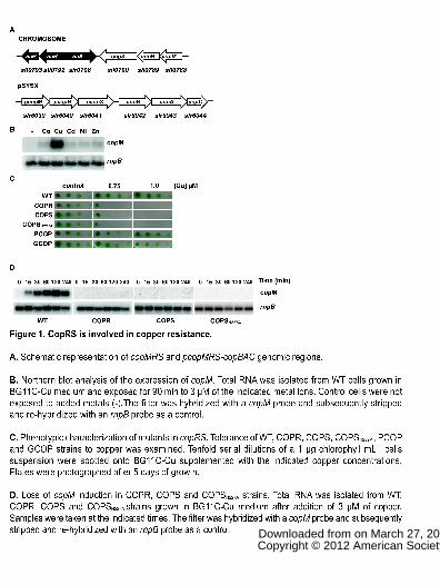
Figure 1. CopRS is involved in copper resistance.
A. Schematic representation of copMRS and pcopMRS-copBAC genomic regions.
B. Northern blot analysis of the expression of copM. Total RNA was isolated from
WT cells grown in BG11C-Cu medium and exposed for 90 min to 3 µM of the
indicated metal ions. Control cells were not exposed to added metals (-).The filter
was hybridized with a copM probe and subsequently stripped and re-hybridized
with an rnpB probe as a control.
C. Phenotypic characterization of mutants in copRS. Tolerance of WT, COPR,
COPS, COPSH227A, PCOP and GCOP strains to copper was examined. Tenfold
serial dilutions of a 1 µg chlorophyll mL-1 cells suspension were spotted onto
BG11C-Cu supplemented with the indicated copper concentrations. Plates were
photographed after 5 days of growth.
D. Loss of copM induction in COPR, COPS and COPSH227A strains. Total RNA
was isolated from WT, COPR, COPS and COPSH227A strains grown in BG11C-Cu
medium after addition of 3 µM of copper. Samples were taken at the indicated
times. The filter was hybridized with a copM probe and subsequently stripped and
re-hybridized with an rnpB probe as a control.
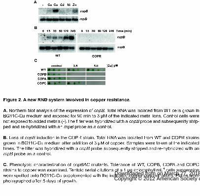
Figure 2. A new RND system involved in copper resistance.
A. Northern blot analysis of the expression of copB. Total RNA was isolated from
WT cells grown in BG11C-Cu medium and exposed for 90 min to 3 µM of the
indicated metal ions. Control cells were not exposed to added metals (-).The filter
was hybridized with a copB probe and subsequently stripped and re-hybridized
with an rnpB probe as a control.
B. Loss of copB induction in the COPR strain. Total RNA was isolated from WT
and COPR strains grown in BG11C-Cu medium after addition of 3 µM of copper.
www.plantphysiol.orgon March 27, 2018 - Published by Downloaded from Copyright © 2012 American Society of Plant Biologists. All rights reserved.
32
Samples were taken at the indicated times. The filter was hybridized with a copB
probe subsequently stripped and re-hybridized with an rnpB probe as a control.
C. Phenotypic characterization of copBAC mutants. Tolerance of WT, COPB,
COPA and COPC strains to copper was examined. Tenfold serial dilutions of a 1
µg chlorophyll mL-1 cells suspension were spotted onto BG11C-Cu supplemented
with the indicated copper concentrations. Plates were photographed after 5 days of
growth.
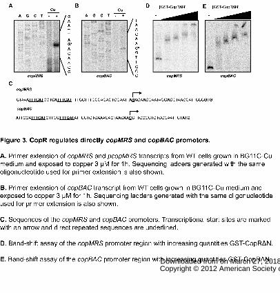
Figure 3. CopR regulates directly copMRS and copBAC promoters.
A. Primer extension of copMRS and pcopMRS transcripts from WT cells grown in
BG11C-Cu medium and exposed to copper 3 μM for 1h. Sequencing ladders
generated with the same oligonucleotide used for primer extension is also shown.
B. Primer extension of copBAC transcript from WT cells grown in BG11C-Cu
medium and exposed to copper 3 μM for 1h. Sequencing ladders generated with
the same oligonucleotide used for primer extension is also shown.
C. Sequences of the copMRS and copBAC promoters. Transcriptional start sites
are marked with an arrow and direct repeated sequences are underlined.
D. Band-shift assay of the copMRS promoter region with increasing quantities
GST-CopRΔN.
E. Band-shift assay of the copBAC promoter region with increasing quantities GST-
CopRΔN.
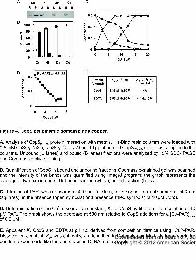
Figure 4. CopS periplasmic domain binds copper.
A. Analysis of CopS(38-183) protein interaction with metals. His-Bind resin columns
were loaded with 0.5 mM CuSO4, NiSO4, ZnSO4, CoCl2. About 10 μg of purified
CopS(38-183) protein was applied to the columns. Unbound (U lanes) and bound (B
lanes) fractions were analyzed by 15% SDS- PAGE and Commassie blue staining.
www.plantphysiol.orgon March 27, 2018 - Published by Downloaded from Copyright © 2012 American Society of Plant Biologists. All rights reserved.

33
B. Quantification of CopS in bound and unbound fractions. Coomassie-stainned gel
was scanned and the intensity of the bands was quantified using ImageJ program;
the graph represents the average of two experiments. Unbound fraction (white),
bound fraction (black).
C. Titration of PAR, which absorbs at 410 nm (circles), to its copper-form absorbing
at 500 nm (squares), in the absence (open symbols) and presence (filled symbols)
of 10 μM CopS.
D. Determinination of the Cu2+ dissociation constant, KD, of CopS by titration into a
solution of 10 μM PAR. The graph shows the decrease at 500 nm relative to CopS
additions for a [Cu-PAR]TOTAL of 0.9 μM.
E. Apparent KD CopS and EDTA at pH 7.5 derived from competition titration using
Cu2+-PAR. Dissociation constant, KD, was estimated as described in Materials and
Methods from four independent experiments like the one shown in D. NA, not
available.
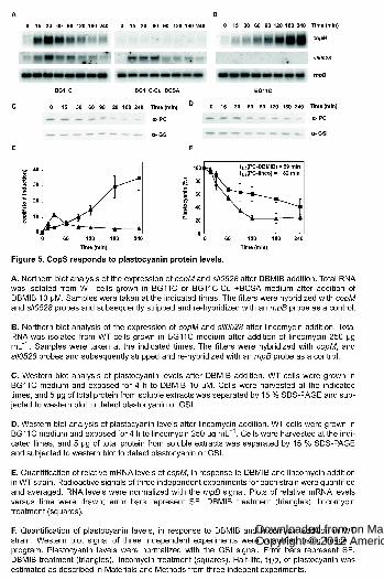
Figure 5. CopS responds to plastocyanin protein levels.
A. Northern blot analysis of the expression of copM and sll0528 after DBMIB
addition. Total RNA was isolated from WT cells grown in BG11C or BG11C-Cu +
BCSA medium after addition of DBMIB 10 μM. Samples were taken at the
indicated times. The filters were hybridized with copM and sll0528 probes and
subsequently stripped and re-hybridized with an rnpB probe as a control.
B. Northern blot analysis of the expression of copM and sll0528 after lincomycin
addition. Total RNA was isolated from WT cells grown in BG11C medium after
addition of lincomycin 250 μg mL-1. Samples were taken at the indicated times. The
filters were hybridized with copM, and sll0528 probes and subsequently stripped
and re-hybridized with an rnpB probe as a control.
www.plantphysiol.orgon March 27, 2018 - Published by Downloaded from Copyright © 2012 American Society of Plant Biologists. All rights reserved.
34
C. Western blot analysis of plastocyanin levels after DBMIB addition. WT cells
were grown in BG11C medium and exposed for 4 h to DBMIB 10 μM. Cells were
harvested at the indicated times, and 5 μg of total protein from soluble extracts was
separated by 15 % SDS-PAGE and subjected to western blot to detect
plastocyanin or GSI.
D. Western blot analysis of plastocyanin levels after lincomycin addition. WT cells
were grown in BG11C medium and exposed for 4 h to lincomycin 250 μg mL-1.
Cells were harvested at the indicated times, and 5 μg of total protein from soluble
extracts was separated by 15 % SDS-PAGE and subjected to western blot to
detect plastocyanin or GSI.
E. Quantification of relative mRNA levels of copM, in response to DBMIB and
lincomycin addition in WT strain. Radioactive signals of three independent
experiments for each strain were quantified and averaged. RNA levels were
normalized with the rnpB signal. Plots of relative mRNA levels versus time were
drawn; error bars represent SE. DBMIB treatment (triangles), lincomycin treatment
(squares).
F. Quantification of plastocyanin levels, in response to DBMIB and lincomycin
addition in WT strain. Western blot signal of three independent experiments were
quantified using Image J program. Plastocyanin levels were normalized with the
GSI signal. Error bars represent SE. DBMIB treatment (triangles), lincomycin
treatment (squares). Half-life, t1/2, of plastocyanin was estimated as described in
Materials and Methods from three indepent experiments.
Figure 6. CopS responds to intracellular copper.
A. Northern blot analysis of the expression of copM, petE and petJ in response to
copper addition in WT, SAS and PETE strains. Total RNA was isolated from WT,
SAS and PETE cells grown in BG11C-Cu medium after addition of copper 200 nM.
www.plantphysiol.orgon March 27, 2018 - Published by Downloaded from Copyright © 2012 American Society of Plant Biologists. All rights reserved.

35
Samples were taken at the indicated times. The filters were hybridized with copM,
petE and petJ probes and subsequently stripped and re-hybridized with an rnpB
probe as a control.
B. Quantification of relative mRNA levels of copM in response to copper addition in
WT, SAS and PETE strains. Radioactive signals of three independent experiments
for each strain were quantified and averaged. RNA levels were normalized with the
rnpB signal in all strains. Plots of relative mRNA levels versus time were drawn;
error bars represent SE. WT strain (triangles) SAS strain (circles), PETE strain
(squares).
Figure 7. CopS is localised to plasma and thylakoid membranes.
A. Membrane localization of CopS. Membrane fractions from COPSHA strain
induced for 4 h with 2 µM of nickel were prepared by sucrose density gradient and
aqueous polymer two-phase partitioning. 5 µg of total protein were loaded and
separated by SDS-PAGE. CopS-3HA, NrtA and PsaC proteins were detected by
western blot. PM, plasma membrane; TM, thylakoid membrane.
B. Quantification of CopS in different membrane fractions. Western blot signal of
three independent experiments were quantified using Image J program and
averaged; error bars represent SE. Plasma membrane (black); thylakoid
membrane (white).
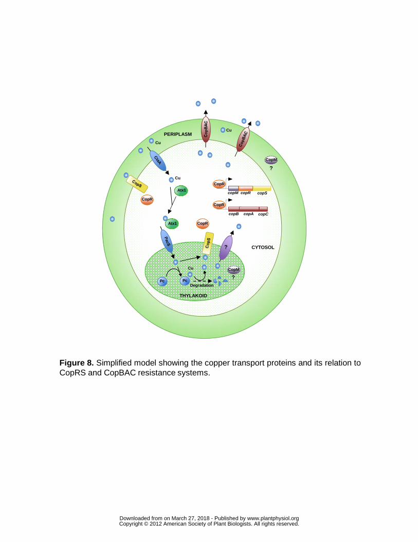
Figure 8. Simplified model showing the copper transport proteins and its
relation to CopRS and CopBAC resistance systems.
www.plantphysiol.orgon March 27, 2018 - Published by Downloaded from Copyright © 2012 American Society of Plant Biologists. All rights reserved.
www.plantphysiol.orgon March 27, 2018 - Published by Downloaded from Copyright © 2012 American Society of Plant Biologists. All rights reserved.
www.plantphysiol.orgon March 27, 2018 - Published by Downloaded from Copyright © 2012 American Society of Plant Biologists. All rights reserved.
www.plantphysiol.orgon March 27, 2018 - Published by Downloaded from Copyright © 2012 American Society of Plant Biologists. All rights reserved.
www.plantphysiol.orgon March 27, 2018 - Published by Downloaded from Copyright © 2012 American Society of Plant Biologists. All rights reserved.
www.plantphysiol.orgon March 27, 2018 - Published by Downloaded from Copyright © 2012 American Society of Plant Biologists. All rights reserved.

www.plantphysiol.orgon March 27, 2018 - Published by Downloaded from Copyright © 2012 American Society of Plant Biologists. All rights reserved.

www.plantphysiol.orgon March 27, 2018 - Published by Downloaded from Copyright © 2012 American Society of Plant Biologists. All rights reserved.
copM copR copS
copB copA copC
CopR
CopR
THYLAKOID
CopR
CopR
CopM
Atx1
Atx1
CYTOSOL
PERIPLASM
DegradationPc Pc
Cu
Cu
Cu
Cu CopM
?
?
?
Figure 8. Simplified model showing the copper transport proteins and its relation toCopRS and CopBAC resistance systems.
www.plantphysiol.orgon March 27, 2018 - Published by Downloaded from Copyright © 2012 American Society of Plant Biologists. All rights reserved.