Evaluation of systolic byDoppler ultrasonography -...
6
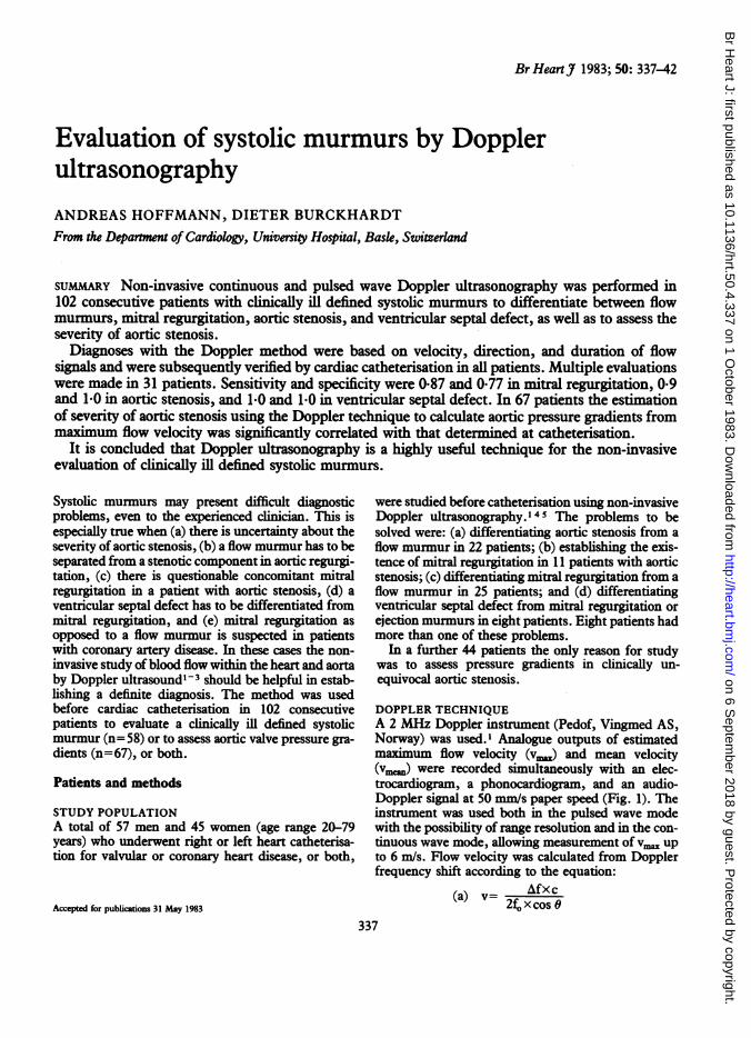
Br Heart J 1983; 50: 337-42 Evaluation of systolic murmurs by Doppler ultrasonography ANDREAS HOFFMANN, DIETER BURCKHARDT From the Deparment of Cardiology, University Hospital, Bask., Switzerland SUMMARY Non-invasive continuous and pulsed wave Doppler ultrasonography was performed in 102 consecutive patients with clinically ill defined systolic murmurs to differentiate between flow murmurs, mitral regurgitation, aortic stenosis, and ventricular septal defect, as well as to assess the severity of aortic stenosis. Diagnoses with the Doppler method were based on velocity, direction, and duration of flow signals and were subsequently verified by cardiac catheterisation in all patients. Multiple evaluations were made in 31 patients. Sensitivity and specificity were 0-87 and 0 77 in mitral regurgitation, 0.9 and 1.0 in aortic stenosis, and 1.0 and 1.0 in ventricular septal defect. In 67 patients the estimation of severity of aortic stenosis using the Doppler technique to calculate aortic pressure gradients from maximum flow velocity was significantly correlated with that determined at catheterisation. It is concluded that Doppler ultrasonography is a highly useful technique for the non-invasive evaluation of clinically ill defined systolic murmurs. Systolic murmurs may present difficult diagnostic problems, even to the experienced clinician. This is especially true when (a) there is uncertainty about the severity of aortic stenosis, (b) a flow murmur has to be separated from a stenotic component in aortic regurgi- tation, (c) there is questionable concomitant mitral regurgitation in a patient with aortic stenosis, (d) a ventricular septal defect has to be differentiated from mitral regurgitation, and (e) mitral regurgitation as opposed to a flow murmur is suspected in patients with coronary artery disease. In these cases the non- invasive study of blood flow within the heart and aorta by Doppler ultrasound'-3 should be helpful in estab- lishing a definite diagnosis. The method was used before cardiac catheterisation in 102 consecutive patients to evaluate a clinically ill defined systolic murmur (n= 58) or to assess aortic valve pressure gra- dients (n=67), or both. Patients and methods STUDY POPULATION A total of 57 men and 45 women (age range 20-79 years) who underwent right or left heart catheterisa- tion for valvular or coronary heart disease, or both, Accepted for publications 31 May 1983 were studied before catheterisation using non-invasive Doppler ultrasonography.'45 The problems to be solved were: (a) differentiating aortic stenosis from a flow murmur in 22 patients; (b) establishing the exis- tence of mitral regurgitation in 11 patients with aortic stenosis; (c) differentiating mitral regurgitation from a flow murmur in 25 patients; and (d) differentiating ventricular septal defect from mitral regurgitation or ejection murmurs in eight patients. Eight patients had more than one of these problems. In a further 44 patients the only reason for study was to assess pressure gradients in clinically un- equivocal aortic stenosis. DOPPLER TECHNIQUE A 2 MHz Doppler instrument (Pedof, Vingmed AS, Norway) was used.I Analogue outputs of estimated maximum flow velocity (v..) and mean velocity (vm.,) were recorded simultaneously with an elec- trocardiogram, a phonocardiogram, and an audio- Doppler signal at 50 mm/s paper speed (Fig. 1). The instrument was used both in the pulsed wave mode with the possibility of range resolution and in the con- tinuous wave mode, allowing measurement of v. up to 6 m/s. Flow velocity was calculated from Doppler frequency shift according to the equation: (a) v= 2£OAfx c (a =2f x cos 0 337 on 6 September 2018 by guest. Protected by copyright. http://heart.bmj.com/ Br Heart J: first published as 10.1136/hrt.50.4.337 on 1 October 1983. Downloaded from
Transcript of Evaluation of systolic byDoppler ultrasonography -...
Br HeartJ 1983; 50: 337-42
Evaluation of systolic murmurs by DopplerultrasonographyANDREAS HOFFMANN, DIETER BURCKHARDTFrom the Deparment of Cardiology, University Hospital, Bask., Switzerland
SUMMARY Non-invasive continuous and pulsed wave Doppler ultrasonography was performed in102 consecutive patients with clinically ill defined systolic murmurs to differentiate between flowmurmurs, mitral regurgitation, aortic stenosis, and ventricular septal defect, as well as to assess theseverity of aortic stenosis.
Diagnoses with the Doppler method were based on velocity, direction, and duration of flowsignals and were subsequently verified by cardiac catheterisation in all patients. Multiple evaluationswere made in 31 patients. Sensitivity and specificity were 0-87 and 0 77 in mitral regurgitation, 0.9and 1.0 in aortic stenosis, and 1.0 and 1.0 in ventricular septal defect. In 67 patients the estimationof severity of aortic stenosis using the Doppler technique to calculate aortic pressure gradients frommaximum flow velocity was significantly correlated with that determined at catheterisation.
It is concluded that Doppler ultrasonography is a highly useful technique for the non-invasiveevaluation of clinically ill defined systolic murmurs.
Systolic murmurs may present difficult diagnosticproblems, even to the experienced clinician. This isespecially true when (a) there is uncertainty about theseverity of aortic stenosis, (b) a flow murmur has to beseparated from a stenotic component in aortic regurgi-tation, (c) there is questionable concomitant mitralregurgitation in a patient with aortic stenosis, (d) aventricular septal defect has to be differentiated frommitral regurgitation, and (e) mitral regurgitation asopposed to a flow murmur is suspected in patientswith coronary artery disease. In these cases the non-invasive study of blood flow within the heart and aortaby Doppler ultrasound'-3 should be helpful in estab-lishing a definite diagnosis. The method was usedbefore cardiac catheterisation in 102 consecutivepatients to evaluate a clinically ill defined systolicmurmur (n= 58) or to assess aortic valve pressure gra-dients (n=67), or both.
Patients and methods
STUDY POPULATIONA total of 57 men and 45 women (age range 20-79years) who underwent right or left heart catheterisa-tion for valvular or coronary heart disease, or both,
Accepted for publications 31 May 1983
were studied before catheterisation using non-invasiveDoppler ultrasonography.'45 The problems to besolved were: (a) differentiating aortic stenosis from aflow murmur in 22 patients; (b) establishing the exis-tence of mitral regurgitation in 11 patients with aorticstenosis; (c) differentiating mitral regurgitation from aflow murmur in 25 patients; and (d) differentiatingventricular septal defect from mitral regurgitation orejection murmurs in eight patients. Eight patients hadmore than one of these problems.
In a further 44 patients the only reason for studywas to assess pressure gradients in clinically un-equivocal aortic stenosis.
DOPPLER TECHNIQUEA 2 MHz Doppler instrument (Pedof, Vingmed AS,Norway) was used.I Analogue outputs of estimatedmaximum flow velocity (v..) and mean velocity(vm.,) were recorded simultaneously with an elec-trocardiogram, a phonocardiogram, and an audio-Doppler signal at 50 mm/s paper speed (Fig. 1). Theinstrument was used both in the pulsed wave modewith the possibility of range resolution and in the con-tinuous wave mode, allowing measurement ofv. upto 6 m/s. Flow velocity was calculated from Dopplerfrequency shift according to the equation:
(a) v= 2£OAfx c(a =2f xcos 0
337
on 6 Septem
ber 2018 by guest. Protected by copyright.
http://heart.bmj.com
/B
r Heart J: first published as 10.1136/hrt.50.4.337 on 1 O
ctober 1983. Dow
nloaded from

338
Fig. 1 Estimated maximuvelocity (v..) simultaneouekctrocardiogram, phonocarsignal at 50mm/s (normal a
Hoffmann, Burckhardt
tional vm. signal-that is, deflections are positive ifthe flow is directed towards and negative if it isdirected away from the transducer.The transducer was placed in the suprasternal
notch (for evaluating aortic stenosis), in the paraster-nal area (mainly for ventricular septal defect), andover the apex (for mitral regurgitation and aorticstenosis) with the patient in the supine or left lateralposition (Fig. 2). The position of the transducer wasadjusted until a pure, high pitched audio signal indi-cated alignment of the ultrasound beam into the bloodjet at an angle close to zero. With an angle close tozero the cosine 0 becoming close to 1 can be ignored inthe Doppler equation (a).The following Doppler findings were used as diag-
nostic criteria: aortic stenosis-v=,, in systole >2.2...............m/s measured within the aortic blood jet (Fig. 3);
mitral regurgitation-high velocity flow signal in sys-tole directed from the apex towards the left atriumlasting beyond S2 (Fig. 4); ventricular septaldefect-high velocity flow signal in systole poiningtowards the transducer positioned at the lower leftsternal border (Fig. 5). For the calculation of Ap=4
onflow velocity (v.,.,) and mean (v.2) only satisfactory v signals were used. V signals
rdiogram, andawhioa were considered unsatisfactory (in four patients) whenorta from suprasternal poppl)e v. was <3.5 m/s despite peak of v. in the secondw*afshalf of systole, as underestimation of v. has to be
assumed in these cases.5wnere aI= ioppier irequency SflUL i=irequencyemitted, c=velocity of sound in blood, 0=anglebetween ultrasound beam and blood jet.
Using a simplified Bernoulli equation145 of thepressure-velocity relation aortic pressure gradients(mmHg) were calculated from vmu (continuous wavemode, m/s) as:
(b) Ap=4(Vax,2).
The direction of flow was determined by the direc-
INVASIVE ASSESSMENT OF DIAGNOSESThe final diagnoses were made by angiography (mitralregurgitation), invasive pressure measurements (aor-tic stenosis), and stepwise analysis of oxygen content(ventricular septal defect).
Sensitivity is defined as true positive/all positivetests; specificity as true negative/all negative tests; anddiagnostic accuracy as (true positive + truenegative)/all tests.
Suprasternal
Parasternal
Fig. 2 Transducer positions forexamining the heart by Dopplerultrasound.
Apiac
"... .X. ...w.........
-1-1
on 6 Septem
ber 2018 by guest. Protected by copyright.
http://heart.bmj.com
/B
r Heart J: first published as 10.1136/hrt.50.4.337 on 1 O
ctober 1983. Dow
nloaded from

Evaluation of systolic murmurs by Doppler ultrasonography
rocGrm..E!ectrc ;r'dvlles , ;.m.,s
P-lSonc.Cr-mm
Phonogran,w
5rv cf
rn I'..
[1 m 1------is ---------
vmsor)
Fig. 3 Estimated maximnflow velocity (va) and meanvelocity (V,aJ.) simultaneously recorded with anelectrocardiogram, phonogram, and audio Doppler signal at50 mmls in aortic stenosis (from suprasternal position).
Fig. 5 Estimated maximumflow velocity (v..) and meanvelocity (v,,,.) simultaneously recorded with anelectrocardiogram, phonogram, and audio Doppler signal at50 mm/s in ventricular septal defect (from lower left sternalborder).
Results
The Table summarises the results in 58 patients withclinically ill defined syttolic murmurs. Eight patientshad more than one problem to be solved.
DIFFERENTIATION OF SYSTOLIC MURMURSAortic stenosis/functionalflow murmurNine of 10 cases with aortic stenosis (Ap >20 mmHg)were correctly identified. In 12 of 12 cases a functionalflow murmur was correctly diagnosed. The predictivevalue in diagnosing aortic stenosis was 09 for a posi-tive finding (sensitivity) and 1 0 for a negative finding(specificity).
-NI
Fig. 4 Estimated maximum flowv velocity (v..9 and meanvelocity (v. simultaneouly recorded wuith anelectrocardiogram, phonogram, and audio Doppler signal at50mm/Is in mitral regurgitation (from apex) concomitant withaortic stenosis.
Mitral regurgitation/ejection type murmurMitral regurgitation was correctly identified in 20 of23 and correctly excluded in 10 of 13 patients. Thepredictive value of a positive finding, therefore, was0-87 and that of a negative finding 077.
DIAGNOSIS OF VENTRICULAR SEPTAL DEFECTThis problem arose in eight patients, five with a ven-tricular septal defect, two with mitral regurgitation,one with pulmonary stenosis. A ventricular septaldefect was correctly diagnosed in all five patients andexcluded in the remaining three cases (predictivevalue 10 each for a positive and negative finding).The overall diagnostic accuracy of the method (truepositive + true negative/all tests) for the assessment ofa systolic murmur was 0*89.
339
..%% _,-,:Ie,,.-. l-, -y-, .- -wcj V)n (,ri rIn
fl\.. /.-l- l,-.1-l.-..-.--,l.......
14 rro. ('.g :, I
'I-
"-l"-\-.I..I //
on 6 Septem
ber 2018 by guest. Protected by copyright.
http://heart.bmj.com
/B
r Heart J: first published as 10.1136/hrt.50.4.337 on 1 O
ctober 1983. Dow
nloaded from

Table Results ofevaluation ofsystolic murmurs by Dopplkr ultrasonography
Probkm No ofpatient Positive resuk Negative resuk Diagnostic accuracy
True False True FalseAortic stenosis 22 9 0 12 1 0-9Mitra regurgitation 36 20 3 10 3 0-83Ventricular septal
defect 8 5 0 3 0 1-0Total 66 34 3 25 4 0-89
120-
100-
FE 80-E
M 60--c
g 40-
20-
0
0
0 Z0 0
,OZ0S*% 0
0 0
* 0'- * 0 00.
0
0
0
0~~~~~~Z* I 0
0 20 40 60 80Catheterisation (mmHg)
Fig. 6 Aortic pressure gradients assessed by thetechnique and at catheterisation in 66 patients withshowing significant correlation (r=0-75, n=64).
ASSESSMENT OF PRESSURE GRADIENT"STENOSISSatisfactory v. signals were obtainedpatients (94%) with aortic stenosis and a
59±15 years. Pressure gradients determDoppler technique were significantly cotthose found at catheterisation (r=0.7'(Fig. 6). If the analysis was restrictedunder the age of 50 years (n=ll) theimproved considerably (r=0.90; p<0-00
Discussion
The Doppler ultrasound method may bean intracardiac stethoscope with spatial r
detects intracardiac flow and determinesand its duration in relation to the events ocycle. Furthermore, localisation of the flidistance from the transducer using the Imode and measurements of high flow vito 6 mIs) using the continuous wave mo
ible. The maximum flow velocity measureaortic blood jet may be used to calculate
gradient across the aortic valve according to a sim-* plified version of Bernoulli's law. ' 45 The method we
* @7 used is not combined with two dimensional echocar-diography and alignment of the ultrasonic beam isattempted solely by acoustic guidance. Some
* difficulty may, therefore, arise for inexperiencedexaminers in localising the Doppler probe which mayresult in too many false negative results. For localisa-tion of the Doppler sample two dimensional echocar-diography is certainly of value, but the combination ofthe two techniques has several limitations itself.Firstly, the combined transducers are heavy, andtherefore proper alignment of the ultrasound beam inthe blood stream is more difficult. Secondly, there isonly one combined instrument commercially available
00 120 which operates both in the pulsed and in the continu-ous wave mode, the others being invaluable for thecalculation of pressure gradients from high velocities.
Doppler Thirdly, the cost of combined equipment amounts tokaortc stenosts a sum five to 10 times that of a Doppler instrument
alone.The diagnostic accuracy of Doppler ultrasound for
the identification of aortic stenosis (pressure gradientS IN AORTIC >20 mmHg) was 0-96 in this series which included
only patients with equivocal clinical findings.in 63 of 67 Although these data were obtained in a small numbermean age of of patients, they are confirmed by the fact that under-muned by the estimation of pressure gradient (<20 mmHg) occur-rrelated with red in only three of 44 patients with unequivocal aor-5; p<0.001) tic stenosis and v. >1-8 m/s was never found into patients more than 30 additional patients with documented
E correlation absence of aortic valve pressure gradients. All these1). patients, however, were not included in this series
because of clear cut clinical findings.The use of continuous wave Doppler method to
assess pressure gradients in aortic stenosis was firstregarded as reported by Hatle4 and good correlations betweenresolution; it pressure gradients assessed by the technique andits direction haemodynamically measured were subsequentlyof the cardiac confirmed by other groups of workers includingiw at a given ourselves.57 In the present series of 67 patients therepulsed wave was a correlation coefficient of 0-75 (Fig. 6). In elderlyelocities (up patients with appreciable post-stenotic dilatation ofde are poss- the aorta or emphysema, or both, aiming for the high-!d within the est velocities in the aortic jet can be very difficult. Thethe pressure fact that in our series the patients were mostly elderly
340 Hoffmann, Burckhardt
on 6 Septem
ber 2018 by guest. Protected by copyright.
http://heart.bmj.com
/B
r Heart J: first published as 10.1136/hrt.50.4.337 on 1 O
ctober 1983. Dow
nloaded from
Evaluation of systolic murmurs by Doppler ultrasonography
(mean age 59 years) explains the rather low correlationcoefficient. Indeed, when only the 11 patients aged<50 years were included in the analysis the correla-tion coefficient was 0.90. The continuous Dopplermethod, therefore, seems to be superior to other non-invasive techniques8 9-including M-mode echocar-diography,'0 cross sectional echocardiography,7 11and the pulsed wave Doppler'2 method-in assessingthe severity of aortic stenosis.The diagnostic accuracy of Doppler ultrasound for
the assessment of mitral regurgitation was 0-83. Thehigh sensitivity of 0-87 is further corroborated by ourexperience in several patients with coronary arterydisease but without audible murmurs, in whom mitralregurgitation was suspected by the Doppler methodand subsequently confirmed by angiography. Thepulsed wave Doppler technique in combination withcross sectional echocardiography has been used byseveral groups of workers for diagnosing aortic regur-gitation3 13 14 and mitral regurgitation,3 15 16 with asensitivity ranging from 0-58 to 0*94. The trans-oesophageal approach was reported to yield a highersensitivity by one group.16 This technique does not,however, fully qualify as non-invasive in our view.The accuracy of diagnosing ventricular septal
defect was 1.0 in our prospective series of eightpatients. In a further eight patients with confirmedventricular septal defect a positive Doppler findingwas also obtained, thus confirming the high sensitivityof the method. The continuous and pulsed wave Dop-pler techniques have both been used to diagnose ven-tricular septal defect, particularly in the setting ofacute myocardial infarction. The results of thesestudies also showed diagnostic accuracies >0-8.17 18The overall diagnostic accuracy of this relatively
simple, inexpensive Doppler technique was 0-89 inour consecutive series of 58 patients with systolicmurmurs of uncertain importance. The Dopplermethod, therefore, seems to be useful in the non-invasive differentiation of mitral regurgitation, ven-tricular septal defect, aortic stenosis, and innocentflow murmurs as well as in estimating pressure gra-dients in aortic stenosis. The limitations of the tech-nique in inexperienced hands may be partly overcomeby combining it with cross sectional echocardiogra-phy.
References
1 Hatle L, Angelsen BA. Doppler ultrasound in cardiology.Philadelphia: Lea and Febiger, 1982.
2 Hatle L, Angelsen BA, Tromsdal A. Non-invasiveassessment of aortic stenosis by Doppler ultrasound. BrHeartJ 1980; 43: 284-92.
3 Quinones MA, Young JB, Waggoner AD, Ostojic MC,
341
Ribeiro LGT, Miller RR. Assessment of pulsed Dopplerechocardiography in detection and quantification of aor-tic and mitral regurgitation. Br HeartJ 1980; 44: 612-20.
4 Hatle L. Non-invasive assessment and differentiation ofleft ventricular outflow obstruction with Dopplerultrasound. Circulation 1981; 64: 381-7.
5 Hoffmann A, Amann FW, Burckhardt D. Nicht-invasive Beurteilung von Druckgradienten bei Aortens-tenose mit Doppler Ultraschall. Schweiz Med Wochenschr1982; 112: 1597-600.
6 Hoffman A, Pfisterer M, Schmitt HE, Burckhardt D.Non invasive assessment of pressure gradients in valvularaortic stenosis by Doppler ultrasound [Abstract]. Circu-lation 1982; 66 (suppl 2): 121.
7 Kwan OL, Waters J, Takeda P, Mazzoleni A, Low R,De Maria A. Relative value of continuous wave Dopplercompared to two-dimensional echocardiography in thequantitation of valvular stenosis [Abstract]. Circulation1982; 66 (suppl 2): 121.
8 Flohr KH, Weir EK, Chesler E. Diagnosis of aorticstenosis in older age groups using external carotid pulserecording and phonocardiography. Br Heart J 1981; 45:577-82.
9 Siegel RJ, Roberts WC. Electrocardiographic observa-tions in severe aortic valve stenosis: correlative necropsystudy to clinical, hemodynamic, and ECG variablesdemonstrating relation of 12-lead QRS amplitude to peaksystolic trans-aortic pressure gradient. Am Heart3J 1982;103: 210-21.
10 Reichek N, Devereux RB. Reliable estimation of peakleft ventricular systolic pressure by M-modeechocardiographic-deterniined end-diastolic relative wallthickness: identification of severe valvular aortic stenosisin adult patients. Am Heart J 1982; 103: 202-9.
11 Godley RW, Green D, Dillon JC, Rogers EW, Feigen-baum H, Weymamn AE. Reliability of two-dimensionalechocardiography in assessing the severity of valvularaortic stenosis. Chest 1981; 79: 657-62.
12 Young JB, Quinones MA, Waggoner AD, Miller RR.Diagnosis and quantification of aortic stenosis withpulsed Doppler echocardiography. Am J Cardiol 1980;45: 987-94.
13 Boughner DR. Assessment of aortic insufficiency bytranscutaneous Doppler ultrasound. Circulation 1975;52: 874-9.
14 Ciobanu M, Abbasi AS, Allen M, Hermer A, SpellbergR. Pulsed Doppler echocardiography in the diagnosisand estimation of severity of aortic insufficiency. Am JCardiol 1982; 49: 339-43.
15 Blanchard D, Diebold B, Peroxlneau P, et al. Non-invasive diagnosis of mitral regurgitation by Dopplerechocardiography. Br Heart3J 1981; 45: 589-93.
16 Schluter M, Laangenstein BA, Hanrath P, Kremer P,Bleifeld W. Assessment of transoesophageal pulsedDoppler echocardiography in the detection of mitral re-gurgitation. Circulation 1982; 66: 784-9.
17 Recusani F, Sgalumbro A, Raisaro A, Tronconi L, Vig-ano M. Doppler echocardiography in differential diag-nosis of mitral insufficiency versus septal rupture com-plicating myocardial infarction. Cardiovascular Applica-tions of Doppler Echocardiography, International Sym-posium INSERM, Versailles, 1982.
on 6 Septem
ber 2018 by guest. Protected by copyright.
http://heart.bmj.com
/B
r Heart J: first published as 10.1136/hrt.50.4.337 on 1 O
ctober 1983. Dow
nloaded from

342 Hoffmann, Burckhardt18 Otterstad JE, Simonsen S, Myhre E. Doppler echocar- Requests for reprints to Dr A Hoffmann, Division of
diography in isolated ventricular septal defect in the Cardiology, University Hospital, CH-4031 Basle,adult. Circulation 1982; 66 (suppl 2): 188. Switzerland.
on 6 Septem
ber 2018 by guest. Protected by copyright.
http://heart.bmj.com
/B
r Heart J: first published as 10.1136/hrt.50.4.337 on 1 O
ctober 1983. Dow
nloaded from