THE OPENING SNAP MITRAL STENOSIS - Heartheart.bmj.com/content/heartjnl/15/2/135.full.pdf · THE...
8
THE OPENING SNAP OF MITRAL STENOSIS BY PATRICK MOUNSEY From the Cardiac Department of the London Hospital Received November 28, 1952 Sixteen years after Laennec (1819) described the murmur of mitral stenosis, Bouillaud (1835) described a reduplication of the second sound in mitral stenosis. The familiar cadence in mitral stenosis, which results from the presence of this added sound, was likened by Durozier (1862) to the syllables ffou-ta-ta-rou. In 1872 Guttman suggested that this sound might be due not to asynchronism of pulmonary and aortic valve closure, as had been held by Durozier, but to vibration at the stenosed mitral valve itself. Sansom (1881) held the same views as Guttman. It was not until 1888 that Rouches introduced the term commonly used today " claquement d'ouverture de la mitrale " or opening snap of the mitral valve, to describe this sound, stating that Potain had used this term in his teaching. He suggested that the sound was produced by sudden stretching of the stenosed mitral valve by blood passing from the left auricle to the left ventricle. Rouches' views on the pathogenesis of the opening snap were not universally accepted and Gallavardin (1905) and Duchosal (1946) postulated other mechanisms. Margolies and Wolferth (1932) made a detailed clinical and phonocardiographic study of this sound and concluded that Rouches' hypothesis as to its mode of production was the most likely. The clinical recognition of the opening snap and its differentiation from two other sounds in early diastole, may sometimes present difficulty. The second component of the second sound when widely split may closely resemble an opening snap. Further, the physiological third heart sound might be confused with the opening snap. The heart sounds were examined clinically and in the phonocardiogram in 33 cases of mitral stenosis to test certain clinical signs that might make easier the differentiation and recognition of these sounds. THE INVESTIGATION DESCRIBED Mitral stenosis was considered to be the principal lesion in all cases and the sole valvular lesion in 28 of them, while in four there was slight aortic incompetence and in another associated mitral incompetence. The ages of the patients varied between 19 and 48, with an average of 34 years. There were 7 men and 26 women. None had gross cardiac enlargement. The electrocardiogram showed evidence of right ventricular preponderance in 24, and auricular fibrillation was present in 12 patients. Fifteen have since had a mitral valvotomy. Phonocardiograms were recorded on a four-channel phonocardiograph using crystal micro- phones, as described by Leatham (1949). An electrocardiogram was used as a reference tracing. INCIDENCE AND RELATIONSHIP OF THE OPENING SNAP An opening snap was diagnosed clinically and confirmed on the phonocardiogram in 28 out of the 33 patients. In four of the remaining five the phonocardiogram recorded an opening snap. In three of these the sound was very soft and therefore difficult to recognize with certainty clinically; in the fourth it followed closely the second heart sound and phonocardiography was necessary to analyse correctly the sequence of sounds. In only one case was an opening snap absent on the phonocardiogram. 135 on 22 May 2018 by guest. Protected by copyright. http://heart.bmj.com/ Br Heart J: first published as 10.1136/hrt.15.2.135 on 1 April 1953. Downloaded from
Transcript of THE OPENING SNAP MITRAL STENOSIS - Heartheart.bmj.com/content/heartjnl/15/2/135.full.pdf · THE...
THE OPENING SNAP OF MITRAL STENOSISBY
PATRICK MOUNSEY
From the Cardiac Department of the London HospitalReceived November 28, 1952
Sixteen years after Laennec (1819) described the murmur of mitral stenosis, Bouillaud (1835)described a reduplication of the second sound in mitral stenosis. The familiar cadence in mitralstenosis, which results from the presence of this added sound, was likened by Durozier (1862) tothe syllables ffou-ta-ta-rou. In 1872 Guttman suggested that this sound might be due not toasynchronism of pulmonary and aortic valve closure, as had been held by Durozier, but to vibrationat the stenosed mitral valve itself. Sansom (1881) held the same views as Guttman. It was notuntil 1888 that Rouches introduced the term commonly used today " claquement d'ouverture dela mitrale " or opening snap of the mitral valve, to describe this sound, stating that Potain had usedthis term in his teaching. He suggested that the sound was produced by sudden stretching of thestenosed mitral valve by blood passing from the left auricle to the left ventricle. Rouches' viewson the pathogenesis of the opening snap were not universally accepted and Gallavardin (1905) andDuchosal (1946) postulated other mechanisms. Margolies and Wolferth (1932) made a detailedclinical and phonocardiographic study of this sound and concluded that Rouches' hypothesis as toits mode of production was the most likely.
The clinical recognition of the opening snap and its differentiation from two other sounds inearly diastole, may sometimes present difficulty. The second component of the second sound whenwidely split may closely resemble an opening snap. Further, the physiological third heart soundmight be confused with the opening snap. The heart sounds were examined clinically and in thephonocardiogram in 33 cases of mitral stenosis to test certain clinical signs that might make easierthe differentiation and recognition of these sounds.
THE INVESTIGATION DESCRIBEDMitral stenosis was considered to be the principal lesion in all cases and the sole valvular lesion
in 28 of them, while in four there was slight aortic incompetence and in another associated mitralincompetence. The ages of the patients varied between 19 and 48, with an average of 34 years.There were 7 men and 26 women. None had gross cardiac enlargement. The electrocardiogramshowed evidence of right ventricular preponderance in 24, and auricular fibrillation was present in12 patients. Fifteen have since had a mitral valvotomy.
Phonocardiograms were recorded on a four-channel phonocardiograph using crystal micro-phones, as described by Leatham (1949). An electrocardiogram was used as a reference tracing.
INCIDENCE AND RELATIONSHIP OF THE OPENING SNAPAn opening snap was diagnosed clinically and confirmed on the phonocardiogram in 28 out of
the 33 patients. In four of the remaining five the phonocardiogram recorded an opening snap.In three of these the sound was very soft and therefore difficult to recognize with certainty clinically;in the fourth it followed closely the second heart sound and phonocardiography was necessary toanalyse correctly the sequence of sounds. In only one case was an opening snap absent on thephonocardiogram.
135
on 22 May 2018 by guest. P
rotected by copyright.http://heart.bm
j.com/
Br H
eart J: first published as 10.1136/hrt.15.2.135 on 1 April 1953. D
ownloaded from
PATRICK MOUNSEYThe term snap well described the quality of the sound heard. On the phonocardiogram the
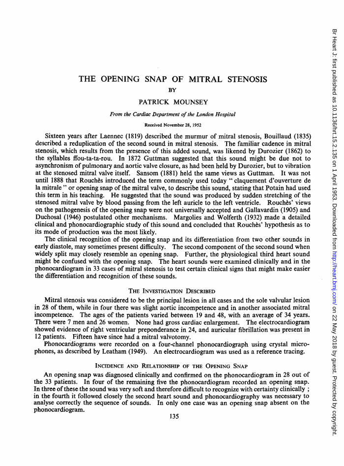
sound was composed chiefly of high frequency vibrations of short duration (0005 to 0-01 sec.).The area where the sound was best heard was determined
by examining its intensity, both on auscultation and onthe phonocardiogram in four selected areas, the mitral
S~2t c<Carea, the pulmonary area, the left sternal edge at thelevel of the fifth intercostal space, and the special supra-mammary area, which lies above and a little internal to
W the left nipple (Fig. 1). The opening snap was best heardin the supramammary area, in 27 out of 32 cases.i)Q
0
Maximum audibility, however, was shared between two10 0 or more areas in the majority, the commonest after
{\{rs ~~the supramammary area being the left sternal edge and3 the mitral area, and the least common the pulmonary
area.
FIG. I.-Diagram to illustrate microphonepositions. (1) Mitral area. (2) Pul-monary area. (3) Left sternal edge.(4) Supramammary area.
The interval between the beginning o0 the secondsound and the opening snap varied widely. The minimuminterval was 003 sec., the maximum 0 14 sec., and theaverage for the group 007 sec. (Fig. 2).
TIME IN Ilioo5 10I
SEC.i5
..1i illOPEN ING
S N A P
.
SECOND COMPONENTOF SPLIT
SECOND SOUND
5 10 15TIME IN 1l0oo SEC.
FIG. 2.-The timing ofthe opening snap and the second component ofa split second sound compared in 33 cases of mitralstenosis. The interval between the beginning of the second sound and the opening snap varied between 0 03and 0-12 sec. (average 007 sec.): that between the beginning or first component of the second sound and thesecond component varied between 0-01 and 005 sec. (average 0 03 sec.). Minimum intervals for the openingsnap and maximum intervals for the second component of a split second sound lay in the same range (0-03 to0 05 sec.). (The maximum interval between the beginning of the second sound and the opening snap (0-14 sec.)mentioned in the text, is not recorded in the Table, where only mean intervals for individual patients are shown.)
10
%n 5 ;tn
u :"
Ui.0
10 -
D
5-Z ii i
136
on 22 May 2018 by guest. P
rotected by copyright.http://heart.bm
j.com/
Br H
eart J: first published as 10.1136/hrt.15.2.135 on 1 April 1953. D
ownloaded from

OPENING SNAP OF MITRAL STENOSIS
In the phonocardiogram a relationship was noted between the diastolic interval on the one handand the interval between the beginning of the second sound and the opening snap on the other inindividual patients in sinus rhythm. An increase in the diastolic interval with slowing of the heartrate was accompanied by a longer interval between the beginning of the second sound and theopening snap. No similar direct correlation was seen between these two variables in the group asa whole. In individual patients with auricular fibrillation, a general relationship was noted betweenthe preceding diastolic interval and the timing of the opening snap that followed, the snap occurringearlier after a short and later after a long diastolic interval (Fig. 3). As in those in sinus rhythmthis relationship, which held generally in most subjects, did not hold for the group as a whole, whereno direct correlation was noted between the diastolic interval and the timing of the opening snap.
1/5 sec I I I 1 1I
MA 0~~IIIwq'u~mm IP IHF IDi I--
M -MF IN I';
2 20°S1§ 20°s2
C R
FIG. 3.-Relationship between the diastolic interval (D) and the interval betweenthe beginning of the second sound and the opening snap (2-OS) in mitralstenosis with auricular fibrillation. A short diastolic interval (DI) isfollowed by an earlier opening snap (OSi): a longer diastolic interval(D2) by a later opening snap (OS2). MA=mitral area. HF and MF=high- and medium- frequency filters. CR=electrocardiographic lead CR1.
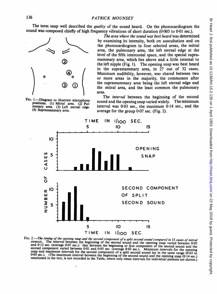
Relationship to the Mid-Diastolic Murmur. In 27 out of 32 cases, the opening snap was closelyfollowed by the mid-diastolic murmur on the phonocardiogram (Fig. 4). In two of the remaindera silent gap was recorded between the opening snap and the murmur (Fig. 5). In three, recordsdid not give clear information about the time of the start of the murmur. In eight cases there wasan initial crescendo to the murmur, which reached its greatest intensity about 005 sec. after theopening snap (Fig. 4). Clinically it was often difficult to time the murmur more accurately than tosay that it followed closely the opening snap.
Relationship between the Opening Snap and the Degree of Mitral Valve Disease Present. Absenceor softness of the opening snap was noted in four cases in the series. In three of these gross calcifi-cation of the mitral valve was present and this was diagnosed radiologically and on palpation atvalvotomy; in the fourth the stenosis was slight, as judged by the absence of symptoms andof generalized cardiac enlargement. One further case of gross calcification of the mitral valvewas met, but in this the opening snap was not unusually soft.
These findings are in agreement with the observations of Rouches (1888) that the opening snapwas not heard when the stenosis was mild or in extreme cases where the valves had lost their supple-ness and ability to snap. Wynn (1952) found an association between gross calcification of themitral valve, softness of the first heart sound, and absence of the opening snap. Sellors (1952)suggests that the snapping quality of the first heart sound in mitral stenosis may be produced at theanterior cusp of the mitral valve and notes that if a finger is placed on this cusp during the operation
137
on 22 May 2018 by guest. P
rotected by copyright.http://heart.bm
j.com/
Br H
eart J: first published as 10.1136/hrt.15.2.135 on 1 April 1953. D
ownloaded from
PATRICK MOUNSEY
1/5 Sec
MAIF
I Il I I
1 2SM < MDM
M,lANiF
C P e h
FIG. 4.-Time relationship between the opening snap (OS) and themid-diastolic murmur (MDM). The murmur follows closely theopening snap. In the first complex there is an initial crescendoin the mid-diastolic murmur. MA=mitral area. MF and LF=medium- and low-frequency filters. 1 and 2=first and secondheart sounds. SM=systolic murmur.
I I I I I I I I I
J z: ^ J i-i 1, i1
1~ ~ ~ ~~~T WR T1MDM
Os2
_AM-CR -
FIG. 5.-Time relationship between the opening snap and the mid-diastolic murmur. A short silent gapseparates the opening snap (OS) from the beginning of the mid-diastolic murmur (MDM). MA=mitralarea. MF=medium-frequency filter. 1 and 2=first and second heart sounds.
of valvotomy, as it balloons into the left atrium at the beginning of systole, the snapping quality ofthe sound, heard by listening directly to the heart with a stethoscope, is temporarily abolished. If,therefore, the diseased mitral valve itself gives rise both to the loud, abrupt, snapping first heartsound and to the opening snap, then the clinical association noted between the intensity of the twosounds in this series is explained.
DIFFERENTIATION OF THE OPENING SNAP FROM SPLITrING OF THE SECOND SOUNDAudible splitting of the second sound is physiological, provided auscultation is carried out in
the pulmonary area; the interval between the two components varies with respiration, tending toincrease with inspiration (Potain, 1866 ; Leatham and Towers, 1951).
Set Il.'ij
.i -,di -i .1 I IMA ON"ii" i? . 11MF
lud",& 1!iillf,WITUNW79"Mml A06 1!.iIwlre-w"r-. ,~ '' ~'~~~~5
I
138
I I
O Lk No
I;1: k.;..1'.1..:i..'v.I.-WOWMV:.'-ICAVAVV14 '*
on 22 May 2018 by guest. P
rotected by copyright.http://heart.bm
j.com/
Br H
eart J: first published as 10.1136/hrt.15.2.135 on 1 April 1953. D
ownloaded from
OPENING SNAP OF MITRAL STENOSIS
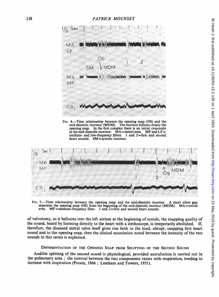
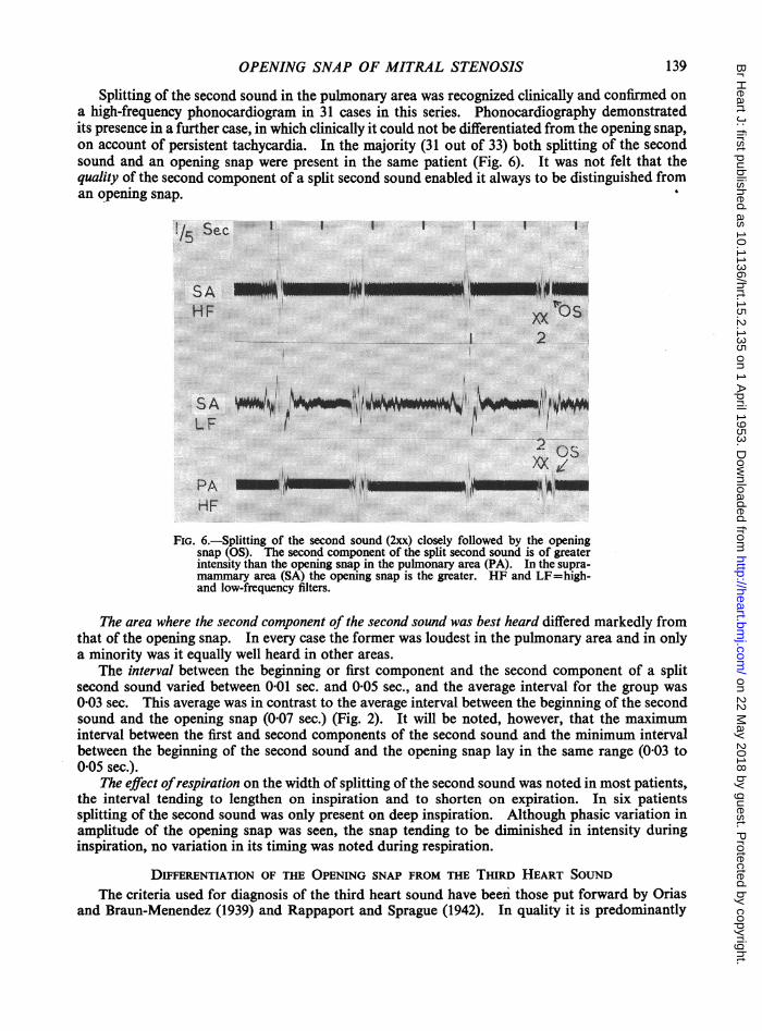
Splitting of the second sound in the pulmonary area was recognized clinically and confirmed ona high-frequency phonocardiogram in 31 cases in this series. Phonocardiography demonstratedits presence in a further case, in which clinically it could not be differentiated from the opening snap,on account of persistent tachycardia. In the majority (31 out of 33) both splitting of the secondsound and an opening snap were present in the same patient (Fig. 6). It was not felt that thequality of the second component of a split second sound enabled it always to be distinguished froman opening snap.
1/5Sec I I I ISee
SA IS EEHF 1x
12
LFi~~
PA EIEE!E E;Iti!iA IEX;in.XX C)S
HF
FIG. 6.-Splitting of the second sound (2xx) closely followed by the openingsnap (OS). The second component of the split second sound is of greaterintensity than the opening snap in the pulmonary area (PA). In the supra-mammary area (SA) the opening snap is the greater. HF and LF=bigh-and low-frequency filters.
The area where the second component of the second sound was best heard differed markedly fromthat of the opening snap. In every case the former was loudest in the pulmonary area and in onlya minority was it equally well heard in other areas.
The interval between the beginning or first component and the second component of a splitsecond sound varied between 0-01 sec. and 005 sec., and the average interval for the group was003 sec. This average was in contrast to the average interval between the beginning of the secondsound and the opening snap (007 sec.) (Fig. 2). It will be noted, however, that the maximuminterval between the first and second components of the second sound and the minimum intervalbetween the beginning of the second sound and the opening snap lay in the same range (003 to0 05 sec.).
The effect ofrespiration on the width of splitting of the second sound was noted in most patients,the interval tending to lengthen on inspiration and to shorten on expiration. In six patientssplitting of the second sound was only present on deep inspiration. Although phasic variation inamplitude of the opening snap was seen, the snap tending to be diminished in intensity duringinspiration, no variation in its timing was noted during respiration.
DIFFERENTIATION OF THE OPENING SNAP FROM THE THIRD HEART SOUNDThe criteria used for diagnosis of the third heart sound have been those put forward by Orias
and Braun-Menendez (1939) and Rappaport and Sprague (1942). In quality it is predominantly
139
on 22 May 2018 by guest. P
rotected by copyright.http://heart.bm
j.com/
Br H
eart J: first published as 10.1136/hrt.15.2.135 on 1 April 1953. D
ownloaded from

PATRICK MOUNSEY
a low-frequency sound as opposed to the higher frequency of the opening snap. The mid-point ofthe third heart sound lies between 0 16 and 0-24 sec. after the beginning of the second sound. Thisis greater than the value of 0-03 to 0*14 sec. for the opening snap in this series. The third heartsound is usually best heard a little internal to the mitral area (Evans, 1942).
In no patient was a clear third heart sound recognized clinically, although a small third heartsound was present in the phonocardiogram in eight (Fig. 7). The rarity of the third heart soundin mitral stenosis is in contrast with the frequency of an opening snap.
i/5 Sec l l l l I
LS E -EEEEESUi!HF PSM
2 3.4 tMDM I
LS E *O0 ItL-F
O5
FIG. 7.-The third heart sound in the presence of an opening snap anda mid-diastolic murmur. A small third heart sound (3) is recordedduring the beginning of the mid-diastolic murmur (MDM). It ispreceded by the opening snap (OS). LSE=left sternal edge.HF and LF=high- and low-frequency filters. 1 and 2=first andsecond heart sounds. PSM=presystolicmurmur. CRI=electro-cardiogram in lead CR1.
Orias and Braun-Menendez (1939) suggested that the third heart sound was due to vibrationsproduced during rapid ventricular filling. In mitral stenosis left ventricular filling is slow, beingimpeded by the stenosis, and so there is diminution or absence of a third heart sound, which isreplaced by the mid-diastolic murmur. When, on the other hand, incompetence is the only lesionin mitral valve disease, Brigden and Leatham (1952) have shown that an obvious third heart soundis a frequent finding: this is explained by rapid filling of the left ventricle through the widely patentmitral valve.
DISCUSSIONIn this series clinical differentiation between an opening snap and the second component of a
split second sound was generally possible, based on the site where the sound was best heard and onthe interval that separated it from the first component of the second sound. The fact that in mostcases both sounds were present was of great value in distinguishing each. Deep breathing was amanceuvre that sometimes aided clinical distinction between the sounds, since with this the splittingof the second sound tended to widen, or in some cases to appear de novo, while the intensity of theopening snap tended to diminish.
Cases with persistent tachycardia presented the greatest difficulty, for in these splitting of thesecond sound and an opening snap lay crowded into an interval of 0-03 sec. When carotid pressureinduced a slower heart rate, differentiation of these three sounds was facilitated. In one patient,in whom this manceuvre failed, phonocardiography with a rapidly moving film showed the presenceof both splitting of the second sound and an opening snap.
The differential diagnosis of the opening snap from a third heart sound seldom caused difficulty.Although a small third heart sound was recorded phonocardiographically in eight cases, clinicallyit was never audible.
140
on 22 May 2018 by guest. P
rotected by copyright.http://heart.bm
j.com/
Br H
eart J: first published as 10.1136/hrt.15.2.135 on 1 April 1953. D
ownloaded from
OPENING SNAP OF MITRAL STENOSIS
The opening snap was found to be most useful for the diagnosis of mitral stenosis in patients inwhom other signs were missing on casual examination. When the opening snap was heard by itself,it stimulated a more thorough search for a latent mid-diastolic murmur.
Although the pathogenesis of the opening snap remains in doubt, there is evidence that it isproduced at the stenosed mitral valve itself. Margolies and Wolferth (1932) showed that theopening snap coincided with the beginning of the descent of the V wave in a jugular pulsetracing. Assuming that the tricuspid and mitral valves open almost simultaneously, the fall injugular venous pressure following the opening snap strongly suggests that this sound is coincidentwith the opening of the mitral valve.
The association of the opening snap with mitral stenosis alone and its absence in other diseasescausing pulmonary hypertension or pulmonary venous congestion is an argument in favour of itsproduction at the stenosed mitral valve. Its tendency to become softer or disappear with extremerigid calcified stenosis suggested that it is not directly related to any increase in left auricular pres-sure, which would remain unrelieved or tend to increase further with increasing stenosis, but ratherto the state of the mitral valve itself. Its infrequency in early mitral valve disease when the diastolicmurmur first appears (Thomas, 1952) suggests that considerable stenosis is required to produce it.Its absence in pure mitral incompetence (Guttman, 1872; Brigden and Leatham, 1952) emphasizesits essential connection with a stenotic lesion.
If it be assumed that the opening snap is produced at the mitral valve, then hemodynamicfactors may be used to explain its variable timing. The timing of the opening snap depends on theinter-relationship between the left atrial pressure and the rate of fall of left ventricular pressureduring the phase of isometric relaxation. When the left ventricular pressure falls below the leftatrial, the mitral valve opens with the production of the opening snap. Messer et al. (1951) haveexplained in the following way the finding that in patients in auricular fibrillation a short cardiaccycle is followed by an early opening snap. A short diastolic interval in mitral stenosis tends tocause an increase in left atrial pressure, due to inefficient emptying of the left atrium. In thesubsequent diastole, therefore, left atrial pressure exceeds left ventricular pressure. slightly earlierand hence the mitral valve opens earlier. The occurrence of an earlier opening snap, with increasingheart rate and hence shorter diastolic interval, in patients in sinus rhythm could be similarly ex-plained. The diastolic interval, however, is only one of many factors affecting the left atrialpressure and left atrial pressure is not the only factor affecting the timing of the opening snap.Although, therefore, a relationship was seen in individual cases in our series between the diastolicinterval and the timing of the opening snap, in the group as a whole no direct relationship wasfound.
SUMMARYA clinical and phonocardiographic study of 33 cases of mitral stenosis has been made. An
opening snap was heard in 28: in a further 4 it was recorded on the phonocardiogram althoughnot detected clinically; in these its soft character or the presence of tachycardia caused it to bemissed clinically. In only one case was an opening snap absent in the phonocardiogram.
Differentiation of the opening snap from splitting of the second sound was made on the timingof the sounds and the area where they were best heard. The second component of a split secondsound was always loudest in the pulmonary area and the opening snap usually loudest in the areaabove and internal to the left breast. The frequency with which both sounds were present (31 outof 33) made their recognition relatively easy. Phonocardiography was necessary to differentiatebetween them in only one case.
The physiological third heart sound differs from the opening snap in its low-pitched quality,the area where it is best heard and its timing. In no patient was a clear third heart sound recognizedclinically, although in eight a small third heart sound was present in the phonocardiogram.
Absence or diminution in intensity of the opening snap was associated with gross calcificationof the mitral valve in three cases and with slight stenosis in a fourth.
141
on 22 May 2018 by guest. P
rotected by copyright.http://heart.bm
j.com/
Br H
eart J: first published as 10.1136/hrt.15.2.135 on 1 April 1953. D
ownloaded from
PATRICK MOUNSEY
The opening snap was found to be a useful diagnostic sign, when other signs of mitral stenosiswere missing on casual examination. Its presence should lead to a more thorough search, whichshould reveal the latent mid-diastolic murmur.
I would like to acknowledge with sincere thanks the help and criticism of Dr. William Evans in the preparation ofthis paper. I wish to thank Mr. W. D. Dicks for technical help with the phonocardiograms.
REFERENCESBouillaud, J. (1835). Traite clinique des Maladies du COur, prdcedd de Recherches nouvelles sur l'Anatomie et la
Physiologie de cet Organe. Bailliere, Paris.Brigden, W. W., and Leatham, A. (1953). Brit. Heart J., 15, 55.Duchosal, P. (1946). Praxis, 24, 385.Durozier, P. (1862). Arch. gdn. Mid. 20, 385.Evans, W. (1942). Brit. Heart J., 5, 205.Gallavardin, L. (1905). Lyon med., 105, 401.Guttman, P. (1872). Lehrbuch der Klinischen Untersuchung Methoden. Hirschwald, Berlin, 257.Laennec, R. T. H. (1819). De l'Auscultation mediate ou Traite du Diagnostic des Maladies des Poumons et du Coeur,
fonde principalement sur ce nouveau Moyen d'Exploration. Brosson et Chaude, Paris.Leatham, A. (1949). Post. Grad. med. J., 25, 568.-, and Towers, M. (1951). Brit. Heart J., 13, 575.Margolies, A., and Wolferth, C. C. (1932). Amer. Heart J., 7, 443.Messer, A. L., Counihan, T. B., Rappaport, M. B., and Sprague, H. B. (1951). Circulation, 4, 576.Orias, O., and Braun-Menendez, E. (1939). The Heart Sounds in Normal and Pathological Conditions. New York.Potain, C. (1866). Bull. Soc. mid. Hop. Paris, 3, 138.Rappaport, M. B., and Sprague, H. B. (1942). Amer. Heart J., 23, 591.Rouches, F. J. M. (1888). These de Paris.Sansom, A. E. (1881). Proc. Med. Soc. Lon., 5, 191.Sellors, T. H. (1952). Personal communication.Thomas, G. T. (1952). Personal communication.Wynn, A. (1952). Brit. Heart J., 14, 296.
142
on 22 May 2018 by guest. P
rotected by copyright.http://heart.bm
j.com/
Br H
eart J: first published as 10.1136/hrt.15.2.135 on 1 April 1953. D
ownloaded from