
Diagnosis of Ebola Virus Disease: Past, Present, and Future · discovered Ebola virus from the...
21
Diagnosis of Ebola Virus Disease: Past, Present, and Future M. Jana Broadhurst, a Tim J. G. Brooks, b Nira R. Pollock c Department of Pathology, Stanford University School of Medicine, Palo Alto, California, USA a ; Public Health England, Porton Down, Salisbury, United Kingdom b ; Department of Laboratory Medicine, Boston Children’s Hospital, Boston, Massachusetts, USA c SUMMARY ..................................................................................................................................................773 INTRODUCTION ............................................................................................................................................773 METHODS FOR DETECTING EBOLA VIRUS INFECTION ....................................................................................................774 Overview .................................................................................................................................................774 Cell Culture ...............................................................................................................................................774 Antibody Detection ......................................................................................................................................775 Protein Antigen Detection ...............................................................................................................................776 Conventional RT-PCR .....................................................................................................................................777 Real-Time RT-PCR.........................................................................................................................................777 FIELD DIAGNOSTIC LABORATORIES IN EBOLA VIRUS OUTBREAKS .......................................................................................778 Importance of Field Diagnostic Capacity .................................................................................................................778 Field Diagnostic Laboratory Efforts in Prior Outbreaks ...................................................................................................778 LABORATORY DIAGNOSIS OF EVD IN THE 2014-2015 EPIDEMIC .........................................................................................778 Overview .................................................................................................................................................778 EVD Diagnostic Tests with Emergency Use Authorization ...............................................................................................779 Standard (nonautomated) real-time RT-PCR tests .....................................................................................................779 Automated real-time RT-PCR tests .....................................................................................................................782 Rapid antigen detection tests .........................................................................................................................783 Specimen Management and Biosafety for Diagnostic Testing ...........................................................................................784 Specimen collection and tracking .....................................................................................................................784 Viral inactivation in diagnostic specimens .............................................................................................................785 Detection of Viral Persistence in Nonblood Body Fluids ..................................................................................................785 Viral persistence in seminal fluid .......................................................................................................................785 Viral persistence in other body fluids ..................................................................................................................786 Viral Sequencing in the 2014-2015 Epidemic.............................................................................................................786 SUMMARY AND FUTURE DIRECTIONS .....................................................................................................................787 ACKNOWLEDGMENT .......................................................................................................................................788 REFERENCES ................................................................................................................................................788 AUTHOR BIOS ..............................................................................................................................................793 SUMMARY Laboratory diagnosis of Ebola virus disease plays a critical role in outbreak response efforts; however, establishing safe and expeditious testing strategies for this high-biosafety-level pathogen in resource-poor environments remains extremely challenging. Since the discovery of Ebola virus in 1976 via tra- ditional viral culture techniques and electron microscopy, di- agnostic methodologies have trended toward faster, more ac- curate molecular assays. Importantly, technological advances have been paired with increasing efforts to support decentral- ized diagnostic testing capacity that can be deployed at or near the point of patient care. The unprecedented scope of the 2014- 2015 West Africa Ebola epidemic spurred tremendous innova- tion in this arena, and a variety of new diagnostic platforms that have the potential both to immediately improve ongoing surveillance efforts in West Africa and to transform future out- break responses have reached the field. In this review, we de- scribe the evolution of Ebola virus disease diagnostic testing and efforts to deploy field diagnostic laboratories in prior out- breaks. We then explore the diagnostic challenges pervading the 2014-2015 epidemic and provide a comprehensive exami- nation of novel diagnostic tests that are likely to address some of these challenges moving forward. INTRODUCTION T he recent outbreak of Ebola virus disease (EVD) in West Africa has highlighted both the importance of rapid and accurate diagnosis of this disease and the challenges around diagnostic test- ing. Throughout the 2014-2015 outbreak, diagnosis relied pri- marily on testing of venipuncture blood samples from symptom- atic individuals in a biocontainment laboratory facility, leading to challenges with specimen collection and data management and often a prolonged turnaround time to final results. Consequently, the need for rapid and, particularly, for point-of-care diagnostics generated an unprecedented surge in development of new diag- nostic methods for EVD. This review summarizes the evolution of laboratory-based methods for EVD diagnosis, the implementa- tion of these methodologies for field-based testing in outbreak Published 13 July 2016 Citation Broadhurst MJ, Brooks TJG, Pollock NR. 2016. Diagnosis of Ebola virus disease: past, present, and future. Clin Microbiol Rev 29:773–793. doi:10.1128/CMR.00003-16. Address correspondence to M. Jana Broadhurst, [email protected], or Nira R. Pollock, [email protected]. Copyright © 2016, American Society for Microbiology. All Rights Reserved. crossmark October 2016 Volume 29 Number 4 cmr.asm.org 773 Clinical Microbiology Reviews on December 23, 2019 by guest http://cmr.asm.org/ Downloaded from
Transcript of Diagnosis of Ebola Virus Disease: Past, Present, and Future · discovered Ebola virus from the...

Diagnosis of Ebola Virus Disease: Past, Present, and Future
M. Jana Broadhurst,a Tim J. G. Brooks,b Nira R. Pollockc
Department of Pathology, Stanford University School of Medicine, Palo Alto, California, USAa; Public Health England, Porton Down, Salisbury, United Kingdomb;Department of Laboratory Medicine, Boston Children’s Hospital, Boston, Massachusetts, USAc
SUMMARY . . . . . . . . . . . . . . . . . . . . . . . . . . . . . . . . . . . . . . . . . . . . . . . . . . . . . . . . . . . . . . . . . . . . . . . . . . . . . . . . . . . . . . . . . . . . . . . . . . . . . . . . . . . . . . . . . . . . . . . . . . . . . . . . . . . . . . . . . . . . . . . . . .773INTRODUCTION . . . . . . . . . . . . . . . . . . . . . . . . . . . . . . . . . . . . . . . . . . . . . . . . . . . . . . . . . . . . . . . . . . . . . . . . . . . . . . . . . . . . . . . . . . . . . . . . . . . . . . . . . . . . . . . . . . . . . . . . . . . . . . . . . . . . . . . . . . . .773METHODS FOR DETECTING EBOLA VIRUS INFECTION . . . . . . . . . . . . . . . . . . . . . . . . . . . . . . . . . . . . . . . . . . . . . . . . . . . . . . . . . . . . . . . . . . . . . . . . . . . . . . . . . . . . . . . . . . . . . . . . . . . .774
Overview . . . . . . . . . . . . . . . . . . . . . . . . . . . . . . . . . . . . . . . . . . . . . . . . . . . . . . . . . . . . . . . . . . . . . . . . . . . . . . . . . . . . . . . . . . . . . . . . . . . . . . . . . . . . . . . . . . . . . . . . . . . . . . . . . . . . . . . . . . . . . . . . .774Cell Culture . . . . . . . . . . . . . . . . . . . . . . . . . . . . . . . . . . . . . . . . . . . . . . . . . . . . . . . . . . . . . . . . . . . . . . . . . . . . . . . . . . . . . . . . . . . . . . . . . . . . . . . . . . . . . . . . . . . . . . . . . . . . . . . . . . . . . . . . . . . . . . .774Antibody Detection . . . . . . . . . . . . . . . . . . . . . . . . . . . . . . . . . . . . . . . . . . . . . . . . . . . . . . . . . . . . . . . . . . . . . . . . . . . . . . . . . . . . . . . . . . . . . . . . . . . . . . . . . . . . . . . . . . . . . . . . . . . . . . . . . . . . . .775Protein Antigen Detection . . . . . . . . . . . . . . . . . . . . . . . . . . . . . . . . . . . . . . . . . . . . . . . . . . . . . . . . . . . . . . . . . . . . . . . . . . . . . . . . . . . . . . . . . . . . . . . . . . . . . . . . . . . . . . . . . . . . . . . . . . . . . . .776Conventional RT-PCR . . . . . . . . . . . . . . . . . . . . . . . . . . . . . . . . . . . . . . . . . . . . . . . . . . . . . . . . . . . . . . . . . . . . . . . . . . . . . . . . . . . . . . . . . . . . . . . . . . . . . . . . . . . . . . . . . . . . . . . . . . . . . . . . . . . . .777Real-Time RT-PCR. . . . . . . . . . . . . . . . . . . . . . . . . . . . . . . . . . . . . . . . . . . . . . . . . . . . . . . . . . . . . . . . . . . . . . . . . . . . . . . . . . . . . . . . . . . . . . . . . . . . . . . . . . . . . . . . . . . . . . . . . . . . . . . . . . . . . . . . .777
FIELD DIAGNOSTIC LABORATORIES IN EBOLA VIRUS OUTBREAKS . . . . . . . . . . . . . . . . . . . . . . . . . . . . . . . . . . . . . . . . . . . . . . . . . . . . . . . . . . . . . . . . . . . . . . . . . . . . . . . . . . . . . . .778Importance of Field Diagnostic Capacity . . . . . . . . . . . . . . . . . . . . . . . . . . . . . . . . . . . . . . . . . . . . . . . . . . . . . . . . . . . . . . . . . . . . . . . . . . . . . . . . . . . . . . . . . . . . . . . . . . . . . . . . . . . . . . . . .778Field Diagnostic Laboratory Efforts in Prior Outbreaks . . . . . . . . . . . . . . . . . . . . . . . . . . . . . . . . . . . . . . . . . . . . . . . . . . . . . . . . . . . . . . . . . . . . . . . . . . . . . . . . . . . . . . . . . . . . . . . . . . .778
LABORATORY DIAGNOSIS OF EVD IN THE 2014-2015 EPIDEMIC . . . . . . . . . . . . . . . . . . . . . . . . . . . . . . . . . . . . . . . . . . . . . . . . . . . . . . . . . . . . . . . . . . . . . . . . . . . . . . . . . . . . . . . . .778Overview . . . . . . . . . . . . . . . . . . . . . . . . . . . . . . . . . . . . . . . . . . . . . . . . . . . . . . . . . . . . . . . . . . . . . . . . . . . . . . . . . . . . . . . . . . . . . . . . . . . . . . . . . . . . . . . . . . . . . . . . . . . . . . . . . . . . . . . . . . . . . . . . .778EVD Diagnostic Tests with Emergency Use Authorization . . . . . . . . . . . . . . . . . . . . . . . . . . . . . . . . . . . . . . . . . . . . . . . . . . . . . . . . . . . . . . . . . . . . . . . . . . . . . . . . . . . . . . . . . . . . . . .779
Standard (nonautomated) real-time RT-PCR tests . . . . . . . . . . . . . . . . . . . . . . . . . . . . . . . . . . . . . . . . . . . . . . . . . . . . . . . . . . . . . . . . . . . . . . . . . . . . . . . . . . . . . . . . . . . . . . . . . . . . .779Automated real-time RT-PCR tests. . . . . . . . . . . . . . . . . . . . . . . . . . . . . . . . . . . . . . . . . . . . . . . . . . . . . . . . . . . . . . . . . . . . . . . . . . . . . . . . . . . . . . . . . . . . . . . . . . . . . . . . . . . . . . . . . . . . .782Rapid antigen detection tests . . . . . . . . . . . . . . . . . . . . . . . . . . . . . . . . . . . . . . . . . . . . . . . . . . . . . . . . . . . . . . . . . . . . . . . . . . . . . . . . . . . . . . . . . . . . . . . . . . . . . . . . . . . . . . . . . . . . . . . . .783
Specimen Management and Biosafety for Diagnostic Testing . . . . . . . . . . . . . . . . . . . . . . . . . . . . . . . . . . . . . . . . . . . . . . . . . . . . . . . . . . . . . . . . . . . . . . . . . . . . . . . . . . . . . . . . . . .784Specimen collection and tracking . . . . . . . . . . . . . . . . . . . . . . . . . . . . . . . . . . . . . . . . . . . . . . . . . . . . . . . . . . . . . . . . . . . . . . . . . . . . . . . . . . . . . . . . . . . . . . . . . . . . . . . . . . . . . . . . . . . . .784Viral inactivation in diagnostic specimens . . . . . . . . . . . . . . . . . . . . . . . . . . . . . . . . . . . . . . . . . . . . . . . . . . . . . . . . . . . . . . . . . . . . . . . . . . . . . . . . . . . . . . . . . . . . . . . . . . . . . . . . . . . . .785
Detection of Viral Persistence in Nonblood Body Fluids. . . . . . . . . . . . . . . . . . . . . . . . . . . . . . . . . . . . . . . . . . . . . . . . . . . . . . . . . . . . . . . . . . . . . . . . . . . . . . . . . . . . . . . . . . . . . . . . . .785Viral persistence in seminal fluid . . . . . . . . . . . . . . . . . . . . . . . . . . . . . . . . . . . . . . . . . . . . . . . . . . . . . . . . . . . . . . . . . . . . . . . . . . . . . . . . . . . . . . . . . . . . . . . . . . . . . . . . . . . . . . . . . . . . . . .785Viral persistence in other body fluids . . . . . . . . . . . . . . . . . . . . . . . . . . . . . . . . . . . . . . . . . . . . . . . . . . . . . . . . . . . . . . . . . . . . . . . . . . . . . . . . . . . . . . . . . . . . . . . . . . . . . . . . . . . . . . . . . .786
Viral Sequencing in the 2014-2015 Epidemic. . . . . . . . . . . . . . . . . . . . . . . . . . . . . . . . . . . . . . . . . . . . . . . . . . . . . . . . . . . . . . . . . . . . . . . . . . . . . . . . . . . . . . . . . . . . . . . . . . . . . . . . . . . . .786SUMMARY AND FUTURE DIRECTIONS . . . . . . . . . . . . . . . . . . . . . . . . . . . . . . . . . . . . . . . . . . . . . . . . . . . . . . . . . . . . . . . . . . . . . . . . . . . . . . . . . . . . . . . . . . . . . . . . . . . . . . . . . . . . . . . . . . . . .787ACKNOWLEDGMENT . . . . . . . . . . . . . . . . . . . . . . . . . . . . . . . . . . . . . . . . . . . . . . . . . . . . . . . . . . . . . . . . . . . . . . . . . . . . . . . . . . . . . . . . . . . . . . . . . . . . . . . . . . . . . . . . . . . . . . . . . . . . . . . . . . . . . . .788REFERENCES . . . . . . . . . . . . . . . . . . . . . . . . . . . . . . . . . . . . . . . . . . . . . . . . . . . . . . . . . . . . . . . . . . . . . . . . . . . . . . . . . . . . . . . . . . . . . . . . . . . . . . . . . . . . . . . . . . . . . . . . . . . . . . . . . . . . . . . . . . . . . . . .788AUTHOR BIOS . . . . . . . . . . . . . . . . . . . . . . . . . . . . . . . . . . . . . . . . . . . . . . . . . . . . . . . . . . . . . . . . . . . . . . . . . . . . . . . . . . . . . . . . . . . . . . . . . . . . . . . . . . . . . . . . . . . . . . . . . . . . . . . . . . . . . . . . . . . . . .793
SUMMARY
Laboratory diagnosis of Ebola virus disease plays a critical rolein outbreak response efforts; however, establishing safe andexpeditious testing strategies for this high-biosafety-levelpathogen in resource-poor environments remains extremelychallenging. Since the discovery of Ebola virus in 1976 via tra-ditional viral culture techniques and electron microscopy, di-agnostic methodologies have trended toward faster, more ac-curate molecular assays. Importantly, technological advanceshave been paired with increasing efforts to support decentral-ized diagnostic testing capacity that can be deployed at or nearthe point of patient care. The unprecedented scope of the 2014-2015 West Africa Ebola epidemic spurred tremendous innova-tion in this arena, and a variety of new diagnostic platformsthat have the potential both to immediately improve ongoingsurveillance efforts in West Africa and to transform future out-break responses have reached the field. In this review, we de-scribe the evolution of Ebola virus disease diagnostic testingand efforts to deploy field diagnostic laboratories in prior out-breaks. We then explore the diagnostic challenges pervadingthe 2014-2015 epidemic and provide a comprehensive exami-nation of novel diagnostic tests that are likely to address someof these challenges moving forward.
INTRODUCTION
The recent outbreak of Ebola virus disease (EVD) in West Africahas highlighted both the importance of rapid and accurate
diagnosis of this disease and the challenges around diagnostic test-ing. Throughout the 2014-2015 outbreak, diagnosis relied pri-marily on testing of venipuncture blood samples from symptom-atic individuals in a biocontainment laboratory facility, leading tochallenges with specimen collection and data management andoften a prolonged turnaround time to final results. Consequently,the need for rapid and, particularly, for point-of-care diagnosticsgenerated an unprecedented surge in development of new diag-nostic methods for EVD. This review summarizes the evolution oflaboratory-based methods for EVD diagnosis, the implementa-tion of these methodologies for field-based testing in outbreak
Published 13 July 2016
Citation Broadhurst MJ, Brooks TJG, Pollock NR. 2016. Diagnosis of Ebola virusdisease: past, present, and future. Clin Microbiol Rev 29:773–793.doi:10.1128/CMR.00003-16.
Address correspondence to M. Jana Broadhurst, [email protected], orNira R. Pollock, [email protected].
Copyright © 2016, American Society for Microbiology. All Rights Reserved.
crossmark
October 2016 Volume 29 Number 4 cmr.asm.org 773Clinical Microbiology Reviews
on Decem
ber 23, 2019 by guesthttp://cm
r.asm.org/
Dow
nloaded from

settings, and recent advances in diagnostic tools that are likely tobenefit future clinical and surveillance efforts. As new diagnostictechnologies become available, it will be increasingly importantfor clinicians to understand both the analytic and practicalstrengths and limitations of each testing platform. Ultimately, theoptimal diagnostic approach for a particular setting will depend uponmultiple factors, including population characteristics and diseaseprevalence, the health care setting (e.g., infrastructure and availabilityof biosafety and infection control measures), training requirements,regional laboratory capacity, regulatory status, and cost.
METHODS FOR DETECTING EBOLA VIRUS INFECTION
Overview
Ebola viruses contain a single-stranded RNA genome that encodesseven viral proteins: nucleoprotein (NP), glycoprotein (GP), poly-merase (L), VP24, VP30, VP35, and VP40. Over the past 25 years,several methods for detecting infection and/or disease with Ebolavirus have been developed that are amenable for use in clinicallaboratory settings (1). These fall into three basic categories: (i)serologic tests that detect host antibodies generated against thevirus, (ii) antigen tests that detect viral proteins, and (iii) molec-ular tests that detect viral RNA sequences (Fig. 1). Specific antivi-ral antibodies can persist for years following infection; however,the variable onset of antibody responses during acute illnessmakes serology minimally useful as a diagnostic tool in the acutesetting. Conversely, antigen detection and molecular tests have
proven very effective for acute diagnosis, as virus levels in theblood typically rise to high levels within the first few days of symp-toms (2). The incubation period following Ebola virus infectiontypically ranges between 3 and 13 days, but may be as long as 21days (3, 4); no tests have yet demonstrated the ability to reliablydetect Ebola virus prior to the onset of symptoms. Some diagnos-tic tests have been designed to broadly detect Ebola virus infection,while others distinguish among the five known Ebola virus species(Zaire/Ebola [EBOV], Sudan [SUDV], Tai Forest [TAFV], Reston[RESTV], and Bundibugyo [BDBV]). Major outbreaks of EVD inhumans have been attributable to EBOV, SUDV, and BDBV; priorto the 2014-2015 epidemic, the origins of EVD outbreaks wererestricted to five African countries: Democratic Republic ofCongo (formerly Zaire), Sudan, Gabon, Uganda, and Republic ofCongo (Fig. 2).
Cell Culture
The traditional gold standard method to confirm the presence ofEbola virus is viral isolation in cell culture, typically using Vero E6African Green monkey kidney cells. Propagated virus can bedirectly visualized by electron microscopy or indirectly visual-ized by immunofluorescence microscopy within 1 to 5 days ofinoculation. While detection of Ebola virus by these methods isdefinitive, these methods require biosafety level 4 (BSL-4) con-tainment and are typically restricted to research and publichealth laboratories (5).
FIG 1 Detection of Ebola virus infection in nonfatal versus fatal cases. Solid lines indicate that the analyte of interest is detected in the majority of cases at thecorresponding time point (days post-symptom onset); dashed lines indicate that the analyte of interest is detected in the minority of cases at that time point. Datafor IgG and IgM detection were compiled from references 9, 10, and 15 to 20. Data for antigen detection were compiled from references 2, 10, 17, and 18. Datafor RNA detection were compiled from references 2, 12, 15, 27, 28, and 35. RT-PCR, reverse transcription-PCR; ELISA, enzyme-linked immunosorbent assay.
Broadhurst et al.
774 cmr.asm.org October 2016 Volume 29 Number 4Clinical Microbiology Reviews
on Decem
ber 23, 2019 by guesthttp://cm
r.asm.org/
Dow
nloaded from

Antibody Detection
Serologic assays for the detection of specific antiviral antibodies inpatient serum have been used to demonstrate current or priorinfection with Ebola virus since the first outbreak investigations ofthis virus in 1976 (6, 7). Indeed, an indirect fluorescent antibodydetection test (IFAT) was used in 1977 to distinguish the newlydiscovered Ebola virus from the closely related Marburg virus,based on the viral antigen specificity of antibodies in convales-cent-phase serum from individuals who had recovered from in-fections with these pathogens (6). For this method, cell culturesinfected with Ebola virus (or antigen suspensions from thesecultures) are irradiated, fixed onto a slide, and incubated with sera
from potentially exposed individuals; bound antibodies are thendetected with a fluorescently labeled secondary antibody (e.g.,rabbit anti-human IgG) and visualized with immunofluorescencemicroscopy (8). Although IFAT played a critical role in establish-ing clinical diagnoses during the first several Ebola outbreaks, itwas considered to have suboptimal sensitivity and specificity (9),and the requirement for BSL-4 biocontainment rendered thismethod unsuitable for large-scale diagnostic efforts.
The development of enzyme-linked immunosorbent assay(ELISA) tests for the detection of Ebola virus-specific IgM and IgGantibodies offered a faster, higher-throughput system for sero-logic testing. These assays, first developed at the U.S. Army Med-
FIG 2 Diagnostic testing in Ebola virus outbreaks. The information provided for each outbreak includes the affected country (or countries), the agenciesprimarily responsible for clinical diagnostic testing during the outbreak, where testing took place (the location is shown in parentheses; field laboratories are alsohighlighted in red), and the primary testing methods used for clinical diagnosis. The size of the box denotes the relative size of the outbreak, categorized in thefollowing groups: �100 cases, 100 to 200 cases, 200 to 300 cases, 300 to 400 cases, and 400 to 500 cases; the West Africa epidemic exceeded 28,600 cases (132). Thecolor of the box denotes the outbreak’s Ebola virus species (EBOV, Zaire/Ebola; SUDV, Sudan; BDBV, Bundibugyo). Abbreviations: DRC, Democratic Republicof Congo; RC, Republic of Congo; MRE, Microbiological Research Establishment; ITMA, Institute of Tropical Medicine, Antwerp; CIRMF, Centre Internationalde Recherches Medicales de Franceville; PHAC, Public Health Agency of Canada; KEMRI, Kenya Medical Research Institute; NICD, National Institute ofCommunicable Diseases; UVRI, Uganda Virus Research Institute; INRB, Institut National de Recherche Biomedicale.
Diagnosis of Ebola Virus Disease
October 2016 Volume 29 Number 4 cmr.asm.org 775Clinical Microbiology Reviews
on Decem
ber 23, 2019 by guesthttp://cm
r.asm.org/
Dow
nloaded from

ical Research Institute of Infectious Diseases (USAMRIID) in1990 (9) and adopted by the CDC, utilize viral antigens preparedfrom inoculated cell cultures to bind antibodies present in patientserum (9, 10). ELISAs (for both antibody and antigen detection;see “Protein Antigen Detection,” below) are compatible withgamma irradiation, allowing for inactivation of infectious virus inclinical specimens and subsequent sample processing underBSL-2 conditions; however, viral inactivation via gamma irradia-tion is available on demand in only a very few specialized institu-tions. Viral inactivation with heat and detergent treatment priorto serologic testing by ELISA has been described previously (11),but data regarding assay performance under these conditions arenot available.
The IgM ELISA in use by the CDC entails an antibody captureplatform, utilizing microtiter plates coated with goat antibodiesthat bind human IgM present in serum samples. After antibodiesfrom a serum sample are captured, Ebola virus-specific IgM isdetected by incubating the plate with a preparation of Ebola viralantigens, followed by polyclonal antibodies from Ebola virus-ex-posed rabbits that bind to captured viral antigens, with final de-tection mediated by horseradish peroxidase (HRP)-conjugatedanti-rabbit antibodies. In contrast, the IgG ELISA in use by theCDC utilizes microtiter plates coated with viral antigens that pulldown Ebola virus-specific antibodies present in serum samples;captured IgG is detected with HRP-conjugated mouse antibodiesspecific for human IgG. A similar IgG ELISA has been developedby the Public Health Agency of Canada and employed in theirrecent field laboratory efforts (12). Given the challenges inherentin generating authentic antigen preparations from viral cultures ina BSL-4 facility, ELISAs that utilize recombinant viral proteinshave been developed (13, 14), but to date they do not appear tohave been validated for clinical use. However, a commerciallyavailable Ebola Zaire virus IgM and IgG ELISA kit (Alpha Diag-nostic International) that utilizes recombinant viral proteins ex-pressed in Escherichia coli has been employed in recent clinicalresearch efforts (15). Limited data are available to assess the sen-sitivity or specificity of these ELISAs. A study evaluating the cross-reactivity of IgM and IgG antibodies in convalescent-phase serafrom outbreaks involving four different species of Ebola virusfound that IgM antibodies are minimally cross-reactive for Ebolavirus antigens from other Ebola virus species, while IgG antibodiesreadily react with antigens from multiple Ebola virus species (16).
IgM and IgG ELISAs were first clinically employed by the CDCduring the 1995 outbreak of Ebola Zaire virus in Kikwit, Demo-cratic Republic of Congo, and have since been a cornerstone ofEVD outbreak investigations. However, these tests have limitedutility in diagnosing acute EVD due to the variable onset of hu-moral responses. In a study of 29 EVD survivors from the 1995Kikwit outbreak, IgM and IgG antibodies appeared between days2 and 10 and days 6 and 19 after symptom onset, respectively (17).In this survivor cohort, IgM was detectable in all patients betweendays 10 and 29 of illness and persisted at least through day 30 andup to day 168 (the latest time point tested). IgG was detectable innearly all patients by day 19 of illness and persisted at least throughday 661 and up to day 749 (the latest time point tested). Similarly,a retrospective analysis of convalescent-phase serum samples col-lected during three outbreaks with different Ebola virus species(1995, Kikwit, Democratic Republic of Congo; 2000, Gulu, Ugan-da; 2007, Bundibugyo, Uganda) showed a loss of IgM in mostpatients by day 80 of illness, with persistence of IgG through the
final time point tested (day 117) (16). Importantly, evaluation ofacutely ill patients from the 1995 Kikwit outbreak as well as the1996 Gabon outbreaks demonstrated that antibody responses areoften not detected during EVD infections with fatal outcomes (10,18, 19). A recent study of acutely ill EVD patients infected duringthe 2014-2015 epidemic and treated in U.S. or European facilitiesshowed the onset of IgM and IgG responses between 6 and 11 daysand 9 and 11 days after symptom onset, respectively (15); differ-ences in antibody responses between fatal and nonfatal infectionsin this cohort were not reported. In survivors, IgG has been shownto persist for years following exposure (9, 10, 20). In summary, thecurrent literature suggests that IgM antibody responses duringEVD infection are variable, with the onset of detection rangingfrom 2 to 11 days following symptom onset and persistingthrough at least day 30 but typically not beyond day 80 in nonfatalinfections (Fig. 1). IgG responses typically become detectable inthe second week of illness in EVD survivors and can persist foryears (Fig. 1), providing a useful tool for population-level sero-prevalence studies.
Protein Antigen Detection
The detection of viral protein antigens circulating in blood pro-vides a reliable method for diagnosing acute EVD in symptomaticpatients, as viral proteins typically accumulate to detectable levelswithin a few days of disease onset. An ELISA for the detection ofEbola virus antigens, first developed at USAMRIID in 1989, uti-lizes a pool of 8 monoclonal mouse antibodies reactive againstEBOV and SUDV for antigen capture and polyclonal antibodiesfrom hyperimmune rabbit serum (reactive against EBOV, SUDV,and RESTV) for antigen detection (21). This assay was first eval-uated by the CDC for clinical use during the 1995 outbreak inKikwit, Democratic Republic of Congo (10), and it performedwell for clinical diagnosis of acute EVD in a field laboratory de-ployed by the CDC during the 2000 outbreak in Gulu, Uganda (2),offering the fastest method of virus detection available at the time(�5 h). By this method, viral antigen can be detected in serum asearly as the first day of symptoms, and detectable antigen is pres-ent in nearly all EVD patients by day 3 of illness (2, 10, 17, 18) (Fig.1). Antigen levels rise throughout the course of disease in fatalcases. During nonfatal infections, antigen levels are comparable tothose in fatal cases during the first 7 to 10 days of illness, afterwhich they typically decline to undetectable levels by day 16 (2, 10,17, 18) (Fig. 1).
The CDC antigen detection ELISA became part of the standarddiagnostic testing suite used by the CDC in subsequent outbreaks;however, limited access to the antibody reagents may have limitedits use by other agencies. ELISA antigen detection tests utilizingmonoclonal antibodies against NP (22), VP40 (23), or GP (24)proteins (generated from mice immunized with purified or re-combinant Ebola virus proteins) have been developed and are inplace at some national reference laboratories (25), but the use ofthese assays for clinical diagnosis has not been reported, as real-time reverse transcription-PCR (RT-PCR) techniques have nowreplaced these tests (discussed in the “Real-Time RT-PCR” sec-tion below). During the recent outbreak, lateral flow immunoas-says (LFIs) emerged as powerful tools for rapid antibody-medi-ated antigen capture that can be performed at the point of care.Novel LFIs for EVD diagnosis are discussed in the “Rapid antigendetection tests” section, below.
Broadhurst et al.
776 cmr.asm.org October 2016 Volume 29 Number 4Clinical Microbiology Reviews
on Decem
ber 23, 2019 by guesthttp://cm
r.asm.org/
Dow
nloaded from

Conventional RT-PCR
Diagnostic RT-PCR tests for Ebola virus, developed by the CDC,were first evaluated on serum samples collected from acutely illpatients during the 1995 Kikwit outbreak (26). These assays usedPCR to amplify the L, GP, and NP genes, followed by size-basedamplicon detection via gel electrophoresis. An important advan-tage of this method was the simple, chemical inactivation of infec-tious virus during the initial steps of RNA extraction by using achaotropic agent such as guanidine thiocyanate, allowing subse-quent sample processing to be carried out on the benchtop. (Ofnote, the efficacy of viral inactivation under such conditions hasrecently been questioned; viral inactivation methods are discussedin more detail in the “Specimen Management and Biosafety forDiagnostic Testing” section). Conventional RT-PCR was found tobe more sensitive than antibody and antigen detection ELISAswhen evaluated over the complete course of symptomatic infec-tion (see below), and in 1999 the CDC recommended its use inconjunction with antigen detection ELISA testing for diagnosis ofacute EVD (26).
Conventional RT-PCR testing for EVD was first evaluated by aregional laboratory during the 1996 outbreaks in Gabon, at theCentre International de Recherches Medicales de Franceville(CIRMF). In this setting, an RT-PCR test detecting the L gene ofEbola virus in peripheral blood mononuclear cells was more sen-sitive than serum IgM or antigen detection by the CDC ELISAs(antigen was detected in 83% of samples that tested positive byRT-PCR, and IgM was positive in 67%) (27). Furthermore, RT-PCR detected viral RNA in two specimens collected on the day ofsymptom onset that were negative for antigen at this time point,suggesting that RT-PCR may detect infection earlier in disease(27). The CDC first used RT-PCR testing of serum samples forclinical diagnosis of acute EVD during the 2000 outbreak in Gulu,Uganda, and similarly found that a nested RT-PCR assay for NPgene detection was able to detect viral RNA up to 72 h beforeantigen became detectable by ELISA and up to 72 h following theloss of antigen detection in convalescent patients (2). Of note, thisstudy also showed that an RT-PCR assay for L gene detectionshowed inferior sensitivity to the nested RT-PCR assay for NPgene detection, warranting caution in RT-PCR assay selection (2).
Importantly, early application of conventional RT-PCR dem-onstrated that it also performed well for detection of virus in otherbody fluids, such as saliva and seminal fluid. Indeed, RT-PCR forthe NP gene was able to detect persistent viral RNA in multiplebody fluids from convalescent patients for several weeks beyondthe cessation of symptoms (28), and in a small study the yield ofRT-PCR for the L and NP genes in saliva samples from acutely illpatients was consistent with that of serum RT-PCR testing (29).(Testing of body fluids is discussed in more detail in the “Detec-tion of Viral Persistence in Nonblood Body Fluids” section.) Fur-thermore, experience soon demonstrated that RT-PCR tests couldbe rapidly developed and adapted to newly identified viral strains,as exemplified during the first known outbreak of BDBV in 2007(30).
Real-Time RT-PCR
Real-time RT-PCR assays utilizing fluorogenic probes designedfor the detection of Ebola virus were first developed (31) andtested in EVD patient serum samples (32) in the early 2000s. In aretrospective evaluation of patient samples collected during the
2000 Gulu outbreak, the CDC found that estimates of viral RNAcopy number based on real-time RT-PCR correlated well withquantification of viral loads by plaque assay, and that lower cyclethreshold (CT) values (and thus higher viral RNA copy numbers)were associated with higher mortality (2). Several reports from the2014-2015 epidemic have confirmed that high viral loads are as-sociated with a poor prognosis, with most studies demonstrating asignificantly higher mortality in patients with �107 RNA cop-ies/ml blood at the time of diagnosis (typically seen as CT values of�25) (4, 12, 33–36). Recent evidence from a longitudinal study offour EVD survivors suggests that CT values greater than �35 in thesetting of convalescence are not associated with infectious virus(37); however, a CT value of �40 is typically used as the cutoff todesignate a positive sample (37). Because existing data indicatethat detection of RNA by real-time RT-PCR is variable in the first72 h of illness, current guidelines recommend that suspected EVDpatients who test negative in this period should be retested after 72h of symptoms (38, 39), or earlier if their condition deteriorates.The reported durations of persistence of detectable RNA in theserum or plasma of EVD survivors appear to depend on the RT-PCR assay used, although differences in study populations be-tween testing centers render comparisons between assays difficult.Field laboratories in the 2014-2015 epidemic (employing a varietyof commercial and laboratory-developed assays) reported vari-able times to clearance of detectable RNA in their survivor popu-lations, ranging from day 13 to 45 of illness (12, 15, 35) (Fig. 1).The duration of RNA detection by RT-PCR during convalescencehas important implications for the reentry of EVD survivors intothe community, and the variable performance of RT-PCR assaysduring this period must be carefully considered when establishingdischarge criteria. The clinical significance of very low levels ofvirus RNA in convalescent patients who are clinically well is un-known.
Compared to conventional RT-PCR, real-time amplicon detec-tion using sequence-specific probes offers greater specificity andmore rapid results (typically 2 to 3 h); however, limited data areavailable regarding the sensitivity and specificity of the variouslaboratory-developed and commercial Ebola virus real-time RT-PCR assays now employed by public health reference laboratories(25). Only one study to date has compared the analytic character-istics of commercially available Ebola virus real-time RT-PCR as-says, and it demonstrated up to 100-fold variations in the limits ofdetection and 1,000-fold variations in the lower limits of quanti-tation (40). Many well-validated, portable thermocyclers withreal-time fluorescence detectors are commercially available (e.g.,models manufactured by Applied Biosystems, Roche Diagnostics,Cepheid, and Bio-Rad) and have been successfully employed infield laboratories in recent outbreak settings.
Despite its potential diagnostic advantages, RT-PCR meth-odology (both conventional and real-time approaches) re-quires significant laboratory infrastructure, electrical power,multiple temperature-sensitive reagents, the operation andmaintenance of specialized equipment, and technical expertisein molecular biology, potentially complicating deployment in re-source-limited settings. The performance of these RT-PCR-basedassays has been found to vary even among national reference lab-oratories (41), and reliable results are contingent upon both ap-propriate sample handling prior to analysis (to avoid RNA degra-dation) and avoidance of cross-contamination; thus, carefuloversight and quality assurance measures are necessary to ensure
Diagnosis of Ebola Virus Disease
October 2016 Volume 29 Number 4 cmr.asm.org 777Clinical Microbiology Reviews
on Decem
ber 23, 2019 by guesthttp://cm
r.asm.org/
Dow
nloaded from

adequate sample integrity and assay performance in field labora-tory settings. Furthermore, assay design must take into accountthe potential for false-negative results due to PCR inhibitors pres-ent across specimen types, as well as sequence variation in novelvirus strains/species.
FIELD DIAGNOSTIC LABORATORIES IN EBOLA VIRUSOUTBREAKS
Importance of Field Diagnostic Capacity
Diagnostic testing during Ebola virus outbreaks can take place in aspectrum of settings that include international reference labora-tories (i.e., samples are shipped out of the country), regional ref-erence laboratories (often requiring the ground transport of sam-ples over long distances), field laboratories situated at or nearpatient care units (which, depending on location, may still requiresubstantial ground transport of samples), and potentially at thepoint of care (see the “Rapid antigen detection tests” section, be-low). Historically, diagnostic testing has been carried out in inter-national reference laboratories (Fig. 2), and nine sites are cur-rently listed as WHO Collaborating Centres for the diagnosis ofViral Hemorrhagic Fevers (VHF): Public Health Agency of Can-ada (PHAC), Institut Pasteur de Lyon (IPL) in France, InstitutPasteur de Dakar (IPD) in Senegal, CIRMF in Gabon, Bernard-Nocht Institute for Tropical Medicine (BNITM) in Germany, Ke-nya Medical Research Institute (KEMRI), National Institute ofCommunicable Diseases (NICD) in South Africa, Uganda VirusResearch Institute (UVRI), and the U.S. CDC (38). (Note thatother national laboratories with the broad designation of being“WHO collaborating centers,” such as the Public Health EnglandVHF laboratory, also offer validated tests for Ebola virus). In ad-dition, some countries have established national reference VHFlaboratories, and these have greatly improved regional diagnosticcapacities. While these laboratories provide outstanding technicalcapacity and rigorous biocontainment, it has become clear thatdelays associated with remote diagnostic testing hinder outbreakresponses (42). Reducing the time to diagnosis has a significantimpact in several aspects of a response effort, as follows.
1. Clinical management of suspected EVD patients. Due to thenonspecific clinical presentation of EVD, many patientswho do not have the disease are admitted to isolation wards,where they may be exposed to those who have EVD (43).Earlier confirmation of EVD (and, in some cases, establish-ing an alternative diagnosis) allows for more effective infec-tion control measures and allocation of limited clinical re-sources.
2. Discharge and community reintegration of EVD survivorsand non-EVD patients. Negative test results are often nec-essary for patients to be accepted back into their communi-ties and to receive health care at non-Ebola facilities.
3. Postmortem testing. Timely testing can allow families toproceed with burial practices as appropriate and can assistin surveillance and contact tracing efforts.
4. Contact tracing. Earlier confirmation of true EVD casesgreatly increases the efficiency of contact tracing efforts.
Although the advantages of expeditious testing are evident, thelogistical and safety challenges of operating a field laboratory dur-ing an outbreak of a pathogen requiring high biosafety level ca-
pacity are daunting. To maintain adequate biosafety and contain-ment, laboratory technicians must either have access to anegative-pressure glove box or comprehensive personal protec-tive equipment. Furthermore, infrastructure for a consistentpower supply, water access, waste disposal, transport and storageof temperature-sensitive reagents, and equipment maintenanceare essential. The logistical challenges involved in operating a fieldmolecular diagnostic laboratory under resource-constrained con-ditions have recently been described (44).
Field Diagnostic Laboratory Efforts in Prior Outbreaks
Despite the challenges, successful field diagnostic laboratory op-erations have been carried out in several past Ebola virus out-breaks (Fig. 2). Notably, a diagnostic laboratory run by the CDCwas set up during the first known Ebola outbreak in 1976 at a localhospital in Zaire, at which immunofluorescence microscopy wasutilized for acute diagnosis. It was not until the 2000 Gulu out-break that another major field diagnostic laboratory operationwas attempted (45). The CDC again set up a lab in a local hospital,which was equipped with antigen-capture ELISA and conven-tional RT-PCR testing. PHAC operated field diagnostic laborato-ries with real-time RT-PCR testing in the 2003 (11), 2007 (46),and 2012 (47) Ebola outbreaks in DRC. Field experience has alsobeen gained from mobile laboratories with molecular testing ca-pacity deployed in outbreaks with other high-biosafety-levelpathogens (e.g., Lassa and Marburg viruses) (48), including thoseoperated by partners of the European Mobile Laboratory Consor-tium, the VHF Consortium, and other members of the WHOGlobal Outbreak Alert and Response Network.
LABORATORY DIAGNOSIS OF EVD IN THE 2014-2015EPIDEMIC
Overview
The unprecedented scope of the West African Ebola epidemicnecessitated a major influx of laboratory resources from the inter-national community. Indeed, following the initial confirmation ofEbola virus infection in specimens from Guinea tested by WHOCollaborating Centers for VHF in Europe (49) and Africa, agen-cies from across the globe ultimately deployed nearly 40 fieldlaboratories to West Africa, with many operating as part of theWHO Emerging and Dangerous Pathogens Laboratory Net-work (EDPLN). While several of the participating agencies hadexperience with field laboratory deployment in outbreak scenar-ios, few had been directly involved with prior Ebola virus out-breaks. Furthermore, although laboratory-developed Ebola virusRT-PCR assays were in routine use at multiple national referencelaboratories, no EVD diagnostic tests had regulatory approval forclinical use at the beginning of the outbreak. To address theseissues, the WHO set forth guidelines for laboratory diagnosis ofEVD to promote standardization of biosafety and quality controlmeasures (38, 50) and initiated an Emergency Use Assessment andListing (EUAL) process for EVD diagnostic tests (51). The FDAalso evaluated EVD diagnostic tests for issuance of Emergency UseAuthorization (EUA) status.
Real-time RT-PCR performed on blood specimens has becomethe standard methodology for diagnosis of acute EVD in an out-break setting, while real-time RT-PCR performed on oral fluidspecimens has become the standard for postmortem testing (38,50). Thus, all field laboratories deployed to West Africa during the
Broadhurst et al.
778 cmr.asm.org October 2016 Volume 29 Number 4Clinical Microbiology Reviews
on Decem
ber 23, 2019 by guesthttp://cm
r.asm.org/
Dow
nloaded from

outbreak were equipped for inactivation of infectious virus underadequate biocontainment, RNA extraction, and real-time RT-PCR (with strict measures to prevent cross-contamination be-tween samples), and they were staffed with laboratory techniciansproficient in molecular biology techniques. Despite the criticalcontribution of these laboratories to the response effort, persistentbarriers around RT-PCR diagnosis (largely attributable to thechallenges in collecting and transporting samples [see “SpecimenManagement and Biosafety for Diagnostic Testing,” below]) haveresulted in prolonged turnaround times that compromise clinicalmanagement and infection control efforts (42). Furthermore, it isunclear how these expatriate-managed resources will be inte-grated into regional laboratory capacity as the epidemic wanes.
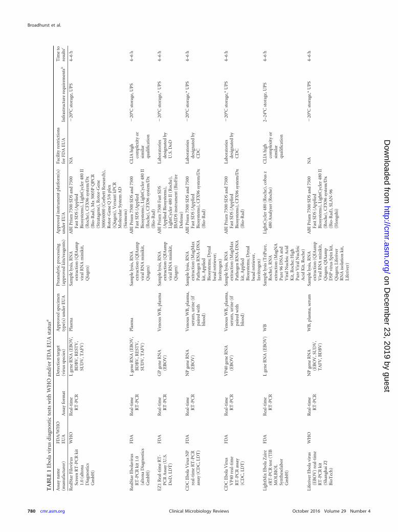
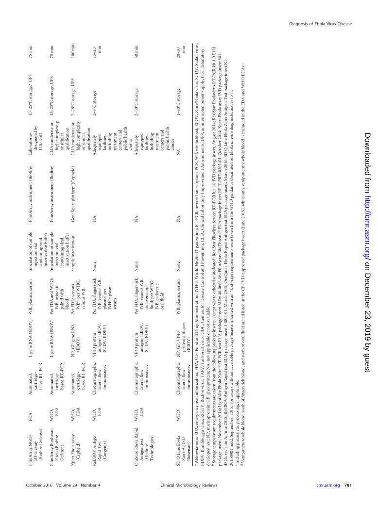
In response to these challenges, and in preparation for estab-lishing sustainable surveillance and rapid response capacity in af-fected countries, the WHO issued a Target Product Profile (TPP)for the development of safe, rapid, and cost-effective EVD diag-nostic tests that can be used at or near the point of care by locallaboratory technicians and health care workers (52). Test featuresprioritized in the TPP include the intended use in decentralizedhealth care facilities with no or minimal laboratory infrastructure,excellent performance characteristics (clinical sensitivity of �95%and specificity of �99%), use of minimally invasive diagnosticspecimens (e.g., capillary blood, oral fluid), and simple test pro-cedures (e.g., no or minimal preanalytic sample processing, min-imal timed and overall procedural steps, preferably no precisevolume transfers, and an integrated internal control). In addition,the TTP describes desirable operational test characteristics, in-cluding long-term reagent stability under tropical conditions withno cold chain requirements, small and portable equipment withminimal or no power requirements or need for maintenance, andminimal training needs. Novel Ebola virus diagnostic tests havenow reached the field, and include automated nucleic acid ampli-fication tests (NAATs) as well as rapid antigen detection tests (53).Those that have received WHO and/or FDA EUA status at thetime of this article are described in Table 1. No ELISA-based testsfor antigen or antibody detection are currently approved for acuteEVD diagnosis.
Of note, all tests with FDA EUA status are approved for “pre-sumptive” testing only; any positive presumptive Ebola virus testin the United States must be confirmed at the CDC (when per-formed at the CDC, the CDC Ebola virus NP and VP40 RT-PCRassays may currently be used for confirmatory testing, although acombination of testing modalities may be employed) (54). Ebolavirus is listed as a Tier 1 Select Agent by the U.S. Department ofHealth and Human Services (55); once a patient specimen hasbeen confirmed to contain infectious Ebola virus by viral culture,all clinical specimens from that patient are subject to Select Agentregulations and must be appropriately destroyed, decontami-nated, or transferred to a Select Agent facility until the patient isshown to have cleared the infection (56). In the United Kingdom,confirmation of infection by any recognized Ebola virus detectionmethod is sufficient to trigger restrictions on sample handling anddisposal (57). Other Western countries also have variants of theseprocedures to maintain the safety of health care workers and forbiosecurity. The WHO does not systematically designate “pre-sumptive” versus “confirmatory” tests in their EUAL; however,WHO guidance documents state that nucleic acid amplificationtests are preferred when feasible and that rapid antigen detectiontests should serve as “presumptive” or “screening” tests in remote
settings without access to immediate molecular testing or to assistin triaging high-risk patients when case loads are high (50, 58).When necessary, confirmatory testing may be performed by a rec-ognized national reference viral hemorrhagic fever (VHF) labora-tory; alternatively, specimens can be sent to one of the nine WHOCollaborating Centres for VHF (38). Therefore, country-specificstrategies for presumptive and confirmatory testing must be es-tablished that take into account the epidemiologic setting andavailable laboratory resources.
EVD Diagnostic Tests with Emergency Use Authorization
Standard (nonautomated) real-time RT-PCR tests. The RealStarFilovirus Screen RT-PCR kit 1.0 (altona Diagnostics GmBH; atwo-target multiplex assay for simultaneous detection of Ebolavirus [all species] and Marburg virus L gene targets; approved foruse in plasma specimens) was the first real-time RT-PCR test toreceive EUA status from the WHO (59) and was widely used byfield laboratories in the most recent epidemic. A laboratory eval-uation of this assay was carried out at the BNITM (this study isbriefly described in the WHO EUAL report [59]; however, detailsof the study have not been published). The analytic sensitivity ofthe RealStar Filovirus assay compared well with two laboratory-developed RT-PCR assays for RNA detection in plasma samplesspiked with RNA extracted from infected cell culture superna-tants, and no cross-reactivity to other viral hemorrhagic fever vi-ruses was observed. Of note, recent studies have raised concernthat the RealStar Filovirus assay, as deployed in the field, is notadequately sensitive (60, 61) (discussed below). A similar assayfrom the same manufacturer, RealStar Ebolavirus RT-PCR kit 1.0(altona Diagnostics GmBH; L gene detection for all Ebola virusspecies; approved for use in plasma specimens) received FDA EUAstatus (62). Data regarding the clinical performance of this assayhave not been published. The second RT-PCR test to be added tothe WHO EUAL was the Liferiver-Ebola virus (EBOV) real-timeRT-PCR kit (Shanghai ZJ BioTech; NP gene detection for EBOV,SUDV, TAFV, and BDBV; approved for use in whole blood[source not specified], plasma, and serum specimens). As de-scribed in the WHO EUAL report (63), the analytic sensitivity ofthis assay was evaluated at the BNITM using whole blood inocu-lated with infectious cell culture supernatants; data regarding clin-ical performance are not available.
Four other standard real-time RT-PCR tests have been grantedEUA status by the FDA: CDC Ebola virus NP and VP40 real-timeRT-PCR assays (U.S. CDC; EBOV NP and VP40 gene detection,respectively; approved for use with venous whole blood, plasma,serum, and urine) (64, 65), DoD EZ1 real-time RT-PCR assay(U.S. Department of Defense; EBOV GP gene detection [50]; ap-proved for use with venous whole blood and plasma) (66), and theLightMix Ebola virus Zaire test (Roche; EBOV L gene detection;approved for use with whole blood [source not specified]) (67).Per the FDA authorizations (64–66), use of the U.S. CDC andDoD assays is restricted to facilities designated by these agencies;thus, these assays are not commercially available to clinical labo-ratories. Deployment of the CDC NP and VP40 assays in a fieldlaboratory in Sierra Leone was recently described (68), althoughan evaluation of assay performance in this setting has not beenprovided. Data regarding clinical performance of the EZ1 andLightMix assays are not available.
Ancillary requirements for each of the standard real-time RT-PCR tests discussed above include capacity for cold chain main-
Diagnosis of Ebola Virus Disease
October 2016 Volume 29 Number 4 cmr.asm.org 779Clinical Microbiology Reviews
on Decem
ber 23, 2019 by guesthttp://cm
r.asm.org/
Dow
nloaded from
TA
BLE
1E
bola
viru
sdi
agn
osti
cte
sts
wit
hW
HO
and/
orFD
AE
UA
stat
usa
Ass
ayn
ame
(man
ufa
ctu
rer)
FDA
/WH
OE
UA
Ass
ayfo
rmat
Det
ecti
onta
rget
(vir
us
spec
ies)
App
rove
dsp
ecim
enty
pe(s
)u
nde
rE
UA
Pre
anal
ytic
proc
essi
ng
(app
rove
dki
ts/r
eage
nts
)A
ppro
ved
inst
rum
ent
plat
form
(s)
un
der
EU
AFa
cilit
yre
stri
ctio
ns
for
FDA
EU
AIn
fras
tru
ctu
rere
quir
emen
tsb
Tim
eto
resu
ltsc
Rea
lSta
rFi
lovi
rus
Scre
enR
T-P
CR
kit
1.0
(alt
ona
Dia
gnos
tics
Gm
bH)
WH
OR
eal-
tim
eR
T-P
CR
Lge
ne
RN
A(E
BO
V,
BD
BV
,RE
STV
,SU
DV
,TA
FV)
Pla
sma
Sam
ple
lysi
s,R
NA
extr
acti
on(Q
IAam
pvi
ralR
NA
min
ikit
,Q
iage
n)
AB
IP
rism
7500
SDS
and
7500
Fast
SDS
(App
lied
Bio
syst
ems)
,Lig
htC
ycle
r48
0II
(Roc
he)
,CFX
96sy
stem
/Dx
(Bio
-Rad
),M
x30
05P
QP
CR
(Str
atag
ene)
,Rot
or-G
ene
3000
/600
0(C
orbe
ttR
esea
rch
),R
otor
-Gen
eQ
5/6
plex
(Qia
gen
),V
ersa
nt
kPC
RM
olec
ula
rSy
stem
AD
(Sie
men
s)
NA
�20
°Cst
orag
e,U
PS
4–6
h
Rea
lSta
rE
bola
viru
sR
T-P
CR
kit
1.0
(alt
ona
Dia
gnos
tics
Gm
bH)
FDA
Rea
l-ti
me
RT
-PC
RL
gen
eR
NA
(EB
OV
,B
DB
V,R
EST
V,
SUD
V,T
AFV
)
Pla
sma
Sam
ple
lysi
s,R
NA
extr
acti
on(Q
IAam
pvi
ralR
NA
min
ikit
,Q
iage
n)
AB
IP
rism
7500
SDS
and
7500
Fast
SDS
(App
lied
Bio
syst
ems)
,Lig
htC
ycle
r48
0II
(Roc
he)
,CFX
96sy
stem
/Dx
(Bio
-Rad
)
CLI
Ah
igh
com
plex
ity
orsi
mila
rqu
alifi
cati
on
�20
°Cst
orag
e,U
PS
4–6
h
EZ
1R
eal-
tim
eR
T-
PC
RA
ssay
(U.S
.D
oD,L
DT
)
FDA
Rea
l-ti
me
RT
-PC
RG
Pge
ne
RN
A(E
BO
V)
Ven
ous
WB
,pla
sma
Sam
ple
lysi
s,R
NA
extr
acti
on(Q
IAam
pvi
ralR
NA
min
ikit
,Q
iage
n)
AB
IP
rism
7500
Fast
SDS
(App
lied
Bio
syst
ems)
,Li
ghtC
ycle
r48
0II
(Roc
he)
,JB
AID
Sin
stru
men
t(B
ioFi
reD
efen
se)
Labo
rato
ries
desi
gnat
edby
U.S
.DoD
�20
°Cst
orag
e,*
UP
S4–
6h
CD
CE
bola
Vir
us
NP
real
-tim
eR
T-P
CR
assa
y(C
DC
,LD
T)
FDA
Rea
l-ti
me
RT
-PC
RN
Pge
ne
RN
A(E
BO
V)
Ven
ous
WB
,pla
sma,
seru
m,u
rin
e(i
fpa
ired
wit
hbl
ood)
Sam
ple
lysi
s,R
NA
extr
acti
on(M
agM
axP
ath
ogen
RN
A-D
NA
kit,
App
lied
Bio
syst
ems;
Dyn
albe
adre
trie
ver;
Invi
trog
en)
AB
IP
rism
7500
SDS
and
7500
Fast
SDS
(App
lied
Bio
syst
ems)
,CFX
96sy
stem
/Dx
(Bio
-Rad
)
Labo
rato
ries
desi
gnat
edby
CD
C
�20
°Cst
orag
e,*
UP
S4–
6h
CD
CE
bola
Vir
us
VP
40re
al-t
ime
RT
-PC
Ras
say
(CD
C,L
DT
)
FDA
Rea
l-ti
me
RT
-PC
RV
P40
gen
eR
NA
(EB
OV
)V
enou
sW
B,p
lasm
a,se
rum
,uri
ne
(if
pair
edw
ith
bloo
d)
Sam
ple
lysi
s,R
NA
extr
acti
on(M
agM
axP
ath
ogen
RN
A-D
NA
kit,
App
lied
Bio
syst
ems;
Dyn
albe
adre
trie
ver,
Invi
trog
en)
AB
IP
rism
7500
SDS
and
7500
Fast
SDS
(App
lied
Bio
syst
ems)
,CFX
96sy
stem
/Dx
(Bio
-Rad
)
Labo
rato
ries
desi
gnat
edby
CD
C
�20
°Cst
orag
e,*
UP
S4–
6h
Ligh
tMix
Ebo
laZ
aire
rRT
-PC
Rte
st(T
IBM
OLB
IOL
Syn
thes
elab
orG
mbH
)
FDA
Rea
l-ti
me
RT
-PC
RL
gen
eR
NA
(EB
OV
)W
BSa
mpl
ely
sis
(Tri
Pu
re,
Roc
he)
,RN
Aex
trac
tion
(Mag
NA
Pu
re96
DN
Aan
dV
iral
Nu
clei
cA
cid
Kit
,Roc
he;
Hig
hP
ure
Vir
alN
ucl
eic
Aci
dK
it,R
och
e)
Ligh
tCyc
ler
480
(Roc
he)
,cob
asz
480
An
alyz
er(R
och
e)C
LIA
hig
hco
mpl
exit
yor
sim
ilar
qual
ifica
tion
2–24
°Cst
orag
e,U
PS
4–6
h
Life
rive
rE
bola
viru
s(E
BO
V)
real
-tim
eR
T-P
CR
kit
(Sh
angh
aiZ
JB
ioT
ech
)
WH
OR
eal-
tim
eR
T-P
CR
NP
gen
eR
NA
(EB
OV
,SU
DV
,T
AFV
,BD
BV
)
WB
,pla
sma,
seru
mSa
mpl
ely
sis,
RN
Aex
trac
tion
(QIA
amp
Vir
alR
NA
min
ikit
,Q
iage
n;Q
IAam
pD
SPvi
rus
Spin
kit,
Qia
gen
;Lif
eriv
erR
NA
isol
atio
nki
t,Li
feri
ver)
AB
IP
rism
7500
SDS
and
7500
Fast
SDS
(App
lied
Bio
syst
ems)
,Lig
htC
ycle
r48
0II
(Roc
he)
,CFX
96sy
stem
/Dx
(Bio
-Rad
),SL
AN
-96
(Hon
gsh
i)
NA
�20
°Cst
orag
e,*
UP
S4–
6h
Broadhurst et al.
780 cmr.asm.org October 2016 Volume 29 Number 4Clinical Microbiology Reviews
on Decem
ber 23, 2019 by guesthttp://cm
r.asm.org/
Dow
nloaded from
Film
Arr
ayN
GD
SB
T-E
assa
y(B
ioFi
reD
efen
se)
FDA
Au
tom
ated
,ca
rtri
dge-
base
dR
T-P
CR
Lge
ne
RN
A(E
BO
V)
WB
,pla
sma,
seru
mIn
ocu
lati
onof
sam
ple
inje
ctio
nvi
alco
nta
inin
gvi
ral
inac
tiva
tion
buff
er
Film
Arr
ayin
stru
men
t(B
iofi
re)
Labo
rato
ries
desi
gnat
edby
U.S
.DoD
15–2
5°C
stor
age,
*U
PS
75m
in
Film
Arr
ayB
ioth
reat
-E
test
(Bio
Fire
Def
ense
)
WH
O,
FDA
Au
tom
ated
,ca
rtri
dge-
base
dR
T-P
CR
Lge
ne
RN
A(E
BO
V)
Per
FDA
and
WH
O:
WB
,uri
ne
(if
pair
edw
ith
bloo
d)
Inoc
ula
tion
ofsa
mpl
ein
ject
ion
vial
con
tain
ing
vira
lin
acti
vati
onbu
ffer
Film
Arr
ayin
stru
men
t(B
iofi
re)
CLI
Am
oder
ate
orh
igh
com
plex
ity
orsi
mila
rqu
alifi
cati
on
15–2
5°C
stor
age,
UP
S75
min
Xpe
rtE
bola
assa
y(C
eph
eid)
WH
O,
FDA
Au
tom
ated
,ca
rtri
dge-
base
dR
T-P
CR
NP
,GP
gen
eR
NA
(EB
OV
)P
erFD
A:v
enou
sW
Bd;p
erW
HO
:ve
nou
sW
B
Sam
ple
inac
tiva
tion
Gen
eXpe
rtpl
atfo
rm(C
eph
eid)
CLI
Am
oder
ate
orh
igh
com
plex
ity
orsi
mila
rqu
alifi
cati
on
2–28
°Cst
orag
e,U
PS
100
min
ReE
BO
VA
nti
gen
Rap
idT
est
(Cor
gen
ix)
WH
O,
FDA
Ch
rom
atog
raph
icla
tera
lflow
imm
un
oass
ay
VP
40pr
otei
nan
tige
n(E
BO
V,
SUD
V,B
DB
V)
Per
FDA
:fin
gers
tick
WB
,ven
ous
WB
,pl
asm
a;pe
rW
HO
:pla
sma,
seru
m
Non
eN
AA
dequ
atel
yeq
uip
ped
faci
litie
s,in
clu
din
gtr
eatm
ent
cen
ters
and
publ
ich
ealt
hcl
inic
s
2–8°
Cst
orag
e15
–25
min
Ora
Sure
Ebo
laR
apid
An
tige
nte
st(O
raSu
reT
ech
nol
ogie
s)
WH
O,
FDA
Ch
rom
atog
raph
icla
tera
lflow
imm
un
oass
ay
VP
40pr
otei
nan
tige
n(E
BO
V,
SUD
V,B
DB
V)
Per
FDA
:fin
gers
tick
WB
,ven
ous
WB
,ca
dave
ric
oral
flu
id;p
erW
HO
:W
B,c
adav
eric
oral
flu
id
Non
eN
AA
dequ
atel
yeq
uip
ped
faci
litie
s,in
clu
din
gtr
eatm
ent
cen
ters
and
publ
ich
ealt
hcl
inic
s
2–30
°Cst
orag
e30
min
SDQ
Lin
eE
bola
Zai
reA
g(S
DB
iose
nso
r)
WH
OC
hro
mat
ogra
phic
late
ralfl
owim
mu
noa
ssay
NP
,GP
,VP
40pr
otei
nan
tige
ns
(EB
OV
)
WB
,pla
sma,
seru
mN
one
NA
NA
1–40
°Cst
orag
e20
–30
min
aA
bbre
viat
ion
s:E
UA
,em
erge
ncy
use
auth
oriz
atio
n;F
DA
,U.S
.Foo
dan
dD
rug
Adm
inis
trat
ion
;WH
O,W
orld
Hea
lth
Org
aniz
atio
n;R
T-P
CR
,rev
erse
tran
scri
ptio
n-P
CR
;WB
,wh
ole
bloo
d;E
BO
V,Z
aire
/Ebo
lavi
rus;
SUD
V,S
uda
nvi
rus;
BD
BV
,Bu
ndi
bugy
ovi
rus;
RE
STV
,Res
ton
viru
s;T
AFV
,Tai
Fore
stvi
rus;
CD
C,C
ente
rsfo
rD
isea
seC
ontr
olan
dP
reve
nti
on;C
LIA
,Clin
ical
Labo
rato
ryIm
prov
emen
tA
men
dmen
ts;U
PS,
un
inte
rru
pted
pow
ersu
pply
;LD
T,l
abor
ator
y-de
velo
ped
test
;NP
,nu
cleo
prot
ein
;GP
,gly
copr
otei
n;N
A,n
otap
plic
able
orn
otav
aila
ble.
bSt
orag
ete
mpe
ratu
rere
quir
emen
tsar
eta
ken
from
the
follo
win
gpa
ckag
ein
sert
s,ex
cept
wh
ere
oth
erw
ise
indi
cate
d:R
ealS
tar
Filo
viru
sSc
reen
RT
-PC
Rki
t1.
0IV
Dpa
ckag
ein
sert
,Au
gust
2014
;Rea
lSta
rE
bola
viru
sR
T-P
CR
kit
1.0
EU
Apa
ckag
ein
sert
,Nov
embe
r20
14;L
igh
tMix
Ebo
laZ
aire
rRT
-PC
Rte
stE
UA
pack
age
inse
rtM
Dx
40-0
666-
96;F
ilmA
rray
Bio
Th
reat
-EE
UA
pack
age
inse
rtR
FIT
-PR
T-0
302-
01,O
ctob
er20
14;X
pert
Ebo
laas
say
IVD
pack
age
inse
rt30
1-48
26,r
evis
ion
A,J
un
e20
15;R
eEB
OV
An
tige
nR
apid
test
EU
Apa
ckag
ein
sert
1400
5-01
,Mar
ch20
15;O
raQ
uic
kE
bola
Rap
idA
nti
gen
test
EU
Apa
ckag
ein
sert
,Mar
ch20
16;S
DQ
Lin
eE
bola
Zai
reA
nti
gen
Tes
tpa
ckag
ein
sert
R1-
2015
0901
.indd
,Sep
tem
ber,
2015
.For
assa
ysw
ith
out
acce
ssib
lepa
ckag
ein
sert
s(m
arke
dw
ith
an*)
,sto
rage
requ
irem
ents
wer
eta
ken
from
the
WH
Ogu
idan
cedo
cum
ent
onE
bola
invi
tro
diag
nos
tic
assa
ys(5
1).
cIn
clu
din
gpr
ean
alyt
icpr
oces
sin
g,if
appl
icab
le.
dV
enip
un
ctu
rew
hol
ebl
ood,
swab
offi
nge
rsti
ckbl
ood,
and
swab
ofor
alfl
uid
are
alll
iste
din
the
CE
-IV
D-a
ppro
ved
pack
age
inse
rt(J
un
e20
15),
wh
ileon
lyve
nip
un
ctu
rew
hol
ebl
ood
isin
clu
ded
inth
eFD
Aan
dW
HO
EU
As.
Diagnosis of Ebola Virus Disease
October 2016 Volume 29 Number 4 cmr.asm.org 781Clinical Microbiology Reviews
on Decem
ber 23, 2019 by guesthttp://cm
r.asm.org/
Dow
nloaded from

tenance, collection and transport of venipuncture blood, samplelysis/inactivation, manual RNA extraction, operation of a thermo-cycler with fluorescence detection, and manual recording and re-porting of results. The estimated time to results including samplepreparation and analytic procedures is 4 to 6 h. Where applicable,data regarding the clinical performance of these tests in compari-son to novel assay platforms are described below.
Automated real-time RT-PCR tests. Several fully automatedPCR platforms have been developed in recent years that integratenucleic acid extraction, PCR amplification, and detection of reac-tion products, typically yielding results in 90 min or less. Theseplatforms are designed for use in more decentralized health caresettings and minimize manual processing, thereby improvingsafety and facilitating use by technicians with minimal training.However, these tests still require specialized instruments and ac-cessory laptop computers, which in turn require electrical powerand equipment maintenance.
The Xpert Ebola assay (Cepheid), which has EUA status fromboth the WHO and FDA (69, 70), is an automated, cartridge-based system for RNA extraction and real-time RT-PCR detectionof EBOV NP and GP genes. Patient samples (of note, venipunc-ture whole blood, swabs of fingerstick blood, and swabs of oralfluid are all listed in the package insert with CE-IVD approval[Xpert Ebola IVD package insert 301-4826, revision A, June 2015],while only venipuncture whole blood is included in the FDA andWHO EUAs) are placed directly into a prefilled sample reagentvial; an aliquot is then loaded into a cartridge and the test is run ona module-format GeneXpert instrument. Kit components for thistest require storage at 2 to 28°C. According to the most recentpackage insert (June 2015), 20 min in the sample reagent com-pletely inactivates up to 4.6 � 106 PFU of EBOV. Ideally, thiswould allow for subsequent sample processing to be carried out ona benchtop, facilitating use at or near the point of care; however,since blood viral loads in acutely ill patients can exceed 108 RNAcopies/ml (4), more information about the efficiency of viral in-activation in such specimens may be needed to consider use of theXpert platform outside a biocontainment laboratory. Notably, theGeneXpert system has been widely used in developing regions formolecular detection of M. tuberculosis complex, though deploy-ment has been limited primarily to district-level labs due in part tothe requirement for a stable and continuous power supply.
An evaluation of the analytical performance of the Xpert Ebolaassay showed a limit of detection of 73 viral genome copies/ml forinactivated virus, 1 PFU/ml for infectious virus, and 232 RNAcopies/ml (71). The analytic and clinical performance character-istics of the Xpert Ebola assay were further evaluated at the NICDin South Africa (72). This study presented a comparison of resultsobtained from the Xpert Ebola assay and a laboratory-developedreal-time RT-PCR assay (L gene target) performed on 281 frozenserum and plasma samples collected from EVD suspect patients inSierra Leone. Agreement between the Xpert Ebola assay and the Lgene RT-PCR assay (as performed on two different thermocy-clers) was 100% in specimens yielding CT values of �35 by the Lgene RT-PCR assay; discrepancies between tests were seen at CT
values of 35 to 45. Viral isolation in cell culture was successful for91/125 specimens that tested positive by either of the moleculartests; importantly, both the Xpert Ebola assay and the L gene RT-PCR assay detected 100% of specimens from which virus could berecovered in culture.
The field performance of the Xpert Ebola assay was recently
evaluated on whole blood and buccal swab samples submitted forroutine clinical RT-PCR testing in a field biocontainment labora-tory in Sierra Leone (73). Compared to the benchmark TrombleyRT-PCR assay (NP gene target) (74), the Xpert assay showed100% sensitivity with 95.8% specificity in fresh venipuncturewhole-blood specimens (n � 211; 22 Ebola virus-positive speci-mens), and 100% sensitivity with 100% specificity in buccal swabspecimens (mixture of fresh and frozen specimens; n � 64; 20Ebola virus-positive specimens). All but one of 8 discordantwhole-blood specimens (Trombley negative/Xpert positive) hadbeen collected from known EVD patients (who had previouslytested positive by the Trombley assay and were under monitoringfor viral clearance), suggesting a revised Xpert assay specificity of99.5%. Of note, a small fraction of specimens failed Xpert testingdue to endogenous/exogenous internal control failure or systemfailure, highlighting both the need for adequate sample and re-agent integrity and the potential requirement for technical exper-tise for troubleshooting and equipment maintenance. The XpertEbola assay performed similarly well in a study carried out at anEbola treatment center near Conakry, Guinea, showing 100% sen-sitivity with 96.0% specificity in fresh venipuncture whole-bloodspecimens (n � 218; 26 Ebola virus-positive specimens) com-pared to an in-house RT-PCR assay (NP gene target) used forroutine clinical diagnostic testing at the national reference labo-ratory at Gamal Abdel Nasser University of Conakry, located nearthe treatment center (75). All discordant specimens (benchmarkRT-PCR negative/Xpert positive) were obtained from knownEVD patients undergoing monitoring for viral clearance, againsuggesting a higher actual specificity for the Xpert assay and high-lighting the performance of this assay in the convalescent stage ofEVD. Importantly, this study demonstrated the feasibility of op-erating the Xpert platform at the site of patient care; however,biosafety and logistical concerns were noted.
The BioFire Defense FilmArray assays (FilmArray Biothreat-Etest and FilmArray NGDS BT-E assay) are automated real-timeRT-PCR tests for detection of the EBOV L gene. The FilmArrayBiothreat-E test has EUA status from both the FDA (76) andWHO (77), with approved use of whole blood (source not speci-fied) and paired urine specimens, while the FilmArray NGDSBT-E test has FDA EUA status (78) for use with whole blood(source not specified), plasma, and serum specimens. For bothFilmArray assays, a pouch preloaded with lyophilized reagents forRNA extraction and real-time RT-PCR is rehydrated, followed bypreparation of the patient sample in a sample injection vial con-taining sample lysis reagents. The contents of the sample injectionvial are then loaded into the pouch and run on a single-assayFilmArray instrument. Per the package insert (BioThreat-E EUAIFU RFIT-PRT-0302-01, October 2014), sample preparation andloading of the pouch are to be performed in a biosafety cabinet. Kitcomponents require storage at 15 to 25°C. Use of the FilmArrayNGDS BT-E test is restricted to laboratories designated by the U.S.DoD (78).
Several studies have evaluated the Filmarray Biothreat-E assayin clinical specimens. The largest of these studies, carried out inPublic Health England laboratories in both Sierra Leone and theUnited Kingdom, compared the results from venipuncture wholeblood tested by the FilmArray Biothreat-E test with paired plasmatested by routine clinical RT-PCR (Trombley assay) (79). The Bio-threat-E test demonstrated 84% sensitivity with 89% specificity inspecimens collected in Sierra Leone (n � 60; 25 Ebola virus-pos-
Broadhurst et al.
782 cmr.asm.org October 2016 Volume 29 Number 4Clinical Microbiology Reviews
on Decem
ber 23, 2019 by guesthttp://cm
r.asm.org/
Dow
nloaded from

itive specimens), and 75% sensitivity with 100% specificity inspecimens collected in the United Kingdom (n � 108; 4 Ebolavirus-positive specimens), compared to the Trombley RT-PCR. Ina smaller study, the Filmarray Biothreat-E assay was tested in clin-ical specimens collected from six EVD patients who were treatedin U.S. hospitals (Emory University and University of NebraskaMedical Center) and compared to clinical RT-PCR results (CDCNP2 and VP40 assays performed by the CDC Viral Special Patho-gens Branch) (80). The Biothreat-E assay showed 86% sensitivitywith 80% specificity compared to benchmark RT-PCR in whole-blood specimens tested by both assays (n � 27) and 89% sensitiv-ity with 75% specificity in urine specimens (n � 13). Twenty ad-ditional whole-blood specimens were tested against paired plasmaassayed by benchmark RT-PCR and showed 100% sensitivity with71% specificity. In a study conducted in a hospital-associated re-search laboratory in Sierra Leone (81), results obtained from on-site testing of whole-blood specimens from EVD suspect patientswith the Filmarray Biothreat-E assay were fully concordant withclinical RT-PCR results (CDC NP and VP40 assays) obtainedfrom plasma specimens collected at a later time and tested in CDCfield laboratories (5 EVD-positive patients and 57 EVD-negativepatients were tested by both assays). Finally, the Filmarray Bio-threat-E assay was utilized at an Ebola treatment center in Guineaon urine (n � 7) and saliva (n � 18) specimens collected fromEVD patients (confirmed by routine RT-PCR testing of venousblood; QuantiTect Probe RT-PCR [Qiagen] and RealStar Filovi-rus Type RT-PCR kit 1.0 [altona Diagnostics]) (82). All urine andsaliva specimens tested positive by the Biothreat-E test, demon-strating the utility of this platform in testing noninvasive speci-men types near the point of patient care.
Rapid antigen detection tests. Three EVD rapid diagnostictests (RDTs) have received WHO and/or FDA EUA status, all ofwhich are lateral flow immunoassays (LFIs) (Table 1). LFIs aredesigned for use at the point of care and have been successfullyused to diagnose other infectious diseases (e.g., HIV, malaria) inresource-poor settings. The ReEBOV Antigen Rapid Test kit(Corgenix, Inc.) was the first LFI for EVD to receive EUA status(both WHO and FDA) (83, 84). This test is a chromatographicdipstick immunoassay for detection of the Ebola virus VP40 ma-trix protein (EBOV, SUDV, BDBV). The FDA EUA allows for thetesting of whole blood (collected by either fingerstick or venipunc-ture) or plasma (83), while the WHO EUA lists whole blood(source not specified), plasma, and serum (84) as acceptable spec-imen types. When used at the bedside, a drop of fingerstick bloodis applied directly to the nitrocellulose test strip, and the strip isthen placed into a tube with buffer to initiate flow of the samplealong the test strip. If present in the sample, VP40 is captured bygold-labeled anti-VP40 antibodies, forming immune complexes thatare subsequently deposited along a stripe of anti-VP40 antibodiesprinted onto the dipstick at a specific location. The gold nanoparticlesproduce a pink-red line that can be visually interpreted in 15 to 25min. No electronic equipment is needed to operate the test, thoughreagents do require refrigeration for storage.
In a recent field validation study in Sierra Leone, the ReEBOVRDT was performed at the bedside on fingerstick blood samplesfrom suspected EVD patients presenting at Ebola care centers, inparallel with collection of venipuncture blood for clinical diagnos-tic testing (RealStar Filovirus Screen RT-PCR kit 1.0 [altona Di-agnostics]) performed in a field reference laboratory; separately,the RDT was also performed on venipuncture blood in the field
reference laboratory (61). This study demonstrated that the RDTwas feasible to perform in restricted patient care areas (red zones)by operators wearing full personal protective equipment and thatinteroperator agreement (for reading the RDT) was high. In bothpoint-of-care settings (n � 105; 28 Ebola virus-positive speci-mens) and reference laboratory settings (n � 277; 45 Ebola virus-positive specimens), the RDT demonstrated 100% sensitivity and92% specificity compared to clinical results obtained with the al-tona RealStar assay. Importantly, most of the EVD patients testedin this study had relatively high viral loads in blood, and the Real-Star assay as performed in the field laboratory during this studywas observed to have imperfect sensitivity (as mentioned above,others have made similar observations regarding the altona assay[60]). Comparison of altona to an alternative RT-PCR benchmarkassay (Trombley) indicated that the altona assay was falsely nega-tive in some samples with a CT of �30 according to the Trombleyassay, leading those authors to recognize that comparison of theReEBOV RDT to an imperfect reference standard (altona) had ledto overestimation of true RDT sensitivity and underestimation oftrue RDT specificity. Therefore, while these results suggest that theReEBOV RDT could be very useful as a rapid point-of-care test forEVD in high-risk populations, its performance in patients withlow viral loads (e.g., those presenting very early or very late in theirdisease course) remains to be ascertained.
The OraQuick Ebola Rapid Antigen Test (OraSure Technolo-gies, Inc.) is a similar chromatographic LFI (detection of VP40matrix protein [EBOV, SUDV, and BDBV]) that recently receivedFDA (85, 86) and WHO (87) EUA status. In addition to wholeblood (venipuncture or fingerstick specimens listed in the FDAEUA [85]; source not specified in the WHO EUA [87]), theOraQuick RDT is the first EVD diagnostic test to receive approvalfor use with cadaveric oral fluid (approved in EUAs from both theFDA [86] and the WHO [87]). For testing whole blood (eitherfingerstick or venous), the specimen is collected into a plastic mi-cropipette provided in the kit and applied to a sample port in anassay device containing the nitrocellulose test strip. Cadaveric oralfluid can be sampled directly by swabbing the oral mucosa withthe flat pad at the end of the collection device. Alternatively, fluidfrom an oral swab stored in viral transport medium can be col-lected into a provided micropipette and applied to the sampleport. The assay device is then placed into a prefilled vial of buffereddeveloper solution. The presence of Ebola virus antigens is visuallydetected by the deposition of gold-labeled antibodies bound toviral proteins along the test line, as described above for the ReE-BOV RDT. The test is interpreted as positive or negative after 30min (OraQuick Ebola EUA package insert, publication 3001-2812, March 2016); per the package insert, the intensity of the testline is not proportional to the amount of virus in the blood. Kitcomponents require storage at 2 to 30°C and can be used at 15 to40°C.
Limited published data are available to evaluate the clinicalperformance of the OraQuick RDT. The WHO EUA report de-scribes a retrospective analysis of frozen venipuncture wholeblood specimens collected from Ebola suspect patients in SierraLeone (87). In this study, the OraQuick RDT demonstrated 84%sensitivity (n � 25 Ebola virus-positive specimens) and 98% spec-ificity (n � 50 Ebola virus-negative specimens) compared to clin-ical real-time RT-PCR testing (RT-PCR assay not specified). Ofnote, the sensitivity of the assay was higher in samples with CT
values �25. The WHO EUA also describes a retrospective analysis
Diagnosis of Ebola Virus Disease
October 2016 Volume 29 Number 4 cmr.asm.org 783Clinical Microbiology Reviews
on Decem
ber 23, 2019 by guesthttp://cm
r.asm.org/
Dow
nloaded from

of frozen cadaveric oral fluid specimens collected in Sierra Leone(87). Compared to the results obtained by retesting with the XpertEbola assay, the OraQuick RDT demonstrated 94% sensitivity(n � 51 Ebola virus-positive specimens) and 100% specificity(n � 193 Ebola virus-negative specimens). Additionally, the pack-age insert (OraQuick Ebola EUA package insert, 3001-2812,March 2016) describes three studies conducted in West Africaevaluating RDT results obtained from direct sampling of cadav-eric oral fluid compared to routine RT-PCR performed on fluidcollected by oral swab and stored in viral transport medium. Allspecimens from these studies tested negative by RT-PCR (total n� 539), and the RDT showed 99 to 100% specificity across studies.The CDC recently evaluated the implementation of the OraQuickRDT for testing febrile patients (n � 1,000) presenting to primarycare facilities in Guinea who were considered to be at low risk forEVD (88). No patients tested positive by the RDT (RT-PCR test-ing was not performed in parallel for comparison). No informa-tion is yet available on the accuracy, operational feasibility, orinterreader reliability of this assay when conducted at the point ofcare in Ebola care centers.
The test most recently added to the WHO EUAL (89) is the SDQ Line Ebola Zaire Ag test (SD Biosensor, Inc.), a chromato-graphic LFI that simultaneously detects NP, GP, and VP40 anti-gens of EBOV in whole blood (source not specified), plasma, orserum. Gold-labeled mouse monoclonal antibodies form com-plexes with antigens present in the specimen and deposit alongthree antigen-specific test lines for visual detection. To performthe test, three drops of specimen are added to a sample port on theassay device, using a provided disposable dropper; alternatively,100 �l of specimen can be added using a precision pipette. The testis visually read at 20 to 30 min, with the appearance of any of thethree test lines interpreted as positive (SD Q Line Ebola Zaire Agtest package insert R1-20150901.indd, September 2015). Kit com-ponents can be stored between 1 and 40°C.
The SD Q Line Ebola Zaire Ag test was evaluated as part of alaboratory-based study in Sierra Leone comparing the perfor-mance of several antigen detection tests for EVD (a brief descrip-tion of the study is provided in the WHO EUAL report for the SDQ Line test [89]; a full account of the study has not been pub-lished). In this study, a total of 446 specimens (100 fresh venouswhole blood, 346 frozen plasma; 126 total Ebola virus-positivespecimens) were tested by rapid antigen detection tests in fourfield laboratories (the other tests included in the study were notspecified). Sensitivity and specificity were evaluated using the Re-alStar Filovirus Screen RT-PCR kit 1.0 (altona Diagnostics) as abenchmark assay. The SD Q Line Ebola Zaire Ag test demon-strated 84.9% sensitivity with 99.7% specificity. No studies haveyet evaluated the performance of this test at the point of care.
Specimen Management and Biosafety for DiagnosticTesting
Specimen collection and tracking. The current standard for real-time RT-PCR testing requires the collection of venipunctureblood into EDTA-coated blood tubes or, when blood collection isnot possible or in the setting of postmortem testing, the collectionof an oral swab into viral transport medium. Guidelines for safespecimen collection, packaging, and transport procedures alongwith lists of necessary materials are provided by the WHO (38, 90,91) and CDC (39). During the 2014-2015 epidemic, chronic re-source limitations and inadequate training often prevented adher-
ence to the WHO guidelines for sample collection and transport.A steady supply chain of the recommended venipuncture andpackaging materials was lacking, requiring phlebotomists to im-provise for these procedures. This compromised the safety of in-dividuals drawing and handling blood samples as well as the in-tegrity of the samples (e.g., hemolysis, cracked tubes, inadequatelabeling), hindering sample analysis and reporting of results. Fur-thermore, as EDTA tubes were often unavailable, testing was notinfrequently performed on serum using assays for which this spec-imen type was not approved. In the future, adequate support forspecimen management systems must be paired with analytic re-sources to enable safe and expeditious EVD diagnostic testing. Theability to use clinical specimens that entail less invasive sampling,such as fingerstick blood or oral fluid swabs, would alleviate someof these challenges. Moving forward, guidelines for the safe col-lection of alternative diagnostic specimens, such as urine and se-men, will be needed (see “Detection of Viral Persistence in Non-blood Body Fluids,” below).
In addition to good practices for sample collection, packaging,and transport, reliable specimen tracking and modes of commu-nication are imperative for accurate and efficient reporting of re-sults to clinicians. Early in the 2014-2015 epidemic, the use ofpatient name, age, and home town to identify specimens fre-quently led to confusion, as this information often did not provideunique patient identification. To address this problem in SierraLeone, suspected Ebola patients and dead bodies undergoingpostmortem screening were identified by local field surveillanceteams and allocated a case number, including a district identifica-tion code. This code was used by diagnostic laboratories as well aslocal and central epidemiology and case control networks to iden-tify each person and their associated samples when reporting re-sults externally. Within each laboratory, samples were identifiedby an internal lab number so that multiple samples from eachpatient could be recorded. Patients who were subsequently admit-ted to a treatment center would receive a hospital identificationnumber, but the district- and country-level data collection sys-tems for case surveillance and geographical distribution relied onthe original case identifier assigned by the surveillance team. Dailydata were collected centrally by the Ministry of Health and Sani-tation, assisted by staff from the CDC, WHO, the United King-dom and Sierra Leone Armed Forces, and staff from other na-tional and international agencies. Several efforts were made tosimplify this system, ranging from preprinted labels for use bylocal staff (with the idea that a set of labels would follow the patientto allow consistent identification of future records and samples) toelectronic medical record systems. In practice, traditional paperforms and identification numbers handwritten onto specimencontainers remained the norm across the network of laboratoriesand treatment centers. The implementation of a systematic caseidentification scheme dramatically improved specimen trackingand reporting; however, unacceptable delays in blood collectioncould occur when surveillance officers were not present to assigncase ID numbers. In these situations, it proved very useful to im-plement an alternative patient/specimen identification systemspecific for a clinical site (and thus managed by the clinical team).Finally, reliable internet connectivity is needed to facilitate thetimely reporting of results via email when treatment centers aresituated remotely from diagnostic laboratories. When such con-nectivity was not available, cell phones were used to convey resultsby text message. In a postoutbreak setting, strategies for efficient
Broadhurst et al.
784 cmr.asm.org October 2016 Volume 29 Number 4Clinical Microbiology Reviews
on Decem
ber 23, 2019 by guesthttp://cm
r.asm.org/
Dow
nloaded from

results communication that are both reliable and maintain patientprivacy must be considered.
Viral inactivation in diagnostic specimens. When utilizing di-agnostic methods that involve manual processing, viral inactiva-tion is critical for the safety of laboratory workers. In addition,rendering specimens noninfectious for high-biocontainmentpathogens allows for subsequent sample processing under BSL-2conditions, which greatly improves laboratory workflow andthroughput. Traditional viral inactivation methods for specimenssuspected to contain hemorrhagic fever viruses include chemicalinactivation, gamma irradiation, and heat treatment (92–94).However, gamma irradiation (typically using a 60Co gamma cell)is usually restricted to public health and research laboratories, andsome forms of chemical inactivation are considered unsafe forlaboratory workers (e.g., beta-propiolactone is potentially carci-nogenic). Regarding viral inactivation in specimens for EVD di-agnosis, careful consideration must be given to whether themethod is compatible with downstream diagnostic tests, with par-ticular concern for maintaining the sensitivity of viral RNA detec-tion by RT-PCR. The feasibility of any given method will varyaccording to the laboratory setting; however, methods involvingchemical inactivation and heat treatment are usually practical forhospital-associated clinical laboratories (54) and field laboratoriesdeployed during an Ebola outbreak (95).
Several studies have evaluated the efficiency of viral inactiva-tion (determined by viral culture of treated specimens) mediatedby common reagents used in the initial processing of specimensfor RT-PCR (commonly referred to as “sample lysis”). The first ofthese studies, carried out by USAMRIID (96), found that TRIzolLS reagent (Invitrogen; contains phenol and the chaotropic saltguanidine isothiocyanate) and buffer AVL (Qiagen; contains gua-nidine isothiocyanate) were each capable of inactivating EbolaZaire virus when the virus stock was diluted with the inactivatingreagent (4 parts reagent:1 part sample; 106 PFU/ml after dilution)and incubated for 10 min at room temperature. Of note, this studydid not test for the inactivation of virus present in whole blood orblood components; thus, the findings may not be generalizable toclinical specimens commonly used for EVD diagnosis. A laterstudy by the CDC (97) found that NC lysis buffer (Applied Bio-systems; contains guanidine hydrochloride) provided completeinactivation of Ebola Zaire virus (1.5 � 105 PFU/ml before dilu-tion) when used at a 3 part reagent:1 part sample dilution for 10min at room temperature, but not when used at the standarddilution for RNA extraction (1:1). By comparison, in the samestudy, Tripure reagent (Roche; contains phenol and a guanidinesalt) provided complete viral inactivation under the same condi-tions at a 1:1 dilution. Again, viral inactivation was not testedin whole blood or blood component specimens. Most recently,an important study from the Defense Science and TechnologyLaboratory (95) showed that buffer AVL, when used at a 4 partsreagent:1 part sample dilution for 10 min at room temperature,did not reliably inactivate Ebola Zaire virus (108 50% tissueculture infective doses [TCID50]/ml in marmoset serum or 106
TCID50/ml in murine blood before dilution). In addition, nei-ther ethanol (4:1 dilution, 10 min at room temperature) nor heat(60°C for 15 min) yielded complete inactivation of the murineblood samples. However, the murine blood samples were com-pletely inactivated following treatment with a combination of buf-fer AVL and ethanol or buffer AVL and heat treatment, with noloss of efficiency in downstream RNA extraction and RT-PCR.
Based on these findings, protocols were adopted in a Public HealthEngland field laboratory in Sierra Leone in which diagnostic se-rum and whole blood specimens were treated with a mixture ofbuffer AVL plus ethanol (11 parts reagents:1 part sample for 10min) or with buffer AVL (8:1 dilution for 10 min) followed by heat(60°C for 15 min) prior to downstream processing on the bench-top (95).
Ideally, viral inactivation methods compatible with ancillarytests that support the clinical management of suspected or con-firmed EVD patients should be defined and selected, includingtests for malaria infection, blood chemistry, complete bloodcount, and coagulation status. In 2005, the CDC published guide-lines recommending the treatment of serum specimens from sus-pected viral hemorrhagic fever patients with a combination ofheat (56°C) and chemical (Triton X-100; Sigma-Aldrich) inacti-vation methods prior to routine handling (treatment length notspecified) (98). This protocol was shown to be compatible with thedetection of Plasmodium falciparum by RT-PCR and with a com-monly used LFI (HRP-2 antigen detection; BinaxNOW; Alere)(99). However, heat treatment was shown to significantly dimin-ish the performance of other Plasmodium antigen detection tests(100). Of note, thin blood smear preparations for morphologicaldiagnosis of malaria are considered noninfectious for hemor-rhagic fever viruses following 15 min of methanol fixation (54,98); this method was recently shown to fully inactivate blood con-taining EBOV at 108 TCID50/ml (101). Heat treatment alone(60°C for 60 min) is compatible with the measurement of thermo-stable blood components, including electrolytes, glucose, and bloodurea nitrogen (94, 102). Data are lacking regarding methods for theinactivation of Ebola virus that are compatible with routine hemocy-tometry, measurement of liver enzymes, or evaluation of coagulationstatus. There is a need for updated formal guidelines in this arena thataccount for recent evidence and experience with viral inactivationmethods and that consider novel diagnostic platforms (both for EVDdiagnosis as well as ancillary testing) and alternative specimen types.When adequate viral inactivation methods are not available, testingmay be performed under BSL-3 biocontainment with the use of abiosafety cabinet and/or appropriate personal protective equipment.Modern closed-path analyzers also provide a high degree of safety ifused appropriately, since they are designed to protect operators fromblood-borne viruses and have suitable cleaning protocols. Smallerplatforms can be adapted for use in a simple cabinet in high-risksituations, and such analyzers were used in several laboratories in thelater stages of the outbreak to measure basic blood chemistry andhematology parameters (54, 103).
Detection of Viral Persistence in Nonblood Body Fluids
As discussed above, the demonstration of viral clearance fromblood is recommended for the discharge of EVD survivors backinto the community. However, viral persistence in other bodyfluids after clearance of viremia may have important implicationsfor disease transmission (104, 105), and laboratory testing in thiscontext is needed to support infection control efforts and to pro-vide appropriate counseling for survivors and their communities.
Viral persistence in seminal fluid. What appears to be the firstevidence that Ebola virus is secreted in seminal fluid was describedin a case report of a laboratory-acquired infection with SUDV thatoccurred during investigations of the 1976 Sudan outbreak (106);infectious virus was detected in semen as late as day 61 followingthe onset of symptoms. Studies of EVD survivors from later out-
Diagnosis of Ebola Virus Disease
October 2016 Volume 29 Number 4 cmr.asm.org 785Clinical Microbiology Reviews
on Decem
ber 23, 2019 by guesthttp://cm
r.asm.org/
Dow
nloaded from

breaks (Democratic Republic of Congo 1995, Gabon 1996, andUganda 2000) demonstrated that Ebola virus is frequently de-tected in seminal fluid after viremia has cleared, with infectiousvirus detected by viral culture in semen specimens collected as lateas day 82 following the onset of symptoms and viral RNA detectedby RT-PCR as late as day 101 (17, 28, 107, 108). These findingswere extended in a recent study of male EVD survivors in SierraLeone, in which semen collected from 46/93 (49%) survivors(ranging from 2 to 10 months following the onset of symptoms)tested positive by RT-PCR (CDC NP and VP40 RT-PCR assays) ina field diagnostic laboratory (109). This study demonstrated adecrease in the prevalence of viral RNA in seminal fluid specimensover time (9/9 [100%] of specimens collected 2 to 3 months aftersymptom onset, 24/40 [65%] of specimens collected 4 to 6 monthsafter symptom onset, and 11/43 [26%] of specimens collected 7 to9 months after symptom onset) as well as increasing CT valuesover time (suggesting dropping viral loads). In this study, viralRNA was detected in semen as late as 284 days (9 months) aftersymptom onset, and the earliest time point at which a semen spec-imen tested negative by RT-PCR was 128 days (4 months) aftersymptom onset. In line with these findings, a recent study inves-tigating viral persistence in a variety of body fluids collected fromEVD survivors in Sierra Leone (described in more detail under“Viral persistence in other body fluids,” below) reported the de-tection of viral RNA by RT-PCR (Trombley assay) in a semenspecimen collected 114 days following discharge from an Ebolatreatment unit (110). The presence of infectious virus in speci-mens collected from the survivor cohorts described in these recentstudies (determined by viral culture) has not yet been reported.Surrogate measures of infectivity based on detecting whole or in-tact virus genome are used for nonculturable viruses such as no-rovirus and could be applied to semen studies to obtain an indi-cation of likely infectious potential.
Despite the apparently high frequency of viral persistence inseminal fluid, the overall rate of sexual transmission of EVD isthought to be very low (109, 111). However, a case of male-to-female sexual transmission in Liberia 6 months after the onset ofsymptoms in the male patient was verified by viral sequencing(112), raising concern that sexual transmission events may occureven after an outbreak is declared to be over (defined by the WHOas 42 days following the resolution of the final case, as determinedby death or two negative RT-PCR results) (113). Accordingly, theWHO currently recommends that male survivors be offered theopportunity to test their semen for Ebola virus by RT-PCR start-ing at 3 months after the onset of illness (testing is not recom-mended in the first 3 months because all semen specimens shouldbe assumed to be positive in this time frame), with repeat testingevery month until two negative RT-PCR results are obtained(114). However, there are currently no guidelines addressing col-lection methods (attention to both biosafety and sample integrityissues is needed) or optimal testing strategies (e.g., viral inactiva-tion, sample processing, assay selection) of seminal fluid speci-mens from survivors. A recent study demonstrated that the DoDEZ1 RT-PCR assay performs similarly for whole blood and semenspecimens spiked with EBOV (115), but none of the EVD diag-nostic tests with EUA status are currently approved for use withseminal fluid specimens.
Viral persistence in other body fluids. Several small studiesand case reports of EVD survivors have documented the persis-tence of infectious virions and/or viral RNA in a variety of body
fluids after clearance of viremia. Persistence of culture-confirmedinfectious virus has been reported in breast milk at day 15 of illness(108), urine at day 26 of illness (116), and ocular fluid at week 14of illness (117); in each case, the finding of persistence was de-scribed for a single patient. Unpublished reports have linked twocases of Ebola in neonates to breast milk from an asymptomaticmother confirmed by PCR to harbor viral RNA (T. Brooks, per-sonal communication). More extensive evidence has accumulatedfor the persistence of viral RNA in body fluids, with detection byRT-PCR in sweat or skin swabs as late as day 40 of illness (15, 116),in vaginal secretions as late as day 33 of illness (15, 28, 118), inurine as late as day 30 of illness (116, 118), in rectal swabs or stoolas late as day 29 of illness (15, 28, 118), in conjunctival fluid as lateas day 28 of illness (28, 118), in saliva as late as day 24 of illness (15,118), and in amniotic fluid (timing unspecified) (119). Samplingmethods and testing strategies have not been consistent acrossstudies, and little is known about the correlation of positive RT-PCR results with risk of viral transmission. Regardless, the existingevidence strongly suggests that virus may persist in multiple bodyfluids for several weeks following the clearance of viremia. Impor-tantly, a recent study conducted at an EVD survivor clinic in SierraLeone showed that 555 body fluid specimens (105 oral swabs, 103axillary swabs, 92 conjunctival swabs, 69 urine specimens, 54 fore-head swabs, 21 vaginal swabs, and 17 rectal swabs) collected from112 EVD survivors (median of 142 days postdischarge from anEbola treatment unit; earliest specimens collected �40 days fol-lowing discharge) all tested negative by RT-PCR (110). The find-ings from this large survivor study are consistent with the datafrom prior outbreaks described above and suggest that body fluidsfrom non-immune-privileged sites are very unlikely to harborpersistent virus after 6 weeks from clearance of viremia. Of note,we emphasize that exposure to body fluids from immune-privi-leged sites (e.g., semen, breast milk, ocular fluid, and cerebrospi-nal fluid) may continue to confer a risk of transmission for anextended period of time. WHO guidelines currently state thatbreast milk from lactating EVD survivors may be tested by RT-PCR if desired by the patient, with retesting every 48 h for thosewho test positive until two consecutive negative results are ob-tained (114). The WHO recommends against testing vaginal fluid(114); recommendations regarding the testing of other body fluidshave not been provided. Validation of EVD diagnostic tests on awider variety of specimen types should be a priority, and guide-lines are needed to address specimen collection and biosafety is-sues for testing of alternative body fluid specimens.
Viral Sequencing in the 2014-2015 Epidemic
Viral sequencing tools hold the potential to benefit EVD diagnos-tic efforts on multiple fronts, including (i) de novo diagnosis in thesetting of an emerging outbreak, (ii) identification of viral strainsresponsible for new transmission chains in an ongoing outbreak,(iii) estimation of viral mutation rate, and (iv) analysis of theimpact of viral mutations on the performance of molecular diag-nostic tests. During the 2014-2015 epidemic, next-generation se-quencing (NGS) platforms enabled the characterization of largenumbers of viral genomes in a relatively short time frame (days toweeks), and advances in portable sequencing tools have made itpossible to acquire sequencing data acutely in field facilities. Initialsample-processing steps for current sequencing strategies parallelthose required for standard RT-PCR testing (i.e., viral inactivation,nucleic acid extraction, reverse transcription, and amplification);
Broadhurst et al.
786 cmr.asm.org October 2016 Volume 29 Number 4Clinical Microbiology Reviews
on Decem
ber 23, 2019 by guesthttp://cm
r.asm.org/
Dow
nloaded from

subsequent steps include the preparation of a sequencing library, gen-eration of raw sequence data, and data analysis. Effective field deploy-ment of portable sequencing devices has been achieved (120) and willlikely play an important role in surveillance and outbreak responseefforts moving forward; however, these efforts remain challenged bythe procedural complexity and the relatively low sensitivity for viraldetection of current sequencing methods.
The emergence of a new EBOV strain in Guinea was determinedby conventional Sanger sequencing of patient specimens collectedin early 2014, performed by WHO Collaborating Centres for VHFin France and Germany (49). As part of this initial effort, whole-genome sequences were characterized from three specimens. Sev-eral large studies using NGS techniques were conducted over thecourse of the 2014-2015 epidemic, representing an internationaleffort that ultimately provided a rich database of hundreds ofEBOV whole-genome sequences. The first of these studies, carriedout in a U.S. laboratory (Illumina platform), sequenced 99 EBOVgenomes in specimens collected from EVD patients in Sierra Le-one from May to June 2014, revealing evidence for a single trans-mission event from a viral reservoir followed by human-to-hu-man transmission (121). In a subsequent study carried out inChina (BGISEQ-100 platform; Ion Proton), EBOV whole-ge-nome sequences were obtained from 175 EVD patient specimenscollected in Sierra Leone from September to November 2014, re-vealing seven novel EBOV sublineages (122). A total of 232 addi-tional EBOV genomes were sequenced in the United States (Illu-mina platform) from patient specimens collected from June 2014to January 2015 in Sierra Leone (123); importantly, the studydemonstrated that no significant mutations had accumulated inknown primer sites for RT-PCR diagnostic tests. The most recentlarge sequencing study, performed in Europe (Illumina platform),provided 179 EBOV genomes from patient specimens collected inGuinea from March 2014 to January 2015 (124). The broad timerange captured in this data set allowed the authors to illustrate theemergence of two distinct EBOV lineages, one of which was re-stricted to Guinea in the early months of the epidemic and a sec-ond lineage which spread from Guinea to Liberia and Sierra Le-one. In an important effort to increase sequencing capacity withinWest Africa, an Illumina Miseq platform was established at theLiberian Institute for Biomedical Research in February 2015 withsupport from USAMRIID (125). This new genomic capacity hasallowed for ongoing monitoring of EBOV sequences within Libe-ria (125) and provided key evidence for a case of sexually trans-mitted EVD (112). Near-real-time sequencing was provided inSierra Leone by Cambridge University (using an Ion Torrent se-quencer) in the closing stages of the outbreak, and this facilitatedrapid contact tracing by identifying the probable location of ex-posure in patients who had traveled home when sick (A. Arias,submitted for publication).
Nanopore sequencing is a relatively new technology that offersadvantages compared to standard NGS methods, including longersequencing reads and the ability to perform real-time sequenceanalysis concurrently with data acquisition. The MinION device(Oxford Nanopore Technologies), a portable nanopore se-quencer, has been evaluated as a diagnostic tool for outbreak re-sponses. Via an unbiased, metagenomic approach for pathogenidentification on the MinION platform, EBOV was correctly iden-tified in RNA extracted from whole-blood specimens collectedduring the 2014 Democratic Republic of Congo Ebola outbreak inunder 6 h (126). The potential for deployment of the MinION
device in a resource-limited setting was demonstrated in a fielddiagnostic laboratory in Liberia, where it was utilized to sequenceEBOV genomes from eight clinical specimens (120). These studieslay an important foundation for the further development of field-ready sequencing tools with the capacity for rapid pathogen iden-tification and in-depth characterization.
SUMMARY AND FUTURE DIRECTIONS
Real-time RT-PCR testing is an accurate and high-throughputmodality and has become the standard for EVD diagnosis. Severalstandard (nonautomated) real-time RT-PCR tests are approvedfor emergency use by the WHO and FDA, and four of these (Re-alStar Filovirus, RealStar Ebolavirus, Liferiver, and LightMix as-says) are commercially available as kits. Diagnosis by standardreal-time RT-PCR in an outbreak setting requires field laborato-ries with substantial infrastructure, operation and maintenance ofcomplex equipment, and expertise in molecular techniques.While such resources were eventually deployed in the 2014-2015epidemic, their integration into sustainable regional laboratorycapacities for ongoing surveillance and response to future out-breaks will be a great challenge. Furthermore, the requirement forcollection and transport of venipuncture blood will continue toconfer additional safety and logistical hurdles. In order to facethese challenges, it is imperative that international partners worktogether with national health ministries to strengthen laboratorycapacity in regions where Ebola is endemic, including the devel-opment of practical improvements to pre- and postanalytic pro-cesses and the training of local laboratory technicians in moleculardiagnostic techniques, biosafety practices, and quality control.
Novel diagnostic platforms, such as automated NAATs andrapid antigen detection tests, that can be used in decentralizedhealth care settings with minimal laboratory infrastructure willlikely play a major role moving forward. More field data areneeded to establish the appropriate use of these novel tests. Exist-ing evidence suggests that RDTs, if thoughtfully integrated intotesting algorithms, could have an immediate impact as point-of-care tests in high-risk populations. Given the persistent challengesin obtaining expeditious RT-PCR results, a point-of-care RDTwith imperfect sensitivity and specificity (especially if used incombination with RT-PCR testing) still stands to confer substan-tial benefits to case management and infection control efforts andshould improve the utilization of limited clinical and public healthresources (127). Accordingly, current WHO guidelines recom-mend initial testing with an RDT when RT-PCR testing is notimmediately available and to assist in triage and case managementwhen clinical and laboratory resources are overwhelmed (58).Three commercially available RDTs (ReEBOV, OraSure, and SDQ Line tests; all are chromatographic LFIs) have received WHOand/or FDA EUA status. In addition, an LFI developed by theDefense Science and Technology Laboratory in the United King-dom performed well (100% sensitivity with 92% specificity com-pared to laboratory testing by RT-PCR) when performed by localstaff at patient bedsides in Sierra Leone (128), further supportingthe utility and feasibility of performing RDTs at the point of carein Ebola treatment centers. Several innovative RDT platforms arein development (53, 127, 129, 130, 131) and have shown promis-ing results in laboratory-based evaluations. Automated NAATs,such as the GeneXpert and Filmarray systems, that provide rapid,sample-to-answer results with minimal operator dependence orpotential for cross-contamination have great potential impacts;
Diagnosis of Ebola Virus Disease
October 2016 Volume 29 Number 4 cmr.asm.org 787Clinical Microbiology Reviews
on Decem
ber 23, 2019 by guesthttp://cm
r.asm.org/
Dow
nloaded from
however, their implementation in decentralized health care set-tings will require careful consideration of biosafety and opera-tional challenges (e.g., access to uninterrupted electricity, temper-ature control, and expertise for test implementation/qualitycontrol and equipment maintenance). As more clinical validationdata become available and practical experience is compiled, localregulatory agencies, in collaboration with the WHO, will be respon-sible for developing updated EVD testing algorithms specific for dif-ferent health care settings in both high-prevalence outbreak and low-prevalence surveillance scenarios. In addition to assay selection foracute diagnostic testing, further guidance is needed to optimize bio-safety practices (e.g., viral inactivation methods) and testing of alter-native specimen types for viral persistence in EVD survivors. Astesting algorithms and guidelines evolve, it remains a criticalresponsibility of health care agencies to ensure means for safe andefficient specimen management, tracking, and reporting.
ACKNOWLEDGMENT
We thank David Heymann (Public Health England) for his helpful com-ments during the preparation of the manuscript.
REFERENCES1. Strong JE, Grolla A, Jahrling PB, Feldmann H. 2006. Filoviruses and
arenaviruses, p 774 –790. In Detrick B, Hamilton RG, Folds JD (ed),Manual of molecular and clinical laboratory immunology, 7th ed. ASMPress, Washington, DC.
2. Towner JS, Rollin PE, Bausch DG, Sanchez A, Crary SM, Vincent M,Lee WF, Spiropoulou CF, Ksiazek TG, Lukwiya M, Kaducu F, Down-ing R, Nichol S. 2004. Rapid diagnosis of Ebola hemorrhagic fever byreverse transcription-PCR in an outbreak setting and assessment of pa-tient viral load as a predictor of outcome. J Virol 78:4330 – 4341. http://dx.doi.org/10.1128/JVI.78.8.4330-4341.2004.
3. Kortepeter MG, Bausch DG, Bray M. 2011. Basic clinical and laboratoryfeatures of filoviral hemorrhagic fever. J Infect Dis 204:S810 –S816. http://dx.doi.org/10.1093/infdis/jir299.
4. Schieffelin JS, Shaffer JG, Goba A, Gbakie M, Gire SK, Colubri A,Sealfon RS, Kanneh L, Moigboi A, Momoh M, Fullah M, Moses LM,Brown BL, Andersen KG, Winnicki S, Schaffner SF, Park DJ, YozwiakNL, Jiang PP, Kargbo D, Jalloh S, Fonnie M, Sinnah V, French I,Kovoma A, Kamara FK, Tucker V, Konuwa E, Sellu J, Mustapha I,Foday M, Yillah M, Kanneh F, Saffa S, Massally JL, Boisen ML, BrancoLM, Vandi MA, Grant DS, Happi C, Gevao SM, Fletcher TE, FowlerRA, Bausch DG, Sabeti PC, Khan SH, Garry RF, KGH Lassa FeverProgram, Viral Hemorrhagic Fever Consortium, WHO Clinical Re-sponse Team. 2014. Clinical illness and outcomes in patients with Ebolain Sierra Leone. N Engl J Med 371:2092–2100. http://dx.doi.org/10.1056/NEJMoa1411680.
5. Feldmann H, Sanchez A, Geisbert TW. 2013. Filoviridae: Marburg andEbola viruses, p 923–962. In Knipe EM, Howley PM (ed), Fields virology,6th ed. Lippincott Williams & Wilkins, Philadelphia, PA.
6. Johnson KM, Lange JV, Webb PA, Murphy FA. 1977. Isolation andpartial characterisation of a new virus causing acute haemorrhagic feverin Zaire. Lancet 310:569 –571. http://dx.doi.org/10.1016/S0140-6736(77)90719-X.
7. WHO International Study Team. 1978. Ebola hemorrhagic fever inSudan, 1976. Bull World Health Organ 56:247–270.
8. Wulff H, Lange J. 1975. Indirect immunofluorescence for the diagnosisof Lassa fever infection. Bull World Health Organ 52:429 – 436.
9. Ksiazek TG, West CP, Rollin PE, Jahrling PB, Peters CJ. 1999. ELISAfor the detection of antibodies to Ebola viruses. J Infect Dis 179:S192–S198. http://dx.doi.org/10.1086/514313.
10. Ksiazek TG, Rollin PE, Williams AJ, Bressler DS, Martin ML, Swane-poel R, Burt FJ, Leman PA, Khan AS, Rowe AK, Mukunu R, SanchezA, Peters CJ. 1999. Clinical virology of Ebola hemorrhagic fever (EHF):virus, virus antigen, and IgG and IgM antibody findings among EHFpatients in Kikwit, Democratic Republic of the Congo, 1995. J Infect Dis179:S177–S187. http://dx.doi.org/10.1086/514321.
11. Grolla A, Lucht A, Dick D, Strong JE, Feldmann H. 2005. Laboratory
diagnosis of Ebola and Marburg hemorrhagic fever. Bull Soc Pathol Exot98:205–209.
12. de La Vega M, Caleo G, Audet J, Qiu X, Kozak R, Brooks JI, Kern S,Wolz A, Sprecher A, Greig J, Lokuge K, Kargbo DK, Kargbo B, DiCaro A, Grolla A, Kobasa D, Strong JE, Ippolito G, Van Herp M,Kobinger GP. 2015. Ebola viral load at diagnosis associates with patientoutcome and outbreak evolution. J Clin Invest 125:4421– 4428. http://dx.doi.org/10.1172/JCI83162.
13. Saijo M, Niikura M, Ikegami T, Kurane I, Kurata T, Morikawa S. 2006.Laboratory diagnostic systems for Ebola and Marburg hemorrhagic fe-vers developed with recombinant proteins. Clin Vaccine Immunol 13:444 – 451. http://dx.doi.org/10.1128/CVI.13.4.444-451.2006.
14. Alpha Diagnostic International. 2016. Zaire-Ebola virus nucleoprotein(EBOV-NP) IgG (AE-320520-1) kit and IgM (AE-320530-1) ELISA kitproduct descriptions. Alpha Diagnostics International, Inc., San Anto-nio, TX.
15. Uyeki TM, Mehta AK, Davey RT, Liddell AM, Wolf T, Vetter P,Schmiedel S, Grunewald T, Jacobs M, Arribas JR, Evans L, Hewlett AL,Brantsaeter AB, Ippolito G, Rapp C, Hoepelman AIM, Gutman J,Working Group of the US-European Clinical Network on ClinicalManagement of Ebola Virus Disease Patients in the US and Europe.2016. Clinical management of Ebola virus disease in the United Statesand Europe. N Engl J Med 374:636 – 646. http://dx.doi.org/10.1056/NEJMoa1504874.
16. MacNeil A, Reed Z, Rollin P. 2011. Serologic cross-reactivity of humanIgM and IgG antibodies to five species of Ebola virus. PLoS Negl Trop Dis5:e1175. http://dx.doi.org/10.1371/journal.pntd.0001175.
17. Rowe AK, Bertolli J, Khan AS, Mukunu R, Muyembe-Tamfum JJ,Bressler D, Williams AJ, Peters CJ, Rodriguez L, Feldmann H, NicholST, Rollin PE, Ksiazek TG. 1999. Clinical, virologic, and immunologicfollow-up of convalescent Ebola hemorrhagic fever patients and theirhousehold contacts, Kikwit, Democratic Republic of the Congo. J InfectDis 179:S28 –S35. http://dx.doi.org/10.1086/514318.
18. Baize S, Leroy EM, Georges-Courbot MC, Capron M, Lansoud-Soukate J, Debre P, Fisher-Hich SP, McCormick JB, Georges AJ. 1999.Defective humoral responses and extensive intravascular apoptosis areassociated with fatal outcomes in Ebola virus-infected patients. Nat Med5:423– 426. http://dx.doi.org/10.1038/7422.
19. Leroy EM, Baize S, Volchkov VE, Fisher-Hoch SP, Georges-CourbotMC, Lansoud-Soukate J, Capron M, Debre P, McCorkmich JB,Georges AJ. 2000. Human asymptomatic Ebola infection and stronginflammatory response. Lancet 355:2210 –2215. http://dx.doi.org/10.1016/S0140-6736(00)02405-3.
20. Wauquier N, Becquart P, Gasquet C, Leroy EM. 2009. Immunoglob-ulin G in Ebola outbreak survivors, Gabon. Emerg Infect Dis 15:1136 –1137. http://dx.doi.org/10.3201/eid1507.090402.
21. Ksiazek TG, Rollin PE, Jahrling PB, Johnson E, Dalgard DW, PetersCJ. 1992. Enzyme immunosorbent assay for Ebola virus antigens in tis-sues of infected primates. J Clin Microbiol 30:947–950.
22. Niikura M, Ikegami T, Saijo M, Kurane I, Miranda ME, Morikawa S.2001. Detection of Ebola viral antigen by enzyme-linked immunosor-bent assay using a novel monoclonal antibody to nucleoprotein. J ClinMicrobiol 39:3267–3271. http://dx.doi.org/10.1128/JCM.39.9.3267-3271.2001.
23. Lucht A, Grunow R, Moller P, Feldmann H, Becker S. 2003. Devel-opment, characterization and use of monoclonal VP40-antibodies forthe detection of Ebola virus. J Virol Methods 111:21–28. http://dx.doi.org/10.1016/S0166-0934(03)00131-9.
24. Lucht A, Grunow R, Otterbein C, Moller P, Feldmann H, Becker S.2004. Production of monoclonal antibodies and development of an an-tigen capture ELISA directed against the envelope glycoprotein GP ofEbola virus. Med Microbiol Immunol 193:181–187. http://dx.doi.org/10.1007/s00430-003-0204-z.
25. Reusken C, Niedrig M, Pas S, Anda P, Baize S, Charrel R, Di Caro A,Drosten C, Fernandez-Garcia MD, Franco L, Gunther S, Leparc-Goffart I, Martina B, Pannetier D, Papa A, Sanchez-Seco MP, Va-palahti L, Koopmans M. 2014. Identification of essential outstandingquestions for an adequate European laboratory response to EbolavirusZaire West Africa 2014. J Clin Virol 72:124 –134.
26. Sanchez A, Ksiazek TG, Rollin PE, Miranda ME, Trappier SG, KhanAS, Peters CJ, Nichol ST. 1999. Detection and molecular characteriza-tion of Ebola viruses causing disease in human and nonhuman primates.J Infect Dis 179:S164 –S169. http://dx.doi.org/10.1086/514282.
Broadhurst et al.
788 cmr.asm.org October 2016 Volume 29 Number 4Clinical Microbiology Reviews
on Decem
ber 23, 2019 by guesthttp://cm
r.asm.org/
Dow
nloaded from

27. Leroy EM, Baize S, Lu CY, McCormick JB, Georges AJ, Georges-Courbot MC, Lansoud-Soukate Fisher-Hoch SP. 2000. Diagnosis ofEbola haemorrhagic fever by RT-PCR in an epidemic setting. J Med Virol60:463– 467. http://dx.doi.org/10.1002/(SICI)1096-9071(200004)60:4�463::AID-JMV15�3.0.CO;2-M.
28. Rodriguez LL, De Roo A, Guimard Y, Trappier SG, Sanchez A,Bressler D, Williams AJ, Rowe AK, Bertolli J, Khan AS, Ksiazek TG,Peters CJ, Nichol ST. 1999. Persistence and genetic stability of Ebolavirus during the outbreak in Kikwit, Democratic Republic of Congo,1995. J Infect Dis 179:S170 –S176. http://dx.doi.org/10.1086/514291.
29. Formenty P, Leroy EM, Epelboin A, Libama F, Lenzi M, Sudeck H,Yaba P, Allarangar Y, Boumandouki P, Nkounkou VB, Drosten C,Grolla A, Feldmann H, Roth C. 2006. Detection of Ebola virus in oralfluid specimens during outbreaks of Ebola virus hemorrhagic fever in theRepublic of Congo. Clin Infect Dis 42:1521–1526. http://dx.doi.org/10.1086/503836.
30. Towner JS, Sealy TK, Khristova ML, Albarino CG, Conlan S, ReederSA, Quan P, Lipkin WI, Downing R, Tappero JW, Okware S, LutwamaJ, Bakamutumaho B, Kayiwa J, Comer JA, Rollin PE, Ksiazek TG,Nichol ST. 2008. Newly discovered Ebola virus associated with hemor-rhagic fever outbreak in Uganda. PLoS Pathog 4:e1000212. http://dx.doi.org/10.1371/journal.ppat.1000212.
31. Gibb TR, Norwood DA, Woollen N, Henchal EA. 2001. Develop-ment and evaluation of a fluorogenic 5= nuclease assay to detect anddifferentiate between Ebola virus subtypes Zaire and Sudan. J ClinMicrobiol 39:4125– 4130. http://dx.doi.org/10.1128/JCM.39.11.4125-4130.2001.
32. Drosten C, Gottig S, Schilling S, Asper M, Panning M, Schmitz H,Gunther S. 2002. Rapid detection and quantification of RNA of Ebolaand Marburg viruses, Lassa virus, Crimean-Congo hemorrhagic fevervirus, Rift Valley fever virus, dengue virus, and yellow fever virus byreal-time reverse transcription-PCR. J Clin Microbiol 40:2323–2330.http://dx.doi.org/10.1128/JCM.40.7.2323-2330.2002.
33. Crowe SJ, Maenner MJ, Kuah S, Erickson BR, Coffee M, Knust M,Klena J, Foday J, Hertz D, Hermans V, Achar J, Calei GM, Van HerpM, Albarino CG, Amman B, Basile AJ, Bearden S, Belser JA, BergeronE, Blau D, Brault AC, Campbell S, Flint M, Gibbons A, Goodman C,McCullan L, Paddock C, Russell B, Salzer JS, Sanchez A, Sealy T, WantD, Saffa B, Turay A, Nichol ST, Towner JS. 2016. Prognostic indicatorsfor Ebola patient survival. Emerg Infect Dis 22:217–223. http://dx.doi.org/10.3201/eid2202.151250.
34. Fitzpatrick G, Vogt F, Moi Gbabai OB, Decroo T, Keane M, De ClerckH, Grolla A, Brechard R, Stinson K, Van Herp M. 2015. The contri-bution of Ebola viral load at admission and other patient characteristicsto mortality in a Medecins Sans Frontieres Ebola case management cen-tre, Kailahun, Sierra Leone, June-October 2014. J Infect Dis 212:1752–1758. http://dx.doi.org/10.1093/infdis/jiv304.
35. Lanini S, Portella G, Vairo F, Kobinger GP, Pesenti A, Langer M,Kabia S, Brogiato G, Amone J, Castilletti C, Miccio R, Zumia A,Capobianchi MR, Di Caro A, Strada G, Ippolito G, INMI-EmergencySierra Leone Study Group. 2015. Blood kinetics of Ebola virus in sur-vivors and nonsurvivors. J Clin Invest 125:4692– 4698. http://dx.doi.org/10.1172/JCI83111.
36. Li J, Duan H, Chen H, Ji Y, Zhang X, Rong Y, Xu Z, Sun L, Zhang J,Liu L, Jin B, Zhang J, Du N, Su H, Teng G, Yuan Y, Qin E, Jia H, WangS, Guo T, Want Y, Mu J, Yan T, Li A, Dong Z, Nie W, Jiang T, Li C,Gao X, Ji D, Zhuang Y, Li L, Wang L, Li W, Duan X, Lu Y, Sun Z,Kanu A, Koroma S, Zhao M, Ji J, Wang F. 2015. Age and Ebola viralload correlate with mortality and survival time in 288 Ebola virus diseasepatients. J Infect Dis 42:34 –39. http://dx.doi.org/10.1016/j.ijid.2015.10.021.
37. Spengler JR, McElroy Harmon JR, Stroher U, Nichol ST, SpiropoulouCF. 2015. Relationship between Ebola virus real-time quantitative poly-merase chain reaction-based threshold cycle value and virus isolationfrom human plasma. J Infect Dis 212:S346 –S349. http://dx.doi.org/10.1093/infdis/jiv187.
38. World Health Organization. 2014. Laboratory diagnosis of Ebola virusdisease. World Health Organization, Geneva, Switzerland. http://apps.who.int/iris/bitstream/10665/134009/1/WHO_EVD_GUIDANCE_LAB_14.1_eng.pdf.
39. Centers for Disease Control and Prevention. 2015. Guidance for col-lection, transport and submission of specimens for Ebola virus testing.
http://www.cdc.gov/vhf/ebola/healthcare-us/laboratories/specimens.html.
40. Cherpillod P, Schibler M, Vielle G, Cordey S, Mamin A, Vetter P,Kaiser L. 2016. Ebola virus disease diagnosis by real-time RT-PCR: acomparative study of 11 different procedures. J Clin Virol 77:9 –14. http://dx.doi.org/10.1016/j.jcv.2016.01.017.
41. Niedrig M, Schmitz H, Becker S, Gunther S, Meulen J, Meyer H,Ellerbrok H, Nitsche A, Gelderblom HR, Drosten C. 2004. First inter-national quality assurance study on the rapid detection of viral agents ofbioterrorism. J Clin Microbiol 42:1753–1755. http://dx.doi.org/10.1128/JCM.42.4.1753-1755.2004.
42. Kelly JD. 2014. Make diagnostic centres a priority for Ebola crisis. Na-ture 513:145. http://dx.doi.org/10.1038/513145a.
43. World Health Organization. 2015. Manual for the care and manage-ment of patients in Ebola care units/community care centers; interimemergency guidance. World Health Organization, Geneva, Switzerland.
44. Wolfel R, Stoecker K, Fleischmann E, Gramsamer B, Wagner M,Molkenthin P, Di Caro A, Gunther S, Ibrahim S, Genzel GH, Ozin-Hofsass AJ, Formenty P, Zoller L. 2015. Mobile diagnostics in outbreakresponse, not only for Ebola: a blueprint for a modular and robust fieldlaboratory. Euro Surveill 20:pii�30055. http://dx.doi.org/10.2807/1560-7917.ES.2015.20.44.30055.
45. World Health Organization. 2001. Outbreak of Ebola haemorrhagicfever, Uganda, August 2000-January 2001. Wkl Epidemiol Rec 76:41– 46.
46. Grolla A, Jones S, Kobinger G, Sprecher A, Girard G, Yao M, Roth C,Artsob H, Feldmann H, Strong JE. 2012. Flexibility of mobile labora-tory unit in support of patient management during the 2007 Ebola-Zaireoutbreak in the Democratic Republic of Congo. Zoonoses Public Health59:151–157. http://dx.doi.org/10.1111/j.1863-2378.2012.01477.x.
47. Centers for Disease Control and Prevention. 2016. Ebola outbreaks2000-2014. Centers for Disease Control and Prevention, Atlanta, GA.http://www.cdc.gov/vhf/ebola/outbreaks/history/summaries.html.
48. Grolla A, Jones SM, Fernando L, Strong JE, Stroher U, Moller P,Paweska JT, Burt F, Palma PP, Sprecher A, Formenty P, Roth C,Feldmann H. 2011. The use of a mobile laboratory unit in support ofpatient management and epidemiological surveillance during the 2005Marburg outbreak in Angola. PLoS Negl Trop Dis 5:e1183. http://dx.doi.org/10.1371/journal.pntd.0001183.
49. Baize S, Pannetier D, Oestereich L, Rieger T, Koivogui L, Magas-souba N, Soropogui B, Sow MS, Keita S, De Clerck H, Tiffany A,Dominguez G, Loua M, Traore A, Kolie M, Malano ER, Heleze E,Bocquin A, Mely S, Raoul H, Caro V, Cadar D, Gabriel M, Pahl-mann M, Tappe D, Schmidt-Chanasit J, Impourna B, Diallo AK,Formenty P, Van Herp M, Gunther S. 2014. Emergence of ZaireEbola virus disease in Guinea. N Engl J Med 371:1418 –1425. http://dx.doi.org/10.1056/NEJMoa1404505.
50. World Health Organization. 2015. Emergency guidance, selectionand use of Ebola in vitro diagnostic (IVD) assays. World HealthOrganization, Geneva, Switzerland. http://www.who.int/csr/resources/publications/ebola/ivd-assays/en/.
51. World Health Organization. 2014. Emergency use assessment and list-ing (EUAL) procedure for Ebola virus disease. World Health Organiza-tion, Geneva, Switzerland. http://www.who.int/diagnostics_laboratory/procurement/purchasing/en/.
52. World Health Organization. 2014. Target product profile for ZaireEbolavirus rapid, simple test to be used in the control of the Ebolaoutbreak in West Africa. World Health Organization, Geneva, Swit-zerland. http://www.who.int/medicines/publications/target-product-profile.pdf.
53. Perkins MD, Kessel M. 2015. What Ebola tells us about outbreak read-iness. Nat Biotechnol 33:464 – 469. http://dx.doi.org/10.1038/nbt.3215.
54. Centers for Disease Control and Prevention. 2015. Guidance for U.S.laboratories for managing and testing routine clinical specimens whenthere is a concern about Ebola virus disease. Centers for Disease Controland Prevention, Atlanta, GA. http://www.cdc.gov/vhf/ebola/healthcare-us/laboratories/safe-specimen-management.html.
55. Centers for Disease Control and Prevention, USDA. 2014. Select agentsand toxins list, Federal Select Agent Program. U.S. Department of Healthand Human Services, Washington, DC. http://www.selectagents.gov/SelectAgentsandToxinsList.html.
56. Centers for Disease Control and Prevention. 19 November 2014. In-terim guidance regarding compliance with select agent regulations forlaboratories handling patient specimens that are known or suspected to
Diagnosis of Ebola Virus Disease
October 2016 Volume 29 Number 4 cmr.asm.org 789Clinical Microbiology Reviews
on Decem
ber 23, 2019 by guesthttp://cm
r.asm.org/
Dow
nloaded from

contain Ebola virus. Centers for Disease Control and Prevention, At-lanta, GA. http://www.cdc.gov/vhf/ebola/healthcare-us/laboratories/select-agent-regulations.html.
57. Department of Health UK, Advisory Committee on Dangerous Patho-gens. 2015. Management of hazard group 4 viral haemorrhagic fevers andsimilar human infectious diseases of high consequence. Public Health Eng-land, London, England. https://www.gov.uk/government/publications/viral-haemorrhagic-fever-algorithm-and-guidance-on-management-of-patients.
58. World Health Organization. 2015. Interim guidance on the use of rapidEbola antigen detection tests. World Health Organization, Geneva, Swit-zerland. http://www.who.int/csr/resources/publications/ebola/ebola-antigen-detection/en/.
59. World Health Organization. 2014. Emergency use assessment andlisting for Ebola virus disease IVDs, public report, product: RealStarfilovirus screen RT-PCR kit 1.0. World Health Organization, Geneva,Switzerland. http://www.who.int/diagnostics_laboratory/procurement/purchasing/en/.
60. Janvier F, Gorbatch S, Queval L, Top J, Vigier C, Cotte J, Foissaud V.2015. Difficulties of interpretation of Zaire Ebola virus PCR results andimplication in the field. J Clin Virol 67:36 –37. http://dx.doi.org/10.1016/j.jcv.2015.04.001.
61. Broadhurst MJ, Kelly JD, Miller A, Semper A, Bailey D, GroppelliE, Simpson A, Brooks T, Hula S, Nyoni W, Sankoh AB, Kanu S,Jalloh A, Ton Q, Sarchet N, George P, Perkins MD, Wonderly B,Murray M, Pollock NR. 2015. ReEBOV antigen rapid test kit forpoint-of-care and laboratory-based testing for Ebola virus disease: afield validation study. Lancet 386:867– 874. http://dx.doi.org/10.1016/S0140-6736(15)61042-X.
62. U.S. Food and Drug Administration. 2014. Emergency use authoriza-tion, RealStar Ebolavirus RT-PCR kit 1.0; letter of authorization. U.S.Food and Drug Administration, Rockville, MD. http://www.fda.gov/MedicalDevices/Safety/EmergencySituations/ucm161496.htm#ebola.
63. World Health Organization. 2015. Emergency use assessment and listingfor Ebola virus disease IVDs, public report, product: Liferiver Ebola virus(EBOV) real time RT-PCR kit. World Health Organization, Geneva,Switzerland. http://www.who.int/diagnostics_laboratory/procurement/purchasing/en/.
64. U.S. Food and Drug Administration. 2015. Emergency use authorization,CDC Ebola virus NP real-time RT-PCR assay; letter of authorization. U.S.Food and Drug Administration, Rockville, MD. http://www.fda.gov/MedicalDevices/Safety/EmergencySituations/ucm161496.htm#ebola.
65. U.S. Food and Drug Administration. 2015. Emergency use authori-zation, CDC Ebola virus VP40 real-time RT-PCR assay; letter of au-thorization. U.S. Food and Drug Administration, Rockville, MD.http://www.fda.gov/MedicalDevices/Safety/EmergencySituations/ucm161496.htm#ebola.
66. U.S. Food and Drug Administration. 2014. Emergency use authoriza-tion, DoD EZ1 real-time RT-PCR assay; letter of authorization. U.S.Food and Drug Administration, Rockville, MD. http://www.fda.gov/MedicalDevices/Safety/EmergencySituations/ucm161496.htm#ebola.
67. U.S. Food and Drug Administration. 2014. Emergency use authoriza-tion, LightMix Ebola Zaire rRT-PCR test; letter of authorization. U.S.Food and Drug Administration, Rockville, MD. http://www.fda.gov/MedicalDevices/Safety/EmergencySituations/ucm161496.htm#ebola.
68. Flint M, Goodman CH, Beardoen S, Blau DM, Amman BR, Basile AJ,Belser JA, Bergeron E, Bowen MD, Brault AC, Campbell S,Chakrabarti AK, Dodd KA, Erickson BR, Freeman MM, Gibbons A,Guerrero LS, Klena JD, Lash RR, Lo MK, McMullan LK, Momoh G,Massally JL, Goba A, Paddock CD, Priestley RA, Pyle M, Rayfield M,Russell J, Salzer JS, Sanchez AJ, Schuh AJ, Sealy TS, Steinau M,Stoddard RA, Taboy C, Turnsek M, Wang D, Zemtsova GE, Zivcec M,Spiropoulou CF, Stroher U, Towner JS, Nichol ST, Bird BH. 2015.Ebola virus diagnostics: the US Centers for Disease Control and Preven-tion laboratory in Sierra Leone, August 2014 to March 2015. J Infect Dis212:S350 –S358. http://dx.doi.org/10.1093/infdis/jiv361.
69. World Health Organization. 2015. Emergency use assessment and list-ing for Ebola virus disease IVDs, public report, product: Xpert Ebolaassay. World Health Organization, Geneva, Switzerland. http://www.who.int/diagnostics_laboratory/procurement/purchasing/en/.
70. U.S. Food and Drug Administration. 2015. Emergency use authoriza-tion, Xpert Ebola assay; letter of authorization. U.S. ood and Drug Ad-
ministration, Rockville, MD. http://www.fda.gov/MedicalDevices/Safety/EmergencySituations/ucm161496.htm#ebola.
71. Pinsky BA, Sahoo MK, Sandlund J, Kleman M, Kulkarni M, GrufmanP, Nygren M, Kwiatkowski R, Baron EJ, Tenover F, Denison B,Higuchi R, Van Atta R, Beer NR, Carrillo AC, Naraghi-Arani P, MireCE, Ranadheera C, Grolla A, Lagerqvist N, Persing DH. 2015. Analyt-ical performance characteristics of the Cepheid GeneXpert Ebola assayfor the detection of Ebola virus. PLoS One 10:e0142216. http://dx.doi.org/10.1371/journal.pone.0142216.
72. Jansen van Vuren P, Grobbelaar A, Storm N, Conteh O, Konneh K,Kamara A, Sanne I, Paweska JT. 2016. Comparative evaluation ofthe diagnostic performance of the prototype Cepheid GeneXpertEbola assay. J Clin Microbiol 54:359 –367. http://dx.doi.org/10.1128/JCM.02724-15.
73. Semper A, Broadhurst MJ, Richards J, Foster G, Simpson A, LogueCH, Kelly JD, Miller A, Brooks TJG, Murray M, Pollock NR. 2016.Perfomance of the GeneXpert Ebola assay for diagnosis of Ebola virusdisease in Sierra Leone: a field evaluation study. PLoS Med 13:e1001980.http://dx.doi.org/10.1371/journal.pmed.1001980.
74. Trombley AR, Wachter L, Garrison J, Buckley-Beason VA, Jahrling J,Hensley LE, Schoepp RJ, Norwood DA, Goba A, Fair JN, Kulesh DA.2010. Comprehensive panel of real-time TaqMan polymerase chainreation assays for detection and absolute quantification of filoviruses,arenaviruses, and new world hantaviruses. Am J Trop Med Hyg 82:954 –960. http://dx.doi.org/10.4269/ajtmh.2010.09-0636.
75. Van den Bergh R, Chaillet P, Sow MS, Amand M, van Vyve C,Jonckheere S, Crestani R, Sprecher A, Van Herp M, Chua A, Piriou E,Koivogui L, Antierens A. 2015. Feasibility of Xpert Ebola assay in Me-decins Sans Frontieres Ebola Program, Guinea. Emerg Infect Dis 22:210 –216. doi 10.3201/eid2202.151238.
76. U.S. Food and Drug Administration. 2014. Emergency use authoriza-tion, FilmArray Biothreat-E test; letter of authorization. U.S. Food andDrug Administration, Rockville, MD. http://www.fda.gov/MedicalDevices/Safety/EmergencySituations/ucm161496.htm#ebola.
77. World Health Organization. 2015. Emergency use assessment and list-ing for Ebola virus disease IVDs, public report, product: FilmArray Bio-threat-E. World Health Organization, Geneva, Switzerland. http://www.who.int/diagnostics_laboratory/procurement/purchasing/en/.
78. U.S. Food and Drug Administration. 2015. Emergency use authoriza-tion, FilmArray NGDS BT-E assay; letter of authorization. U.S. Food andDrug Administration, Rockville, MD. http://www.fda.gov/MedicalDevices/Safety/EmergencySituations/ucm161496.htm#ebola.
79. Weller SA, Bailey D, Matthews S, Lumley S, Sweed A, Ready D,Eltringham G, Richards J, Vipond R, Lukaszewski R, Payne PM,Aarons E, Simpson AJ, Hutley EJ, Brooks T. 2016. Evaluation of theBiofire FilmArray Biothreat E-test (v2.5) for rapid identification of Ebolavirus disease in heat-treated blood samples obtained in Sierra Leone andUnited Kingdom. J Clin Microbiol 54:114 –119. http://dx.doi.org/10.1128/JCM.02287-15.
80. Southern TR, Racsa LD, Albarino CG, Fey PD, Hinrichs SH, MurphyCN, Herrera VL, Sambol AR, Hill CE, Ryan EL, Kraft S, CampbellSealy TK, Schuh A, Ritchie JC, Lyon ML, III, Mehta AK, Varkey JB,Ribner BS, Brantly KP, Stroher U, Iwen PC, Burd EM. 2015. Com-parison of FilmArray and quantitative real-time reverse transcriptasePCR for detection of Zaire ebolavirus from contrived and clinical speci-mens. J Clin Microbiol 53:2956 –2960. http://dx.doi.org/10.1128/JCM.01317-15.
81. Leski TA, Ansumana R, Taitt CR, Lamin JM, Bangura U, Lahai J,Mbayo G, Kanneh MB, Bawo B, Bockarie AS, Scullion M, Phillips CL,Horner CP, Jacobsen KH, Stenger DA. 2015. Use of the FilmArraysystem for detection of Zaire Ebolavirus in a small hospital in Bo, SierraLeone. J Clin Microbiol 53:2368 –2370. http://dx.doi.org/10.1128/JCM.00527-15.
82. Magassouba N, Gay-Andrieu F, Picot V, Peyrefitte C, Dacosta B,Bedin F, Dore A, Kourouma F, Machuron J, Longuet C, Phillips C,Scullion M, Miller M. 2016. Clinical performance of the FilmarrayBiothreat-E test for diagnosing Ebola virus disease in “alternate” speci-men types: urine and saliva. Open Forum Infect Dis 2(Suppl 1):S103.http://dx.doi.org/10.1093/ofid.ofv133.106.
83. U.S. Food and Drug Administration. 2015. Emergency use authoriza-tion, ReEBOV antigen rapid test; letter of authorization. U.S. Food andDrug Administration, Rockville, MD. http://www.fda.gov/MedicalDevices/Safety/EmergencySituations/ucm161496.htm#ebola.
Broadhurst et al.
790 cmr.asm.org October 2016 Volume 29 Number 4Clinical Microbiology Reviews
on Decem
ber 23, 2019 by guesthttp://cm
r.asm.org/
Dow
nloaded from

84. World Health Organization. 2015. Emergency use assessment and list-ing for Ebola virus disease IVDs, public report, product: ReEBOV anti-gen rapid test kit. World Health Organization, Geneva, Switzerland. http://www.who.int/diagnostics_laboratory/procurement/purchasing/en/.
85. U.S. Food and Drug Administration. 2015. Emergency use authoriza-tion, OraQuick Ebola rapid antigen test; letter of authorization. U.S.Food and Drug Administration, Rockville, MD. http://www.fda.gov/MedicalDevices/Safety/EmergencySituations/ucm161496.htm#ebola.
86. U.S. Food and Drug Administration. 2016. Emergency use authoriza-tion, OraQuick Ebola rapid antigen test; letter of authorization. U.S.Food and Drug Administration, Rockville, MD. http://www.fda.gov/MedicalDevices/Safety/EmergencySituations/ucm161496.htm#ebola.
87. World Health Organization. 2016. Emergency use assessment andlisting for Ebola virus disease IVDs; public report; product: OraQuickEbola rapid antigen test kit (cadaveric oral fluid and whole blood).World Health Organization, Geneva, Switzerland. http://www.who.int/diagnostics_laboratory/procurement/purchasing/en/.
88. Huang JY, Louis FJ, Dixon MG, Sefu M, Kightlinger L, Martel LD,Jayaraman GC, Gueye AS. 2016. Baseline assessment of the use of Ebolarapid diagnostic tests: Forecariah, Guinea, October–November 2015.MMWR Morb Mortal Wkly Rep 65:328 –329. http://dx.doi.org/10.15585/mmwr.mm6512a4.
89. World Health Organization. 2015. Emergency use assessment and list-ing for Ebola virus disease IVDs, public report, product: SD Q Line EbolaZaire Ag. World Health Organization, Geneva, Switzerland. http://www.who.int/diagnostics_laboratory/procurement/purchasing/en/.
90. World Health Organization. 2014. Laboratory: how to safely collectblood samples from persons suspected to be infected with highly infec-tious blood-borne pathogens (e.g., Ebola). World Health Organization,Geneva, Switzerland. http://who.int/csr/resources/publications/ebola/blood-collect-en.pdf.
91. World Health Organization. 2014. How to safely collect oral swabs(saliva) from deceased patients suspected to be infected with Ebola; in-terim guideline. World Health Organization, Geneva, Switzerland. http://who.int/csr/resources/publications/ebola/safely-collect-oral-swabs/en.pdf.
92. Freeman R, Codd AA, Selkon JB. 1982. Effect of beta-propriolactone inblood on routine haematological, biochemical, and serological investi-gations. Lancet 319:1048 –1049. http://dx.doi.org/10.1016/S0140-6736(82)92102-X.
93. Elliott LH, McCormick JB, Johnson KM. 1982. Inactivation of Lassa,Marburg, and Ebola viruses by gamma irradiation. J Clin Microbiol 16:704 –708.
94. Mitchell SW, McCormick JB. 1984. Physiochemical inactivation ofLassa, Ebola, and Marburg viruses and effect on clinical laboratory anal-yses. J Clin Microbiol 20:486 – 489.
95. Smither SJ, Weller SA, Phelps A, Eastaugh L, Ngugi S, O’Brien LM,Steward J, Lonsdale SG, Lever MS. 2015. Buffer AVL alone does notinactivate Ebola virus in a representative clinical sample type. J Clin Mi-crobiol 53:3148 –3154. http://dx.doi.org/10.1128/JCM.01449-15.
96. Blow JA, Dohm DJ, Negley DL, Mores CN. 2004. Virus inactivation bynucleic acid extraction reagents. J Virol Methods 119:195–198. http://dx.doi.org/10.1016/j.jviromet.2004.03.015.
97. Towner JS, Sealy TS, Ksiazek TG, Nichol ST. 2007. High-throughputmolecular detection of hemorrhagic fever virus threats with applicationsfor outbreak settings. J Infect Dis 196:S205–S212. http://dx.doi.org/10.1086/520601.
98. Centers for Disease Control and Prevention. 2005. Interim guidancefor managing patients with suspected viral hemorrhagic fever in U.S.hospitals. Centers for Disease Control and Prevention, Atlanta, GA.
99. Lau R, Wang A, Chong-Kit A, Ralevski F, Boggild A. 2015. Evaluationof Ebola virus inactivation procedures for Plasmodium falciparum ma-laria diagnostics. J Clin Microbiol 53:1387–1390. http://dx.doi.org/10.1128/JCM.00165-15.
100. Loutfy MR, Assmar M, Burgess DCH, Kain KC. 1998. Effects of viralhemorrhagic fever inactivation methods on the performance of rapiddiagnostic tests for Plasmodium falciparum. J Infect Dis 178:1852–1855.http://dx.doi.org/10.1086/314524.
101. Cutts T, Cook B, Poliquin G, Strong J, Theriault S. 2016. Inacti-vating Zaire Ebolavirus in whole-blood thin smears used for malariadiagnosis. J Clin Microbiol 54:1157–1159. http://dx.doi.org/10.1128/JCM.02960-15.
102. Bhagat CI, Lewer M, Prins A, Beilby JP. 2000. Effects of heating plasma
at 56°C for 30 min and at 60°C for 60 min on routine biochemistryanalytes. Ann Clin Biochem 37:802– 804. http://dx.doi.org/10.1258/0004563001899997.
103. Hunt L, Gupta-Wright A, Simms V, Tamba F, Knott V, Tambu K,Heisenberg-Mansaray S, Tambu E, Sheriff A, Conteh S, Smith T,Tobin S, Brooks T, Houlihan C, Cummings R, Fletcher T. 2015.Clinical presentation, biochemical, and haematological parameters andtheir association with outcome in patients with Ebola virus disease: anobservational cohort study. Lancet Infect Dis 15:1292–1299. http://dx.doi.org/10.1016/S1473-3099(15)00144-9.
104. Chughtai AA, Barnes M, Macintyre CR. 2016. Persistence of Ebola virusin various body fluids during convalescence: evidence and implicationsfor disease transmission and control. Epidemiol Infect 144:1652–1660.http://dx.doi.org/10.1017/S0950268816000054.
105. Thorson A, Formenty P, Lofthouse C, Broutet N. 2016. Systematicreview of the literature on viral persistence and sexual transmission fromrecovered Ebola survivors: evidence and recommendations. BMJ Open6:e008859. http://dx.doi.org/10.1136/bmjopen-2015-008859.
106. Emond RTD, Evans B, Bowen ETW, Lloyd G. 1977. A case of Ebolavirus infection. Br Med J 2:541–544. http://dx.doi.org/10.1136/bmj.2.6086.541.
107. Richards G, Murphy S, Reeve J, Mer M, Zinman C, Taylor R, Swane-poel R, Duse A, Sharp G, De La Rey I, Kassianides C. 2000. Unex-pected Ebola virus in a teriary care setting: clinical and epidemiologicalaspects. Crit Care Med 28:240 –244. http://dx.doi.org/10.1097/00003246-200001000-00041.
108. Bausch DG, Towner JS, Dowell SF, Kaducu F, Lukwiya M, Sanchez A,Nichol ST, Ksiazek TG, Rollin PE. 2007. Assessment of the risk of Ebolavirus transmission from bodily fluids and fomites. J Infect Dis 196:S142–S147. http://dx.doi.org/10.1086/520545.
109. Deen GF, Knust B, Broutet N, Sesay FR, Formenty P, Ross C, ThorsonAE, Massaquoi TA, Marrinan JE, Ervin E, Jambai A, McDonald SLR,Bernstein K, Wurie AH, Dumbuya MS, Abad N, Idriss B, Wi T,Bennett SD, Davies T, Ebrahim FK, Meites E, Naidoo D, Smith S,Banerjee A, Erickson BR, Brault A, Durski KN, Winter J, Sealy T,Nichol ST, Lamunu M, Stroher U, Morgan O, Sahr F. 14 October 2015.Ebola RNA persistence in semen of Ebola virus disease survivors: prelim-inary report. N Engl J Med http://dx.doi.org/10.1056/NEJMoa1511410.
110. Green E, Hunt L, Ross JCG, Nissen NM, Curran T, Badhan A,Sutherland KA, Richards J, Lee JS, Allen SH, Laird S, Blackman M,Collocott I, Parker PA, Walbridge A, Phillips R, Sellu SJ, Dama A,Sheriff AK, Sombo J, Ngegba D, Wurie AH, Checchi F, Brooks TJ. 16May 2016. Viraemia and Ebola virus secretion in survivors of Ebola virusdisease in Sierra Leone: a cross-sectional cohort study. Lancet Infect Dishttp://dx.doi.org/10.1016/S1473-3099(16)30060-3.
111. Sprecher A. 14 October 2015. Handle survivors with care. N Engl J Medhttp://dx.doi.org/10.1056/NEJMe1512928.
112. Mate SE, Kugelman JR, Nyenswah TG, Ladner JT, Wiley MR, Cordier-Lassalle T, Christie A, Schroth GP, Gross SM, Davies-Wayne GJ,Shinde SA, Murugan R, Sieh SB, Badio M, Fakoli L, Tawey F, de WitE, van Doremalen N, Munster VJ, Pettitt J, Prieto K, HumrighouseBW, Stroher U, DiClaro JW, Hensley LE, Schoepp RJ, Safronetz D,Fair J, Kuhn JH, Lackley DJ, Laney AS, Williams DE, Lo T, GasasiraA, Nichol ST, Formenty P, Kateh FN, De Cock KM, Bolay F, Sanchez-Lockhart M, Palacios G. 2015. Molecular evidence of sexual transmis-sion of Ebola virus. N Engl J Med 373:2448 –2454. http://dx.doi.org/10.1056/NEJMoa1509773.
113. World Health Organization. 2015. Criteria for declaring the end of theEbola outbreak in Guinea, Liberia or Sierra Leone. World Health Orga-nization, Geneva, Switzerland. http://www.who.int/csr/disease/ebola/declaration-ebola-end/en/.
114. World Health Organization. 2016. Clinical care for survivors of Ebolavirus disease; interim guidance; updated April 2016. World HealthOrganization, Geneva, Switzerland. http://www.who.int/csr/resources/publications/ebola/guidance-survivors/en/.
115. Pettitt J, Higgs ES, Adams RD, Jahrling PB, Hensley LE. 2016. Use ofexisting diagnostic reverse-transcription polymerase chain reaction as-says for detection of Ebola virus RNA in semen. J Infect Dis 213:1237–1239. http://dx.doi.org/10.1093/infdis/jiv454.
116. Kreuels B, Wichmann D, Emmerich P, Schmidt-Chanasit J, de Heer G,Kluge S, Sow A, Renne T, Cunther S, Lohse AW, Addo MM, Schmie-del S. 2014. A case of severe Ebola virus infection complicated by gram-
Diagnosis of Ebola Virus Disease
October 2016 Volume 29 Number 4 cmr.asm.org 791Clinical Microbiology Reviews
on Decem
ber 23, 2019 by guesthttp://cm
r.asm.org/
Dow
nloaded from

negative septicemia. N Engl J Med 371:2394 –2401. http://dx.doi.org/10.1056/NEJMoa1411677.
117. Varkey JB, Shantha JG, Crozier I, Kraft CS, Lyon M, Mehta AK,Kumar G, Smith JR, Kainulainen MH, Whitmer S, Stroher U, UyekiTM, Ribner BS, Yeh S. 2015. Persistence of Ebola virus in ocular fluidduring convalescence. N Engl J Med 372:2423–2427. http://dx.doi.org/10.1056/NEJMoa1500306.
118. Mora-Rillo M, Arsuaga M, Ramirez-olivencia G, de la Calle F,Borobia AM, Sanchez-Seco P, Lago M, Figueira JC, Fernandez-Puntero B, Viejo A, Negredo A, Nunez C, Flores E, Carcas AJ,Jimenez-Yuste V, Lasala F, Garcia-de Lorenzo A, Arnalich F, Arri-bas JR. 2015. Acute respiratory distress syndrome after convalescentplasma use: treatment of a patient with Ebola virus disease contractedin Madrid, Spain. Lancet Respir Med 3:554 –562. http://dx.doi.org/10.1016/S2213-2600(15)00180-0.
119. Baggi FM, Taybi A, Kurth A, Van Herp M, Di Caro A, Wolfel R,Gunther S, Decroo T, Declerch H, Jonckheere. 2014. Management ofpregnant women infected with Ebola virus in a treatment centre inGuinea, June 2014. Euro Surveill 19:pii�20983. http://dx.doi.org/10.2807/1560-7917.ES2014.19.49.20983.
120. Hoenen T, Groseth A, Rosenke K, Fischer RJ, Hoenen A, Judson SD,Martellaro C, Falzarano D, Marzi A, Squires RB, Wollenberg KR, deWit E, Prescott J, Safronetz D, van Doremalen N, Bushmaker T,Feldmann F, McNally K, Bolay FK, Fields B, Sealy T, Rayfield M,Nichol ST, Zoon KC, Massaquoi M, Munster VJ, Feldmann H. 2016.Nanopore sequencing as a rapidly deployable Ebola outbreak tool. EmergInfect Dis 22:331–334. http://dx.doi.org/10.3201/eid2202.151796.
121. Gire SK, Goba A, Andersen KG, Sealfon RSG, Park DJ, Kanneh L,Jalloh S, Momoh M, Fullah M, Dudas G, Wohl S, Moses L, YozwiakNL, Winnicki S, Matranga CB, Malboeuf CM, Qu J, Gladden AD,Schaffner SF, Yang Z, Jiang P, Nekoui M, Colubri A, Coomber MR,Fonnie M, Moigboi A, Gbakie M, Kamara FK, Tucher V, Konuwa E,Saffa S, Sellu J, Jalloh A, Kovoma A, Joninga J, Mustapha I, Kargbo K,Foday M, Yillah M, Kanneh F, Robert W, Massally JLB, Chapman SB,Bochicchio J, Murphy C, Musbaum C, Young S, Birren BW, Grant DS,Scheiffelin JS, Lander ES, Happi C, Gevao SM, Gnirke A, Rambaut A,Garry RF, Khan SH, Sabeti PC. 2014. Genomic surveillance elucidatesEbola virus origin and transmission during the 2014 outbreak. Science345:1369 –1372. http://dx.doi.org/10.1126/science.1259657.
122. Tong Y, Shi W, Liu D, Qian J, Liang L, Bo X, Liu J, Ren H, Fan H, NiM, Sun Y, Jin Y, Teng Y, Li Z, Kargbo D, Dafae F, Kanu A, Chen C,Lan Z, Jiang H, Luo Y, Lu H, Zhang X, Yang F, Hu Y, Cao Y, Deng Y,Su H, Sun Y, Lui W, Want Z, Wang C, Bu A, Guo Z, Zhang L, Nie W,Bai C, Sun C, An Z, Zu P, Zhang Z, Huang Y, Mi Z, Yu D, Yao H, FengY, Xia Z, Zheng X, Yang S, Lu B, Jiang J, Karbo B, He F, Gao G, CaoW. 2015. Genetic diversity and evolutionary dynamics of Ebola virus inSierra Leone. Nature 524:93–96. http://dx.doi.org/10.1038/nature14490.
123. Park DJ, Dudas G, Wohl S, Goba A, Whitmer SLM, Andersen KG,Sealfon RS, Ladner ST, Kugelman JR, Matranga CB, Winnicki SM, QuJ, Gire S, Gladden-Young A, Jalloh S, Nosamiefan D, Yozwiak NL,Moses LM, Jiang P, Lin A, Schaffner SF, Bird B, Towner J, Mamoh M,Gbakie M, Kanneh L, Kargbo D, Massally JLB, Kamara FK, KonuwaE, Sellu J, Jalloh AA, Mustapha I, Foday M, Yillah M, Erickson BR,Sealy T, Blau D, Paddock C, Brault A, Amman B, Basile J, Bearden S,Belser J, Bergeron E, Campbell S, Chakrabarti A, Didd K, et al. 2015.Ebola virus epidemiology, transmission, and evolution during seven
months in Sierra Leone. Cell 161:1516 –1526. http://dx.doi.org/10.1016/j.cell.2015.06.007.
124. Carroll MW, Matthews DA, Hiscox JA, Elmore MJ, Pollakis G,Rambaut A, Hewson R, Garcia-Dorival I, Bore JA, Koundouno R,Abdellati S, Afrough B, Aiyepada J, Akhilomen P, Asogun D,Atkinson B, Badusche M, Bah A, Bate S, Baumann J, Becker D,Becker-Ziaja B, Bocquin A, Borremans B, Bosworth A, Boettcher J,Cannas A, Carletti F, Castilletti C, Clark S, Colavita F, Diederich S,Donatus A, Duraffour S, Ehichioya D, Ellerbrok H, Fernandez-Garcia MD, Fizet A, Fleischmann E, Gryseels S, Hermelink A,Hinzmann J, Hopf-Guevara U, Ighodalo Y, Jameson L, KelterbaumA, Kis Z, Kloth S, et al. 2015. Temporal and spatial analysis of the2014-2015 Ebola virus outbreak in West Africa. Nature 524:97–102.http://dx.doi.org/10.1038/nature14594.
125. Kugelman JR, Wiley MR, Mate S, Ladner JT, Beitzel B, Fakoli L,Taweh F, Prieto K, Diclaro JW, Minogue T, Schoepp RJ, SchaecherKE, Pettitt J, Bateman S, Fair J, Kuhn JH, Hensley L, Park DJ, SebetiPC, Sanchez-Lockhart M, Bolay FK, Palacios G. 2015. Monitoring ofEbola virus Makona evolution through establishment of advancedgenomic capability in Liberia. Emerg Infect Dis 21:1135–1143. http://dx.doi.org/10.3201/eid2107.150522.
126. Greninger AL, Naccache SN, Federman S, Yu G, Mbala P, Bres V,Stryke D, Bouquet J, Somasekar S, Linnen JM, Dodd R, MulembakaniP, Schneider BS, Muyembe-Tamfum J, Stramer SL, Chiu C. 2015.Rapid metagenomic identification of viral pathogens in clinical samplesby real-time nanopore sequencing analysis. Genome Med 7:99 –112.http://dx.doi.org/10.1186/s13073-015-0220-9.
127. Nouvellet P, Garske T, Mills HL, Nedjati-Gilani G, Hinsley W, BlakeIM, Van Kerkhove MD, Cori A, Dorigatti I, Jombart T, Riley S, FraserC, Donnelly CA, Ferguson NM. 2015. The role of rapid diagnostics inmanaging Ebola epidemics. Nature 528:S109 –S116. http://dx.doi.org/10.1038/nature16041.
128. Walker NF, Brown CS, Youkee D, Baker P, Williams N, Kalawa A,Russell K, Samba AF, Bentley N, Koroma F, King MB, Parker BE,Thompson M, Boyles T, Healey B, Kargbo B, Bash-Taqi D, SimpsonAJ, Kamara A, Kamara TB, Lado M, Johnson O, Brooks T. 2015.Evaluation of a point-of-care blood test for identification of Ebola virusdisease at Ebola holding units, Western Area, Sierra Leone, January toFebruary 2015. Euro Surveill 20:pii�21073. http://dx.doi.org/10.2807/1560-7917.ES2015.20.12.21073.
129. Baca JT, Severns V, Lovato D, Branch DW, Larson RS. 2015. Rapiddetection of Ebola virus with a reagent-free point-of-care biosensor. Sen-sors 15:8605– 8614. http://dx.doi.org/10.3390/s150408605.
130. Yen C, Puig H, Tam JO, Gomez-Marquez J, Bosch I, Hamad-SchifferliK, Gehrke L. 2015. Multicolored silver nanoparticles for multiplexeddisease diagnostics: distinguishing dengue, yellow fever, and Ebola vi-ruses. Lab Chip 15:1638 –1641. http://dx.doi.org/10.1039/c5lc00055f.
131. Kurosaki Y, Magassouba N, Oloniniyi OK, Cherif MS, Sakabe S,Takada A, Hirayama K, Yasuda J. 2016. Development and evaluation ofreverse transcription-loop-mediated isothermal amplification (RT-LAMP) assay coupled with a portable device for rapid diagnosis of Ebolavirus disease in Guinea. PLoS Negl Trop Dis 10:e0004472. http://dx.doi.org/10.1371/journal.pntd.0004472.
132. World Health Organization. 2016. WHO Ebola situation report, 30March 2016. World Health Organization, Geneva, Switzerland. http://apps.who.int/ebola/current-situation/ebola-situation-report-30-march-2016.
Continued next page
Broadhurst et al.
792 cmr.asm.org October 2016 Volume 29 Number 4Clinical Microbiology Reviews
on Decem
ber 23, 2019 by guesthttp://cm
r.asm.org/
Dow
nloaded from

M. Jana Broadhurst, M.D., Ph.D., completedher M.D.-Ph.D. training at the University ofCalifornia, San Francisco, and is currently a res-ident physician in the Department of Pathologyat the Stanford University School of Medicine.Dr. Broadhurst served as the Lead for Diagnos-tics for Partners In Health in Sierra Leone dur-ing the 2014-2015 EVD epidemic. In this role,Dr. Broadhurst collaborated with the SierraLeone Ministry of Health and Sanitation andWHO to improve practices pertaining to speci-men collection and handling for EVD diagnosis at Partners In Health patientcare centers and worked extensively alongside local health care workerswithin these facilities. Dr. Broadhurst collaborated with international labo-ratory partners to facilitate mobile laboratory deployment within SierraLeone and served as the field lead for two studies evaluating novel EVDdiagnostic tests. Her long-term research interests include diagnostics de-velopment for parasitic diseases and immune regulation during chronic in-fection with helminth parasites.
Tim Brooks, FRCPath, CBE, is Head of the Rareand Imported Pathogens Laboratory (RIPL) atPublic Health England (PHE), which is based atPorton Down in the United Kingdom. RIPL is aWHO collaborating laboratory for High Conse-quence Pathogens and provides diagnostic andclinical advice for a wide range of unusual bac-terial and viral pathogens. Tim Brooks is one ofthe leading partners in the national ImportedFever Service, which combines the clinical skillsof the Liverpool and London Tropical Infec-tious Disease Hospitals with RIPL services. The IFS offers 24-hour service foracutely ill travellers arriving in the United Kingdom from anywhere in theworld. Dr. Brooks’ research interests range from environmental detection ofmicroorganisms and clinical diagnostics, through aerobiology and decon-tamination, to disease pathogenesis and work for the European SpaceAgency. He led the PHE Ebola laboratories in Sierra Leone and is workingwith local officials to establish regional laboratory services.
Nira R. Pollock, M.D., Ph.D., D(ABMM), is theAssociate Medical Director of the InfectiousDiseases Diagnostic Laboratory at Boston Chil-dren’s Hospital and a member of the Division ofInfectious Diseases at Beth Israel DeaconessMedical Center (Boston, MA). Dr. Pollock hasan active research program focused on the de-velopment and evaluation of diagnostic tests forinfectious diseases and related applications. Dr.Pollock’s diagnostics research has spanned arange of diseases, including Clostridium difficileinfection, active and latent tuberculosis, influenza, Lyme disease, and Ebolavirus disease (EVD), and has involved many different technologies, rangingfrom simple paper-based lateral flow and microfluidic platforms to novelautomated platforms for protein and nucleic acid detection. Duringthe 2014-2015 EVD outbreak, Dr. Pollock led two evaluations of novelEVD diagnostics in Sierra Leone, in collaboration with Partners In Health(Boston, MA) and Public Health England.
Diagnosis of Ebola Virus Disease
October 2016 Volume 29 Number 4 cmr.asm.org 793Clinical Microbiology Reviews
on Decem
ber 23, 2019 by guesthttp://cm
r.asm.org/
Dow
nloaded from