Ebola virus hf
30
Ebola Hemorrhagic Fever • Dr. Gopalrao Jogdand, M.D. Ph.D. • Professor & Head • Department of Community Medicine
-
Upload
chalmeda-anandrao-institute-of-medical-sciences -
Category
Healthcare
-
view
111 -
download
0
Transcript of Ebola virus hf

Ebola Hemorrhagic Fever
• Dr. Gopalrao
Jogdand, M.D.
Ph.D.
• Professor & Head
• Department of
Community
Medicine

Historical background
• The first outbreak of Ebola HF occurred in 1976, in
Zaire
• In the same year the disease broke out in Sudan
• It was characterized by high mortality
• In the recent past outbreaks were noticed in African
subcontinent till 1990
• In the new millennium (2014) severe outbreaks were
noticed in several African Countries
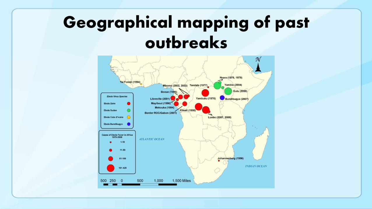
Geographical mapping of past outbreaks

Geographical distribution 2014

Ebola virusVirus belongs to the Filoviridae family.
Genus Ebola.
There are five strains of Ebola of which
four are pathological to humans.
They are Zaire Ebola Virus, Sudan virus,
Tai forest Virus, bundibugyo virus and
reston virus which do not cause human
disease.
The natural reservoir host is unknown.
The virus was first discovered in 1976 in
the Democratic Republic of Congo

Ebola Virus Transmission
Virus present in high quantity in blood, body fluids, and excreta of symptomatic EVD-infected patients
Opportunities for human-to-human transmission
Direct contact (through broken skin or unprotected mucous membranes) with an EVD-infected patient’s blood or body fluids
Sharps injury (with EVD-contaminated needle or other sharp)
Direct contact with the corpse of a person who died of EVD
Indirect contact with an EVD-infected patient’s blood or body fluids via a contaminated object (soiled linens or used utensils)
Ebola can also be transmitted via contact with blood, fluids, or meat of an infected animal
Limited evidence that dogs become infected with Ebola virus
No reports of dogs or cats becoming sick with or transmitting Ebola
6
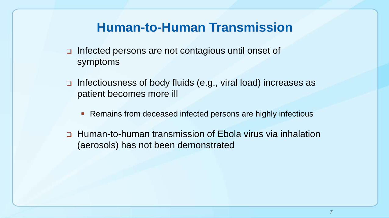
Human-to-Human Transmission
Infected persons are not contagious until onset of
symptoms
Infectiousness of body fluids (e.g., viral load) increases as
patient becomes more ill
Remains from deceased infected persons are highly infectious
Human-to-human transmission of Ebola virus via inhalation
(aerosols) has not been demonstrated
7

Ebola Virus Pathogenesis
Direct infection of tissues
Immune dysregulation
Hypovolemia and vascular collapse
Electrolyte abnormalities
Multi-organ failure, septic shock
Disseminated intravascular coagulation (DIC) and coagulopathy
Lancet. Mar 5, 2011; 377(9768): 849–862.
8


Early Clinical Presentation
Acute onset; typically 8–10 days after exposure
(range 2–21 days)
Signs and symptoms
Initial: Fever, chills, myalgias, malaise, anorexia
After 5 days: GI symptoms, such as nausea, vomiting, watery
diarrhea, abdominal pain
Other: Headache, conjunctivitis, hiccups, rash, chest pain,
shortness of breath, confusion, seizures
Hemorrhagic symptoms in 18% of cases
Other possible infectious causes of symptoms
Malaria, typhoid fever, meningococcemia, Lassa fever and other
bacterial infections (e.g., pneumonia) – all very common in Africa
10

Clinical Features
Nonspecific early symptoms progress to:
Hypovolemic shock and multi-organ failure
Hemorrhagic disease
Death
Non-fatal cases typically improve 6–11 days after
symptoms onset
Fatal disease associated with more severe early
symptoms
Fatality rates of 70% have been reported in rural Africa
Intensive care, especially early intravenous and electrolyte
management, may increase the survival rate
11

Clinical Manifestations by Organ System
in West African Ebola Outbreak
Organ System Clinical Manifestation
General Fever (87%), fatigue (76%), arthralgia (39%), myalgia (39%)
Neurological Headache (53%), confusion (13%), eye pain (8%), coma (6%)
Cardiovascular Chest pain (37%),
Pulmonary Cough (30%), dyspnea (23%), sore throat (22%), hiccups (11%)
Gastrointestinal Vomiting (68%), diarrhea (66%), anorexia (65%), abdominal pain
(44%), dysphagia (33%), jaundice (10%)
Hematological Any unexplained bleeding (18%), melena/hematochezia (6%),
hematemesis (4%), vaginal bleeding (3%), gingival bleeding
(2%), hemoptysis (2%), epistaxis (2%), bleeding at injection site
(2%), hematuria (1%), petechiae/ecchymoses (1%)
Integumentary Conjunctivitis (21%), rash (6%)
WHO Ebola Response team. NEJM. 2014
12

Examples of Hemorrhagic Signs
Bleeding at IV Site
Hematemesis
Gingival bleeding
13

Enhancement of Ebola virus infection

Laboratory Findings
Thrombocytopenia (50,000–100,000/mL range)
Leukopenia followed by neutrophilia
Transaminase elevation: elevation serum aspartate amino-
transferase (AST) > alanine transferase (ALT)
Electrolyte abnormalities from fluid shifts
Coagulation: PT and PTT prolonged
Renal: proteinuria, increased creatinine
15

Ebola Virus Diagnosis
Real Time PCR (RT-PCR)
Used to diagnose acute infection
More sensitive than antigen detection ELISA
Identification of specific viral genetic fragments
Performed in select CLIA-certified laboratories
RT-PCR sample collection
Volume: minimum volume of 4mL whole blood
Plastic collection tubes (not glass or heparinized tubes)
Whole blood preserved with EDTA is preferred
• Whole blood preserved with sodium polyanethol sulfonate (SPS), citrate, or with clot activator is
acceptable
16

Other Ebola Virus Diagnostics
Virus isolation
Requires Biosafety Level 4 laboratory;
Can take several days
Immuno-histochemical staining and histopathology
On collected tissue or dead wild animals; localizes viral antigen
Serologic testing for IgM and IgG antibodies (ELISA)
Detection of viral antibodies in
specimens, such as blood, serum,
or tissue suspensions
Monitor the immune response
in confirmed EVD patients
17

Packaging & Shipping Clinical Specimens to CDC for Ebola
Testing
http://www.cdc.gov/vhf/ebola/hcp/packaging-diagram.html
18

Clinical Management of EVD:
Supportive, but Aggressive
Hypovolemia and sepsis physiology Aggressive intravenous fluid resuscitation
Hemodynamic support and critical care management if
necessary
Electrolyte and acid-base abnormalities Aggressive electrolyte repletion
Correction of acid-base derangements
Symptomatic management of fever and gastrointestinal
symptoms Avoid NSAIDS
Multisystem organ failure can develop and may require Oxygenation and mechanical ventilation
Correction of severe coagulopathy
Renal replacement therapyReference: Fowler RA et al. Am J Respir Crit Care Med. 2014
19
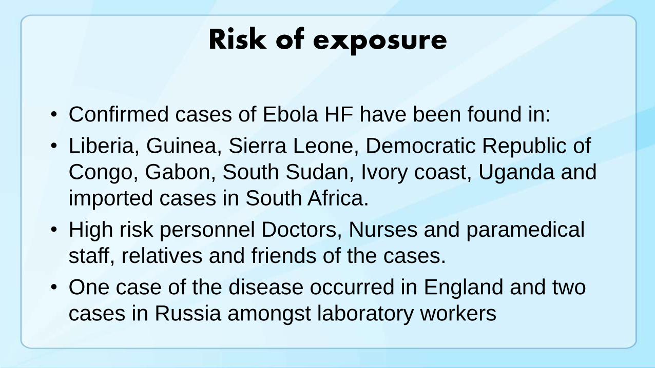
Risk of exposure
• Confirmed cases of Ebola HF have been found in:
• Liberia, Guinea, Sierra Leone, Democratic Republic of
Congo, Gabon, South Sudan, Ivory coast, Uganda and
imported cases in South Africa.
• High risk personnel Doctors, Nurses and paramedical
staff, relatives and friends of the cases.
• One case of the disease occurred in England and two
cases in Russia amongst laboratory workers

Threat to India
• Threat to our country is of Imported cases
• Nearly 47000 Indian nationals are working in Ebola
infested countries
• Since there is no vaccine or satisfactory treatment
against the disease
• Strict implementation of International health regulations
and quarantine procedure is mandatory
• Strict surveillance is advised by W.H.O. for travelers
coming from these African countries

Ebola (HF)
• It is postulated that the disease may be zoonotic one as it is found in Monkeys, Gorillas and chimpanzees.
• The onset is abrupt in nature, incubation period 2 to 21 days
• Mode of transmission: direct contact with the case, body fluids and secretions of the case
• Signs and Symptoms:
• Fever, headache, Joint and muscular pain, weakness, diarrhea, vomiting and stomach pain.
• Some patients may experience, rash, red eyes, hiccups, cough, sore throat, chest pain, difficulty in swallowing and breathing, internal and external bleeding

Diagnosis
• Within few days of onset of clinical symptoms:
• ELISA testing
• Later in the course of the disease and recovery:
• IgM and IgG anti bodies testing
• Retrospectively in deceased patients:
• Immunohistochemistry testing
• PCR
• Virus isolation

Treatment
• There is no satisfactory treatment or vaccine against
the disease
• Symptomatic treatment of cases is the only alternative
• Maintenance of fluid and electrolyte balance
• Maintenance of oxygen and blood pressure of the
patient
• Barrier nursing
• Isolation of the suspected and confirmed cases of the
disease

Prevention
• Isolation of suspected and confirmed cases
• Use of masks, gloves, gowns and goggles
• Health education of the population
• Health ADVISORY for travelers
• Social support mobilization
• Maintenance of environmental sanitation
• Strict implementation of surveillance procedure
• Rigid implementation of International Health Regulations
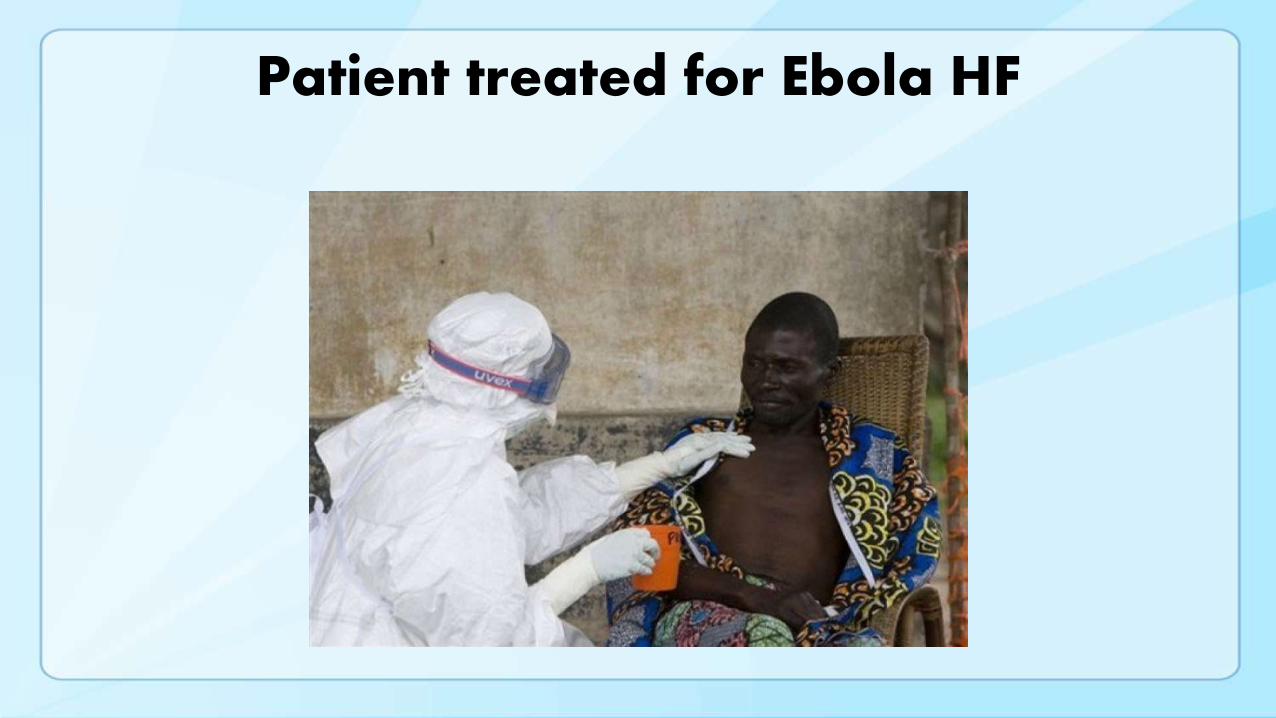
Patient treated for Ebola HF

Cutaneous bleeding

Barrier nursing

Health education

Thank you