Herpes simplex 2 how to test for herpes, can you cure herpes

1
t
Development of HSV-1 lacking the immunodominant gB498-505 epitope and analyses of the
alternative CD8+ T cell response in the murine ocular infection model
by
Sarah Bidula
B.S. Biology, Allegheny College, 2007
M.S. Biology, New York University, 2009
Submitted to the Graduate Faculty of
School of Medicine in partial fulfillment
of the requirements for the degree of
Master of Science
University of Pittsburgh
2012
2
UNIVERSITY OF PITTSBURGH
School of Medicine
This thesis was presented
by
Sarah Bidula
It was approved by
Robert L. Hendricks, Ph.D.,
Professor, Department of Ophthalmology
Frank J. Jenkins, Ph.D., Associate Professor, Department of Pathology
Russell D. Salter, Ph.D.,
Professor, Department of Immunology
Paul R. Kinchington, Ph.D. Dissertation Advisor
Professor, Department of Ophthalmology

3
Copyright © by Sarah Bidula
2012

4
Herpes simplex virus type 1 (HSV-1) establishes latent infections in sensory ganglia such
as the trigeminal ganglia (TG), and periodically reactivates to cause repeated corneal infections.
These may trigger an immune-mediated corneal disease known as herpes stromal keratitis
(HSK), which results in corneal scarring, and eventually blindness. HSK is the most common
infectious cause of blindness in the US, affecting over a quarter of a million people annually.
Herpes simplex virus type 1 (HSV-1) latent infections in trigeminal ganglia (TG) are associated
with persistent CD8+ T cell infiltrate. The ability of such CD8+ T cells to block HSV-1
reactivation from ex vivo ganglionic cultures establish the potential to control reactivation in
vivo. As such, targets of CD8+ T cells in the TG will guide vaccination strategies aimed to block
reactivation. In latently infected C57BL/6 mice, ~50% of the CD8+ T cell infiltrate in the TG is
directed to an immunodominant epitope (residues 498-505) on viral glycoprotein B (gB-CD8).
gB-CD8 can block reactivation from latency. Remaining CD8+
ABSTRACT
T cells in the TG are virus
Development of HSV-1 lacking the immunodominant gB498-505 epitope and analyses of
the alternative CD8+ T cell response in the murine ocular infection model
Sarah Bidula, M.S.
University of Pittsburgh, 2012
5
specific, can block reactivation, but are directed to unknown antigens. Given that CD8+ T cells
contribute to maintenance of the HSV-1 latent state, it is important to understand viral antigens
targeted by these cells and factors influencing dominance hierarchy. Here, we assessed the CD8+
T cell target specificity in TG of C57BL/6 mice infected with HSV-1 lacking and ectopically
restored for the gB498-505 epitope. Epitope mutants with near wild type pathogenicity were
isolated, and found to induce TG associated CD8+ T cell infiltrates of size similar to that
induced by wild type HSV-1, but with little gB498-505-specificity. The nature of the compensated
response reflected increase of CD8+ T cell populations directed to most known subdominant
epitopes seen in wild type HSV-1 infection. However, a gB498-505 dominated CD8+ T cell
response developed following infection with HSV with gB498-505 epitope-mutation that expressed
short gB494-509 peptides at an ectopic (gC) locus. We conclude that loss of the HSV dominant
epitope does not alter the size of the HSV-specific CD8+
T cell response nor broaden the TCR
repertoire, but rather results in a broader dominance hierarchy of subdominant epitopes rising to
co-dominance. We further conclude that immunodominance is not a result of properties of the
HSV-1 gB protein itself or its genomic locus.

6
TABLE OF CONTENTS
1.0 INTRODUCTION ........................................................................................................................................... 9
1.1.1 HSV-1 DISEASE AND EPIDEMIOLOGY ..................................................................................................................... 10
1.2 HSV-1 STRUCTURE ................................................................................................................................... 12
1.3 HSV-1 LIFE CYCLE ..................................................................................................................................... 13
1.3.1 ENTRY ........................................................................................................................................................................... 13
1.3.2 TEMPORAL GENE EXPRESSION ................................................................................................................................. 14
1.3.3 LATENCY ....................................................................................................................................................................... 16
1.3.4 REACTIVATION ............................................................................................................................................................ 18
1.4 IMMUNITY TO HSV-1 INFECTION ........................................................................................................ 19
1.5 MOUSE MODEL OF LATENCY ..................................................................................................................... 22
2.0 SPECIFIC AIMS ........................................................................................................................................... 25
2.1 RATIONALE ................................................................................................................................................ 25
2.2 SPECIFIC AIM 1 .......................................................................................................................................... 27
2.3 SPECIFIC AIM 2 .......................................................................................................................................... 28
3.0 GENERAL MATERIALS AND METHODS ............................................................................................. 29
3.1.2 MULTISTEP IN VITRO GROWTH KINETICS ................................................................................................................ 29
7
3.1.3 MICE .............................................................................................................................................................................. 30
3.1.4 TISSUE PREPARATION AND GANGLIA DISSOCIATION ............................................................................................. 30
3.1.5 CD8+ T CELL EXPANSION AND RECOGNITION OF MUTANT PROTEINS. ............................................................... 31
3.1.6 ACTIVATION ANALYSIS AND STAINING OF T CELLS ............................................................................................... 31
3.1.7 REAGENTS AND FLOW CYTOMETRY. ......................................................................................................................... 32
3.1.8 QUANTITATIVE REAL-TIME PCR .............................................................................................................................. 33
3.1.9 EX VIVO TG CULTURES FOR REACTIVATION ........................................................................................................... 34
4.0 GANGLIONIC CD8+ T CELL SPECIFICITY INDUCED BY HERPES SIMPLEX VIRUS TYPE 1
LACKING AND ECTOPICALLY RESTORED FOR THE GB498-505
MURINE IMMUNODOMINANT
EPITOPE ................................................................................................................................................................. 36
4.1 ABSTRACT .................................................................................................................................................. 37
4.2 INTRODUCTION .............................................................................................................................................. 38
4.3 MATERIALS AND METHODS ................................................................................................................. 42
4.4 RESULTS ...................................................................................................................................................... 47
4.5 DISCUSSION .................................................................................................................................................... 60
5.0 SUBDOMINANT CD8 + T CELL ACTIVATION PHENOTYPE DURING LATENCY IN THE
ABSENSE OF GB SPECIFIC CD8+ T CELLS ..................................................................................................... 65
5.1 ABSTRACT .................................................................................................................................................. 66
5.2 INTRODUCTION .............................................................................................................................................. 67
5.3 MATERIALS AND METHODS ................................................................................................................. 69
5.3.1 BRDU STAINING .......................................................................................................................................................... 69
5.4 RESULTS ...................................................................................................................................................... 71

8
5.5 DISCUSSION ................................................................................................................................................... 78
6.0 GENERAL DISCUSSION AND CONCLUSIONS ..................................................................................... 81
BIBLIOGRAPHY .................................................................................................................................................... 88
9
LIST OF FIGURES
Figure 1.1 Representation of the HSV-1 virion. ........................................................................... 12
Figure 1.2 HSV-1 gene expression kinetics ……………………………………………………..16
Figure 4.4.1 Construction of gBnull virus and of HSV-1 with gB498-505 mutations……………..45
Figure 4.4.2………………………………………………………………………………………48
Figure 4.4.3………………………………………………………………………………………50
Figure 4.4.4………………………………………………………………………………………51
Figure 4.4.5………………………………………………………………………………………53
Figure 4.4.6………………………………………………………………………………………54
Figure 4.4.7………………………………………………………………………………………56
Figure 4.4.8………………………………………………………………………………………58
Figure 4.4.9………………………………………………………………………………………59
Figure 5.4.1………………………………………………………………………………………72
Figure 5.4.2………………………………………………………………………………………73
Figure 5.4.3………………………………………………………………………………………75
Figure 5.4.4………………………………………………………………………………………76

10
1.0 INTRODUCTION
1.1 HERPES SIMPLEX VIRUS TYPE 1
1.1.1 HSV-1 Disease and Epidemiology
Herpes simplex virus type 1 (HSV-1) is a member of the Herpesviridae family, and can be
further classified as Alphaherpesvirinae, along with HSV-2 and varicella zoster virus (VZV). All
the human Alphaherpesviruses are neurotropic, infecting multiple cell types during primary and
reactivated disease and establishing latency in neurons sensory ganglia that innervate the site of
primary infection [1]. It is currently thought that around 80% of the world’s population is latently
infected with HSV-1 by adulthood [2,3,4]. In the United States, 57% of individuals between the
ages of 14 and 49 are seropositive for HSV-1 with the number rising to 90% by the age of 60
[5,6]. Reactivated disease can have variable presentation, ranging from asymptomatic viral
shedding to clinical presentation of cold sores, herpetic whitlow, genital lesions, more serious
diseases such as neonatal systemic infections, encephalitis as well as blinding stromal keratitis
[4].
11
1.1.2 Herpes Stromal Keratitis Disease
Herpes stromal keratitis (HSK) is caused by Herpes Simplex Virus Type-1 (HSV-1).
HSK is the most frequent and serious type of eye infection in the United States causing vision
loss in over a quarter of a million people annually [7]. HSK is the result of reactivation of HSV-
1 and subsequent viral reactivation at the corneal surface. This reactivation causes an immune
infiltrate in the eye that is initially aimed at controlling this HSV-1 replication but subsequently
shows lack of specificity to HSV-1 antigens. These immune cells release factors such as
interferon-γ (IFNγ) and interleukin-2 (IL-2), and other compounds, including nitrous oxide (NO)
that can cause destruction and disorganization of the collagen matrix of the stroma [8,9,10].
There is also a cascade of non-specific responses including neutrophils that recruit macrophages,
plasma cells, and CD4+
Despite advances in understanding immune regulation of HSV-1 infection, there is still
no vaccine. Current therapies for HSK include antiviral acyclovir drops and corticosteroids that
can help to reduce viral replication and inflammation [14,15]. However, these treatments are
only effecting during ongoing viral replication and HSK is still able to develop without the
presence of infectious virus in the cornea [16]. Once corneal blindness occurs, a corneal
transplant is the single option available with the possibility of restoring vision.
T cells that regulate proinflammatory cytokines [11,12]. It is the build-up
of scar tissue over time that ultimately results in a loss of vision from the cornea [11,13].
We argue that the only way to prevent HSK is to stop reactivation of HSV-1 and prevent
the resultant tissue scarring. The studies in this proposal address the expression of HSV-1

12
proteins and their immunogenicity during acute infection and latency with the aim of creating
vaccines designed to stop HSV-1 from reactivating.
1.2 HSV-1 STRUCTURE
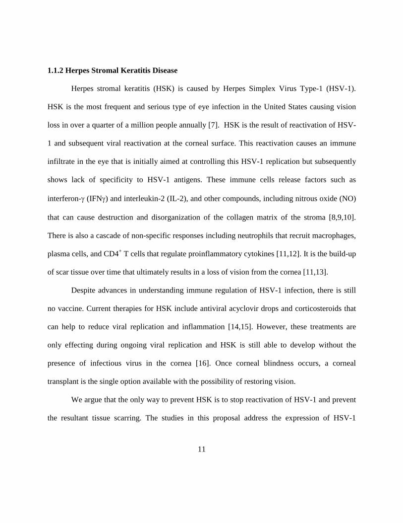
The HSV virion has four compartments: 1) an outer envelope that contains the glycoproteins, 2)
the tegument layer that contains about 20-30 viral proteins and some cellular proteins, 3) a capsid
layer, and 4) an electron core that encompasses the HSV genome (Figure 1.1) [4]. HSV-1
genome is a linear double stranded DNA of 152 kb genome that is packed inside of the
icosahedron shaped capsid. The genome can encode for the expression of about 80 viral proteins
[1,17].
Glycoprotein spikes
Envelope
Capsid
Tegument
Viral DNA
Figure 1.1 Representation of the HSV-1 virion.
13
The HSV-1 nucleocapsid contains one genome of double stranded viral DNA. The capsid is surrounded by a tegument layer which is enclosed within a host derived envelope containing viral glycoprotein spikes.
1.3 HSV-1 LIFE CYCLE
Most HSV-1 infections occur between the ages of 6 months and 3 years in the human population,
usually causing mild or asymptomatic primary infections. The life cycle of HSV-1 is defined in
three types: lytic replication, latency, and reactivation. Here lytic infection will be considered
first.
1.3.1 Entry
Viral replication begins when HSV-1 is able to infect and enter into a permissible host cell.
There are three viral glycoproteins (gB, gC, and gD) that mediate viral attachment to the host cell
but others may be involved. Glycoprotein C mediates the initial attachment of the HSV-1 virion
by binding to heparin sulfate moieties on the cell surface [18,19]. However, gC is not essential
and gB is able to bind in the absence of gC [20]. Once gC binds to the heparin sulfates, gD is
now able to bind with better efficiency to other cell surface receptors such as nectin 1,
Herpesvirus entry mediator (HVEM) or O-sulfated heparin sulfate. HSV-1 then enters the cells
by a fusion event involving gB, gD, and the gH/gL complex [21]. Entry may involve direct
14
plasma membrane fusion, or uptake as endosomes and fusion from them to gain cytoplasmic
entry.
1.3.2 Temporal Gene Expression
During the lytic phase over 80 genes are expressed in kinetic temporal cycles that can be defined
experimentally: immediate early, early and late (Fig. 1.2). After the virus enters the hose cell the
viral tegument protein, VP16, and the two hos6 transcription factors, host cell factor (HCF) and
Oct1 bind immediate early promoters (IE or α) and promote their transcription.
The immediate early genes are the first to be expressed. These can be transcribed in the
absence of de novo viral protein synthesis, which is distinct from the early and late genes that are
expressed only following the expression of early proteins. The IE genes encode proteins that are
necessary for the transcription of other HSV-1 genes. Early genes encode proteins responsible
for viral DNA replication and late genes are necessary for encoding proteins for glycoproteins,
assembly, and maturation. Once the expression of all classes of proteins has occurred, HSV-1
virions are able to be assembled and released from the infected cell [1].
α gene expression peaks around 2-4 hours post infection, with five α proteins being
encoded by HSV-1: ICP0, ICP4, ICP22, ICP27, and ICP47. All of these proteins except for
ICP47 are needed to regulate gene expression and transcription of the other gene classes. ICP0 is
a multifunctional protein with E3 ubiquitin ligase activity that degrades host ND10-PML nuclear
domains to prepare the host cell for viral gene expression. It is also important for efficient
reactivation from latency [22] and for helping to suppressing the IFNγ response [23,24,25,26,27].
15
ICP4 is a protein that interacts with the host transcriptional machinery and is necessary for
activation of early and late genes [28]. ICP27 is also important for HSV-1 replication, and is also
important in inhibiting mRNA splicing and RNA binding [17]. ICP47 is important in allowing
HSV-1 to evade the immune system by down regulating antigen presentation by blocking the
transporter associated with antigen processing (TAP) [29,30]. One potential role for ICP22 may
be altering the localization of cellular proteins into distinct nuclear foci containing ubiquitinated
proteins and proteasomal components. [31].
The early or β genes are expressed after accumulation of α proteins. Early gene
expression peaks at 5 to 7 h.p.i. These proteins are mainly responsible for further adaptation of
the cell and DNA replication. They include DNA polymerase, ribonuclotide reductase, thymidine
kinase, the single stranded DNA binding protein, and the viral protein kinases. The late genes (L
or γ) are subclassified as γ1 and γ2. γ1 genes are expressed in low amounts during early
infection and do not peak until after viral DNA synthesis, while γ2 genes are not transcribed until
viral DNA synthesis has initiated. These genes generally encode structural proteins and
glycoproteins needed for virion assembly and egress [17].

16
Figure 1.2. HSV-1 gene expression kinetics.
This graph represents the temporal cascade of HSV-1 lytic gene expression. α genes (purple) are expressed immediately after infection is initiated followed by the β genes (blue). After accumulation of β proteins, DNA replication (dotted line) occurs followed by γ gene expression. γ1 genes are made prior to DNA replication while γ2 genes require DNA synthesis before they can be expressed. Progeny virus (red) can be assembled and released from the infected cell once all of the protein classes have accrued.
1.3.3 Latency
After infection of an epithelial cell, HSV-1 gains access to sensory neurons that innervate
the initial site of infection. This allows the virion to be transported via retrograde transport to the
nerve body of innervating neurons where it may briefly replicate and then establish latency.
During latency, HSV-1 lies dormant within the sensory ganglia of the initial site of infection and
no productive infectious virus is made.
The mouse model is the preferred method to study HSK and HSV-1 latency. This is due
to the fact that mice can be infected through the eye, flank, and nose and also maintain latency

17
virus in the TG. Mice also rarely spontaneously reactivate, allowing them to be a prime
candidate for studying latency [32,33]. After a murine ocular infection, virus is able to access the
nerve termini of the neurons that innervate the mouse cornea. HSV-1 is then able to travel by
retrograde transport to the neuronal body of the sensory neurons and can be detected in the TG
within 2 dpi [34]. Evidence suggests some neurons replicate the virus, while others immediately
support a latent state. When latency is established, most lytic gene expression is silenced except
for latency (LAT) transcripts that continue to express and accumulate. These LAT transcripts are
non poly A, non capped, RNA transcripts that are found in latently infected neurons in the TG
[34]. LAT transcripts can be found in both human and murine TG that are latently infected
[35,36,37]. LAT null viruses are unable to establish latency with the frequency that wild-type
HSV-1 viruses are, although they do enter latency [38]. LAT-null viruses also reactivate with
reduced efficiency [39]. LAT null viruses also causes more pathology and induce neuronal death
that is not seen in wild-type viruses, leading to the conclusion that one of the functions of LAT is
to prevent apoptosis of latency infected neurons and enhance latency establishment [40,41].
Chromatin regulation is also important during latency. The viral genome circularizes and
is maintained as an episome within the nucleus [42]. It has been shown that episomal HSV-1
DNA is associated with host nucleosomes and histones [43]. It is thought that latency is
controlled through chromatin regulation of these histones. The LAT promoter has increased
levels of histone H3 as compared to the ICP0 gene, indicating that the LAT promoter is
maintained in an activate state [44] while lytic promoters are associated with heterochromatin
[45]. However, current evidence suggest the chromatin state is somewhat in a state of flux, in

18
which several factors may contribute to changes in its state that leads to de-repression of viral
genes [46,47].
1.3.4 Reactivation
Classic reactivation occurs when the viral genome is able to enter the lytic stage after a period of
latency. As a result, progeny virus is assembled and released from the latently infected neuron.
When HSV-1 reactivates, the envelope proteins and capsids are thought to travel separately via
anterograde transport to the nerve termini. It is here that the progeny virus is assembled [48]. The
ability of the host to control infection at the site of latency is an important factor in maintaining
HSV-1 in latency. However, in some cases limited gene expression has been seen that has been
termed “molecular reactivation” and this may correspond to an abortive reactivation attempt by
HSV-1 that is able to be quashed before any infectious virus can be produced or which may
represent a partial de-repression of gene expression without progression to full virus production.
As such, gene expression during latency was, until recently, thought to be silent except
for the expression of LAT’s. However, current studies have shown that lytic gene products are
seen in latently infected TGs [49,50]. During reactivation events, it is possible that the “typical”
α-β-γ temporal cascade is not followed, and instead there is a two phase gene expression model.
A recent study used nerve growth factor depleted TG and showed low levels of deregulated gene
expression prior to reactivation events [46]. A second study showed that VP16 may be the
limited factor in what initiates the full temporal gene cascade [47]. However, both of these
models were ex vivo, and need to be substantiated in an in vivo model.

19
In the murine model, several stimuli have been shown to induce full virus reactivation.
These include UV, stress, hyperthermia and hypothermia, as well as various chemicals such as
sodium butyrate or dexamethasone [51,52,53,54,55]. Physical dislocation of the ganglia also
results in reactivation in an ex vivo model [56]. In the mouse model, HSV-1 reactivation is
limited to only a small number of infected neurons within the infected TG [56] and the rate of
reactivation is proportional to the number of genomes that are latent in the infected ganglia [57].
Reactivation in vivo results in anterograde axonal transport of infectious virions to the primary
site of infection and in the ocular infection model this results in repeated replication in the
cornea. Depending on host and viral genetics, subsequent infiltration of the stroma leads to
destruction of the corneal matrix and HSK. Gene expression during reactivation is different than
during a primary lytic infection. For example, transcriptional activation of HSV-1 genes occurs
during reactivation in the absence of VP16 [58]. It has also been suggested that early gene
transcripts can be detected before immediately early transcripts in a TG explant model [59].
However, these issues are difficult to resolve and remain quite controversial.
1.4 IMMUNITY TO HSV-1 INFECTION
HSV-1 infection provokes a cellular innate immune response at the initial site of infection. This
control is mediated by type 1 interferons (IFN-α and IFN-β) [60]. IFN-α and IFN-β are able to
attract neutrophils, macrophages, and natural killer cells, all of which produce antiviral
compounds such as NO, IFNs, and interleukin-12 [61,62]. However, while this innate immune
20
response is able to control the virus in the cornea, HSV-1 can still gain access to nerve termini
and infect the TG. In the murine model, HSV-1 reaches the TG within 2 dpi. Macrophages in the
TG produce NO and tumor necrosis factor (TNF-α), which helps to reduce the amount of viral
spread in the TG initially [63].
However, the innate response is not enough to control viral replication in the TG as
evidences by persistent replication in RAG/SCID mice. The adaptive response is one component
that drives latency establishment and then maintains the virus in a latent state. The initial
response is typified by inflammation caused by CD4+ T cells. These cells produce multiple
antiviral cytokines, and are necessary to generate working CD8+ T cells at the site of infection, or
in the murine model, in the TG [64]. CD8+
In the murine mouse model, HSV-1 can be detected in the TG by 2 dpi and the virus titer
declines until latency is established at 8 dpi. Virus specific CD4
T cells are able to clear virus through cytokine and
lytic granule production [65,66,67]. There are neutralizing antibodies made to viral antigens,
especially to the glycoproteins (such as gB and gD) [68,69,70]. However, it is as yet unknown
how they control latent HSV-1 infections.
+ and CD8+ T cells are able to
detected at 5 dpi [71]. These cells originate and are expanded in the draining lymph nodes (DLN)
where professional antigen presenting cells (APC) are able to deliver HSV-1 antigens from the
periphery to cross present to naïve CD8+ T cells [72]. Antigens are thought to be acquired by
DCs in the cornea. HSV-1 proteins present within dendritic cells (DC) are processed by a
proteosomal pathway into short peptide fragments that are 8-10 amino acids long [73,74,75],
which are then transported to the ER lumen by the TAP transporter and processed and assembled
into the MHC-I complex. Surface presentation in conjunction with MHC-I is required to

21
stimulate and expand CD8+ T cells. After activation, HSV-1 specific CD8+ T cells migrate from
the DLN into the primary site of infection and the TG, coinciding with the effector phase of the
immune response in the TG, which peaks at 8 dpi in the C57BL/6 mouse model. The contraction
phase is next, in which most of the CD8+ T cells are lost as most viral antigen is surpressed.
However, the cells maintained at low levels after 34 dpi are considered a resident effector
memory population [76,77,78,79]. The human TG also sees a memory population, and latently
infected neurons are surrounded by CD8+
Concerning CD8
T cells [80,81].
+ T in the TG, CD8+ T cells block reactivation through two main
activities, namely IFNγ and lytic granules. IFNγ has multiple functions, including its ability to
upregulate antigen presentation, help to inhibit viral translation, and also upregulate proteins that
can induce apoptosis [82,83,84,85]. Lytic granules are important due to their ability to act
through perforin, granulysin, and granzymes, all of which can be cytotoxic to virally infected
cells [86,87,88]. Granzyme B is important for inducing caspase-dependent apoptosis [89].
However, evidence suggests it works with perforin to mediate CD8+
Studies have shown that CD8
T cell blockage of HSV-1
reactivation [66] without causing apoptosis or destruction of any neurons [66,67].
+ T cells remain in close contact with latently infected TG
neurons, suggesting that they may play a significant role in the preservation of the HSV-1 latent
state [76]. CD8+ T cell receptors polarize towards neurons in in situ latently infected ganglia,
indicating that there is recognition of these neurons through MHC-I receptors [90], and these
CD8+ T cells also maintain expression of cytokines such as IFNγ, IL-10 and TNF-α [91]. This
suggests that there is antigen exposure to retain the TG CD8+ T cell population throughout
latency [81,92], and therefore implies that HSV-1 latency is not antigenically silent. Studies have

22
detected transcripts of α, β, and γ genes in the TG during periods of latency in the absence of
infectious virions, supporting spontaneous molecular reactivation events [49,50]. This leads to
the hypothesis that antigen expression during periodic attempts by the virus to reactivate can be
sensed by surrounding CD8+
T cells, which then act to prevent reactivation.
1.5 C57BL/6 MOUSE MODEL OF LATENCY
Much of the evidence that the latent HSV-1 genome intermittently reactivates to provide
sufficient viral antigens for mounting a cellular immune infiltrate has come from work with the
C57BL/6 (B6) mouse model. HSV-1 infection in the mouse results in both an ocular disease and
latent state that resemble what is observed in humans. Epithelial skin disease is seen initially
upon infection in a mouse, but this is resolved quickly. Stromal keratitis develops within 7 days
post infection (dpi), and follows through 21 dpi although viral pathogenicity, infection results in
virus replication in the corneal epithelial for 1 to 4 days [93].
It is necessary to identify the viral antigens to which CD8+ T cells are targeted in order to
design a vaccine. CD8+ T cell populations are usually directed to one or a few immunodominant
epitopes that display a hierological order. This may be due to many factors such as the binding
affinity of the peptide epitope for MHC-I, the precursor frequency of the CD8+ T cells that are
able to react to the epitope, the concentration of the epitope, or the kinetics of expression and the
type of infection [94]. These immunodominant populations often are complemented by minor

23
populations of CD8+ T cells that can recognize subdominant epitopes to a much lower frequency.
In the latently infected TG of B6 mice, 50% of the CD8+ T infiltrate is directed to the dominant
gB498-505 epitope [76] and 5-20% of CD8+ T cells are directed to residues 822-829 of HSV-1
ribonucleotide reductase subunit 1 [95]. gB is an important γ1 gene, and has been detected as
early as 2 hr post infection by CD8+ T cells [96]. CD8+ T cells specific for gB498-505 are
selectively retained and depletion of these T cells in an ex vivo culture results in reactivation.
Studies show that this population has indicators of recent antigen exposure, such as the
expression of granzyme B and surface markers of activation (CD25, CD44, CD69) [76,97] These
CD8+ T cells are also able to rapidly produce IFNγ after HSV-1 antigen exposure [76,97]. Clones
of the CD8+ T cells specific for gB498-505 from the mouse TG are able to block reactivation in ex
vivo, reactivating ganglionic cultures through MHC-I, establishing that the γ1 protein gB is
recognized early and can mediate a CD8+
Previously HSV-1 latency was considered to be antigenically silent, however there is
evidence to show that in latently infected TG, gB-CDs polarize their TCR to the surface of
infected neurons to release IFNγ and lytic granules and form an immunological synapse [66,76].
However, it is difficult to quantify how much MHC-I is expressed by these latently infected
neurons, since expression becomes so low during the latent phase [98,99]. Granzyme B, a marker
of activation, is also seen during latency in these HSV-1 specific CD8
T cell prevention of HSV-1 reactivation.
+ T cells, suggesting that
there may be antigen exposure during latency [71,76], and these cells can also be stimulated to
quickly produce IFNγ [76,100]. Together, this suggests that CD8+ T cells recognize antigen and
block reactivation before infectious virus is produced. The question of the hierarchical

24
populations of virus specific CD8+
T cells and the effect of the dominant population on the
subdominant population remained at the initiation of this work, undefined.

25
2.0 SPECIFIC AIMS
2.1 RATIONALE
Herpes simplex virus type 1 (HSV-1) is a neurotropic alphaherpesvirus that causes primary and
reactivated secondary diseases. HSV-1 can trigger a severe immune-mediated corneal disease
known as herpes stromal keratitis (HSK), which causes corneal scarring and eventually leads to
blindness. In C57BL/6 mice, latency is accompanied with a T cell infiltrate in the trigeminal
ganglion (TG) that is maintained during the life of the mice. Many of the virus specific CD8+ T
cells were specific to an immunodominant protein known as glycoprotein B (gB) which is
recognized by CD8+ T cells during lytic and latent phases [96]. During active infection, up to
65% of the cytotoxic T-lymphocyte (CTL) response to HSV-1 is directed against gB [101], and
50% of the CD8+ T cells that remained in the TG during latency were gB specific [76]. Most
persisting CD8+ T cells remained in close contact to the neurons during latency, which suggests
that they may play a role in blocking reactivation [102,103]. The viral antigens that these CD8+ T
cells are targeted to can be manipulated to boost the immune response against existing latent
HSV-1 by eliminating the dominant epitope. This forces recognition of an undefined epitope,
causing expansion of a different CD8 specific T cell population [101]. As just detailed,
knowledge of the subdominant populations was skant and not defined at the start of this work.

26
However, as will e shown, it appears that the dominant population does affect the subdominant
hierarchy, and that there is a complex interplay of the populations of CD8+
T cells that develops
in HSV-1 latent infections.
27
2.2 SPECIFIC AIM 1
Characterize the alternate HSV-1 specific CD8+ T cell response in the trigeminal ganglion
when the gB498-505 epitope is removed and separate the gB498-505 epitope from the expression
of gB and place it ectopically in the HSV-1 genome to establish the basis to study of
expression of the gB498-505
To better understand which viral antigens CD8
epitope and the proportion of gB-CD8s in latently infected TG.
+ T cells respond to in order to block reactivation,
we will identify the targets of the non gB-CD8s in the absence of the gB498-505 epitope. Different
HSV-1 viruses with one amino acid change in the gB epitope have been created. These mutant
HSV-1s lacking the wild type gB498-505 epitope should stimulate a different HSV-1 specific CD8+
Secondly, a peptide virus will be created such that the gB epitope is expressed outside of
the gB locus in multiple copies to determine if the gB-CD8
T cell response in the murine model than native HSV-1.
+ T cell response can be restored. This
would allow the influence of expression kinetics and viral promoter activity on the epitope and
its hierarchical dominance to be assessed without modulating the expression of the key gB
glycoprotein. We expect this to induce a gB-CD8 infiltrate that is comparable to or greater than
that induced by wild type HSV-1. Since mutant-gB HSV-1 does not drive the formation of the
gB498-505 specific CD8+ T cells in animals, the peptide virus will be created to determine if a
gB498-505 epitope peptide multimer restores the gB-CD8+ T cell specificity. It is also possible that
the expression of the gB plasmid alone may be able to increase the amount of gB-CD8+ T cells
infiltrating in the TG beyond 50% after latency. As a result, this may help to prevent reactivation
through better vaccine design.

28
2.3 SPECIFIC AIM 2
Determine the hierarchical order of subdominant CD8+ T cells that are matained during
latency, and if they are able to retain an activated phenotype and are capable of protecting
against reactivation during latency in the absence of gB specific CD8+
The gB mutant HSV-1 can establish latent infections in TG as well as elicit a robust CD8
T cells.
+ T cell
response. Therefore, there may be a varying virus-specific CD8+ T cell infiltrate that is blocking
reactivation in the TG. The activation state of these CD8+
T cells will be addressed by flow
cytometry. Epitope-specific cells can also be selectively expanded out of a latently infected TG
to determine if they are able to protect against reactivation events ex vivo. This will allow us to
further define how the immune response effectively counteracts reactivation using these multiple
antigens.

29
3.0 GENERAL MATERIALS AND METHODS
3.1.1 Virus and cells.
Vero cells (ATCC, Manassas, Virginia), B6WT3 fibroblast cells (MHC-I compatible with
C57BL/6 mice; ATCC), gB-Vero (Vero cells transfected with a plasmid expressing gB from the
native gB promoter; a kind gift of William Goins, University of Pittsburgh) were grown in
Dulbecco's modified Eagle's medium (DMEM) supplemented with 10% fetal bovine serum
(FBS), Penicillin-G (100 units/ml), Streptomycin (100 mg/ml) and Fungizone (250 mg/ml). The
wild type RE strain of HSV-1 (HSV-1 RE) was used as the basis for all recombinant viruses.
3.1.2 Multistep in vitro growth kinetics
Vero cells were infected with different HSV-1 viruses at an MOI of 0.01 PFU/cell, and incubated
as stated in each study. Cells and supernatants were combined for harvest, and infectious virus
released following three freeze–thaw cycles was detected by standard plaque assay on Vero cells
or gB-Veros for any gB-knock out viruses.

30
3.1.3 Mice
Six to eight week old female C57BL/6 mice were anesthetized by intraperitoneal (i.p.) injection
of 2.0 mg ketamine hydrochloride and 0.04 mg xylazine (Phoenix Scientific, San Marcos, CA) in
0.2 mL PBS (Sigma-Aldrich, St. Louis, MO). Anesthetized mice had the epithelial layers of
their corneas scratched and then infected with 1 x 10⁵
3.1.4 Tissue Preparation and ganglia dissociation
PFU of the respective HSV-1 in 3 µl RPMI
(Invitrogen). All animal experiments were conducted in accordance with guidelines established
by the University of Pittsburgh Institutional Animal Care and Use Committee.
At various times post infection as defined in the text, anesthetized mice were injected i.p. with
0.3 ml of 1000 U/ml heparin (Sigma-Aldrich, St. Louis, MO) and euthanized by exsanguination.
The trigeminal ganglia were harvested by surgery and digested in 200 µl per two ganglion of
RPMI (Invitrogen) containing 10% fetal bovine serum (FBS) (Invitrogen) with 400 U/ml
collagenase type I (Sigma-Aldich) for 1 hr at 37oC. TG were then triturated into single-cell
suspension. Spleens were dispersed mechanically and filtered through a 40 µm nylon cell
strainer (BD Biosciences, Bedford, MA). Spleens were treated with red blood cell lysis buffer for
three minutes prior to use.

31
3.1.5 CD8+
gB-CD8
T cell expansion and recognition of mutant proteins.
+ T cells used in this work were expanded from TG of day 8 post infection HSV-1
acutely infected mice, as detailed previously [102]. Briefly, collagen dissociated suspensions of
latently infected TG were cultured with B6WT3 fibroblasts transfected with plasmids expressing
full length WT gB for 10 days, followed by MACS bead purification of CD8+ T cells. Resulting
populations were >95% CD3ε, CD8α, and positive for gB498-505/H-2KB tetramer. To assess
the recognition of mutant gB proteins, B6WT3 (1 x 10⁵ cells) were transfected with 5µg of
plasmids expressing each gB epitope mutant protein or wild type gB under the CMV-IE
promoter. At 8 hours post transfection, 5 x 10⁴ gB-CD8 were added and cultured for 6h in the
presence of GolgiplugR
3.1.6 Activation Analysis and staining of T cells
. T cells were subsequently surface stained for CD45, CD8 and
intracellular IFN-γ, as detailed below.
T cell phenotypic characterization was performed essentially as detailed previously [104]. Single
cell suspensions of TGs and spleens were stained with anti-CD45, CD8α, and the respective
dimer or tetramer for 1 hr at room temperature, and then fixed for 20 minutes with
Cytofix/Cytoperm (BD Biosciences, Bedford, MA), stained for intracellular antigens, washed
and then analyzed by flow cytometry. CD8+ T cell recognition of target antigens was determined
by pulsing B6WT3 fibroblast with the respective peptide at a concentration of 1 µg/ml for 30
min at 37oC/5% CO2. The dispersed TG or spleen cells were added to peptide-pulsed fibroblasts

32
in the presence of Golgi-plug (BG Biosciences, Bedford, MA) for 6 hr at 37oC/5% CO2
3.1.7 Reagents and flow cytometry.
. After
stimulation, cells were stained for surface expression of anti-CD45, CD-107a and CD8α,
permeabilized and fixed for 20 minutes using Cytofix/Cytoperm (BD Biosciences) and then
subjected to intracellular stain for IFN-γ and TNF-α. The peptides used in this work were
detailed previously [104]. For each peptide, TGs from a total of four mice were separately
analyzed for reactivity to each peptide.
Phycoerythrin (PE)-conjugated H-2Kb tetramers complexed with the gB498-505, RR1982-989,
RR1822-829, or ICP8876-883, peptide and PE-conjugated H-2Db tetramers complexed with the
ICP8168-176, RR1372-380, or RR2279-287
peptide were provided by the National Institute of Allergy
and Infectious Diseases Tetramer Core Facility (Emory University Vaccine Center, Atlanta,
GA). Rat anti-mouse Pacific-Blue-conjugated anti-CD8α (clone 53-6.7), APC-conjugated anti-
IFN-γ (XMG1.2), PerCP-conjugated anti-CD45 (30-F11), PE-Cy7-conjugated anti-TNFα (MP6-
XT22), APC-conjugated anti-granzyme B (anti-GrzB) (GB11), and BD Cytofix/Cytoperm
Fixation/Permeabilization Solution Kit were purchased from BD Pharmingen (San Diego, CA).
The appropriate isotype control Abs were purchased from the same company used for the
reactive antibody. All flow cytometry samples were collected on a FACSAria cytometery and
analyzed by FACSDiva software (BD Biosciences).

33
3.1.8 Quantitative real-time PCR
DNA from latently infected TG was extracted using the Qiagen DNAeasy™ Tissue Kit as per
manufacturer’s instructions. In short, collagenase-treated TG were resuspended in 200 μl PBS
per sample, then treated with 20 μl proteinase K and 180 μl of Buffer ATL, mixed thoroughly,
and incubated 10 min at 56ºC on a heating block. Samples were then treated with 200μl of
Buffer AL, then 100% EtOH and transferred to a mini-spin column and centrifuged. The
columns were treated with 500 μl of Buffer AW1 and Buffer AW2, with spins between each
treatment, and finally, samples were eluted in 100 μl Buffer AE (Ambion, Austin, TX). DNA
was quantitated using a SpectraMax Plus 384 spectrophotometer (Molecular Devices, Sunnyvale,
CA) using SoftMax Pro 4.3 software (Molecular Devices) and 100 ng DNA per sample was
resuspended.
12.5 ng of DNA or water control was mixed in duplicate with 11.25 μl of TaqMan
Universal PCR Master Mix (Roche, Branchburg, NJ) and 1.25 μl of the HSV-1 glycoprotein H
(gH)-specific primer-probe set, custom designed and synthesized by ABI Assays-by-Design
service (Applied Biosystems, Foster City, CA). Samples (25 μl/well) were assayed in 96-well
plates with an ABI Prism 7700 sequence detector. ABI Primer Express v1.5a software default
settings were used for instrument control and data analysis. The gH sequences were: forward
primer (5’-GACCACCAGAAAACCCTCTTT-3’), reverse primer
(5’ACGCTCTCGTCTAGATCAAAGC-3’), and probe [5’-(FAM)TCCGGACCATTTTC(NFQ)-
3’]. The HSV-1 genome contains a single copy of the gH gene; therefore, viral genome copy
number can be determined quantitatively by comparing the experimental CT
value observed from

34
the gH primer-probe assay with CT
3.1.9 Ex vivo TG cultures for Reactivation
values of known concentrations of gH-containing plasmid
standards.
Single-cell latently infected TG suspensions were plated at one-fifth TG equivalents per well in
48-well culture plates in 400 μl of DMEM containing 10% FBS, 10 mM HEPES buffer
(GIBCO), 10 U/ml recombinant murine IL-2 (R and D Systems), and 50 μM 2-mercaptoethanol.
Cultures of TG were monitored for reactivation in one or two ways depending on the study.
Virus production was assessed by testing culture supernatant fluid for live virus by standard viral
plaque assays by sampling 100 μl of the supernatant every other day for use in a standard plaque
assay to determine the presence of infectious virus. If a fluorescent virus was used in a study,
virus production was monitored by observing the wells under a fluorescence microscope at low
magnification (4X objective) for expression of EGFP and/or RFP in neurons and spread to
surrounding fibroblasts. Scanning for fluorescence was performed at low magnification to
minimize the amount of time the cultures were exposed to uncontrolled conditions outside of the
37 °C, 5% CO2
In studies requiring depletion of endogenous CD8
incubator, and to minimize UV exposure. Each assessment was conducted every
two days for a total of eight to ten days in culture. Data are represented as the percent of wells
that were positive for viral reactivation.
+ T cells (Chapter 5), latently infected
TG suspensions (34 d.p.i) were depleted of endogenous CD8α T cells by antibody/complement
mediated lysis using Low-Tox M Rabbit Complement (Cedarlane). Efficiency of depletion was
assessed by flow cytometry. Single-cell TG suspensions were plated at one-fifth TG equivalents
per well in 48-well culture plates in 400 µl of DMEM containing 10% FBS, 10 mM HEPES

35
buffer (GIBCO), 10 U/ml recombinant murine IL-2 (R&D Systems), and 50 µM 2-
mercaptoethanol. Where indicated, cultures were supplemented with exogenous gB-CD8 at 2 x
104
gB-CD8/well. TG cultures were monitored for reactivation by testing culture supernatant
fluid for live virus by standard viral plaque assays as described above. Supernatants were tested
every two days for a total of ten days in culture. Data are represented as the percent of wells that
were positive for viral reactivation.

36
4.0 GANGLIONIC CD8+ T CELL SPECIFICITY INDUCED BY HERPES SIMPLEX
VIRUS TYPE 1 LACKING AND ECTOPICALLY RESTORED FOR THE GB498-505
MURINE IMMUNODOMINANT EPITOPE
Sarah Bidula, Srividya Ramachandran, Anthony St. Ledger, Alex Sette, Robert
Hendricks, and Paul R. Kinchington
37
4.1 ABSTRACT
Latency of herpes simplex virus type 1 (HSV-1) in human and murine trigeminal ganglia
(TG) is associated with a persistent cellular immune infiltrate, which includes virus specific
CD8+ T cells surrounding latently infected neurons. In the HSV-1 ocular infected C57Bl6
mouse latency model, over 50% of the TG associated CD8+ T cells are dominantly skewed to
one epitope (amino acids 498-505) on HSV-1 glycoprotein B. Given that CD8 T cells contribute
to maintenance of the HSV-1 latent state, it is important to understand viral antigens targeted by
these cells and factors influencing dominance hierarchy. Here, we assessed the CD8+ T cell
target specificity in TG of C57BL/6 mice infected with HSV-1 lacking and ectopically restored
for the gB498-505 epitope. Epitope mutants with near wild type pathogenicity were isolated, and
found to induce TG associated CD8+ T cell infiltrates of size similar to that induced by wild type
HSV-1, but with little gB498-505-specificity. The nature of the compensated response reflected
increase of CD8+ T cell populations directed to most known subdominant epitopes seen in wild
type HSV-1 infection. However, a gB498-505 dominated CD8+ T cell response developed
following infection with HSV with gB498-505 epitope-mutation that expressed short gB494-509
peptides at an ectopic (gC) locus. We conclude that loss of the HSV dominant epitope does not
alter the size of the HSV-specific CD8+
T cell response nor broaden the TCR repertoire, but
rather results in a broader dominance hierarchy of subdominant epitopes rising to co-dominance.
We further conclude that immunodominance is not a result of properties of the HSV-1 gB
protein itself or its genomic locus.

38
4.2 INTRODUCTION
Primary mucosal, ocular or skin infections by herpes simplex virus type 1 (HSV-1) invariably
lead to infection of axonal termini of innervating sensory neurons, retrograde transport to the
sensory ganglionic nuclei and the establishment of a latent state. Latency is characterized by no
virus production, repression of lytic gene expression, the entry of the genome into a circular
heterochromatin regulated state, and the expression of latency-associated RNA transcripts (LAT)
in many HSV-1 genome-positive neurons [96,101,105]. These non-coding RNAs give rise to
miRNAs that are thought to contribute to the latency [106]. How the latent state is maintained is
not yet clear, but it likely involves both intrinsic activities within the host cell neuron (e.g.,
chromatin regulation) and extracellular influences such as cellular immunity. Periodically,
multiple and varied stimuli trigger release of the latent state and, if complete, virus delivery at
the periphery and recurrent disease at or near the site of primary infection. Of particular clinical
significance is recurrent infection of the corneal epithelium. This may trigger an immune
mediated infiltration of the corneal stroma leading to collagen disorganization and deposition of
scarring tissue. Repeated infections progressively opacify the transparent stroma [107,108] to
cause herpes stromal keratitis (HSK), a potentially blinding disease that affects some 200,000-
300,000 people in the United States annually [7]. HSK is responsible for approximately 10% of
corneal transplants in the United States [109].
One approach to prevent HSK is to block HSV reactivation from latency by improving
immune surveillance. The TG associated cellular immune infiltrate in the murine ocular infection
model peaks near the onset of viral latency, and includes both CD4+ and virus specific CD8+ T

39
cells. These populations contract as long term quiescence is established, but reach a low
persisting level that then remains for the life of the host. Ganglionic CD8+ T cells remain in
close apposition to latently infected neurons and show an activated granzyme B+ memory state
[97] as well as polarization of their receptors towards neurons in an apparent immunological
synapse that strongly suggests that latency is not antigenically silent. This is in agreement with
the finding that rare neurons in the murine latently infected ganglia are positive for HSV mRNA
and proteins without virus delivery to the periphery [33,49]. Maintaining the viability and
function of the associated CD8+ T cell populations in ex vivo culture TG homogenates reduced
or prevented reactivation [76]. Hoshino et al (2007) established that reactivation frequency in ex
vivo TG cultures correlates inversely with the number of CD8+ T cells in the ganglia [110].
These observations support a model in which sporadic reactivation events are contained by the
cellular immune infiltrate, and has raised the possibility that manipulating the environment to
increase number, functionality or retention of ganglionic CD8+T cells could better prevent
reactivation. To date, most HSV vaccine strategies reaching clinical trials have been designed
to induce protective antibody responses, and these have been largely unsuccessful. Vaccine
strategies aimed at inducing T cell responses may be more effective, but only limited information
is available on the identity of the HSV-1 epitopes targeted by CD8+ T cells in human disease and
how the ganglionic CD8+
The specificity of the HSV-1 CD8
T cell population can be manipulated. Obviously, the knowledge of the
targets of such T cell populations is important to future vaccine strategies.
+ T cell responses in the murine model has recently
been examined. The potential number of targets for cellular immunity is large: HSV-1 is capable
of generating some 80 viral proteins in a productive infection, which are expressed in a

40
coordinated cascade composed of viral immediate early genes (α), followed by early genes (β)
and DNA replication, and then late protein synthesis (γ) that peaks after viral DNA replication
has begun [111,112]. Similar to other viruses and pathogens, CD8+ T cells to HSV-1 are directed
to a very limited number of the potential antigenic pool that typically falls into a dominance
hierarchy. C57BL/6 mice infected with HSV-1 have an infiltrate of CD8+ T cells highly skewed
(>50%) to a single immunodominant epitope located on the critical glycoprotein B (gB), an
HSV-1 early-late gene that functions in entry, membrane fusion and egress, but is nevertheless
expressed within two hours of infection. The dominance to gB498-505 [96,101] persists in the
CD8 T cell populations associated with both the peak infiltrate in the ganglia and that found at
long term latency [76]. Remaining CD8+ T cells in the acutely infected TG are HSV-1 specific
and directed to a subdominant epitope repertoire on 11 viral proteins, the majority of which are
expressed before viral DNA synthesis. Of significance to this work is that both gB-CD8s and the
gB498-505
We have previously sought to manipulate the gB dominant CD8
non-specific populations can block HSV-1 reactivation from latency [102,103].
+ T cell response by
developing HSV-1 that expressed gB as a true late, viral DNA replication-dependent gene. The
CD8 T cell response to this virus contained reduced immunodominance of gB498-505 during
latency, without affecting global numbers of the infiltrate, suggesting compensation in the
response for the loss of the gB response [113]. A compensatory CD8 T cell response was also
seen in dorsal root ganglia following infection with HSV lacking the dominant epitope [101].
Here, we addressed the identity of viral antigens targeted by the CD8+ T cell response in the
HSV-1 acutely infected TG in the absence of the immunodominant epitope gB498-505. We show
that gB498-505-negative HSV-1 induces a CD8+ T cell response in the trigeminal ganglia
41
following ocular infection that is similar in size to that induced by wild type virus. The
compensated response was accounted for by increase of CD8+ T cell populations directed to most
of the 14 subdominant epitopes targeted by the response to a wild type HSV-1 infection [104],
suggesting no emergence of a cryptic epitope to dominance. However, expression of the gB498-
505 peptide sequences alone in the background of HSV-1 S1L gB498-505 recombinant virus
restored the gB498-505 dominated CD8+
T cell response. The broader specificity of the immune
response could provide the basis for development of multivalent vaccines. The separation of the
epitope from gB expression indicates that the context of the gB protein itself or its genomic locus
are not primary drivers of the dominant response.
42
4.3 MATERIALS AND METHODS
DNA constructs and virus derivatives containing gB498-505
DNAs generated for these studies were amplified by polymerase chain reactions using the
proofreading polymerase “Expand” (Roche) under hot start conditions and in reactions
containing 4% DMSO, as detailed previously [113]. The plasmid made to derive gB-GFP null
virus was modified from a pUC19 based plasmid detailed previously (Ramachandran et al 2010)
[113] that contained HSV-1 DNA sequence representing part of the gB promoter and coding
region from 54810 to 56801 (with reference to the HSV-1 17 sequence), with EcoR1 site
added to 54810, HindIII added to 56801, and a coding-silent AvrII site at HSV-1 bp 58812
upstream of the gB ORF start. The AvrII-EcoRI fragment containing the gB coding sequence
was replaced with an EcoRI-AvrII digested PCR fragment (gB residues 507 to the stop codon),
generated using the primers gBBackF 5’ GCGCCTAGGCTCGGATCCCAGTTTACGTACAAC
3’ and gBBackR 5’ GAGCGGAATTCATTTACAACAAACCCCCCATCA 3’. (pgBp-gBend”).
EGFP was PCR amplified from pEGFP-C3 (Clontech Inc). using the following primers: gBF-
EGFP 5’ CCC TAG GCT ACC TGA CGG CGG GCA CGA CGG 3’ and gBR-EGFP 5’ TTG
TAC GTA GGA TCC TTA CTT GTA CAG CTC GTC 3’ and the BamHI-AvrII digested
product was inserted into the first construct to place EGFP under control of the gB promoter
(gBp), followed by gB coding sequence after amino acid 507. This construct was used to
generate HSV-1 gB-EGFP null virus on gB-Vero cells, selecting based on gain of EGFP, using
methods outlined previously [113]. Plaque purified virus was verified for inability to replicate in
mutations

43
non complementing Vero cells, and correct DNA insertion was confirmed by PCR and Southern
blotting. Two independently isolated gB null viruses (designated “30” and “G’) were isolated
that appeared genetically identical.
To develop plasmids with mutations in the gB498-505 epitope, the gBp-EGFP-gB plasmid
just outlined was digested with AvrII and SnaBI to remove DNA encoding EGFP, and the gB
coding region was restored by placing SnaBI-AvrII digested PCR fragments of the gB protein
coding sequence from 1 to 509 with altered epitope sequence. PCR using the forward primers
gBFrontF: 5’ GCCCTAGGCTACCTGACGGGGGGCACGACGGGCCCCCGTAG 3’, and the
primers listed in Table 1 to generate the mutations. Each PCR fragment was initially cloned into
pGEM-T Easy and DNA sequenced for verification. Resulting constructs contain a full length
gB coding sequence with mutations in the gB498-505
Expression plasmids for each mutant gB protein were created by cloning the AvrII-EcoRI
fragment (encoding the gB coding sequence) into the vector EGFP C3, digested with NheI and
EcoRI to remove EGFP coding sequence. This allowed expression of the gB proteins from the
human cytomegalovirus (hCMV IE) Immediate early promoter.
region and a coding-silent marker AvrII site
to differentiate recombinants from wild type virus. HSV-1 recombinants were obtained by
cotransfecting linearized plasmids with HSV-1-gBnull-GFP infectious DNA on Vero cells,
followed by isolation and purification of non fluorescent plaques. All viruses were confirmed for
gain of the AvrII site and the expected mutations. Two independently isolated viruses containing
gB L505A (HSV L8A) and gB S498L (designated HSV S1L) were isolated and found to be
similar.

44
Table 4.4.1. Primer sequences (complementary to the coding sequence) used in PCR to generate mutations in the gB epitope (SSIEFARL) region
Name Resulting Mutation
Reverse primers used
WT SSIEFARL (none)
5’ GTT GTA CGT AAA CTG CAG CCT GGC GAA CTC GAT GGA GGA GGT GGT CTT GAT GCG CTC CA 3’
L8A SSIEFARA 5 ‘ GTT GTA CGT AAA CTG agc CCT GGC GAA CTC GAT GGA GGA GGT GGT CTT GAT GCG CTC CA 3’
F5L SSIELARL 5’ GTT GTA CGT AAA CTG CAG CCT GGC cAA CTC GAT GGA GGT GGT CTT GAT GCG CTC CA 3’
S1G GSIEFARL 5’ GTT GTA CGT AAA CTG CAG CCT GGC GAA CTC GAT GGA ccc GGT CTT GAT GCG CTC CA 3’
S1L LSIEFARL 5’ GTT GTA CGT AAA CTG CAG CCT GGC GAA CTC GAT GGA caa GGT GGT CTT GAT GCG CTC CA 3’
S1G/L8A GSIEFARA 5’ GTT GTA CGT AAA CGT agc CCT GGC GAA CTC GAT GGA ccc GGT GGT CTT GAT GCG CTC CA 3’
S1G/I3A GSAEFARL 5’ GTT GTA CGT AAA CTG CAG CCT GGC GAA CTC Ggc GGA ccc GGT GGT CTT GAT GCG CTC CA 3’
L8A/R7K SSIEFAKA 5’ GTT GTA CGT AAA CGT agc CtT GGC GAA CTC GAT GGA GGA GGT GGT CTT GAT GCG CTC CA 3’
S1G/I3N/F5L/E4S GSNSLARL 5’ GTT GTA CGT AAA Ctg CAG CCT GGC cAA gct GtT GGA ccc GGT GGT CTT GAT GCG CTC CA 3’
Ectopic gB498-505
Head to tail multimers of the gB 494-509 peptide region were generated from the
oligonucleotides 5’ P- GAT CCC ACC ATG GCG ATC AAG ACC ACC TCC TCC ATC GAG
TTC GCC AGG CTG CAG TTT ACG TAC ACC CAC AAA-3’, which was hybridized to the
oligonucleotide (5’P-
GATCTTTGTGGGTGTACGTAAACTGCAGCCTGGCGAACTCGATGGAGGAGGTGGTCT
TGATCGCCATGGTGG-3’. Resultant double stranded oligonucleotides contain 5’ GATC
overhangs representing a cut BamHI at the beginning and BglII at the end of the coding strand.
These underwent a series of sequential ligations, followed by digestion with BamHI and BglII to
restoration in HSV-1 S1L in gC locus

45
separate head-to-head and tail-to-tail joins. Multimers were resolved on 2% agarose TBE gels,
eluted and cloned into BamHI site of pEGFP N1. A plasmid in which four repeats of the
oligonucleotide were in frame with N terminus of EGFP was then digested with BamHI –NotI,
and cloned into the BamHI and NotI-cut plasmid pgC-mRFP: gB-EGFP, detailed previously
[103]. The resulting plasmid contains the gB promoter driving four in-frame copies of peptide
encoding the residues -(asp-pro-thr) -Met-ala-Ile-lys-thr-thr-ser-ser-ile-glu-phe-ala-arg-leu-gln-
phe-thr-tyr-thr-his-lys (gB sequence underlined: immunodominant peptide, double-underlined) in
frame with EGFP. Recombinant virus containing the plasmid was derived by co transfecting
linearized plasmid with infectious HSV-1 S1L DNA, and virus was selected and purified based
on gain of mRFP expression from the gC promoter (Figure 1).
Figure 4.4.1. Construction of gBnull virus and of HSV-1 with gB498-505 mutations. Line I represents the parental plasmid detailed previously (Ramachandran et al 2010), and the replacement of the gB ORF with EGFP followed by the remaining part of the gB ORF from

46
residue 509 to the end (gB ORF Back). Line ii represents the HSV genome and the approximate coding position and direction of the gene for gB, and line iii represents the recombination into the gB ORF at the GFP locus to give gBnull EGFP viruses. Line iv represents the replacement gB genes and the site of the epitope mutations with respect to the SnabI site used for derivation, as detailed in the text. AvrII and SnaBI are restriction sites used are shown.
47
4.4 RESULTS
4.4.1 Mutation of the HSV-1 gB498-505 epitope. The primary goals of this work were to
determine the antigenic specificity of the CD8+ T cell response to HSV-1 in the absence of the
gB498-505 immunodominant epitope in C57BL/6 mice. A secondary goal was to test the feasibility
of separating the expression of the immunodominant epitope from that of the gB protein, in order
to examine the contribution of viral promoter influence and expression kinetics on the specificity
of the gB498-505 CD8+ T response. Both goals required the development of virus that was no
longer able to induce a gB498-505 directed CD8 T cell response. Stock et al (2007) detailed a
mutation in the gB epitope (L505A, or L8A) that effectively abrogated gB-CD8 development
[101]. We generated this change in our virus background, as well as several additional mutants
in the gB epitope, for reasons that will become apparent below. Our strategy (Fig, 4.4.1) used
mutagenic primers (Table 4.4.1) with select changes in the 498-505 epitope region that amplified
the portion of the gB gene from amino acids 1 to 509, which could subsequently be restored into
the full gB ORF. As the gB498-505 epitope lies adjacent to an α−helical linker region spanning
two regions thought to be important to gB fusogenic activities, it was expected that some
changes in this domain would not be compatible with the essential function of gB. Indeed,
deleting the 498-505 epitope entirely resulted in an unstable gB protein that did not complement
gB null virus (data not shown). However, eight gB proteins with point mutations in the gB498-505
region expressed a gB protein of size indistinguishable from wild type (Fig 4.4.2) that also
complemented gB null HSV-1 in culture (data not shown).

48
To determine if these mutations abrogated gB CD8+ T cell recognition, each mutant gB
protein was expressed in B6WT3 plasmid transfected fibroblasts from the HCMV IE promoter,
and the ability of gB CD8 T cells (expanded from the TG of an HSV-1 infected B6 mouse) to
recognize the cells was assessed by flow cytometry following intracellular IFNγ staining. As
expected (Fig. 4.4.2), gB CD8s added to cells expressing wild type gB sequences (gB parental
plasmid and one in the gB protein had undergone the entire mutagenesis scheme with WT
unmutated sequences) resulted in production of IFNγ by approximately 12-15% of gB-CD8s in a
three hour recognition period, indicating MHC-I -antigen restricted recognition by gB CD8s. In
contrast, the eight gB498-505
epitope mutant gB proteins were unable to stimulate IFNγ production
by gB-CD8s above control levels. We conclude that the changes induced in the peptide region all
effectively abrogated gB CD8 T cell recognition and subsequent activation.
Figure 4.4.2. gB498-505 epitope mutant constructs or wild type were transfected into B6WT3 (1 x 10⁵ cells) at 5µg. 18 hours post transfection, cells were combined with 5 x 10⁴ gB-CD8 and stimulated for 6h in the presence of golgiplug. Cells were surface stained for CD45 and CD8,

49
and stained for intracellular IFN-γ. gB proteins S1G and L8A/R7K showed lower expression levels in this assay, but not others. The graph depicts the mean percent of IFN-γ positive cells (n = 2/group) and standard error of the mean for each stimulation. The bottom panel represents an immunoblot for gB confirming protein expression within each transfection.
Recombinant Virus development and evaluation in vitro.
HSV-1 recombinants containing each mutation were developed by rescue of an HSV-1 gB
deficient virus derived on gB Vero cells containing EGFP in place of the N-terminal half of the
gB protein (Fig. 4.4.1, i-iii), in a manner similar to that detailed previously [114]. All HSV with
mutations except HSV containing gB with the SIFE mutation (Table 1) formed plaques
indistinguishable from wild type on non-complementing Vero cells, indicating the mutations
were compatible with essential gB functions. HSV with the gB SIFE change (the equivalent of
the corresponding VZV gB sequence, residues 397-405) produced very small plaques suggesting
impaired functional restoration, so this virus was not characterized further. All viruses replicated
in Vero cell culture to the same level of wild type, but this was not the case in vivo. We assessed
virus levels in the TG of B6 ocularly infected mice at day 4 post infection, and found only HSV
with gB containing S498L (S1L) and L505A (L8A) were as robust as wild type virus (data not
shown). HSV-1 KOS-based virus with L8A change, in which an MHC-I anchoring residue was
altered, was previously detailed by Stock et al (2007), whereas S1L represents a novel mutation
predicted to be at the beginning of the peptide region presented to CD8+ T cells in the MHC-I
complex. In both multi-step (infected at MOI of 0.01) and single step (infected at MOI of 10)
growth curves in Vero cell culture, these viruses were as robust as wild type (Fig. 4.4.3). No

50
difference in growth was seen in other cell types, including primary human foreskin and corneal
fibroblast culture (data not shown).
Figure 4.4.3. Monolayer cultures of Vero cells were infected at an MOI of 10 PFU/cell or 0.01 PFU/cell respectively with HSV-1 RE, S1L, or L8A. At the indicated hours post-infection (h.p.i), cells and supernatants were harvested, pooled, subjected to three freeze–thaw cycles and the viral titers were determined. The results are shown as mean numbers of PFU/culture ± standard error of the means (SEM).
HSV gB498-505
Inoculation of C57BL/6 mice at the cornea with 1 x 10
mutant virus growth and Pathogenesis in vivo.
5 PFU/eye revealed that both S1L and
L8A epitope mutant viruses accessed the ganglia and replicated as efficiently as wild type in the
TG at 4 d.p.i (which represents the peak of viral replication). There was no significant difference
in the levels observed for each mutant virus or wild-type HSV-1 (Fig 4.4.4A), although a slight
trend of L8A to replicate at lower levels was seen. These results indicate that the L8A and S1L
mutations in the gB epitope did not impair ganglionic access and replication prior to the onset of
latency. We also found ganglionic copy number of the two mutant viruses at day 34 post
infection, representing the latent state, were not significantly different from that of wild-type

51
HSV-1 RE (data not shown).
We also more extensively assessed these viruses for the ability to induce stromal keratitis
(HSK), which is highly dependent on both the viral and host genetic backgrounds. As HSK is
mild and can be difficult to assess in the B6 mouse due to iris pigmentation, Balb/C mice were
utilized, as this strain develops HSK at a high frequency following corneal scarification and
HSV-1 inoculation. We recognize that Balb/C have different MHC-I and likely do not exhibit the
immunodominance for the gB498-505
epitope. HSV-1 RE infected mice developed scores of
stromal keratitis that progressed to a level that required sacrifice at day 17 post infection
following recommendations of animal care staff. Interestingly, despite an almost equivalent
replication in every parameter evaluated, HSV-1 L8A virus isolates were found to be impaired in
the ability to induce stromal disease, and did not give clinical scores above background.
However, HSV-1 S1L caused moderate HSK that was only marginally delayed as compared to
that induced by the wild type parental virus at 17 dpi. These results suggest that HSK is an
appropriate evaluation of viral pathogenesis, and that HSV-1 gB S1L virus was more robust than
L8A virus in ocular viral pathogenesis.
Figure 4.4.4. TG viral growth in B6 Mice (A) and ocular pathogenicity scores in Balb/C mice (B) following ocular scarification and infection with 1 x 10⁵ PFU with HSV-1 WT (RE), HSV-

52
1 L8A, or HSV-1 S1L. A) TG were harvested and subjected to three freeze thaw cycles and infectious virus released into the supernatant was titrated on Vero cells. The graph represents the mean virus titer for each virus ± SEM of the mean (n = 5 mice). B) mice were monitored for 21 dpi for the development of stromal disease. The data are represented as the mean +/- SEM of the mean. (n=5 mice).
A third assessment for robustness was assessed for S1L, the more robust HSV-1 mutant,
by determining its ability to reactivate from latency, using a modification of the ganglionic
explant reactivation system detailed by Decman et al 2005 [115]. In this assay, B6 mice latently
infected with parental HSV-1 or HSV-1 S1L virus through the ocular route were sacrificed at
day 34 post infection, and then depleted of endogenous CD8 T cells as detailed previously [102].
Ganglionic equivalents at 1/5th latent infected TG/well were cultured in 48 well plates and
monitored for reactivation by sampling the supernatant for infectious virus every two days. A
portion of these cultures were also treated with 2 x 10⁴ gB CD8 T cells/well, which has
previously been shown to prevent reactivation from latency [102]. The prediction was that gB
CD8 T cells would be able to prevent reactivation frequency of HSV-1 but not HSV-1 S1L.
Indeed, in the absence any CD8 T cells, similar levels of cultures reactivated parental virus and
the S1L, with over half of the cultures at day 10 showing reactivation. The addition of gB CD8
T cells prevented most of the reactivation of wild type virus, and at 10 days post explant, only
8% of the cultures had undergone reactivation. In contrast, HSV-1 S1L in the presence of gB
CD8s reactivated at the same level as either virus without gB CD8 T cells present, consistent
with the expectation that the S1L change fully abrogated recognition of targets by gB CD8 T
cells. We conclude that HSV S1L is robust and near wild type in most aspects of pathogenesis,
and yet is completely lacking the ability to be recognized by gB CD8 T cells (Figure 4.4.5).

53
Figure 4.4.5. Ganglionic reactivation of HSV-1 parental virus (RE) and HSV-1 containing S1L in gB498-505. Day 34 latently infected B6 mice infected following ocular scarification were sacrificed, the TGs obtained and dispersed. Endogenous CD8 T cells were depleted, and 1/5th equivalents of ganglia/well were incubated under conditions to maintain T cell viability and function, either alone or with 2 x 10⁴
gB CD8s/ well. Samples of culture fluid were harvested every two days and titrated for the presence of virus.
HSV lacking the gB498-505 epitope induce a compensatory CD8 T cell infiltrate in the TG at
day 8 post infection that lacks gB Specificity The HSV-1 L8A and S1L viruses were used to
address the consequences of mutation of the gB498-505 immunodominant epitope on the
ganglionic CD8+ T cell response following ocular infection. Stock et al [101] reported that flank
infection of B6 mice with an HSV-1 KOS gB L8A mutant resulted in an equivalent CD8+ T cell
response in the dorsal root ganglion that was of size similar to that induced by wild type virus,
but the nature of the change was not assessed. In ocular infected B6 mice at day 8 post infection
(the peak level of infiltrate in the TG), total CD8+ T cell infiltrates to HSV-1 L8A and S1L were
not statistically different from wild type infection (Fig 4.4.6a). However, simultaneous staining
of CD8+ T cells with gB498-505 H2-KB tetramers revealed that while wild type virus induced a

54
mean gB498-505 specific CD8+ T cell response of 52%, responses to HSV-1 L8A and S1L
infection lacked almost any gB498-505 specificity. Tetramer bound gB-CD8 to HSV-1 L8A was
4.8%, while that to S1L was only 3.2% (Fig 4.4.6b). Analyses of the spleen revealed a near
absence of CD8+ T cells marked by the gB tetramers (data not shown), suggesting virus S1L and
L8A mutants were unable to stimulate a gB498-505 specific CD8+
T responses. This work indicates
a compartmental compensation in CD8 T cell response which must therefore have altered
specificity.
Figure 4.4.6. Mice were infected with 1 x 10⁵ PFU/eye with HSV-1 WT, HSV-1 L8A, or HSV-1 S1L. TG were excised at 8 d.p.i., dissociated into single cell suspensions and surface stained the expression of gB498-505
. Cells were analyzed by flow cytometry. The data are represented as the mean +/- SEM of the mean. (n=5 mice)
Nature of the compensatory CD8+
That HSV-1 S1L induces an equivalent absolute number of CD8
T cell response
+ T cells infiltrating the
ganglia with almost no reactivity to the gB epitope suggests that the gB498-505 compartment is
replaced. Possibilities included 1) a CD8+ T cell response developed to a previously
unrecognized epitope, 2) the CD8+ T cell response increased to a previously minor HSV-1
55
subdominant epitope which rose to immunodominance, 3) that CD8+ T cells recognize the
mutant S1L gB epitope LSIEFARL, or 4) that some or many of the subdominant epitopes rose to
co-dominance. We first addressed the recognition of the mutated epitope, by pulsing B6
fibroblasts with either the S1L, L8A or WT peptides (LSIEFARL, SSIEFARA SSIEFARL,
respectively) and then assessing the ability of ganglionic CD8 cells to produce IFNγ (as a marker
of activation). Ganglionic CD8 T cells induced by a wild type HSV-1 ocular infection expressed
IFNγ following stimulation with SSIEFARL peptide pulsed fibroblasts, but did not become
activated by B6 cells pulsed with S1L and L8A peptides. The HSV-1 S1L induced ganglionic
CD8+ T cell population did not produce IFNγ to fibroblasts pulsed with any of the 3 peptides,
S1L, L8A, or wild-type (data not shown). As such, the alternative response is not due to
compensation of CD8+
We then evaluated the subdominant epitopes for recognition, by HSV-1 S1L day 8
induced ganglionic CD8
T cells to the mutant peptides due to recognition of them.
+ T cells, and compared the levels to wild type. The epitope repertoire
was recently determined in the B6 background, and shown to be restricted to 18 HSV-specific
epitopes (out of 376 predicted targets evaluated) on 11 viral proteins [116] [104] simultaneous
comparisons confirmed that a wild type HSV-1 infection induced half of the CD8+ T cells in the
TG at day 8 as gB498-505 specific, while S1L did not. However, using the 18 subdominant
peptides individually to pulse B6 fibroblasts, we found the fraction of ganglionic CD8+ T cells
responding to each target from the TG of S1L infected mice at 8 dpi (by IFNγ staining) were
almost all increased over the levels from wild type infections (Fig 4.4.7). The degree of increase
was variable, with less than a two fold increase for some peptides to well over 11 fold for
UL25338-345 over those levels seen following a wild type HSV-1 infection. Only a few (such as
56
gG and RR2 83-90) remained relatively unaffected in the S1L induced response. In total, the
increase in the fractions to these 18 epitopes accounted for more than the total compensated
CD8+
T cell response (119.7% ± 32.6%), suggesting there was not an additional epitope to
account for a large fraction of the compensatory response. Thus the compensation is due to a
broad increase of those populations directed to most of the subdominant epitopes and does not
involve a dominant response to a new cryptic or single subdominant epitope.
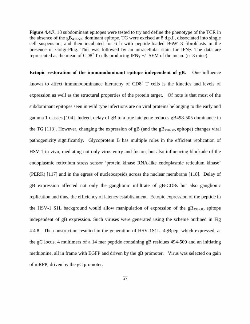
57
Figure 4.4.7. 18 subdominant epitopes were tested to try and define the phenotype of the TCR in the absence of the gB498-505 dominant epitope. TG were excised at 8 d.p.i., dissociated into single cell suspension, and then incubated for 6 h with peptide-loaded B6WT3 fibroblasts in the presence of Golgi-Plug. This was followed by an intracellular stain for IFNγ. The data are represented as the mean of CD8+
T cells producing IFNγ +/- SEM of the mean. (n=3 mice).
Ectopic restoration of the immunodominant epitope independent of gB. One influence
known to affect immunodominance hierarchy of CD8+ T cells is the kinetics and levels of
expression as well as the structural properties of the protein target. Of note is that most of the
subdominant epitopes seen in wild type infections are on viral proteins belonging to the early and
gamma 1 classes [104]. Indeed, delay of gB to a true late gene reduces gB498-505 dominance in
the TG [113]. However, changing the expression of gB (and the gB498-505 epitope) changes viral
pathogenicity significantly. Glycoprotein B has multiple roles in the efficient replication of
HSV-1 in vivo, mediating not only virus entry and fusion, but also influencing blockade of the
endoplasmic reticulum stress sensor ‘protein kinase RNA-like endoplasmic reticulum kinase’
(PERK) [117] and in the egress of nucleocapsids across the nuclear membrane [118]. Delay of
gB expression affected not only the ganglionic infiltrate of gB-CD8s but also ganglionic
replication and thus, the efficiency of latency establishment. Ectopic expression of the peptide in
the HSV-1 S1L background would allow manipulation of expression of the gB498-505 epitope
independent of gB expression. Such viruses were generated using the scheme outlined in Fig
4.4.8. The construction resulted in the generation of HSV-1S1L. 4gBpep, which expressed, at
the gC locus, 4 multimers of a 14 mer peptide containing gB residues 494-509 and an initiating
methionine, all in frame with EGFP and driven by the gB promoter. Virus was selected on gain
of mRFP, driven by the gC promoter.

58
Figure 4.4.8. Construction of gBnull virus and of HSV-1 with gB498-505 mutations. Line I represents the parental plasmid detailed above, and the insertion of the gC promoter in frame with mRFP followed by the gB promoter in frame with four gB494-509
repeats and EGFP. Line ii represents the HSV genome and the approximate coding position and direction of the plasmid.
B6 mice inoculated by the ocular route using 1 x 10⁵ PFU/per eye were compared in
parallel to mice infected with either the parental RE strain, or with the S1L mutant (Fig 4.4.9).
The ganglionic titers for each virus were statistically insignificant, as were total absolute number
of CD8+ T cells infiltrating the ganglia at day 8 post infection. However, gB-CD8s were
detected in the HSV-1 S1L-4gBpep infected animals that were not statistically significant from
the gB498-505 specific CD8+ T cell response to wild type HSV-1 RE in the same experiment.
This data indicates the ectopic expression of the epitope, independent of gB, is sufficient to drive
the development of the gB498-505 dominated CD8+
T cell response in the C57Bl6 mouse. Thus
the dominance of the epitope is not a result of their structural features of the gB protein itself.
i)

59
Figure 4.4.9. Characterization of the ectopic gB virus
(A) TG viral growth in B6 Mice (B) Absolute number of CD8+ T cells at 8 d.p.i. (C) %gB Specific CD8+ T cells at 8 d.p.i. A) TG were harvested 4 d.p.i. and subjected to three freeze thaw cycles and infectious virus released into the supernatant was titrated on Vero cells. The graph represents the mean virus titer for each virus ± SEM of the mean (n = 5 mice). (B) and (C) Mice were infected with 1 x 10⁵ PFU/eye with HSV-1 WT, HSV-1 S1L, or the peptide virus. TG were excised at 8 d.p.i., dissociated into single cell suspensions and surface stained the expression of gB498-505
. Cells were analyzed by flow cytometry. The data are represented as the mean +/- SEM of the mean. (n=5 mice)

60
4.5 DISCUSSION
This work is designed to understand aspects of the development of the CD8+ T cell
repertoire to HSV-1 in the B6 murine model, particularly pertaining to the immune infiltrate into
the trigeminal ganglion that is concomitant with the establishment of the latent state. The
immunodominance of the gB498-505 epitope, to which a majority (>50%) of HSV-1 specific CD8+
T cells are directed following HSV-1 infection of the B6 mouse, has been well established, and
reagents that identify this population have allowed the probing of many aspects of the cellular
immune response following HSV infection and the latent state. This includes the observations
that gB498-505 CD8+ T cells express granzyme B and surface markers of activation (CD25, CD44,
CD69) [76,97] during latency, and rapidly produce IFNγ, IL10 and TNFα after HSV-1 antigen
exposure [76,91,97]. The receptor polarization towards neurons during latency in B6 mice [90]
has strongly suggested that gB is sporadically expressed during latency. Importantly, clones of
the CD8+ T cells specific for gB498-505 from the mouse TG are able to block reactivation in ex
vivo models establishing that the γ1 protein gB is recognized early and can mediate CD8+ T cell
blockade of the HSV-1 reactivation process. More is becoming known of the nature of the
remaining CD8+ T cells and their role in infection. The CD8+ T cell population specific to
subdominant epitopes can also block reactivation from latency [116]. The studies described here
were initiated to address the nature of the CD8+
The S1L mutant did not induce a gB
T cell response to infectious viruses lacking this
dominant epitope. From this work we can draw several important conclusions.
498-505 dominated response, and yet was robust in
pathogenesis. This was deemed important as virus replication in the TG could affect the antigens
61
presented and the recruitment and retention of the CD8+
The loss of the gB
T cells in the ganglia. Many mutations
in this region were developed in culture, but were attenuated in mice suggesting that the region
defined functions in vivo that are not important in vitro. The natures of these specific roles are
not yet known. Aside from its role in fusion and entry, gB is also involved in the modulation of
the PERK activity and has roles in promoting nuclear egress of the nucleocapsid that may affect
pathogenensis in vivo. However, S1L and L8A viruses showed near wild type levels in most
respects. L8A mutants have been evaluated previously in the flank skin infection model
C57BL/6 mice, and no differences were found when compared to wild type infection [101].
However, these authors did not assess their virus in the ocular infection model, where we found a
very low induction of stromal disease in a different mouse strain where the dominance of the
epitope would not contribute. This suggests further, as yet unknown, roles of this region of gB
affecting pathogenesis.
498-505 immunodominant epitope results in the development of a CD8+
T cell response equivalent in size infiltrating the TG that compensates for the loss of half of the
CD8+ T cells seen in HSV-1. This confirms the observation of Stock et al (2007), who found a
similar ganglionic compensation in the CD8+ T cell response following flank skin infection with
HSV-1 containing the L8A mutation that also blocks development of CD8+ T cells with gB498-505
specificity [101]. Here we have established the nature of this compensation in the ganglia,
which intriguingly suggests that the TG has mechanisms to regulate the CD8+ infiltrate at the
initial infection. The nature of this is not yet known: it is possible that gB CD8+ T cells regulate
the subdominant CD8+ T cell populations, and the epitope loss results in other T cell populations
filling the ganglionic compartment. The nature of this compensation is an increase of the

62
fraction of CD8+ T cells directed to minor subdominant epitopes targeted by the natural
developing CD8+ T cell response to HSV-1. This repertoire is directed to a very limited subset
of HSV-1 proteins. The majority of these were of the early or early late class [104]. This work
shows that loss of the epitope does not result in an antigenic expansion seen by CD8+ T cells or
emergence of one or a few epitopes to dominance. The S1L infection resulted in upregulation of
most of the 18 epitopes without the presence of a novel subdominant epitope that appear to
identify the entire repertoire specific to HSV S1L in C57BL/6 mice. From this we can conclude
that the CD8+ T cell response is being compensated for by a broad increase of the subdominant
populations. Since it has been shown that the non-gB498-505 specific population induced by wild
type HSV-1 infection can block reactivation [116], we consider it highly probable that this
alternative CD8+
The CD8
T cell TG infiltrate will block reactivation efficiently.
+ T cell response seen in an HSV-S1L infection also seems to be selective for
proteins that are produced prior to HSV DNA synthesis, which is also seen in the wild-type
infection [104]. Early proteins are the most frequently targeted epitopes. There is no known
mechanism for why there is a dominance hierarchy, but there are many factors that may play a
role in selection such as viral gene expression during reactivation events, [119] precursor
frequency, and the ability of the peptide to bind MHC [94]. Further characterization will develop
our understanding on how CD8+ T cells specific to immunodominant epitopes influence the
infiltration and retention of those specific to non-immunodominant epitopes. It will also help us
understand the dynamics of the development and maintenance of CD8+ T cell responses in the
murine ganglia. Furthermore, the identity of the alternate CD8+ T cell epitopes will also shed
light on which viral antigens CD8+ T cells respond to and the mechanisms involved in blocking

63
reactivation. The identity of these viral antigens may be the basis for the development of a new
multivalent vaccine to induce protection against reactivation to HSV-1.
In this work, we also effectively separated the epitope from the gB protein by expressing
the epitope ectopically from at the gC locus in the S1L background. This work leads to the
conclusion that the immunodominance of the gB498-505 in this mouse strain is not the
consequence of the gB protein itself, nor the genomic locus from which it is expressed. It is
conceivable that the immunodominance was the result of a structural definition of the protein
that may have rendered the dominant peptide more easily generated from the gB protein. The gB
locus and the epigenetic conditions that regulate its expression could have also contributed to
immunodominance, but this does not seem to be the case. Ectopic expression of a peptide
multimer alone resulted in a gB498-505 dominated response that was identical to that of the wild
type virus. Ruling out these scenarios, it is possible that the immunodominance is dictated in part
by the precursor frequency of the CD8+
While these studies will help us understand the dynamics of how CD8
T cells or another unknown mechanism.
+ T cell responses
develop and are maintained in the murine ganglia, translation of this work to human studies will
prove to be more challenging. In the murine model we show a proof of principle that by altering
expression of the immunodominant CD8+ T cell epitope, we can augment the immune response
to that antigen and subsequently prevent reactivation. However, since humans may not react to
the same CD8+ T cell epitopes, the challenge facing scientists is to be able to predict or identify
viral epitopes that are reactive in humans. Also, since therapeutic vaccines would need to be
delivered locally to the site of latency, an effective and safe method of delivery is needed. The
use of live virus may not be a viable option due to safety concerns. While the use of a replication

64
defective HSV-1 viral vector would be useful in that there would be constant antigen
presentation to surrounding CD8+
T cells, there is still the risk of viral reactivation from the
vaccine virus, which could exacerbate any existing disease. Use of an appropriate adjuvant plays
a critical and important part in the efficacy of a vaccine. While peptide vaccines with a strong
adjuvant would boost the immune response temporarily, repeated vaccinations may be necessary.
However, knowledge of the importance of the cellular immune response in the control of HSV-1
infection and reactivation has led to several animal studies involving vaccines aimed at eliciting
such a response, such as DNA vaccines and vaccines encoding cytokines. The future of HSV-1
therapeutic vaccines may lie in these new techniques and targets.

65
5.0 SUBDOMINANT CD8 + T CELL ACTIVATION PHENOTYPE DURING
LATENCY IN THE ABSENSE OF GB SPECIFIC CD8+
T CELLS

66
5.1 ABSTRACT
Latency of HSV-1 in human and murine trigeminal ganglia (TG) is associated with a persistent
virus specific cellular immune infiltrate. It has been shown that in the C57BL/6 mouse model,
mice infected with HSV-1 lacking the gB498-505 epitope develop an alternative CD8+ T cell
repertoire and this repertoire is still able to block viral reactivation from latency. In chapter 4 we
identified the global nature of the CD8+ T cell repertoire at the peak response. However, the
activation phenotype of CD8s recognizing these subdominant epitopes at latency is unknown.
Here, we assessed the activation phenotype of these CD8s and their specificity. CD8s specific to
select subdominant epitopes maintained hierarchy that was somewhat different from the
responses at the peak of the infiltrate. They also showed ratios of IFNγ staining at 30 dpi as seen
in an acute infection, as well as an activated state interpreted through GrzB and PD-1 staining.
We conclude that loss of the HSV dominant epitope results in a retained functionality of the
HSV-specific CD8+
T cell repertoire during latency, and contrasts to the exhausted state seen for
subdominants in the presence of the gB population.

67
5.2 INTRODUCTION
Latency is initially established when replicating virus is cleared from the ganglion, leaving only
latent HSV-1 genomes in a chromatin regulated gene expression repressed state. In the C57BL/6
mouse, the persistent infiltrate of CD8+ T cells maintain, in part, these genomes in latency. CD8+
T cell numbers reach their peak at 8 dpi, coinciding with the onset of viral latency, but contract
to a memory population of virus-specific CD8+ T cells that remain in the TG for the life of the
animal [71,76,77,78,79]. There are many questions about how these cells maintain latency. This
chapter focuses on whether or not these cells are able to remain functional in the absence of the
gB498-505
It is as yet unclear why the CD8
epitope and how their specificity alters to that of the peak response. We examine if the
subdominant infiltrate remains, and if the HSV-1 SIL peptide virus can ectopically restore a gB-
CD8 population during latency.
+ T cell population in C57BL/6 mice dominates on the
gB498-505 dominant epitope. Immune dominance can be dictated by multiple factors, such as the
epitope concentration presented, the binding affinity of the peptide to the MHC-I complex, the
precursor frequency of the CD8+ T cells that are able to react to gB498-505 as well as the kinetics
and level of expression. We have shown that in the absence of this epitope the subdominant
population is able to rise and fill the void for the missing CD8+
CD8
T cell population. However, how
this subdominant population changes or how the dominant population can be manipulated in
order to help prevent reactivation is as yet undetermined.
+ T cells act through IFNγ production and lytic granule release [66,78]. Consistent
with others’ observations (10,12,32), gB-CD8+ T cells that produce IFNγ were found during all

68
stages of latency, and this population showed indications of recent antigen exposure through the
expression of granzyme B, the ability to quickly produce IFNγ following antigen exposure, and
through surface markers of activation such as CD44 and CD69 (40,95). gB-CD8+ T cells contain
lytic granules that are composed of multiple proteins, including perforin, Fas ligand, granulysin,
and granzymes, all of which are all cytotoxic to infected cells [86,87,88]. Granzymes are
important because they can cleave the proteins of intracellular and extracellular pathogens [120].
Granzyme B also induces caspase-dependent apoptosis, and with the help of perforin can induce
CD8+
There is evidence to suggest that HSV-1 latency is not antigencally silent. In a latently
infected TG, gB-CD8s polarize their TCR to the surface of neurons, and through this
immunological synapse release IFNγ and lytic granules [66,76]. This indicates in vivo
recognition of neurons through an MHC-1 manner and suggests that low level antigen exposure
retains the TG CD8
T cell blocking of HSV-1 reactivation without apoptosing a neuron [66,67,89].
+ T cell population during latency. Studies have also shown that CD8+ T
cells only need one MHC-I to peptide complex in order to be stimulated, so it is possible that low
levels subdominant protein epitope expression during abortive reactivation events can be enough
stimulus for CD8+
T cells to block reactivation. [121,122].

69
5.3 MATERIALS AND METHODS
5.3.1 BrdU Staining
A FITC BrdU Flow Kit was purchased from BD Pharmingen (San Diego, CA).
Phycoerythrin (PE)-conjugated H-2Kb tetramers complexed with the gB498-505, RR1982-989,
RR1822-829, or ICP8876-883 peptide and PE-conjugated H-2Db tetramers complexed with the
ICP8168-176, RR1372-380, or RR2279-287
Latently infected mice with either wild type HSV-1 or the S1L HSV-1 were pulsed for
two days prior to harvest with 1mg/mouse of BrdU in 0.1ml PBS via i.p. injection. At 34 days
post infection the TG were harvested and cells were prepared for flow cytometric analysis. TG
cells were washed twice with FACS buffer (1% FBS, 0.1% sodium azide in PBS) prior to
staining. Cells were incubated for 10 minutes with F
peptide were provided by the National Institute of Allergy
and Infectious Diseases Tetramer Core Facility (Emory University Vaccine Center, Atlanta,
GA). All other antibodies were purchased as described previously.
C block prior to surface staining with the
appropriate antibodies. Following surface staining, cells were permeabilized with
Cytofix/Cytoperm solution for 20 min on ice and then incubated with Cytofix/Cytoperm plus for
10 minutes on ice. After being washed with 1X BD Perm/Wash solution, cells were treated with
30 µg of DNAse for 1hr at 37°C. Cells were washed again and then incubated for 30 minutes in
BrdU antibody diluted in 1X BD Perm/Wash. After labeling, suspensions were washed with 1X
Perm/Wash and resuspended in FACS buffer for analysis by flow cytometry. Flow cytometry

70
was performed on a BD FACSAria Cell Sorter using FACSDiva 4.1 or 5.0.2 software (BD
Pharmingen).

71
5.4 RESULTS
5.4.1 Subdominant Epitope Hierarchy of the CD8+
Eight days post infection, HSV-1 S1L induces an equivalent absolute number of CD8
T cell response during latency
+ T
cells infiltrating the ganglia with almost no reactivity to the gB epitope. It was determined that
this response was not due to compensation of CD8+ T cells to the mutant gB498-505
We wanted to evaluate the nature and hierarchy of the CD8
peptides, but
instead was compensated for by an increase in the number of CD8s specific to subdominant
epitopes as described in Chapter 4.
+ T cell response to the
subdominant epitopes for recognition by HSV-1 S1L in a latently infected animal to determine if
these subdominant CD8+ T cells remained in the same hierological order. It has been seen in
studies with our collaborator, Anthony St. Leger, that CD8+ T cells responding to subdominant
epitopes in a wild-type infection show signs of exhaustion, such as high levels of PD-1 and low
levels of IFNγ secretion. However, in an S1L infection, it seemed that subdominant epitopes
were still able to maintain some functionality, and this needed to be studied further. Due to
constraints in the number of CD8+ T cells in the gangalia at latency, only a select representative
population of the peptides was assayed. We found the fraction of ganglionic CD8+ T cells
responding to each peptide from the TG of S1L infected mice at 30 dpi (by IFNγ staining)
remained increased over the levels from wild type infections (Fig 5.4.1). The ratio of cells
responding to each target also appeared to remain consistent with the relative ratios from 8 dpi
infected animals suggesting a similar hierarchical order. We compared this with data from a wild
type infection (A. St. Leger and R Hendricks, unpublished data). This showed that the CD8+ T
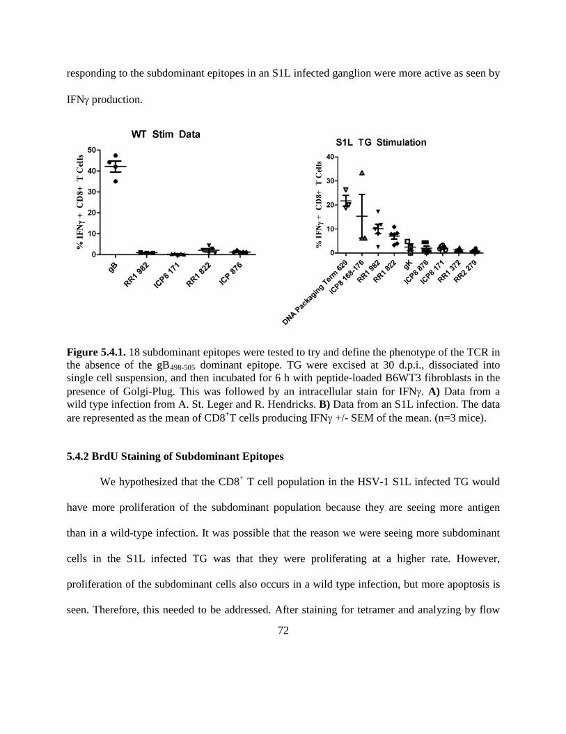
72
responding to the subdominant epitopes in an S1L infected ganglion were more active as seen by
IFNγ production.
Figure 5.4.1. 18 subdominant epitopes were tested to try and define the phenotype of the TCR in the absence of the gB498-505 dominant epitope. TG were excised at 30 d.p.i., dissociated into single cell suspension, and then incubated for 6 h with peptide-loaded B6WT3 fibroblasts in the presence of Golgi-Plug. This was followed by an intracellular stain for IFNγ. A) Data from a wild type infection from A. St. Leger and R. Hendricks. B) Data from an S1L infection. The data are represented as the mean of CD8+
T cells producing IFNγ +/- SEM of the mean. (n=3 mice).
5.4.2 BrdU Staining of Subdominant Epitopes
We hypothesized that the CD8+ T cell population in the HSV-1 S1L infected TG would
have more proliferation of the subdominant population because they are seeing more antigen
than in a wild-type infection. It was possible that the reason we were seeing more subdominant
cells in the S1L infected TG was that they were proliferating at a higher rate. However,
proliferation of the subdominant cells also occurs in a wild type infection, but more apoptosis is
seen. Therefore, this needed to be addressed. After staining for tetramer and analyzing by flow

73
cytometry, we determined how many tetramer specific CD8+ T cells were also positive for BrdU.
The level of proliferation in the population as assessed by BrdU incorporation in the HSV-1 S1L
CD8+
T cells was similar to that of the HSV-1 WT infected mice (Fig. 5.4.2). This suggested that
proliferation may not be an important factor in distinguishing the subdominant population or
their activation state.
Figure 5.4.2. BrdU was given intraperitoneally (i.p.) 48 and 24 hours before harvest. TG were then harvested and stained for CD8, CD45, tetramer specific T cell receptor, and BrdU incorporation. There is only a 1% gB population seen in the S1L infected TG as compared to 50% seen in the WT infected TG. Data represent each specific animal and standard error of 6 mice from 2 experiments. Significance was determined by one way ANOVA. 5.4.3 PD-1/GrzB Activation State of Epitopes at Latency
Studies have shown that a CD8+ T cell can act through gB and block reactivation through
a non-cytolytic pathway, which may involve IFNγ and GrzB [102,115]. Determining which cells
from a HSV-1 S1L infection are GrzB positive will identify which antigens are being presented
and recognized in the absence of the dominant epitope. Programmed death receptor-1, or PD-1,

74
is another important marker that can indicate both recent exposure of CD8+ T cells to antigen but
can also indicate exhaustion. We wanted to compare the activation state of the CD8+ T cells in
latently infected mice with either wild type HSV-1 or HSV-1 S1L. B6 mice were inoculated
using 1 x 10⁵ per eye of either wild type HSV-1 or HSV-1 S1L (Figure 5.4.3). At 30 dpi, TG
were then excised and stained for CD8, CD45, GrzB and PD-1 and analyzed by flow cytometry
to determine the activation state of the CD8+ T cell population. There appears to be a large
amount of variation in the activation states between the WT subdominant epitopes as compared
to the F5L subdominant epitopes. Although there is more GrzB in the WT CD8+ T cells, there
also seems to be more PD-1, which may mean that there is more exhaustion in these cell
populations or that they have recently been exposed to their antigen at higher levels since it has
been shown that these CD8+
T cells are able to protect against reactivation. Further studies need
to be performed to determine what determines differences in the dominant versus subdominant
cells.
WT S1L

75
Figure 5.4.3. Characterization of the activation state of subdominant epitopes
Mice were infected with 1 x 10⁵
PFU/eye with HSV-1 WT or HSV-1 S1L. TG were excised at 30 d.p.i., dissociated into single cell suspensions and surface stained for the expression of a respective subdominant epitope. Cells were analyzed by flow cytometry. The data are represented as the mean of the total tetramer positive population (n=3 mice). Four examples are shown.
5.4.4 Response to the ectopically restored Peptide Virus at latency
One stimulus affecting immunodominance hierarchy of CD8+ T cells is the kinetics and
levels of expression of the protein target. As our lab has shown, delay of gB expression to a true
late gene effectively leads to reduced gB498-505 dominance in the TG [113] at latency changing
the expression of gB (and the gB498-505 epitope) significantly changes viral pathogenicity. We
showed in Chapter 4 that ectopic expression of the gB498-505 peptide in the HSV-1 SIL epitope-

76
negative background allows for manipulation of expression of the gB498-505 epitope independent
of gB expression during acute infection. It was deemed important to verify that this expression
was maintained through a latent infection. B6 mice were inoculated using 1 x 10⁵ per eye and
compared to mice infected with either the WT strain or the S1L mutant (Figure 5.4.4). The
absolute CD8 count is similar in all viruses and all viruses are similarly virulent as shown in
chapter 4. Not only does the total CD8+ T cell count reach similar numbers but the number of
gB-CD8s detected in the HSV-1 S1L-4gBpep infected mice were also similar to that of the WT
infected animal. This signifies that ectopic expression of the gB498-505 epitope maintains the
dominant CD8+
The specificity at the subdominants was also assessed by comparing two subdominant
epitopes, RR1 982 and RR1 882, between these three viruses. In animals infected with HSV-1
S1L-4gBpep, the ability to respond to subdominant epitopes seems to restore to that of a WT
infected animal, while animals infected with HSV-1 SIL still respond to subdominant epitopes at
an increased level. Both of these studies show that dominance of the epitope is not a result of the
structural features of the gB protein itself or its locus.
T cell response in the B6 mouse model throughout latency as compared to wild
type.

77
Figure 5.4.4. Characterization of the ectopic gB virus at latency
(A) Absolute number of CD8+ T cells at 30 d.p.i. (B) %gB specific CD8+ T cells at 30 d.p.i. (A) and (B) Mice were infected with 1 x 10⁵ PFU/eye with HSV-1 WT, HSV-1 S1L, or the peptide virus. TG were excised at 30 d.p.i., dissociated into single cell suspensions and surface stained for the expression of gB498-505
. Cells were analyzed by flow cytometry. The data are represented as the mean +/- SEM of the mean. (n=6 mice)

78
5.5 DISCUSSION
Recurrent HSK resulting from reactivation of latent HSV-1 in neurons of the TG lead to
progressive corneal scarring and loss of vision. Strategies to block HSV-1 reactivation from
latency before virus is delivered to the periphery can prevent inflammation and scarring, but to
date, no vaccine is available. Until recently, HSV-1 latency was believed to be antigenically
silent and invisible to the immune system, making vaccine development difficult. However, there
has been a shift in paradigm and our data, along with multiple other studies, show that the
interaction between the infected ganglion, the sporadically reactivating HSV-1 genome, and the
CD8+
Previous to this work, a strong role for CD8
T cells control, at least in part, the reactivation process [123,124]. Understanding and
manipulating this interaction may be the key to controlling HSV-1 reactivation and preventing
HSK.
+ T cells being able to control viral latency
was established but it was unclear how HSV-1 antigen expression influenced the way these cells
responded to the virus during this period [67,125,126]. We show that CD8+
We determined that HSV-1 S1L induced a similar CD8
T cells responding to
subdominant epitopes are retained in similar ratios during latency as they are found during an
acute infection, and that they are able to act quickly as demonstrated by their ability to produce
IFNγ. This shows that antigen exposure is important for the retention and functionality of these
cells during latency. This work has major implications towards designing a better therapeutic
vaccine to prevent HSV-1 reactivation in the trigeminal ganglia.
+ T cell infiltrate at latency as
compared to a wild type virus. The next step was to perform ex vivo proliferation assays as well
79
as stain for GrzB and PD-1 to determine if these subdominant epitope populations were still
functionally competent in an HSV-1 S1L infected TG. While more GrzB was seen in the WT
CD8+
Using the SIL peptide virus, we compared the number of gB-CD8’s retained in a peptide
virally infected TG in parallel with an S1L infected animal and a wild type infected animal. We
showed that ectopic expression of the gB
T cells, there also seems to be more PD-1, which may mean that there is more exhaustion
in these cell populations.
498-505 epitope can maintain the dominant CD8+ T cell
response in the mouse model throughout latency. We also compared two subdominant epitopes,
RR1 982 and RR1 822 between these three viruses. In animals infected with HSV-1 S1L-
4gBpep, the ability to respond to subdominant epitopes seems to return to that of a WT infected
animal. This work leads to the conclusion that the immunodominance of the gB498-505
It was conceivable that the immunodominance was the result of a structural definition of
the protein that may have rendered the dominant peptide more easily generated from the gB
protein. The gB locus and the epigenetic conditions that regulate its expression could also have
contributed to immunodominance, but this does not seem to be the case. The separation and
ectopic expression of the epitope now establishes the feasibility of this approach to assess the
contribution of the kinetics of viral gene expression to dominance of the gB-CD8 T cell
responses, since we can now use different classes of viral promoters to drive the multipeptide.
For example, we have previously found the true late expression of gB (from the gC protein)
results in the development of an altered dominance in that the 50% population in the TG is
in this
mouse strain is not the consequence of the gB protein itself, nor the genomic locus from which it
is expressed.

80
reduced, and depletes over time [113]. It is known that immunodominance can be influenced by
the kinetics of viral gene expression as well as the load of antigen presented at the priming stages
as well as the level of antigen expressed at persistent stages of infection [127]. For example,
with vaccinia virus it was recently demonstrated that altered expression of a late antigen to an
immediate early antigen resulted in the enhancing of acute and memory CD8+ T cell responses
[128]. It may be that specific promoters are able to more effectively express a viral antigen. This
may induce a more effectively retained infiltrate in the ganglia that can better block reactivation
if they are functional. T cells at latency to subdominant populations appear to express
considerably less granzyme B [71], and it has been suggested that periodic sporadic reactivation
events may drive abnormal inflation of CD8+ T cells following systemic administration of HSV -
1 [129]. Studies on the T cell specificity of the retained CD8+ T cell infiltrate and their
functionality during latency are in progress for both HSV -1 and HSV-1 lacking the dominant
epitope. However, the low levels of CD8+
This work has given us considerable insight into the retention of antigen specific CD8
T cells that are retained in the ganglia at latency
confound the assessment of T cell specificity.
+ T
cells in the latently infected TG. Translating this work to human studies will require a large
amount of work; however, this gives us a solid groundwork for which immunogens to focus on,
such as the immediate early and early proteins that seem to be seen more frequently by CD8+
T
cells, and will allow us to begin development of an effect vaccine against HSV-1 reactivation.
81
6.0 GENERAL DISCUSSION AND CONCLUSIONS
The CD8+ T cell response in the HSV-1 infected ganglia is an important component of host
control of reactivation during latency. The immunodominance of the gB498-505 epitope in CD8+ T
cells in the C57BL/6 mouse model is impressive, accounting for half of the HSV-1 specific
systemic CD8+ T response, the CD8+ T cell infiltrate in the acutely infected TG [125], and the
persisting contracted CD8+
gB-CD8s in the HSV-1 latently infected TG express surface markers of activation
(CD25, CD44, CD69) [76,97], are granzyme B positive, and rapidly produce IFNγ and TNFα
upon HSV-1 antigen exposure [82,91,125,131]. They also demonstrate T cell receptor
polarization towards neurons during latency in an immunological synapse [90] and the ability to
block reactivation ex vivo [102,103]. It is likely that these gB-CD8s respond to sporadic gB
antigen expression during latency and block progression to reactivation. This is consistent with
reports that show HSV-1 gene and protein expression in the latently infected mouse and human
TG without any productive virus production [34,49].
T population associated with latency [130].
Recent work has focused more on the HSV-1 specific non-gB498-505 CD8 T cell
populations, since these also have the potential to prevent HSV-1 reactivation [116]. They
recognize a very limited repertoire of 18 epitopes on 14 viral proteins [104]. This study

82
investigated the viral specificity of CD8+ T cells that infiltrate the acutely infected TG in the
absence of the dominating gB epitope in order to understand the relationship of CD8+
We developed two mutant HSV-1 viruses that lack the gB epitope but which retained
near wild type pathogenicity levels in the mouse model. Full pathogenicity was deemed
necessary as reduced viral replication in the TG could affect the level of antigens presented and
the recruitment of the CD8
T cell
repertoires responding to HSV-1 ocular and ganglionic infection. The hierarchy of the immune
response is affected by multiple factors, including MHC affinity, T cell precursor frequency, and
antigen expression timing and structure [90,132,133].
+ T cell population in the ganglia, as seen in the LCMV model [127].
HSV-1 L8A had been shown in the flank skin infection model in C57BL/6 mice to be similar to
wild type in pathogenesis and did not generate a dominating gB-CD8 population [101].
However, we favored HSV-1 S1L in our studies because L8A showed a reduced CD8 infiltrate
in the mouse ganglia over that generated to S1L and wild type (Fig 4.4.6), which was also seen
by Stock et. al (2007). Interestingly, neither mutation resulted in development of CD8+ T cell
populations to the mutant epitopes, suggesting they prevented stimulation and maturation of the
gB498-505
The loss of the dominant gB
precursor populations that account for part of the immunodominance [104].
498-505 epitope resulted in an equivalent CD8+ T cell response
infiltrating the TG that compensated for the loss of half of the CD8+ T cells seen in an HSV-1
infected TG. It is intriguing how this compensatory response is controlled since its size is almost
identical to that of the wild type. Factors that may contribute include the antigen presenting cells
that, without the dominant epitope, may better present the subdominant epitopes and more
effectively stimulate the T cell populations. It may also be that gB-CD8s control the infiltration,

83
as it has long been known that IFNγ production is one factor that regulates the CD8+
Our work now shows the nature of the compensation in the TG occurs by parallel
increase of the fraction of CD8
T cell
hierarchy, and gB-CD8s rapidly express IFNγ upon stimulation [76,102].
+ T cells directed to most of the minor subdominant epitopes
targeted by the natural developing CD8+ T cell response to HSV-1. In our data, there was no
emergence of one or only a few peptides or a new epitope to dominance or co-dominance, and as
the total CD8+ T cell numbers in the ganglia is similar to the sum of the fractions of the total
compensation to each epitope, we consider it unlikely that the compensation is due to significant
antigenic expansion of CD8+
There are some consistencies in the hierarchy of dominance between wild type and S1L
infections, but some changes as well. For example, one of the highest subdominant targets to
wild type infection is the ribonucleotide reductase, of which two epitopes 982 and 822 are among
the highest in the S1L infection as well. In particular, we observed a 4-5 fold increase in the
ribonucleotide reductase 982 subdominant epitope (RR1 982), which contrasts with the absence
of an increase in the subdominant epitope in the study of Stock et. al. [101]. The reason for this is
not clear, as we have also found with our L8A that the CD8s to the RR1 822 epitope also
increase (data not shown). It is also possible that the site of infection between the two studies
may influence subdominant alternative responses. Stock et al. argued that the compensatory
response was due to the emergence of cryptic epitopes, which is in direct contrast to our
conclusion that it is upregulation of the next best ranking epitopes that seems more likely to
T cells to peptides outside of the tested targets. Thus the loss of the
immundominant epitope results in a broader T cell repertoire, which may be more effective at
sensing the sporadic reactivation events that occur during latency and preventing reactivation.
84
occur in the acutely infected TG. It is clear that some subdominant populations arise to
codominance from while other higher ranking epitopes show much less of an increase following
the loss of the gB epitope, such as that to ICP8 976, which shows only a modest 1.2 fold
increase, while a few epitopes that have very minor populations in a wild type infection (UL25
for example) show relative large fold increases. This is inconsistent with the emergence of
cryptic epitopes seen for Influenza and Listeria, but in some ways is similar to the pattern seen in
other systems. For example, loss of epitope specific responses in LCMV resulted in
compensatory responses against other subdominant epitopes, without changing the hierarchy and
compensated responses leading to a rise in hierarchical importance of other viral targets can still
mediate effective targeting by CD8+ T cells responses [134]. Serial upranking of minor
subdominant epitopes following sequential deletion of the dominant epitopes has also been seen
for HIV epitopes delivered by vaccinia vectors [134]. The broadening of the CD8+ T cell
repertoire following loss of the dominant epitope has also been seen with other pathogens [135].
The dominant epitope is often replaced by several subdominant epitopes. The drivers of the
HSV dominance hierarchy and the changes induced by the S1L mutation are not yet clear, but
are likely complex. It is known that a specific population of CD8+ T cells can affect others in
such a way that dominance hierarchy is a contextual series of events in which the dominance of a
specific epitope is dependent upon other epitopes and the response to them [136]. Thus it is
possible that gB-CD8 development may suppress the development and infiltration of non gB-
CD8s in our HSV model. Since gB-CD8s express IFNγ in the ganglia, and IFNγ is predicted to
influence CD8+ T cell hierarchy and functionality [137], it is possible that the loss of the
dominating gB-CD8s relieves the suppressor effect on other epitope-specific populations and
85
allows the increased proliferation of the subdominant-specific CD8+
It is interesting that the CD8
T cell precursors [138].
Recent work suggests IFNγ release by gB-CD8 may up regulate PD-L1 expression in neurons
that may drive exhaustion of PD-1 positive subdominant CD8s (S Jeong and RL Hendricks,
unpublished data). If so, the rise in subdominant CD8s may be a direct result of the loss of the
major CD8 dominant population.
+ T cell response seen in an HSV-S1L infection also seems to
be selective for proteins that are produced prior to HSV DNA synthesis, which is also seen in the
wild-type infection [104]. Most of the epitope targets are directed to early proteins, with the
strongest compensatory increase being directed to epitopes on ribonucleotide reductase (an early
protein), and two gamma1 regulated proteins, gB and the DNA terminase subunit. It is notable
that there is very little evidence of any immediate early protein recognition and only a few
epitopes from proteins expressed only after DNA replication are seen. Levels and timing of
antigen are known to greatly influence memory cell development and functionality [138]. The
contribution of viral kinetics to dominance hierarchy is largely unexplored in HSV-1, but recent
evidence has strongly suggested the classical cascade of HSV-1 seen in lytic infected cells does
not necessarily apply to sporadic reactivation events [46,47]. There are many factors that may
play a role in selection of targets for CD8+ T cell development, such as kinetics and levels of
viral gene expression during reactivation events [119], precursor frequency, and the ability of the
peptide to bind MHC [94]. Further characterization of the contribution to CD8+ T cell
development will help us develop our understanding of how CD8+ T cells specific to
immunodominant epitopes influence the infiltration and retention of those directed to non-
immunodominant epitopes. The identity of the alternate CD8+ T cell epitopes may also reveal

86
viral antigens to which CD8+
This work also has additional relevance to vaccine design, since it adds to the wealth of
information that indicates CD8
T cells can respond to in blocking reactivation, which could then
be developed as T cell generating vaccines.
+ T cell induction is an important goal of any vaccine. Most
current HSV-1 vaccines are based on viral-encoded surface glycoprotein subunits designed to
boost the neutralizing antibody response, and have largely failed to meet their criteria in clinical
trials for prevent infection or disease. We argue that an effective vaccine strategy that boosts
CD8+ T cell levels in the ganglia and surrounding the sensory neurons could prevent spread in
the ganglia upon infection, or prevent reactivation from latency if used therapeutically. In the
murine model, our goal was to show proof of principle that manipulating the CD8+ T cell
response by removing the dominant epitope and expressing multiple copies of it expands the
immune response to a particular antigen. While these studies will help us understand the
dynamics of how CD8+ T cell responses develop and are maintained in the murine ganglia,
translation of this work to human studies will prove to be more challenging, because the diverse
HLA types will dictate that the identity of specific antigens to which CD8+ T cells are directed.
This might make it difficult to predict which viral epitopes are reactive in a human HSV-1
infection. Interestingly, recent genome wide screening of CD8+ T cell targets for seven diverse
individuals suggest that some HSV-1 proteins appear to be targeted more frequently than others,
with the large subunit of ribonucleotide reductase showing a prominence in six individuals to
both CD4 and CD8+ T cells [139]. This correlates with the main dominant epitope seen in the
S1L infected B6 mice. Thus it is likely that additional proteins outside of the usual glycoprotein

87
candidates are valid potential vaccines for induction of a protective T cell immune response that
is optimally intended to prevent reactivation.
88
BIBLIOGRAPHY
1. Roizman BaK, D.M (2001) Herpes simplex viruses and their replication; B.N. Fields DMK, P.M. Howley, S.E. Straus, and M.A. Martin., editor. Philadelphia: Lippincott - Raven. 2399 - 2459 p.
2. Aherne A, Kennan A, Kenna PF, McNally N, Lloyd DG, et al. (2004) On the molecular pathology of neurodegeneration in IMPDH1-based retinitis pigmentosa. Hum Mol Genet 13: 641-650.
3. Smith Jennifer S, Robinson N J (2002) Age-Specific Prevalence of Infection with Herpes Simplex Virus Types 2 and 1: A Global Review. The Journal of Infectious Diseases 186: S3-S28.
4. Whitley R (1996) Fields Virology; Knipe DM HP, Griffin D, Lamb R, Martin M, Straus S, editor. Philadelphia: Lippincott, Williams & Wilkins.
5. Schillinger J, Xu, F, Sternberg, MR, Armstrong, GL, Lee, FK, Namias, AJ, McQuillan, GM, Louis, ME, Markowitz, LE (2004) National Seroprevalance and trends in Herpes simplex virus type 1 in the United States, 1976-1994. Sexually Transmitted Diseases 31: 753 - 760.
6. Toma HS, Murina AT, Areaux RG, Neumann DM, Bhattacharjee PS, et al. (2009) Ocular HSV-1 Latency, Reactivation and Recurrent Disease. Seminars in Ophthalmology 23: 249-273.
7. Liesegang TJ (2001) Herpes simplex virus epidemiology and ocular importance. Cornea 20: 1-13.
8. Lee S, Zheng M, Kim B, Rouse BT (2002) Role of matrix metalloproteinase-9 in angiogenesis caused by ocular infection with herpes simplex virus. The Journal of Clinical Investigation 110: 1105-1111.
9. Mistry SK, Zheng M, Rouse BT, Morris SM (2001) Induction of arginases I and II in cornea during herpes simplex virus infection. Virus Research 73: 177-182.
10. Biswas PS, Rouse BT (2005) Early events in HSV keratitis--setting the stage for a blinding disease. Microbes and Infection 7: 799-810.
11. Carr DJ, Tomanek L (2006) Herpes simplex virus and the chemokines that mediate the inflammation. Curr Top Microbiol Immunol 303: 47-65.
12. Lepisto AJ, Frank GM, Hendricks RL (2007) How herpes simplex virus type 1 rescinds corneal privilege. Chem Immunol Allergy 92: 203-212.
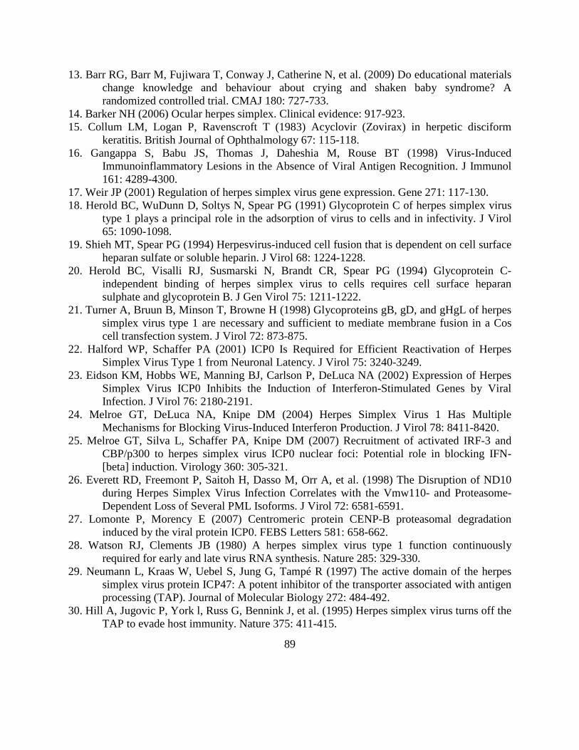
89
13. Barr RG, Barr M, Fujiwara T, Conway J, Catherine N, et al. (2009) Do educational materials change knowledge and behaviour about crying and shaken baby syndrome? A randomized controlled trial. CMAJ 180: 727-733.
14. Barker NH (2006) Ocular herpes simplex. Clinical evidence: 917-923. 15. Collum LM, Logan P, Ravenscroft T (1983) Acyclovir (Zovirax) in herpetic disciform
keratitis. British Journal of Ophthalmology 67: 115-118. 16. Gangappa S, Babu JS, Thomas J, Daheshia M, Rouse BT (1998) Virus-Induced
Immunoinflammatory Lesions in the Absence of Viral Antigen Recognition. J Immunol 161: 4289-4300.
17. Weir JP (2001) Regulation of herpes simplex virus gene expression. Gene 271: 117-130. 18. Herold BC, WuDunn D, Soltys N, Spear PG (1991) Glycoprotein C of herpes simplex virus
type 1 plays a principal role in the adsorption of virus to cells and in infectivity. J Virol 65: 1090-1098.
19. Shieh MT, Spear PG (1994) Herpesvirus-induced cell fusion that is dependent on cell surface heparan sulfate or soluble heparin. J Virol 68: 1224-1228.
20. Herold BC, Visalli RJ, Susmarski N, Brandt CR, Spear PG (1994) Glycoprotein C-independent binding of herpes simplex virus to cells requires cell surface heparan sulphate and glycoprotein B. J Gen Virol 75: 1211-1222.
21. Turner A, Bruun B, Minson T, Browne H (1998) Glycoproteins gB, gD, and gHgL of herpes simplex virus type 1 are necessary and sufficient to mediate membrane fusion in a Cos cell transfection system. J Virol 72: 873-875.
22. Halford WP, Schaffer PA (2001) ICP0 Is Required for Efficient Reactivation of Herpes Simplex Virus Type 1 from Neuronal Latency. J Virol 75: 3240-3249.
23. Eidson KM, Hobbs WE, Manning BJ, Carlson P, DeLuca NA (2002) Expression of Herpes Simplex Virus ICP0 Inhibits the Induction of Interferon-Stimulated Genes by Viral Infection. J Virol 76: 2180-2191.
24. Melroe GT, DeLuca NA, Knipe DM (2004) Herpes Simplex Virus 1 Has Multiple Mechanisms for Blocking Virus-Induced Interferon Production. J Virol 78: 8411-8420.
25. Melroe GT, Silva L, Schaffer PA, Knipe DM (2007) Recruitment of activated IRF-3 and CBP/p300 to herpes simplex virus ICP0 nuclear foci: Potential role in blocking IFN-[beta] induction. Virology 360: 305-321.
26. Everett RD, Freemont P, Saitoh H, Dasso M, Orr A, et al. (1998) The Disruption of ND10 during Herpes Simplex Virus Infection Correlates with the Vmw110- and Proteasome-Dependent Loss of Several PML Isoforms. J Virol 72: 6581-6591.
27. Lomonte P, Morency E (2007) Centromeric protein CENP-B proteasomal degradation induced by the viral protein ICP0. FEBS Letters 581: 658-662.
28. Watson RJ, Clements JB (1980) A herpes simplex virus type 1 function continuously required for early and late virus RNA synthesis. Nature 285: 329-330.
29. Neumann L, Kraas W, Uebel S, Jung G, Tampé R (1997) The active domain of the herpes simplex virus protein ICP47: A potent inhibitor of the transporter associated with antigen processing (TAP). Journal of Molecular Biology 272: 484-492.
30. Hill A, Jugovic P, York l, Russ G, Bennink J, et al. (1995) Herpes simplex virus turns off the TAP to evade host immunity. Nature 375: 411-415.
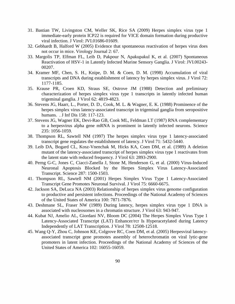
90
31. Bastian TW, Livingston CM, Weller SK, Rice SA (2009) Herpes simplex virus type 1 immediate-early protein ICP22 is required for VICE domain formation during productive viral infection. J Virol: JVI.01686-01609.
32. Gebhardt B, Halford W (2005) Evidence that spontaneous reactivation of herpes virus does not occur in mice. Virology Journal 2: 67.
33. Margolis TP, Elfman FL, Leib D, Pakpour N, Apakupakul K, et al. (2007) Spontaneous Reactivation of HSV-1 in Latently Infected Murine Sensory Ganglia. J Virol: JVI.00243-00207.
34. Kramer MF, Chen, S. H., Knipe, D. M. & Coen, D. M. (1998) Accumulation of viral transcripts and DNA during establishment of latency by herpes simplex virus. J Virol 72: 1177-1185.
35. Krause PR, Croen KD, Straus SE, Ostrove JM (1988) Detection and preliminary characterization of herpes simplex virus type 1 transcripts in latently infected human trigeminal ganglia. J Virol 62: 4819-4823.
36. Stevens JG, Haarr, L., Porter, D. D., Cook, M. L. & Wagner, E. K. (1988) Prominence of the herpes simplex virus latency-associated transcript in trigeminal ganglia from seropositive humans. . J Inf Dis 158: 117-123.
37. Stevens JG, Wagner EK, Devi-Rao GB, Cook ML, Feldman LT (1987) RNA complementary to a herpesvirus alpha gene mRNA is prominent in latently infected neurons. Science 235: 1056-1059.
38. Thompson RL, Sawtell NM (1997) The herpes simplex virus type 1 latency-associated transcript gene regulates the establishment of latency. J Virol 71: 5432-5440.
39. Leib DA, Bogard CL, Kosz-Vnenchak M, Hicks KA, Coen DM, et al. (1989) A deletion mutant of the latency-associated transcript of herpes simplex virus type 1 reactivates from the latent state with reduced frequency. J Virol 63: 2893-2900.
40. Perng G-C, Jones C, Ciacci-Zanella J, Stone M, Henderson G, et al. (2000) Virus-Induced Neuronal Apoptosis Blocked by the Herpes Simplex Virus Latency-Associated Transcript. Science 287: 1500-1503.
41. Thompson RL, Sawtell NM (2001) Herpes Simplex Virus Type 1 Latency-Associated Transcript Gene Promotes Neuronal Survival. J Virol 75: 6660-6675.
42. Jackson SA, DeLuca NA (2003) Relationship of herpes simplex virus genome configuration to productive and persistent infections. Proceedings of the National Academy of Sciences of the United States of America 100: 7871-7876.
43. Deshmane SL, Fraser NW (1989) During latency, herpes simplex virus type 1 DNA is associated with nucleosomes in a chromatin structure. J Virol 63: 943-947.
44. Kubat NJ, Amelio AL, Giordani NV, Bloom DC (2004) The Herpes Simplex Virus Type 1 Latency-Associated Transcript (LAT) Enhancer/rcr Is Hyperacetylated during Latency Independently of LAT Transcription. J Virol 78: 12508-12518.
45. Wang Q-Y, Zhou C, Johnson KE, Colgrove RC, Coen DM, et al. (2005) Herpesviral latency-associated transcript gene promotes assembly of heterochromatin on viral lytic-gene promoters in latent infection. Proceedings of the National Academy of Sciences of the United States of America 102: 16055-16059.

91
46. Du T, Zhou G, Roizman B (2011) HSV-1 gene expression from reactivated ganglia is disordered and concurrent with suppression of latency-associated transcript and miRNAs. Proc Natl Acad Sci U S A 108: 18820-18824.
47. Kim JY, Mandarino A, Chao MV, Mohr I, Wilson AC (2012) Transient reversal of episome silencing precedes VP16-dependent transcription during reactivation of latent HSV-1 in neurons. PLoS Pathog 8: e1002540.
48. Snyder A, Bruun B, Browne HM, Johnson DC (2007) A Herpes Simplex Virus gD-YFP Fusion Glycoprotein Is Transported Separately from Viral Capsids in Neuronal Axons. J Virol 81: 8337-8340.
49. Feldman LT, Ellison AR, Voytek CC, Yang L, Krause P, et al. (2002) Spontaneous molecular reactivation of herpes simplex virus type 1 latency in mice. Proc Natl Acad Sci U S A 99: 978-983.
50. Kramer MF, Coen DM (1995) Quantification of transcripts from the ICP4 and thymidine kinase genes in mouse ganglia latently infected with herpes simplex virus. J Virol 69: 1389-1399.
51. Walker J, Laycock KA, Pepose JS, Leib DA (1998) Postexposure vaccination with a virion host shutoff defective mutant reduces UV-B radiation-induced ocular herpes simplex virus shedding in mice. Vaccine 16: 6-8.
52. Cook SD, Paveloff MJ, Doucet JJ, Cottingham AJ, Sedarati F, et al. (1991) Ocular herpes simplex virus reactivation in mice latently infected with latency-associated transcript mutants. Invest Ophthalmol Vis Sci 32: 1558-1561.
53. Neumann DM, Bhattacharjee PS, Hill JM (2007) Sodium Butyrate: a Chemical Inducer of In Vivo Reactivation of Herpes Simplex Virus Type 1 in the Ocular Mouse Model. J Virol 81: 6106-6110.
54. Freeman ML, Sheridan BS, Bonneau RH, Hendricks RL (2007) Psychological Stress Compromises CD8+ T Cell Control of Latent Herpes Simplex Virus Type 1 Infections. J Immunol 179: 322-328.
55. Sawtell NM, Thompson RL (1992) Rapid in vivo reactivation of herpes simplex virus in latently infected murine ganglionic neurons after transient hyperthermia. J Virol 66: 2150-2156.
56. Sawtell NM, Thompson RL (2004) Comparison of Herpes Simplex Virus Reactivation in Ganglia In Vivo and in Explants Demonstrates Quantitative and Qualitative Differences. J Virol 78: 7784-7794.
57. Hoshino Y, Pesnicak L, Cohen JI, Straus SE (2007) Rates of Reactivation of Latent Herpes Simplex Virus from Mouse Trigeminal Ganglia Ex Vivo Correlate Directly with the Viral Load and Inversely with the Number of Infiltrating CD8+T Cells. J Virol: JVI.00474-00407.
58. Thompson RL, Preston CM, Sawtell NM (2009) De Novo Synthesis of VP16 Coordinates the Exit from HSV Latency In Vivo. PLoS Pathog 5: e1000352.
59. Tal-Singer R, Lasner TM, Podrzucki W, Skokotas A, Leary JJ, et al. (1997) Gene expression during reactivation of herpes simplex virus type 1 from latency in the peripheral nervous system is different from that during lytic infection of tissue cultures. J Virol 71: 5268-5276.
92
60. Malmgaard L (2004) Induction and regulation of IFNs during viral infections. Journal of Interferon and Cytokine Research 24: 439-454.
61. Daheshia M, Kanangat S, Rouse BT (1998) Production of Key Molecules by Ocular Neutrophils Early After Herpetic Infection of the Cornea. Experimental Eye Research 67: 619-624.
62. Kanangat S, Thomas J, Gangappa S, Babu JS, Rouse BT (1996) Herpes simplex virus type 1-mediated up-regulation of IL-12 (p40) mRNA expression. Implications in immunopathogenesis and protection. J Immunol 156: 1110-1116.
63. Kodukula P, Liu T, Rooijen NV, Jager MJ, Hendricks RL (1999) Macrophage Control of Herpes Simplex Virus Type 1 Replication in the Peripheral Nervous System. J Immunol 162: 2895-2905.
64. Frank GM, Lepisto AJ, Freeman ML, Sheridan BS, Cherpes TL, et al. (2010) Early CD4+ T Cell Help Prevents Partial CD8+ T Cell Exhaustion and Promotes Maintenance of Herpes Simplex Virus 1 Latency. J Immunol 184: 277-286.
65. Decman V, Kinchington PR, Harvey SAK, Hendricks RL (2005) Gamma Interferon Can Block Herpes Simplex Virus Type 1 Reactivation from Latency, Even in the Presence of Late Gene Expression. J Virol 79: 10339-10347.
66. Knickelbein JE, Khanna KM, Yee MB, Baty CJ, Kinchington PR, et al. (2008) Noncytotoxic Lytic Granule-Mediated CD8+ T Cell Inhibition of HSV-1 Reactivation from Neuronal Latency. Science 322: 268-271.
67. Liu T, Khanna KM, Chen X, Fink DJ, Hendricks RL (2000) CD8+ T Cells Can Block Herpes Simplex Virus Type 1 (HSV-1) Reactivation from Latency in Sensory Neurons. J Exp Med 191: 1459-1466.
68. Long D, Madara TJ, Ponce de Leon M, Cohen GH, Montgomery PC, et al. (1984) Glycoprotein D protects mice against lethal challenge with herpes simplex virus types 1 and 2. Infect Immun 43: 761-764.
69. Peng T, Ponce-de-Leon M, Jiang H, Dubin G, Lubinski JM, et al. (1998) The gH-gL Complex of Herpes Simplex Virus (HSV) Stimulates Neutralizing Antibody and Protects Mice against HSV Type 1 Challenge. J Virol 72: 65-72.
70. Cantin EM, Eberle R, Baldick JL, Moss B, Willey DE, et al. (1987) Expression of herpes simplex virus 1 glycoprotein B by a recombinant vaccinia virus and protection of mice against lethal herpes simplex virus 1 infection. Proceedings of the National Academy of Sciences of the United States of America 84: 5908-5912.
71. Sheridan BS, Cherpes TL, Urban J, Kalinski P, Hendricks RL (2009) Reevaluating the CD8 T-Cell Response to Herpes Simplex Virus Type 1: Involvement of CD8 T Cells Reactive to Subdominant Epitopes. J Virol 83: 2237-2245.
72. Bedoui S, Whitney PG, Waithman J, Eidsmo L, Wakim L, et al. (2009) Cross-presentation of viral and self antigens by skin-derived CD103+ dendritic cells. Nat Immunol 10: 488-495.
73. Stern LJ, Wiley DC (1994) Antigenic peptide binding by class I and class II histocompatibility proteins. Structure 2: 245-251.
74. Kloetzel P-M (2004) The proteasome and MHC class I antigen processing. Biochimica et Biophysica Acta (BBA) - Molecular Cell Research 1695: 225-233.

93
75. Lopez D, Del Val M (1997) Cutting Edge: Selective involvement of proteasomes and cysteine proteases in MHC class I antigen presentation. J Immunol 159: 5769-5772.
76. Khanna KM, Bonneau RH, Kinchington PR, Hendricks RL (2003) Herpes Simplex Virus-Specific Memory CD8+ T Cells Are Selectively Activated and Retained in Latently Infected Sensory Ganglia. Immunity 18: 593-603.
77. van Lint AL, Kleinert L, Clarke SRM, Stock A, Heath WR, et al. (2005) Latent Infection with Herpes Simplex Virus Is Associated with Ongoing CD8+ T-Cell Stimulation by Parenchymal Cells within Sensory Ganglia. J Virol 79: 14843-14851.
78. Liu T, Tang Q, Hendricks RL (1996) Inflammatory infiltration of the trigeminal ganglion after herpes simplex virus type 1 corneal infection. J Virol 70: 264-271.
79. Shimeld C, Whiteland JL, Nicholls SM, Grinfeld E, Easty DL, et al. (1995) Immune cell infiltration and persistence in the mouse trigeminal ganglion after infection of the cornea with herpes simplex virus type 1. Journal of Neuroimmunology 61: 7-16.
80. Theil D, Derfuss T, Paripovic I, Herberger S, Meinl E, et al. (2003) Latent herpesvirus infection in human trigeminal ganglia causes chronic immune response. Am J Pathol 163: 2179-2184.
81. Verjans GM, Hintzen RQ, van Dun JM, Poot A, Milikan JC, et al. (2007) Selective retention of herpes simplex virus-specific T cells in latently infected human trigeminal ganglia. Proc Natl Acad Sci U S A 104: 3496-3501.
82. Kimura T, Nakayama K, Penninger J, Kitagawa M, Harada H, et al. (1994) Involvement of the IRF-1 transcription factor in antiviral responses to interferons. Science 264: 1921-1924.
83. Schroder K, Hertzog PJ, Ravasi T, Hume DA (2004) Interferon-{gamma}: an overview of signals, mechanisms and functions. J Leukoc Biol 75: 163-189.
84. Shirayoshi Y, Burke PA, Appella E, Ozato K (1988) Interferon-induced transcription of a major histocompatibility class I gene accompanies binding of inducible nuclear factors to the interferon consensus sequence. Proc Natl Acad Sci U S A 85: 5884-5888.
85. Wong AH-T, Tam NWN, Yang Y-L, Cuddihy AR, Li S, et al. (1997) Physical association between STAT1 and the interferon-inducible protein kinase PKR and implications for interferon and double-stranded RNA signaling pathways. EMBO J 16: 1291-1304.
86. Griffiths G, Isaaz S (1993) Granzymes A and B are targeted to the lytic granules of lymphocytes by the mannose-6-phosphate receptor. J Cell Biol 120: 885-896.
87. Peters P, Borst J, Oorschot V, Fukuda M, Krahenbuhl O, et al. (1991) Cytotoxic T lymphocyte granules are secretory lysosomes, containing both perforin and granzymes. J Exp Med 173: 1099-1109.
88. Barry M, Bleackley RC (2002) Cytotoxic T lymphocytes: all roads lead to death. Nat Rev Immunol 2: 401-409.
89. Trapani JA, Sutton VR (2003) Granzyme B: pro-apoptotic, antiviral and antitumor functions. Curr Opin Immunol 15: 533-543.
90. Liu T, Khanna KM, Chen X, Fink DJ, Hendricks RL (2000) CD8(+) T cells can block herpes simplex virus type 1 (HSV-1) reactivation from latency in sensory neurons. J Exp Med 191: 1459-1466.

94
91. Halford WP, Gebhardt BM, Carr DJ (1996) Persistent cytokine expression in trigeminal ganglion latently infected with herpes simplex virus type 1. J Immunol 157: 3542-3549.
92. Hufner K, Derfuss T, Herberger S, Sunami K, Russell S, et al. (2006) Latency of alpha-herpes viruses is accompanied by a chronic inflammation in human trigeminal ganglia but not in dorsal root ganglia. J Neuropathol Exp Neurol 65: 1022-1030.
93. Brandt CR (2005) The role of viral and host genes in corneal infection with herpes simplex virus type 1. Exp Eye Res 80: 607-621.
94. Kotturi MF, Scott I, Wolfe T, Peters B, Sidney J, et al. (2008) Naive precursor frequencies and MHC binding rather than the degree of epitope diversity shape CD8+ T cell immunodominance. J Immunol 181: 2124-2133.
95. Wallace ME, Keating R, Heath WR, Carbone FR (1999) The cytotoxic T-cell response to herpes simplex virus type 1 infection of C57BL/6 mice is almost entirely directed against a single immunodominant determinant. J Virol 73: 7619-7626.
96. Mueller SN, Jones CM, Chen W, Kawaoka Y, Castrucci MR, et al. (2003) The early expression of glycoprotein B from herpes simplex virus can be detected by antigen-specific CD8+ T cells. J Virol 77: 2445-2451.
97. van Lint AL, Kleinert L, Clarke SR, Stock A, Heath WR, et al. (2005) Latent infection with herpes simplex virus is associated with ongoing CD8+ T-cell stimulation by parenchymal cells within sensory ganglia. J Virol 79: 14843-14851.
98. Pereira RA, Simmons A (1999) Cell Surface Expression of H2 Antigens on Primary Sensory Neurons in Response to Acute but Not Latent Herpes Simplex Virus Infection In Vivo. J Virol 73: 6484-6489.
99. Pereira RA, Tscharke DC, Simmons A (1994) Upregulation of class I major histocompatibility complex gene expression in primary sensory neurons, satellite cells, and Schwann cells of mice in response to acute but not latent herpes simplex virus infection in vivo. J Exp Med 180: 841-850.
100. Lang A, Nikolich-Zugich J (2005) Development and Migration of Protective CD8+ T Cells into the Nervous System following Ocular Herpes Simplex Virus-1 Infection. J Immunol 174: 2919-2925.
101. Stock AT, Jones CM, Heath WR, Carbone FR (2006) CTL response compensation for the loss of an immunodominant class I-restricted HSV-1 determinant. Immunol Cell Biol 84: 543-550.
102. Knickelbein JE, Khanna KM, Yee MB, Baty CJ, Kinchington PR, et al. (2008) Noncytotoxic lytic granule-mediated CD8+ T cell inhibition of HSV-1 reactivation from neuronal latency. Science 322: 268-271.
103. Ramachandran S, Knickelbein JE, Ferko C, Hendricks RL, Kinchington PR (2008) Development and pathogenic evaluation of recombinant herpes simplex virus type 1 expressing two fluorescent reporter genes from different lytic promoters. Virology 378: 254-264.
104. St Leger AJ, Peters B, Sidney J, Sette A, Hendricks RL (2011) Defining the Herpes Simplex Virus-Specific CD8+ T Cell Repertoire in C57BL/6 Mice. J Immunol.
105. Knipe DM, Cliffe A (2008) Chromatin control of herpes simplex virus lytic and latent infection. Nat Rev Microbiol 6: 211-221.

95
106. Umbach JL, Kramer MF, Jurak I, Karnowski HW, Coen DM, et al. (2008) MicroRNAs expressed by herpes simplex virus 1 during latent infection regulate viral mRNAs. Nature 454: 780-783.
107. Mistry SK, Zheng M, Rouse BT, Morris SM, Jr. (2001) Induction of arginases I and II in cornea during herpes simplex virus infection. Virus Res 73: 177-182.
108. Biswas PS, Rouse BT (2005) Early events in HSV keratitis--setting the stage for a blinding disease. Microbes Infect 7: 799-810.
109. Remeijer L, Duan R, van Dun JM, Wefers Bettink MA, Osterhaus AD, et al. (2009) Prevalence and clinical consequences of herpes simplex virus type 1 DNA in human cornea tissues. J Infect Dis 200: 11-19.
110. Hoshino Y, Pesnicak L, Cohen JI, Straus SE (2007) Rates of reactivation of latent herpes simplex virus from mouse trigeminal ganglia ex vivo correlate directly with viral load and inversely with number of infiltrating CD8+ T cells. J Virol 81: 8157-8164.
111. Toma HS, Murina AT, Areaux RG, Jr., Neumann DM, Bhattacharjee PS, et al. (2008) Ocular HSV-1 latency, reactivation and recurrent disease. Semin Ophthalmol 23: 249-273.
112. Miller CS, Danaher RJ, Jacob RJ (1998) Molecular aspects of herpes simplex virus I latency, reactivation, and recurrence. Crit Rev Oral Biol Med 9: 541-562.
113. Ramachandran S, Davoli KA, Yee MB, Hendricks RL, Kinchington PR (2010) Delaying the expression of herpes simplex virus type 1 glycoprotein B (gB) to a true late gene alters neurovirulence and inhibits the gB-CD8+ T-cell response in the trigeminal ganglion. J Virol 84: 8811-8820.
114. Desai P, Homa FL, Person S, Glorioso JC (1994) A genetic selection method for the transfer of HSV-1 glycoprotein B mutations from plasmid to the viral genome: preliminary characterization of transdominance and entry kinetics of mutant viruses. Virology 204: 312-322.
115. Decman V, Kinchington PR, Harvey SA, Hendricks RL (2005) Gamma interferon can block herpes simplex virus type 1 reactivation from latency, even in the presence of late gene expression. J Virol 79: 10339-10347.
116. Sheridan BS, Cherpes TL, Urban J, Kalinski P, Hendricks RL (2009) Reevaluating the CD8 T-cell response to herpes simplex virus type 1: involvement of CD8 T cells reactive to subdominant epitopes. J Virol 83: 2237-2245.
117. Mulvey M, Arias C, Mohr I (2007) Maintenance of endoplasmic reticulum (ER) homeostasis in herpes simplex virus type 1-infected cells through the association of a viral glycoprotein with PERK, a cellular ER stress sensor. J Virol 81: 3377-3390.
118. Wisner TW, Wright CC, Kato A, Kawaguchi Y, Mou F, et al. (2009) Herpesvirus gB-induced fusion between the virion envelope and outer nuclear membrane during virus egress is regulated by the viral US3 kinase. J Virol 83: 3115-3126.
119. Yewdell JW (2006) Confronting complexity: real-world immunodominance in antiviral CD8+ T cell responses. Immunity 25: 533-543.
120. Bots M, Medema JP (2006) Granzymes at a glance. J Cell Sci 119: 5011-5014. 121. Purbhoo MA, Irvine DJ, Huppa JB, Davis MM (2004) T cell killing does not require the
formation of a stable mature immunological synapse. Nat Immunol 5: 524-530.

96
122. Sykulev Y, Joo M, Vturina I, Tsomides TJ, Eisen HN (1996) Evidence that a Single Peptide-MHC Complex on a Target Cell Can Elicit a Cytolytic T Cell Response. Immunity 4: 565-571.
123. Decman V, Freeman ML, Kinchington PR, Hendricks RL (2005) Immune control of HSV-1 latency. Viral Immunol 18: 466-473.
124. Sheridan BS, Knickelbein JE, Hendricks RL (2007) CD8 T cells and latent herpes simplex virus type 1: keeping the peace in sensory ganglia. Expert Opin Biol Ther 7: 1323-1331.
125. Khanna K, Bonneau, RH, Kinchington, PR, andHendricks, RL (2003) Herpes Simplex Virus-Specific Memory CD8+ T Cells Are Selectively Activated and Retained in Latently Infected Sensory Ganglia. Immunity 18: 593-603.
126. Liu T, Khanna KM, Carriere BN, Hendricks RL (2001) Gamma Interferon Can Prevent Herpes Simplex Virus Type 1 Reactivation from Latency in Sensory Neurons. J Virol 75: 11178-11184.
127. Probst HC, Lagnel J, Kollias G, van den Broek M (2003) Inducible transgenic mice reveal resting dendritic cells as potent inducers of CD8+ T cell tolerance. Immunity 18: 713-720.
128. Baur K, Brinkmann K, Schweneker M, Patzold J, Meisinger-Henschel C, et al. (2010) Immediate-early expression of a recombinant antigen by modified vaccinia virus ankara breaks the immunodominance of strong vector-specific B8R antigen in acute and memory CD8 T-cell responses. J Virol 84: 8743-8752.
129. Lang PA, Cervantes-Barragan L, Verschoor A, Navarini AA, Recher M, et al. (2009) Hematopoietic cell-derived interferon controls viral replication and virus-induced disease. Blood 113: 1045-1052.
130. Sheridan BS, Khanna KM, Frank GM, Hendricks RL (2006) Latent Virus Influences the Generation and Maintenance of CD8+ T Cell Memory. J Immunol 177: 8356-8364.
131. Halford WP, Schaffer PA (2001) ICP0 is required for efficient reactivation of herpes simplex virus type 1 from neuronal latency. Journal of virology 75: 3240-3249.
132. Vinitsky A, Anton LC, Snyder HL, Orlowski M, Bennink JR, et al. (1997) The generation of MHC class I-associated peptides is only partially inhibited by proteasome inhibitors: involvement of nonproteasomal cytosolic proteases in antigen processing? J Immunol 159: 554-564.
133. Bennett EM, Bennink JR, Yewdell JW, Brodsky FM (1999) Cutting edge: adenovirus E19 has two mechanisms for affecting class I MHC expression. J Immunol 162: 5049-5052.
134. Gi M, Im W, Hong S (2009) Dendritic cells as danger-recognizing biosensors. Sensors (Basel) 9: 6730-6751.
135. Dominguez MR, Silveira EL, de Vasconcelos JR, de Alencar BC, Machado AV, et al. (2011) Subdominant/cryptic CD8 T cell epitopes contribute to resistance against experimental infection with a human protozoan parasite. PLoS One 6: e22011.
136. Yewdell JW, Bennink JR (1999) Mechanisms of viral interference with MHC class I antigen processing and presentation. Annu Rev Cell Dev Biol 15: 579-606.
137. Yu XG, Addo MM, Rosenberg ES, Rodriguez WR, Lee PK, et al. (2002) Consistent patterns in the development and immunodominance of human immunodeficiency virus

97
type 1 (HIV-1)-specific CD8+ T-cell responses following acute HIV-1 infection. J Virol 76: 8690-8701.
138. Wherry EJ, Blattman JN, Murali-Krishna K, van der Most R, Ahmed R (2003) Viral persistence alters CD8 T-cell immunodominance and tissue distribution and results in distinct stages of functional impairment. J Virol 77: 4911-4927.
139. Jing L, Haas J, Chong TM, Bruckner JJ, Dann GC, et al. (2012) Cross-presentation and genome-wide screening reveal candidate T cells antigens for a herpes simplex virus type 1 vaccine. J Clin Invest 122: 654-673.