Herpes simplex virus - fac.ksu.edu.sa · Introduction: Herpes simplex viruses (HSV) more commonly...
25
Department of Microbiology Faculty of Science, Women Students King Saud University, Riyadh Kingdom of Saudi Arabia Herpes simplex virus 2016-2017 450Mic – Medical Virology Supervised by: Norah Aldosari By: Maryam Muthanna Malak Alshatwi Razan Althuwaikh Hasna Alhresi
Transcript of Herpes simplex virus - fac.ksu.edu.sa · Introduction: Herpes simplex viruses (HSV) more commonly...
Department of Microbiology
Faculty of Science, Women Students
King Saud University, Riyadh
Kingdom of Saudi Arabia
Herpes simplex virus
2016-2017
450Mic – Medical Virology
Supervised by:
Norah Aldosari
By:
Maryam Muthanna
Malak Alshatwi
Razan Althuwaikh
Hasna Alhresi
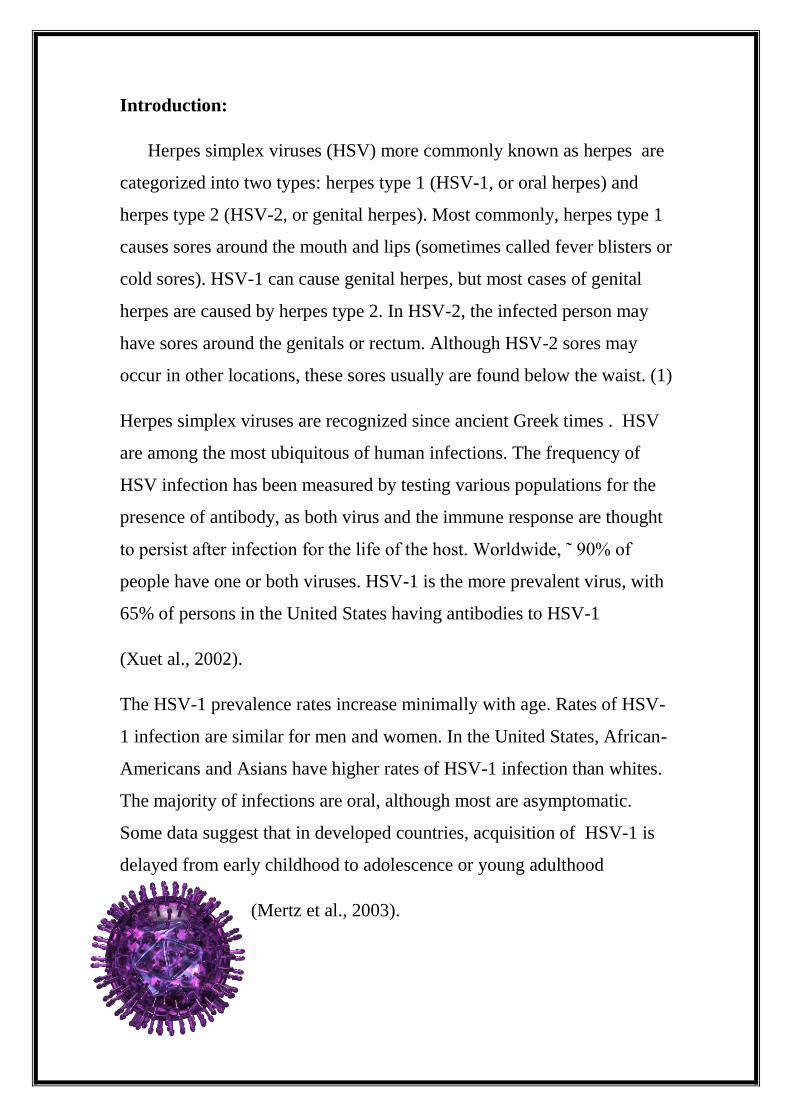
Introduction:
Herpes simplex viruses (HSV) more commonly known as herpes are
categorized into two types: herpes type 1 (HSV-1, or oral herpes) and
herpes type 2 (HSV-2, or genital herpes). Most commonly, herpes type 1
causes sores around the mouth and lips (sometimes called fever blisters or
cold sores). HSV-1 can cause genital herpes, but most cases of genital
herpes are caused by herpes type 2. In HSV-2, the infected person may
have sores around the genitals or rectum. Although HSV-2 sores may
occur in other locations, these sores usually are found below the waist. (1)
Herpes simplex viruses are recognized since ancient Greek times . HSV
are among the most ubiquitous of human infections. The frequency of
HSV infection has been measured by testing various populations for the
presence of antibody, as both virus and the immune response are thought
to persist after infection for the life of the host. Worldwide, ˜ 90% of
people have one or both viruses. HSV-1 is the more prevalent virus, with
65% of persons in the United States having antibodies to HSV-1
(Xuet al., 2002).
The HSV-1 prevalence rates increase minimally with age. Rates of HSV-
1 infection are similar for men and women. In the United States, African-
Americans and Asians have higher rates of HSV-1 infection than whites.
The majority of infections are oral, although most are asymptomatic.
Some data suggest that in developed countries, acquisition of HSV-1 is
delayed from early childhood to adolescence or young adulthood
(Mertz et al., 2003).
Genital herpes caused by HSV-2 is a global issue, and an estimated 417
million people worldwide were living with the infection in 2012.
Prevalence of HSV-2 infection was estimated to be highest in Africa
(31.5%), followed by the Americas (14.4%). It was also shown to
increase with age, though the highest numbers of people newly-infected
were adolescents. (2)
More women are infected with HSV-2 than men; in 2012 it was estimated
that 267 million women and 150 million men were living with the
infection. This is because sexual transmission of HSV is more efficient
from men to women than from women to men. (2)
Classification:
Order: Group I ( dsDNA )
Family: Herpesviridae
Subfamily: Alphaherpesvirinae
Genus: Simplexvirus
Species: Herpes simplex virus 1 (HSV-1)
Herpes simplex virus 2 (HSV-2) (3)
Structure and genome:
HSV-1 and HSV-2 contain a large double-stranded DNA linear
molecule.
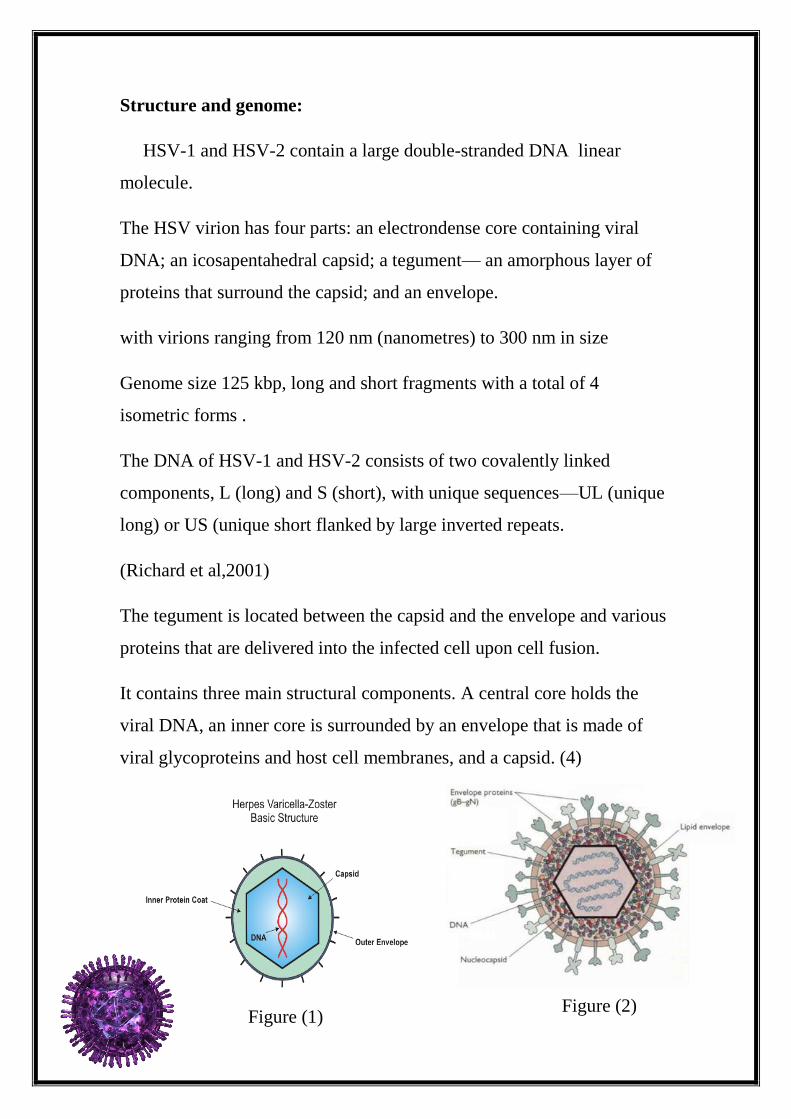
The HSV virion has four parts: an electrondense core containing viral
DNA; an icosapentahedral capsid; a tegument— an amorphous layer of
proteins that surround the capsid; and an envelope.
with virions ranging from 120 nm (nanometres) to 300 nm in size
Genome size 125 kbp, long and short fragments with a total of 4
isometric forms .
The DNA of HSV-1 and HSV-2 consists of two covalently linked
components, L (long) and S (short), with unique sequences—UL (unique
long) or US (unique short flanked by large inverted repeats.
(Richard et al,2001)
The tegument is located between the capsid and the envelope and various
proteins that are delivered into the infected cell upon cell fusion.
It contains three main structural components. A central core holds the
viral DNA, an inner core is surrounded by an envelope that is made of
viral glycoproteins and host cell membranes, and a capsid. (4)
Figure (2) Figure (1)
Virulence factors:
There are about 40 to 50 different proteins in the virion ("virion
structural proteins"). Some of these proteins make up the icosahedral
capsid, some make up the tegumet, and some are the glycoproteins in the
envelope,When the virus is attached, the viral envelope will fuse with the
cellular membrane, leading to the release of proteins from the tegument
and from the nucleocapsid into the cytoplasm the viral genome is rapidly
circularized in the absence of any viral protein synthesis suggesting a
mechanism under dependence of cellular proteins and/or structural viral
proteins. The transcription of the herpesviruses’ genome then proceeds.
(Thomas C. Mettenleiter,2002).
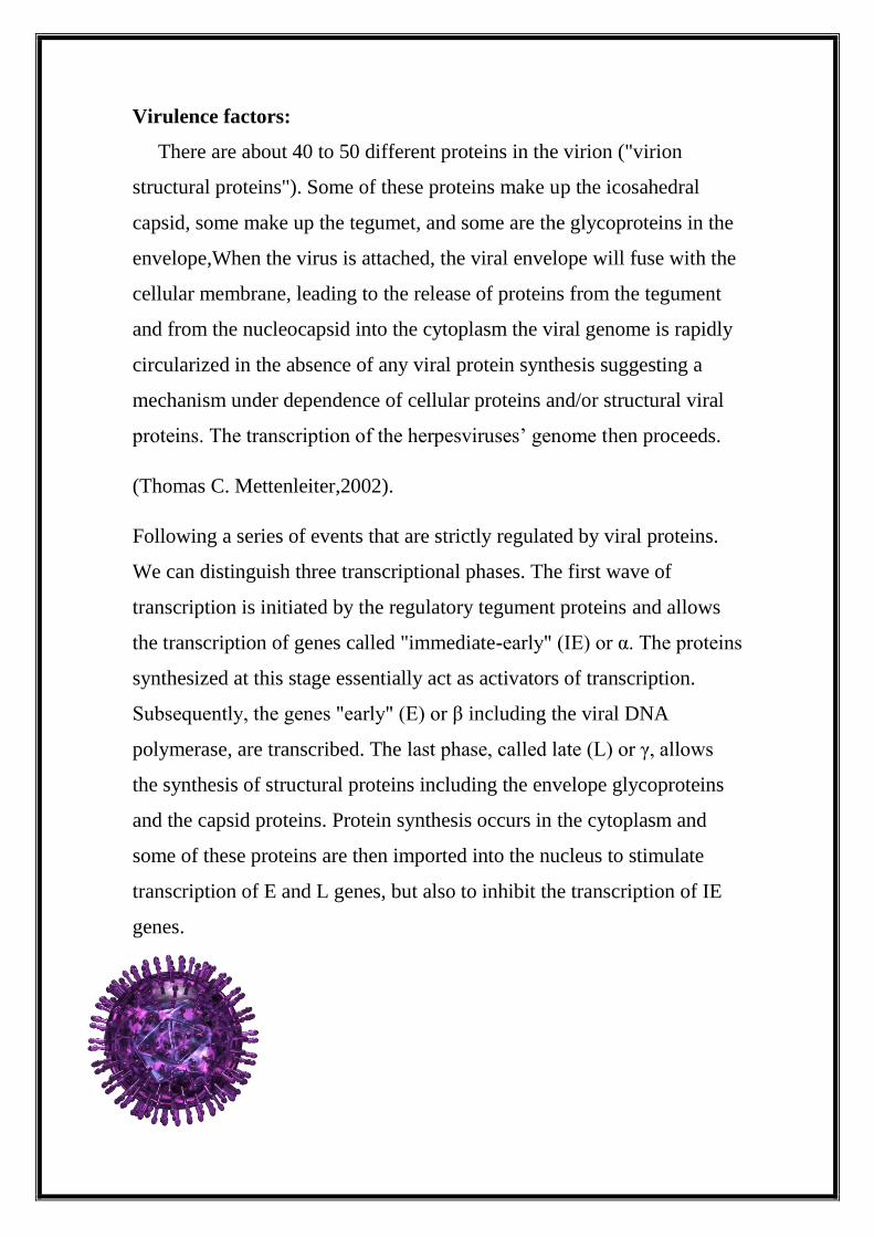
Following a series of events that are strictly regulated by viral proteins.
We can distinguish three transcriptional phases. The first wave of
transcription is initiated by the regulatory tegument proteins and allows
the transcription of genes called "immediate-early" (IE) or α. The proteins
synthesized at this stage essentially act as activators of transcription.
Subsequently, the genes "early" (E) or β including the viral DNA
polymerase, are transcribed. The last phase, called late (L) or γ, allows
the synthesis of structural proteins including the envelope glycoproteins
and the capsid proteins. Protein synthesis occurs in the cytoplasm and
some of these proteins are then imported into the nucleus to stimulate
transcription of E and L genes, but also to inhibit the transcription of IE
genes.
These steps are explained in the following picture :
Figure (4)
Figure (3)
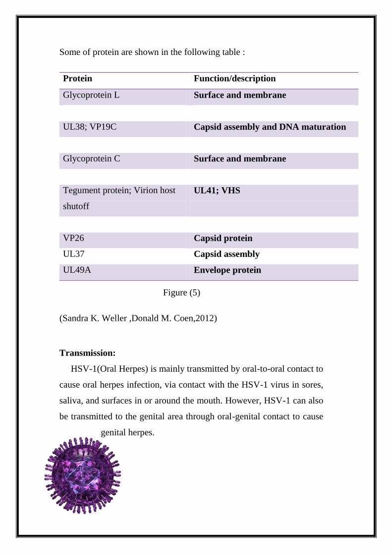
Some of protein are shown in the following table :
(Sandra K. Weller ,Donald M. Coen,2012)
Transmission:
HSV-1(Oral Herpes) is mainly transmitted by oral-to-oral contact to
cause oral herpes infection, via contact with the HSV-1 virus in sores,
saliva, and surfaces in or around the mouth. However, HSV-1 can also
be transmitted to the genital area through oral-genital contact to cause
genital herpes.
Function/description Protein
Surface and membrane Glycoprotein L
Capsid assembly and DNA maturation UL38; VP19C
Surface and membrane Glycoprotein C
UL41; VHS Tegument protein; Virion host
shutoff
Capsid protein VP26
Capsid assembly UL37
Envelope protein UL49A
Figure (5)
HSV-1 can be transmitted from oral or skin surfaces that appear
normal and when there are no symptoms present. However, the
greatest risk of transmission is when there are active sores.
Individuals who already have HSV-1 oral herpes infection are unlikely
to be subsequently infected with HSV-1 in the genital area.
In rare circumstances, HSV-1 infection can be transmitted from a
mother with genital HSV-1 infection to her infant during delivery.
HSV-2 (Genital Herpes) infections often have no symptoms, or mild
symptoms that go unrecognized. Most infected people are unaware that
they have the infection. Typically, about 10-20% of people with HSV-
2 infection report a prior diagnosis of genital herpes.
When symptoms do occur, genital herpes is characterized by one or
more genital or anal blisters or open sores called ulcers. In addition to
genital ulcers, symptoms of new genital herpes infections often include
fever, body aches, and swollen lymph nodes.
After an initial genital herpes infection with HSV-2, recurrent
symptoms are common but often less severe than the first outbreak.
The frequency of outbreaks tends to decrease over time. People
infected with HSV-2 may experience sensations of mild tingling or
shooting pain in the legs, hips, and buttocks before the occurrence of
genital ulcers.(2)
Penetration:
Attachment For HSV cell surface heparin sulphateis major binding factor.
Removal of HS does not remove attachment completely.
Most herpes viruses use more than one attachment pathway
Penetration Mediated by viral surface proteins –fusion of viral envelope
with cell plasma membrane.
gB, gD and gH are all involved in fusion.
Transport Release of viral DNA into the nucleus is mediated by an
unidentified viral function. (5)
Animation of penetration. (6)
Replication cycle:
Penetration , The nucleocapsid enters the cell by direct membrane fusion
plasma membrane .
In the lytic cycle, HSV infects epithelial cells located in the mucosa,
replicates, and causes epithelial cell death . HSV-1 most frequently
invades oral and ocular epithelial cells while HSV-2 infects the genital
areas, but both strains have the ability to cause infection in either area of
the body. (4)
Viral DNA into the nucleus. HSV replicates by three rounds of
transcription that yield: (immediate early) proteins that mainly regulate
viral replication; (early) proteins that synthesize and package DNA; and
(late) proteins, most of which are virion proteins.
Of the 84 known polypeptides, at least 47 are not needed for viral
replication in cultured 3 cells .
These 47 genes are not completely dispensable.
Richard et al ,2001) )
Central nervous system HSV replication in the brain causes encephalitis
This is a life-threatening disease with a high mortality that causes
permanent neurological damage in those who survive primary infection
or secondary infection, and it is unclear how the virus enters the central
nervous system.
Fortunately, the condition is rare (with an incidence of approximately 1
in 200 000 in the USA) and, if diagnosed at an early stage, responds to
treatment with antiviral agents; these have significantly reduced the
associated mortality and morbidity There is also epidemiological
evidence that the presence of HSV-1 DNA in the brain might be
associated with Alzheimer’s disease, although no molecular mechanism
for such a link has been proposed Congenital and neonatal infection
Generalised HSV infection of the neonate has a very high mortality .
Infection is often acquired during passage through the birth canal but can
also be acquired in utero. Caesarian section has been used in mothers
with genital ulcers in an attempt to minimise the risks of peripartum
transmission.
a) Herpes simplex virus (HSV) is shown undergoing the lytic cycle
(entry, uncoating, viral transcription and DNA replication in the nucleus,
particle assembly, exit from the cell) in epithelial cells of the skin to
cause a primary infection.
(b) Some virus enters the sensory neuron terminals and travels
retrogradely to the nucleus where it establishes latency.
(c) Periodic reactivation results in anterograde transport of viral particles,
shedding from the neuron, and re-infection of epithelial cells, which leads
to asymptomatic shedding or recurrent lesions .
expression is dependent, to varying degrees, on prior synthesis of viral
DNA. The products of the L genes are mostly structural components of
the virion Viral protein synthesis occurs in the cytoplasm but the
nucleocapsids are assembled within the nucleus. These
nucleocapsidsrecognise the nascent viral genomes, which are cleaved into
monomers as they are packaged. The completed nucleocapsids exit the
nucleus by budding through the nuclear membrane.
The virus then exits the cell by traversing several membrane-bound
organelles, where it acquires its mature envelope and glycoproteins
Eventual cell lysis is the inevitable outcome of lytic infection with HSV-
in tissue culture cells.
(Robin . 2003)
Animation of replication cycle: (7)
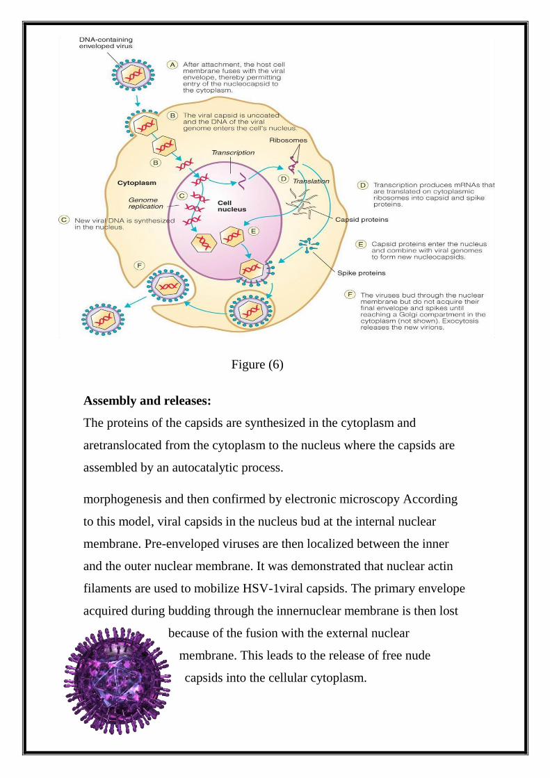
Assembly and releases:
The proteins of the capsids are synthesized in the cytoplasm and
aretranslocated from the cytoplasm to the nucleus where the capsids are
assembled by an autocatalytic process.
morphogenesis and then confirmed by electronic microscopy According
to this model, viral capsids in the nucleus bud at the internal nuclear
membrane. Pre-enveloped viruses are then localized between the inner
and the outer nuclear membrane. It was demonstrated that nuclear actin
filaments are used to mobilize HSV-1viral capsids. The primary envelope
acquired during budding through the innernuclear membrane is then lost
because of the fusion with the external nuclear
membrane. This leads to the release of free nude
capsids into the cellular cytoplasm.
Figure (6)
Capsids then transit in the cytoplasm to acquire, on one hand, tegument
proteins and, on the other hand, envelope glycoproteins by budding in
Golgi apparatus vesicles. Mature virions are then released at the surface
of the cell by exocytosis.
(Sandra K. Weller ,Donald M. Coen,2012)
Signs and Symptoms:
Many people who get the virus that causes herpes never see or feel
anything since after the first infection, the virus goes to sleep (becomes
dormant) in the nerve tissues . Sometimes, the virus later wakes up
(reactivates), causing cold sores. (1)
Oral Herpes Symptoms:
There are three stages to oral herpes after being infected:
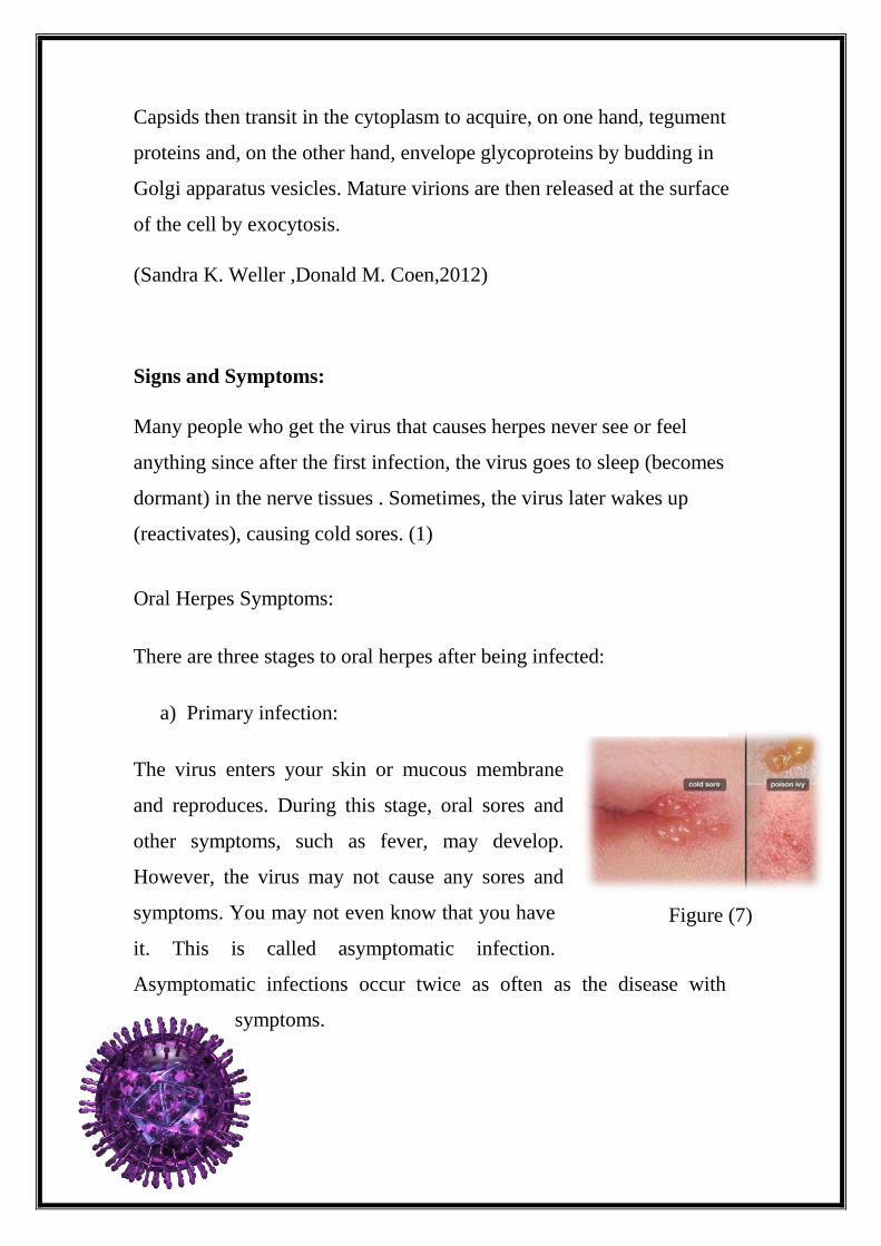
a) Primary infection:
The virus enters your skin or mucous membrane
and reproduces. During this stage, oral sores and
other symptoms, such as fever, may develop.
However, the virus may not cause any sores and
symptoms. You may not even know that you have
it. This is called asymptomatic infection.
Asymptomatic infections occur twice as often as the disease with
symptoms.
Figure (7)
b) Latency:
From the infected site, the virus moves to a mass of nervous tissue in
spine called the dorsal root ganglion. There, the virus reproduces again
and becomes inactive.
c) Recurrence:
When you experience certain emotional or physical stresses, the virus
may reactivate and cause new sores and symptoms. One such stress
may be a viral illness such as the common cold, hence the frequently
used name of cold sores.(8)
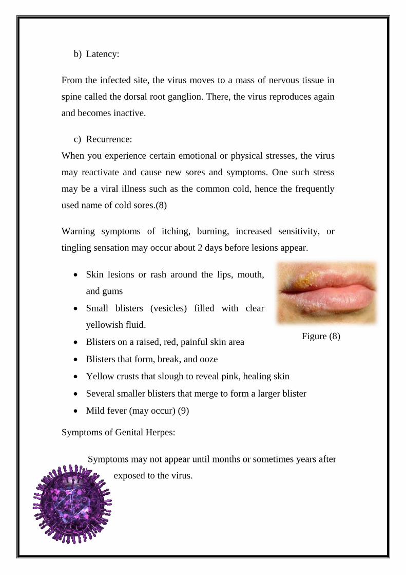
Warning symptoms of itching, burning, increased sensitivity, or
tingling sensation may occur about 2 days before lesions appear.
Skin lesions or rash around the lips, mouth,
and gums
Small blisters (vesicles) filled with clear
yellowish fluid.
Blisters on a raised, red, painful skin area
Blisters that form, break, and ooze
Yellow crusts that slough to reveal pink, healing skin
Several smaller blisters that merge to form a larger blister
Mild fever (may occur) (9)
Symptoms of Genital Herpes:
Symptoms may not appear until months or sometimes years after
exposed to the virus.
Figure (8)
If symptoms was for the first infected, they usually appear four to seven
days after exposed to the virus. The symptoms are usually more severe
first time around than in cases of recurrent infections.
a) Primary infection:
The symptoms of genital herpes for the first time include:
small blisters that burst to leave red, open sores around genitals,
rectum (back passage), thighs and buttocks
blisters and ulcers on the cervix (lower part of the womb) in
women
vaginal discharge in women
pain when you pass urine
a general feeling of being unwell, with aches, pains and flu-like
symptoms
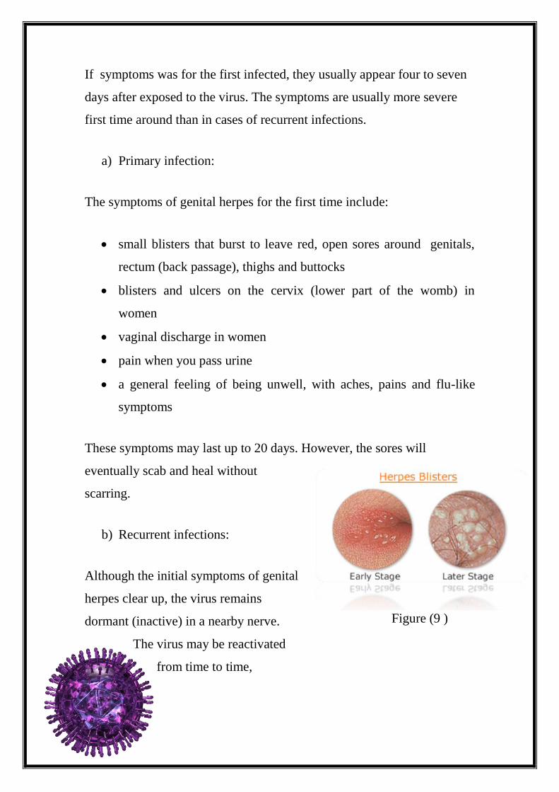
These symptoms may last up to 20 days. However, the sores will
eventually scab and heal without
scarring.
b) Recurrent infections:
Although the initial symptoms of genital
herpes clear up, the virus remains
dormant (inactive) in a nearby nerve.
The virus may be reactivated
from time to time,
Figure (9 )
travelling back down the nerve to skin and causing recurrent outbreaks.
Symptoms of a recurrent outbreak may include:
a tingling, burning or itching sensation around genitals, and
sometimes down to leg, before blisters appear
painful red blisters that soon burst to leave sores around genitals,
rectum (back passage), thighs and buttocks
blisters and ulcers on the cervix (lower part of the womb) in
women.
Recurrent outbreaks are usually shorter and less severe. This is because
the body has produced protective antibodies (proteins that fight infection)
in reaction to the previous infection. Your body now recognises the virus
and mounts a response that is able to fight HSV more effectively.
Over time, any recurrent genital herpes infections become less frequent
and less severe. (10)
Diagnosis and Cytopathic effect:
1. Laboratory tests:
Traditionally, tests involve taking a swab from an active lesion, growing
the virus in the laboratory, and using colour-coded antibodies to pinpoint
whether HSV-1 or HSV-2 is the culprit.
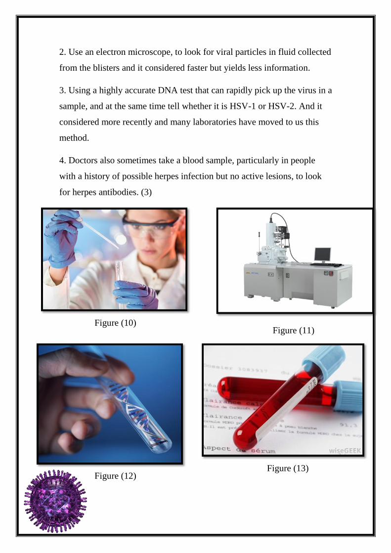
2. Use an electron microscope, to look for viral particles in fluid collected
from the blisters and it considered faster but yields less information.
3. Using a highly accurate DNA test that can rapidly pick up the virus in a
sample, and at the same time tell whether it is HSV-1 or HSV-2. And it
considered more recently and many laboratories have moved to us this
method.
4. Doctors also sometimes take a blood sample, particularly in people
with a history of possible herpes infection but no active lesions, to look
for herpes antibodies. (3)
Figure (10) Figure (11)
Figure (13) Figure (12)
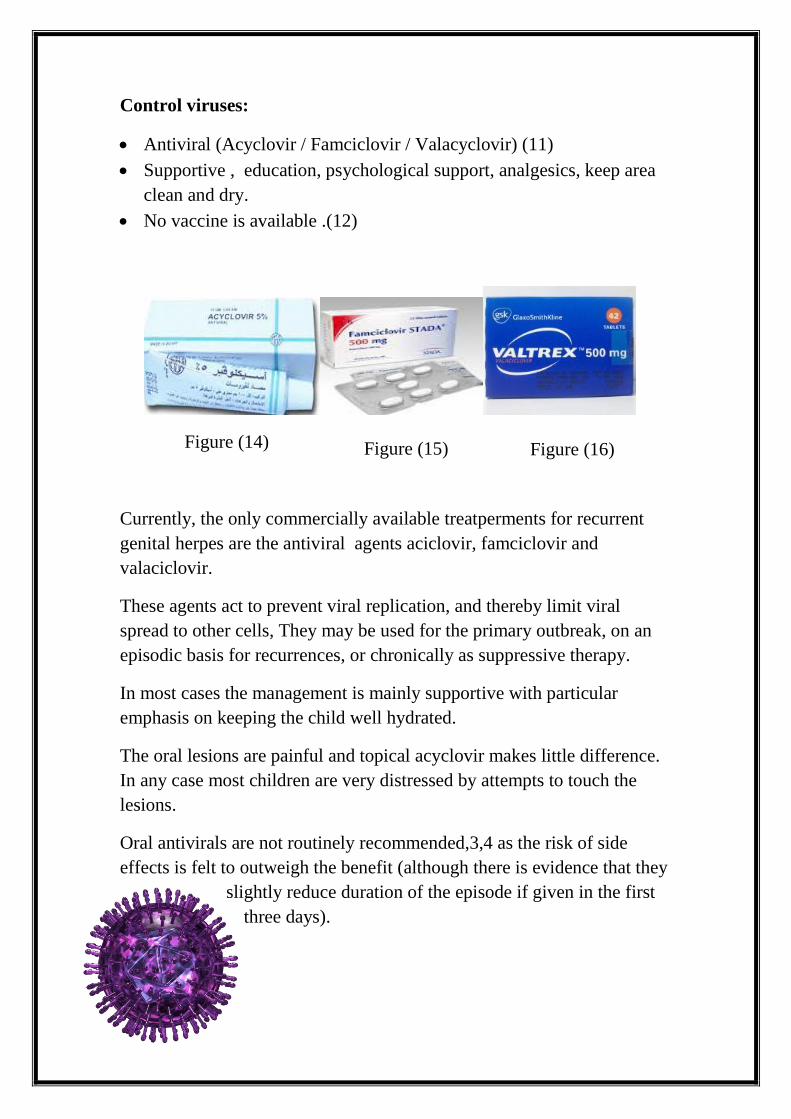
Control viruses:
Antiviral (Acyclovir / Famciclovir / Valacyclovir) (11)
Supportive , education, psychological support, analgesics, keep area
clean and dry.
No vaccine is available .(12)
Currently, the only commercially available treatperments for recurrent
genital herpes are the antiviral agents aciclovir, famciclovir and
valaciclovir.
These agents act to prevent viral replication, and thereby limit viral
spread to other cells, They may be used for the primary outbreak, on an
episodic basis for recurrences, or chronically as suppressive therapy.
In most cases the management is mainly supportive with particular
emphasis on keeping the child well hydrated.
The oral lesions are painful and topical acyclovir makes little difference.
In any case most children are very distressed by attempts to touch the
lesions.
Oral antivirals are not routinely recommended,3,4 as the risk of side
effects is felt to outweigh the benefit (although there is evidence that they
slightly reduce duration of the episode if given in the first
three days).
Figure (14) Figure (16) Figure (15)
They are sometimes used for children admitted to hospital with
dehydration secondary to gingivostomatitis.
(Lowth et al, 2014)
Recent discoveries:
The initial study population comprised 351,865 individuals, of whom
42,954 zoster cases with incident ischemic stroke and 24,237 zoster cases
with acute MI fulfilled the eligibility criteria and were included in the
primary analysis (Figure 17). Characteristics of these individuals are
presented in Table 18. The median age at zoster diagnosis was 80 y
(interquartile range [IQR] 74–86 y), and the median observation period
was 5 y (IQR 4–5 y). The majority of participants were female (71% of
zoster cases with stroke, 64% of zoster cases with MI); 89% of
participants were white (88% of strokes, 90% of MIs), 5% were black,
and the remaining 6% were Asian (2%), Hispanic (2%), or of
other/unknown ethnicity (2%). In all, 16% of zoster cases had HZO; the
remaining 84% had zoster of an unspecified site. Also, 34% of cases were
of low income, and 90% had evidence of preexisting CVD before zoster
diagnosis.
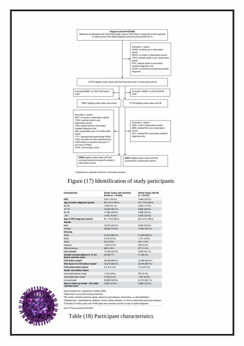
Figure (17) Identification of study participants
Table (18) Participant characteristics
A small minority of cases received the zoster vaccine before developing
zoster (3% of cases with stroke, 2% of cases with MI), 6% received the
vaccine after zoster diagnosis, and 91% were unvaccinated throughout the
observation period. Characteristics of individuals included in the analyses
stratified by zoster vaccination status are given in S1 Table. Vaccinated
and unvaccinated individuals had similar median age, gender, and
preexisting CVD risk profiles, although unvaccinated individuals were
more than twice as likely to be of low income and received more
prescriptions in the year leading up to their vascular event compared to
vaccinated individuals. Ethnicity data among vaccinated individuals
could not be reported because some numbers were small enough to be
restricted by the CMS small-sized-cell privacy policy.
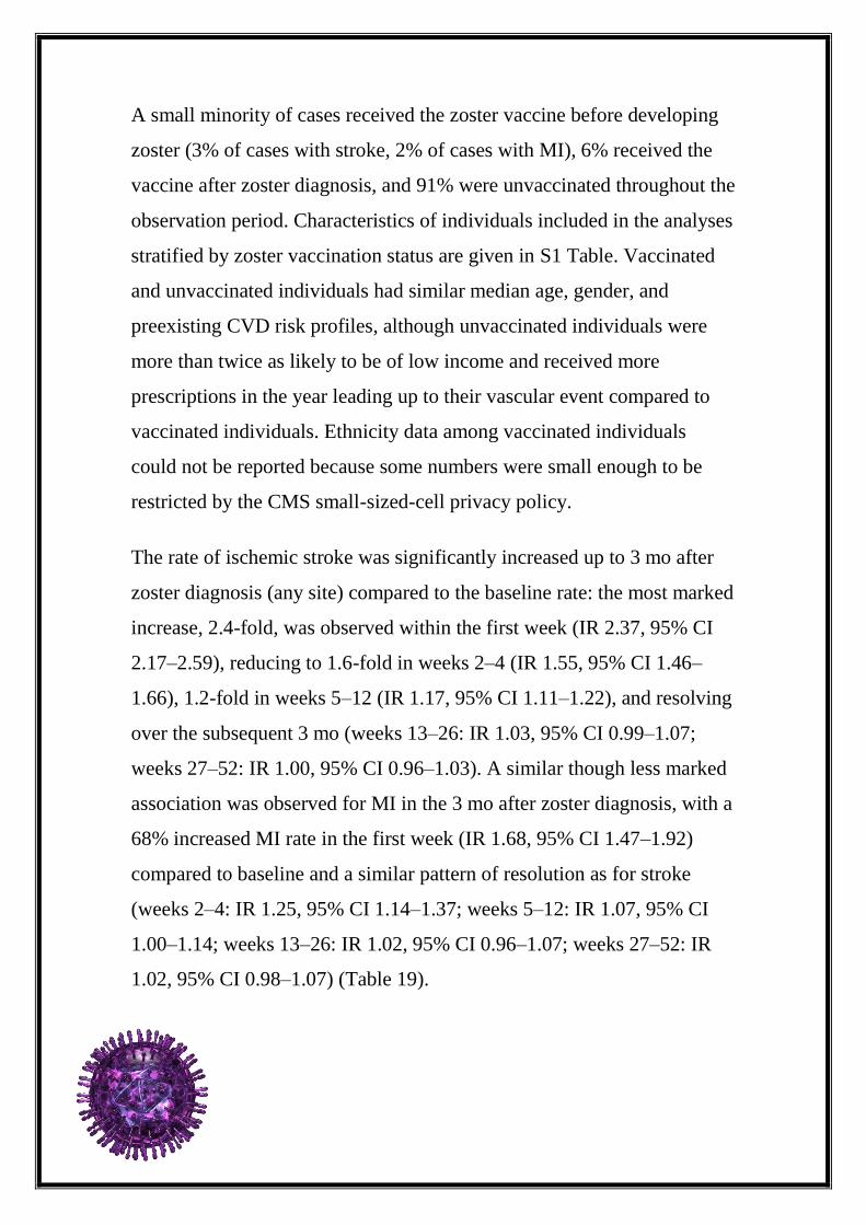
The rate of ischemic stroke was significantly increased up to 3 mo after
zoster diagnosis (any site) compared to the baseline rate: the most marked
increase, 2.4-fold, was observed within the first week (IR 2.37, 95% CI
2.17–2.59), reducing to 1.6-fold in weeks 2–4 (IR 1.55, 95% CI 1.46–
1.66), 1.2-fold in weeks 5–12 (IR 1.17, 95% CI 1.11–1.22), and resolving
over the subsequent 3 mo (weeks 13–26: IR 1.03, 95% CI 0.99–1.07;
weeks 27–52: IR 1.00, 95% CI 0.96–1.03). A similar though less marked
association was observed for MI in the 3 mo after zoster diagnosis, with a
68% increased MI rate in the first week (IR 1.68, 95% CI 1.47–1.92)
compared to baseline and a similar pattern of resolution as for stroke
(weeks 2–4: IR 1.25, 95% CI 1.14–1.37; weeks 5–12: IR 1.07, 95% CI
1.00–1.14; weeks 13–26: IR 1.02, 95% CI 0.96–1.07; weeks 27–52: IR
1.02, 95% CI 0.98–1.07) (Table 19).
Analyses restricted to cases with HZO (n = 6,971 with ischemic stroke,
3,946 with MI) yielded associations comparable to those of the primary
analysis (week 1 after HZO diagnosis: stroke IR 2.73, 95% CI 2.22–3.35;
MI IR 2.06, 95% CI 1.52–2.79) that resolved over the same time period
(Table 20).
Stratifying by zoster vaccination status revealed no evidence for a
reduced IR for ischemic stroke during the first 4 wk after zoster diagnosis
among individuals who received the zoster vaccine (n = 843) (IR 1.14,
95% CI 0.75–1.74) compared to unvaccinated individuals (n = 40,724)
(IR 1.78, 95% CI 1.68–1.88) (p-value for interaction = 0.28). The overall
IR combining vaccinated and unvaccinated individuals for the same 4-wk
post-zoster period was 1.76 (95% CI 1.67–1.86). There was no evidence
that the IR for MI after zoster diagnosis varied according to zoster
vaccination status (p = 0.44): the IR in weeks 1–4 after zoster diagnosis
was 1.36 (95% CI 0.78–2.39) in vaccinated individuals (n = 400) and
Table (19) Primary analysis: age-adjusted incidence ratios for ischemic stroke and
myocardial infarction in risk periods after zoster diagnosis.
Table (20) Age-adjusted incidence ratios for ischemic stroke and
myocardial infarction in risk periods after herpes zoster ophthalmicus.
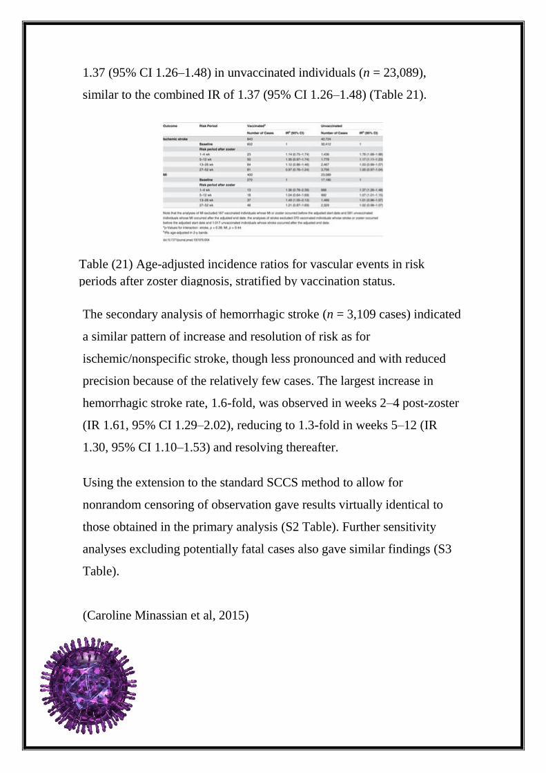
1.37 (95% CI 1.26–1.48) in unvaccinated individuals (n = 23,089),
similar to the combined IR of 1.37 (95% CI 1.26–1.48) (Table 21).
The secondary analysis of hemorrhagic stroke (n = 3,109 cases) indicated
a similar pattern of increase and resolution of risk as for
ischemic/nonspecific stroke, though less pronounced and with reduced
precision because of the relatively few cases. The largest increase in
hemorrhagic stroke rate, 1.6-fold, was observed in weeks 2–4 post-zoster
(IR 1.61, 95% CI 1.29–2.02), reducing to 1.3-fold in weeks 5–12 (IR
1.30, 95% CI 1.10–1.53) and resolving thereafter.
Using the extension to the standard SCCS method to allow for
nonrandom censoring of observation gave results virtually identical to
those obtained in the primary analysis (S2 Table). Further sensitivity
analyses excluding potentially fatal cases also gave similar findings (S3
Table).
(Caroline Minassian et al, 2015)
Table (21) Age-adjusted incidence ratios for vascular events in risk
periods after zoster diagnosis, stratified by vaccination status.
References:
Caroline Minassian, Sara L. Thomas, Liam Smeeth, Ian Douglas, Ruth
Brauer, Sinead M.Langan, Acute Cardiovascular Events after Heroes
Zoster: A Self-Controlled Case Series Analysis in Vaccinated and
Unvaccinated Older Residents of the United States, 2015.
Lowth. Mary, Practice Nurse, 2014.
Mertz G. J., Rosenthal S. L., Stanberry L. R, Is herpes simplex virus
type 1 (HSV-1) now more common than HSV-2 in first episodes of
genital herpes? Sex. Transm. Dis, 2003.
Richard J Whitley, Bernard Roizman, Copyright review, 2001.
Robin Lachmann, expert reviews, 2003.
Thomas C. Mettenleiter, Herpesvirus Assembly and Egress,2002.
Sandra K. Weller ,Donald M. Coen, REPLICATION OF Dsdna
GENOME VIRUSES A. Herpesviruses, 2012.
Xu F., Schillinger J. A., Sternberg M. R., Seroprevalence and
coinfection with herpes simplex virus type 1 and type 2 in the United
States, Infect. Dis, 2002.
1- http://www.webmd.com/genital-herpes/pain-management-herpes
2- http://www.who.int/mediacentre/factsheets/fs400/en/
3- http://slideplayer.com/slide/5713283/
4-
http://www.bio.davidson.edu/people/sosarafova/assets/bio307/jehodge/pa
ge01.html
5-
http://slideplayer.com/slide/3863503/
6- https://www.youtube.com/watch?v=moBtPyuPXrE
7-
http://www.sumanasinc.com/webcontent/animations/content/herpessimpl
ex.html
8- http://www.ashasexualhealth.org/stdsstis/herpes/oral-herpes/
9- http://www.pkids.org/diseases/herpes/oral_herpes.html
10- http://www.nhs.uk/Conditions/Genital-
herpes/Pages/Symptoms.aspx
11-
http://courses.washington.edu/medch401/pdf_text/401_07_Herpes.pdf
12-
http://www.google.com.sa/url?sa=t&rct=j&q=&esrc=s&source=web&cd
=1&ved=0ahUKEwjN_4mWpP7LAhVIHxoKHUBGCN8QFggbMAA&
url=http%3A%2F%2Fwww.infectiousdiseases.utoronto.ca%2FAssets%2
FInfectious%2BDiseases%2BDigital%2BAssets%2FInfectious%2BDisea
ses%2FInfectious%2BDiseases%2BDigital%2BAssets%2FHalf-
day%2Bseminars%2FHerpesviridae1.ppt&usg=AFQjCNGuCwJXIe_xG
XsKw_8vpfw5hMWWwg&sig2=7tzIMA-sHkqbc94OiRmPeA