Bones, Joints and Soft tissue tumors
13
Pathology MSS Done By Samah Freihat , Tasneem Al Oqaily Corrected By Dana Yousef
Transcript of Bones, Joints and Soft tissue tumors

PathologyMSS
Done By Samah Freihat , Tasneem Al Oqaily
Corrected By Dana Yousef

Lecture 6
we talked in lec #5 about bone and cartilaginous tumor, we mentioned that there are certain bone tumors in which we don't know their cell of origin .
Example, EWING SARCOMA
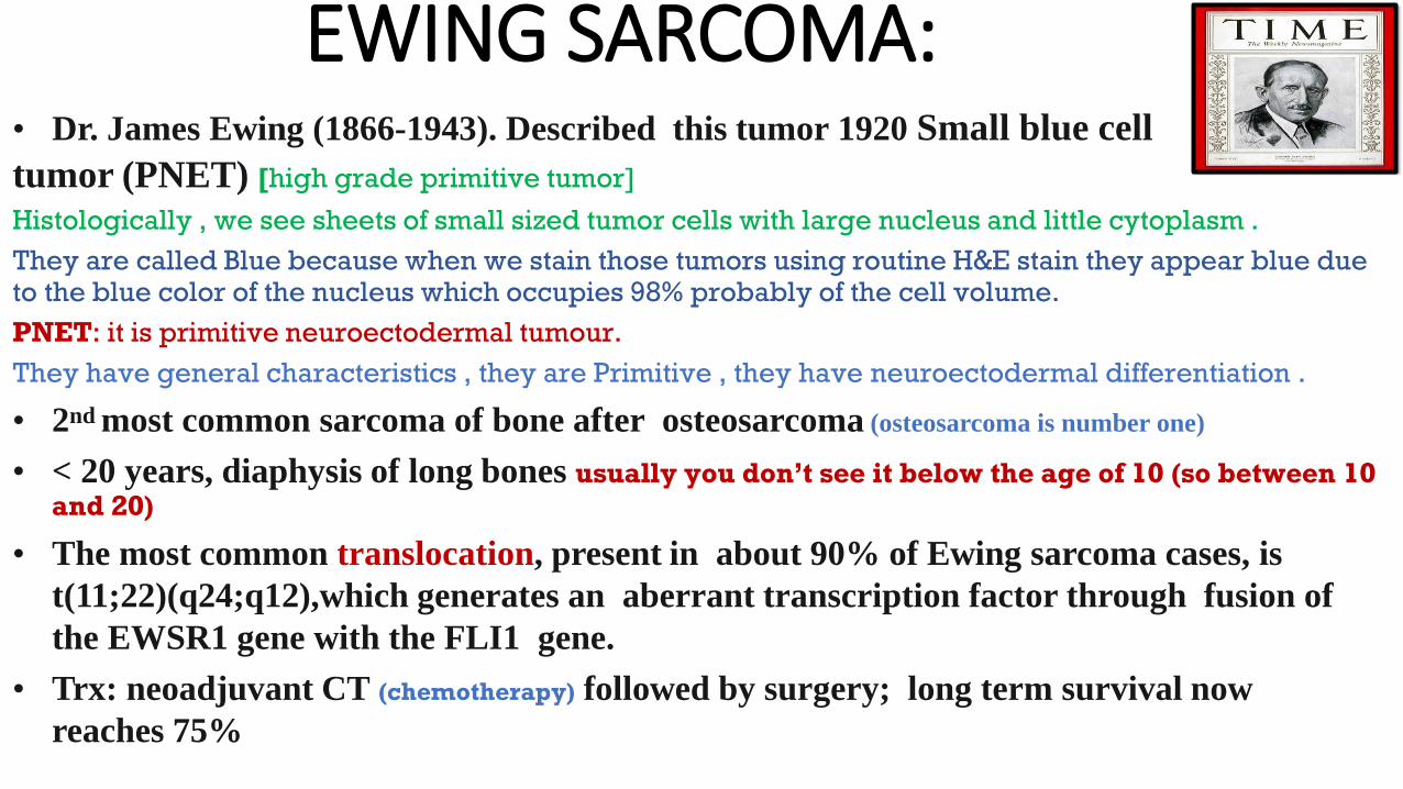
EWING SARCOMA:• Dr. James Ewing (1866-1943). Described this tumor 1920 Small blue cell
tumor (PNET) [high grade primitive tumor]
Histologically , we see sheets of small sized tumor cells with large nucleus and little cytoplasm .
They are called Blue because when we stain those tumors using routine H&E stain they appear blue due to the blue color of the nucleus which occupies 98% probably of the cell volume.
PNET: it is primitive neuroectodermal tumour.
They have general characteristics , they are Primitive , they have neuroectodermal differentiation .
• 2nd most common sarcoma of bone after osteosarcoma (osteosarcoma is number one)
• < 20 years, diaphysis of long bones usually you don’t see it below the age of 10 (so between 10 and 20)
• The most common translocation, present in about 90% of Ewing sarcoma cases, is
t(11;22)(q24;q12),which generates an aberrant transcription factor through fusion of
the EWSR1 gene with the FLI1 gene.
• Trx: neoadjuvant CT (chemotherapy) followed by surgery; long term survival now
reaches 75%

ES FEATURES:This is the diaphysis of the humerus, notice that this tumor infiltrates the soft tissue and elevates the periosteum causing codman triangle which helps you understand that codman triangle isn't specific for osteosarcoma only
a lot of blue cell tumor destroying the bone shown in this histological section
This is translocation t (11,22), there is 2 of them actually(EWS FLI1), (EWS FLI2).this is a picture of fish
analysis, so this is probably the most sensitive test for Ewing sarcoma using florescent In SituHybridization (FISH).
this is the old method by classic cytogenetic analysis, where the translocation occurs between the chromosome 11 & chromosome 22 (11 is a bigger chromosome compared to 22) And the fusion protein product from this translocation.
So these are radiologic, molecular and histologic features of Ewing sarcoma.

GIANT CELL TUMOR OF BONE
- Locally aggressive neoplasm of adults you don’t see it in
children and you don’t see it at the age of 15 or even 20
- Epiphyses of long bones
- Osteoclast-like giant cells
- Rare malignant behavior 95% of the cases it behaves in a benign
fashion (you can cure it; you can resect it)
- contain high levels of RANKL (which stimulates the
differentiation of osteoclasts)
- Trx: curetting and put a bone cement or resection
we don't know the cell of origin the location and the age are important to help you narrow your diagnosis

histological appearance of benign tumor: bubble appearance expanding the cortex of the bonewithout infiltration to the extra cortical spaceand if the patient is 45 years old, this is the most Common differential of giant cell tumor of bone, so they go in and take a biopsy and sometimes they just Resect it.
histologically you see sheets , wall to wall multi nucleated giant cells or osteoclast-like giant cells
so the tumor cells are the giant cells and the one in between (single mononuclear cells)
sometimes it is called osteoclastoma because the primary histology is composed of numerous wall to wall osteoclast like multi-nucleated giant cells
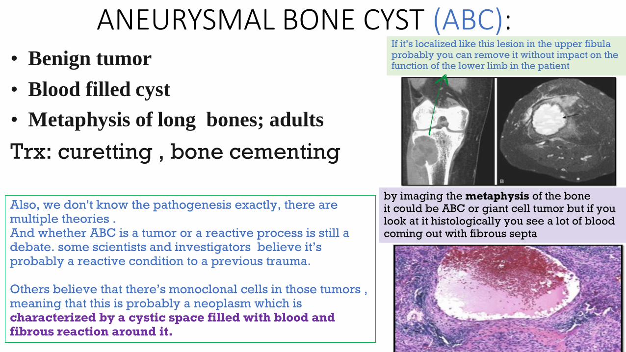
ANEURYSMAL BONE CYST (ABC):
• Benign tumor
• Benign tumor
• Blood filled cyst
• Metaphysis of long bones; adults
Also, we don't know the pathogenesis exactly, there are multiple theories .And whether ABC is a tumor or a reactive process is still a debate. some scientists and investigators believe it’s probably a reactive condition to a previous trauma.
Others believe that there’s monoclonal cells in those tumors , meaning that this is probably a neoplasm which is characterized by a cystic space filled with blood and fibrous reaction around it.
Trx: curetting , bone cementing
by imaging the metaphysis of the boneit could be ABC or giant cell tumor but if you look at it histologically you see a lot of blood coming out with fibrous septa
If it’s localized like this lesion in the upper fibula probably you can remove it without impact on the function of the lower limb in the patient

NONOSSIFYING FIBROMA:• Benign lesion, maybe reactive not a true
neoplasm (other names: FCD, MFD
• Metaphysis
• Histology: bland fibroblastic
proliferation
• May resolve spontaneously
fibroma in the bone
looks like a benign fibroma or a fibroblast .There are some multi nucleated giant cells but they’re not common (there is just five or six in here)
This is the bone (tibia) and there is a lesion here which is not destroying the surrounding structure, it is not elevating the periosteum and it is well circumscribed.

FIBROUS DYSPLASIA (FD):
• Not a real tumor; rather a developmental abnormality of bone
genesis due to mutations in GNAS1 gene (cAMP mediated
osteoblast differentiation) and this will lead to abnormal bone formation. So some people
believe this is a tumor or a neoplasm, others believe this is rather a developmental dysplastic abnormal disease.
• Forms of FD: FD is actually a group of diseases or syndromes
– Monostotic: affecting one bone (common bones affected by this FD are the maxillary & mandibular
bones of the face causing what we call cherubism in children
– Polystotic: multiple bones
– Mazabraud syndrome: FD (monostotic or polyostotic) + soft tissue myxoma (which is not a
common tumor of soft tissue)
– McCune-Albright syndrome: polystotic FD + café- au-lait skin pigmentation
(multiple brownish pigmentation of the skin ) + endocrine abnormalities (precocious
puberty)
important

McCUNE-ALBRIGHT SYNDROME:
This is an abnormal bone feature . If you look at it , pagetdisease should also be considered
The radiology is slightly similar to paget disease , biopsy shows the appearance of Chinese letter
McCune Albright syndrome patient is a patient with polyostotic FD + café-au-lait skin pigmentation (and this is how it diffuse)+ endocrine abnormalities (precociouspuberty)
And this is cherubism in children , this patient has dysplasia of the maxillary bone
The femur, endocrine abnormality Biopsy, polyostotic fibrous dysplasia
if you take a biopsy and look at the abnormal trabeculae , you’ll notice This appearance of haphazard arrangement of bone trabeculae & the fibrous matrix in between , it reminds us with the Chinese letter , so this appearance is called Chinese letter appearance in histologySo the classic characteristic microscopic feature of fibrous dysplasia is Chinese letter appearance with abnormal development of the bone trabeculae and matrix So, you can differentiate histologically between Paget disease & McCune Albright syndrome
café- au-lait skin pigmentation
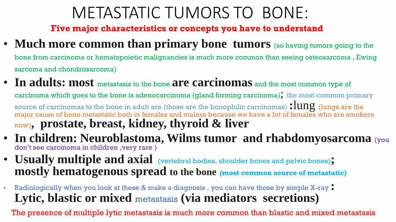
METASTATIC TUMORS TO BONE:
• Much more common than primary bone tumors (so having tumors going to the
bone from carcinoma or hematopoietic malignancies is much more common than seeing osteosarcoma , Ewing
sarcoma and chondrosarcoma)
• In adults: most metastasis to the bone are carcinomas and the most common type of
carcinoma which goes to the bone is adenocarcinoma (gland forming carcinoma); the most common primary
source of carcinomas to the bone in adult are (those are the bonophilic carcinomas) :lung (lungs are the major cause of bone metastatic both in females and maless because we have a lot of females who are smokers
now), prostate, breast, kidney, thyroid & liver
• In children: Neuroblastoma, Wilms tumor and rhabdomyosarcoma (you don’t see carcinoma in children ,very rare )
• Usually multiple and axial (vertebral bodies, shoulder bones and pelvic bones);mostly hematogenous spread to the bone (most common source of metastatic)
• Radiologically when you look at these & make a diagnosis , you can have those by simple X-ray : Lytic, blastic or mixed metastasis (via mediators secretions)
Five major characteristics or concepts you have to understand
The presence of multiple lytic metastasis is much more common than blastic and mixed metastasis

BLASTIC METASTASIS LYTIC METASTASIS
This is a patient with hundreds of blastic metastasis . The primary source here was the prostate, the prostate is commonly associated with blastic metastasis.When a Prostate tumor goes to the bone , it can cause blastic metastasis & it can also cause lytic, but the blastic is much more common than lytic in prostate malignancies
This is another patient were the bone is eaten in multiple areas (vertebral body, pelvic bone and the femur), this is bad prognosis so this is lytic metastasis . the most common primary of those is probably carcinoma (adenocarcinoma in lungs)this is stage 4 (bad prognosis), after this appearance most of patient don’t survive beyond 6-12 months.
This is how it look in x-ray
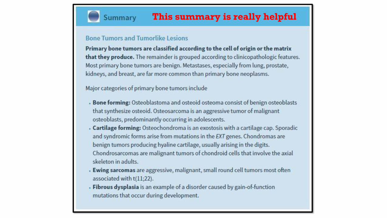
This summary is really helpful