‘Rischio residuo di anomalie cromosomiche dopo test di ... · Francesca Romana GRATI, Ph.D.,...
63
Francesca Romana GRATI, Ph.D., ErCLG R&D Director TOMA Advanced Biomedical Assays, S.p.A. [email protected] ‘Rischio residuo di anomalie cromosomiche dopo test di screening negativo per le maggiori aneuploidie’
Transcript of ‘Rischio residuo di anomalie cromosomiche dopo test di ... · Francesca Romana GRATI, Ph.D.,...

Francesca Romana GRATI, Ph.D., ErCLG
R&D Director
TOMA Advanced Biomedical Assays, S.p.A.
‘Rischio residuo di anomalie cromosomiche
dopo test di screening negativo per le maggiori
aneuploidie’

OUTLINES
Discuss on the ‘a priori’ and ‘post-test’ or ‘residual risk’ after a
negative test result
Implications of the residual risk for pre-test counseling before
screening test for common aneuploidies
Support to women’s decision autonomy
Resources
Tools
Education

Prenatal diagnosis of chromosome abnormalities
AF and CVS are carried out for a variety of reasons:
• fetal US abnormality/ies
• previous affected fetus/child
• parent carrier of a chromosome abnormality
• …
The current standard test is the karyotype/CMA on fetal samples:
• Chorionic villi sampling (CVS): 11-13wg
• Amniotic fluid sampling (AF): 16-18wg
Main indication: diagnosis of fetal aneuploidies, primarily trisomy 21

MATERNAL AGE AND TRISOMIES
An association between maternal age and trisomies: proneness of older
oocytes to maternal meiosis I and II non-disjunction errors.
Hassold et al, Ann Hum Genet. 1980 Jul;44(Pt 1):29-36; Hassold T, Hunt PA, Sherman S. Curr Opin Genet Dev. 1993 Jun;3(3):398-403;
Lamb et al Human Molecular Genetics, 1997, 1391–1399; Am. J. Hum. Genet. 76:91–99, 2005; Ferreira, Grati FR et al, Prenat Diagn.
2016 Dec;36(12):1146-1155;
Screening programs for T21
Developed starting from 70’s: evolved considerably in the last few decades
Recent developments in the cfDNA testing: DR ~99%; FPR<0.1%
1960 1980 1990 2005 2011
De
tect
ion
Ra
te
(%)

cfDNA testing performances: a meta-analysis
Gil et al, Ultrasound Obstet Gynecol. 2017 Apr 11. doi: 10.1002/uog.17484. [Epub
ahead of print]
DR FPR
n n n (95% CI) (95% CI)
99.7% 0.04%
(99.1-99.9) (0.02-0.08)
98.2% 0.05%
(95.5-99.2) (0.03-0.07)
99.0% 0.04%
(65.8-100) (0.02-0.07)
95.8% 0.14%
(70.3-99.5) (0.05-0.38)
100.0% 0.003%
(83.6-100) (0-0.07)
DR FPR
(95% CI) (95% CI)
100.0% 0%
(95.2-100) (0-0.003)
*peer-review studies reporting on clinical validation or implementation of maternal cfDNA testing in screening for
aneuploidies, in which data on pregnancy outcome were provided for more than 85% of the study population
(January 2011-31 December 2016)
Type of
aneuploidy
Twin pregnancies: weighted pooled
T21 8 24 1,111
45,X 23 36 7,677
other SCA 11 17 5,383
T18 25 560 212,019
T13 18 119 212,883
Type of
aneuploidy
number of
studies
trisomic
cases
non-trisomic
cases
Singleton pregnancies: weighted pooled
T21 30 1,963 225,032

CfDNA TESTING CANNOT DETECT ALL FETAL CHROMOSOME ABNORMALITIES
Ferreira, Grati FR et al, Prenat Diagn. 2016 Dec;36(12):1146-1155; SIEOG 2017 - TEST DI SCREENING PER LA TRISOMIA
21 MEDIANTE ANALISI DEL cfDNA SUL SANGUE MATERNO E POTENZIALI PROBLEMATICHE MEDICO-LEGALI
CfDNA INFORMED CONSENT DISCLOSURES:
• Many fetal karyotype abnormalities cannot be identified
• Residual risk (RR) still remains
• It is crucial to provide accurate information on the actual rates of
karyotype anomalies and RR at all maternal and gestational ages

A priori and residual risk
Risk before test
Test is “negative”
Residual risk after
“negative” test
Courtesy: Thomas J Musci
Any type of test

Fetal chromosomal risks from previous studies
Only for major aneuploidies that are obvious at birth (T21 and 18)
Inferred the risk for chromosome abnormalities in women <35y at birth
Not take into account sonography, which is now a routine tool in prenatal
care
fetuses with anatomical abnormalities may have been included in these older
datasets
skewed risk towards a higher range
Hook EB. Lancet. 1976 Jul 3;2(7975):33-4; Morris JK et al, Prenat Diagn. 2005 Apr;25(4):275-8; Morris
JK et al J Med Screen. 2002;9(1):2-6; Ferreira, Grati FR et al, Prenat Diagn. 2016 Dec;36(12):1146-1155

Determination of the fetal chromosomal risks stratified according to MA and GA
Enrolled population
Unbiased retrospective analysis anonymized, database-stored cytogenetic
diagnostic results on 129,263 samples of CVS (n=41,782) and AF (n=87,481);
Indication: MA, anxiety or elective decision (≥35y and <35y)
NO other pretest risk factors aside from MA (no increased serum screening, negative
family history)
NO obvious sonographic abnormalities detected prior to the procedure
TOMA lab institutional review board approval (#0000015)
Ferreira, Grati FR et al, Prenat Diagn. 2016 Dec;36(12):1146-1155
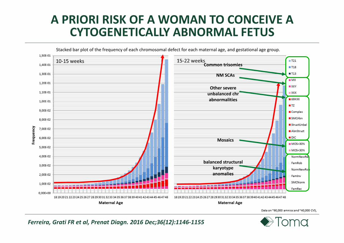
A PRIORI RISK OF A WOMAN TO CONCEIVE A CYTOGENETICALLY ABNORMAL FETUS
Ferreira, Grati FR et al, Prenat Diagn. 2016 Dec;36(12):1146-1155
Stacked bar plot of the frequency of each chromosomal defect for each maternal age, and gestational age group.
balanced structural
karyotype
anomalies
Common trisomies
Other severe
unbalanced chr
abnormalities
NM SCAs
Mosaics

Ferreira, Grati FR et al, Prenat Diagn. 2016 Dec;36(12):1146-1155
Overall risk for cytogenetic abn at >15GA (including WHITE box)
18y : 1/301
48y: 1/9
18y -1/301
48y - 1/9
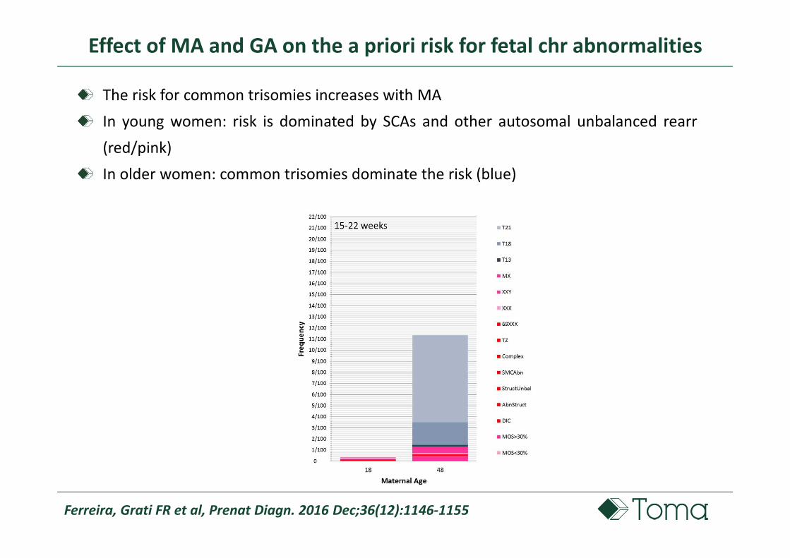
Effect of MA and GA on the a priori risk for fetal chr abnormalities
Ferreira, Grati FR et al, Prenat Diagn. 2016 Dec;36(12):1146-1155
Effect of MA and GA on the a priori risk for fetal chr abnormalities
The risk for common trisomies increases with MA
In young women: risk is dominated by SCAs and other autosomal unbalanced rearr
(red/pink)
In older women: common trisomies dominate the risk (blue)

Ferreira, Grati FR et al, Prenat Diagn. 2016 Dec;36(12):1146-1155
Results
Lower frequency of the common trisomies than reported from
previous studies, in which sonographic findings were not
available
Frequency of chromosomal aneuploidies is significantly higher
in earlier GA
CVS 2.63% (1100/41782) VS AF 1.82% (1596/87481); OR 0.6873 (95%CI 0.659-0.7428)
Ferreira, Grati FR et al, Prenat Diagn. 2016 Dec;36(12):1146-1155
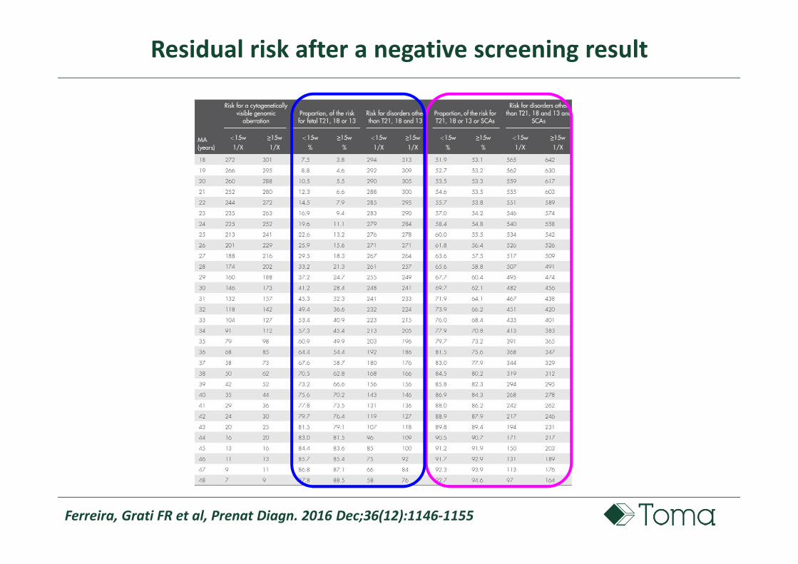
Residual risk after a negative screening result
Ferreira, Grati FR et al, Prenat Diagn. 2016 Dec;36(12):1146-1155
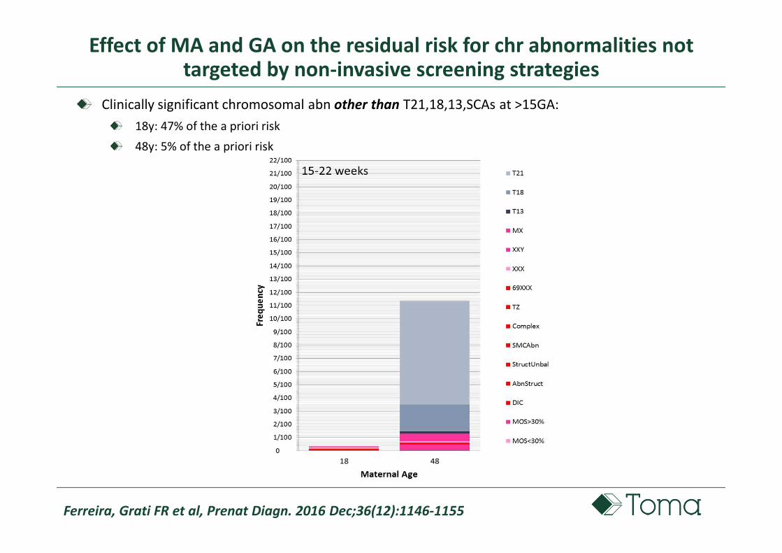
Effect of MA and GA on the residual risk for chr abnormalities not targeted by non-invasive screening strategies
Clinically significant chromosomal abn other than T21,18,13,SCAs at >15GA:
18y: 47% of the a priori risk
48y: 5% of the a priori risk

Ferreira, Grati FR et al, Prenat Diagn. 2016 Dec;36(12):1146-1155; Wapner et al, N
Engl J Med 2012;367(23):2175–2184
Newer technologies impact the epidemiology of fetal chr abn:
pCNV or likely pCNVs prevalence (by CMA) in women with anatomically normal fetuses with
normal karyotypes is 1.65% (1/61)
Clearly pCNVs: 0.5% - 95th% CI 0.2–0.8
pCNVs with variable expressivity: 0.6% – 95th% CI 0.3–1.1
Likely pVOUS: 0.6% – 95th% CI 0.3–1.1
Lack of association with MA, serum screening analyte levels or GA
prevalence fixed at 1.65% (1/61) in all women
SUBMICROSCOPIC CHROMOSOME ABNORMALITIES
A-PRIORI RISK

Ferreira, Grati FR et al, Prenat Diagn. 2016 Dec;36(12):1146-1155; Wapner et al, N Engl J Med
2012;367(23):2175–2184
TOMA lab DataBase
A PRIORI RISK OF A WOMAN TO CONCEIVE A
CHROMOSOMICALLY ABNORMAL FETUS
NICHD
Clinical
Trial
pCNVS
Stacked bar plot of the frequency of each genomic defect for each maternal age, and gestational age group (no balanced rearr)

EFFECT OF MATERNAL AGE ON THE A PRIORI RISK
In younger ages the non-age-
dependent pCNVs dominate fetal risk
CNVs represent the main component
of the a priori risk for fetal genomic
abnormalities in younger women:
80% of the risk in 18y
15% of the risk in 48y
1/50
1/8
Ferreira, Grati FR et al, Prenat Diagn. 2016 Dec;36(12):1146-1155

Ferreira, Grati FR et al, Prenat Diagn. 2016 Dec;36(12):1146-1155
Effect of MA and GA on the residual risk for chr abnormalities not targeted by non-invasive screening strategies
1/51
1/34
1/44
1/55
Residual risk for other ‘off-target’ clinically significant genomic abn
other than T21,18,13
other than T21,18,13, homogeneous SCAs
1/50
1/55
1/30
1/37
10-14 wks 15-22 wks

Ferreira, Grati FR et al, Prenat Diagn. 2016 Dec;36(12):1146-1155
After the exclusion of T21,18,13&SCAs, the RR for other pathogenic
GENOMIC abnormalities is still consistent
Not so much different in young (~1/50) and old women (~1/40)
Even excluding pCNVs with variable expressivity and likely pVOUS, a
residual risk of 0.5% (95th% CI 0.2–0.8) for pCNVs with highly penetrant
phenotypes still remains: level of risk to justify offering invasive testing
PROPORTION OF DEFECTS DETECTED BY THE DIFFERENT TESTING STRATEGY

PROPORTION OF DEFECTS DETECTED BY THE DIFFERENT TESTING STRATEGY
‘… Le gestanti che , per scelta personale , in assenza di una indicazione che conferisca loro
un rischio “ a priori” elevato per le microdelezioni/microduplicazioni, decidano di
sottoporsi ad una diagnosi prenatale invasiva, dovrebbero essere informate dell’esistenza
del CMA come tecnica di approfondimento diagnostico, ad integrazione del cariotipo
fetale. La sua applicazione in questa popolazione dovrebbe rispondere all’obiettivo di
ridurre il rischio di sindromi note da microdelezione/microduplicazione associate a fenotipi
clinici gravi….’

SUPPORTING WOMEN’S AUTONOMY IN PRENATAL TESTING
‘Early and noninvasive fetal genetic sequencing is on the horizon. Such expanded prenatal
testing could offer patients substantial benefits. But current practices in prenatal screening
and the complex nature of genomic science and technology create the risk that these tests
will be integrated into care without the robust, evidence-based informed consent processes
necessary for respecting women’s autonomy. If that happens, patients will be asked to
decide whether to undergo invasive diagnostic testing and then to consider whether to
terminate or continue their pregnancy without a full understanding of the results. …’
Johnston, Farrell, and Parens. NEJM 377;6 August 10, 2017

‘The need for fully informed consent in prenatal screening and testing has never been more
urgent. Meeting this need will require adoption of reimbursement policies and
professional practice guidelines that support clinicians in breaking with current routine
practices, which too often involve dispensing with or failing to adequately carry out an
informed consent process. It will also require funding for development of approaches to
pretest and posttest education and counseling that empower patients to decide whether
to be tested and what to do after receiving their results.’ …
‘Only with these practices and policies in place can women’s decisions about prenatal
screening, diagnostic testing, and termination or continuation of pregnancy be truly free
and informed.’
Johnston, Farrell, and Parens. NEJM 377;6 August 10, 2017
HOW TO SUPPORT WOMEN’S AUTONOMY?
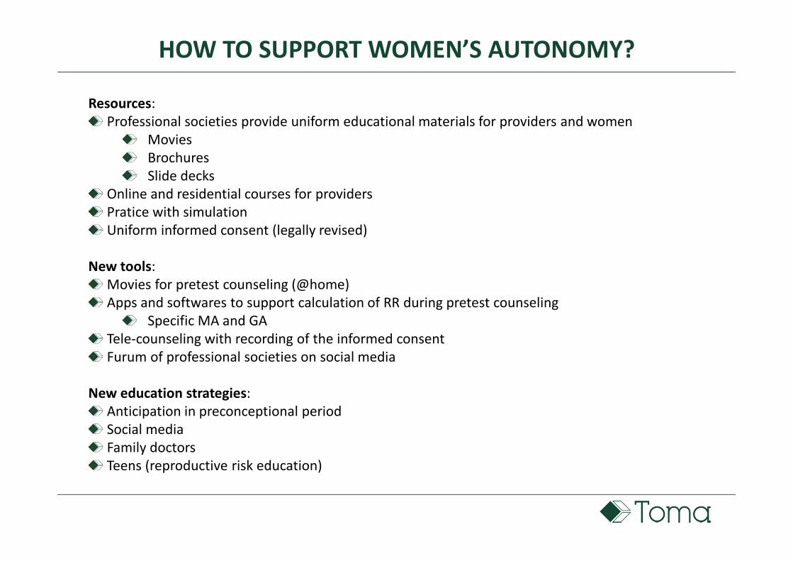
Resources:
Professional societies provide uniform educational materials for providers and women
Movies
Brochures
Slide decks
Online and residential courses for providers
Pratice with simulation
Uniform informed consent (legally revised)
New tools:
Movies for pretest counseling (@home)
Apps and softwares to support calculation of RR during pretest counseling
Specific MA and GA
Tele-counseling with recording of the informed consent
Furum of professional societies on social media
New education strategies:
Anticipation in preconceptional period
Social media
Family doctors
Teens (reproductive risk education)
HOW TO SUPPORT WOMEN’S AUTONOMY?
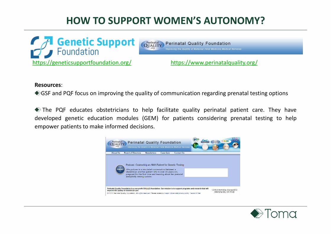
Resources:
GSF and PQF focus on improving the quality of communication regarding prenatal testing options
The PQF educates obstetricians to help facilitate quality perinatal patient care. They have
developed genetic education modules (GEM) for patients considering prenatal testing to help
empower patients to make informed decisions.
HOW TO SUPPORT WOMEN’S AUTONOMY?
https://geneticsupportfoundation.org/ https://www.perinatalquality.org/

HOW TO SUPPORT WOMEN’S AUTONOMY?

ISPD Global Updates (July/August 2017) – Genetic Counseling SIG
http://ispdhome.org/ISPD/Special_Interest_Groups/Genetic_Counseling/ISPD/SIGs/Geneti
c_Counseling.aspx?utm_source=Informz&utm_medium=Email&utm_campaign=eBlasts#G
U817
HOW TO SUPPORT WOMEN’S AUTONOMY?

Susan Gross
Komal Bajaj Jose Ferreira

QUESTION
CfDNA vs. CVS in the high risk patients?

Increased a-priori risk for genetic abnormalities in pregnancies with U/S abnormalities
Type of genomic disorder Type of test on CVS/AF Resolution
Cytogenetic abnormalities Karyotyping >5-7Mb
Submicroscopic dels/dups CMA Kb--> <5Mb
Monogenic disorders WES bp
Imprinting disorders Different molecular tests Epigenetic

Fetal chromosome abnormalities in pregnancies with U/S abnormalities
Grati et al, Am J Med Genet Part A 152A:1434–1442.
* OR 15.58, 95%CI 13.71-17.70
** OR 31.22, 95%CI 24.09-40.46
*** OR 8.98, 95%CI 7.76-10.39
**** OR 21.15, 95%CI 17.16-26.08
* **
**** ***

Grati et al, Am J Med Genet Part A 152A:1434–1442; Grati et al, Unpublished data
Chromosome abnormalities in CVS of pregnancies with U/S abnormalities
• Increased NT (54%)
• CH-Hydrops-Oedema (18%)
• IUGR (2%)
• Omphalocoele (2.4%)
• Fetal malformations ndd (19%)
HOMOGENEOUS
• De novo (majority)
• Inherited from a parent carrier
of a balanced rearrangement
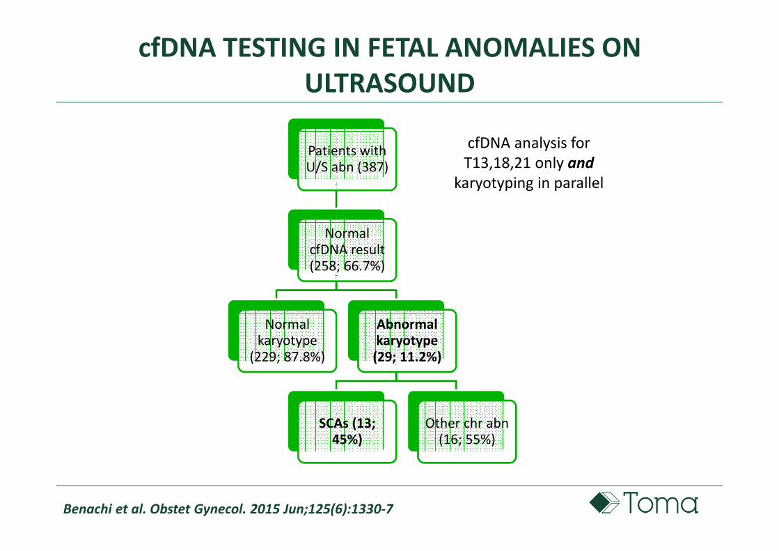
cfDNA TESTING IN FETAL ANOMALIES ON
ULTRASOUND
Benachi et al. Obstet Gynecol. 2015 Jun;125(6):1330-7
Patients with U/S abn (387)
Normal cfDNA result (258; 66.7%)
Normal karyotype
(229; 87.8%)
Abnormal karyotype
(29; 11.2%)
SCAs (13; 45%)
Other chr abn(16; 55%)
cfDNA analysis for
T13,18,21 only and
karyotyping in parallel

Increased a-priori risk for genetic abnormalities in pregnancies with U/S abnormalities
Type of genomic disorder Type of test on CVS/AF Resolution
Cytogenetic abnormalities Karyotyping >5-7Mb
Submicroscopic dels/dups CMA Kb--> <5Mb
Monogenic disorders WES bp
Imprinting disorders Different molecular tests Epigenetic

INCREMENTAL YIELD BY MICROARRAY WITH NORMAL FETAL KARYOTYPE
De Wit et al., UOG 2014
• 5.6% (95% CI 4.7-6.6) structural ultrasound anomaly restricted to
one anatomical system and a normal karyotype
• 9.1% (95% CI 7.5-10.8) poly-malformed fetuses
Isolated anomaliesCardiac Resp CNS Facial MSK
Pooled prevalence(95% CI)
22/4764.6%
(2.7-6.5)
5/816.2%
(0.9-11.4)
35/5636.2%
(4.2-8.2)
6/1135.3%
(1.2-9.4)
24/3057.9%
(4.8-10.9)
Isolated anomalies
GIT UrogenitalNT
>3.5 mmCystic
hygromaTotal
Pooled prevalence(95% CI)
7/1056.7%
(1.9-11.4)
9/1535.9%
(2.2-9.6)
5/1623.1%
(0.4-5.7)
12/2624.6%
(2.0-7.1)
125/22205.6%
(4.7-6.6)

High post-test residual risk for fetal pCNVs
Wapner et al, NEJM 2012; Yaron et al, Obstet Gynecol. 2015 Nov;126(5):1095-9; Grati FR,
Ultrasound Obstet Gynecol. 2016 May 31. doi: 10.1002/uog.15975
5-6 CNVs represent only a portion (~20%) of the overall pCNVs that can affect
the fetus
All
possible
CNVs
(1.7%)
CNVs CNVs CNVs CNVs
targeted by targeted by targeted by targeted by
cfDNA testcfDNA testcfDNA testcfDNA test
False reassurance to patients – consistent residual risk

Increased a-priori risk for genetic abnormalities in pregnancies with U/S abnormalities
Type of genomic disorder Type of test on CVS/AF Resolution
Cytogenetic abnormalities Karyotyping >5-7Mb
Submicroscopic dels/dups CMA Kb--> <5Mb
Monogenic disorders WES bp
Imprinting disorders Different molecular tests Epigenetic

WHOLE EXOME SEQUENCING IN FETAL ANOMALIES ON U/S – ON THE HORIZON
Drury et al, Prenatal Diagnosis 2015, 35, 1010–1017; Pangalos et al, DOI 10.7717/peerj.1955;
ACOG and sMFM Committee opinion Number 682, December 2016
WES examines coding regions (exons) of the genome

WHOLE EXOME SEQUENCING IN FETAL ANOMALIES ON U/S – ON THE HORIZON
Drury et al, Prenatal Diagnosis 2015, 35, 1010–1017; Pangalos et al, DOI 10.7717/peerj.1955;
ACOG and sMFM Committee opinion Number 682, December 2016
Aimed to identify the etiology for fetal U/S abnormalities
Actually not recommended outside of the context of clinical trials
Offered on research basis in some labs or for specific clinical indications in
other labs (recurrent or lethal fetal anomalies)
Limited published data on prenatal application of WES
Monogenic diseases may be identified in up to 20-30% of fetuses with
multiple anomalies suggestive of a genetic disorder for which karyotyping and
CMA are normal
Provide options of PGD or early prenatal diagnosis in a future pregnancy

Increased a-priori risk for genetic abnormalities in pregnancies with U/S abnormalities
Type of genomic disorder Type of test on CVS/AF Resolution
Cytogenetic abnormalities Karyotyping >5-7Mb
Submicroscopic dels/dups CMA Kb--> <5Mb
Monogenic disorders WES bp
Imprinting disorders Different molecular tests Epigenetic
Normal fetal karyotype and normal CMA!!

Imprinting Syndromes and fetal U/S abnormalities
Beckwith–Wiedemann syndrome (BWS) in fetuses with:
• isolated omphalocoele
• overgrowth
• polydramnios
• enlarged placenta
• distended abdomen
• visceromegaly
• macroglossia
Grati et al, J. Med. Genet. 2007;44;257-263
UPD11pat or other related imprinting defects

Imprinting Syndromes and fetal U/S abnormalities
Silver Russell syndrome (SRS) in fetuses with:
• IUGR
• Micrognathia
• CHD
• clinodactyly
• Partial or total asymmetry
Miozzo, Grati et al, Placenta (2001), 22, 813–821; OMIM #180860
UPD7mat or other imprinting defects

cfDNA TESTING IN FETAL ANOMALIES ON
ULTRASOUND
Beulen et al, Ultrasound Obstet Gynecol. 2016 Aug 12. doi: 10.1002/uog.17228
Patients with U/S abn (251)
Normal cfDNAresult (224;
89.2%)
Genetic testing not performed (191; 85.3%)
Normal genetic testing (21/28; 75%)
Abnormal genetic testing
(7/28; 25%)
Cytogenetic abn. (T13, MX)
(28.6%)
2 pCNVs(28.6%)
2 monogenic disorders (28.6%)
1 imprinting disorder (14.3%)
GW cfDNA analysis for all autosomic
partial and whole chromosome
aneuploidy (no SCAs) and karyotyping
in parallel

Residual Risk
Risk before test
Test is “negative”
Residual risk after
“negative” test
Increased a priori risk for
different types of genetic
abnormalities
Courtesy: Thomas J Musci
RR is dependent on:
• detection rate of applied test
• genetic aetiology of the fetal malformation
• 20-30% RR for cytogenetic imbalances
• 6-9% pCNVs (submicroscopic)
• 20-30% monogenic disorders
• imprinting disorders
Any cfDNA testing

Ultrasound abnormality: is there a role for NIPT?
Grati FR and Benn P, accepted reply letter to Fiorentino et al, Prenat Diagn. 2017
Jun;37(6):593-601; Courtesy: Thomas J Musci;
Limited clinical utility in high risk cases
NIPT with
microdeletion
panel
Invasive test
• Confirmatory testing recommended
• Extremely low PPV for rare
conditions
Invasive test
• Significant residual risk
• Detection rates low or unknown
• Many genetic conditions/
microdeletions not addressed by NIPT
“Positive” NIPT
“Negative” NIPT
Potential for delayed diagnosis, additional cost, and anxiety for patients
Ultrasound
anomaly

Differential Diagnosis
R/O chromosomal or genetic etiology
Patient counseling
Prognosis
Treatment options
Ultrasound abnormality: is there a role for NIPT?
Courtesy: Thomas J Musci

QUESTION
Which screening strategy?

Ferreira, Grati FR et al, Prenat Diagn. 2016 Dec;36(12):1146-1155; Grati et al, manuscript in
preparation
COMPARISON OF DIFFERENT SCREENING STRATEGIES FOR THE DETECTION OF THE OVERALL FETAL CYTOGENETIC
ABNORMALITIES AT BIRTH
Acronym Screening stategy First-tier test Second-tier test Third-tier test
FTSCombined first
trimester Combined FTS Karyotype if risk is ≥1/270 //
CON Contingent Combined FTS
Karyotype or cfDNA-T for HR (>1/10);
CfDNA-T or nothing else for IR (1/10-1/1000);
Nothing else for LR (<1/1000)
Karyotype if cfDNA
risk is ≥1/100 or
‘high risk’
SEQ Sequential Combined FTS CfDNA-T for ≥1/270;
Nothing else for <1/270
Karyotype if cfDNA
risk is ≥1/100 or
‘high risk’
cfDNA-TUniversal cfDNA
for T13,18,21
cfDNA test for
T21,18,13Karyotype if cfDNA risk is ≥1/100 or ‘high risk’ //
cfDNA-TXY
Universal cfDNA
for T13,18,22,
SCAs
cfDNA test for
T21,18,13 and
SCAs
Karyotype if cfDNA risk is ≥1/100 or ‘high risk’ //
QUAD QUAD test
Second
trimester
(T18,21)
Karyotype if risk is ≥1/270 //
INT Integrated Integrated test Karyotype if risk is ≥1/270 //

Grati et al, manuscript in preparation
PROPORTION OF CHR DEFECTS DETECTED BY THE DIFFERENT TESTING STRATEGIES
The distribution and prevalence of the chr abn are different at different MA
Although two strategies may show approximately the same overall DR, one may favor
the detection of a different subset of chr abn compared with another one

WHICH SCREENING STRATEGY?
• Low residual risk for the targets of the test
At the cost of:
• a very low sensitivity for off-target chr abn
• expenses for cfDNA tests
• Detection for large array of off-target chr abn
At the cost of:
• a high residual risk for the targets of the test
• expenses for invasive procedures
BudgetMaternal age distribution
and stratificationTechnological
resources
Medical/scientific
resources
Grati et al, manuscript in preparation

False positive rate of screenings
Compared with traditional serum±ultrasound screening (TSS), cfDNA tests have a
much lower FPR for T21,18,13
The higher FPR of TSS was often considered a limitation
Distinct advantage with TSS due to NT’s ability to pick up additional chromosomal
abnomalies (‘off-target’) in addition to the higher reflex invasive testing rate
AIM: present detection rates of all (target and off-target) fetal karyotype
abnormalities at birth by different screening strategies including cfDNA test and TSS
Grati FR et al, manuscript in preparation; Norton et al, Obstet Gynecol 2014;124:979–86; Syngelaki et al, Fetal
Diagn Ther 2014;35:174–184

A PRIORI RISK OF A WOMAN TO CONCEIVE A CYTOGENETICALLY ABNORMAL FETUS
Ferreira, Grati FR et al, Prenat Diagn. 2016 Dec;36(12):1146-1155
Stacked bar plot of the frequency of each chromosomal defect for each maternal age, and gestational age group.
balanced structural
karyotype
anomalies
Common trisomies
Other severe
unbalanced chr
abnormalities
NM SCAs
Mosaics

Ferreira, Grati FR et al, Prenat Diagn. 2016 Dec;36(12):1146-1155; *Cuckle et al, Clin Biochem.
2015;48(15):932-41; ^ Gil et al, Ultrasound Obstet Gynecol. 2017 Apr 11. doi: 10.1002/uog.17484; *Nicolaides
K, Prenat Diagn 2011; 31: 7–15; *Sonek and Cuckle Ultrasound Obstet Gynecol 2014; 44: 621–630; °Nicolaides
K, American Journal of Obstetrics and Gynecology (2004) 191, 45e67
Methods
Prior risks for each defect derived from TOMA lab Dataset of ≈130K prenatal dx on CVS (n=43K)
and AF (n=87K) with an indication of AMA, anxiety and elective decision (reported by clinicians)
Fetal loss rate at birth for T13,18,21°
Sensitivities and specificities for common aneuploidies and triploidy abstracted from the
published literature:
Serum screenings for T21,18,13, MX, triploids: from prior seminal studies* (5% cumulative FPR)
cfDNA testing: 0.13% cumulative FPR for T21,18,13; 0.273% cumulative FPR for T21,18,13+SCAs^
Sensitivity for other karyotype abnormalities correspond to the FPR of TSS or cfDNA tests
No result rate with cfDNA testing of 1%: the DR for all chr abnormalities was adjusted downward
as a 1% of ‘no result’ cases by cfDNA are actually undetected karyotype abnormalities
PROPORTION OF THE CHROMOSOMAL DEFECTS OCCURRING AT BIRTH DETECTABLE BY DIFFERENT TESTING STRATEGIES

THE ROLE OF ‘MATERNAL AGE’ TODAY
Maternal age should have a central role to rationalize resources to
obtain the most efficient cost-benefit

Encephalocoele� 46,XX,rec(5)dup(5q)inv(5)(p15.2q32)
Fetal chromosome abnormalities in pregnancies with U/S abnormalities

Ventriculomegaly� 47,XY,+15
Fetal chromosome abnormalities in pregnancies with U/S abnormalities

Hydrocephaly�69,XXY
Fetal chromosome abnormalities in pregnancies with U/S abnormalities

Anencephaly� 47,XX,+9
Fetal chromosome abnormalities in pregnancies with U/S abnormalities
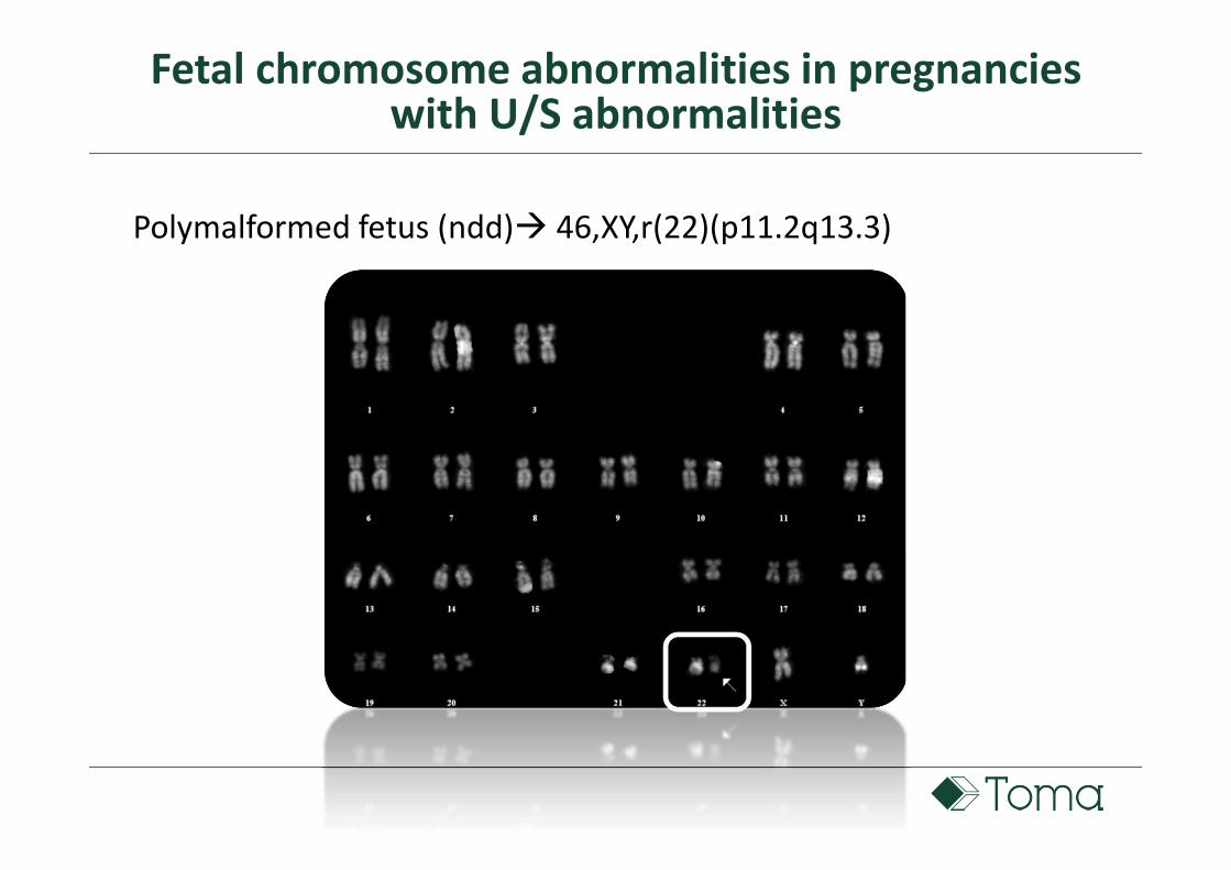
Polymalformed fetus (ndd)� 46,XY,r(22)(p11.2q13.3)
Fetal chromosome abnormalities in pregnancies with U/S abnormalities

INCREMENTAL YIELD BY MICROARRAY
WITH NORMAL FETAL KARYOTYPE
Jansen et al, UOG 2015
Pooled analysis: 7.0% (95% CI, 5.3–8.6%) incremental yield by
CMA (excluding 22q11 microdeletion cases);
Incremental yield increases to 12% (95% CI, 7.6–16%) when
22q11 deletion cases were included
Stratified analysis: incremental yield
• 3.4% (95%CI 0.3–6.6%) for isolated CHD
• 9.3% (95%CI, 6.6–12%) when additional extracardiac
malformations were present
Fetuses with CHD ± extracardiac defects (systematic meta-analysis)

ROLE OF CMA IN ANATOMICALLY ABNORMAL FETUSES AND NORMAL KARYOTYPE
De Wit et al., UOG 2014; ^ISUOG consensus statement, Ultrasound Obstet Gynecol. 2016 Nov 27. doi:
10.1002/uog.17324
• 1 in every 20 anatomically abnormal fetuses with a normal karyotype shows a
submicroscopic CNV that explains its phenotype and provides prognostic
information
• Professional societies recommend prenatal invasive diagnosis with CMA as
first-tier test on AF/CVS in CHD^– Many different submicroscopic and monogenic causes for CHD
– Association between CHD and neurodevelopmental delay

Ferreira, Grati FR et al, Prenat Diagn. 2016 Dec;36(12):1146-1155; Grati et al, manuscript in preparation
PROPORTION OF THE CHROMOSOMAL DEFECTS OCCURRING AT BIRTH DETECTABLE BY DIFFERENT TESTING STRATEGIES
25y 35y 45y
cfDNA-TXY has the highest DR at all MA;
SEQ has the lowest DR, approximating the QUAD only at older MA: with SEQ the second-tier cfDNA-T drops down by 40-folds (from
5% to 0.13%) the FPR of the strategy, thereby reducing the likelihood of finding other off-target chr abn
CON is always better than SEQ thanks to the larger population performing follow up karyotyping
Among TSS, INT has the highest DR, at the cost of a late GA reporting
cfDNA-T equals or is better than CON or FTS only at older MA, when trisomies dominate the risk