An Electrophysiological and Pharmacological Study of the ...
18
University of the Pacific University of the Pacific Scholarly Commons Scholarly Commons School of Pharmacy Faculty Articles Thomas J. Long School of Pharmacy 7-31-2021 An Electrophysiological and Pharmacological Study of the An Electrophysiological and Pharmacological Study of the Properties of Human iPSC-Derived Neurons for Drug Discovery Properties of Human iPSC-Derived Neurons for Drug Discovery Robert F. Halliwell University of the Pacific, rhalliwell@pacific.edu Hamed Salmanzadeh University of the Pacific Leanne Coyne University of the Pacific William S. Cao University of the Pacific, wcao@pacific.edu Follow this and additional works at: https://scholarlycommons.pacific.edu/phs-facarticles Part of the Pharmacy and Pharmaceutical Sciences Commons Recommended Citation Recommended Citation Halliwell, R. F., Salmanzadeh, H., Coyne, L., & Cao, W. S. (2021). An Electrophysiological and Pharmacological Study of the Properties of Human iPSC-Derived Neurons for Drug Discovery. Cells, 10(8), 1–17. DOI: 10.3390/cells10081953 https://scholarlycommons.pacific.edu/phs-facarticles/579 This Article is brought to you for free and open access by the Thomas J. Long School of Pharmacy at Scholarly Commons. It has been accepted for inclusion in School of Pharmacy Faculty Articles by an authorized administrator of Scholarly Commons. For more information, please contact mgibney@pacific.edu.
Transcript of An Electrophysiological and Pharmacological Study of the ...

University of the Pacific University of the Pacific
Scholarly Commons Scholarly Commons
School of Pharmacy Faculty Articles Thomas J. Long School of Pharmacy
7-31-2021
An Electrophysiological and Pharmacological Study of the An Electrophysiological and Pharmacological Study of the
Properties of Human iPSC-Derived Neurons for Drug Discovery Properties of Human iPSC-Derived Neurons for Drug Discovery
Robert F. Halliwell University of the Pacific, [email protected]
Hamed Salmanzadeh University of the Pacific
Leanne Coyne University of the Pacific
William S. Cao University of the Pacific, [email protected]
Follow this and additional works at: https://scholarlycommons.pacific.edu/phs-facarticles
Part of the Pharmacy and Pharmaceutical Sciences Commons
Recommended Citation Recommended Citation Halliwell, R. F., Salmanzadeh, H., Coyne, L., & Cao, W. S. (2021). An Electrophysiological and Pharmacological Study of the Properties of Human iPSC-Derived Neurons for Drug Discovery. Cells, 10(8), 1–17. DOI: 10.3390/cells10081953 https://scholarlycommons.pacific.edu/phs-facarticles/579
This Article is brought to you for free and open access by the Thomas J. Long School of Pharmacy at Scholarly Commons. It has been accepted for inclusion in School of Pharmacy Faculty Articles by an authorized administrator of Scholarly Commons. For more information, please contact [email protected].

cells
Article
An Electrophysiological and Pharmacological Study of theProperties of Human iPSC-Derived Neurons forDrug Discovery
Robert F. Halliwell *, Hamed Salmanzadeh, Leanne Coyne and William S. Cao
Citation: Halliwell, R.F.;
Salmanzadeh, H.; Coyne, L.; Cao, W.S.
An Electrophysiological and
Pharmacological Study of the
Properties of Human iPSC-Derived
Neurons for Drug Discovery. Cells
2021, 10, 1953. https://doi.org/
10.3390/cells10081953
Academic Editors: James Adjaye and
Nina Graffmann
Received: 28 June 2021
Accepted: 29 July 2021
Published: 31 July 2021
Publisher’s Note: MDPI stays neutral
with regard to jurisdictional claims in
published maps and institutional affil-
iations.
Copyright: © 2021 by the authors.
Licensee MDPI, Basel, Switzerland.
This article is an open access article
distributed under the terms and
conditions of the Creative Commons
Attribution (CC BY) license (https://
creativecommons.org/licenses/by/
4.0/).
Department of Physiology & Pharmacology, Thomas J. Long School of Pharmacy, University of the Pacific,Stockton, CA 95211, USA; [email protected] (H.S.); [email protected] (L.C.);[email protected] (W.S.C.)* Correspondence: [email protected]; Tel.: +1-209-946-2074
Abstract: Human stem cell-derived neurons are increasingly considered powerful models in drugdiscovery and disease modeling, despite limited characterization of their molecular properties. Here,we have conducted a detailed study of the properties of a commercial human induced PluripotentStem Cell (iPSC)-derived neuron line, iCell [GABA] neurons, maintained for up to 3 months in vitro.We confirmed that iCell neurons display neurite outgrowth within 24 h of plating and label for thepan-neuronal marker, βIII tubulin within the first week. Our multi-electrode array (MEA) recordingsclearly showed neurons generated spontaneous, spike-like activity within 2 days of plating, whichpeaked at one week, and rapidly decreased over the second week to remain at low levels up toone month. Extracellularly recorded spikes were reversibly inhibited by tetrodotoxin. Patch-clampexperiments showed that iCell neurons generated spontaneous action potentials and expressedvoltage-gated Na and K channels with membrane capacitances, resistances and membrane potentialsthat are consistent with native neurons. Our single neuron recordings revealed that reduced spikingobserved in the MEA after the first week results from development of a dominant inhibitory tonefrom GABAergic neuron circuit maturation. GABA evoked concentration-dependent currents thatwere inhibited by the convulsants, bicuculline and picrotoxin, and potentiated by the positiveallosteric modulators, diazepam, chlordiazepoxide, phenobarbital, allopregnanolone and mefenamicacid, consistent with native neuronal GABAA receptors. We also show that glycine evoked robustconcentration-dependent currents that were inhibited by the neurotoxin, strychnine. Glutamate,AMPA, Kainate and NMDA each evoked concentration-dependent currents in iCell neurons thatwere blocked by their selective antagonists, consistent with the expression of ionotropic glutamatereceptors. The NMDA currents required the presence of the co-agonist glycine and were blocked in ahighly voltage-dependent manner by Mg2+ consistent with the properties of native neuronal NMDAreceptors. Together, our data suggest that such human iPSC-derived neurons may have significantvalue in drug discovery and development and may eventually largely replace the need for animaltissues in human biomedical research.
Keywords: neurotransmitter receptors; ion channels; multi-electrode array; patch-clamp
1. Introduction
Galvani (1737–1798) and Volta (1745–1827) in the late eighteenth century establishedbioelectricity and the field of electrophysiology [1]. The instruments used to investigateanimal electricity have since evolved radically from the differential rheotome built byBernstein (1868) to the patch-clamp technique invented by Nobel Laureates, Neher andSakmann (1976). Notwithstanding these major advances in technology, studies of thenervous system have relied almost exclusively on animal-derived tissues (particularly thefrog excised nerve-muscle preparation) even when the primary questions have revolved
Cells 2021, 10, 1953. https://doi.org/10.3390/cells10081953 https://www.mdpi.com/journal/cells

Cells 2021, 10, 1953 2 of 17
around human brain functions. Indeed, when mice and rats are included, over 100 millionanimals are used annually in biomedical research worldwide [2].
Electrophysiological and neuropharmacological studies of human neurons have, untilrecently, been technically, legally and ethically difficult and often conducted on small sec-tions (slices) of neocortex resected from patients undergoing neurosurgery to treat epilepsy,or to remove arterio-vascular malformations or neoplasms (e.g., [3]). This model retainssome of the complexity of neural networks and cell diversity but may or may not representthe ‘normal’ properties of human neurons. Alternative approaches have used recombinanthuman receptors and ion channels expressed in human cell lines or Xenopus oocytes toinvestigate receptor pharmacology or drug interactions on these systems (e.g., [4]) but thecomplex neural networks and diverse neuronal cell types are absent in this approach.
Significant advances in stem cell technology over the last decade are now providingneural cells from diverse human stem cell populations including embryonic, fetal, adult,induced pluripotent stem cells (hiPSCs) and from the direct conversion of terminally differ-entiated fibroblasts into induced neurons, termed iNeurons (for review see [5]). Humanstem cell derived neurons are therefore increasingly advanced as powerful new tools indrug discovery [6] despite their characterization often being limited to changes in cellmorphology, immunocytochemical labeling and/or a description of minimal electrophysio-logical properties [5]. There are now several commercially available human stem cell linesand iPSC-derived neural cells, including iCell neurons (renamed iCell GABA Neurons,Fujifilm Cellular Dynamics Inc. FCDI). iCells neurons are a pure population of humanneurons derived from IPSCs using proprietary differentiation and purification protocolsand terminally differentiated into mature, cortical forebrain-like neurons [7,8]. Based onflow cytometry, gene expression and immunolabeling they are 85–90% GABAergic neuronsand 10–15% glutamatergic neurons; there are no glial cells present (FCDI). In an earlyreport, these human iCell neurons were shown to express voltage-gated sodium, potassiumand L-type calcium channels and GABA evoked responses that were inhibited by bicu-culline [9–11]. Using fluorescence-based calcium imaging, Dage and colleagues (2014) alsoreported the expression of AMPA and NMDA receptors in iCell neurons [7]. In the presentstudy, we have conducted a more complete characterization of the neuropharmacologicalproperties of iCell neurons to determine their value in drug receptor studies. Specifically,we have utilized multi-electrode arrays (MEA) and conventional patch-clamp electrophys-iology, along with microscopy and immunocytochemistry to determine the expressionand properties of voltage- and ligand-gated ion channels, as well as the development ofsynaptic activity in iCell neurons maintained for up to 3 months in vitro.
2. Materials and Methods2.1. Cell Culture
Frozen vials of iCell Neurons (Fujifilm Cellular Dynamics Inc. [FCDI], Madison, WI,USA) were thawed as per the manufacturer’s instructions and, in preparation for immuno-cytochemistry and patch-clamp electrophysiology, seeded at a density of 75,000 cells perwell onto 12 mm glass coverslips in 24-well plates. Prior to cell seeding, coverslips werecoated with 0.01% poly-L-ornithine (Sigma-Aldrich, St. Louis, MO, USA) for 1 h at roomtemperature, washed twice with phosphate-buffered saline (PBS) and coated with a 3.3µg/mL solution of laminin (Sigma-Aldrich) for 1 h in a 37 C incubator. The laminin wasaspirated immediately before the addition of cell suspensions. Cells were maintained ina 37 C incubator (5% CO2/95% humidified air) in iCell Neurons Maintenance Mediumcontaining iCell Neurons Medium Supplement (FCDI). Following an initial 100% mediachange at 24 h in culture, 50% media changes were then conducted every 4–7 days. Thenumber of days in vitro refers to the days from first thawing the vial of frozen iCells andplating them on coverslips or the MEA plates.

Cells 2021, 10, 1953 3 of 17
2.2. Immunocytochemistry
For immunolabelling, cells were cultured on glass coverslips in 24-well plates, asdescribed above. The cells were first washed in phosphate buffered saline (PBS) and thenfixed in 4% paraformaldehyde in PBS. The fixative was aspirated, and cells were washedin PBS and incubated at 4 C overnight in a blocking solution (5% normal donkey serumand 0.3% triton X-100 in PBS). Thereafter, cells were placed in mouse anti-β-III Tubulinprimary antibody (Millipore, Burlington, MA, USA) diluted 1:500 in blocking solutionand incubated at 4 C overnight. The following day, cells were washed once in PBS andtwice in blocking solution. At the completion of the final wash, cells were left in blockingsolution for an additional 30 min. The blocking solution was then aspirated, and the cellsincubated in a 1:200 dilution of Cy3-conjugated donkey anti-mouse IgG secondary antibody(Millipore) for 2 h at room temperature. The cells were subsequently washed in deionizedwater and counterstained with 100 ng/mL DAPI (Sigma-Aldrich) before mounting ontomicroscope slides using anti-fade mounting solution (Prolong Gold® Antifade Reagent;Invitrogen, Waltham, MA, USA). Immunolabeled cells were visualized through an invertedfluorescent microscope (Nikon Eclipse TE200, Melville, NY, USA) with an attached CCDcamera (PCO 1300; PCO-Tech, Willmington, DE, USA). 600–660 nm Cy3 fluorescenceemission from 540–580 nm excitation was observed using a 505-nm dichroic mirror, while435–485 nm DAPI emission from 340–380 nm excitation was observed using a 400-nmdichroic mirror.
2.3. Multi-Electrode Array Electrophysiology
iCell Neurons were plated at 100,000 cells per well on 24-well MEA plates (CytoviewMEA24, Axion Biosystems, Atlanta, GA, USA) previously coated with Polyethyleneimine(Sigma). Spontaneous electrical activity was acquired using the Axion Maestro Edge MEAsystem and Axion’s Integrated Studio (AXIS) software, v. 2.0.4.21 (Axion Biosystems) over28 days. All recordings were performed at a constant temperature of 37 C and 5% CO2.Each well of the 24-well MEA plate contains 16 individual microelectrodes, giving a total of384 integrated recording electrodes per plate. Prior to a 10 min baseline recording period,the MEA plates were placed in the Maestro MEA platform and equilibrated for 5 min. AXISsoftware was used to control the heating system and to monitor the recordings, whichinvolves simultaneous sampling of the channels at 12.5 kHz per channel with a gain of1200× and band pass filters of 200–3 KHz. After recording, the RAW files were re-recordedwith AXIS to convert the data into Microsoft Excel files (version 16), which includes spiketiming and profile information. Spikes were detected using a threshold set to 6 times theestimated standard deviation of the rms-noise on each channel. Electrodes with activityhigher than 0.05 spikes/s at least once over the recording time were only included fordata analysis.
2.4. Patch-Clamp Electrophysiology
Agonist and voltage-evoked currents were recorded from cells with a neuronal-likemorphology (phase bright with neurite processes), using the whole-cell configuration ofthe patch-clamp technique, as we have described previously (Coyne et al., 2007). Briefly,patch electrodes were made from borosilicate glass pipettes (World Precision Instruments,Sarasota, FL, USA) on a Narishige PP-830 (Amityville, NY, USA) electrode puller and hadtip resistances of 2–5 MΩ. Currents were recorded using an Axopatch 200B amplifier andheadstage (Axon Instruments) and low-pass filtered at 10 kHz before digitization via aNational Instruments DAQ card and a National Instruments BNC-2090 interface boardand stored on a computer running WinWCP software (v. 4.4.3, University of Strathclyde,Glasgow, UK). Whole cell currents were monitored on the desktop computer. Seriesresistance, pipette capacitance, and whole-cell capacitance were cancelled electronically.
Cells were perfused with a bath solution containing the following (in mM): NaCl(142.0), KCl (5.0), CaCl2 (2.0), glucose (10.0), MgCl2 (2.0), HEPES (5.0). The internal solutioncontained the following (in mM): KCl or CsCl (140.0), MgCl2 (2.0), EGTA (11.0), HEPES

Cells 2021, 10, 1953 4 of 17
(10.0), Mg-ATP (2.0). Solutions were titrated to pH 7.4 and electrode solutions filteredbefore use with 0.2µm disposable filters (Millipore). All experiments were conducted atambient room temperature (22–25 C). Neural-like cells were voltage-clamped at a holdingpotential of −60 mV or current-clamped at an equilibrium potential of −70 mV, unlessotherwise stated. For determination of agonist-evoked current-to-voltage relationships, themembrane potential was stepped between −120 mV and 60 mV in 20 mV increments. Toactivate voltage-gated currents, 20 ms voltage steps were applied from −80 to 40 mV in10 mV increments from a holding potential of −80 mV. For recording potassium currents,CsCl was replaced with KCl or K-gluconate in the electrode solution. The bath solutions forinvestigating voltage-activated currents also contained 2 mM CoCl2, to prevent potentialcalcium channel activation.
2.5. Drugs and Their Application
All drugs were obtained from Sigma-Aldrich (St. Louis, MO, USA) except tetrodotoxincitrate (TTX), kynurenic acid, and 6,7-dinitroquinoxaline-2,3-dione disodium salt (DNQX),which were purchased from Tocris Bioscience (Ellisville, MO, USA). Stock solutions ofbicuculline methyl bromide, allopregnanolone, and picrotoxin were dissolved in DMSO,while stock solutions of kynurenic acid and mefenamic acid were made up in 0.1 M sodiumhydroxide. The final bath concentrations of these solvents did not exceed 0.1% of therecording solutions. Stock solutions of DNQX, phenobarbital sodium salt, chlordiazepox-ide hydrochloride, D-2-amino-5-phosphonovalerate (AP5), tetraethylammonium chloride(TEA), and strychnine hydrochloride were made up in bath solution. All drug solutionswere made fresh immediately prior to experiments except for TTX, which was stored at−20 C in small aliquots of stock solution made up in deionized water. Drugs were applieddirectly to cells under voltage-clamp from the tip of a 250-µm pipette. Fresh bath solutionwas also perfused through the bath (at 1–2 mL/min) using a gravity-feed system to preventany build-up of drug solutions in the bath. At least 2 or 3 control responses were recordedbefore the addition of any drug. Drugs were washed out once a stable effect was observedand control responses then re-established before further drug applications.
2.6. Data Analysis
Data are expressed as mean ± SEM and were analyzed by either the Student’s t test(two-tailed) or one-way analysis of variance (ANOVA) using Prism 9 software (GraphPadSoftware, San Diego, CA, USA). Tukey’s or Dunnett’s post hoc test for significance wereused when variance between means was found with ANOVA. Concentration-responsecurves were analyzed using variable-slope nonlinear regression. Data was obtained fromat least 3 individual cells per experiment, and statistical significance was set at p ≤ 0.05 forall analyses.
3. Results
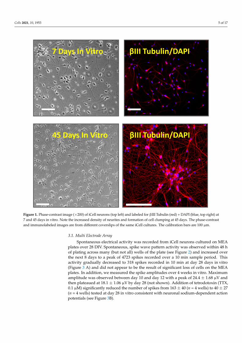
iCell neurons were successfully cultured and maintained for up to 70 days in vitro(DIV). The neurons were phase-bright with spherical or pyramidal cell bodies and multipleneurite processes that became more elaborate and denser over time in vitro. Cells stainedpositively with the DNA dye, DAPI, and were positive for the pan-neuronal marker βIII-tubulin, a microtubule stabilizing protein found in neuronal cell bodies and axons. Wenoted that cells formed small clumps or islands with longer-term culture (see Figure 1).
Cells 2021, 10, 1953 5 of 17Cells 2021, 10, x 5 of 18
Figure 1. Phase-contrast image (×200) of iCell neurons (top left) and labeled for βIII Tubulin (red) + DAPI (blue, top right) at 7 and 45 days in vitro. Note the increased density of neurites and formation of cell clumping at 45 days. The phase-contrast and immunolabeled images are from different coverslips of the same iCell cultures. The calibration bars are 100 μm.
3.1. Multi Electrode Array Spontaneous electrical activity was recorded from iCell neurons cultured on MEA
plates over 28 DIV. Spontaneous, spike wave pattern activity was observed within 48 h of plating across many (but not all) wells of the plate (see Figure 2) and increased over the next 8 days to a peak of 4723 spikes recorded over a 10 min sample period. This activity gradually decreased to 318 spikes recorded in 10 min at day 28 days in vitro (Figure 3 A) and did not appear to be the result of significant loss of cells on the MEA plates. In addi-tion, we measured the spike amplitudes over 4 weeks in vitro. Maximum amplitude was observed between day 10 and day 12 with a peak of 24.4 ± 1.68 μV and then plateaued at 18.1 ± 1.06 μV by day 28 (not shown). Addition of tetrodotoxin (TTX, 0.1 μM) significantly reduced the number of spikes from 163 ± 40 (n = 4 wells) to 40 ± 27 (n = 4 wells) tested at day 28 in vitro consistent with neuronal sodium-dependent action potentials (see Figure 3B).
Figure 1. Phase-contrast image (×200) of iCell neurons (top left) and labeled for βIII Tubulin (red) + DAPI (blue, top right) at7 and 45 days in vitro. Note the increased density of neurites and formation of cell clumping at 45 days. The phase-contrastand immunolabeled images are from different coverslips of the same iCell cultures. The calibration bars are 100 µm.
3.1. Multi Electrode Array
Spontaneous electrical activity was recorded from iCell neurons cultured on MEAplates over 28 DIV. Spontaneous, spike wave pattern activity was observed within 48 hof plating across many (but not all) wells of the plate (see Figure 2) and increased overthe next 8 days to a peak of 4723 spikes recorded over a 10 min sample period. Thisactivity gradually decreased to 318 spikes recorded in 10 min at day 28 days in vitro(Figure 3 A) and did not appear to be the result of significant loss of cells on the MEAplates. In addition, we measured the spike amplitudes over 4 weeks in vitro. Maximumamplitude was observed between day 10 and day 12 with a peak of 24.4 ± 1.68 µV andthen plateaued at 18.1 ± 1.06 µV by day 28 (not shown). Addition of tetrodotoxin (TTX,0.1 µM) significantly reduced the number of spikes from 163 ± 40 (n = 4 wells) to 40 ± 27(n = 4 wells) tested at day 28 in vitro consistent with neuronal sodium-dependent actionpotentials (see Figure 3B).

Cells 2021, 10, 1953 6 of 17Cells 2021, 10, x 6 of 18
Figure 2. Multi-Electrode Array (MEA) recordings from iCell neurons. Top left shows extracellular voltage trace from an active electrode. Top right corner shows spike activity over baseline noise. Center panel is a color-coded heat map with a snapshot from a 24 well plate with blue indicating lower (circa 1Hz) spike frequency and red/white high (10Hz) activity. The bottom panel is a Raster plot showing individual electrodes and spike frequency at day 8 post-plating (with the elec-trode numbers shown on the left side).
Figure 2. Multi-Electrode Array (MEA) recordings from iCell neurons. Top left shows extracellular voltage trace froman active electrode. Top right corner shows spike activity over baseline noise. Center panel is a color-coded heat mapwith a snapshot from a 24 well plate with blue indicating lower (circa 1 Hz) spike frequency and red/white high (10 Hz)activity. The bottom panel is a Raster plot showing individual electrodes and spike frequency at day 8 post-plating (withthe electrode numbers shown on the left side).

Cells 2021, 10, 1953 7 of 17Cells 2021, 10, x 7 of 18
Figure 3. (A) a histogram summary of the total number of spikes recorded from iCell neurons in a 10-min recording period, every two days over 28 days in vitro. (B) a MEA spike trace before (baseline) and in the presence of tetrodotoxin (TTX, 0.1 μM) with a histogram showing that it significantly (* p ≤ 0.05) reduced the number of spikes recorded over a 10 min period. The bars and lines represent the mean ± SEM (n = 4).
3.2. Patch-Clamp Electrophysiology In cells sampled over the course of our experiments, the membrane potential of neu-
rons increased from −33 ± 1.8 mV (n = 27) in the first week of cell culture to −41 ± 1.4 mV (n = 31, range −31 to −57 mV) in the fourth week of culture to −47 ± 2.2 mV (n = 19, range −35 to −65 mV, p < 0.05) in the eighth week in culture. The cell membrane capacitance recorded from sampled cells throughout ten weeks in culture was 15.1 ± 0.5 pF, the mem-brane resistance was 870 ± 80 MΩ and series resistance was 12.8 ± 0.5 MΩ (n = 95). These values are well within the ranges for immature neural cells derived from human stem cells in 2D monoculture reported by other groups [5,10,12,13].
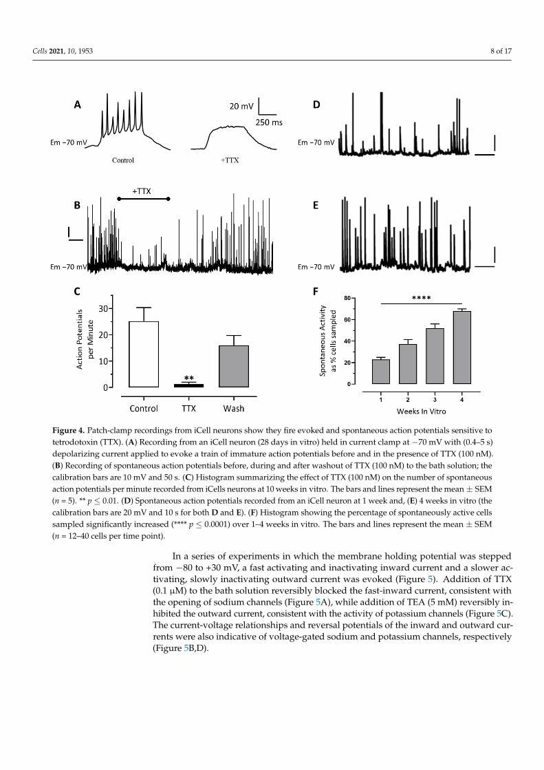
Consistent with our MEA observations, iCell neurons held in current-clamp at −70 mV fired spontaneous action potentials (Figure 4 B) that were reversibly inhibited by the sodium channel blocking drug, TTX (0.1 μM, Figure 4 B and C). Action potentials could also be evoked in current-clamped cells by the injection of (0.5–0.8 s) depolarizing current (Figure 4 A). These evoked action potentials were also blocked by the addition of TTX (0.1 μM, Figure 4 A). Under our recording conditions the frequency of action potential dis-charge slightly increased (Figure 4 D and E) and the proportion of cells that were sponta-neously active significantly increased from 25% ± 5% in the first week of culture to 67% ± 2% in the fourth week of culture (n = 16 to 40 cells per week, Figure 4F).
Figure 3. (A) a histogram summary of the total number of spikes recorded from iCell neurons in a 10-min recording period,every two days over 28 days in vitro. (B) a MEA spike trace before (baseline) and in the presence of tetrodotoxin (TTX,0.1 µM) with a histogram showing that it significantly (* p ≤ 0.05) reduced the number of spikes recorded over a 10 minperiod. The bars and lines represent the mean ± SEM (n = 4).
3.2. Patch-Clamp Electrophysiology
In cells sampled over the course of our experiments, the membrane potential of neu-rons increased from −33 ± 1.8 mV (n = 27) in the first week of cell culture to −41 ± 1.4 mV(n = 31, range −31 to −57 mV) in the fourth week of culture to −47 ± 2.2 mV (n = 19, range−35 to −65 mV, p < 0.05) in the eighth week in culture. The cell membrane capacitancerecorded from sampled cells throughout ten weeks in culture was 15.1 ± 0.5 pF, the mem-brane resistance was 870 ± 80 MΩ and series resistance was 12.8 ± 0.5 MΩ (n = 95). Thesevalues are well within the ranges for immature neural cells derived from human stem cellsin 2D monoculture reported by other groups [5,10,12,13].
Consistent with our MEA observations, iCell neurons held in current-clamp at −70 mVfired spontaneous action potentials (Figure 4B) that were reversibly inhibited by the sodiumchannel blocking drug, TTX (0.1 µM, Figure 4B,C). Action potentials could also be evoked incurrent-clamped cells by the injection of (0.5–0.8 s) depolarizing current (Figure 4A). Theseevoked action potentials were also blocked by the addition of TTX (0.1 µM, Figure 4A). Un-der our recording conditions the frequency of action potential discharge slightly increased(Figure 4D,E) and the proportion of cells that were spontaneously active significantlyincreased from 25% ± 5% in the first week of culture to 67% ± 2% in the fourth week ofculture (n = 16 to 40 cells per week, Figure 4F).
Cells 2021, 10, 1953 8 of 17
Cells 2021, 10, x 8 of 18
Figure 4. Patch-clamp recordings from iCell neurons show they fire evoked and spontaneous action potentials sensitive to tetrodotoxin (TTX). (A) Recording from an iCell neuron (28 days in vitro) held in current clamp at −70mV with (0.4–5 s) depolarizing current applied to evoke a train of immature action potentials before and in the presence of TTX (100 nM). (B) Recording of spontaneous action potentials before, during and after washout of TTX (100 nM) to the bath solution; the calibration bars are 10 mV and 50 s. (C) Histogram summarizing the effect of TTX (100 nM) on the number of spontaneous action potentials per minute recorded from iCells neurons at 10 weeks in vitro. The bars and lines represent the mean ± SEM (n = 5). ** p ≤ 0.01. (D) Spontaneous action potentials recorded from an iCell neuron at 1 week and, (E) 4 weeks in vitro (the calibration bars are 20 mV and 10 s for both D and E). (F) Histogram showing the percentage of spontaneously active cells sampled significantly increased (**** p ≤ 0.0001) over 1–4 weeks in vitro. The bars and lines represent the mean ± SEM (n = 12–40 cells per time point).
In a series of experiments in which the membrane holding potential was stepped from −80 to +30 mV, a fast activating and inactivating inward current and a slower acti-vating, slowly inactivating outward current was evoked (Figure 5). Addition of TTX (0.1 μM) to the bath solution reversibly blocked the fast-inward current, consistent with the opening of sodium channels (Figure 5 A), while addition of TEA (5 mM) reversibly inhib-ited the outward current, consistent with the activity of potassium channels (Figure 5 C). The current-voltage relationships and reversal potentials of the inward and outward cur-rents were also indicative of voltage-gated sodium and potassium channels, respectively (Figure 5B,D).
Figure 4. Patch-clamp recordings from iCell neurons show they fire evoked and spontaneous action potentials sensitive totetrodotoxin (TTX). (A) Recording from an iCell neuron (28 days in vitro) held in current clamp at −70 mV with (0.4–5 s)depolarizing current applied to evoke a train of immature action potentials before and in the presence of TTX (100 nM).(B) Recording of spontaneous action potentials before, during and after washout of TTX (100 nM) to the bath solution; thecalibration bars are 10 mV and 50 s. (C) Histogram summarizing the effect of TTX (100 nM) on the number of spontaneousaction potentials per minute recorded from iCells neurons at 10 weeks in vitro. The bars and lines represent the mean ± SEM(n = 5). ** p ≤ 0.01. (D) Spontaneous action potentials recorded from an iCell neuron at 1 week and, (E) 4 weeks in vitro (thecalibration bars are 20 mV and 10 s for both D and E). (F) Histogram showing the percentage of spontaneously active cellssampled significantly increased (**** p ≤ 0.0001) over 1–4 weeks in vitro. The bars and lines represent the mean ± SEM(n = 12–40 cells per time point).
In a series of experiments in which the membrane holding potential was steppedfrom −80 to +30 mV, a fast activating and inactivating inward current and a slower ac-tivating, slowly inactivating outward current was evoked (Figure 5). Addition of TTX(0.1 µM) to the bath solution reversibly blocked the fast-inward current, consistent withthe opening of sodium channels (Figure 5A), while addition of TEA (5 mM) reversibly in-hibited the outward current, consistent with the activity of potassium channels (Figure 5C).The current-voltage relationships and reversal potentials of the inward and outward cur-rents were also indicative of voltage-gated sodium and potassium channels, respectively(Figure 5B,D).

Cells 2021, 10, 1953 9 of 17
Cells 2021, 10, x 9 of 18
Figure 5. iCell neurons show robust voltage-activated currents. (A) whole-cell (sodium) currents evoked by a step from −80 to −40 mV (black trace) that was inhibited by TTX (100 nM, red trace,). (B) plot of the peak inward currents against voltage steps from −80 to +40 mV in the absence ( black line) and presence ( red line) of TTX (100 nM). (C) whole-cell (potassium) current evoked by a step from −80 to +40 mV (black trace) was inhibited by TEA (5 mM, red trace). (D) plot of the peak inward currents against voltage step in the absence ( black line) and presence ( red line) of TEA (5 mM).
Inhibitory Ligand-Gated Currents GABA is the most abundant inhibitory neurotransmitter in the mammalian brain,
with iCell neurons being described as primarily GABAergic. Application of GABA to neu-rons under whole-cell voltage clamp evoked concentration-dependent currents with an EC50 of 8 μM [6–9, 95% CI] (n = 11) (Figure 6 A and D). The GABA (3 μM) current-voltage relationship displayed a slight outward rectification and reversed at approximately 0 mV, consistent with the equilibrium potential of −1.9 mV for chloride ions under our recording conditions (not shown).
Figure 5. iCell neurons show robust voltage-activated currents. (A) whole-cell (sodium) currents evoked by a step from−80 to −40 mV (black trace) that was inhibited by TTX (100 nM, red trace). (B) plot of the peak inward currents againstvoltage steps from −80 to +40 mV in the absence (• black line) and presence (# red line) of TTX (100 nM). (C) whole-cell(potassium) current evoked by a step from −80 to +40 mV (black trace) was inhibited by TEA (5 mM, red trace). (D) plot ofthe peak inward currents against voltage step in the absence (
Cells 2021, 10, x 9 of 18
Figure 5. iCell neurons show robust voltage-activated currents. (A) whole-cell (sodium) currents evoked by a step from −80 to −40 mV (black trace) that was inhibited by TTX (100 nM, red trace,). (B) plot of the peak inward currents against voltage steps from −80 to +40 mV in the absence ( black line) and presence ( red line) of TTX (100 nM). (C) whole-cell (potassium) current evoked by a step from −80 to +40 mV (black trace) was inhibited by TEA (5 mM, red trace). (D) plot of the peak inward currents against voltage step in the absence ( black line) and presence ( red line) of TEA (5 mM).
Inhibitory Ligand-Gated Currents GABA is the most abundant inhibitory neurotransmitter in the mammalian brain,
with iCell neurons being described as primarily GABAergic. Application of GABA to neu-rons under whole-cell voltage clamp evoked concentration-dependent currents with an EC50 of 8 μM [6–9, 95% CI] (n = 11) (Figure 6 A and D). The GABA (3 μM) current-voltage relationship displayed a slight outward rectification and reversed at approximately 0 mV, consistent with the equilibrium potential of −1.9 mV for chloride ions under our recording conditions (not shown).
black line) and presence ( red line) of TEA (5 mM).
Inhibitory Ligand-Gated Currents
GABA is the most abundant inhibitory neurotransmitter in the mammalian brain, withiCell neurons being described as primarily GABAergic. Application of GABA to neuronsunder whole-cell voltage clamp evoked concentration-dependent currents with an EC50 of8 µM [6–9, 95% CI] (n = 11) (Figure 6A,D). The GABA (3 µM) current-voltage relationshipdisplayed a slight outward rectification and reversed at approximately 0 mV, consistentwith the equilibrium potential of −1.9 mV for chloride ions under our recording conditions(not shown).

Cells 2021, 10, 1953 10 of 17Cells 2021, 10, x 10 of 18
Figure 6. iCell neurons express GABAA receptor gated chloride channels. (A) GABA evokes concentration-dependent cur-rents. (B) sub-maximal currents are potentiated by chlordiazepoxide (CDZ) and (C) inhibited by bicuculline (BIC). The holding potential of cells was −60 mV. (D) A semi-log-linear plot of the GABA concentration-response relationship rec-orded from iCell neurons. The symbols represent the mean ± SEM of n = 11 cells. (E) a histogram summarizing the effects of diazepam (DIAZ, 10 μM), chlordiazepoxide (CDZ, 10 μM), mefenamic acid (MFA, 30 μM), phenobarbital (PHB, 100 μM), allopregnanolone (ALLO, 0.2 μM), picrotoxin (PICRO, 10 μM) and bicuculline (BIC, 10 μM) on sub-maximal GABA-evoked whole-cell currents. Each bar represents the mean ± SEM of 4–6 cells. * p ≤ 0.05, ** p ≤ 0.01; **** p ≤ 0.0001.
A diverse array of clinically important agents act as positive allosteric modulators of the GABAA receptor and help to define it’s subunit composition [14,15]. These drugs in-clude the benzodiazepines, barbiturates, neurosteroids and the fenamate non-steroidal anti-inflammatory drugs (NSAIDs, [4]). Addition of the benzodiazepine diazepam (10 μM) or chlordiazepoxide (10 μM, Figure 6 B) potentiated GABA (EC20) currents to 258% ± 31% (n = 5) and 160% ± 15% (n = 5) of control, while the barbiturate phenobarbital (100 μM), the neurosteroid allopregnanolone (0.2 μM), and the fenamate NSAID, mefenamic acid (MFA, 30 μM) potentiated these currents to 433% ± 46% (n = 5), 180% ± 22% (n = 4) and 246% ± 38% (n = 5) of control, respectively (Figure 6 E). In contrast, the non-competi-tive GABAA chloride channel blocker, picrotoxin (10 μM) inhibited GABA (EC60) currents to 20% ± 5% (n = 4) of control and the competitive GABAA receptor antagonist, bicuculline (10 μM, Figure 6C) inhibited these currents to 13% ± 2% (n = 4) of control (see Figure 6E).
Like GABAA receptors, glycine receptors (GlyR) are anion-selective ligand-gated ion channels with activation leading to rapid increases in chloride ion permeability, mem-brane hyperpolarization and a reduction in neuronal excitability [16]. Here, we discovered
Figure 6. iCell neurons express GABAA receptor gated chloride channels. (A) GABA evokes concentration-dependentcurrents. (B) sub-maximal currents are potentiated by chlordiazepoxide (CDZ) and (C) inhibited by bicuculline (BIC). Theholding potential of cells was −60 mV. (D) A semi-log-linear plot of the GABA concentration-response relationship recordedfrom iCell neurons. The symbols represent the mean ± SEM of n = 11 cells. (E) a histogram summarizing the effects ofdiazepam (DIAZ, 10 µM), chlordiazepoxide (CDZ, 10 µM), mefenamic acid (MFA, 30 µM), phenobarbital (PHB, 100 µM),allopregnanolone (ALLO, 0.2 µM), picrotoxin (PICRO, 10 µM) and bicuculline (BIC, 10 µM) on sub-maximal GABA-evokedwhole-cell currents. Each bar represents the mean ± SEM of 4–6 cells. * p ≤ 0.05, ** p ≤ 0.01; **** p ≤ 0.0001.
A diverse array of clinically important agents act as positive allosteric modulatorsof the GABAA receptor and help to define it’s subunit composition [14,15]. These drugsinclude the benzodiazepines, barbiturates, neurosteroids and the fenamate non-steroidalanti-inflammatory drugs (NSAIDs, [4]). Addition of the benzodiazepine diazepam (10 µM)or chlordiazepoxide (10 µM, Figure 6B) potentiated GABA (EC20) currents to 258% ± 31%(n = 5) and 160% ± 15% (n = 5) of control, while the barbiturate phenobarbital (100 µM),the neurosteroid allopregnanolone (0.2 µM), and the fenamate NSAID, mefenamic acid(MFA, 30 µM) potentiated these currents to 433% ± 46% (n = 5), 180% ± 22% (n = 4) and246% ± 38% (n = 5) of control, respectively (Figure 6E). In contrast, the non-competitiveGABAA chloride channel blocker, picrotoxin (10 µM) inhibited GABA (EC60) currents to20% ± 5% (n = 4) of control and the competitive GABAA receptor antagonist, bicuculline(10 µM, Figure 6C) inhibited these currents to 13% ± 2% (n = 4) of control (see Figure 6E).
Like GABAA receptors, glycine receptors (GlyR) are anion-selective ligand-gated ionchannels with activation leading to rapid increases in chloride ion permeability, membranehyperpolarization and a reduction in neuronal excitability [16]. Here, we discovered thatapplication of glycine evoked concentration-dependent currents with an EC50 of 60 µM

Cells 2021, 10, 1953 11 of 17
[51–70, 95% CI] (n = 15) (Figure 7A,B). The glycine (30 µM) current-voltage relationship wasapproximately linear (Ohmic) and reversed at around 0 mV, consistent with the equilibriumpotential of −1.9 mV for chloride ions under our recording conditions. Addition of theglycine receptor antagonist, strychnine led to a reversible and concentration-dependent in-hibition of glycine (EC30) currents with an IC50 of 50 nM [34–74, 95% CI] (n = 4, Figure 7C,D).These data are consistent with those described for native glycine receptor-gated chloridecurrents in mammalian central nervous system neurons [16–18].
Cells 2021, 10, x 11 of 18
that application of glycine evoked concentration-dependent currents with an EC50 of 60 μM [51–70, 95% CI] (n = 15) (Figure 7A,B). The glycine (30 μM) current-voltage relation-ship was approximately linear (Ohmic) and reversed at around 0 mV, consistent with the equilibrium potential of −1.9 mV for chloride ions under our recording conditions. Addi-tion of the glycine receptor antagonist, strychnine led to a reversible and concentration-dependent inhibition of glycine (EC30) currents with an IC50 of 50 nM [34–74, 95% CI] (n = 4, Figure 7 C and D). These data are consistent with those described for native glycine receptor-gated chloride currents in mammalian central nervous system neurons [16–18].
Figure 7. Glycine evokes robust concentration-dependent currents in iCell neurons. (A) glycine evoked inward currents recorded from a single iCell neuron held in voltage-clamp at −60 mV. (B) semi-log-linear plot of the glycine dose–response relationship recorded from iCell neurons. The symbols represent the mean ± SEM of 15 cells. (C) strychnine (10 μM) re-versibly inhibits glycine (EC30) currents in a cell held at −60 mV. (D) a graph summarizing the effects of strychnine (0.001–10 μM) on submaximal (EC30) glycine evoked currents recorded from iCell neurons. The symbols represent the mean ± SEM of 4 cells.
3.3. Excitatory Ligand-Gated Currents Glutamate is the major excitatory neurotransmitter of the mammalian central nerv-
ous system and interacts with both ligand-gated and G-protein couple receptors [19,20]. Application of glutamate (0.3–1000 μM) to neurons under whole-cell voltage clamp evoked concentration-dependent currents with an EC50 of 8 μM [6–10, 95% CI] (n = 7, Fig-ure 8B). Addition of the broad-spectrum glutamate receptor antagonist, kynurenic acid (1mM) reversibly inhibited glutamate (EC80) currents to 19% ± 6% (n = 6) of control (Figure 8A).
Figure 7. Glycine evokes robust concentration-dependent currents in iCell neurons. (A) glycine evoked inward currentsrecorded from a single iCell neuron held in voltage-clamp at −60 mV. (B) semi-log-linear plot of the glycine dose–responserelationship recorded from iCell neurons. The symbols represent the mean ± SEM of 15 cells. (C) strychnine (10 µM)reversibly inhibits glycine (EC30) currents in a cell held at −60 mV. (D) a graph summarizing the effects of strychnine(0.001–10 µM) on submaximal (EC30) glycine evoked currents recorded from iCell neurons. The symbols represent themean ± SEM of 4 cells.
3.3. Excitatory Ligand-Gated Currents
Glutamate is the major excitatory neurotransmitter of the mammalian central nervoussystem and interacts with both ligand-gated and G-protein couple receptors [19,20]. Ap-plication of glutamate (0.3–1000 µM) to neurons under whole-cell voltage clamp evokedconcentration-dependent currents with an EC50 of 8 µM [6–10, 95% CI] (n = 7, Figure 8B).Addition of the broad-spectrum glutamate receptor antagonist, kynurenic acid (1 mM)reversibly inhibited glutamate (EC80) currents to 19% ± 6% (n = 6) of control (Figure 8A).

Cells 2021, 10, 1953 12 of 17Cells 2021, 10, x 12 of 18
Figure 8. iCell neurons express ionotropic glutamate, NMDA, AMPA and kainate receptors. (A) A recording of submaxi-mal (EC80) glutamate currents that are rapidly and reversibly inhibited by kynurenic acid. The histogram to the right of the trace is the mean ± SEM of 6 similar experiments. (B–E) The concentration-response curves for glutamate, AMPA, NMDA and kainate, respectively. The x-axis is the agonist concentration on a log scale, and the y-axis is the response, normalized to the maximal current. (F) Histogram summarizing NMDA (30 μM) responses before, in the presence, and following washout of AP5 (100 μM). (G) Histogram summarizing Kainate (100 μM) responses before, in the presence, and following washout of DNQX (10 μM). (H) Histogram summarizing AMPA (30 μM) responses before, in the presence, and following washout of DNQX (10 μM). All the bars and error lines represent the mean ± SEM of 4–6 cells; *** p ≤ 0.001. Neurons were voltage-clamped at −60 mV.
We were also able to evoke concentration-dependent currents from voltage-clamped cells using kainic acid (1–3000 μM) with an EC50 of 361 μM ([232–562, 95% CI] n = 6, Figure 8 E). Additionally, α-amino-3-hydroxy-5-methyl-4-isoxazolepropionic acid (AMPA, 1–300 μM) also evoked concentration-dependent currents in voltage-clamped neurons with an EC50 of 21 μM [15–31, 95% CI] n = 4, Figure 8 C). The AMPA/kainate receptor antago-nist, DNQX (10 μM), reversibly inhibited kainate (EC30) and AMPA (EC60) evoked currents to 14% ± 6% (n = 9) and 3% ± 3% (n = 4) of control, respectively (see Figure 8 G and H), consistent with the expression of kainate and AMPA receptors in iCell neurons.
Application of N-methyl-D-aspartate (NMDA, 1–1000 μM) to neurons under whole-cell voltage clamp, with glycine (1 μM) included in the extracellular recording solution and in the absence of magnesium ions, evoked concentration-dependent currents with an EC50 of 57 μM [44–73, 95% CI] n = 4, Figure 8 D). Addition of the NMDA receptor antago-nist (2R)-amino-5-phosphonovaleric acid (AP5, 100 μM) reversibly inhibited NMDA (EC30) currents to 8% ± 3% (n = 4) of control (Figure 8 F). In the absence glycine, NMDA currents were diminished below c. 25 pA confirming that glycine is a required co-agonist. We also determined that the NMDA (30 μM) current-voltage relationship was approxi-mately linear in the absence of Mg2+ but displayed strong voltage-dependent block at neg-
Figure 8. iCell neurons express ionotropic glutamate, NMDA, AMPA and kainate receptors. (A) A recording of submaximal(EC80) glutamate currents that are rapidly and reversibly inhibited by kynurenic acid. The histogram to the right of thetrace is the mean ± SEM of 6 similar experiments. (B–E) The concentration-response curves for glutamate, AMPA, NMDAand kainate, respectively. The x-axis is the agonist concentration on a log scale, and the y-axis is the response, normalizedto the maximal current. (F) Histogram summarizing NMDA (30 µM) responses before, in the presence, and followingwashout of AP5 (100 µM). (G) Histogram summarizing Kainate (100 µM) responses before, in the presence, and followingwashout of DNQX (10 µM). (H) Histogram summarizing AMPA (30 µM) responses before, in the presence, and followingwashout of DNQX (10 µM). All the bars and error lines represent the mean ± SEM of 4–6 cells; *** p ≤ 0.001. Neurons werevoltage-clamped at −60 mV.
We were also able to evoke concentration-dependent currents from voltage-clampedcells using kainic acid (1–3000 µM) with an EC50 of 361 µM ([232–562, 95% CI] n = 6,Figure 8E). Additionally, α-amino-3-hydroxy-5-methyl-4-isoxazolepropionic acid (AMPA,1–300 µM) also evoked concentration-dependent currents in voltage-clamped neurons withan EC50 of 21 µM [15–31, 95% CI] n = 4, Figure 8C). The AMPA/kainate receptor antagonist,DNQX (10 µM), reversibly inhibited kainate (EC30) and AMPA (EC60) evoked currents to14% ± 6% (n = 9) and 3% ± 3% (n = 4) of control, respectively (see Figure 8G,H), consistentwith the expression of kainate and AMPA receptors in iCell neurons.
Application of N-methyl-D-aspartate (NMDA, 1–1000 µM) to neurons under whole-cell voltage clamp, with glycine (1 µM) included in the extracellular recording solution andin the absence of magnesium ions, evoked concentration-dependent currents with an EC50of 57 µM [44–73, 95% CI] n = 4, Figure 8D). Addition of the NMDA receptor antagonist(2R)-amino-5-phosphonovaleric acid (AP5, 100 µM) reversibly inhibited NMDA (EC30)currents to 8% ± 3% (n = 4) of control (Figure 8F). In the absence glycine, NMDA currentswere diminished below c. 25 pA confirming that glycine is a required co-agonist. Wealso determined that the NMDA (30 µM) current-voltage relationship was approximatelylinear in the absence of Mg2+ but displayed strong voltage-dependent block at negativepotentials, with a region of negative slope conductance above −20 mV holding potential

Cells 2021, 10, 1953 13 of 17
(Figure 9A,B). Together, our data show that iCell Neurons express functional NMDA-typeglutamate receptors with several of the complex pharmacological properties described fornative CNS neuron receptors.
Cells 2021, 10, x 13 of 18
ative potentials, with a region of negative slope conductance above −20 mV holding po-tential (Figure 9A,B). Together, our data show that iCell Neurons express functional NMDA-type glutamate receptors with several of the complex pharmacological properties described for native CNS neuron receptors.
Figure 9. Mg2+ inhibits NMDA-evoked currents recorded from iCell neurons in a highly voltage-dependent fashion. (A) recording of responses to short (400 ms) pulses of NMDA (30 μM) applied to a neuron voltage-clamped at +60 and −100 mV in the absence (black line) and presence (red line) of Mg2+ (2 mM). (B) the current-voltage relationship for NMDA (30 μM) responses in the absence () and presence () of magnesium ions. Note the negative slope conductance above −20 mV when magnesium is included in the bath solution. The x-axis shows the holding potential (Vh) and the y-axis is the NMDA current normalized to +60 mV. The graph is the mean of 4 cells.
4. Discussion Human iPSC-derived neurons are increasingly promoted as tools in drug discovery
(e.g., [6,21–23], even when their molecular pharmacological properties are not fully char-acterized [5]. We have previously reported on the electrophysiological characteristics of neurons derived from a human stem cell line that is simple and inexpensive to culture and differentiate [24,25]. Here, we determined the physiological and pharmacological proper-ties of the voltage- and ligand-gated ion channels expressed in a commercially available population of iPSC-derived neurons (iCell neurons) maintained for up to 10 weeks in vitro, using both multi-electrode array and patch-clamp approaches.
Our data showed that iCell neurons display neurite outgrowth within 24 h of plating and label for the pan-neuronal marker, βIII tubulin within the first week consistent with the morphological and immunocytochemical features of neurons. Our MEA recordings also showed that these iPSC-derived neurons generated spontaneous spike-like activity within 2 days of plating which peaked after one week, and then rapidly decreased over the second week to remain at low levels up to one month in vitro. The reduced spike activity was not associated with a coincidental rapid loss of cells from the MEA plate. A previous study that used MEA to record from iCell neurons for 9 days post-plating also reported they were most active at day 7 to 8 [26] consistent with our recording from these cells over 28 days post-plating. Odawara and colleagues (2014) similarly reported that
Figure 9. Mg2+ inhibits NMDA-evoked currents recorded from iCell neurons in a highly voltage-dependent fashion.(A) recording of responses to short (400 ms) pulses of NMDA (30 µM) applied to a neuron voltage-clamped at +60 and−100 mV in the absence (black line) and presence (red line) of Mg2+ (2 mM). (B) the current-voltage relationship for NMDA(30 µM) responses in the absence (•) and presence (#) of magnesium ions. Note the negative slope conductance above−20 mV when magnesium is included in the bath solution. The x-axis shows the holding potential (Vh) and the y-axis is theNMDA current normalized to +60 mV. The graph is the mean of 4 cells.
4. Discussion
Human iPSC-derived neurons are increasingly promoted as tools in drug discovery(e.g., [6,21–23], even when their molecular pharmacological properties are not fully char-acterized [5]. We have previously reported on the electrophysiological characteristics ofneurons derived from a human stem cell line that is simple and inexpensive to culture anddifferentiate [24,25]. Here, we determined the physiological and pharmacological proper-ties of the voltage- and ligand-gated ion channels expressed in a commercially availablepopulation of iPSC-derived neurons (iCell neurons) maintained for up to 10 weeks in vitro,using both multi-electrode array and patch-clamp approaches.
Our data showed that iCell neurons display neurite outgrowth within 24 h of platingand label for the pan-neuronal marker, βIII tubulin within the first week consistent withthe morphological and immunocytochemical features of neurons. Our MEA recordingsalso showed that these iPSC-derived neurons generated spontaneous spike-like activitywithin 2 days of plating which peaked after one week, and then rapidly decreased overthe second week to remain at low levels up to one month in vitro. The reduced spikeactivity was not associated with a coincidental rapid loss of cells from the MEA plate. Aprevious study that used MEA to record from iCell neurons for 9 days post-plating alsoreported they were most active at day 7 to 8 [26] consistent with our recording from thesecells over 28 days post-plating. Odawara and colleagues (2014) similarly reported thatspontaneous activity rapidly decreased in iCells after 8 days in vitro unless co-cultured withrat astrocytes or in glial cell conditioned media [27]. The extracellularly recorded spikes

Cells 2021, 10, 1953 14 of 17
were inhibited by the sodium channel blocker, tetrodotoxin, consistent with the generationof neuronal action potentials, and in keeping with a short report by Kasteel and colleagues(2017) [28]. Together, these data indicate that this population of iPSC-derived neurons,in standard culture conditions, may be useful for shorter-term experiments, particularlywhen spontaneous neuronal activity is a focus of research.
Our patch-clamp recordings showed that the passive membrane properties of iCellsneurons, including their membrane capacitance, membrane resistance and membranepotential are consistent with native neurons [29], embryonic stem cell derived neurons(e.g., [12]), and iPSC-derived neurons (e.g., [10,30]). Cells also generated evoked andspontaneous action potentials within the first week of plating, which increased over onemonth in culture, along with a significant increase in the proportion of cells that werespontaneously active, indicating that such pre-differentiated neurons continue to maturein vitro. Our single cell recordings showing increased spiking over the first month thereforediffered from our MEA recordings showing a decrease. The decrease in spontaneousactivity, around 6–8 days in vitro is likely the result of some migration of cells on the MEAchip (see also Odawara et al., 2014) but, most significantly, the development of a dominantinhibitory tone from dense GABAergic neuron circuit maturation suppressing spontaneousspike activity. In support of this hypothesis, FCDI reported that the GABAA receptorantagonist, GABAZINE increased bursting rate in iCell neurons (https://fujifilmcdi.com/assets/CDI_iCellNC_AxionMEA_AP-NRCAXN1405281.pdf accessed on 28 July 2021). Insupplementary experiments (Supplementary Figure S1) we determined that iCell neurons,under current clamp, and depolarized by glutamate, leads to the generation of only a fewaction potentials, even in the continued presence of glutamate. In contrast, pre-applyingthe GABAA receptor antagonist, bicuculline (to block inhibitory [GABA] tone) and thenevoking depolarization with glutamate leads to a train of action potentials that are wellmaintained throughout application of glutamate (see Supplementary Figure S1) Thesedata therefore strongly support the hypothesis that iCells are under powerful GABAergicinhibitory tone and the reason for the reduction in spike activity beginning around 6–8 dayson the MEA plate. The voltage-step protocols also confirmed that both voltage-gatedsodium and potassium ion channels are strongly expressed, can be blocked by TTX andTEA, respectively, and underlie the recorded action potentials, consistent with previousreports [9,10,28]. Our observations support the value of iCell neurons as a model for studiesof neuronal voltage-gated sodium and potassium channels, and the study of glutamate-evoked action potentials.
In addition, our patch-clamp recordings revealed several important neuropharma-cological properties of iCells. First, neurons responded to the application of GABA (inkeeping with the manufacturers (re)designation of iCells as iCell GABANeurons) from2 days post-plating and for a further 3 months in vitro. Previous studies using automatedpatch-clamp instruments to record from ensembles of cells reported that GABA currents iniCells were inhibited by bicuculline [9] and modulated by diazepam [11]. In the presentstudy, we show that GABA responses were reversibly inhibited by both the competitiveand the non-competitive GABAA antagonists, bicuculline and picrotoxin, respectively.Moreover, we demonstrate for the first time that these GABA currents are potentiatedby a diverse range of positive allosteric modulators of the GABAA receptor includingchlordiazepoxide, diazepam, phenobarbital, allopregnanolone and mefenamic acid. Inhi-bition of GABA currents by bicuculline and picrotoxin indicates a receptor conformationto include β-α subunits forming the GABA and bicuculline binding site as well as thetransmembrane domain site for picrotoxin [31]. Sensitivity to the benzodiazepines, chlor-diazepoxide and diazepam, is conferred by the presence of the γ2 receptor subunit inthe GABAA receptor complex [32,33] whilst sensitivity to the NSAID, mefenamic acid isconferred by the presence of the β2/β3 subunits in the receptor complex [4,34]. We cantherefore deduce from our electrophysiological data that the GABAA receptor isoformsexpressed by iCells are likely composed of αxβ2/3γ2 subunits (where x is one of severalpossible α subunits) to enable the rich and complex pharmacological responses observed

Cells 2021, 10, 1953 15 of 17
to these clinically important agents and consistent with native GABAA receptor pharmacol-ogy [35]. Measurements of mRNA expression levels of GABAA receptor subunits in iCellneurons are also entirely consistent with our patch-clamp data [7,11]. Together these dataindicate that these iPSC-derived human neurons may be valuable models for studies of theGABAA receptor chloride channel complex in drug discovery and development.
This study also revealed for the first time that the inhibitory neurotransmitter glycineevoked robust and concentration-dependent responses in iCell neurons and that thesecurrents were inhibited by the competitive antagonist of glycine receptors, strychnine [36].Like GABAA receptors, glycine receptors are members of the Cys-loop receptor family andmediate synaptic inhibition in the spinal cord, brainstem, and other caudal regions of thebrain [37]. Glycine receptors are involved in nociception and implicated in several neuro-logical diseases, including hyperekplexia (startle disease) and autism; they are inhibited bydrugs such as picrotoxin and potentiated by general anesthetics, ethanol and endogenouscannabinoids [38]. iCell neurons may therefore serve as a useful tool to investigate thepharmacological properties of human glycine receptors.
Finally, we determined extended agonist concentration range responses to glutamate,AMPA, kainate and NMDA from iCell neurons, providing new information on saturatingand non-saturating agonist concentrations that are essential for studies of positive andnegative allosteric modulators of these major excitatory receptors (e.g., [8]). Our data areconsistent with the manufacturer’s description that iCell neurons are a mixed population ofpost-mitotic neural subtypes comprised of both GABAergic and glutamatergic neurons. Theagonist-evoked currents were inhibited by antagonists consistent with the expression of theionotropic AMPA, kainate and NMDA glutamate receptors. Additionally, we determinedthat the NMDA currents, like native neuronal receptors, were inhibited in a highly voltage-dependent manner by magnesium ions [39] and required the presence of the co-agonist,glycine in the extracellular recording solution [40]. Consistent with our current findings,Dage and colleagues (2014) showed that application of AMPA or NMDA evoked transientcalcium fluxes in iCell neurons. In addition, and based on their gene expression profile,the NMDA receptors expressed in iCell neurons were reported to be composed of GluN1and GluN2B heterodimeric subunits, similar to that found in neonatal cortex [7,8]. Overall,these data are therefore consistent with the many of the physiological and pharmacologicalproperties of those described for native ionotropic glutamate receptors [20,41].
In summary, our electrophysiological observations on iCell neurons show they expressseveral of the major voltage- and ligand-gated ion channels with the neurophysiologicaland neuropharmacological properties consistent with native neurons. We also show thatthese cells remain viable in long-term culture for experimentation. These new data thereforesuggest their value for pharmacological research and drug development, especially giventhe role of GABAA and ionotropic glutamate receptors in normal brain functions of fastsynaptic neurotransmission and synaptic plasticity to pathological conditions such asepilepsy, anxiety, depression, neurodegeneration and stroke [41,42]. Moreover, recentstudies have indicated that iPSC-derived neurons and glial cells may also have value andgreater sensitivity than rodent neurons in neurotoxicology studies [43–45]. Future work inthis lab, however, will determine the functional properties of iPSC-derived neurons andglia in 3D cerebral neurospheres (brain organoids) in an effort to better understand theirvalidity as more complex models of the human nervous system in vitro.
Supplementary Materials: The following are available online at https://www.mdpi.com/article/10.3390/cells10081953/s1, Figure S1: (A) A single iCell neuron held in current clamp at −70 mV,transiently exposed to glutamate (30 µM) depolarizes the cell and evokes 4–5 spikes. (B) A single iCellneuron with bicuculline (3 µM) included in the bath solution transiently exposed to glutamate (30 µM)evokes depolarization and a train of action potential throughout the membrane depolarization. (C) Ahistogram of the number of action potentials recorded over 100 s when cells are depolarized in theabsence (control) or presence of bicuculline.

Cells 2021, 10, 1953 16 of 17
Author Contributions: Conceptualization, R.F.H.; Data curation, H.S., L.C. and W.S.C.; Formalanalysis, R.F.H., H.S., L.C. and W.S.C.; Funding acquisition, R.F.H.; Investigation, H.S., L.C. andW.S.C.; Methodology, H.S., L.C. and W.S.C.; Project administration, R.F.H.; Writing—original draft,R.F.H.; L.C. and W.S.C.; Writing—review and editing, R.F.H., H.S. All authors have read and agreedto the published version of the manuscript.
Funding: This research received no external funding.
Institutional Review Board Statement: Not applicable.
Informed Consent Statement: Not applicable.
Data Availability Statement: Data available on request.
Conflicts of Interest: The authors declare no conflict of interest.
References1. Piccolino, M. Animal Electricity and the Birth of Electrophysiology: The Legacy of Luigi Galvani. Brain Res. Bull. 1998, 46, 381–407.
[CrossRef]2. Rai, J.; Kaushik, K. Reduction of Animal Sacrifice in Biomedical Science & Research through Alternative Design of Animal
Experiments. Saudi Pharm. J. 2018, 26, 896–902. [CrossRef]3. Avoli, M.; Williamson, A. Functional and Pharmacological Properties of Human Neocortical Neurons Maintained in Vitro. Prog.
Neurobiol. 1996, 48, 519–554. [CrossRef]4. Halliwell, R.F.; Thomas, P.; Patten, D.; James, C.H.; Martinez-Torres, A.; Miledi, R.; Smart, T.G. Subunit-Selective Modulation
of GABAA Receptors by the Non-Steroidal Anti-Inflammatory Agent, Mefenamic Acid. Eur. J. Neurosci. 1999, 11, 2897–2905.[CrossRef]
5. Halliwell, R.F. Electrophysiological Properties of Neurons Derived from Human Stem Cells and INeurons in Vitro. Neurochem.Int. 2017, 106, 37–47. [CrossRef]
6. Bonaventura, G.; Iemmolo, R.; Attaguile, G.A.; La Cognata, V.; Pistone, B.S.; Raudino, G.; D’Agata, V.; Cantarella, G.; Barcellona,M.L.; Cavallaro, S. IPSCs: A Preclinical Drug Research Tool for Neurological Disorders. Int. J. Mol. Sci. 2021, 22, 4596. [CrossRef][PubMed]
7. Dage, J.L.; Colvin, E.M.; Fouillet, A.; Langron, E.; Roell, W.C.; Li, J.; Mathur, S.X.; Mogg, A.J.; Schmitt, M.G.; Felder, C.C.; et al.Pharmacological Characterisation of Ligand- and Voltage-Gated Ion Channels Expressed in Human IPSC-Derived ForebrainNeurons. Psychopharmacology 2014, 231, 1105–1124. [CrossRef] [PubMed]
8. Neagoe, I.; Liu, C.; Stumpf, A.; Lu, Y.; He, D.; Francis, R.; Chen, J.; Reynen, P.; Alaoui-Ismaili, M.H.; Fukui, H. The GluN2BSubunit Represents a Major Functional Determinant of NMDA Receptors in Human Induced Pluripotent Stem Cell-DerivedCortical Neurons. Stem Cell Res. 2018, 28, 105–114. [CrossRef]
9. Haythornthwaite, A.; Stoelzle, S.; Hasler, A.; Kiss, A.; Mosbacher, J.; George, M.; Brüggemann, A.; Fertig, N. CharacterizingHuman Ion Channels in Induced Pluripotent Stem Cell-Derived Neurons. J. Biomol. Screen. 2012, 17, 1264–1272. [CrossRef]
10. Berry, B.J.; Akanda, N.; Smith, A.S.T.; Long, C.J.; Schnepper, M.T.; Guo, X.; Hickman, J.J. Morphological and FunctionalCharacterization of Human Induced Pluripotent Stem Cell-Derived Neurons (ICell Neurons) in Defined Culture Systems.Biotechnol. Prog. 2015, 31, 1613–1622. [CrossRef]
11. Yuan, N.Y.; Poe, M.M.; Witzigmann, C.; Cook, J.M.; Stafford, D.; Arnold, L.A. Characterization of GABAA Receptor Ligandswith Automated Patch-Clamp Using Human Neurons Derived from Pluripotent Stem Cells. J. Pharmacol. Toxicol. Methods 2016,82, 109–114. [CrossRef]
12. Johnson, M.A.; Weick, J.P.; Pearce, R.A.; Zhang, S.-C. Functional Neural Development from Human Embryonic Stem Cells:Accelerated Synaptic Activity via Astrocyte Coculture. J. Neurosci. 2007, 27, 3069–3077. [CrossRef] [PubMed]
13. Risner-Janiczek, J.R.; Ungless, M.A.; Li, M. Electrophysiological Properties of Embryonic Stem Cell-Derived Neurons. PLoS ONE2011, 6, e24169. [CrossRef] [PubMed]
14. Olsen, R.W. Allosteric Ligands and Their Binding Sites Define γ-Aminobutyric Acid (GABA) Type A Receptor Subtypes. Adv.Pharmacol. 2015, 73, 167–202. [CrossRef] [PubMed]
15. Olsen, R.W.; Sieghart, W. International Union of Pharmacology. LXX. Subtypes of γ-Aminobutyric AcidA Receptors: Classificationon the Basis of Subunit Composition, Pharmacology, and Function. Update. Pharmacol. Rev. 2008, 60, 243–260. [CrossRef]
16. Burgos, C.F.; Yévenes, G.E.; Aguayo, L.G. Structure and Pharmacologic Modulation of Inhibitory Glycine Receptors. Mol.Pharmacol. 2016, 90, 318–325. [CrossRef] [PubMed]
17. Siebler, M.; Pekel, M.; Köller, H.; Müller, H.W. Strychnine-Sensitive Glycine Receptors in Cultured Primary Neurons from RatNeocortex. Brain Res. Dev. Brain Res. 1993, 73, 289–292. [CrossRef]
18. Wang, L.; Li, W.-G.; Huang, C.; Zhu, M.X.; Xu, T.-L.; Wu, D.-Z.; Li, Y. Subunit-Specific Inhibition of Glycine Receptors byCurcumol. J. Pharmacol. Exp. Ther. 2012, 343, 371–379. [CrossRef] [PubMed]
19. Mayer, M.L. Structural Biology of Glutamate Receptor Ion Channel Complexes. Curr. Opin. Struct. Biol. 2016, 41, 119–127.[CrossRef]

Cells 2021, 10, 1953 17 of 17
20. Kew, J.N.C.; Kemp, J.A. Ionotropic and Metabotropic Glutamate Receptor Structure and Pharmacology. Psychopharmacology 2005,179, 4–29. [CrossRef] [PubMed]
21. Chang, C.-Y.; Ting, H.-C.; Liu, C.-A.; Su, H.-L.; Chiou, T.-W.; Lin, S.-Z.; Harn, H.-J.; Ho, T.-J. Induced Pluripotent Stem Cell(IPSC)-Based Neurodegenerative Disease Models for Phenotype Recapitulation and Drug Screening. Molecules 2020, 25, 2000.[CrossRef]
22. Silva, M.C.; Haggarty, S.J. Human Pluripotent Stem Cell-Derived Models and Drug Screening in CNS Precision Medicine. Ann.N. Y. Acad. Sci. 2020, 1471, 18–56. [CrossRef]
23. Qian, L.; Tcw, J. Human IPSC-Based Modeling of Central Nerve System Disorders for Drug Discovery. Int. J. Mol. Sci. 2021,22, 1203. [CrossRef]
24. Stewart, R.; Coyne, L.; Lako, M.; Halliwell, R.F.; Przyborski, S.A. Human Embryonal Carcinoma Stem Cells Expressing GreenFluorescent Protein Form Functioning Neurons in Vitro: A Research Tool for Co-Culture Studies. Stem Cells Dev. 2004, 13, 646–657.[CrossRef]
25. Coyne, L.; Shan, M.; Przyborski, S.A.; Hirakawa, R.; Halliwell, R.F. Neuropharmacological Properties of Neurons Derived fromHuman Stem Cells. Neurochem. Int. 2011, 59, 404–412. [CrossRef] [PubMed]
26. Tukker, A.M.; de Groot, M.W.G.D.M.; Wijnolts, F.M.J.; Kasteel, E.E.J.; Hondebrink, L.; Westerink, R.H.S. Is the Time Right for inVitro Neurotoxicity Testing Using Human IPSC-Derived Neurons? ALTEX Altern. Anim. Exp. 2016, 33, 261–271. [CrossRef]
27. Odawara, A.; Saitoh, Y.; Alhebshi, A.H.; Gotoh, M.; Suzuki, I. Long-Term Electrophysiological Activity and PharmacologicalResponse of a Human Induced Pluripotent Stem Cell-Derived Neuron and Astrocyte Co-Culture. Biochem. Biophys. Res. Commun.2014, 443, 1176–1181. [CrossRef] [PubMed]
28. Kasteel, E.E.J.; Westerink, R.H.S. Comparison of the Acute Inhibitory Effects of Tetrodotoxin (TTX) in Rat and Human NeuronalNetworks for Risk Assessment Purposes. Toxicol. Lett. 2017, 270, 12–16. [CrossRef] [PubMed]
29. Hille, B. Ion Channels of Excitable Membranes, 3rd ed.; Sinauer Associates Inc.: Sunderland, MA, USA, 2001; p. 11.30. Prè, D.; Nestor, M.W.; Sproul, A.A.; Jacob, S.; Koppensteiner, P.; Chinchalongporn, V.; Zimmer, M.; Yamamoto, A.; Noggle, S.A.;
Arancio, O. A Time Course Analysis of the Electrophysiological Properties of Neurons Differentiated from Human InducedPluripotent Stem Cells (IPSCs). PLoS ONE 2014, 9, e103418. [CrossRef] [PubMed]
31. Kim, J.J.; Hibbs, R.E. Direct Structural Insights into GABAA Receptor Pharmacology. Trends Biochem. Sci. 2021, 46, 502–517.[CrossRef]
32. Pritchett, D.B.; Sontheimer, H.; Shivers, B.D.; Ymer, S.; Kettenmann, H.; Schofield, P.R.; Seeburg, P.H. Importance of a NovelGABAA Receptor Subunit for Benzodiazepine Pharmacology. Nature 1989, 338, 582–585. [CrossRef] [PubMed]
33. Sigel, E.; Baur, R.; Trube, G.; Möhler, H.; Malherbe, P. The Effect of Subunit Composition of Rat Brain GABAA Receptors onChannel Function. Neuron 1990, 5, 703–711. [CrossRef]
34. Coyne, L.; Su, J.; Patten, D.; Halliwell, R.F. Characterization of the Interaction between Fenamates and Hippocampal NeuronGABA(A) Receptors. Neurochem. Int. 2007, 51, 440–446. [CrossRef] [PubMed]
35. Castellano, D.; Shepard, R.D.; Lu, W. Looking for Novelty in an “Old” Receptor: Recent Advances toward Our Understanding ofGABAARs and Their Implications in Receptor Pharmacology. Front. Neurosci. 2020, 14, 616298. [CrossRef]
36. Young, A.B.; Snyder, S.H. Strychnine Binding Associated with Glycine Receptors of the Central Nervous System. Proc. Natl. Acad.Sci. USA 1973, 70, 2832–2836. [CrossRef]
37. Betz, H.; Laube, B. Glycine Receptors: Recent Insights into Their Structural Organization and Functional Diversity. J. Neurochem.2006, 97, 1600–1610. [CrossRef]
38. Dutertre, S.; Becker, C.-M.; Betz, H. Inhibitory Glycine Receptors: An Update. J. Biol. Chem. 2012, 287, 40216–40223. [CrossRef][PubMed]
39. Mayer, M.L.; Westbrook, G.L.; Guthrie, P.B. Voltage-Dependent Block by Mg2+ of NMDA Responses in Spinal Cord Neurones.Nature 1984, 309, 261–263. [CrossRef] [PubMed]
40. Johnson, J.W.; Ascher, P. Glycine Potentiates the NMDA Response in Cultured Mouse Brain Neurons. Nature 1987, 325, 529–531.[CrossRef]
41. Fu, H.; Chen, Z.; Josephson, L.; Li, Z.; Liang, S.H. Positron Emission Tomography (PET) Ligand Development for IonotropicGlutamate Receptors: Challenges and Opportunities for Radiotracer Targeting N-Methyl-d-Aspartate (NMDA), α-Amino-3-Hydroxy-5-Methyl-4-Isoxazolepropionic Acid (AMPA), and Kainate Receptors. J. Med. Chem. 2019, 62, 403–419. [CrossRef][PubMed]
42. Yuan, H.; Low, C.-M.; Moody, O.A.; Jenkins, A.; Traynelis, S.F. Ionotropic GABA and Glutamate Receptor Mutations and HumanNeurologic Diseases. Mol. Pharmacol. 2015, 88, 203–217. [CrossRef] [PubMed]
43. Wing, C.; Komatsu, M.; Delaney, S.M.; Krause, M.; Wheeler, H.E.; Dolan, M.E. Application of Stem Cell Derived Neuronal Cellsto Evaluate Neurotoxic Chemotherapy. Stem Cell Res. 2017, 22, 79–88. [CrossRef] [PubMed]
44. Liu, L.; Koo, Y.; Russell, T.; Gay, E.; Li, Y.; Yun, Y. Three-Dimensional Brain-on-Chip Model Using Human IPSC-DerivedGABAergic Neurons and Astrocytes: Butyrylcholinesterase Post-Treatment for Acute Malathion Exposure. PLoS ONE 2020,15, e0230335. [CrossRef] [PubMed]
45. Tukker, A.M.; Wijnolts, F.M.J.; de Groot, A.; Westerink, R.H.S. Applicability of HiPSC-Derived Neuronal Cocultures and RodentPrimary Cortical Cultures for In Vitro Seizure Liability Assessment. Toxicol. Sci. 2020, 178, 71–87. [CrossRef]