Clinical, electrophysiological, and immunopathological...
15
Introduction Lepr Rev (2001) 72, 35-49 Clinical, electrophysiological, and immunopathological study of peripheral nerves in Hansen's disease WAFAA RAMADAN,* BASMA MOURAD,* WAEL FADEL,** & ENAS GHORABA* Deparents of *Deatology and **Neulo, Facul of Medicine, Tanta University, Tanta, Egypt Accepted for publication 18 October 2000 Summa Ransen' s disease is a disease of peripheral nerves. Some patients deve10p peripheral neuropathy before the diagnosis of the disease, and others develop these complications after starting therapy. Electrophysiological (EP) studies were caied out in Ransen' s disease patients. This work studied the neural deficits, electromyo- graphy (EMG) and motor nerve conduction (MNC) variables in different types of leprosy and the immunopathology of sural nerve tissue in patients with severe neural deficits. Forty leprosy patients had neurological exnations and EP study. Histo- pathological and immunopathological study of sural nerve biopsy specimens was performed for 10 patients with severe neural deficits. The results of the neurological study showed that there was involvement of cranial nerves, muscular system, motor reflexes and sensory system and trophic and vasomotor changes. EP study showed significant changes in EMG of abductor digiti minimi in patients as compared to controls. MNC variables of common peroneal nerve were abnormal in 80% of alI patients, MNC of median nerve was abnormal in 72.5%, while MNC of ulnar nerve was abnormal in 70% and SNC of ulnar nerve was abnormal in 77.5% of the total. ln conclusion, electrophysiological investigations have an important role in the detec- tion of muscle denervation and neuropathic changes in leprosy patients. These investigations are safe, rapid and non-invasive techniques. On the other hand immunopathological study revealed that the degree of immune positivity correlated with the degree of nerve fibrosis. Although its overall prevalence is decreasing, leprosy continues to be a major cause of neuropathy worldwide, l as Mycobacterium leprae has the capacity to invade the peripheral nervous system and cause neuropathy. 2 Leprous neuropathy is characterized by involvement of dermal nerves and superficial peripheral nerve trunks in cooler body regions. I There are four possible mechanisms of nerve damage in all forms of HD: (1) the presence of M. lepe or its antigens at cooler sites; (2) trauma of superficially placed nerve trunks; (3) increased Correspondence to: W. Ramadan 0305-75 18/011072035+ 15 $1.00 © Lepra 35
Transcript of Clinical, electrophysiological, and immunopathological...

Introduction
Lepr Rev (200 1 ) 72, 35-49
Clinical, electrophysiological, and
immunopathological study of peripheral nerves in
Hansen's disease
WAFAA RAMADAN , * B A S M A M O URAD , *
WAEL FADEL, * * & E N A S GHORAB A *
Departments of *Dennatology and * *Neurology, Faculty of Medicine, Tanta University, Tanta, Egypt
Accepted for publication 1 8 October 2000
Summary Ransen' s disease is a disease of peripheral nerves. Some patients deve10p
peripheral neuropathy before the diagnosis of the disease, and others develop these
complications after starting therapy. Electrophysiological (EP) studies were carried
out in Ransen' s disease patients. This work studied the neural deficits, electromyo
graphy (EMG) and motor nerve conduction (MNC) variables in different types of
leprosy and the immunopathology of sural nerve tissue in patients with severe neural
deficits . Forty leprosy patients had neurological exarninations and EP study. Histo
pathological and immunopathological study of sural nerve biopsy specimens was
performed for 10 patients with severe neural deficits. The results of the neurological
study showed that there was involvement of cranial nerves, muscular system, motor
reflexes and sensory system and trophic and vasomotor changes. EP study showed
significant changes in EMG of abductor digiti minimi in patients as compared to
controls . MNC variables of common peroneal nerve were abnormal in 80% of alI
patients, MNC of median nerve was abnormal in 72.5%, while MNC of ulnar nerve
was abnormal in 70% and SNC of ulnar nerve was abnormal in 77.5% of the total. ln conclusion, electrophysiological investigations have an important role in the detec
tion of muscle denervation and neuropathic changes in leprosy patients. These
investigations are safe, rapid and non-invasive techniques. On the other hand
immunopathological study revealed that the degree of immune positivity correlated with the degree of nerve fibrosis.
Although its overall prevalence is decreasing, leprosy continues to be a major cause of neuropathy worldwide, l as Mycobacterium leprae has the capacity to invade the peripheral nervous system and cause neuropathy.2 Leprous neuropathy is characterized by involvement of dermal nerves and superficial peripheral nerve trunks in cooler body regions. I There are four possible mechanisms of nerve damage in all forms of HD: ( 1 ) the presence of M. leprae or its antigens at cooler sites; (2) trauma of superficially placed nerve trunks; (3) increased
Correspondence to: W. Ramadan
0305-75 1 8/011072035+15 $ 1 .00 © Lepra 35
36 W. Ramadan et aI.
intraneural pressure; and (4) vascular changes in intraneural blood vesse1s. 3 HD leads to change in muscle function resulting in muscle imbalances with deformity of soft tissues and joints.4 The precise pathophysiological mechanism of peripheral neuropathy is not yet clear. It is not due to the invasion of nerves by living bacilli but it may be due to a later immune response from antigen-antibody reaction to dead bacilli and the peripheral nerves become fibrosed from mechanical stress .4
A I M OF THE W O R K
This work studied the neural deficits and electrophysiological changes in different types of leprosy and the immunopathological status of the neural tis sue in patients with severe neural deficits, to increase our insight into the pathophysiology of neural involvement in leprosy.
Materiais and methods
Forty treated (received MDT) leprosy patients (5 PN, 2 TI, 1 6 BT, 2 BL, and 1 5 LL) and 1 0 normal healthy persons (matching the sarne age and sex of the patients) serving as control were the subject of this study. The patients were 26 males and 1 4 females and their age ranged fmm 1 9 to 80 years with a mean value of 44·9 ± 1 4·57. The duration of the disease ranged from 6 months to 40 years . Eighteen of these patients had experienced leprosy reactions (7 LL, 1 0 BT and 1 BL) .
M E T H O D S
All patients and controls had complete history taking, thorough clinical and dermatological examinations, and full neurological exarninations: exarnination of cranial nerves, peripheral nerves, motor system (muscles and motor reftexes) and sensory system (superficial and deep sensations).
Cranial nerve assessment
This was carried out by the detection of clinical manifestations of nerve impairment, e.g. optic nerve (distributed visual acuity, and colour vision), trigeminal nerve (hypothesia of the face, wasting of pterygoid, masseter and temporalis muscles and decreased or lost corneal reftex), facial nerve (lower motor neuron facial paralysis), cochleovestibular nerve (decreased acuity of hearing), glossopharyngeal nerve (decreased pharyngeal reftex) and vagus nerve (hoarseness of voice and dysphagia) .
Examination 01 specific peripheral nerves
The ulnar, median and common peroneal nerves were palpated at the elbow, wrist and knee respectively.5
Muscle power assessment
This was performed by testing the contraction of the muscle against gravity and resistance.

Sensory system assessment
Peripheral nerves in Hansen 's disease 37
Superficial sensory system was assessed by using the pin prick test for pain sensation, hot test tube for temperature and a piece of thin paper, a feather or a wisp of cotton-wool for light touch sensation in both upper and lower limbs. Testing joint, vibration and pressure sensations assessed the deep sensory system.
I N V E S T I G A T I O N S
• Slit skin smear for alI patients for the confirmation of diagnosis and classification of patients .
• Electrophysiological �tudy including:
Needle EMG: The detection and recording of the electrical activity from a portion of a muscle (recording of motor unit potentials). The apparatus used was the Dantec -Neuromatic 2000 C.2-channel. The muscles examined were the abductor polIicis brevis for testing the integrity of the median nerve, abductor digiti minimi for testing the ulnar nerve and extensor digitorum brevis for testing the common peroneal nerve. For each muscle EMG was done in three phases : during insertion of needle, at rest and at fulI volition.6
Normal EMG: Complete electric silence at rest. With minimal voluntary contraction, individual motor unit potentials are seen, this represents the summation of membrane action potentials of many muscle fibres, which innervated by the sarne anterior hom celI (the motor unit) . With increasing contraction, more units are recruited and the firing rate increases . Since individual motor units cannot be distinguished, this is referred to as a complete interference pattem.6
Abnormal EMG: Changes with neuropathy: these include, fibrillation, fasciculation, giant motor unit potentials and reduced interference or recruitment pattem.6
FibrilIation: the contraction of a single muscle fibre, which appears when the muscle fibre has lost its nerve supply.6
Fasciculation: the spontaneous or involuntary contraction of a motor unit or a smalI group of motor units . The waveforms are bizarre and irregular, occurring most often in motor neuron disease and may occur in normal persbns after exhausted exercise or with general fatigue. 6
Giant motor unit potentials: occur in chronic neuropathy, where there is incomplete denervation. The surviving axons from sprouts, which reinnervate neighbouring denervated muscle fibre, leads to enlargement of the motor units .6
Reduced interference pattem: the reduction in the number of motor units in a denervated muscle diminishes the interference pattem on voluntary muscle contraction.6
Changes with myopathy: motor unit potential is lower in amplitude and shorter in duration than normal, and there is reduced interference pattem.6
Motor nerve conduction (MNe) variables: These include motor nerve conduction velocity (MCV), distal latency (DL), amplitude (amp) and stimulus strength (st. st) . The apparatus used for EMG measures them.6
MCV: recording muscle action potential from the stimulation of motor nerves.6
DL: the time from the onset of stimulus artifact to the onset of response.6
St.st. : measures the strength of muscle contraction due to e1ectric stimulation of

38 W. Ramadan et aI.
motor nerves. The stimulating electrodes are placed on the left end of the nerve, and the recording leads are on the right end.
Figure 1 illustrates the recording situation. Stimulation is started gradually. At the beginning there is no action potential, because the stimulus is subthreshold. Increasing the stimulus strength elicits the appearance of a small action potential when the nerve threshold is reached. By still further increasing the intensity of stimulation, the action potential of the nerve begins to grow in amplitude, up to a maximum. This is the maximal response, evoked by the maximal stimulation. Points between threshold and maximal stimulation are called either suprathreshold or submaximal. Stimulating with weak currents, activates the most excitable fibres in the nerve, with increasing the stimulus strength, fibres of lower excitability or higher threshold begin to recruit.7
Sensory nerve conduction (SNC) variables of ulnar nerve only: These include sensory conduction velocity (SCV), DL, amp. , and st. st. This was done in a similar manner to MNC.6
SCV: recording nerve action potential from the stimulation of sensory nerves.6
• Histopathological study: Nerve biopsy specimens of sural nerve were taken from 1 0 patients with severe neurological deficits (diagnosed clinically by detecting the impairment of motor, sensory anel/or autonomic nerve function and by EP to detect the extent of nerve impairment) . The age of these patients ranged from 50 to 80 years and the duration of the disease ranged from 1 5 to 40 years. The nerve biopsy specimen was taken from the most affected limbo Each specimen was divided into two halves, one half for histopathological study [stained with H&E stain and Mallory ' s phosphotungistic acid haematoxylin (PTAH) stain] ,8 and the other half for immunopathological study.
• Immunopathological study: Streptavidin-biotin immunoperoxidase (ABIP) stammg and Immustain Polyclonal Rabbit Primary Antibody set for IgG and IgM (DPC Diagnostic Products Corporation) .9
1 . ----�------------- S U BTH RESHOLD
2 . ----��"'���------- TH RESHOLD
3. S U BMAXIMAL
4. MAXIMAL
Figure 1. Effects of different stimulus strength on the amplitude of the action potential during stimulation of a whole nerve.

Peripheral nerves in Hansen 's disease 39
Method 9
1 . Prepare buffered wash working solution. 2. Prepare duplicate slides for each specimen. 3 . Removal of paraffin and rehydration of sections. 4. Place blocking endogenous peroxidase on each slide. 5 . Trypsinization of tis sue sections in 0. 1 trypsin solution. 6. Exposure to primary antibody B 1 Immustain Polyc1onal Rabbit Primary Antibody set
for IgG and DPC' s Immustain Polyc1onal Rabbit antibody set for IgM. 7. Exposure to polyc1onal linking reagent. 8 . Exposure for streptavidin enzyme. 9. Colour development: put several drops of DI-arnino-benzidine working colour reagent
on each slide, then counter stain with Mayer' s haematoxylin. 1 0. Interpretation of the results : view the slides on bright-field microscope. The
intensity of staining was graded as follows: + (mild), ++ (moderate) and + + + (marked) .
Results
C L l N I C A L R E S U L T S
Table I shows the distribution of leprosy patients according to age, duration of the disease and type of leprosy.
N E U R O L O G I C A L R E S U L T S
Table 2 shows leprosy patients with impairment of:
• Cranial nerves : the trigeminal nerve was the most common cranial nerve impaired, being involved in 19 patients (47 ·5%) . The predominant manifestations of trigeminal nerve
Table 1. Distribution of leprosy patients studied according to age, duration of the disease, and type of leprosy
Types of leprosy patients (n = 40)
LL (n = 15) BL (n = 2) BT (n = 1 6) TI (n = 2) PN (n = 5) Total (n = 40)
Age/years <20 1 2 20-40 3 4 2 4 14 41 -60 10 9 19 7 1 -80 4 1 5
Duration/years < 1 2 3 1 -5 4 2 3 1 0 6- 10 2 5 9 1 1 - 1 5 1 4 5 1 6-20 3 1 4 >20 5 4 9

40 W. Ramadan et alo
affection were mainly sensory manifestations in the form of hypothesia of the face in 12 patients, anaesthesia of the face in seven patients and loss of corneal reflex in alI of them. Motor manifestations were detected in 1 0 patients only (wasting and weakness of temporalis and masseter muscles in seven patients, and deviation of the lower jaw due to the affection of pterygoid muscle in three patients) .
• Muscular system: the commonest disabilities in muscles were manifested by: claw hand(s) in 1 3 patients (32·5%), wasting of hand(s) anel/or forearm(s) in 3 1 patients (77 ·5%) [unilateral distal 1/3 wasting towards ulnar side (flexors of wrist, long flexors of 4th and 5th digits and intrinsic hand muscles) in 1 0 cases, distal 1/3 wasting towards radial side (triceps, brachioradialis, wrist, finger and thumb extensars) in three cases, unilateral wasting of hypothenar muscles in four cases, bilateral distal 2/3 wasting in five cases and bilateral distal 1/3 wasting in nine cases] and lower limb(s) wasting in 2 1 patients (52·5%) [wasting of distal leg muscles towards peroneal side in four cases, wasting of foot muscles in three cases, bilateral wasting of distal 1/3 of legs in three cases and bilateral distal 2/3 wasting of legs in 1 1 cases] , hypotonia of upper and lower limbs in 1 8 patients (45%) and 1 3 patients (32·5%), respectively, and decreased muscle power of upper and lower limbs in 1 2 patients (30%) and 1 1 patients (27 ·5%), respectively.
• Motor reflexes : supinator reflex was the most affected, dirninished in 1 8 patients (45%); least affected were triceps and knee reflexes, each dirninished in seven patients (17 ·5%).
• Sensory system: superficial and deep sensations were affected as shown. • Trophic and vasomotor changes: the most common manifestations were resorption of
fingers in 1 8 patients (45%), anhidrosis in 17 patients (42·5%) and resorption of toes in 1 3 patients (32·5%) .
With regard to nerve thickness, thickened ulnar nerve was found in 17 patients, thickened median nerve in five patients, thickened radial nerve in two patients, thickened great auricular nerve in nine patients and thickened common peroneal nerve in 1 0 patients.
ELECTROPH Y S l O L O G I C A L R E S U L T S
EMG results
Table 3 shows EMG results of some muscles in leprosy patients. There was abnormal EMG of abductor pollicis brevis in 29 patients (two had fibrilIation potentials, 27 had reduced or discrete recruitment pattern), abnormal EMG of abductor digiti rninirni in 28 patients (four had fibrilIation potentials, 24 had reduced ar discrete recruitment pattern) and abnormal EMG of extensor digitorum brevis in 24 patients (two had fibrillation potentials, 22 had reduced or discrete recruitment pattern) .
Table 4 shows statistical analysis of EMG amplitude of some muscles in leprosy patients and controls . There was a non-significant reduction in EMG amp. of abductor pollicis brevis in patients as compared to controls, significant reduction in EMG amp. of abductor digiti rninirni and non-significant reduction in EMG amp. of extensor digitorum brevis.
Nerve conduction results
Tables 5, 6 and 7 show statistical analysis of MNe variables of median nerve, ulnar nerve (Figure 2) and common peroneal nerve, respectively. There was a statisticalIy significant

Peripheral nerves in Hansen 's disease 4 1
Table 2 . Neurological manifestations i n different types o f leprosy patients studied
Types of leprosy patients (n = 40)
Neurological manifestations LL BL BT TI PN Total
Cranial nerves affected: Optical nerve 7 8 Trigerninal nerve 17 1 19 Facial nerve 5 2 7 Glossopharyngeal 3 3 6 Vagus 7 6 1 3 Cochleovestibular 3 1 5
Muscular system: Disability:
Claw hand 3 7 2 1 3 Hand drop 1 1 Flexion deforrnity (toes) 2 3
Muscle wasting of: Upper limb(s) 1 2 1 3 2 3 3 1 Lower limb(s) 1 2 5 1 3 2 1
Hypotonia of: Upper limb(s) 6 1 1 1 1 8 Lower limb(s) 7 4 2 13
Decreased muscle power of: Upper limb(s) 5 7 1 2 Lower limb(s) 7 4 1 1
Motor reflexes: Dirninished
Biceps reflex 5 3 8 Supinator reflex 7 10 18 Triceps reflex 4 3 7 Knee reflex 5 2 7 Ankle reflex 6 2 2 10
Loss of: Ankle reflex 4 2 6
Sensory system: Superficial sensation:
Mononeuropathy on: Ulnar side 3 7 1 3 Radial side 1 2 Peroneal side 4 1 6 Glove and stocking hypothesia 8 6 1 5 Glove hypothesia 1 3 Stocking hypothesia 2 1 3 Maculoanaesthetic patches
Deep sensation: Dirninished joint & vibration sensation 8 5 1 3
Trophic & vasomotor changes: Resorption of fingers 8 1 0 1 8 Resorption o f toes 9 4 1 3 Fissuring foot 3 6 3 1 3 Plantar ulceration 3 3 Joint deforrnity 4 2 6 Osteoarthritis 2 3 5 Anhidrosis 5 8 3 17

42 W. Ramadan et ai.
Table 3. EMG results of sorne rnusc1es in leprosy patients
EMG of
Abductor pollicis brevis Abductor digiti minimi Extensor.digitorum brevis
Normal
1 1 1 2 16
Patients (n = 40)
Fibrillation potentials
2 4 2
Abnormal
Reduced or discrete recruitrnent panem
27 24 22
Table 4. Statistical analysis of EMG amplitude of sorne musc1es in leprosy patients and controls. rnv = millivolt
Abductor pollicis brevis
Range Mean ± SD T P
Patients n = 40
0·3-2· 1 mv 1 · 1 1 0·58 0·71 0·05
* Significant.
Control n = 10
0·5-2· 1 mv 1 ·25 0·53
EMG amplitude of
Abductor digiti minimi Extensor digitorum brevis
Patients n = 40
0·2- 1 · 8 rnv 0·91 0·67 3 ·63 0·0 1 *
Control n = 10
0·8-2·3 mv 1 ·59 0·49
Patients n = 40
0·32-2·0 mv 1 ·09 0·54 0·86 0·05
03 1 0ms/D - - - - - - - - - - - - - - - - - iõõ - - - - -
1 1 . 6 msD - - - - - - - TS:Z - frisC
- - - - - - - - - - - - - - - - - - - �e - rrrrn 27.5 m/s
Control n = 10
0·5-0·2 mv 1 ·25 0·57
Figure 2. Motor nerve conduction study of ulnar nerve in a patient with LL showing decreased NCV and Amp. with increased DL and St. st.
reduction in MCV, significant prolongation of DL, and significant reduction of amp. of median nerve in 72.5% of patients, of ulnar nerve in 70% and of common peroneal nerve in 80% of patients. There was a significant increase of St. st of a1l previous nerves in 100% of patients.
Table 8 shows statistical analysis of SNC variables of ulnar nerve. There was a significant
Peripheral nerves in Hansen 's disease 43
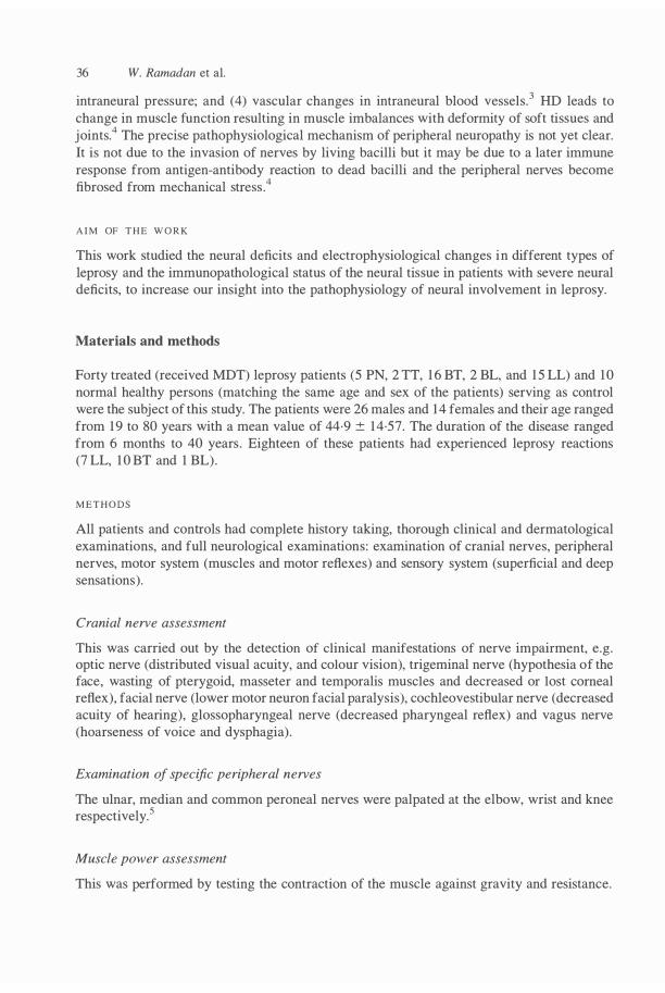
Table 5. Statistical analysis of motor nerve conduction variables of median nerve in leprosy patients and controls. ms = millisecond, m/s = meter/second, /LV = microvolt
Patients (n = 40) Controls (n = 10)
Median motor nerve variables Range Mean ±SD Range Mean ± SD P
CV m/s 0-5 1 ·6 3 1 · 1 1 17 ·33 48·2-58·2 52·01 3 ·03 7-20 <0·00 1 * D.L. ms 0- 15 ·6 7·23 4·75 3 ·04-4·22 3 ·91 0 ·38 4·37 <0·001 * Amp mv 0- 10 6·94 3·22 1 1 ·5 - 1 5 1 3 ·05 1 ·04 10·09 <0·001 * St. st. /LV 37-99·9 90·82 2 1 ·26 8 · 1 3 10·50 1 ·96 23·50 <0·00 1 *
* Significant.
Table 6. Statistical analysis of motor nerve conduction variables of ulnar nerve in leprosy patients and controls
Patients Controls n = 40 n = 1 0
Ulnar motor nerve variable Range Mean ±SD Range Mean ± SD P
CV m/s O-50 26· 14 17·65 5 1 ·6-66·9 58 · 1 3 4·88 10·03 <0·001 * D.L. ms 0- 1 8 8 ·29 5 ·97 2·36-3· 1 2 2·79 0·23 5 ·82 <0·001 * Amp mv 0-44 3 030 7·01 1 1 ·3 - 1 5 1 3 ·02 1 · 27 4·34 <0·00 1 * St. St. /LV 80-99·9 97·9 1 6·05 8 · 1 3 l HO 1 ·9 1 44·54 <0·00 1 *
* Significant.
Table 7. Statistical analysis of motor nerve conduction variables of common peroneal nerve in leprosy patients and controls
Patients Controls n = 40 n = 10
Common peroneal motor nerve variable Range Mean ± SD Range Mean ± SD P
CV m/s 0-4 1 .3 28·27 1 3 -98 45·4-5 1 · 8 47·84 2·29 8·42 <0·001 * D.L. ms 0- 15 .2 7·77 4·85 3 ·76-5·44 4·63 0·63 3 ·96 <0·001 * Amp mv 0-4 2·29 1 ·35 9·5- 1 5 1 2·48 1 ·99 15 ·32 <0·00 1 * St. St. /LV 76-99·9 96·23 8 · 1 0 9- 1 3 1 1 -00 1 ·76 6 1 ·04 <0·001 *
* Significant.
Table 8. Statistical analysis of sensory nerve conduction variables of ulnar nerve in leprosy patients and controls
Patients Controls n = 40 n = 1 0
Ulnar sensory nerve variable Range Mean ± SD Range Mean ± SD P
CV m/s 0-36·6 16 ·61 14·25 47·3-54·2 49·92 2·65 1 3 ·86 <0·001 * D.L. ms 0- 1 5 7·87 5·56 2·2-3·08 2·68 0·30 5·87 <0·001 * Amp mv 0-60 24· 1 3 2 1 ·00 17-100 42· 1 3 30·77 2·20 <0·001 * St. St./LV 40-93·5 47·25 8 ·38 8 ·8- 1 2-4 10·08 1 ·27 13 -88 <0·001 *
* Significant.

44 W. Ramadan et aI .
Table 9. Evaluation of MNC variables of different nerves and SNC variables of ulnar nerve
Nerve conduction variables
Normal Abnormal
Conducting Non-conducting Nerve nerve
Nerve n % n % n %
Median 1 1 27'S 23 S7'S 6 I S 'O Ulnar (motor) 1 2 30·0 20 SO·O 8 20·0 Common peroneal 8 22·0 28 60·0 4 1 0·0 Ulnar (sensory) 9 22·S 19 47'S 1 2 30·0
Total n %
29 72-S 28 70·0 32 80·0 3 1 77-S
reduction of SCV, significant prolongation of DL, significant reduction of amp. in 77.5% of patients and significant increase of st. st. in 1 00% of patients.
Table 9 shows that MNC of median nerve was abnormal in 29 patients (72.5%) (the nerve was non-conducting in six patients) . MNC of ulnar nerve was abnormal in 28 patients (70%) of the total (non-conducting nerve in eight patients) and MNC of common peroneal was abnormal in 32 patients (80%) of the total (non-conducting nerve in four patients) . SNC of ulnar nerve was abnormal in 3 1 patients (77 .5%) (non-conducting nerve in 1 2 patients), while st. st. was abnormal in all patients.
H I S T O P A T H O L O G I C A L R E S U L T S
Table 10 shows the distribution of the patients who were included in the histopathological study according to age, duration of the disease and type of leprosy.
Table 1 1 shows the histopathological changes of sural nerve biopsy specimens in leprosy patients by using different stains .
Table 1 0 . Distribution of leprosy patients who were used for histopathological study according to age, duration of disease and type of disease
Patients (n = 1 0)
LL BT Total (n = 1 0)
Age (years) SO-60 4 2 6 6 1 -70 1 2 3 >70 1 1
Duration (years) 20-2S 3 2 S 26-30 2 2 4 >30 1 1
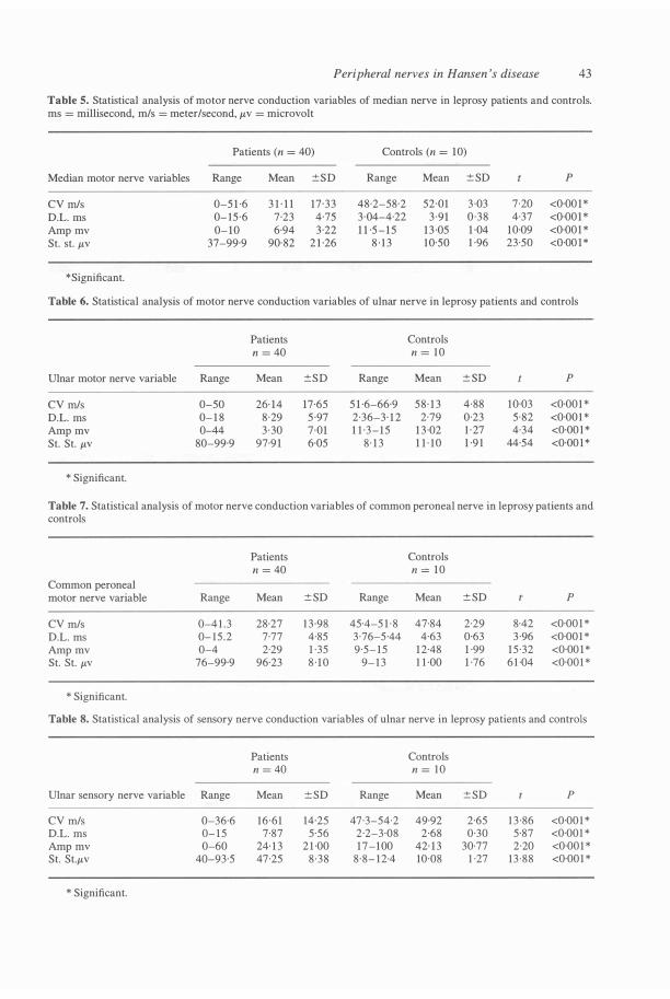
H & E S T A I N
Peripheral nerves in Hansen 's disease 45
Figure 3. Marked nerve fibrosis replacing the endoneural tissue in BT (H&E x 1 25).
There was fibrosis of the intraneural tissue, [marked fibrosis of endoneural tissue in two patients (20%) out of 1 0 (Figure 3) , moderate in six patients (60%) and miId in two patients (20%)] , destruction of the perineurium, areas of breakdown of myeIin sheath, degenerative changes of Schwann cells and no evidence of invading inftammatory cell infiItrate in sural nerve biopsy specimens in most patients (TabIe 1 1 ) .
Table 1 1 . Histopathological changes o f sural nerve biopsy specimens in leprosy patients by using different stains
Leprosy patients (n = 1 0)
Histopathological changes LL (n = 6) BT (n = 4)
H & E: Mild fibrosis 2 Moderate fibrosis 4 2 Severe fibrosis 2
Mallory' s PTAH: Fibrosis 6 No fibrosis 4
ABIP: IgG deposition + 2 ++ 4 2 + + + 2
IgM deposition + 2 ++ 1 2 + + + 5

46 W. Ramadan et aI.
Figure 4. Extensive nerve fibrosis with scattered scanty myelinated nerve fibres in BT (Mal1ory' s x 125) .
H I S T O C H E M I C A L S T A I N ( M A L L O R Y ' S P T A H )
There was predominant tibrosis of the nerve trunk in six cases (60%) out of 1 0 (Figure 4 and Table 1 1 ) .
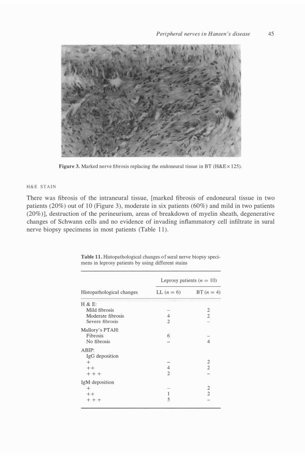
I M M U N O P A T H O L O G I C A L R E S U L T S ( A B I P S T A I N )
There was mild deposition (+) of IgG in two patients (20%), moderate (++) in six patients (60%) (Figure 5), and marked (+ + +) in two patients (20%) .
IgM deposition was mild in three patients (30%), moderate in two patients (20%), and marked in tive patients (50%).
Figure 5. Moderate deposits of IgG (++) in sural nerve section in PN (ABIP x 250).

Peripheral nerves in Hansen 's disease 47
There were areas of uneven distribution of IgG and IgM in the neural tissue in most
specimens. The antigens (lgG and IgM) deposited and appeared as homogenous and reddish patches in the endoneural areas (Table 1 1 ) .
Discussion
The mechanism of nerve damage in HD remains diverse and unclarified. 1 O It may be intrafascicular, intraneural, extrafascicular or extraneural lesions. 1 1 Peripheral nerve involvement is usualIy more and appears earlier in TI than in LL and also certain nerves are affected more than others in HD. I2 Croft et al. I 3 found that the most commonly affected nerve by function impairment was the posterior tibial (sensory) folIowed by the ulnar nerve.
ln this study, the cranial nerves were affected in this order of sequence, the trigeminal folIowed by optic, then facial, vagus, glossopharyngeal, cochleovestibular and finalIy spinal accessory nerves. Cranial nerve impairments were more in LL except the facial nerve, which was affected only in BT. This finding was similar to that mentioned by Lubbers et ai. 14
Claw hand was the most common disability among patients in the present study; this indicates that ulnar nerve is the most affected nerve in leprosy patients. The impairments were noticed more in LL, and this can be attributed to long duration of the disease, the presence of large number of skin lesions, occurrence of more invasive type and frequent episodes of reaction in those patients. This agrees with the findings of other studies. 1 5 , 1 6
ln the present study, alI sensory modalities (superficial and deep) were affected except deep pressure sensation, which was diminished in the advanced cases only. This agrees with the results of Andersen.5 Trophic and vasomotor changes were manifested in the form of resorption of fingers anel/or toes, anhidrosis, fissuring of foot, joint deformity, osteoarthritis and plantar u1ceration.
Mshana et ai. 12 mentioned that some nerves that appeared to be clinicalIy normal have been shown to have pathological changes.
Van Brakel and Khawas l7 recommended that alI leprosy patients should have a nerve function assessment at every visit to the clinic at least during their first year of treatment and regular nerve function assessment was essential to detect silent neuropathy at an early stage and to prevent permanent impairment of nerve function. 17
ln the present study EMG results showed a neuropathic pattem, which could be attributed to the sequelae of nerve degeneration rather than muscle degeneration. Thus it can be suggested that the wasting of muscles encountered among leprosy patients with neuropathY may be due to neurogenic atrophy.
Wemeck et ai. 1 8 stated that the involvement of skeletal striated muscles in leprosy was considered secondary due to peripheral neuropathy, and others attributed it to a primary muscle lesiono ln the present study, MNC variables showed that ulnar nerve was more affected than median and common peroneal nerves; this was in agreement with the results of Antia et ai. 1 9
ln this study, there was a markedly significant increase of st.st. in alI patients even in cases with normal conduction velocity. This means that st. st is a very important and sensitive test for early detection of nerve involvement, which becomes abnormal before any noticeable changes in other motor nerve conduction variables .
Samant et al.2o reported that nerve conduction m�asurements proved to be more sensitive
48 W. Ramadan et al.
in detecting nerve function impairrnent, but the combination of physical palpation for nerve thickening and graded nylon test was closely comparable to measurement of nerve conduction.
ln the present study, SCV of ulnar nerve was slower than MCV, indicating that the sensory system was more affected than the motor system in leprosy patients. This agrees with the findings of other studies.20,2 1
Regarding the hl.stopathological results, there was fibrosis of the intraneural tissues, destruction of the perineurium, areas of breakdown of myelin sheath, degenerative changes of Schwann celIs and no evidence of invading inflammatory celI infiltrate in sural nerve biopsy specimens in most patients. Van Brakel and Khawas22 found nearly similar results.
On the other hand, Mafoyane et al.23 found chronic granulomatous infiltrate within the nerve bundles and extensive destruction and fibrosis of the nerves in patients with primary neuritic leprosy. This variation may be due to the fact that alI the studied patients were chosen from those with advanced neural deficits, so fibrosis was the end result of inflammatory changes.
As regards the immunopathological results, in our study, there was marked deposition of IgG and/or IgM in specimens with extensive neural fibrosis . IgG and IgM deposition in the neural tissues may be attributed to the transmission of these deposits through the endoneural blood vessels .
Barros et aI. 24 found high antigenic load, demonstrated by using anti-BCG antibody (peroxidase-antiperoxidase technique) in the nerve specimens of their patients (in treated LL patients and in untreated TI patients) .
ln this study, the neural tissues showed areas of unequal distribution of IgG and IgM, this may be attributed to uneven involvement of nerve fascicles or may be related to the chronological occurrence of nerve darnage among different nerve fibres or fascicles in the sarne nerve, this point needs further studies on different nerve biopsy specimens and different grades of nerve darnage in a sufficient number of leprosy patients.
References
I Nations SP, Katz JS, Lyde CB, Barohn RJ. Leprous neuropathy: an American perspective. Semin Neurol, 1998; 18: 1 1 3- 1 24.
2 Rambukana L. How does Mycobacterium leprae target the peripheral nervous system? Trends Microbiol, 2000; 8: 23-28.
3 Job CK, Nerve damage in Hansen' s disease. Part II. The Star, 199 1 ; 50: 5-7. 4 Krotoski JB . Peripheral neuropathy and examination of the hands. The Star, 199 1 ; 50: 1 -5 . 5 Andersen JG. Sensory testing under field conditions. The Star, 1990; 50: 6-7. 6 Gabr M. Clinical, biochemical, and electrical study of skeletal muscles in patients with diabetic neuropathy. MD
Thesis, Faculty of Medicine, Tanta University, Tanta, Egypt, 1989. 7 Eyzaguirre C, Fidone JS. Physiology ofthe nervous system. An introductory texto Part 1: general neurophysiology.
The nerve. YearBook Medical Publishers lncorporated Chicago, 1975; pp. 20-30. 8 Bancroft ID, Stevens A. Histopathological stains and their diagnostic uses. Churchill Livingstone, Edinburgh,
New York, 1974, pp 3-17. 9 Taylor CR, K.ledisek G. Immunohistologic techniques in surgical pathology - a spectrum of new special stain.
Hum Pathol, 12: 590-595. 1 0 Antia NH, Shetty VP. Nerve damage in leprosy. Int J Lepr, 1988; 56: 619-62 1 . I I Charosky CB, Gatti JC, Cardama JE. Neuropathies in Hansen' s disease. Int J Lepr, 1983 ; 51: 576-586. 1 2 Mshana RN, Humber DP, Harboe M, Belehu A. Demonstration of mycobacterial antigens in nerve biopsies from
leprosy patients using peroxidase-antiperoxidase immunoenzyme technique. Clin Immunol lmmunopathol, 1983; 29: 359-368.
1 3 Croft RP, Richardus lH, Nicholls PG, Smith Wc. Nerve function impairment in leprosy: design, methodology,

Peripheral nerves in Hansen 's disease 49
and intake status of a prospective cohort study of 2664 new leprosy cases in Bangladesh (The Bangladesh Acute Nerve Damage Study). Lepr Rev, 1 999; 70: 140- 1 59.
1 4 Lubbers WJ, Schopper A, Hogeweg M, Soldenhoff R. Paralysis of facial muscles in leprosy patients with lagophthalmos. lnt J Lepr, 1 994; 66: 220-224.
1 5 Smith WCS. The epidemiology of disability in leprosy including risk factors. Lepr Rev, 1 992; 63: 23-30. 1 6 Chopra NK, Arawal JS. Hansen ' s disease deforrnities: an epidemiological study in a multidrug therapy project.
Star, 1 990; 49: 9 - 1 2. 1 7 Van Brakel WH, Khawas ffi. Silent neuropathy in leprosy: an epidemiological description. Lepr Rev, 1 994; 65:
350-360. 1 8 Wemeck LC, Teive HA, Scola RH. Muscle involvement in leprosy. Study of the anterior tibial muscle in 40
patients. Arq Neuropsiquiatr Sep. 1 999; 57: 723-734. 1 9 Antia NH, Pandya SS, Dastur DK. Nerves in the arm in leprosy. Clinical, electrodiagnostic and operative aspects.
lnt J Lepr, 1 970; 38: 12-28. 20 Samant G, Shetty VP, Uplekar MW, Antia NH. Clinical and electrophysiological evaluation of nerve function
impairment following cessation of multidrug therapy in leprosy. Lepr Rev, 1 999; 70: 1 0-20. 2 1 Vegrhese M, Ittimani KV, Satyanarayan KR. A study of the conduction velocity of the motor fibres of ulnar and
median nerves in leprosy. lnt J Lepr, 1 970; 38: 27 1 -277. 22 Van Brakel WH, Khawas ffi. Nerve damage in leprosy: An epidemiological and clinical study of 396 patients in
west Nepal. Part I . Definitions, methods and frequencies. Lepr Rev, 1994; 65: 204-221 . 23 Mafoyane NA, Lucas SB, Khawas ffi . Primary neuritic leprosy in a black South Africa. Lepr Rev, 1992; 63: 277-28 1 . 24 Barros U , Shetty VP, Antia NH . Demonstration of mycobacterial antigens i n nerves i n tuberculoid leprosy. Acta
Neuropathol, 1 987; 73: 387-392.