Alma Mater Studiorum – Università di Bologna EMERGING ...
97
Alma Mater Studiorum – Università di Bologna EMERGING CONTAMINANTS IN AGRICULTURAL ECOSYSTEMS: IMPACT OF SELECTED PHARMACEUTICALS ON WATER AND SOIL ECOLOGY AND PRACTICAL IMPLICATIONS DOTTORATO DI RICERCA Doctor Europaeus in Colture erbacee, Genetica agraria, Sistemi agroterritoriali Ciclo XXII Settore scientifico disciplinare di afferenza: AGR/02 Presentata da: Maria Ludovica Saccà Coordinatore Dottorato Tutore Prof. Giovanni Dinelli Prof. Alberto Vicari Co-tutori Dott. Cesare Accinelli Prof. Margarita Martín Aprile 2010
Transcript of Alma Mater Studiorum – Università di Bologna EMERGING ...
Alma Mater Studiorum – Università di Bologna
EMERGING CONTAMINANTS IN AGRICULTURAL ECOSYSTEMS: IMPACT OF SELECTED
PHARMACEUTICALS ON WATER AND SOIL ECOLOGY AND PRACTICAL IMPLICATIONS
DOTTORATO DI RICERCA Doctor Europaeus
in Colture erbacee, Genetica agraria, Sistemi agroterritoriali
Ciclo XXII
Settore scientifico disciplinare di afferenza: AGR/02 Presentata da: Maria Ludovica Saccà Coordinatore Dottorato Tutore Prof. Giovanni Dinelli Prof. Alberto Vicari
Co-tutori Dott. Cesare Accinelli
Prof. Margarita Martín
Aprile 2010
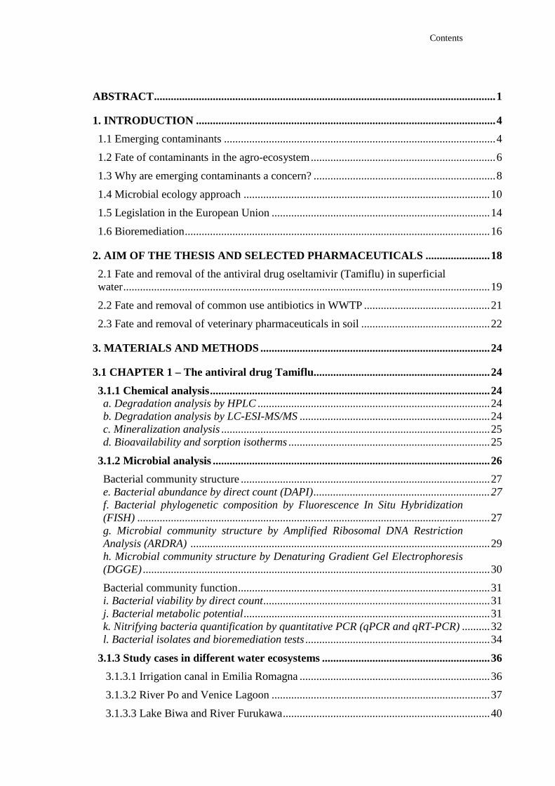
Contents
ABSTRACT..........................................................................................................................1
1. INTRODUCTION ...........................................................................................................4
1.1 Emerging contaminants .................................................................................................4
1.2 Fate of contaminants in the agro-ecosystem..................................................................6
1.3 Why are emerging contaminants a concern? .................................................................8
1.4 Microbial ecology approach ........................................................................................10
1.5 Legislation in the European Union ..............................................................................14
1.6 Bioremediation.............................................................................................................16
2. AIM OF THE THESIS AND SELECTED PHARMACEUTICALS .. .....................18
2.1 Fate and removal of the antiviral drug oseltamivir (Tamiflu) in superficial water...................................................................................................................................19
2.2 Fate and removal of common use antibiotics in WWTP .............................................21
2.3 Fate and removal of veterinary pharmaceuticals in soil ..............................................22
3. MATERIALS AND METHODS ..................................................................................24
3.1 CHAPTER 1 – The antiviral drug Tamiflu...............................................................24
3.1.1 Chemical analysis....................................................................................................24 a. Degradation analysis by HPLC ...................................................................................24 b. Degradation analysis by LC-ESI-MS/MS ....................................................................24 c. Mineralization analysis ................................................................................................25 d. Bioavailability and sorption isotherms ........................................................................25
3.1.2 Microbial analysis ...................................................................................................26
Bacterial community structure .........................................................................................27 e. Bacterial abundance by direct count (DAPI)...............................................................27 f. Bacterial phylogenetic composition by Fluorescence In Situ Hybridization (FISH) ..............................................................................................................................27 g. Microbial community structure by Amplified Ribosomal DNA Restriction Analysis (ARDRA) ...........................................................................................................29 h. Microbial community structure by Denaturing Gradient Gel Electrophoresis (DGGE) ............................................................................................................................30
Bacterial community function..........................................................................................31 i. Bacterial viability by direct count.................................................................................31 j. Bacterial metabolic potential........................................................................................31 k. Nitrifying bacteria quantification by quantitative PCR (qPCR and qRT-PCR) ..........32 l. Bacterial isolates and bioremediation tests ..................................................................34
3.1.3 Study cases in different water ecosystems ............................................................36
3.1.3.1 Irrigation canal in Emilia Romagna ....................................................................36
3.1.3.2 River Po and Venice Lagoon ..............................................................................37
3.1.3.3 Lake Biwa and River Furukawa..........................................................................40
Contents
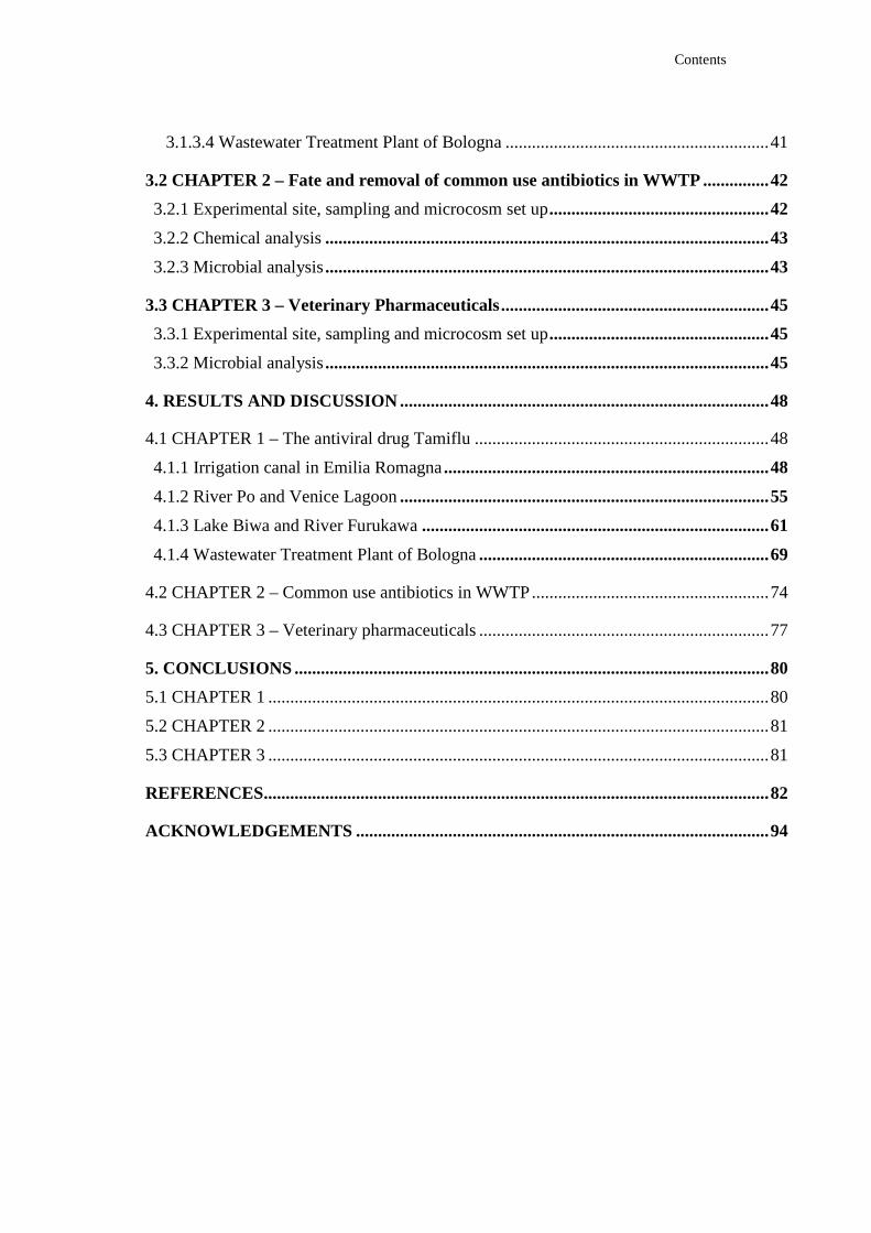
3.1.3.4 Wastewater Treatment Plant of Bologna ............................................................41
3.2 CHAPTER 2 – Fate and removal of common use antibiotics in WWTP ...............42
3.2.1 Experimental site, sampling and microcosm set up..................................................42
3.2.2 Chemical analysis.....................................................................................................43
3.2.3 Microbial analysis.....................................................................................................43
3.3 CHAPTER 3 – Veterinary Pharmaceuticals.............................................................45
3.3.1 Experimental site, sampling and microcosm set up..................................................45
3.3.2 Microbial analysis.....................................................................................................45
4. RESULTS AND DISCUSSION....................................................................................48
4.1 CHAPTER 1 – The antiviral drug Tamiflu ...................................................................48
4.1.1 Irrigation canal in Emilia Romagna..........................................................................48
4.1.2 River Po and Venice Lagoon....................................................................................55
4.1.3 Lake Biwa and River Furukawa...............................................................................61
4.1.4 Wastewater Treatment Plant of Bologna..................................................................69
4.2 CHAPTER 2 – Common use antibiotics in WWTP......................................................74
4.3 CHAPTER 3 – Veterinary pharmaceuticals ..................................................................77
5. CONCLUSIONS ............................................................................................................80
5.1 CHAPTER 1 ..................................................................................................................80
5.2 CHAPTER 2 ..................................................................................................................81
5.3 CHAPTER 3 ..................................................................................................................81
REFERENCES...................................................................................................................82
ACKNOWLEDGEMENTS ..............................................................................................94
Abstract
1
ABSTRACT
Pharmaceuticals are useful tools to prevent and treat human and animal diseases.
Following administration, a significant fraction of pharmaceuticals is excreted
unaltered into faeces and urine and may enter the aquatic ecosystem and
agricultural soil through irrigation with recycled water, constituting a significant
source of emerging contaminants into the environment. Understanding major
factors influencing their environmental fate is consequently needed to value the
risk, reduce contamination, and set up bioremediation technologies.
The antiviral drug Tamiflu (oseltamivir carboxylate, OC) has received recent
attention due to the potential use as a first line defence against H5N1 and H1N1
influenza viruses. Research has shown that OC is not removed during
conventional wastewater treatments, thus having the potential to enter surface
water bodies. A series of laboratory experiments investigated the fate and the
removal of OC in surface water systems in Italy and Japan and in a municipal
wastewater treatment plant.
A preliminary laboratory study investigated the persistence of the active antiviral
drug in water samples from an irrigation canal in northern Italy (Canale Emiliano
Romagnolo). After an initial rapid decrease, OC concentration slowly decreased
during the remaining incubation period. Approximately 65% of the initial OC
amount remained in water at the end of the 36-day incubation period. A negligible
amount of OC was lost both from sterilized water and from sterilized
water/sediment samples, suggesting a significant role of microbial degradation.
Stimulating microbial processes by the addition of sediments resulted in reduced
OC persistence. Presence of OC (1.5 µg mL-1) did not significantly affect the
metabolic potential of the water microbial population, that was estimated by
glyphosate and metolachlor mineralization. In contrast, OC caused an initial
transient decrease in the size of the indigenous microbial population of water
samples.
A second laboratory study focused on basic processes governing the
environmental fate of OC in surface water from two contrasting aquatic
ecosystems of northern Italy, the River Po and the Venice Lagoon. Results of this
study confirmed the potential of OC to persist in surface water. However, the
Abstract
2
addition of 5% of sediments resulted in rapid OC degradation. The estimated half-
life of OC in water/sediment of the River Po was 15 days. After three weeks of
incubation at 20 °C, more than 8% of 14C-OC evolved as 14CO2 from
water/sediment samples of the River Po and Venice Lagoon. OC was moderately
retained onto coarse sediments from the two sites. In water/sediment samples of
the River Po and Venice Lagoon treated with 14C-OC, more than 30% of the 14C-
residues remained water-extractable after three weeks of incubation. The low
affinity of OC to sediments suggests that the presence of sediments would not
reduce its bioavailability to microbial degradation.
Another series of laboratory experiments investigated the fate and the removal of
OC in two surface water ecosystems of Japan and in the municipal wastewater
treatment plant of the city of Bologna, in Northern Italy. The persistence of OC in
surface water ranged from non-detectable degradation to a half-life of 53 days.
After 40 days, less than 3% of radiolabeled OC evolved as 14CO2. The presence of
sediments (5%) led to a significant increase of OC degradation and of
mineralization rates. A more intense mineralization was observed in samples of
the wastewater treatment plant when applying a long incubation period (40 days).
More precisely, 76% and 37% of the initial radioactivity applied as 14C-OC was
recovered as 14CO2 from samples of the biological tank and effluent water,
respectively. Two bacterial strains growing on OC as sole carbon source were
isolated and used for its removal from synthetic medium and environmental
samples, including surface water and wastewater. Inoculation of water and
wastewater samples with the two OC-degrading strains showed that
mineralization of OC was significantly higher in both inoculated water and
wastewater, than in uninoculated controls. Denaturing gradient gel electrophoresis
and quantitative PCR analysis showed that OC would not affect the microbial
population of surface water and wastewater.
The capacity of the ligninolytic fungus Phanerochaete chrysosporium to degrade
a wide variety of environmentally persistent xenobiotics has been largely reported
in literature. In a series of laboratory experiments, the efficiency of a formulation
using P. chrysosporium was evaluated for the removal of selected
pharmaceuticals from wastewater samples. Addition of the fungus to samples of
the wastewater treatment plant of Bologna significantly increased (P < 0.05) the
removal of OC and three antibiotics, erythromycin, sulfamethoxazole, and
Abstract
3
ciprofloxacin. Similar effects were also observed in effluent water. OC was the
most persistent of the four pharmaceuticals. After 30 days of incubation,
approximately two times more OC was removed in bioremediated samples than in
controls. The highest removal efficiency of the formulation was observed with the
antibiotic ciprofloxacin.
The studies included environmental aspects of soil contamination with two
emerging veterinary contaminants, such as doramectin and oxibendazole, wich are
common parasitic treatments in cattle farms.
Introduction
4
1. INTRODUCTION 1.1 Emerging contaminants
During the past decade, the increasing introduction in the market of new
chemicals, and the development of more accurate analytical methods, added a
variety of new environmental ‘emerging’ contaminants to the already large array
of pollutants. Emerging contaminants are defined as any synthetic or naturally
occurring chemical that is not commonly monitored in the environment, though
having the potential to enter soil and aquatic ecosystems, causing known or
suspected adverse ecological and/or human health effects (USGS, 2009).
Recent concern are receiving chemicals that have been detected in varied water
sources, such as antibiotics, anti-depressants, tranquilizers, endocrine disrupting
chemicals, personal care products, illicit drugs, fluorinated compounds and
nanomaterials. Although present in the environment at low concentrations, in the
range of ng L-1, most of these ‘micropollutants’ raise considerable toxicological
concern, particularly if present as components of complex mixtures
(Schwarzenbach et al., 2006). Emerging contaminants can enter the environment
by a variety of sources, such as sewage treatment plants (STPs), runoff from
agricultural land uses, aquaculture and livestock farming, industrial wastes and
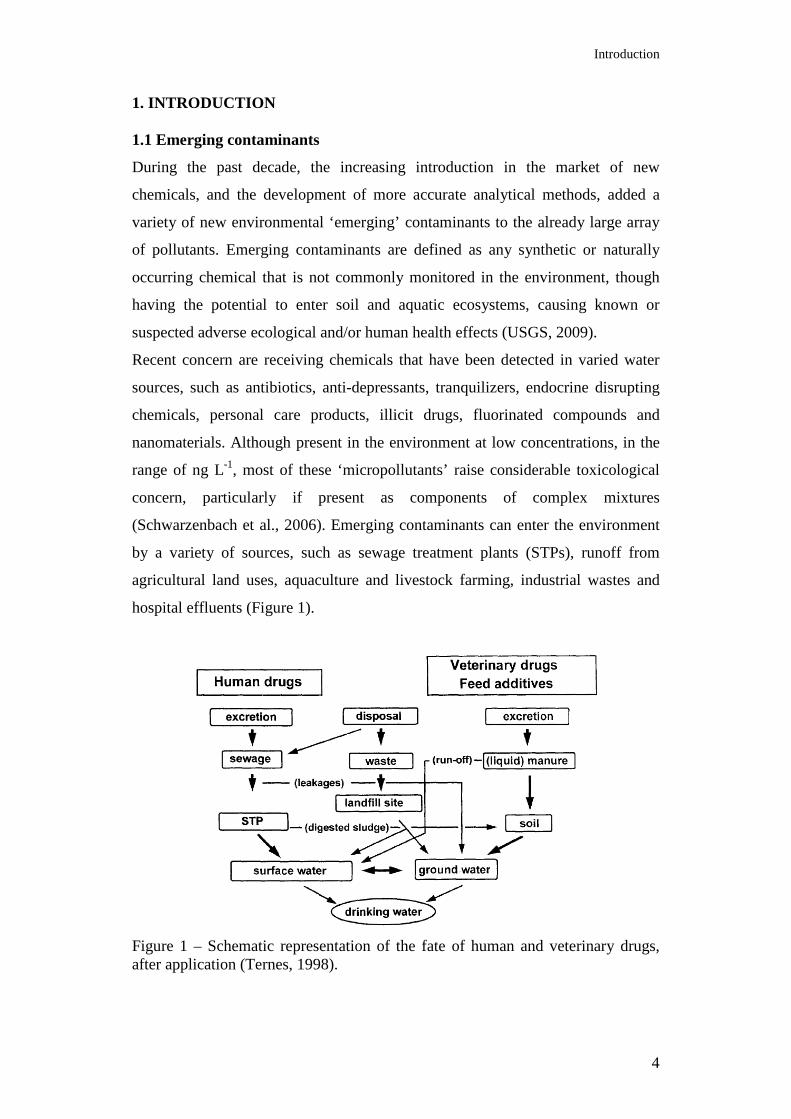
hospital effluents (Figure 1).
Figure 1 – Schematic representation of the fate of human and veterinary drugs, after application (Ternes, 1998).
Introduction
5
The occurrence of pharmaceuticals in the environment is a recent issue. Research
on this topic started in the 1990s, when Ternes, a German chemist, investigated
the environmental fate of a group of prescribed medicines after exctretion (Ternes,
1998). These were the first results of monitoring studies of pharmaceutical
measurements in local STPs and rivers.
Thousands of different pharmaceutically active compounds are actually used in
high quantities to treat or to prevent diseases (Kümmerer et al., 2009a; Bottoni et
al., 2010). Following therapeutic administration, a great percentage of
pharmaceuticals is excreted in urine and faeces (Carlsson et al., 2006) as parent
compound and/or metabolites and enters the sewage treatment system, where they
are often only partially removed (Halling-Sørensen et al., 1998). In a study
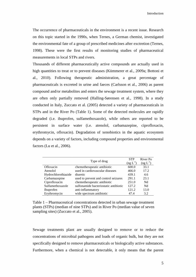
conducted in Italy, Zuccato et al. (2005) detected a variety of pharmaceuticals in
STPs and in the River Po (Table 1). Some of the detected molecules are rapidly
degraded (i.e. ibuprofen, sulfamethoxazole), while others are reported to be
persistent in surface water (i.e. atenolol, carbamazepine, ciprofloxacin,
erythromycin, ofloxacin). Degradation of xenobiotics in the aquatic ecosystem
depends on a variety of factors, including compound properties and environmental
factors (Lu et al., 2006).
Type of drug
STP (ng L-1)
River Po (ng L-1)
Ofloxacin chemotherapeutic antibiotic 600.0 33.1 Atenolol used in cardiovascular diseases 466.0 17.2 Hydrochlorothiazide diuretic 439.1 4.6 Carbamazepine used to prevent and control seizures 291.1 23.1 Ciprofloxacin chemotherapeutic antibiotic 251.0 Nd Sulfamethoxazole sulfonamide bacteriostatic antibiotic 127.2 Nd Ibuprofen anti-inflammatory 121.2 13.0 Erythromycin wide spectrum antibiotic 47.4 3.2
Table 1 – Pharmaceutical concentrations detected in urban sewage treatment plants (STPs) (median of nine STPs) and in River Po (median value of seven sampling sites) (Zuccato et al., 2005).
Sewage treatments plant are usually designed to remove or to reduce the
concentrations of microbial pathogens and loads of organic bulk, but they are not
specifically designed to remove pharmaceuticals or biologically active substances.
Furthermore, when a chemical is not detectable, it only means that the parent
Introduction
6
compound has been removed from the compartment of interest, and eventually it
has only been transformed in a degradation product. Degradation of a parent
molecule consists in reactions of oxidation, reduction or hydrolysis, and its
transformation products are often more reactive and sometimes more toxic than
the parent drug. By changing the physico-chemical behaviour of the substance,
degradation processes can modify its water solubility (usually an increase of water
solubility is expected) with respect to the parent compounds (Halling-Sørensen et
al., 1998). Present level of knowledge about the degradation pathways in STPs is
not always exhaustive, and wastewater is one of the major sources of
micropollutants in the environment (Schwarzenbach et al., 2006).
The two major mechanisms involved in the removal of substances from the
incoming waste stream in STPs are the following: microbial degradation and
sorption onto solid particles. The removal rate of pharmaceuticals in STPs is
affected by several factors, including their physico-chemical properties, the
adopted treatment process, hydraulic and sludge retention time, environmental
parameters, and properties of the influent (O'Brien and Dietrich, 2004). Sludge
retention time has been considered as one of the most important process
parameters. For highly polar substances, as most pharmaceuticals are, the most
important removal process is biological transformation or mineralization by
microorganisms; if the residence time is too short, it will not implement an
efficient biodegradation. Wastewater treatment technologies for the removal of
emerging contaminants are membrane bioreactors, ozonation and photocatalytic
processes, constructed wetlands, advanced sorbents and nanotechnology, artificial
recharge (Barceló et al., 2008; Radjenovic et al., 2009). None of these processes
though is entirely successful in the complete removal of contaminants. Ozone
treatment, for example, typically transforms chemical compounds but does not
mineralize them entirely (Stalter et al., 2009).
1.2 Fate of contaminants in the agro-ecosystem
Application of municipal biosolids on agricultural lands, as a source of crop
nutrients and organic matter, is a common farming practice in many countries and
jurisdictions (European Commission, 2001; Mantovi et al., 2005; Edwards et al.,
2009). Furthermore, municipal wastewater reuse, through irrigation of agricultural
land with reclaimed water, is an important supplement to water scarcity
Introduction
7
worldwide, especially in arid regions. During the dry season, streams rely almost
entirely on STPs effluents for flow, and effluents are extensively used in irrigation
and even for recharging drinking water aquifers. However fields amended with
biosolids and irrigated with reclaimed water risk to affect water quality by runoff
of contaminants of emerging interest (Lee et al., 2007; Topp et al., 2008). The
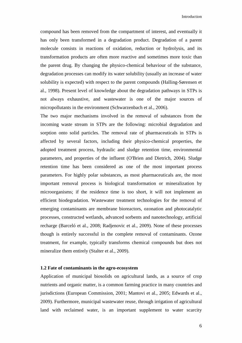
most worrying consequence is that these contaminants are continuously exposed
to humans, with different possible pathways: ingestion of food plants cultivated
on land irrigated with reclaimed water, ingestion of meat/animal products from
animal pasture on contaminated land, ingestion of drinking water produced from
groundwater polluted by reclaimed water, inhalation of volatile contaminants
during irrigation processes (Figure 2).
Figure 2 - Exposure pathway of chemicals to humans via agricultural irrigation (Weber et al., 2006).
It has been reported that the use of reclaimed water for soil irrigation can result in
the presence of pharmaceuticals in soil, in concentrations that vary through the
irrigation season. Some compounds persist for months after irrigation, and
accumulate in soil. It was demonstrated that soil samples collected before the
irrigation season contained pharmaceuticals, presumably left over from the
previous year’s irrigation, including erythromycin, carbamazepine, fluoxetine (an
antidepressant), and diphenhydramine (a common antihistamine). Several of the
pharmaceuticals detected increased in concentration during the study, suggesting
that the soil retained or adsorbed the pharmaceuticals (Kinney et al., 2006).
Agricultural land receives also other types of organic waste, such as solid and
liquid manure from intensive livestock farming sites, and effluents from intensive
aquaculture systems, in order to recycle nutrients and water for crop production.
However hormones, antibiotics and veterinary medicines are used extensively in
livestock production and, after application to animals, the drug may be adsorbed
and partially metabolized before being excreted with urine and faeces. Once the

Introduction
8
resulting manure or slurry is applied to land, the medicines and their metabolites
may run off into surface waters or leach to groundwater, where they may impact
the environment as well as human health (Koschorreck et al., 2002). The re-use of
manure is therefore a significant source of emerging contaminants in agricultural
land (Kolpin et al., 2002; Christian et al., 2003; Kumar et al., 2004). Aquaculture
systems, where the use of veterinary drugs is ordinary and necessary, also
contribute to the dispersion of pharmaceuticals into soil (Kupka-Hansen et al.,
1992).
1.3 Why are emerging contaminants a concern?
For most emerging contaminants there is currently little information regarding
their potential toxicological significance in ecosystems. The very low
concentrations in the environment, far below the doses employed for medical
treatments, avoid the detection of any biological effects with acute toxicity tests
(Boxall et al., 2003). The effects of these contaminants are especially related to
long-term and low-level environmental exposures, and they do not need to be
persistent to cause negative effects, due to the continuous introduction of assumed
drugs into the environment. The chronic nature of exposure to trace
concentrations of pharmaceuticals, the synergistic effects of mixtures of unrelated
chemicals (Cleuvers, 2003), and to what extent drugs can be transferred to
humans through food-chain biomagnification, are mostly unknown and advise
caution.
The reason why pharmaceuticals are problematic as environmental
micropollutants, is that they are developed with the intention of performing a
biological effect. Certain pharmaceuticals are designed to modulate endocrine and
immune systems and cellular signal transduction and as such have obvious
potential as endocrine disruptors in the environment. Antibiotics are meant to
produce direct effects on bacteria, and consequently have the potential to alter the
microbial community structure, change the growth, enzyme activity and diversity
of microbes (Schiermeier, 2003), and select for those few resistant bacteria in any
given population, which then reproduce and create an increasingly resistant
population through successive generations (Castiglioni et al., 2008; Farrell, 2009).
The use of biological systems for the treatment of antibiotic production
wastewater creates an ecosystem that contains much higher concentrations of
Introduction
9
antibiotics than normal aquatic environments, and thus may be an important
reservoir of antibiotic-resistant bacteria. In a survey of a wastewater treatment
plant that received effluent from a penicillin G production facility, Li et al. (2009)
demonstrated that, compared with upstream samples, effluent and downstream
samples showed significantly high levels of resistance for almost all the tested
antibiotics.
The effects of pharmaceuticals on water bacterial communities are principally a
reduction from 50 to 70% of the bacterial number in water and sediments and
therefore inhibition of bacteria responsible of anaerobic degradation of organic
matter: reduction of nitrification processes and of sulphate-reducing activity.
Concentrations of 12.5-75 mg L-1 of oxytetracycline, a broad spectrum antibiotic,
have been found to be inhibitive of nitrification, and lead to a build-up of toxic
ammonia and nitrite (Klaver et al., 1994). Oxytetracycline and flumequine
inactive completely sulphate-reducing bacteria after 7 days of medication (Kupka-
Hansen et al., 1992; Smith et al., 1994).
Exposure to waterborne pollutants may cause health risks, such as contamination
of aquatic food sources and of agricultural products (Weber et al., 2006). Hence,
any measures taken to prevent the chemical pollution of surface and groundwater
resources will not only improve ecosystem health, but will also benefit both the
production of clean water and safe food for human consumption (Schwarzenbach
et al., 2006). Understanding the sources, transport, and fate of emerging
contaminants is therefore essential to provide information to eventually expand
the range of pollutants that should be monitored in effluent discharges and the
implementation of the guidelines.
Environment contamination with new pollutants may result in changes in the
microbial ecology, possibly changing the types of bacteria that carry out important
ecosystem processes such as nutrient transformations and biomass decomposition.
Microbial biodiversity has in fact a functional importance in the maintenance of
soil and water biological processes, because most of the transformations involved
in biogeochemical cycles are mediated exclusively by microorganisms. It has
been reported that shifts in microbial community structure, associated with a
reduction in microbial biodiversity, lead to losses of functional stability (Griffiths
et al., 2004; Girvan et al., 2005).
Introduction
10
Nitrogen is an essential element for crop growth and a key agricultural input. The
fixation of N2 from the atmosphere, in which it is reduced to ammonia in an
energy-demanding process, is due principally to microbial activity. The oxidation
of ammonia to nitrate via nitrite by autotrophic nitrifying bacteria is a key process
in agricultural/natural ecosystems and wastewater treatment (Jordan et al., 2005).
The first step, the oxidation of ammonia to hydroxylamine, is catalyzed by aerobic
chemoautotrophic ammonia oxidizing bacteria (AOB) and nitrite-oxidizing
bacteria (NOB). Activity and dynamics of the nitrifying bacterial population have
been largely used in environmental studies (Hermansson and Lindgren, 2001;
Limpiyakorn et al., 2005; Molina et al., 2007). Studies have shown that AOB and
NOB are less competitive than the heterotrophic bacteria for oxygen and growing
space and are sensitive to environmental inhibition (Van Benthum et al., 1997;
Juliastuti et al., 2003; Limpiyakorn et al., 2005; Pagga et al., 2006). Ammonium
oxidation by autotrophic ammonia-oxidizing bacteria (AOB) is a key process in
agricultural ecosystems and wastewater treatment. Denitrification occurs in many
distantly related species of microorganisms (Zumft, 1992), thus also bacteria with
this physiological capability may be used as functional markers for ecological
studies (Gregory et al., 2003). Bacterial communities constitute the basis of food
webs and are responsible for organic matter transformations and mineral
recycling. Ecosystem functions that depend on microbial activities can suffer from
chemical exposures if microorganisms are sensitive to the toxic effects of
pollutants (Ogunseitan, 2000). Given the various sensitivities of different
microorganisms to toxic chemicals, there is a growing interest in microbial
toxicity testing at the community or ecosystem level, and in including bacterial
community responses in the environmental risk assessment of toxic pollutants
(Brandt et al., 2004).
1.4 Microbial ecology approach
Traditional parameters used for the Environmental Risk Assessment (ERA) of
pharmaceuticals are cheimodynamic and physico-chemical properties such as
solubility, Kow (octanol/water partition constant), Kd (soil-water partition
constant), Koc (soil organic carbon/water partition constant) and DT50 (degradation
half-life) in soil and water, PEC (predicted environmental concentration), PNEC
(predicted no-effect concentration). Furthermore ecotoxicological effects of
Introduction
11
pharmaceuticals on non-target organisms in water and soil are assessed with
standard acute and chronic ecotoxicologic tests on freshwater and marine
organisms (e.g. Daphnia magna, rainbow trout) and soil organisms (e.g. Eisenia
fetida, Enchytraeus crypticus, Caenorhabditis elegans, Folsomia candida).
Pollution may influence soil and water quality and productivity but little is known
on the effects on microbial communities, and consequent impacts on functioning.
Due to their small size, large numbers, and ubiquitous distribution in the
environment, microorganisms are valuable indicators of the occurrence of
disturbances due to exogenous physico-chemical stressors. The study of bacterial
abundance, vitality and community structure are among the most useful tools
developed in microbial ecology for direct characterization of target populations, in
their natural environment, avoiding cultivation. The assessment of variations in
microbial community structure is of basic importance to permit to evaluate the
impact of an environmental stressor. At the organism level, the presence of a
certain indicator bacteria can indicate sources of pollution into an environment,
but the molecular-level responses of autochthonous microorganisms to changes in
ambient conditions are more critical for ecosystem health assessment. There is a
wide array of molecules, including nucleic acids, lipids, and proteins, that is
useful for diagnosing microbial responses to pollution and for monitoring
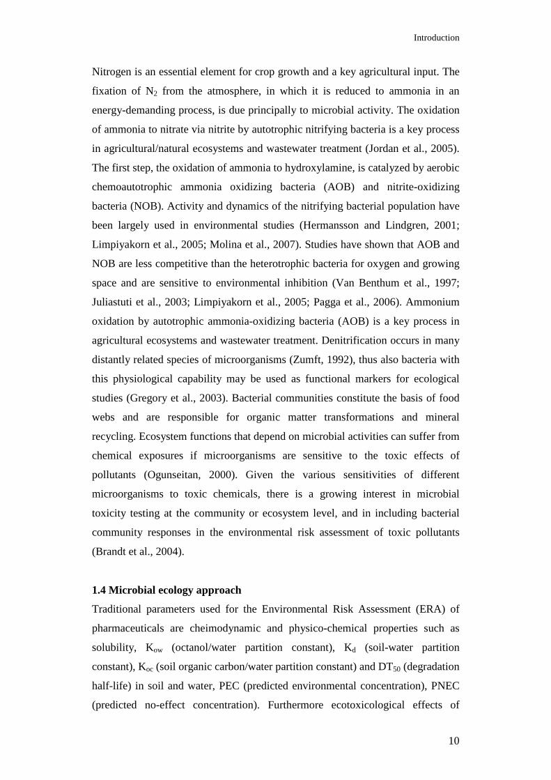
environmental management strategies. The presence of toxic chemicals in
microbial ecosystems, for example, induces the synthesis of detoxifying or
degradative enzymes and certain stress proteins (Figure 3). Effects due to
chemical toxicity tend to narrow the spectrum of microbial diversity because
organisms that are not capable of resisting the toxic effects either die or enter a
static metabolic phase, leaving those that have evolved resistance mechanisms,
that are capable of utilizing the excess chemicals as nutrients, to proliferate and
become dominant members of the impacted ecosystem (Ogunseitan, 2000).
Protein molecules mediate these effects by virtue of the ability of each species to
synthesize degradative enzymes or otherwise engage in repair mechanisms
through the activities of stress proteins and modified structural components
(Ogunseitan, 2000). Monitoring these proteins provides information on toxic
chemical fates (biodegradative enzymes) and effects (toxicity-induced changes in
protein profiles). Complex microbial communities may therefore serve as ideal
and ecologically relevant toxicity indicators (Brandt et al., 2004).
Introduction
12
A number of microbiologically driven processes has been proposed to evaluate
the effects of xenobiotics on ecosystems (Wagner-Döbler et al., 1992; Nazaret et
al., 1994; Moyer et al., 1994; Griebler and Slezak, 2001). Proteins, genes,
metabolites, or lipids that, when expressed, present a pattern of molecular change
in an organism in response to a specific environmental stressor, can be defined as
environmental biomarkers.
Figure 3 – Schematic representation of microbial community analysis in response to environmental perturbations. Toxic chemicals, for example, can cause changes in microbial population densities and diversity (Ogunseitan, 2000).
The evaluation of bacterial biodiversity is mainly limited by their small size, by
the absence of distinguishing phenotypic characters, and by the fact that most of
these organisms cannot be cultivated (Torsvik et al., 2002). The number of
techniques to study microbial communities has increased exponentially over the
last 20 years and the advent of culture-independent methods, such as molecular
biological techniques, has changed the view of microbial diversity (Rossello-Mora
and Amann, 2001). Among these techniques it is possible to distinguish between
those which are primarily based on the use of Polymerase Chain Reaction (PCR),
Introduction
13
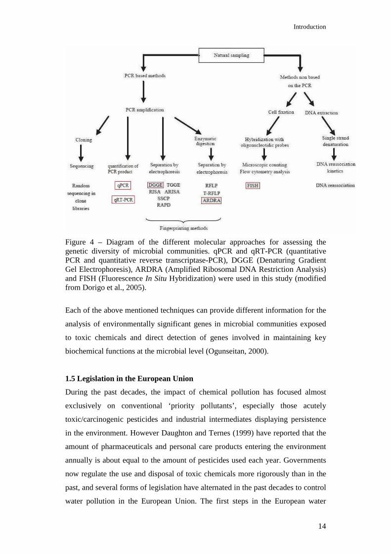
and those that are non-PCR-based (Figure 4). PCR uses specific primers to
amplify a DNA target sequence. The bacterial 16S rDNA gene is today the most
commonly used for assessing overall diversity in microbial communities and for
studying the phylogeny of microorganisms. Sequence variations in the PCR
fragments are detected either by a cloning/sequencing analysis, which provides a
complete characterization of the fragments, or by an electrophoretic analysis,
which provides a visual separation of the mixture of fragments. Fragments
separation is based on sequence polymorphism, in Denaturing Gradient Gel
Electrophoresis (DGGE) or length polymorphism, in Terminal-Restriction
Fragment Length Polymorphism (T-RFLP) and Automated Ribosomal Intergenic
Spacer Analysis (ARISA). DGGE is frequently used in environmental studies
(Ibekwe et., 2001; Guo et al., 2009). Quantitative Polymerase Chain Reaction
(qPCR and qRT-PCR) has become a commonly used technique for the detection
and quantification of microorganisms in the environment for its high sensitivity at
low concentrations (Dionisi et al., 2003; Devers et al., 2004; Zhang and Fang,
2006; Kim et al., 2007). It can be used to detect changes in gene expression
patterns induced by adverse conditions, also not requiring prior knowledge of
expected contaminants, using non-specific stress responses as general indicators
of deleterious conditions (Van Dyk et al., 1995).
Non-PCR-based methods commonly used in environmental studies are
epifluorescence microscopy techniques, such as direct count of bacterial
abundance (DAPI count) and vitality (Live/Dead cell viability assay), and
Fluorescence In Situ Hybridization (FISH). FISH uses rRNA-targeted fluorescent
probes to investigate the overall taxonomic composition of bacterial communities.
Probes can be designed to be complementary to species-, group-, or kingdom-
specific target sites.
Introduction
14
Figure 4 – Diagram of the different molecular approaches for assessing the genetic diversity of microbial communities. qPCR and qRT-PCR (quantitative PCR and quantitative reverse transcriptase-PCR), DGGE (Denaturing Gradient Gel Electrophoresis), ARDRA (Amplified Ribosomal DNA Restriction Analysis) and FISH (Fluorescence In Situ Hybridization) were used in this study (modified from Dorigo et al., 2005).
Each of the above mentioned techniques can provide different information for the
analysis of environmentally significant genes in microbial communities exposed
to toxic chemicals and direct detection of genes involved in maintaining key
biochemical functions at the microbial level (Ogunseitan, 2000).
1.5 Legislation in the European Union
During the past decades, the impact of chemical pollution has focused almost
exclusively on conventional ‘priority pollutants’, especially those acutely
toxic/carcinogenic pesticides and industrial intermediates displaying persistence
in the environment. However Daughton and Ternes (1999) have reported that the
amount of pharmaceuticals and personal care products entering the environment
annually is about equal to the amount of pesticides used each year. Governments
now regulate the use and disposal of toxic chemicals more rigorously than in the
past, and several forms of legislation have alternated in the past decades to control
water pollution in the European Union. The first steps in the European water
Introduction
15
legislation have focused mainly on quality standards for certain types of waters
(bathing waters, aquaculture and drinking waters), leading to the stipulation of the
Drinking Water Directive and the Bathing Water Directive.
Within the European Union, the quality of water for human consumption is
determined by the Drinking Water Directive (Council Directive 98/93/EC). Of the
48 parameters within the directive, none is related to pharmaceuticals.
The nutrients dimension was then added to water protection with the Urban
Wastewater Treatment Directive of 1991 (Council Directive 91/271/EEC),
concerning urban wastewater treatment.
The Nitrates Directive (91/676/EEC) sets out clear rules for nitrates pollution
from agriculture, one of the main sources of groundwater pollution as well as of
eutrophication of surface waters in many regions of Europe.
In 2000, the Water Framework Directive (2000/60/EC) has expanded EU water
policy to all waters and addresses all sources of impacts. It defines the ecological
quality according to hydro morphological, physico-chemical and biological
(biodiversity to the three levels: genetic, of population, of community)
parameters, and priority pollutants concentrations in water, sediments and
organisms. The Directive on Priority Substances (2008/105/EC) identifies 33
substances or groups of substances, which have been shown to be of major
concern for European Waters, for the adoption of control measures over the next
20 years (http://ec.europa.eu/environment/water/water-
framework/priority_substances.htm). Further 14 substances were identified as
being subject to review for identification as possible priority hazardous
substances. The list includes selected chemicals, plant protection products,
biocides, metals and other groups like Polyaromatic Hydrocarbons (PAH) that are
mainly incineration by-products and Polybrominated Biphenylethers (PBDE) that
are used as flame retardants. Additionally member countries have undertaken their
own national reviews to identify emerging future contaminants. The much wider
range of emerging pollutants that are now widely used is not included in the list,
however the priority substance list will be updated every 4 years and has
identified future emerging priority candidates.
Since 2007, regulation on chemicals and their safe use is established by the
European Community (EC 1907/2006) with the REACH legislation (Registration,
Evaluation, Authorization of Chemicals). REACH regulates the large number of
Introduction
16
substances that have entered the market in Europe in the last years, sometimes in
very high amounts, for yet there is insufficient information on the hazards that
they pose to human health and the environment.
Furthermore, European guidelines for use of reclaimed water are generally limited
to defining risks associated with microbial organisms, bulk parameters such as
chemical oxygen demand (COD), biochemical oxygen demand (BOD), pH and
total suspended solids (TSS). These parameters exclude the monitoring of specific
chemical concentrations.
The European Agency for the Evaluation of Medicinal Products (EMEA)
coordinates the existing scientific resources of the Member States of the EU in
order to evaluate and supervise medicinal products for both human and veterinary
use throughout the entire EU.
1.6 Bioremediation
Molecular ecological information is especially useful for the development of
strategies to improve bioremediation, in which the metabolic potential of
microorganisms is used to clean up contaminated environments (Watanabe,
2001). In the last years molecular tools have facilitated the study of natural
microbial populations without cultivation, including the fraction of
microorganisms that have the ability to degrade certain xenobiotics.
Bioremediation employs living organisms, most often microorganisms, plants, or
both to degrade, detoxify, or sequester toxic chemicals from natural waters and
soils. It can be used to treat soil, sediment, sludge, water, or even air. Treatments
can be either ex situ, involving the removal of contaminated materials from a
polluted site prior to treatment, or in situ, if contaminants are treated without
moving them to a treatment facility. Bioremediation treatments include:
bioaugmentation, by augmenting natural systems with exogenous biological
materials, usually natural microorganisms or plants grown to large numbers in
fermenters or greenhouses; biostimulation, the use of nutrients, substrates or
environmental conditions to stimulate the naturally occurring organisms that can
perform bioremediation; bioreactors, treatment of a contaminated substance in a
large tank containing organisms or enzymes; bioventing, involves the venting of
oxygen through soil to stimulate the growth or natural and introduced
bioremediation organisms; composting, involves mixing contaminated materials
Introduction
17
with compost containing bioremediation organisms; land farming, the use of
farming tilling and soil amendment techniques to encourage the growth of
bioremediation organisms in a contaminated area. Finally, abiotic processes
sometimes can be used in combination with biotic processes to degrade
particularly recalcitrant molecules. Examples of abiotic catalysts that may
enhance biodegradative processes include ultraviolet light, inorganic reductants,
and Fenton reagent (iron and hydrogen peroxide). The bioremediation industry
has developed many novel approaches for monitoring and quantifying
bioremediation processes (Crawford, 2006) to offer an efficient, cheap and
biocompatible option for decontamination of polluted ecosystems.
The biodegradative environmental fate of contaminants can be determined
through the integration of field, laboratory, and modelling efforts (Hooper et al.,
2002). The National Research Council (1993) has recommended three criteria for
demonstrating intrinsic remediation: documenting a decrease in contaminant
concentrations at the site, showing experimentally that microorganisms in site
samples have the potential to transform the contaminants under expected site
conditions, developing evidence showing that the biodegradation potential is
actually realized in the field.
Among bacteria, the degradation of recalcitrant pollutants is of great
environmental significance. A wide variety of bacteria able to utilize xenobiotics
as a source of energy and capable of degrading a broad range of pollutants has
been isolated (Gu and Berry, 1992; Topp et al., 2000; Gu and Mitchell, 2006;
Singh and Walker, 2006; Yoon et al., 2006; Miyauchi et al., 2008), and many
have been exploited in pollutant biodegradation and wastewater treatment (Bryers,
1994; Osswald et al., 1995; Sharp et al., 1998). Pseudomonas sp. ADP is one of
the best studied s-triazine-degrading bacteria (Mandelbaum et al., 1995; Martinez
et al., 2001; Moràn et al., 2006), Arthrobacter aurescens TC1 is able to degrade a
variety of pollutants, among which the herbicide glyphosate, mixed bacterial
cultures in a consortium can show degradation ability of various pollutants, even
though their individual components can be unable to utilize the chemical as
energy source (Mandelbaum et al., 1993; De Souza et al., 1993).
Furthermore, among the genus Basidiomycetes, the so called white rot fungi
(WRF) are capable of degrading a lignocellulose substrate by producing three
types of extracellular enzymes, often referred to as Lignin Modifying Enzymes
Introduction
18
(LMEs), and they are Lignin Peroxidase (LiP), Manganese-Dependent Peroxidase
(MnP) and Laccase (Lac). LiP oxidises methoxyl groups on aromatic rings, MnP
and Lac are able to oxidise phenolic substrates. As the enzymes are non-specific,
they have been found capable of degrading a wide variety of chemical compounds
like DDT, PCB, lindane, dioxin, benzopyrene, cyanides, azides, CCl4 and
pentachlorophenol (Singh et al., 1999; Lu et al., 2009). The main fungus studied is
Phanerochaete chrysosporium, and also studied extensively are Trametes
versicolor, Pleurotus ostreatus, Phanerochaete sordida, Trametes hirsutus, and
Fusarium culmorum.
2. AIM OF THE THESIS AND SELECTED PHARMACEUTICALS
The aim of the present thesis is to assess the impact of selected emerging
contaminants on the microbial community of different water and soil ecosystems,
selected for the study. This work is structured in three parts, each regarding the
fate of different pharmaceuticals.
Chapter 1 includes the main part of the work, it regards the fate and removal of
the antiviral Tamiflu (oseltamivir carboxylate, OC), recommended for the
treatment of cases of avian and swine influenza. Tamiflu is predicted to reach the
water system because resistant to biodegradation in wastewater treatment plants.
Contrasting environmental samples were chosen for laboratory experiments: three
surface water ecosystems of Italy, such as an irrigation canal Canale Emiliano
Romagnolo (paragraph 1), River Po and Venice Lagoon (paragraph 2); two
surface water ecosystems of Japan, River Furukawa and Lake Biwa (paragraph
3); and samples of activated-sludge-mixed liquor from the municipal wastewater
treatment plant of the city of Bologna and the effluent water of the plant
(paragraph 4). Besides degradation and mineralization of OC during incubation
time, the effect of OC on the bacterial community structure was determined by
fingerprinting techniques (ARDRA and DGGE), qPCR, qRT-PCR, and
epifluorescence microscopy techniques (FISH, bacterial abundance and vitality).
Furthermore, bacterial strains growing on oseltamivir as sole carbon source were
isolated and tested for degradation capacities. A bioremediation strategy was
performed to evaluate the capability of a white rot fungus, P. crysosporium, to
degrade the antiviral.
Introduction
19
In Chapter 2 the destiny of common use human and veterinary antibiotics, such
as ciprofloxacine, erythromycin, sulfamethoxazole, was monitored in the afore-
mentioned wastewater treatment plant and effluent. Bioremediation with P.
chrysosporium was tested for degradation processes acceleration.
Chapter 3 concerns the fate of two veterinary pharmaceuticals, doramectin and
oxibendazol, common parasitic treatments in farms, in contaminated soils, and the
effect on the bacterial community structure.
The work for this thesis was conducted principally in the University of Bologna
(Department of Agroenvironmental Sciences and Technologies), with the
contribution of national and international collaborations. A part of the research
regarding the antiviral Tamiflu (Chapter 1) was conducted with the Water
Research Institute (IRSA, CNR of Rome), in particular epifluorescence
microscopy techniques. Collaboration with the Department of Chemistry of Umeå
University (Dr. Jerker Fick) regarded chemical analysis conducted in Chapter 1
and 2. The work in Chapter 3 was conducted during a 9 month period (January
2007 - April 2007; September 2008 - March 2009) spent in Complutense
University of Madrid, in the Faculty of Veterinary, under the supervision of Prof.
Margarita Martin.
2.1 Fate and removal of the antiviral drug oseltamivir (Tamiflu) in
superficial water
During the past years influenza A viral infections have posed serious risks to
public health. Since 2003, 286 cases of ‘avian’ influenza H5N1 human deaths
have been confirmed by the WHO, and worldwide more than 213 countries have
reported laboratory confirmed cases of ‘swine’ influenza H1N1, including at least
16226 deaths. The available options to control influenza A viruses are limited.
Health agencies all over the world have been forced to adopt strategies for
containing the viruses and to protect the health of the public. Although
vaccination is the primary strategy for prevention, neuraminidase inhibitors are
the drugs of choice for the treatment and the prevention of influenza A illness in
children and adults. The World Health Organization (WHO, 2006) has
recommended the use of the antiviral drug Tamiflu (oseltamivir phosphate),

Introduction
20
produced and marketed by F. Hoffman-La Roche Ltd (Basel, Switzerland), for the
treatment and post-exposure prophylaxis in a pandemic scenario. Hundreds of
million of course of Tamiflu have been stockpiled worldwide since 2003, and in
the last year sales of the antiviral have further exploded. Oseltamivir phosphate
(OP) [ethyl-(3R,4R,5S)-4-acetamido-5-amino-3-(1-ethylpropoxy)-1-cyclohexene-
1-carboxylate] is the prodrug of the active metabolite oseltamivir carboxylate
(OC) [(3R,4R,5S)-4-acetamido-5-amino-3-(1-ethylpropoxy)-1-cyclohexene-1-
carboxylic acid], a specific inhibitor of influenza A and B virus neuraminidase
(Kim et al., 1997; Li et al., 1998). OC reduces viral infection by binding to the
highly conserved active site of the neuraminidase of the virus, inhibiting the
release of progeny virions from the surface of infected cells (Bardsley-Elliot and
Noble 1999). OC has been shown to be clinically active for the treatment and
chemoprophylaxis of influenza in adults and in children (Ward et al., 2005).
Pharmacological studies have demonstrated that after oral administration of OP
and absorption in the gastrointestinal tract, it is converted by the hepatic esterases
to OC (Figure 5). More than 80% of each oral dose of OP is eliminated by renal
excretion as OC (Ward et al., 2005).
Figure 5 – Structure of the prodrug oseltamivir phosphate (OP) and the active form oseltamivir carboxylate (OC) (Singer et al., 2007).
The OC molecule has amine and carboxylate groups that impart hydrophilicity, a
low partition coefficient (log P 1.1), and high water solubility (588 mg mL-1 at
25°C) (American Hospital Formulary Service, 2006). These physico-chemical
features minimize loss by sorption to sewage sludge during wastewater treatment.
A recent study conducted in Sweden by Fick et al. (2007) demonstrated that OC is
not completely removed during conventional sewage water treatments which
include mechanical, chemical and biological (activated-sludge) processes.
Consequently, in case of urban areas with a large number of patients receiving
Introduction
21
Tamiflu, the potential risk of OC to contaminate the aquatic ecosystem is
expected (Fick et al., 2007; Singer et al., 2007, 2008). Predicted environmental
concentration (PEC) of OC, calculated in catchments with particularly low flow
and high populations, would be over 20 µg L-1, which is significantly higher than
that observed for most other pharmaceutical contaminants (Singer et al., 2008).
This assumption was recently confirmed in two studies conducted in Japan, OC
was detected in a conventional activated-sludge–based STP discharge in the
concentration of 293.3 ng L-1, and in the receiving river water samples in the
concentration of 6.6–190.2 ng L-1 during the peak of the flu season (Ghosh et al.,
2010). Söderstrom et al., (2009) detected no OC in Japanese surface water
systems before the flu-season, but 2–58 ng L-1 were found in the samples taken
during the flu season.
Chronic ecotoxicity tests, conducted in light of the 2006 EMEA guidelines on
environmental risk assessment for human pharmaceuticals, revealed that the level
of concern regarding ecotoxicity is quite low. The preliminary no observed effects
concentrations (NOECs) resulted in a PNEC of 100 µg L-1, applying an
assessment factor of 10 (Singer et al., 2007). Considerable concerns are the
potential inhibition of non-target neuraminidases in different organisms than
influenza viruses, and the fact that the presence of OC into rivers can be a risk for
the generation of OC resistance in influenza viruses. Recent analysis of isolated A
viruses revealed a high increase in Tamiflu-resistant strains, in different countries
worldwide (Hurt et al., 2009).
2.2 Fate and removal of common use antibiotics in WWTP
Antibiotics are used extensively in human and veterinary medicine, as well as in
aquaculture, for preventing or treating microbial infections. The risk to
contaminate soil or surface water run-off after application of manure, or in the use
of sewage sludge for land amendment, is high, increasing the selective pressure
for resistant bacteria (Kümmerer et al., 2009b). Common use antibiotics of
emerging concern, most likely to cause environmental problems (Zuccato et al.,
2006), have been used in this work, for a degradation and bioremediation study
(Figure 6).
Ciprofloxacin is a broad-spectrum fluoroquinolone antibacterial drug, commonly
used for the treatment of bacterial infections, in particular of the urinary and

Introduction
22
respiratory tracts. Erythromycin is a macrolide antibiotic, used to treat several
types of infections (respiratory tract infections, skin infections, acute pelvic
inflammatory disease, erythrasma, etc.). Sulfamethoxazole is a sulfonamide
bacteriostatic antibiotic, often used in combination with trimethoprim. Excretion
rates and further details on properties and metabolism of antibiotics can be found
on http://pubchem.ncbi.nlm.nih.gov/.
Figure 6 - Human and veterinary common use antibiotics used in the study.
2.3 Fate and removal of veterinary pharmaceuticals in soil
Two veterinary antimicrobials were selected in this study to assess their effects on
a contaminated soil bacterial community: doramectin and oxibendazole (Figure
7).
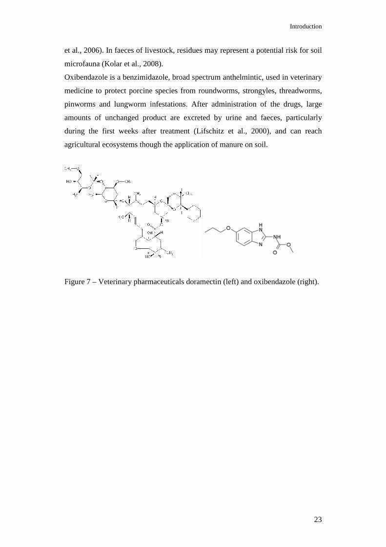
Doramectin is a macrocyclic lactone, potent anthelmintic, for the treatment of
parasites such as gastrointestinal roundworms, lungworms, eyeworms, grubs,
sucking lice and mange mites in cattle. It is an endectocide molecule, for the
activity against ecto- and endo-parasites (Shoop et al., 1995). It has low
mammalian toxicity and formulations are convenient to use, hence it is
extensively used worldwide in veterinary medicine. However the occurrence and
persistence of residues of the drug brings the need for continued monitoring of its
fate. Residues of doramectin (79.8 µg kg-1) have been found in sheep milk at 3
days post-treatment, and were still detectable for 30 days after treatment (Danaher
Introduction
23
et al., 2006). In faeces of livestock, residues may represent a potential risk for soil
microfauna (Kolar et al., 2008).
Oxibendazole is a benzimidazole, broad spectrum anthelmintic, used in veterinary
medicine to protect porcine species from roundworms, strongyles, threadworms,
pinworms and lungworm infestations. After administration of the drugs, large
amounts of unchanged product are excreted by urine and faeces, particularly
during the first weeks after treatment (Lifschitz et al., 2000), and can reach
agricultural ecosystems though the application of manure on soil.
Figure 7 – Veterinary pharmaceuticals doramectin (left) and oxibendazole (right).
Materials and Methods
24
3. MATERIALS AND METHODS
3.1 CHAPTER 1 – The antiviral drug Tamiflu
3.1.1 Chemical analysis
a. Degradation analysis by HPLC
Oseltamivir carboxylate (OC) concentration in incubated water samples of the
CER irrigation canal was determined by HPLC after derivatization with 20 mM
naphthalene-2,3-dialdehyde (Sigma-Aldrich Italia s.r.l., Milan, Italy) and 20 mM
potassium cyanide (Ultra Scientific Italia s.r.l., Bologna, Italy) as described in
Eisenberg and Cundy (1998). Aliquots of the derivatized mixtures were analysed
by a chromatography system equipped with a 250 x 0.46 mm Prodigy ODS-2
column (Phenomenex Inc., Torrance, CA), and an RF-10AXL spectrofluorometric
detector (Shimadzu Italia s.r.l., Milan, Italy). Isocratic elution was carried out at
40 °C, and the eluent flow was set at 1.0 mL min-1 with 50 mM sodium acetate in
acetonitrile/water (27 : 73, v/v). Detection of OC was achieved by setting the
detector at excitation and emission wavelengths of 420 and 472 nm, respectively.
OC was quantified on the basis of external standards. OC was obtained from
analytical grade OP (≥ 99% purity; Sequoia Research Product, Pangbourne, UK)
by chemical hydrolysis at elevated pH. Samples from flasks containing water and
sediments were extracted with ethanol, centrifuged at 5000 g for 10 min,
redissolved in 50 mM monosodium phosphate and analysed as described above.
Recoveries of OC from water and water/sediment samples were 97.1 and 87.7%,
respectively.
b. Degradation analysis by LC-ESI-MS/MS
OC degradation in the further experiments was assessed following the procedure
described in Fick et al. (2007), samples were extracted by solid phase extraction
and analyzed by liquid chromatography/electro spray tandem mass spectrometry.
Briefly, samples were acidified to pH 3, filtered through a 0.45-µm filter and
loaded on a Strata-X-C (200 mg, 6 mL) column (Phenomenex Inc. Torrence, CA,
USA). Eluate was concentrated and reconstituted in acetonitrile/H2O (1:1)
containing 0.1% formic acid. Aliquots (10 µL) were injected into a LC-ESI-
Materials and Methods
25
MS/MS equipped with C18 column (YMC Inc. Wilmington, NC), a P40000 HPLC
pump (Thermo Scientific Inc., Waltham, MA, USA) and a LCQ Duo ion-trap
mass spectrometer (Thermo Scientific Inc.). Oseltamivir carboxylate (OC) was
obtained from F. Hoffmann-La Roche Ltd (Basel, Switzerland).
c. Mineralization analysis
Mineralization of [U-ring-14C]-OC was measured in biometers, which consisted in
250-mL flasks equipped with suspended glass vials containing 4 mL of a 1 M
NaOH solution to trap 14CO2. Aliquots of water (50 mL) were transferred into
each flask under aseptic conditions and spiked with a solution of unlabeled
(chemical purity > 98%) and 14C-OC (radiopurity > 97.9%, specific activity 4.96
MBq mg-1) to give a final concentration of 20 µg L-1 (1.5 kBq mL-1). Radiolabeled
OC was provided by F. Hoffmann-La Roche Ltd (Basel, Switzerland). Water
samples containing 5 and 10% (w/v) sediments were included in the study.
Samples consisting in autoclaved water and water/sediments were used as
controls. Samples were incubated for 21 days at 20 °C on an orbital shaker (125
rpm) in the dark. Trapped 14CO2 was determined by mixing a 1-mL aliquot of
NaOH solution with 4 mL of Hi Safe 3 scintillation cocktail (PerkinElmer,
Boston, MA, USA), and the amount of radioactivity was measured by liquid
scintillation counting (LSC) using a Wallac 1490 liquid scintillation counter
(Wallac Oy, Turku, Finland). Prior to analysis, samples were kept in the dark for
12 hours.
The experiment was conducted in triplicate. Data of the degradation and
mineralization study were analyzed by analysis of variance. Means were separated
by Fisher’s least significant difference (LSD) and significant differences were
detected at the P = 0.05 level.
d. Bioavailability and sorption isotherms
At the end of the incubation period, samples of the mineralization study were
transferred into 50-mL centrifuge tubes, shaken for 1 hour and centrifuged at 5000
g for 10 min. The total volume of supernatant was measured and total
radioactivity determined by LSC, mixing triplicate 1-mL aliquots with 4 mL of
HiSafe 3 Scintillation Cocktail. Pellets were sequentially extracted with 0.1 M
CaCl2, and acetonitrile. For each extraction, pellets were dispersed by vortexing,
Materials and Methods
26
shaken for 3 hours and centrifuged at 5000 g for 10 min. Total radioactivity in the
supernatants was determined by LSC. Finally, remaining 14C-residues were
determined by combusting triplicate subsamples of ACN-extracted pellet using a
Packard 306 (Packard. Instrument Co., Sterling, VA, USA). Sorption isotherms of
OC on sediments of the River Po and Venice Lagoon were determined by the
batch equilibrium method. Aliquots ( 2 g, air-dried basis) of each sediment were
weighed into 50-mL glass centrifuge tubes and a 10-mL aliquot of 14C-OC
solution, prepared in 0.01 M CaCl2, was added. Sorption isotherms were
determined using triplicate samples at five initial OC concentrations, ranging from
20 to 100 µg mL-1. Radiolabeled OC was added to unlabeled solutions to give an
initial radioactivity of approximately 3 x 10⁻³ µCi mL-1. Tubes were sealed with
teflon-lined caps, mechanically shaken at 20 °C for 14 hours, and samples were
centrifuged at 5000 g for 10 min. Aliquots (5 mL) of supernatant were removed,
filtered through a 0.2-µm filter, and radioactivity in 1-mL fractions was
determined by LSC. Preliminary investigations showed that equilibrium was
attained in less than 14 hours and that there was no significant OC sorption to
centrifuge tubes. The amount of sorbed OC was calculated from the concentration
differences between the supernatant of the equilibrated solutions and those of the
corresponding initial solutions. Sorption data were fitted to the log form of the
Freundlich equation:
logCs = logKf + (1/n) logCe
where Cs is the concentration of OC sorbed (µg g-1 sediment), Ce is the
equilibrium concentration (µg mL-1 solution) and Kf and 1/n are the empirical
Freundlich constants. Values of Kf and 1/n were estimated by linear regression
after a log–log transformation.
3.1.2 Microbial analysis
In this work, microbial community level toxicity was tested using a polyphasic
approach, involving a range of molecular-based methods, targeting both structure
and function of the tested microbial communities.
Potential effects of OC on basic microbiological aspects of the indigenous
microbial community of water and sediments were investigated using DNA-based
Materials and Methods
27
approaches. All the microbiological analyses were conducted using aliquots taken
from samples of the OC degradation study.
Bacterial community structure
e. Bacterial abundance by direct count (DAPI)
The size of the bacterial population in water samples was estimated by direct
count, using a fluorescent dye. Total bacterial abundance was calculated by fixing,
at different time intervals, aliquots (1 mL) of water with the same amount of
phosphate-buffer saline (PBS) containing formaldehyde (2% w/v), Tween 20 (0.5
v/v) and sodium pyrophosphate (0.1 M). In order to separate water bacterial
aggregates, a gentle sonication (10 sec, 15 W using a Microson XL2000 ultrasonic
liquid processor) was performed on each sub-sample. Samples were then treated
with the DNA-binding fluorescent stain 4’-6-diamino-2-phenylindole (DAPI) (1
µg mL-1), and filtered onto a 0.22 mm black polycarbonate filter. Cells were
enumerated using an epifluorescence microscope (DM LB30, Leica GmBH,
Heideberg, Germany), as described in Barra Caracciolo et al. (2005).
f. Bacterial phylogenetic composition by Fluorescence In Situ Hybridization
(FISH)
In order to investigate the effects of OC on the bacterial community and to assess
if it could be involved in degradation, Florescence In Situ Hybridization (FISH)
was performed on OC treated and untreated sub-samples collected from
degradation microcosms (2 replicates). The phylogenetic composition of the OC-
treated and control samples was analyzed at different sampling times (0, 14, 21
and 36 days). For each condition, four sub-samples (1 mL each) were fixed (1:1)
with a solution composed of phosphate-buffered saline: 130 mM NaCl; 7 mM
Na2HPO4; 3 mM NaH2PO4; 2% formaldehyde; 0.5% Tween 20 and 100 mM
sodium pyrophosphate. After sonication, samples were filtered on a 0.2 µm
polycarbonate membrane. Filters were stored at -20 °C until further processing.
FISH of the harvested cells was performed using probes for the identification of
the major bacterial phylogenetic divisions found in freshwater (Zwart et al.,
2002), such as the Bacteria domain, and the phyla of α-Proteobacteria, β-
Proteobacteria, γ-Proteobacteria, Planctomycetes, Cytophaga-Flavobacterium,
Firmicutes. For this purpose, the Cy3-labelled oligonucleotide probes described in
Materials and Methods
28
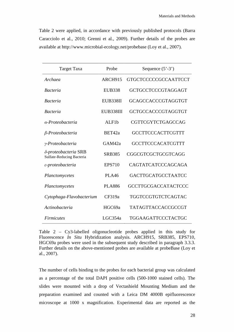
Table 2 were applied, in accordance with previously published protocols (Barra
Caracciolo et al., 2010; Grenni et al., 2009). Further details of the probes are
available at http://www.microbial-ecology.net/probebase (Loy et al., 2007).
Target Taxa Probe Sequence (5’-3’)
Archaea ARCH915 GTGCTCCCCCGCCAATTCCT
Bacteria EUB338 GCTGCCTCCCGTAGGAGT
Bacteria EUB338II GCAGCCACCCGTAGGTGT
Bacteria EUB338III GCTGCCACCCGTAGGTGT
α-Proteobacteria ALF1b CGTTCGYTCTGAGCCAG
β-Proteobacteria BET42a GCCTTCCCACTTCGTTT
γ-Proteobacteria GAM42a GCCTTCCCACATCGTTT
δ-proteobacteria SRB Sulfate-Reducing Bacteria
SRB385 CGGCGTCGCTGCGTCAGG
ε-proteobacteria EPS710 CAGTATCATCCCAGCAGA
Planctomycetes PLA46 GACTTGCATGCCTAATCC
Planctomycetes PLA886 GCCTTGCGACCATACTCCC
Cytophaga-Flavobacterium CF319a TGGTCCGTGTCTCAGTAC
Actinobacteria HGC69a TATAGTTACCACCGCCGT
Firmicutes LGC354a TGGAAGATTCCCTACTGC
Table 2 – Cy3-labelled oligonucleotide probes applied in this study for Fluorescence In Situ Hybridization analysis. ARCH915, SRB385, EPS710, HGC69a probes were used in the subsequent study described in paragraph 3.3.3. Further details on the above-mentioned probes are available at probeBase (Loy et al., 2007). The number of cells binding to the probes for each bacterial group was calculated
as a percentage of the total DAPI positive cells (500-1000 stained cells). The
slides were mounted with a drop of Vectashield Mounting Medium and the
preparation examined and counted with a Leica DM 4000B epifluorescence
microscope at 1000 x magnification. Experimental data are reported as the
Materials and Methods
29
number of cells mL-1, calculated by multiplying the total cell abundance and the
percentage of cells detected by each specific probe.
Data were obtained from the mean of four sub-samples. Statistical analysis of the
data was done using Kruskal-Wallis One Way Analysis of Variance on Ranks,
with significant differences at level of P < 0.01.
g. Microbial community structure by Amplified Ribosomal DNA Restriction
Analysis (ARDRA)
Samples of the degradation study were used to determine bacterial population
changes in response to the presence of OC. Total DNA was isolated from
incubated samples using the DNA PowerSoil Isolation Kit (MoBio Laboratories
Inc., Solana Beach, CA). Duplicates 100-mL aliquots of water were passed
through a 0.22-µm nylon filter (GE Water & Technologies, Trevose, PA). Filters
were transferred into PowerBead tubes provided with the kit and then processed
following the instructions of the manufacturer. The effects on the structure of the
bacterial community were estimated by Amplified Ribosomal DNA Restriction
Analysis (ARDRA). ARDRA is a method based on restriction endonuclease
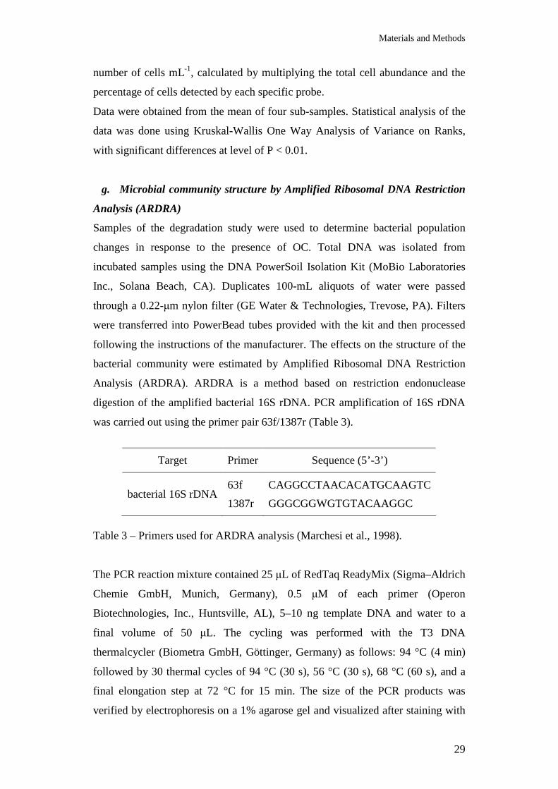
digestion of the amplified bacterial 16S rDNA. PCR amplification of 16S rDNA
was carried out using the primer pair 63f/1387r (Table 3).
Target Primer Sequence (5’-3’)
bacterial 16S rDNA 63f
1387r
CAGGCCTAACACATGCAAGTC
GGGCGGWGTGTACAAGGC
Table 3 – Primers used for ARDRA analysis (Marchesi et al., 1998). The PCR reaction mixture contained 25 µL of RedTaq ReadyMix (Sigma–Aldrich
Chemie GmbH, Munich, Germany), 0.5 µM of each primer (Operon
Biotechnologies, Inc., Huntsville, AL), 5–10 ng template DNA and water to a
final volume of 50 µL. The cycling was performed with the T3 DNA
thermalcycler (Biometra GmbH, Göttinger, Germany) as follows: 94 °C (4 min)
followed by 30 thermal cycles of 94 °C (30 s), 56 °C (30 s), 68 °C (60 s), and a
final elongation step at 72 °C for 15 min. The size of the PCR products was
verified by electrophoresis on a 1% agarose gel and visualized after staining with
Materials and Methods
30
SYBR Green I (Sigma–Aldrich). Aliquots of amplified 16S rDNA products (10
µL) were digested with 10 U of AluI and EcoRI (Sigma–Aldrich) in a total
volume of 40 µL at 37 °C for 2 hours. Digested products were resolved by vertical
non-denaturing 8% polyacrylamide gel electrophoresis and visualized by SYBR
Green I staining. Data were computed with the software GelCompar II version
5.10 (Applied Maths, Sint-Martens-Latem, Belgium) to cluster the data and
construct the similarity matrix to make comparisons of bacterial communities of
the differently treated sample.
h. Microbial community structure by Denaturing Gradient Gel
Electrophoresis (DGGE)
The structure and diversity of the bacterial community was estimated by a two-
step nested-PCR denaturing gradient gel electrophoresis (DGGE) analysis. DGGE
permits to see how bacterial sequences change over time and treatment.
Prefiltered (0.45 µm) aliquots of water and water/sediment samples were passed
through a sterile 0.22-µm nylon filter (GE Water & Technologies, Trevose, PA).
Filters were transferred into PowerBead tubes provided with the kit PowerSoil
DNA Isolation Kit (MoBio Laboratories Inc., Solana Beach, CA) and then
processed following the instructions of the manufacturer. Total DNA was first
amplified using the primer pairs P63f and P518r (Table 4) in a 50 µl reaction
mixture consisting of 5-10 ng of DNA, 5 U of AmpliTaq DNA polymerase
(Invitrogen, Carlsbad, CA), 10x reaction buffer, 4 mM MgCl2, 0.5 mM of each
dNTP, 0.8 µM of each primer and nuclease-free water. Reaction conditions were
the following: denaturation at 94 °C for 5 min, followed by 31 cycles of
denaturation at 94 °C for 60 s, annealing at 53 °C for 60 s, extension at 72 °C for
2 min, and final extension at 72 °C for 10 min. Amplicons were used for the
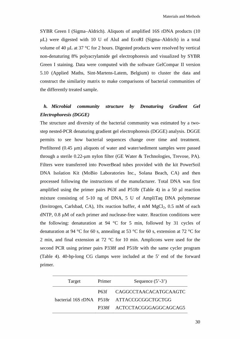
second PCR using primer pairs P338f and P518r with the same cycler program
(Table 4). 40-bp-long CG clamps were included at the 5′ end of the forward
primer.
Target Primer Sequence (5’-3’)
bacterial 16S rDNA
P63f
P518r
P338f
CAGGCCTAACACATGCAAGTC
ATTACCGCGGCTGCTGG
ACTCCTACGGGAGGCAGCAG5
Materials and Methods
31
Table 4 – Primers used for DGGE analysis. After quantification of amplified products, equal amounts of amplicons (250-300
ng) were loaded onto DGGE gel. Gel contained 8% (w/v) polyacrylamide gels
(37.5:1 acrylamide:bisacrylamide) with a urea/formamide denaturing gradient of
40–60% (where 100% denaturant contains 7 M urea 8 and 40% v/v formamide).
A 10-mL stacking gel containing no denaturants was added before polymerization
was complete. Gels were run for 16 hours at 60 °C, with a constant voltage of 65
mV in 1 x TAE buffer. DGGE analysis was performed in DCode system (Bio-Rad
Laboratories, Hercules, CA, USA). Bands were visualized after staining with
GelRed (Biotum Inc., Hayward, CA). Band profiles were analyzed using the
GelCompare II package (Applied Maths, Kortrijk, Belgium). Dendrograms were
constructed using the DICE coefficients and were subjected to unweighted pair
group method cluster analysis (UPGMA).
Bacterial community function
i. Bacterial viability by direct count
The relative abundance of viable bacteria in water samples was estimated by
direct count, using fluorescent dyes. Cell viability was estimated using a cell
viability kit (Live/Dead®, BacLightTM), following the method proposed by
Haglund et al. (2003). Two different fluorescent dyes were used, SYBR Green II,
and propidium iodide, respectively as viability and membrane-compromised cell
markers. Aliquots (1 mL) of water samples were incubated in the presence of
SYBR Green II (1/10,000 dilution; Sigma-Aldrich, Germany) and propidium
iodide (20 mM). After incubation, samples were filtered through a black
polycarbonate filter (0.22 mm pore size) and viable (green) and dead (red)
bacteria were enumerated by direct count using a Leica DM 4000B
epifluorescence microscope at 1000 x magnification. Live cell abundance was
calculated, as the number of live bacteria mL-1, from the total cell abundance,
obtained by DAPI counts, multiplied by % of live cells/live+dead.
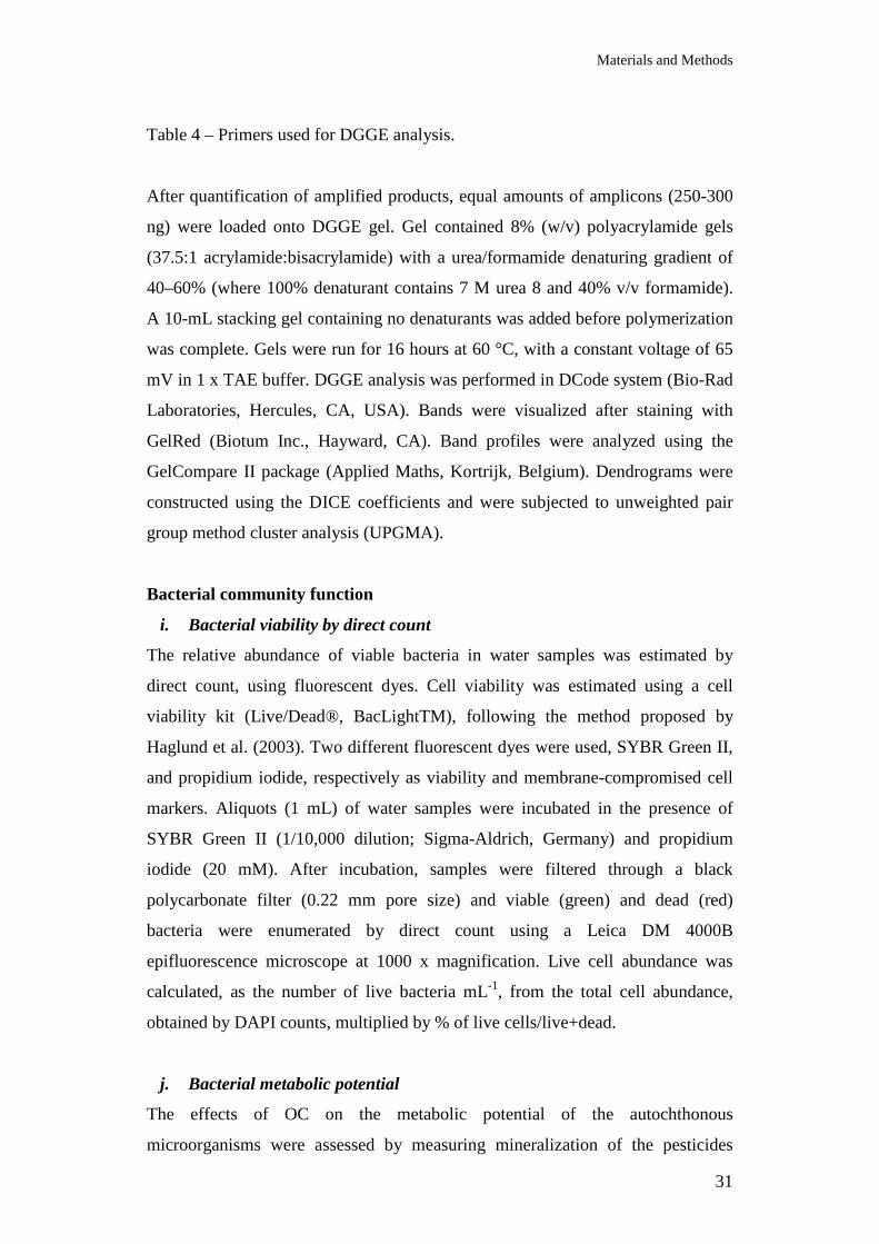
j. Bacterial metabolic potential
The effects of OC on the metabolic potential of the autochthonous
microorganisms were assessed by measuring mineralization of the pesticides
Materials and Methods
32
glyphosate and metolachlor in water samples of the CER irrigation canal. Samples
of non-sterilized and sterilized water containing OC (1.5 µg mL-1) were prepared
as described above. Glyphosate and metolachlor were applied as water solutions
using a mixture of unlabelled and 14C-labelled compound in order to obtain a final
concentration of 1 µg a.i. L-1. Unlabelled glyphosate (purity > 99%) and 14C-
glyphosate (N-phosphonomethyl-2-14C-glycine; radiopurity > 99%, specific
activity 5.4 mCi mmol-1) were purchased from Sigma-Aldrich Italia (Milan, Italy).
Unlabelled metolachlor (purity > 96%) and 14C-metolachlor (2-chloro-N-(2-ethyl-
6-methyl-[U-14C]phenyl)-N-(2-methoxy-1-methyl-ethyl)acetamide; radiopurity >
99%, specific activity 13 mCi mmol-1) were donated by Syngenta Crop Protection
AG (Basel, CH). Treated water samples were incubated at 20 °C on an orbital
shaker (125 rpm) in the dark. Metolachlor and glyphosate mineralization was
monitored by trapping the evolved 14CO2 in vials containing 4 mL of a 1 M NaOH
solution. The NaOH solution was replaced at sampling, facilitating flask aeration.
Aliquots (1 mL) of NaOH solution were mixed with 4 mL of HiSafe 3 liquid
scintillation cocktail (PerkinElmer, Boston, MA) and radioactivity quantified
using a Wallac 1490 liquid scintillation counter (Wallac Oy, Turku, Finland).
Samples were kept in the dark for 12 hours prior to analysis. Experiment was
conducted in triplicate, and untreated samples (control) were included.
Experiment was repeated with samples consisting of water/sediment mixture
prepared as described above. Metabolic potential was expressed as the percentage
of added glyphosate and metolachlor mineralized.
k. Nitrifying bacteria quantification by quantitative PCR (qPCR and qRT-
PCR)
Quantitative PCR (Heid et al., 1996; Schmittgen, 2001) is a technique that permits
a very accurate quantitative determination of DNA and RNA. It is based on
detecting and quantifying a fluorescent probe or DNA-binding agent. By
recording the amount of fluorescence at each cycle, it is possible to monitor the
PCR reaction during the exponential phase, when the first significant increase in
the amount of PCR product is correlated to the initial amount of target template.
The more template is present at the beginning of the PCR reaction, the fewer
cycles it takes to reach the point at which the fluorescent signal is first detected as
being significantly greater than the background.
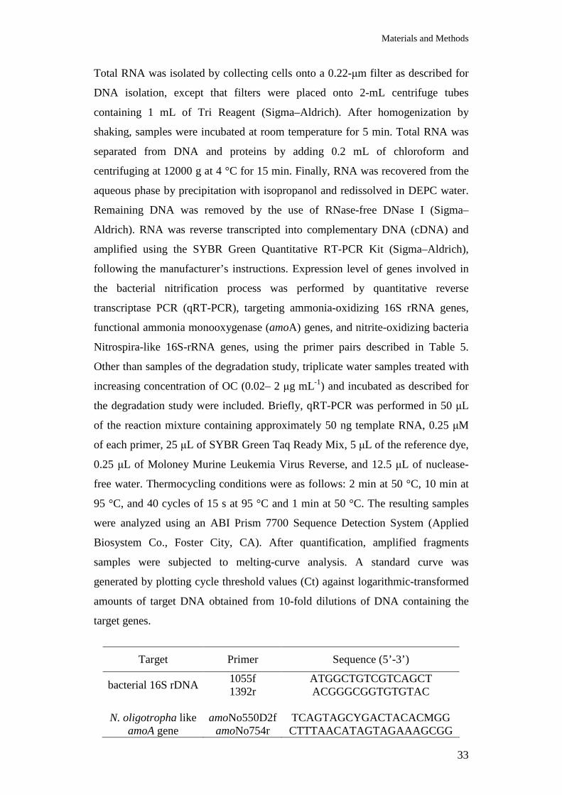
Materials and Methods
33
Total RNA was isolated by collecting cells onto a 0.22-µm filter as described for
DNA isolation, except that filters were placed onto 2-mL centrifuge tubes
containing 1 mL of Tri Reagent (Sigma–Aldrich). After homogenization by
shaking, samples were incubated at room temperature for 5 min. Total RNA was
separated from DNA and proteins by adding 0.2 mL of chloroform and
centrifuging at 12000 g at 4 °C for 15 min. Finally, RNA was recovered from the
aqueous phase by precipitation with isopropanol and redissolved in DEPC water.
Remaining DNA was removed by the use of RNase-free DNase I (Sigma–
Aldrich). RNA was reverse transcripted into complementary DNA (cDNA) and
amplified using the SYBR Green Quantitative RT-PCR Kit (Sigma–Aldrich),
following the manufacturer’s instructions. Expression level of genes involved in
the bacterial nitrification process was performed by quantitative reverse
transcriptase PCR (qRT-PCR), targeting ammonia-oxidizing 16S rRNA genes,
functional ammonia monooxygenase (amoA) genes, and nitrite-oxidizing bacteria
Nitrospira-like 16S-rRNA genes, using the primer pairs described in Table 5.
Other than samples of the degradation study, triplicate water samples treated with
increasing concentration of OC (0.02– 2 µg mL-1) and incubated as described for
the degradation study were included. Briefly, qRT-PCR was performed in 50 µL
of the reaction mixture containing approximately 50 ng template RNA, 0.25 µM
of each primer, 25 µL of SYBR Green Taq Ready Mix, 5 µL of the reference dye,
0.25 µL of Moloney Murine Leukemia Virus Reverse, and 12.5 µL of nuclease-
free water. Thermocycling conditions were as follows: 2 min at 50 °C, 10 min at
95 °C, and 40 cycles of 15 s at 95 °C and 1 min at 50 °C. The resulting samples
were analyzed using an ABI Prism 7700 Sequence Detection System (Applied
Biosystem Co., Foster City, CA). After quantification, amplified fragments
samples were subjected to melting-curve analysis. A standard curve was
generated by plotting cycle threshold values (Ct) against logarithmic-transformed
amounts of target DNA obtained from 10-fold dilutions of DNA containing the
target genes.
Target Primer Sequence (5’-3’)
bacterial 16S rDNA
1055f 1392r
ATGGCTGTCGTCAGCT ACGGGCGGTGTGTAC
N. oligotropha like
amoA gene amoNo550D2f
amoNo754r TCAGTAGCYGACTACACMGG CTTTAACATAGTAGAAAGCGG
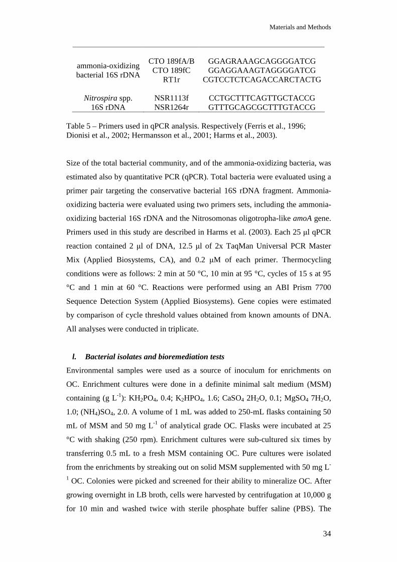
Materials and Methods
34
ammonia-oxidizing bacterial 16S rDNA
CTO 189fA/B CTO 189fC
RT1r
GGAGRAAAGCAGGGGATCG GGAGGAAAGTAGGGGATCG
CGTCCTCTCAGACCARCTACTG
Nitrospira spp. 16S rDNA
NSR1113f NSR1264r
CCTGCTTTCAGTTGCTACCG GTTTGCAGCGCTTTGTACCG
Table 5 – Primers used in qPCR analysis. Respectively (Ferris et al., 1996; Dionisi et al., 2002; Hermansson et al., 2001; Harms et al., 2003). Size of the total bacterial community, and of the ammonia-oxidizing bacteria, was
estimated also by quantitative PCR (qPCR). Total bacteria were evaluated using a
primer pair targeting the conservative bacterial 16S rDNA fragment. Ammonia-
oxidizing bacteria were evaluated using two primers sets, including the ammonia-
oxidizing bacterial 16S rDNA and the Nitrosomonas oligotropha-like amoA gene.
Primers used in this study are described in Harms et al. (2003). Each 25 µl qPCR
reaction contained 2 µl of DNA, 12.5 µl of 2x TaqMan Universal PCR Master
Mix (Applied Biosystems, CA), and 0.2 µM of each primer. Thermocycling
conditions were as follows: 2 min at 50 °C, 10 min at 95 °C, cycles of 15 s at 95
°C and 1 min at 60 °C. Reactions were performed using an ABI Prism 7700
Sequence Detection System (Applied Biosystems). Gene copies were estimated
by comparison of cycle threshold values obtained from known amounts of DNA.
All analyses were conducted in triplicate.
l. Bacterial isolates and bioremediation tests
Environmental samples were used as a source of inoculum for enrichments on
OC. Enrichment cultures were done in a definite minimal salt medium (MSM)
containing (g L-1): KH2PO4, 0.4; K2HPO4, 1.6; CaSO4 2H2O, 0.1; MgSO4 7H2O,
1.0; (NH4)SO4, 2.0. A volume of 1 mL was added to 250-mL flasks containing 50
mL of MSM and 50 mg L-1 of analytical grade OC. Flasks were incubated at 25
°C with shaking (250 rpm). Enrichment cultures were sub-cultured six times by
transferring 0.5 mL to a fresh MSM containing OC. Pure cultures were isolated
from the enrichments by streaking out on solid MSM supplemented with 50 mg L-
1 OC. Colonies were picked and screened for their ability to mineralize OC. After
growing overnight in LB broth, cells were harvested by centrifugation at 10,000 g
for 10 min and washed twice with sterile phosphate buffer saline (PBS). The
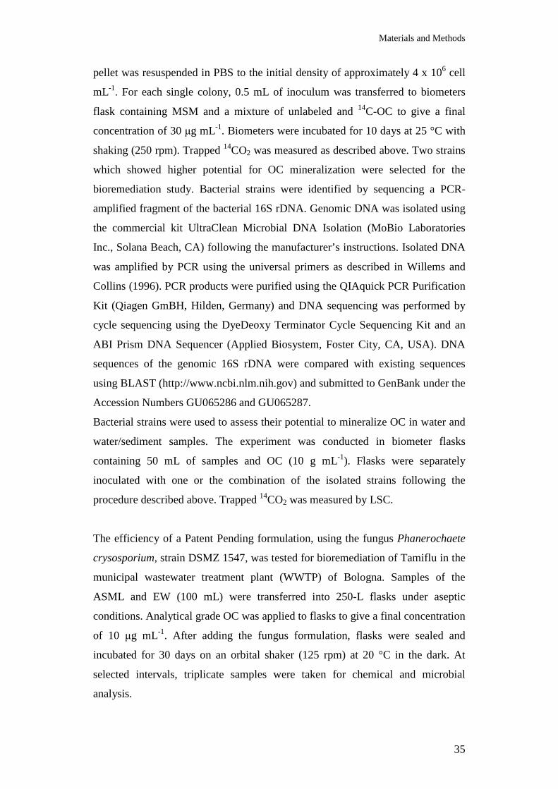
Materials and Methods
35
pellet was resuspended in PBS to the initial density of approximately 4 x 10⁶ cell
mL-1. For each single colony, 0.5 mL of inoculum was transferred to biometers
flask containing MSM and a mixture of unlabeled and 14C-OC to give a final
concentration of 30 µg mL-1. Biometers were incubated for 10 days at 25 °C with
shaking (250 rpm). Trapped 14CO2 was measured as described above. Two strains
which showed higher potential for OC mineralization were selected for the
bioremediation study. Bacterial strains were identified by sequencing a PCR-
amplified fragment of the bacterial 16S rDNA. Genomic DNA was isolated using
the commercial kit UltraClean Microbial DNA Isolation (MoBio Laboratories
Inc., Solana Beach, CA) following the manufacturer’s instructions. Isolated DNA
was amplified by PCR using the universal primers as described in Willems and
Collins (1996). PCR products were purified using the QIAquick PCR Purification
Kit (Qiagen GmBH, Hilden, Germany) and DNA sequencing was performed by
cycle sequencing using the DyeDeoxy Terminator Cycle Sequencing Kit and an
ABI Prism DNA Sequencer (Applied Biosystem, Foster City, CA, USA). DNA
sequences of the genomic 16S rDNA were compared with existing sequences
using BLAST (http://www.ncbi.nlm.nih.gov) and submitted to GenBank under the
Accession Numbers GU065286 and GU065287.
Bacterial strains were used to assess their potential to mineralize OC in water and
water/sediment samples. The experiment was conducted in biometer flasks
containing 50 mL of samples and OC (10 g mL-1). Flasks were separately
inoculated with one or the combination of the isolated strains following the
procedure described above. Trapped 14CO2 was measured by LSC.
The efficiency of a Patent Pending formulation, using the fungus Phanerochaete
crysosporium, strain DSMZ 1547, was tested for bioremediation of Tamiflu in the
municipal wastewater treatment plant (WWTP) of Bologna. Samples of the
ASML and EW (100 mL) were transferred into 250-L flasks under aseptic
conditions. Analytical grade OC was applied to flasks to give a final concentration
of 10 µg mL-1. After adding the fungus formulation, flasks were sealed and
incubated for 30 days on an orbital shaker (125 rpm) at 20 °C in the dark. At
selected intervals, triplicate samples were taken for chemical and microbial
analysis.
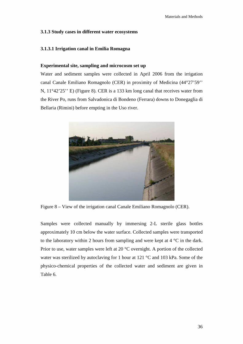
Materials and Methods
36
3.1.3 Study cases in different water ecosystems
3.1.3.1 Irrigation canal in Emilia Romagna
Experimental site, sampling and microcosm set up
Water and sediment samples were collected in April 2006 from the irrigation
canal Canale Emiliano Romagnolo (CER) in proximity of Medicina (44°27’59’’
N, 11°42’25’’ E) (Figure 8). CER is a 133 km long canal that receives water from
the River Po, runs from Salvadonica di Bondeno (Ferrara) downs to Donegaglia di
Bellaria (Rimini) before empting in the Uso river.
Figure 8 – View of the irrigation canal Canale Emiliano Romagnolo (CER).
Samples were collected manually by immersing 2-L sterile glass bottles
approximately 10 cm below the water surface. Collected samples were transported
to the laboratory within 2 hours from sampling and were kept at 4 °C in the dark.
Prior to use, water samples were left at 20 °C overnight. A portion of the collected
water was sterilized by autoclaving for 1 hour at 121 °C and 103 kPa. Some of the
physico-chemical properties of the collected water and sediment are given in
Table 6.

Materials and Methods
37
Water
pH DO
(mg L-1) DOC
(mg L-1) HCO3
- (mg L-1)
F- (mg L-1)
Cl- (mg L-1)
NO2-
(mg L-1) NO3
- (mg L-1)
PO43-
(mg L-1) SO4
2- (mg L-1)
8.3 13.1 4.7 158.0 0.17 28.5 0.07 13.2 <0.02 83.8
Sediments Sand Silt Clay Organic Carbon 7.3% 38.9% 53.8% 2.8%
Table 6– Physico-chemical properties of water and sediment from irrigation canal Canale Emiliano Romagnolo (CER). DO: dissolved oxygen. DOC dissolved organic carbon.
Water samples (80 mL) were transferred into 250-mL sterile Erlenmeyer flasks
under aseptic conditions. Samples were treated with oseltamivir carboxylate (OC)
dissolved in 50 mM NaH2PO4 to give a final concentration of 1.5 µg mL-1. Flasks
were sealed, and samples were incubated at 20 °C on an orbital shaker (125 rpm)
in the dark. At selected time intervals, duplicate 1-mL aliquots were taken for
analytical or microbiological analysis. All operations were conducted under sterile
conditions. Control samples, consisting of untreated water and samples containing
5% (w/v) of sterilized or non-sterilized sediments taken from the same irrigation
canal, were included. Sediments were air-dried and passed through a 5-mm sieve.
The whole experiment was conducted in triplicate.
Chemical analysis
Degradation of OC was assessed by HPLC, as described in paragraph a.
Microbial analysis
Bacterial abundance (e), viability (i), phylogenetic composition using the probes
for the identification of the bacterial taxa: Bacteria, α-Proteobacteria, β-
Proteobacteria, γ-Proteobacteria, Planctomycetes, Cytophaga-Flavobacterium,
Firmicutes (f), and metabolic potential (j) were assessed as described in the
mentioned paragraphs.
3.1.3.2 River Po and Venice Lagoon

Materials and Methods
38
Experimental site, sampling and microcosm set up
Samples of water and sediment were collected in November 2007 from the River
Po (Occhiobello, Italy) and the Venice Lagoon (in proximity of St. Mark’s
Square, Venice, Italy) (Figure 9).
Figure 9 – Sampling points in River Po (above) and Venice Lagoon (below).
In both sites, samples were collected manually by immersing 2-L sterilized glass
bottles approximately 10 cm below the water surface. Sediments were collected
using a sterilized stainless steel corer. Samples were transported to the laboratory
within 2 hours from sampling and kept at 4 °C in the dark for no longer than three
days. Prior to use, water samples were left at 20 °C overnight. Sediments were left
to dry at room temperature for two days, homogenized by passing through a 4-mm
sieve and the remaining water content was determined gravimetrically. A portion
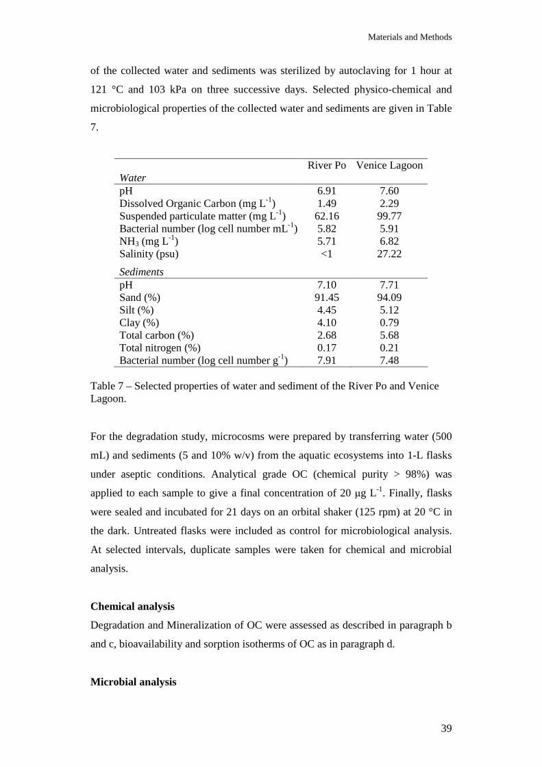
Materials and Methods
39
of the collected water and sediments was sterilized by autoclaving for 1 hour at
121 °C and 103 kPa on three successive days. Selected physico-chemical and
microbiological properties of the collected water and sediments are given in Table
7.
River Po Venice Lagoon Water pH 6.91 7.60 Dissolved Organic Carbon (mg L-1) 1.49 2.29 Suspended particulate matter (mg L-1) 62.16 99.77 Bacterial number (log cell number mL-1) 5.82 5.91 NH3 (mg L-1) 5.71 6.82 Salinity (psu) <1 27.22
Sediments pH 7.10 7.71 Sand (%) 91.45 94.09 Silt (%) 4.45 5.12 Clay (%) 4.10 0.79 Total carbon (%) 2.68 5.68 Total nitrogen (%) 0.17 0.21 Bacterial number (log cell number g-1) 7.91 7.48
Table 7 – Selected properties of water and sediment of the River Po and Venice Lagoon. For the degradation study, microcosms were prepared by transferring water (500
mL) and sediments (5 and 10% w/v) from the aquatic ecosystems into 1-L flasks
under aseptic conditions. Analytical grade OC (chemical purity > 98%) was
applied to each sample to give a final concentration of 20 µg L-1. Finally, flasks
were sealed and incubated for 21 days on an orbital shaker (125 rpm) at 20 °C in
the dark. Untreated flasks were included as control for microbiological analysis.
At selected intervals, duplicate samples were taken for chemical and microbial
analysis.
Chemical analysis
Degradation and Mineralization of OC were assessed as described in paragraph b
and c, bioavailability and sorption isotherms of OC as in paragraph d.
Microbial analysis
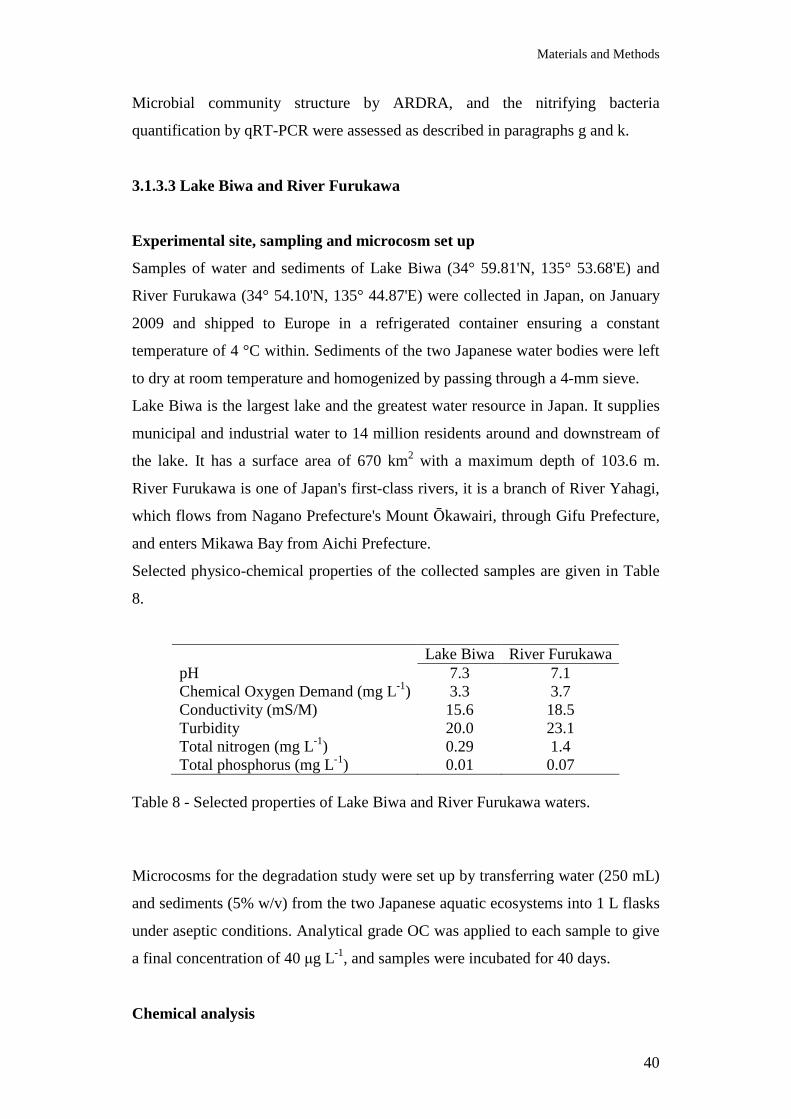
Materials and Methods
40
Microbial community structure by ARDRA, and the nitrifying bacteria
quantification by qRT-PCR were assessed as described in paragraphs g and k.
3.1.3.3 Lake Biwa and River Furukawa
Experimental site, sampling and microcosm set up
Samples of water and sediments of Lake Biwa (34° 59.81'N, 135° 53.68'E) and
River Furukawa (34° 54.10'N, 135° 44.87'E) were collected in Japan, on January
2009 and shipped to Europe in a refrigerated container ensuring a constant
temperature of 4 °C within. Sediments of the two Japanese water bodies were left
to dry at room temperature and homogenized by passing through a 4-mm sieve.
Lake Biwa is the largest lake and the greatest water resource in Japan. It supplies
municipal and industrial water to 14 million residents around and downstream of
the lake. It has a surface area of 670 km2 with a maximum depth of 103.6 m.
River Furukawa is one of Japan's first-class rivers, it is a branch of River Yahagi,
which flows from Nagano Prefecture's Mount Ōkawairi, through Gifu Prefecture,
and enters Mikawa Bay from Aichi Prefecture.
Selected physico-chemical properties of the collected samples are given in Table
8.
Lake Biwa River Furukawa pH 7.3 7.1 Chemical Oxygen Demand (mg L-1) 3.3 3.7 Conductivity (mS/M) 15.6 18.5 Turbidity 20.0 23.1 Total nitrogen (mg L-1) 0.29 1.4 Total phosphorus (mg L-1) 0.01 0.07
Table 8 - Selected properties of Lake Biwa and River Furukawa waters.
Microcosms for the degradation study were set up by transferring water (250 mL)
and sediments (5% w/v) from the two Japanese aquatic ecosystems into 1 L flasks
under aseptic conditions. Analytical grade OC was applied to each sample to give
a final concentration of 40 µg L-1, and samples were incubated for 40 days.
Chemical analysis
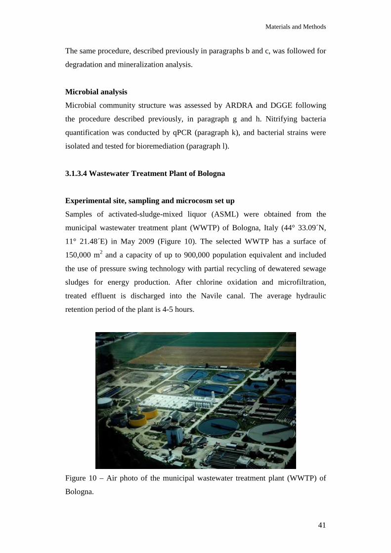
Materials and Methods
41
The same procedure, described previously in paragraphs b and c, was followed for
degradation and mineralization analysis.
Microbial analysis
Microbial community structure was assessed by ARDRA and DGGE following
the procedure described previously, in paragraph g and h. Nitrifying bacteria
quantification was conducted by qPCR (paragraph k), and bacterial strains were
isolated and tested for bioremediation (paragraph l).
3.1.3.4 Wastewater Treatment Plant of Bologna
Experimental site, sampling and microcosm set up
Samples of activated-sludge-mixed liquor (ASML) were obtained from the
municipal wastewater treatment plant (WWTP) of Bologna, Italy (44° 33.09´N,
11° 21.48´E) in May 2009 (Figure 10). The selected WWTP has a surface of
150,000 m2 and a capacity of up to 900,000 population equivalent and included
the use of pressure swing technology with partial recycling of dewatered sewage
sludges for energy production. After chlorine oxidation and microfiltration,
treated effluent is discharged into the Navile canal. The average hydraulic
retention period of the plant is 4-5 hours.
Figure 10 – Air photo of the municipal wastewater treatment plant (WWTP) of
Bologna.
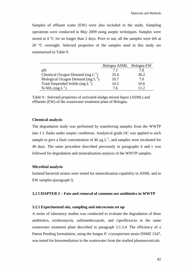
Materials and Methods
42
Samples of effluent water (EW) were also included in the study. Sampling
operations were conducted in May 2009 using aseptic techniques. Samples were
stored at 4 °C for no longer than 2 days. Prior to use, all the samples were left at
20 °C overnight. Selected properties of the samples used in this study are
summarized in Table 9.
Bologna ASML Bologna EW pH 7.1 7.8 Chemical Oxygen Demand (mg L-1) 35.4 36.2 Biological Oxygen Demand (mg L-1) 10.7 7.0 Total Suspended Solids (mg L-1) 14.5 10.6 N-NH3 (mg L-1) 7.6 11.2
Table 9 - Selected properties of activated-sludge-mixed liquor (ASML) and effluents (EW) of the wastewater treatment plant of Bologna.
Chemical analysis
The degradation study was performed by transferring samples from the WWTP
into 1 L flasks under aseptic conditions. Analytical grade OC was applied to each
sample to give a final concentration of 40 µg L-1, and samples were incubated for
40 days. The same procedure described previously in paragraphs b and c was
followed for degradation and mineralization analysis of the WWTP samples.
Microbial analysis
Isolated bacterial strains were tested for mineralization capability in ASML and in
EW samples (paragraph l).
3.2 CHAPTER 2 – Fate and removal of common use antibiotics in WWTP
3.2.1 Experimental site, sampling and microcosm set up
A series of laboratory studies was conducted to evaluate the degradation of three
antibiotics, erythromycin, sulfamethoxazole, and ciprofloxacin in the same
wastewater treatment plant described in paragraph 3.1.3.4. The efficiency of a
Patent Pending formulation, using the fungus P. crysosporium strain DSMZ 1547,
was tested for bioremediation in the wastewater from the studied pharmaceuticals.
Materials and Methods
43
All antibiotics were of analytical grade (> 98 %) and were obtained from Sigma-
Aldrich Chemie GmbH (Steinheim, Germany). Samples of the incubation study
were prepared by transferring ASML or EW (100 mL) into 250-L flasks under
aseptic conditions. Analytical grade pharmaceuticals were separately applied to
each flask to give a final concentration of 10 µg mL-1. After adding the fungal
formulation, flasks were sealed and incubated for 30 days on an orbital shaker
(125 rpm) at 20 °C in the dark. At selected intervals, triplicate samples were taken
for chemical and microbial analysis.
3.2.2 Chemical analysis
Samples were extracted by solid phase extraction and analyzed by liquid
chromatography/electro spray tandem mass spectrometry. Analysis of the three
pharmaceuticals were performed using a triple stage quadrupole MS/MS TSQ
Quantum Ultra EMR (Thermo Fisher Scientific, San Jose, CA, USA) coupled
with an Accela LC pump (Thermo Fisher Scientific, San Jose, CA, USA) and a
PAL HTC autosampler (CTC Analytics AG, Zwingen, Switzerland). Samples (20
µL) were loaded onto a Hypersil GOLD aQ TM column (50 mm x 2.1 mm ID x 5
µm particles, Thermo Fisher Scientific, San Jose, CA, USA). Elution conditions
were programmed as follows: water/methanol (90/10) for 1 min, then composition
was changed to water/acetonitrile/methanol (30/10/60) for 8 min and then the
column was washed for 9 min by acetonitrile/methanol (60/40). Heated
electrospray in positive ion mode was used for ionisation of target compounds.
Key parameters were the following: ionisation voltage 3.5 kV, sheath gas 50 and
auxiliary gas 35 arbitrary units, vaporiser temperature 100 °C, capillary
temperature 325 °C, collision gas was argon at 1.5 mL min-1. Both first and third
quadrupole were operated at resolution 0.7 FWHD. Three SRM transitions were
monitored for each analyte. Samples were quantified using internal standards.
Prior to analysis, samples were kept in the dark for 12 hours. The whole
incubation study experiment was conducted in triplicate. Data of the degradation
study were analyzed by analysis of variance. Means were separated by Fisher’s
least significant difference (LSD) and significant differences were detected at the
P = 0.05 level.
3.2.3 Microbial analysis
Materials and Methods
44
Triplicates samples prepared as for the incubation study were used for
microbiological analysis. Growth of the introduced bioremediation fungus was
monitored by quantitative PCR (qPCR) using the primer pair Cu1F/Cu2R
targeting the laccase gene (Luis et al., 2004). Granules used for amending ASML
and EW samples were periodically removed and total DNA was isolated using the
CTAB method (Doyle and Doyle, 1990). Briefly, triplicates granules were
removed from each incubated sample, dried at 40 °C for 2 hours, vortexed for 5
min to remove adhering organic particles and air-flushed by high-pressure air.
Surface-cleaned granules were transferred to a 2-mL microcentrifuge tube
containing 500 µL of CTAB buffer and glass beads (425-600 µm; Sigma-
Aldrich). After vortexing for 2 min, tubes were incubated at 65 °C for 15 min, and
an equivalent volume of chloroform:isoamyl alchol (24:1) was added to tubes.
Tubes were gently shaken and centrifuged at 10,000 x g for 5 min before the
addition of 2/3 volume of isopropanol/ammonium acetate to precipitate the DNA.
The pellet was rinsed with 70% ethanol, air dried, resuspended in 100 µL of TE
buffer and used for qPCR. Each 25 µL qPCR reaction contained 2 µL of DNA,
12.5 µL of 2× TaqMan Universal PCR Master Mix (Applied Biosystems, CA),
and 0.2 µM of each primer. Thermocycling conditions were as follows: 2 min at
50 °C, 10 min at 95 °C, cycles of 15 s at 95 °C and 1 min at 60 °C. Reactions
were performed using an ABI Prism 7700 Sequence Detection System (Applied
Biosystems). Gene copies were estimated by comparison of cycle threshold values
obtained from known amounts of DNA. All analyses were conducted in triplicate.
Structure of the bacterial and fungal communities of incubated samples was
investigated by PCR- denaturing gradient gel electrophoresis (DGGE) analysis.
Prefiltered (0.45 µm) aliquots of ASML and EW samples were passed through a
sterile 0.22-µm nylon filter (GE Water & Technologies, Trevose, PA). Filters
were transferred into PowerBead tubes provided with the UltraClean Soil DNA
Isolation Kit (MoBio Laboratories Inc., Solana Beach, CA) and then processed
following the instructions of the manufacturer. For bacterial PCR-DGGE, total
DNA was amplified using the primer pair GC-968f/1401r targeting the V6-V8
region of the 16S rDNA (Heuer et al., 1999). Amplification reactions were
performed in a 50 µL reaction mixture consisting of 5-10 ng of DNA, 1.25 U of
BioTaq DNA polymerase (Bioline Abcys, Paris, France), 10× PCR buffer, 2.5
mM MgCl2, 200 µM of each dNTP, 0.5 µM of each primer, 250 ng µL-1 bovine
Materials and Methods
45
serum albumin and nuclease-free water. Reaction conditions were the following:
denaturation at 94 °C for 5 min, followed by 35 cycles of denaturation at 97 °C
for 45 s, annealing at 58 °C for 60 s, extension at 72 °C for 45 s and final
extension at 72 °C for 5 min. For fungal PCR, a semi-nested approach targeting
the internal transcribed spacer region (ITS) was adopted. Primer pairs and PCR
conditions are described elsewhere (Crouzet et al., 2010). Equal amounts of
amplicons (800 ng) were loaded onto DGGE gel. Gel contained 8% (w/v)
polyacrylamide gels (37.5:1 acrylamide:bisacrylamide) with a urea/formamide
denaturing gradient of 45 – 65% for bacteria and 25–55% for fungi (where 100%
denaturant contains 7 M urea and 40% v/v formamide). Gels were run for 16
hours at 60 °C, with a constant voltage of 140 V for bacteria and 70 V for fungi.
DGGE was performed in DCode system (Bio-Rad Laboratories, Hercules, CA,
USA). Bands were visualized after staining with Gel Star Nucleic acid gel stain
and digitized using the Versa Doc Imaging System (Bio-Rad). Band profiles were
analyzed using the GelCompare II package (Applied Maths, Kortrijk, Belgium).
Dendrograms were constructed using the DICE coefficients and were subjected to
unweighted pair group method cluster analysis (UPGMA).
3.3 CHAPTER 3 – Veterinary Pharmaceuticals
3.3.1 Experimental site, sampling and microcosm set up
Soil from an olive tree grove in Aranjuez, in central Spain, was sampled in 2008.
The agricultural soil is routinely amended with livestock residues from a pig farm.
Two sets of microcosms were arranged by adding to the soil the veterinary drugs
oxibendazole, to give a final concentration of 5 µg mL-1, and doramectin, with 20
µg mL-1 final concentration. Samples were incubated at 20 °C on an orbital shaker
(125 rpm) in the dark. After respectively 21 and 23 days of incubation, soil
aliquots were used for analytical and microbiological analysis. All operations
were conducted under sterile conditions. Control samples, consisting of untreated
soil, were included.
3.3.2 Microbial analysis In order to investigate the effects of oxibendazole and doramectin on the
phylogenetic structure of the soil bacterial community, filters with cells, fixed as
Materials and Methods
46
described previously in paragraph (f), were analysed by Fluorescence In Situ
Hybridization. Phylogenetic probes were applied for the identification of the
following bacterial taxa: Archaea, Bacteria, α-Proteobacteria, β-Proteobacteria,
γ-Proteobacteria, δ-proteobacteria Sulfate-Reducing Bacteria, ε-proteobacteria,
Cytophaga-Flavobacterium, Actinobacteria, Firmicutes.
Furthermore, because of the importance of nitrate-reducing bacteria in the
nitrogen cycle, the effects of the two veterinary drugs on this functional group
were investigated. Denitrification involves the reduction of nitrate, via nitrite and
nitric oxide, to nitrous oxide or dinitrogen gas by a respiratory process under
oxygen-limiting conditions. In particular in this work, the nitrate reductase gene
narG, was of special interest to provide information on the denitrifying bacteria in
the environment. Denitrification was induced by adding sodium nitrate (10 mM)
to soil, and simultaneously shifting to O2-limited conditions by sealing the sample
with a paraffin layer and avoiding shaker rotation.
The presence of the narG gene in soil samples was checked by PCR amplification
from extracted DNA, prior to analysis of gene expression. Total DNA was
isolated from incubated samples using Ultra Clean Soil DNA Kit (MoBio
Laboratories, Inc. Solana Beach, CA, USA). A set of partially degenerated
primers for the amplification of the narG gene (Table 10), in yet uncultivated
bacteria in the environment, was used for PCR and qRT-PCR assays. Total RNA
was isolated from soil using the FastRNA® Pro Soil-Direct Kit (MP Biomedicals,
Solon, OH). Samples were processed following the instructions of the
manufacturer. Reverse transcriptase was obtained by utilization of the
iScript™cDNA Synthesis Kit (Bio-Rad Laboratories, Hercules, CA), and
amplification was obtained by the use of SYBR Green PCR mix (iTaqTM
SYBR® Green Supermix with ROX, Bio-Rad). Conditions described in (López-
Gutiérrez et al., 2004) were used for the qRT-PCR assay.
Target Primer/Probe Sequence (5’-3’)
narG gene 1960m2f TA(CT)GT(GC)GGGCAGGA(AG)AAACTG
narG gene 2050m2r CGTAGAAGAAGCTGGTGCTGTT
Table 10 – narG primers and probe used in the study for qPCR and FISH analysis (López-Gutiérrez et al., 2004).
Materials and Methods
47
The sequence of the narG primer 2050m2r was used to build a specific fam-
labelled probe, for the detection of the narG gene by Fluorescence in Situ
Hybridization in the incubated and non-incubated soil samples (paragraph f).
Conditions described previously were used for the FISH analysis (Martin et al.,
2008). Percentages of narG-harbouring cells were calculated based on the total
number of cells stained with DAPI.
Isolation of bacterial strains
Bacterial strains, able to grow on the veterinary drugs as sole carbon source, were
isolated from the soil sample by enrichment culture in minimal medium MB
(K2HPO4: 1.6 g L-1; KH2PO4: 0.4g L-1; CaSO4 x 2 H2O: 0.1 g L-1; MgSO4 x 7
H2O: 1.0 g L-1; FeSO4 x 7 H2O: 0.02 g L-1; (NH4)2SO4: 2 g L-1; Agar: 15 g L-1)
supplemented with doramectin (100 µg mL-1) and oxibendazole (100 µg mL-1).
DNA from each isolate was extracted using the method described in Casas et al.
(1995). Amplification of rDNA gene was performed by PCR using the universal
primer ARI (5’ GAGAGTTTGATCCTGGCTCAGGA 3’), and reverse pH (5’
AAGGAGGTGATCCAGCCGCA 3’). The amplified fragments were sequenced
and compared with sequences available in the GenBank/EMBLdatabases
(http://www.ncbi.nlm.nih.gov).
Results and Discussion
48
4. RESULTS AND DISCUSSION
4.1 CHAPTER 1 – The antiviral drug Tamiflu
4.1.1 Irrigation canal in Emilia Romagna
Chemical analysis
Degradation of OC
Degradation of OC in water and water/sediment samples over the course of the
36-day incubation period is shown in Figure 11. Degradation of OC in water did
not adequately fit the first-order model (r2 ≤ 0.80). After a rapid decrease, OC
concentrations slowly decreased during the remaining incubation period.
Approximately 65% of the applied amount degraded in water samples within 36
days. These findings suggested that degradation of OC in water is a complex
process, not simply described by the linear model. OC was less persistent in
samples containing sediments (5% w/v). In contrast to water samples, the linear
model gave a strong fit (r2>0.96) to the degradation of OC in water/sediment
mixtures. The estimated half-life of OC in the water/sediment microcosm was 21
days. Chemical analysis revealed that approximately 5% of the applied OC was
degraded within 36 days in sterilized water samples (Figure 11). Similar values
were observed in the sterilized water/sediment mixture (data not shown). This
information provides supporting evidence that OC degradation was mainly driven
by microbial processes. Considering the size of the cultivable bacterial population
(7.9±0.28 log CFUs g-1 air-dried sediments), the effect of sediments is compatible
with the increasing microbial abundance and metabolic potential of the
microcosm. Enhanced biodegradation of xenobiotics in the presence of sediments
has been reported for a number of compounds, including pesticides and
antimicrobials (Walker et al., 1984; Pritchard et al., 1987; Kim et al., 2004). This
effect could result from a greater number of microorganisms on the surface of
sediment particles, an increased activity of microorganisms in the presence of
sediments due to greater availability of nutrient, or an ability of sediments to
concentrate chemical through sorption (Walker et al., 1984). Under some
circumstances, abiotic processes (i.e. hydrolysis and photolysis) can have an
important role in the degradation of pharmaceuticals in water (Liu et al., 2001).
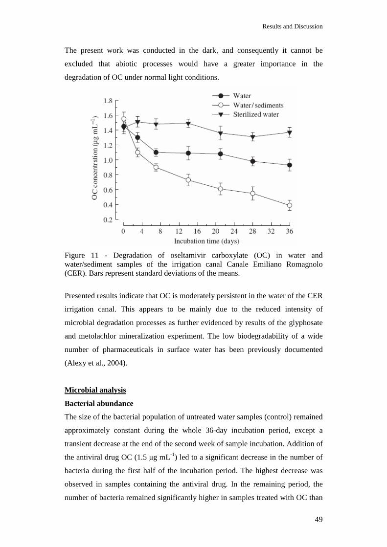
Results and Discussion
49
The present work was conducted in the dark, and consequently it cannot be
excluded that abiotic processes would have a greater importance in the
degradation of OC under normal light conditions.
Figure 11 - Degradation of oseltamivir carboxylate (OC) in water and water/sediment samples of the irrigation canal Canale Emiliano Romagnolo (CER). Bars represent standard deviations of the means.
Presented results indicate that OC is moderately persistent in the water of the CER
irrigation canal. This appears to be mainly due to the reduced intensity of
microbial degradation processes as further evidenced by results of the glyphosate
and metolachlor mineralization experiment. The low biodegradability of a wide
number of pharmaceuticals in surface water has been previously documented
(Alexy et al., 2004).
Microbial analysis
Bacterial abundance
The size of the bacterial population of untreated water samples (control) remained
approximately constant during the whole 36-day incubation period, except a
transient decrease at the end of the second week of sample incubation. Addition of
the antiviral drug OC (1.5 µg mL-1) led to a significant decrease in the number of
bacteria during the first half of the incubation period. The highest decrease was
observed in samples containing the antiviral drug. In the remaining period, the
number of bacteria remained significantly higher in samples treated with OC than
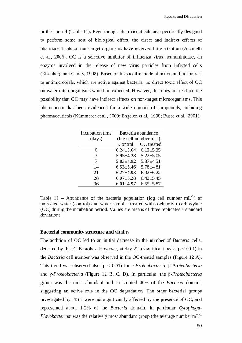
Results and Discussion
50
in the control (Table 11). Even though pharmaceuticals are specifically designed
to perform some sort of biological effect, the direct and indirect effects of
pharmaceuticals on non-target organisms have received little attention (Accinelli
et al., 2006). OC is a selective inhibitor of influenza virus neuraminidase, an
enzyme involved in the release of new virus particles from infected cells
(Eisenberg and Cundy, 1998). Based on its specific mode of action and in contrast
to antimicrobials, which are active against bacteria, no direct toxic effect of OC
on water microorganisms would be expected. However, this does not exclude the
possibility that OC may have indirect effects on non-target microorganisms. This
phenomenon has been evidenced for a wide number of compounds, including
pharmaceuticals (Kümmerer et al., 2000; Engelen et al., 1998; Busse et al., 2001).
Incubation time (days)
Bacteria abundance (log cell number ml-1)
Control OC treated 0 6.24±5.64 6.12±5.35 3 5.95±4.28 5.22±5.05 7 5.83±4.92 5.37±4.51 14 6.53±5.46 5.78±4.81 21 6.27±4.93 6.92±6.22 28 6.07±5.28 6.42±5.45 36 6.01±4.97 6.55±5.87
Table 11 – Abundance of the bacteria population (log cell number mL-1) of untreated water (control) and water samples treated with oseltamivir carboxylate (OC) during the incubation period. Values are means of three replicates ± standard deviations. Bacterial community structure and vitality
The addition of OC led to an initial decrease in the number of Bacteria cells,
detected by the EUB probes. However, at day 21 a significant peak (p < 0.01) in
the Bacteria cell number was observed in the OC-treated samples (Figure 12 A).
This trend was observed also (p < 0.01) for α-Proteobacteria, β-Proteobacteria
and γ-Proteobacteria (Figure 12 B, C, D). In particular, the β-Proteobacteria
group was the most abundant and constituted 40% of the Bacteria domain,
suggesting an active role in the OC degradation. The other bacterial groups
investigated by FISH were not significantly affected by the presence of OC, and
represented about 1-2% of the Bacteria domain. In particular Cytophaga-
Flavobacterium was the relatively most abundant group (the average number mL-1
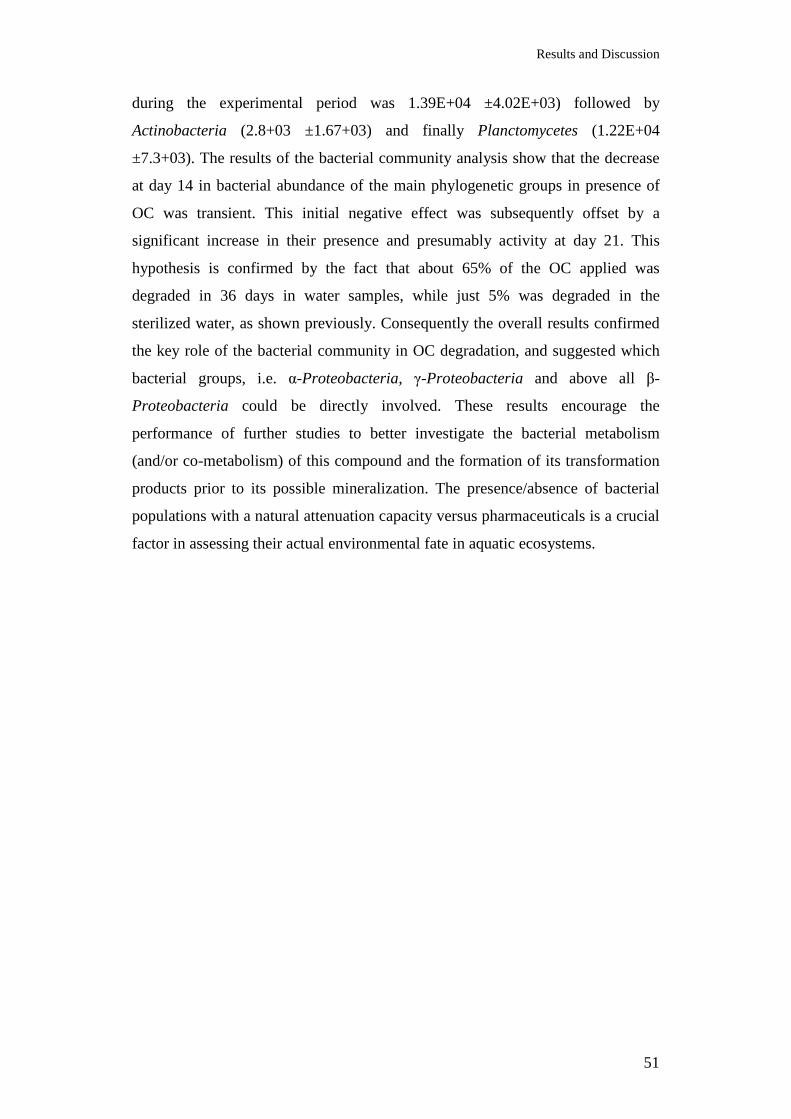
Results and Discussion
51
during the experimental period was 1.39E+04 ±4.02E+03) followed by
Actinobacteria (2.8+03 ±1.67+03) and finally Planctomycetes (1.22E+04
±7.3+03). The results of the bacterial community analysis show that the decrease
at day 14 in bacterial abundance of the main phylogenetic groups in presence of
OC was transient. This initial negative effect was subsequently offset by a
significant increase in their presence and presumably activity at day 21. This
hypothesis is confirmed by the fact that about 65% of the OC applied was
degraded in 36 days in water samples, while just 5% was degraded in the
sterilized water, as shown previously. Consequently the overall results confirmed
the key role of the bacterial community in OC degradation, and suggested which
bacterial groups, i.e. α-Proteobacteria, γ-Proteobacteria and above all β-
Proteobacteria could be directly involved. These results encourage the
performance of further studies to better investigate the bacterial metabolism
(and/or co-metabolism) of this compound and the formation of its transformation
products prior to its possible mineralization. The presence/absence of bacterial
populations with a natural attenuation capacity versus pharmaceuticals is a crucial
factor in assessing their actual environmental fate in aquatic ecosystems.
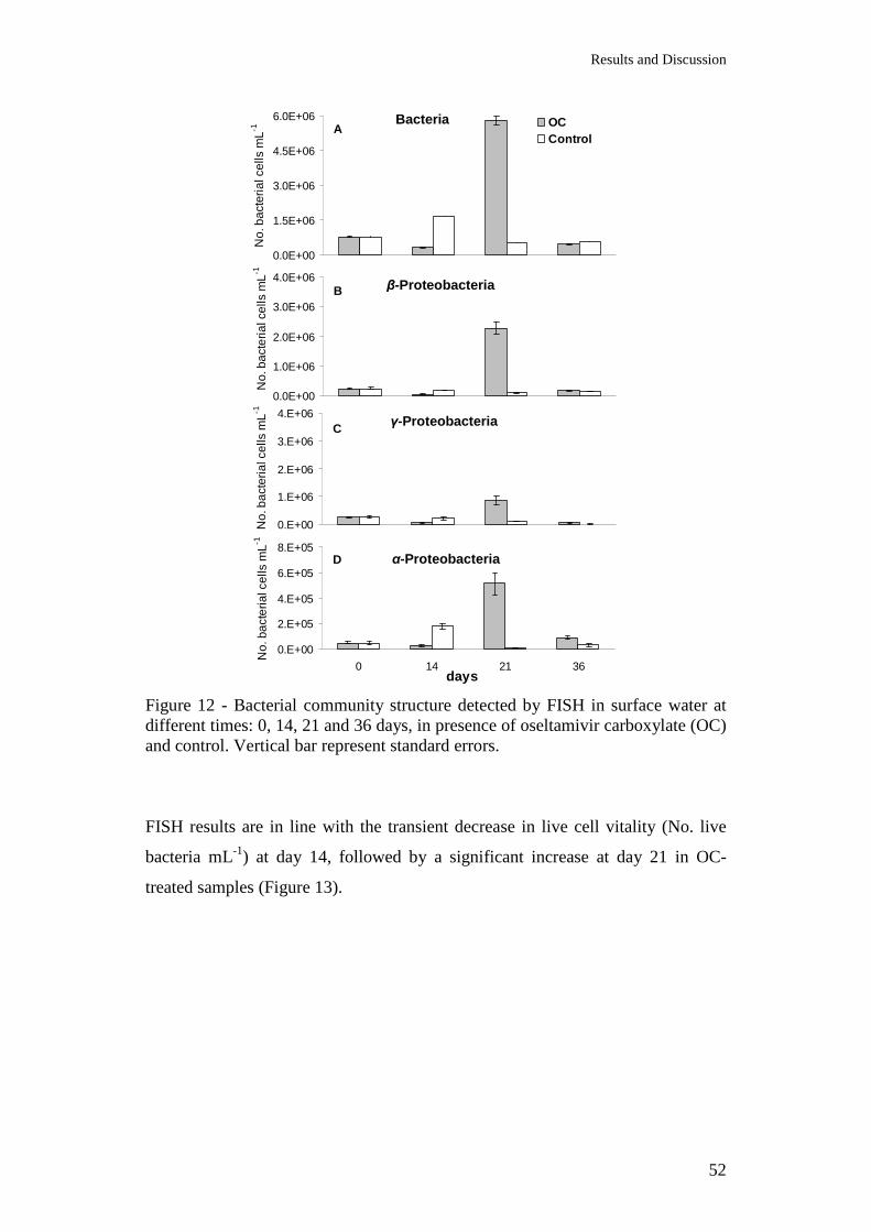
Results and Discussion
52
0.0E+00
1.5E+06
3.0E+06
4.5E+06
6.0E+06 OCControl
Bacteria A
0.E+00
2.E+05
4.E+05
6.E+05
8.E+05
0 14 21 36
α-Proteobacteria D
0.0E+00
1.0E+06
2.0E+06
3.0E+06
4.0E+06β-Proteobacteria B
0.E+00
1.E+06
2.E+06
3.E+06
4.E+06γ-Proteobacteria C
days
No.
bac
teria
l cel
ls m
L -1…
.
No.
bac
teria
l cel
ls m
L -1…
.
No.
bac
teria
l cel
ls m
L -1…
.
No.
bac
teria
l cel
ls m
L -1…
.
Figure 12 - Bacterial community structure detected by FISH in surface water at different times: 0, 14, 21 and 36 days, in presence of oseltamivir carboxylate (OC) and control. Vertical bar represent standard errors. FISH results are in line with the transient decrease in live cell vitality (No. live
bacteria mL-1) at day 14, followed by a significant increase at day 21 in OC-
treated samples (Figure 13).
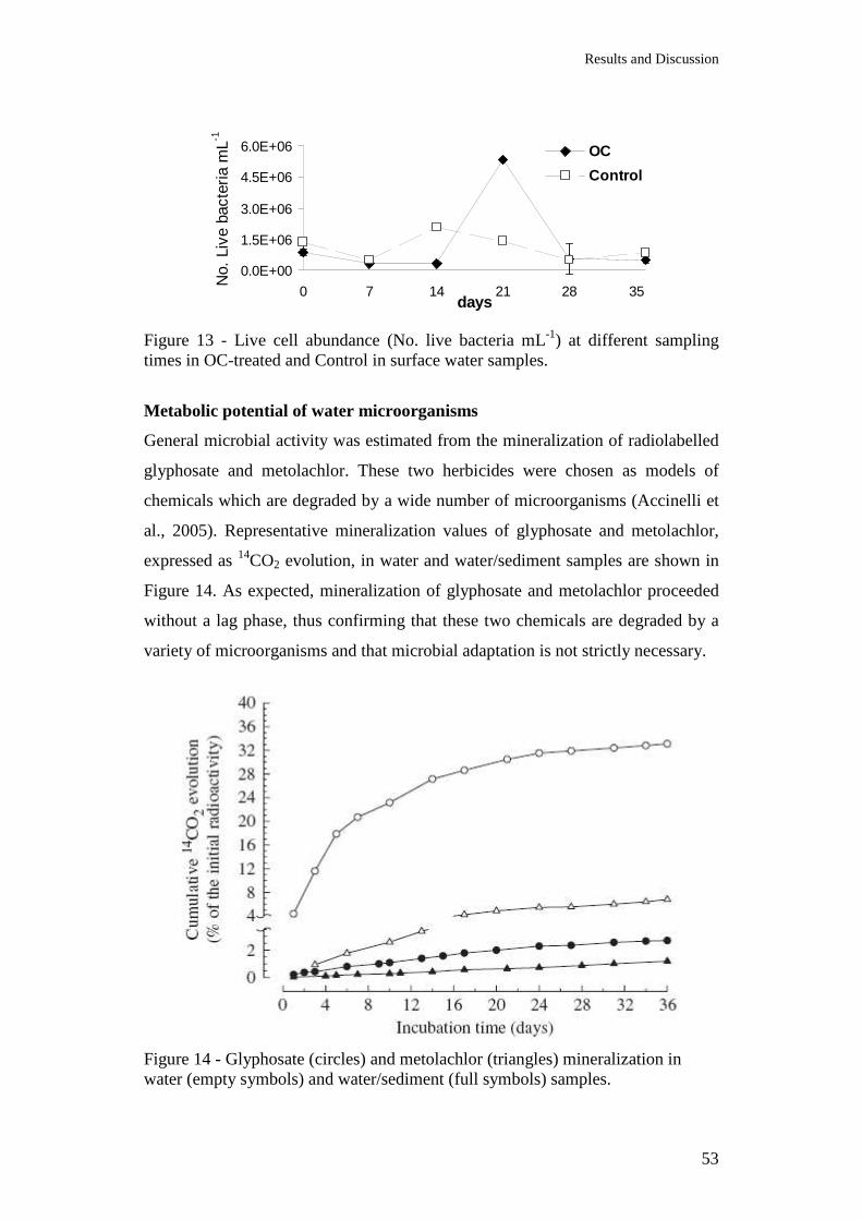
Results and Discussion
53
0.0E+00
1.5E+06
3.0E+06
4.5E+06
6.0E+06
0 7 14 21 28 35days
OC
Control
No.
Liv
e ba
cter
ia m
L -1…
..
Figure 13 - Live cell abundance (No. live bacteria mL-1) at different sampling times in OC-treated and Control in surface water samples.
Metabolic potential of water microorganisms
General microbial activity was estimated from the mineralization of radiolabelled
glyphosate and metolachlor. These two herbicides were chosen as models of
chemicals which are degraded by a wide number of microorganisms (Accinelli et
al., 2005). Representative mineralization values of glyphosate and metolachlor,
expressed as 14CO2 evolution, in water and water/sediment samples are shown in
Figure 14. As expected, mineralization of glyphosate and metolachlor proceeded
without a lag phase, thus confirming that these two chemicals are degraded by a
variety of microorganisms and that microbial adaptation is not strictly necessary.
Figure 14 - Glyphosate (circles) and metolachlor (triangles) mineralization in water (empty symbols) and water/sediment (full symbols) samples.

Results and Discussion
54
During the 36-day incubation period, cumulative 14CO2 evolution in water
samples accounted for 2.9 and 0.9% of the total applied 14C as glyphosate and
metolachlor, respectively (Table 12). Even though mineralization of glyphosate
can vary among environmental samples, these values are considerably lower than
those reported for soil ecosystems (Accinelli et al., 2005; Getenga and Kengara,
2004; Strange-Hansen et al., 2004). Addition of sediments resulted in an intense
increase in glyphosate and metolachlor mineralization. At the end of the
incubation period, mineralization of both glyphosate and metholachlor in samples
containing 5% sediments was approximately 10 times higher than that observed in
water samples. These findings confirmed the important role of microorganisms in
glyphosate and metolachlor mineralization. Moreover, the lack of herbicide
mineralization in sterilized water suggested that chemical degradation is not a
major pathway of degradation of these two chemicals (Table 12). Results from
this mineralization experiment reinforced the concept that a major factor limiting
a more rapid degradation of OC in surface water of the CER irrigation canal is
represented by the low metabolic potential of this ecosystem. Information
concerning degradation and other environmental aspects of these two chemicals in
surface water is scarce (Tsui and Chu, 2003). Based on the results from a recent
monitoring investigation conducted in the USA, Kolpin et al. (2006) speculated
that glyphosate would be much more persistent in surface waters than in soil. The
presence of a low concentration (1.5 µg mL-1) of the antiviral drug OC did not
reduce the potential of water from the irrigation canal to mineralize the two
studied herbicides (Table 12). These are the first data concerning environmental
aspects of the antiviral drug OC or other neuraminidase antivirals. Considering
that degradation of glyphosate and metolachlor is to some extent related to the
size of indigenous bacteria, these findings suggest that the moderate persistence of
OC in water was mainly caused by the low metabolic potential of the water
microbial community rather than indirect effects of OC on microorganisms.
Cumulative 14CO2 evolution 14C-Glyphosate 14C-Metolachlor Control OC treated Control OC treated
Water 2.85±0.15 2.91±0.22 0.85±0.07 0.81±0.09 Steril water 0.82±0.06 0.88±0.06 0.11±0.02 0.09±0.02 Water/sediment 38.22±3.98 36.35±4.11 9.91±1.21 8.75±1.39

Results and Discussion
55
Table 12– Effect of oseltamivir carboxylate (OC) on 14CO2 evolution from radio-labelled glyphosate and metholachlor within the 36-day incubation period (% of initial radioactivity). 4.1.2 River Po and Venice Lagoon
Chemical analysis
Concentrations of OC in samples of the River Po over the course of the 21-day
incubation period are shown in Figure 15. No appreciable degradation of OC was
observed in water. In contrast, addition of sediments promoted OC degradation.
More specifically, OC degradation in samples containing 5% sediments was
adequately described by the first order kinetic model (r2 = 0.91) with a calculated
half-life of 15 days. Degradation of OC was not further stimulated by raising the
sediment content to 10%.
Figure 15 - Degradation of oseltamivir carboxylate (OC) in water and water/sediment samples of the River Po and in water of the Venice Lagoon. Bars represent standard deviations of the means.
These findings are consistent with those of the mineralization study. As indicated
in Figure 16, less than 1% of 14CO2 was evolved from water samples. Cumulative 14CO2 evolution was approximately 10-times higher in samples containing 5% of
sediments. Addition of a higher amount of sediments did not lead to a significant
increase of 14CO2 evolution. Since no appreciable OC mineralization was
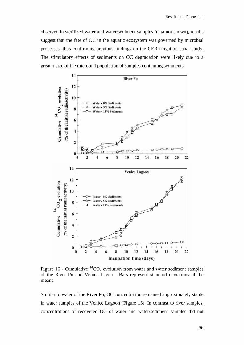
Results and Discussion
56
observed in sterilized water and water/sediment samples (data not shown), results
suggest that the fate of OC in the aquatic ecosystem was governed by microbial
processes, thus confirming previous findings on the CER irrigation canal study.
The stimulatory effects of sediments on OC degradation were likely due to a
greater size of the microbial population of samples containing sediments.
Figure 16 - Cumulative 14CO2 evolution from water and water sediment samples of the River Po and Venice Lagoon. Bars represent standard deviations of the means.
Similar to water of the River Po, OC concentration remained approximately stable
in water samples of the Venice Lagoon (Figure 15). In contrast to river samples,
concentrations of recovered OC of water and water/sediment samples did not
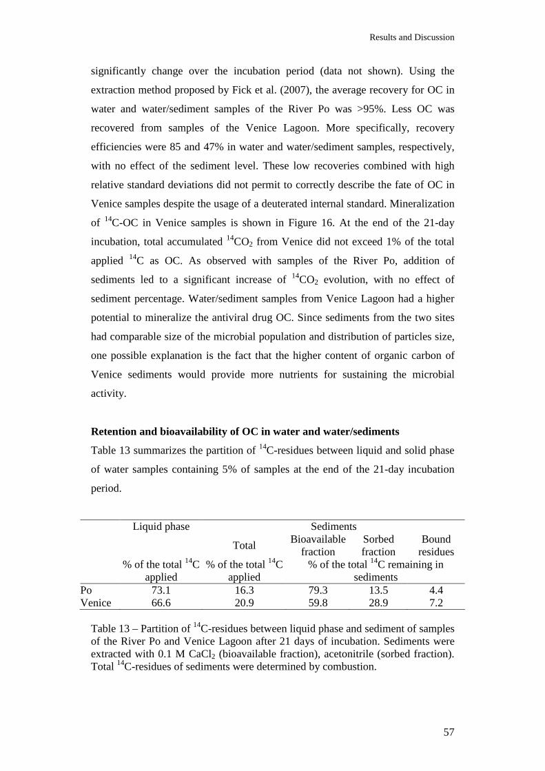
Results and Discussion
57
significantly change over the incubation period (data not shown). Using the
extraction method proposed by Fick et al. (2007), the average recovery for OC in
water and water/sediment samples of the River Po was >95%. Less OC was
recovered from samples of the Venice Lagoon. More specifically, recovery
efficiencies were 85 and 47% in water and water/sediment samples, respectively,
with no effect of the sediment level. These low recoveries combined with high
relative standard deviations did not permit to correctly describe the fate of OC in
Venice samples despite the usage of a deuterated internal standard. Mineralization
of 14C-OC in Venice samples is shown in Figure 16. At the end of the 21-day
incubation, total accumulated 14CO2 from Venice did not exceed 1% of the total
applied 14C as OC. As observed with samples of the River Po, addition of
sediments led to a significant increase of 14CO2 evolution, with no effect of
sediment percentage. Water/sediment samples from Venice Lagoon had a higher
potential to mineralize the antiviral drug OC. Since sediments from the two sites
had comparable size of the microbial population and distribution of particles size,
one possible explanation is the fact that the higher content of organic carbon of
Venice sediments would provide more nutrients for sustaining the microbial
activity.
Retention and bioavailability of OC in water and water/sediments
Table 13 summarizes the partition of 14C-residues between liquid and solid phase
of water samples containing 5% of samples at the end of the 21-day incubation
period.
Liquid phase Sediments
Total Bioavailable
fraction Sorbed fraction
Bound residues
% of the total 14C
applied % of the total 14C
applied % of the total 14C remaining in
sediments Po 73.1 16.3 79.3 13.5 4.4 Venice 66.6 20.9 59.8 28.9 7.2
Table 13 – Partition of 14C-residues between liquid phase and sediment of samples of the River Po and Venice Lagoon after 21 days of incubation. Sediments were extracted with 0.1 M CaCl2 (bioavailable fraction), acetonitrile (sorbed fraction). Total 14C-residues of sediments were determined by combustion.
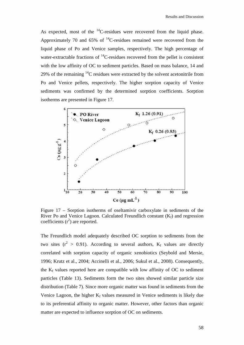
Results and Discussion
58
As expected, most of the 14C-residues were recovered from the liquid phase.
Approximately 70 and 65% of 14C-residues remained were recovered from the
liquid phase of Po and Venice samples, respectively. The high percentage of
water-extractable fractions of 14C-residues recovered from the pellet is consistent
with the low affinity of OC to sediment particles. Based on mass balance, 14 and
29% of the remaining 14C residues were extracted by the solvent acetonitrile from
Po and Venice pellets, respectively. The higher sorption capacity of Venice
sediments was confirmed by the determined sorption coefficients. Sorption
isotherms are presented in Figure 17.
Figure 17 – Sorption isotherms of oseltamivir carboxylate in sediments of the River Po and Venice Lagoon. Calculated Freundlich constant (Kf) and regression coefficients (r2) are reported.
The Freundlich model adequately described OC sorption to sediments from the
two sites (r2 > 0.91). According to several authors, Kf values are directly
correlated with sorption capacity of organic xenobiotics (Seybold and Mersie,
1996; Krutz et al., 2004; Accinelli et al., 2006; Sukul et al., 2008). Consequently,
the Kf values reported here are compatible with low affinity of OC to sediment
particles (Table 13). Sediments form the two sites showed similar particle size
distribution (Table 7). Since more organic matter was found in sediments from the
Venice Lagoon, the higher Kf values measured in Venice sediments is likely due
to its preferential affinity to organic matter. However, other factors than organic
matter are expected to influence sorption of OC on sediments.
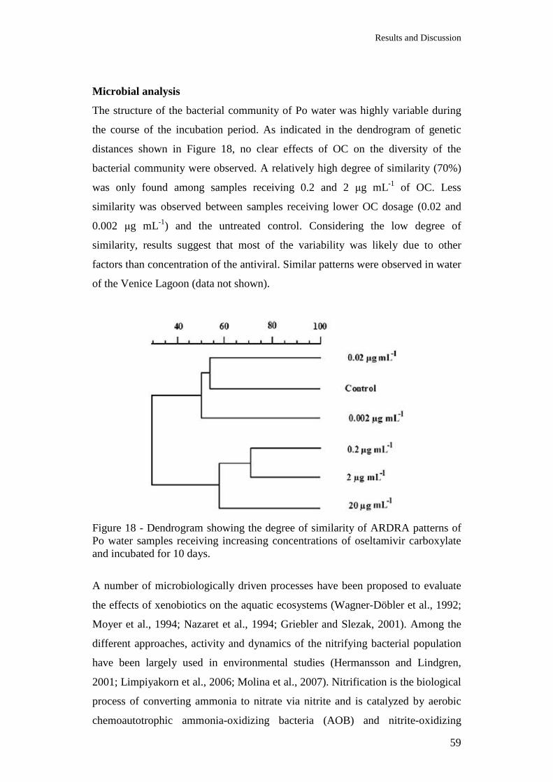
Results and Discussion
59
Microbial analysis
The structure of the bacterial community of Po water was highly variable during
the course of the incubation period. As indicated in the dendrogram of genetic
distances shown in Figure 18, no clear effects of OC on the diversity of the
bacterial community were observed. A relatively high degree of similarity (70%)
was only found among samples receiving 0.2 and 2 µg mL-1 of OC. Less
similarity was observed between samples receiving lower OC dosage (0.02 and
0.002 µg mL-1) and the untreated control. Considering the low degree of
similarity, results suggest that most of the variability was likely due to other
factors than concentration of the antiviral. Similar patterns were observed in water
of the Venice Lagoon (data not shown).
Figure 18 - Dendrogram showing the degree of similarity of ARDRA patterns of Po water samples receiving increasing concentrations of oseltamivir carboxylate and incubated for 10 days.
A number of microbiologically driven processes have been proposed to evaluate
the effects of xenobiotics on the aquatic ecosystems (Wagner-Döbler et al., 1992;
Moyer et al., 1994; Nazaret et al., 1994; Griebler and Slezak, 2001). Among the
different approaches, activity and dynamics of the nitrifying bacterial population
have been largely used in environmental studies (Hermansson and Lindgren,
2001; Limpiyakorn et al., 2006; Molina et al., 2007). Nitrification is the biological
process of converting ammonia to nitrate via nitrite and is catalyzed by aerobic
chemoautotrophic ammonia-oxidizing bacteria (AOB) and nitrite-oxidizing
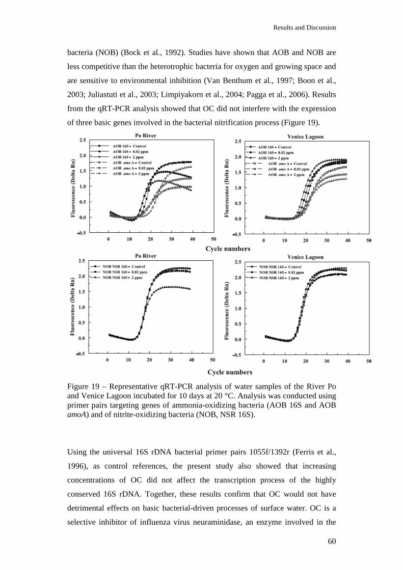
Results and Discussion
60
bacteria (NOB) (Bock et al., 1992). Studies have shown that AOB and NOB are
less competitive than the heterotrophic bacteria for oxygen and growing space and
are sensitive to environmental inhibition (Van Benthum et al., 1997; Boon et al.,
2003; Juliastuti et al., 2003; Limpiyakorn et al., 2004; Pagga et al., 2006). Results
from the qRT-PCR analysis showed that OC did not interfere with the expression
of three basic genes involved in the bacterial nitrification process (Figure 19).
Figure 19 – Representative qRT-PCR analysis of water samples of the River Po and Venice Lagoon incubated for 10 days at 20 °C. Analysis was conducted using primer pairs targeting genes of ammonia-oxidizing bacteria (AOB 16S and AOB amoA) and of nitrite-oxidizing bacteria (NOB, NSR 16S).
Using the universal 16S rDNA bacterial primer pairs 1055f/1392r (Ferris et al.,
1996), as control references, the present study also showed that increasing
concentrations of OC did not affect the transcription process of the highly
conserved 16S rDNA. Together, these results confirm that OC would not have
detrimental effects on basic bacterial-driven processes of surface water. OC is a
selective inhibitor of influenza virus neuraminidase, an enzyme involved in the
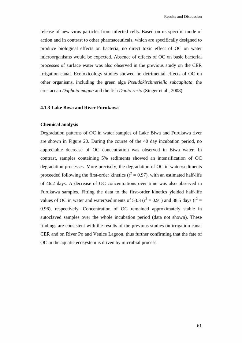
Results and Discussion
61
release of new virus particles from infected cells. Based on its specific mode of
action and in contrast to other pharmaceuticals, which are specifically designed to
produce biological effects on bacteria, no direct toxic effect of OC on water
microorganisms would be expected. Absence of effects of OC on basic bacterial
processes of surface water was also observed in the previous study on the CER
irrigation canal. Ecotoxicology studies showed no detrimental effects of OC on
other organisms, including the green alga Pseudokirchneriella subcapitata, the
crustacean Daphnia magna and the fish Danio rerio (Singer et al., 2008).
4.1.3 Lake Biwa and River Furukawa
Chemical analysis
Degradation patterns of OC in water samples of Lake Biwa and Furukawa river
are shown in Figure 20. During the course of the 40 day incubation period, no
appreciable decrease of OC concentration was observed in Biwa water. In
contrast, samples containing 5% sediments showed an intensification of OC
degradation processes. More precisely, the degradation of OC in water/sediments
proceeded following the first-order kinetics (r2 = 0.97), with an estimated half-life
of 46.2 days. A decrease of OC concentrations over time was also observed in
Furukawa samples. Fitting the data to the first-order kinetics yielded half-life
values of OC in water and water/sediments of 53.3 (r2 = 0.91) and 38.5 days (r2 =
0.96), respectively. Concentration of OC remained approximately stable in
autoclaved samples over the whole incubation period (data not shown). These
findings are consistent with the results of the previous studies on irrigation canal
CER and on River Po and Venice Lagoon, thus further confirming that the fate of
OC in the aquatic ecosystem is driven by microbial process.
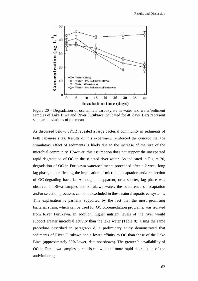
Results and Discussion
62
Figure 20 - Degradation of oseltamivir carboxylate in water and water/sediment samples of Lake Biwa and River Furukawa incubated for 40 days. Bars represent standard deviations of the means.
As discussed below, qPCR revealed a large bacterial community in sediments of
both Japanese sites. Results of this experiment reinforced the concept that the
stimulatory effect of sediments is likely due to the increase of the size of the
microbial community. However, this assumption does not support the unexpected
rapid degradation of OC in the selected river water. As indicated in Figure 20,
degradation of OC in Furukawa water/sediments proceeded after a 2-week long
lag phase, thus reflecting the implication of microbial adaptation and/or selection
of OC-degrading bacteria. Although no apparent, or a shorter, lag phase was
observed in Biwa samples and Furukawa water, the occurrence of adaptation
and/or selection processes cannot be excluded in these natural aquatic ecosystems.
This explanation is partially supported by the fact that the most promising
bacterial strain, which can be used for OC bioremediation programs, was isolated
from River Furukawa. In addition, higher nutrient levels of the river would
support greater microbial activity than the lake water (Table 8). Using the same
procedure described in paragraph d, a preliminary study demonstrated that
sediments of River Furukawa had a lower affinity to OC than those of the Lake
Biwa (approximately 30% lower; data not shown). The greater bioavailability of
OC in Furukawa samples is consistent with the more rapid degradation of the
antiviral drug.
Results and Discussion
63
OC mineralization
Results of the mineralization study are summarized in Figure 21. Mineralization
of OC in water of Lake Biwa, expressed as 14CO2 evolution, accounted for <1% of
the initial 14C added as 14C-OC. During the same 40-day incubation period, a
higher mineralization was observed in water samples from River Furukawa.
Similarly to what was observed in the OC degradation study, the addition of 5%
sediments led to an increase of 14CO2 evolution. More precisely, the cumulative
mineralization accounted for 12.8% and 21.0% in Biwa and Furukawa samples,
respectively (Figure 21). No appreciable 14CO2 evolution was observed in sterile
samples (data not shown). Differences of OC mineralization among samples of
the two Japanese aquatic environments are compatible with results of the OC
degradation study. Negligible mineralization of OC in surface water had been also
observed in River Po and Venice samples, and by other authors (Straub, 2009).
Slow degradation rates in surface water are commonly reported for a number of
xenobiotics, including pesticides (Accinelli et al., 2007).
Figure 21 - Cumulative 14CO2 evolution from water and water sediment samples of the Lake Biwa and River Furukawa. Samples were incubated for 40 days. Bars represent standard deviations of the means.
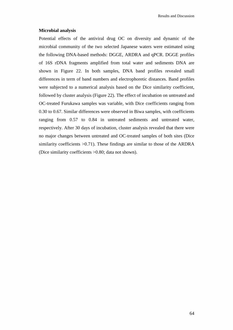
Results and Discussion
64
Microbial analysis
Potential effects of the antiviral drug OC on diversity and dynamic of the
microbial community of the two selected Japanese waters were estimated using
the following DNA-based methods: DGGE, ARDRA and qPCR. DGGE profiles
of 16S rDNA fragments amplified from total water and sediments DNA are
shown in Figure 22. In both samples, DNA band profiles revealed small
differences in term of band numbers and electrophoretic distances. Band profiles
were subjected to a numerical analysis based on the Dice similarity coefficient,
followed by cluster analysis (Figure 22). The effect of incubation on untreated and
OC-treated Furukawa samples was variable, with Dice coefficients ranging from
0.30 to 0.67. Similar differences were observed in Biwa samples, with coefficients
ranging from 0.57 to 0.84 in untreated sediments and untreated water,
respectively. After 30 days of incubation, cluster analysis revealed that there were
no major changes between untreated and OC-treated samples of both sites (Dice
similarity coefficients >0.71). These findings are similar to those of the ARDRA
(Dice similarity coefficients >0.80; data not shown).

Results and Discussion
65
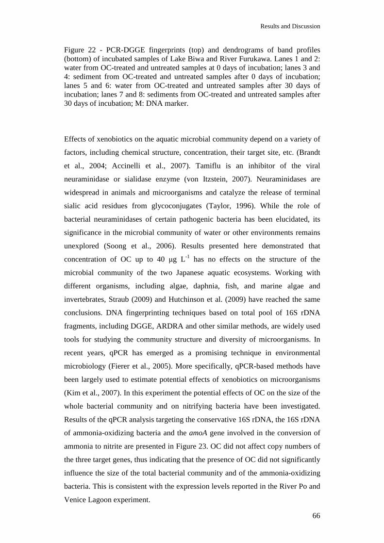
Results and Discussion
66
Figure 22 - PCR-DGGE fingerprints (top) and dendrograms of band profiles (bottom) of incubated samples of Lake Biwa and River Furukawa. Lanes 1 and 2: water from OC-treated and untreated samples at 0 days of incubation; lanes 3 and 4: sediment from OC-treated and untreated samples after 0 days of incubation; lanes 5 and 6: water from OC-treated and untreated samples after 30 days of incubation; lanes 7 and 8: sediments from OC-treated and untreated samples after 30 days of incubation; M: DNA marker.
Effects of xenobiotics on the aquatic microbial community depend on a variety of
factors, including chemical structure, concentration, their target site, etc. (Brandt
et al., 2004; Accinelli et al., 2007). Tamiflu is an inhibitor of the viral
neuraminidase or sialidase enzyme (von Itzstein, 2007). Neuraminidases are
widespread in animals and microorganisms and catalyze the release of terminal
sialic acid residues from glycoconjugates (Taylor, 1996). While the role of
bacterial neuraminidases of certain pathogenic bacteria has been elucidated, its
significance in the microbial community of water or other environments remains
unexplored (Soong et al., 2006). Results presented here demonstrated that
concentration of OC up to 40 µg L-1 has no effects on the structure of the
microbial community of the two Japanese aquatic ecosystems. Working with
different organisms, including algae, daphnia, fish, and marine algae and
invertebrates, Straub (2009) and Hutchinson et al. (2009) have reached the same
conclusions. DNA fingerprinting techniques based on total pool of 16S rDNA
fragments, including DGGE, ARDRA and other similar methods, are widely used
tools for studying the community structure and diversity of microorganisms. In
recent years, qPCR has emerged as a promising technique in environmental
microbiology (Fierer et al., 2005). More specifically, qPCR-based methods have
been largely used to estimate potential effects of xenobiotics on microorganisms
(Kim et al., 2007). In this experiment the potential effects of OC on the size of the
whole bacterial community and on nitrifying bacteria have been investigated.
Results of the qPCR analysis targeting the conservative 16S rDNA, the 16S rDNA
of ammonia-oxidizing bacteria and the amoA gene involved in the conversion of
ammonia to nitrite are presented in Figure 23. OC did not affect copy numbers of
the three target genes, thus indicating that the presence of OC did not significantly
influence the size of the total bacterial community and of the ammonia-oxidizing
bacteria. This is consistent with the expression levels reported in the River Po and
Venice Lagoon experiment.
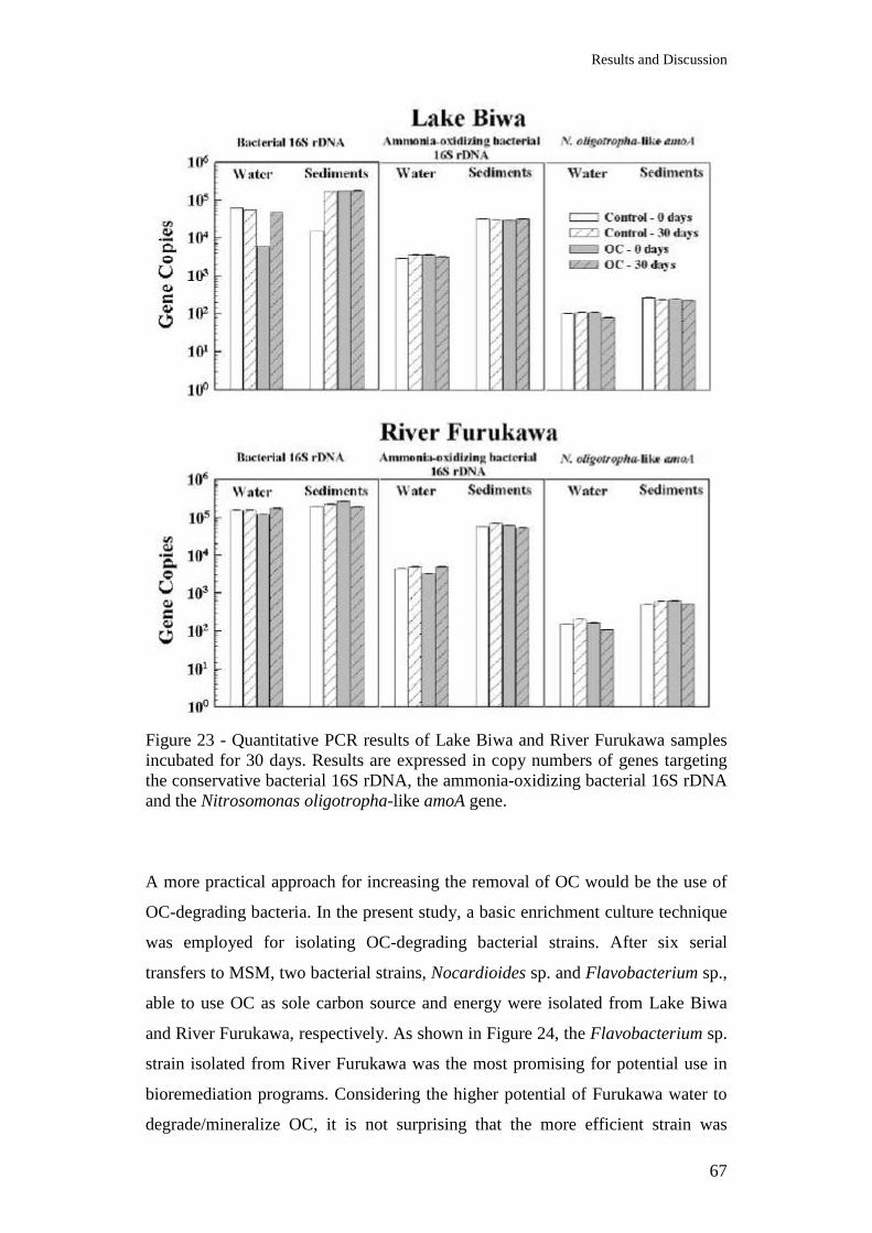
Results and Discussion
67
Figure 23 - Quantitative PCR results of Lake Biwa and River Furukawa samples incubated for 30 days. Results are expressed in copy numbers of genes targeting the conservative bacterial 16S rDNA, the ammonia-oxidizing bacterial 16S rDNA and the Nitrosomonas oligotropha-like amoA gene.
A more practical approach for increasing the removal of OC would be the use of
OC-degrading bacteria. In the present study, a basic enrichment culture technique
was employed for isolating OC-degrading bacterial strains. After six serial
transfers to MSM, two bacterial strains, Nocardioides sp. and Flavobacterium sp.,
able to use OC as sole carbon source and energy were isolated from Lake Biwa
and River Furukawa, respectively. As shown in Figure 24, the Flavobacterium sp.
strain isolated from River Furukawa was the most promising for potential use in
bioremediation programs. Considering the higher potential of Furukawa water to
degrade/mineralize OC, it is not surprising that the more efficient strain was
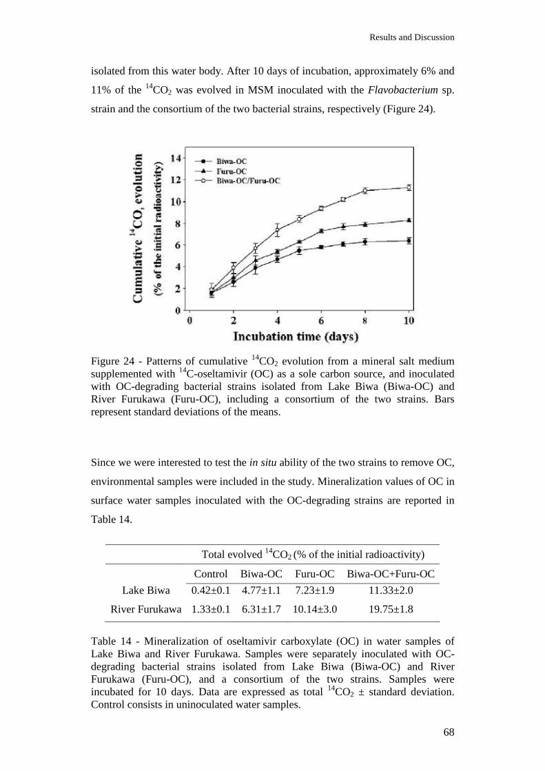
Results and Discussion
68
isolated from this water body. After 10 days of incubation, approximately 6% and
11% of the 14CO2 was evolved in MSM inoculated with the Flavobacterium sp.
strain and the consortium of the two bacterial strains, respectively (Figure 24).
Figure 24 - Patterns of cumulative 14CO2 evolution from a mineral salt medium supplemented with 14C-oseltamivir (OC) as a sole carbon source, and inoculated with OC-degrading bacterial strains isolated from Lake Biwa (Biwa-OC) and River Furukawa (Furu-OC), including a consortium of the two strains. Bars represent standard deviations of the means.
Since we were interested to test the in situ ability of the two strains to remove OC,
environmental samples were included in the study. Mineralization values of OC in
surface water samples inoculated with the OC-degrading strains are reported in
Table 14.
Total evolved 14CO2 (% of the initial radioactivity)
Control Biwa-OC Furu-OC Biwa-OC+Furu-OC
Lake Biwa 0.42±0.1 4.77±1.1 7.23±1.9 11.33±2.0
River Furukawa 1.33±0.1 6.31±1.7 10.14±3.0 19.75±1.8
Table 14 - Mineralization of oseltamivir carboxylate (OC) in water samples of Lake Biwa and River Furukawa. Samples were separately inoculated with OC-degrading bacterial strains isolated from Lake Biwa (Biwa-OC) and River Furukawa (Furu-OC), and a consortium of the two strains. Samples were incubated for 10 days. Data are expressed as total 14CO2 ± standard deviation. Control consists in uninoculated water samples.
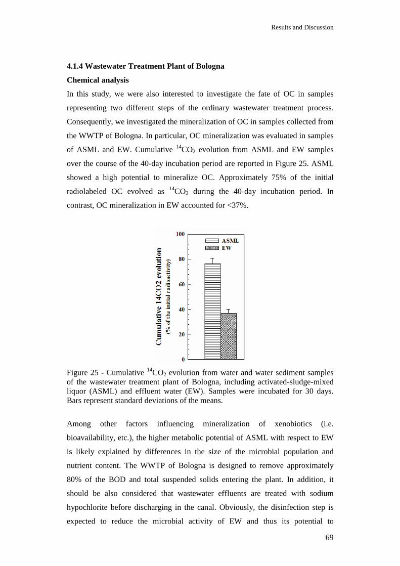
Results and Discussion
69
4.1.4 Wastewater Treatment Plant of Bologna
Chemical analysis
In this study, we were also interested to investigate the fate of OC in samples
representing two different steps of the ordinary wastewater treatment process.
Consequently, we investigated the mineralization of OC in samples collected from
the WWTP of Bologna. In particular, OC mineralization was evaluated in samples
of ASML and EW. Cumulative 14CO2 evolution from ASML and EW samples
over the course of the 40-day incubation period are reported in Figure 25. ASML
showed a high potential to mineralize OC. Approximately 75% of the initial
radiolabeled OC evolved as 14CO2 during the 40-day incubation period. In
contrast, OC mineralization in EW accounted for <37%.
Figure 25 - Cumulative 14CO2 evolution from water and water sediment samples of the wastewater treatment plant of Bologna, including activated-sludge-mixed liquor (ASML) and effluent water (EW). Samples were incubated for 30 days. Bars represent standard deviations of the means.
Among other factors influencing mineralization of xenobiotics (i.e.
bioavailability, etc.), the higher metabolic potential of ASML with respect to EW
is likely explained by differences in the size of the microbial population and
nutrient content. The WWTP of Bologna is designed to remove approximately
80% of the BOD and total suspended solids entering the plant. In addition, it
should be also considered that wastewater effluents are treated with sodium
hypochlorite before discharging in the canal. Obviously, the disinfection step is
expected to reduce the microbial activity of EW and thus its potential to
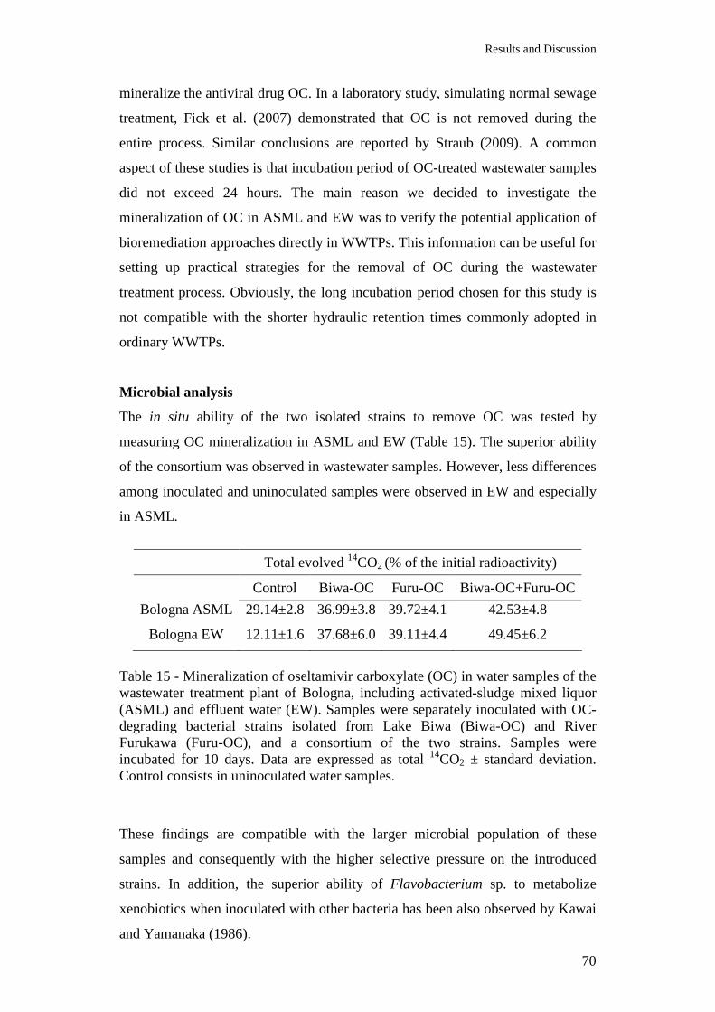
Results and Discussion
70
mineralize the antiviral drug OC. In a laboratory study, simulating normal sewage
treatment, Fick et al. (2007) demonstrated that OC is not removed during the
entire process. Similar conclusions are reported by Straub (2009). A common
aspect of these studies is that incubation period of OC-treated wastewater samples
did not exceed 24 hours. The main reason we decided to investigate the
mineralization of OC in ASML and EW was to verify the potential application of
bioremediation approaches directly in WWTPs. This information can be useful for
setting up practical strategies for the removal of OC during the wastewater
treatment process. Obviously, the long incubation period chosen for this study is
not compatible with the shorter hydraulic retention times commonly adopted in
ordinary WWTPs.
Microbial analysis
The in situ ability of the two isolated strains to remove OC was tested by
measuring OC mineralization in ASML and EW (Table 15). The superior ability
of the consortium was observed in wastewater samples. However, less differences
among inoculated and uninoculated samples were observed in EW and especially
in ASML.
Total evolved 14CO2 (% of the initial radioactivity)
Control Biwa-OC Furu-OC Biwa-OC+Furu-OC
Bologna ASML 29.14±2.8 36.99±3.8 39.72±4.1 42.53±4.8
Bologna EW 12.11±1.6 37.68±6.0 39.11±4.4 49.45±6.2
Table 15 - Mineralization of oseltamivir carboxylate (OC) in water samples of the wastewater treatment plant of Bologna, including activated-sludge mixed liquor (ASML) and effluent water (EW). Samples were separately inoculated with OC-degrading bacterial strains isolated from Lake Biwa (Biwa-OC) and River Furukawa (Furu-OC), and a consortium of the two strains. Samples were incubated for 10 days. Data are expressed as total 14CO2 ± standard deviation. Control consists in uninoculated water samples.
These findings are compatible with the larger microbial population of these
samples and consequently with the higher selective pressure on the introduced
strains. In addition, the superior ability of Flavobacterium sp. to metabolize
xenobiotics when inoculated with other bacteria has been also observed by Kawai
and Yamanaka (1986).
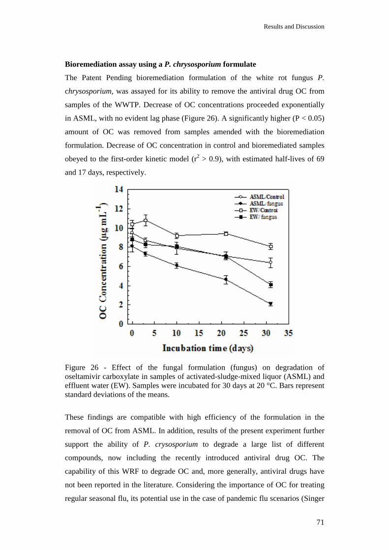
Results and Discussion
71
Bioremediation assay using a P. chrysosporium formulate
The Patent Pending bioremediation formulation of the white rot fungus P.
chrysosporium, was assayed for its ability to remove the antiviral drug OC from
samples of the WWTP. Decrease of OC concentrations proceeded exponentially
in ASML, with no evident lag phase (Figure 26). A significantly higher (P < 0.05)
amount of OC was removed from samples amended with the bioremediation
formulation. Decrease of OC concentration in control and bioremediated samples
obeyed to the first-order kinetic model (r2 > 0.9), with estimated half-lives of 69
and 17 days, respectively.
Figure 26 - Effect of the fungal formulation (fungus) on degradation of oseltamivir carboxylate in samples of activated-sludge-mixed liquor (ASML) and effluent water (EW). Samples were incubated for 30 days at 20 °C. Bars represent standard deviations of the means.
These findings are compatible with high efficiency of the formulation in the
removal of OC from ASML. In addition, results of the present experiment further
support the ability of P. crysosporium to degrade a large list of different
compounds, now including the recently introduced antiviral drug OC. The
capability of this WRF to degrade OC and, more generally, antiviral drugs have
not been reported in the literature. Considering the importance of OC for treating
regular seasonal flu, its potential use in the case of pandemic flu scenarios (Singer
Results and Discussion
72
et al., 2008), the proposed formulation would represent a practical alternative for
future bioremediation programs.
Degradation of OC in EW (Figure 26) did not fit the first-order kinetic model.
However, and as discussed below, even considering that OC was more persistent
in EW, removal of OC was significantly stimulated (P < 0.05) in bioremediated
samples.
After 30 days of incubation, the cumulative evolution of 14CO2 accounted for 40
and 69% of the initial radioactivity in control and bioremediated ASML samples,
respectively (Figure 27). Less 14CO2 evolved from EW samples (Figure 27).
These findings are consistent with the previous experiments, that demonstrated
that OC degradation and mineralization in water and wastewater is mainly driven
by microbial process. Before entering the receiving canal, water from the WWTP
is subjected to microfiltration and chlorine oxidation treatments. In addition to
microbial oxidation occurring in the biological tank, these two latter treatments,
are expected to further reduce the microbial activity of EW.
Figure 27 - Effect of the fungal formulation (fungus) on cumulative 14CO2 evolution of oseltamivir carboxylate from samples of activated-sludge-mixed liquor (ASML) and effluent water (EW). Samples were incubated for 30 days at 20 °C. Bars represent standard deviations of the means. Analysis of the bacterial 16S rDNA PCR products by DGGE (Figure 29)
indicated the structure of the microbial community of ASML and EW over the
incubation period. The introduced formulation did not cause major changes in the
Results and Discussion
73
bacterial community diversity. And as expected, high degree of similarity was
observed within bacterial populations in OC-treated and non-treated samples, thus
confirming the low impact of OC on indigenous bacteria populations.
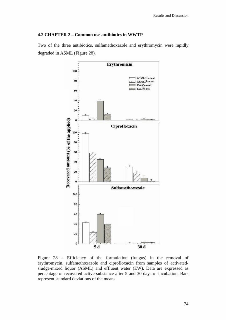
Results and Discussion
74
4.2 CHAPTER 2 – Common use antibiotics in WWTP Two of the three antibiotics, sulfamethoxazole and erythromycin were rapidly
degraded in ASML (Figure 28).
Figure 28 – Efficiency of the formulation (fungus) in the removal of erythromycin, sulfamethoxazole and ciprofloxacin from samples of activated-sludge-mixed liquor (ASML) and effluent water (EW). Data are expressed as percentage of recovered active substance after 5 and 30 days of incubation. Bars represent standard deviations of the means.
Results and Discussion
75
The effect of the bioremediation formulation was clearly distinguishable after 5
days of incubation in both ASML and EW samples. In the case of the antibiotic
ciprofloxacin, this positive effect was also distinguishable at the end of the
incubation period. In particular, after 30 days of incubation, 30 and 18% of
ciprofloxacin were removed from bioremediated and control ASML samples,
respectively. In contrast to the other pharmaceuticals, more ciprofloxacin was
removed in EW than in ASML, with a significant stimulatory effect of the
formulation. The results of this experiment demonstrated the efficacy of the
bioremediation approach.
PCR amplification of the conservative bacterial 16S rDNA produced fragments of
the expected size (data not shown). Separation of PCR products by DGGE
resulted in distinct band profiles (Figure 29). Analysis of DGGE profiles indicated
that the structure of the microbial community of ASML and EW changed over the
incubation period. Band profiles of samples receiving the same pharmaceutical
were generally similar, thus suggesting that major changes were caused by
pharmaceuticals rather than the introduced formulation. Similarity within the
bacterial community was less pronounced at the end of the 30-day incubation
period.

Results and Discussion
76
Figure 29 - PCR-DGGE patterns (below) and similarity analysis (above) of amplified 16S gene from total DNA of activated-sludge-mixed liquor (ASML) and effluent water (EW) samples at the initial stage (5 days) and at the end of the incubation period (30 days). Samples were singularly spiked with the four pharmaceuticals listed below and amended (*) or not amended with the fungal formulation. OC: oseltamivir carboxylate, ER: erythromycin, SU: sulfamethoxazole, CI: ciprofloxacin.
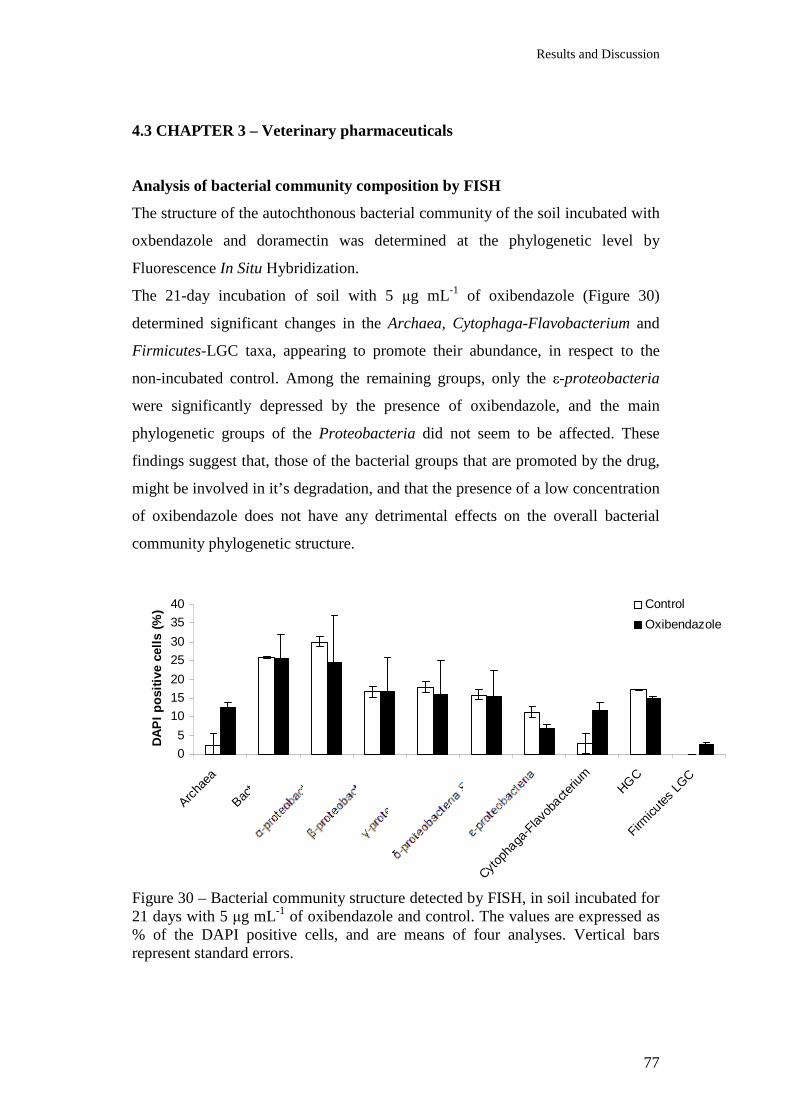
Results and Discussion
77
4.3 CHAPTER 3 – Veterinary pharmaceuticals
Analysis of bacterial community composition by FISH
The structure of the autochthonous bacterial community of the soil incubated with
oxbendazole and doramectin was determined at the phylogenetic level by
Fluorescence In Situ Hybridization.
The 21-day incubation of soil with 5 µg mL-1 of oxibendazole (Figure 30)
determined significant changes in the Archaea, Cytophaga-Flavobacterium and
Firmicutes-LGC taxa, appearing to promote their abundance, in respect to the
non-incubated control. Among the remaining groups, only the ε-proteobacteria
were significantly depressed by the presence of oxibendazole, and the main
phylogenetic groups of the Proteobacteria did not seem to be affected. These
findings suggest that, those of the bacterial groups that are promoted by the drug,
might be involved in it’s degradation, and that the presence of a low concentration
of oxibendazole does not have any detrimental effects on the overall bacterial
community phylogenetic structure.
0
5
10
15
20
25
30
35
40
Archa
ea
Bacte
ria
Cytop
haga
-Flav
obacte
rium
HGC
Firmicu
tes L
GC
DA
PI
po
siti
ve c
ells
(%
) Control
Oxibendazole
Figure 30 – Bacterial community structure detected by FISH, in soil incubated for 21 days with 5 µg mL-1 of oxibendazole and control. The values are expressed as % of the DAPI positive cells, and are means of four analyses. Vertical bars represent standard errors.

Results and Discussion
78
The presence of 20 µg mL-1 of doramectin affected the bacterial community
phylogenetic structure by depressing significantly (p < 0.01) the Bacteria and α-,
γ-, ε-, δ-proteobacteria groups (Figure 31). On the contrary the β-proteobacteria
cluster seems to be enhanced by the presence of the veterinary drug in soil.
05
101520253035404550
Archa
ea
Bacte
ria
Cytop
haga
-Flav
obacte
rium
HGC
Firmicu
tes L
GC
DA
PI
po
siti
ve c
ells
(%
) Control
Doramectin
Figure 31 – Bacterial community structure detected by FISH, in soil incubated for 23 days with 20 µg mL-1 of doramectin and control. The values are expressed as % of the DAPI positive cells, and are means of four analyses. Vertical bars represent standard errors.
Furthermore FISH analysis, using the narG probe, permitted the gene detection in
incubated and non-incubated soil samples. The presence of both, oxibendazole
and doramectin caused a significant (p < 0.05) increase in the narG positive % of
the DAPI stained cells (Table 16) in comparison to the non-incubated control.
This result suggests that the bacterial community might use the chemical drug as a
substrate for metabolism, possibly involving its degradation.
narG positive cells (%DAPI) Control 4.64 ± 2.58 Oxibendazole 11.70 ± 9.88 Control 1.04 ± 1.23 Doramectin 22.73 ± 17.23
Table 16 – Number of narG positive cells (% DAPI) in incubated soils and control, analyzed by FISH. Values are the means of four replicates ± standard deviations.
Results and Discussion
79
Analysis of narG gene by PCR and qRT-PCR
The presence and expression level of the narG gene in soil samples, incubated
with the veterinary drugs and controls, was further confirmed by classical PCR
amplification, from extracted DNA in all the samples, indicating that the gene was
ubiquitous. The change in expression of the narG gene in the sample was
evaluated by comparison to the control (no treatment) as a ratio, using the 16S
gene a reference. The presence of oxibendazole, did not affect the expression of
the narG gene, that remained constant in the incubated soils and in the control.
This is in line with FISH analysis of the bacterial community that revealed no
specific negative effects. On the contrary the presence of doramectin caused a
significant increase in narG/16S mRNA ratios, from 12 to 55 times. The increase
of the narG/16S mRNA ratio is possibly due to the global decrease of 16S
mRNA, while the portion of remaining bacteria contribute to the denitrification
process.
Isolation of bacterial strains
Eleven different bacterial isolates were obtained from the oxibendazole-treated
microcosms, and classified as Bacillus pumilus, Bacillus imples, Bacillus
nealsonii, Bacillus thuringiensis, Bacillus benzoevorans, Bacillus anthracis,
Acetobacter pasteurianus, Bacillus cereus, Arthrobacter crystallopoietes,
Rhodococcus rhodochrous, and Klebsiella planticola. Two different strains were
obtaines from doramectin-treated microcosms, classified as Bacillus subtilis and
Bacillus megaterium. Strains were tested for their degrading capabilities and qRT-
PCR was performed to monitor the expression of narG gene in presence/absence
of the two veterinary drugs (work in progress, data not shown).
Conclusions
80
5. CONCLUSIONS
5.1 CHAPTER 1
The first study on CER irrigation canal samples showed the important role played
by microbial processes in the degradation of the antiviral drug oseltamivir
carboxylate (OC) in surface water. Even though the low amount of 1.5 µg mL-1 of
OC was lost from surface water samples, the potential of OC degradation was
significantly greater in water samples containing sediments.
For better understanding the degradation of OC in other environmental scenarios
and to elucidate potential effects of this antiviral drug on the structure of the
microbial community of surface water, a second laboratory study was conducted
on two different aquatic environments located in northern Italy, the River Po and
the Venice Lagoon. This study demonstrated the potential of the antiviral to
persist in surface water, showing also that in both river and saline water, rapid OC
removal can be achieved by addition of a low amount (5%) of sediments to water,
which promotes microbial degradation processes. Since OC showed a low
sorption affinity to sediments, no reduction of bioavailability is expected after
stimulating microbial processes by addition of sediments to environmental
samples. Concentrations of OC up to 20 µg mL-1 did not affect the structure of the
microbial community and bacterial nitrification processes. Considering that
environmental concentrations of OC in highly populated catchments during an
influenza pandemic are predicted to be in excess of 20 µg mL-1, no detrimental
effects of OC on the microbial community are expected.
Further laboratory experiments conducted using samples from the Japanese
surface water and sediments of Lake Biwa and River Furukawa, and wastewater
from the treatment plant of the city of Bologna indicated that OC degradation is
variable and not easily predictable. Major factors influencing the degradation rate
of this antiviral drug include intrinsic environmental properties and most
importantly the size of the indigenous microbial community. Although slow
biotransformation rates were observed in surface water, degradation of OC
proceeded more rapidly in wastewater. This suggests that bioremediation
techniques would be more successful when applied to wastewater and effluent.
Using different DNA-based approaches, it may be concluded that OC would not
affect the structure of the microbial community and the size of the whole bacterial
Conclusions
81
community, including the group of ammonium-oxidizing bacteria. In the present
study the potential use of OC-degrading bacteria was explored for its removal
from water and wastewater. Two bacterial strains able to grow using OC as sole
carbon and energy sources were isolated. Inoculation of environmental samples
with these two bacterial strains demonstrated the feasibility of this technology,
especially in surface water and effluents of a wastewater treatment plant.
Furthermore the tested bioremediation strategy using a formulation with the
fungus P. chrysosporium showed that after 30 days of incubation, the formulation
removed more than half the initial amount of OC in both activated-sludge-mixed
liquor (ASML) and effluents (EW).
Taken together, these information suggest that prolonged residential time of OC in
the biological degradation step of wastewater treatment plants, and the
bioremediation approach can be useful strategies for reducing the risk of OC to
enter the aquatic ecosystem.
5.2 CHAPTER 2
The bioremediation formulation of P. chrysosporium was capable to remove
efficiently also three different antibiotics from wastewater, including samples of
ASML and EW. In the case of the less persistent pharmaceutical, the antibiotic
erythromycin, almost 80% of the applied amount was removed in bioremediated
ASML and EW samples within only 5 days of incubation.
5.3 CHAPTER 3
The presence of two veterinary pharmaceuticals, doramectin and oxibendazole, in
an agricultural soil affected the overall structure of the bacterial community,
analyzed by FISH promoting some phylogenetic groups rather than depressing
others. The two drugs were possibly used as a substrate for bacterial metabolism,
as suggested by the increase in the narG positive cells. Furthermore the isolation
of a number of bacterial strains capable to grow on oxibendazole and doramectin
was promising for the development of further bioremediation strategies.
References
82
REFERENCES
Accinelli C, Koskinen WC, Seebinger JD, Vicari A, Sadowsky MJ (2005) Environmental Fate of Two Sulfonamide Antimicrobial Agents in Soil. J Agric Food Chem, 53, 4110 Accinelli C, Hashim M, Epifani R, Schneider RJ, Vicari A (2006) Effects of the antimicrobial agent sulfamethazine on metolachlor persistence and sorption in soil. Chemosphere, 63, 1539 Accinelli C, Koskinen WC, Becker JM, Sadowsky MJ (2007) Environmental fate of two sulfonamide antimicrobial agents in soil. J Agric Food Chem, 55(7), 2677-82 Alexy R, Kümpel T, Kümmerer K (2004) Assessment of degradation of 18 antibiotics in the Closed Bottle Test. Chemosphere, 57, 505 American Hospital Formulary Service (2006) Oseltamivir. In: AHFS Drug Information, 8:1828: Available: http://www.ashp.org/ahfs/first_rel/Revised-75oseltamivir_nov2006.pdf Bardsley-Elliot A and Noble S (1999) Oseltamivir. Drugs 58(5):851–860. BBC. 2006. Bird Flu: Country Preparations. Available: http://news.bbc.co.uk/1/hi/health/4380014stm Barra Caracciolo A, Grenni P, Cupo C, Rossetti S (2005) In situ analysis of native microbial communities in complex samples with high particulate loads. FEMS Microbiol Lett, 253(1), 55-8 Barra Caracciolo A, Grenni P, Saccà ML, Amalfitano S, Ciccoli R, Martín M, Gibello A (2010) The role of a groundwater bacterial community in the degradation of the herbicide terbuthylazine. FEMS Microbiol Ecol, 71, 127-136 Bock E, Koops HP, Ahlers B, Harms H (1992) The genus Nitrobacter and related genera. In: Balows A, Trüper HG, Dworkin M, Harder W, Schleifer KH (Eds.), The Prokaryotes. Springer-Verlag, New York, pp. 414–430 Boon N, Top EM, Verstraete W, Siciliano SD (2003) Bioaugmentation as a tool to protect the structure and function of an activated-sludge microbial community against a 3-chloroaniline shock load. Appl Environ Microbiol, 69, 1511–1520 Bottoni P, Caroli S, Barra Caracciolo A (2010) Pharmaceuticals as priority water contaminants. Toxicological & Environmental Chemistry, 92 (3) 549-565 Barceló D, Petrovic M (2008) Emerging Contaminants from Industrial and Municipal Waste, Removal Technologies. The Handbook of Environmental Chemistry (Eds: O. Hutzinger D. Barceló ·A. Kostianoy) Volume 5 Water Pollution, Part S/2 270-274
References
83
Brandt KK, Jørgensen NOG, Nielsen TH, Winding A (2004) Microbial community-level toxicity testing of linear alkylbenzene sulfonates in aquatic microcosms. FEMS Microbiology Ecology, 49 (2) 229-241 Bryers JD (1994) Biofilms and the technological implications of microbial cell adhesion. Colloid Surf. B: Biointerf, 2, 9–23 Busse MD, Ratcliff AW, Shestak CJ, Powers RF (2001) Glyphosate toxicity and the effects of long-term vegetation control on soil microbial communities. Soil Biol Biochem, 33, 1777 Carlsson C, Johansson AK, Alvan G, Bergman K, Kuhler T (2006) Are pharmaceuticals potent environmental pollutants? Part II: Environmental risk assessments of selected pharmaceutical excipients. Science of the Total Environment 364, 88–95 Casas I, Powell L, Klapper PE, Cleator GM (1995) New method for the extraction of viral RNA and DNA from cerebrospinal fluid for use in the polymerase chain reaction assay. J Virol Methods, 53, 25-36 Castiglioni S, Pomati F, Miller K, Burns BP, Zuccato E, Calamari D, Neilan BA (2008) Novel homologs of the multiple resistance regulator marA in antibiotic-contaminated environments. Water Research, 42 (16) 4271-4280 Cleuvers M (2003) Aquatic ecotoxicity of pharmaceuticals including the assessment of combination effects. Toxicol Lett, 142, 185–194 Cleuvers T, Schneider RJ, Farber HA, Skutlarek D, Meyer MT, Goldbach (2003) Determination of Antibiotic Residues in Manure, Soil, and Surface Waters. Acta Hydrochim Hydrobiol, 31, 36-44 Crawford RL (2006) Bioremediation In: Dworkin M., Falkow S., Rosenberg E., Schleifer K.H. and Stackebrandt E. (Eds.), The Prokaryotes: A Handbook on the Biology of Bacteria. Volume 1: Symbiotic associations, Biotechnology and Applied Microbiology. New York, USA: Springer-Verlag, pp. 850-863 Danaher M, Howells LC, Crooks SRH, Cerkvenik-Flajs V, O’Keeffe M (2006) Review of methodology for the determination of macrocyclic lactone residues in biological matrices. Journal of Chromatography B, 844 (2) 175-203 De Souza ML, Newcombe D, Alvey S, Crowley DE, Hay A, Sadowsky MJ & Wackett LP (1993) Molecular basis of a bacterial consortium: interspecies catabolism of atrazine. Appl Environ Microbiol, 64, 178–184 Devers M, Soulas G, Martin-Laurent F (2004) Real-time reverse transcription PCR analysis of expression of atrazine catabolism genes in two bacterial strains isolated from soil. Journal of Microbiological Methods, 56, 3–15 Dionisi HM, Layton AC, Harms G, Gregory IR, Robinson KG, Sayler GS (2002) Quantification of Nitrosomonas oligotropha-like ammonia-oxidizing bacteria and
References
84
Nitrospira spp. from full-scale wastewater treatment plants by competitive PCR. Appl Environ Microb, 68, 245–253 Dionisi HM, Harms G, Layton AC, Gregory IR, Parker J, Hawkins SA, Robinson KG, Sayler GS (2003) Power analysis for real-time PCR quantification of genes in activated sludge and analysis of the variability introduced by DNA extraction. Appl Environ Microbiol, 69 (11) 6597-604 Dorigo U, Volatier L, Humbert JF (2005) Molecular approaches to the assessment of biodiversity in aquatic microbial communities. Water Res, 39, 2207-2218 Edwards M, Topp E, Metcalfe CD, Li H, Gottschall N, Bolton P, Curnoe W, Payne M, Beck A, Kleywegt S, Lapen DR (2009) Pharmaceutical and personal care products in tile drainage following surface spreading and injection of dewatered municipal biosolids to an agricultural field. Science of The Total Environment, 407 (14) 4220-4230 Eisenberg EJ and Cundy KC (1998) High-performance liquid chromatographic determination of GS4071, a potent inhibitor of influenza neuraminidase, in plasma by precolumn fluorescence derivatization with naphthalenedialdehyde. J Chromatogr B Biomed Sci Appl, 716(1-2) 267-73 Engelen B, Meinken K, von Wintzingerode F, Heuer H, Malkomes HP, Backhaus H (1998) Monitoring impact of a pesticide treatment on bacterial soil communities by metabolic and genetic fingerprinting in addition to conventional testing procedures. Appl Environ Microbiol, 64, 2814 European Commission (2001) Organic contaminants in sewage sludge for agricultural use. Report Coordinated by the European Commission Joint Research Centre, Institute for Environment and Sustainability, Soil and Waste Unit, October, 2001, pp 73 Farrell NR (2009) The Landscape of Antibiotic Resistance. Environ Health Perspect, 117, A244-A250 Ferris MJ, Muyzer G, Ward DM (1996) Denaturing gradient gel electrophoresis profiles of 16S rRNA-defined populations inhabiting a hot spring microbial mat community. Appl Environ Microb 62, 340–346 Fick J, Lindberg RH, Tysklind M, Haemig PD, Waldenström J, Wallensten A, Olsen B (2007) Antiviral oseltamivir is not removed or degraded in normal sewage water treatment: implications for development of resistance by influenza A virus. PLoS ONE 3 2 (10), e986 Fierer N, Jackson JA, Vilgalys R, Jackson RB (2005) Assessment of soil microbial community structure by use of taxon-specific quantitative PCR assays. Appl Environ Microbiol 71, 4117–4120 Getenga ZM and Kengara FO (2004) Mineralization of Glyphosate in Compost-Amended Soil Under Controlled Conditions. Bull Environ Contam Toxicol, 72, 266
References
85
Ghosh GC, Nakada N, Yamashita N, Tanaka H (2010) Oseltamivir Carboxylate, the Active Metabolite of Oseltamivir Phosphate (Tamiflu), Detected in Sewage Discharge and River Water in Japan. Environ Health Persp, 118, 103–107 Girvan MS, Campbell CD, Killham K, Prosser JI, Glover LA (2005) Bacterial diversity promotes community stability and functional resilience after perturbation. Environ Microbiol, 7 (3) 301-13 Gregory LG, Bond PL, Richardson DJ, Spiro S (2003) Characterization of a nitrate-respiring bacterial community using the nitrate reductase gene (narG) as a functional marker. Microbiology, 149, 229–237 Grenni P, Gibello A, Barra Caracciolo A, Fajardo C, Nande M, Vargas R, Saccà ML, Martínez-Iñigo MJ, Ciccoli R, Martín M (2009) A new fluorescent oligonucleotide probe for in situ detection of s-triazine-degrading Rhodococcus wratislaviensis in contaminated groundwater and soil samples. Water Res, 43, 2999-3008 Griebler C and Slezak D (2001) Microbial activity in aquatic environments measured by dimethyl sulfoxide reduction and intercomparison with commonly used methods. Appl Environ Microb, 67, 100–109 Griffiths BS, Kuan HL, Ritz K, Glover LA, McCaig AE, Fenwick C (2004) The relationship between microbial community structure and functional stability, tested experimentally in an upland pasture soil. Microb Ecol, 47 (1) 104-13 Gu JD and Berry DF (1992) Metabolism of 3-methylindole by a methanogenic consortium. Appl Environ Microbiol, 58, 2667–2669 Gu JD and Mitchell R (2006) Biodeterioration. In: Dworkin M, Falkow S, Rosenberg E, Schleifer KH and Stackebrandt E (Eds.), The Prokaryotes: A Handbook on the Biology of Bacteria. Volume 1: Symbiotic associations, Biotechnology and Applied Microbiology. New York, USA: Springer-Verlag, pp. 864-903 Guo H, Chen G, Lv Z, Zhao H, Yang H (2009). Alteration of microbial properties and community structure in soils exposed to napropamide. J Environ Sci, 21 (4) 494-502 Haglund AL, Lantz P, Törnblom E, Tranvik L (2003) Depth distribution of active bacteria and bacterial activity in lake sediment. FEMS Microbiol Ecol, 46 (1) 31-8 Halling-Sørensen B, Nors Nielsen S, Lanzky PF, Ingerslev F, Holten Lützhøft HC, Jørgensen SE (1998) Occurrence, fate and effects of pharmaceutical substances in the environment- A review. Chemosphere, 36 (2) 357-393 Harms G, Layton AC, Dionisi HM, Gregory IR, Garrett VM, Hawkins SA, Robinson KG, Sayler GS (2003) Real-time PCR quantification of nitrifying bacteria in a municipal wastewater treatment plant. Environ Sci Technol, 37 (2) 343-51
References
86
Hermansson A and Lindgren PE (2001) Quantification of ammonia-oxidizing bacteria in arable soil by real-time PCR. Appl Environ Microbiol, 67 (2) 972-6 Hooper SW, Daye KJ, Shuttle KA, Williams RA (2002) Subsurface Contaminant Fate Determination Through Integrated Studies of Intrinsic Remediation. In: Scow KM, Fogg GE, Hinton DE, Johnson ML (Eds.), Integrated Assessment of Ecosystem Health. CRC Press. Chap. 4, pp. 63-70 Hurt AC, Ernest J, Deng YM, Iannello P, Besselaar TG, Birch C, Buchy P, Chittaganpitch M, Chiu SC, Dwyer D, Guigon A, Harrower B, Kei IP, Kok T, Lin C, McPhie K, Mohd A, Olveda R, Panayotou T, Rawlinson W, et al. (2009) Emergence and spread of oseltamivir-resistant A(H1N1) influenza viruses in Oceania, South East Asia and South Africa. Antiviral Research, 83 (1) 90-93 Hutchinson TH, Beesley A, Frickers PE, Readman JW, Shaw JP, Straub JO (2009) Extending the environmental risk assessment for oseltamivir (Tamiflu®) under pandemic use conditions to the coastal marine compartment. Environ Int, 35, 931–936 Ibekwe AM, Papiernik SK, Gan J, Yates SR, Yang CH, Crowley DE (2001) Impact of Fumigants on Soil Microbial Communities. Appl Environ Microbiol, 67, 3245–3257 Jordan FL, Cantera JJL, Fenn ME, Stein LY (2005) Autotrophic ammonia-oxidizing bacteria contribute minimally to nitrification in a nitrogen-impacted forested ecosystem. Appl Environ Microbiol, 71, 197–206 Juliastuti SR, Baeyens J, Creemers C, Bixio D, Lodewyckx E (2003) The inhibitory effects of heavy metals and organic compounds on the net maximum specific growth rate of the autotrophic biomass in activated sludge. Journal of Hazardous Materials, 100, 1-3, 271-283 Kawai F and Yamanaka H (1986) Biodegradation of polyethylene glycol by symbiotic mixed culture (obligate mutualism). Arch Microbiol 146, 125–129 Kim CU, Lew W, Williams MA, Liu H, Zhang L, Swaminathan S, et al. (1997) Influenza neuraminidase inhibitors possessing a novel hydrophobic interaction in the enzyme active site: design, synthesis, and structural analysis of carbocyclic sialic acid analogues with potent anti-influenza activity. J Am Chem Soc, 119 (4) 681–690 Kim YH, Pak K, Pothuluri V, Cerniglia CE (2004) Mineralization of erythromycin A in aquaculture sediments. FEMS Microbiol Lett, 234, 169 Kim YS, Min J, Hong HN, Park JH, Park KS, Gu MB (2007) Gene expression analysis and classification of mode of toxicity of polycyclic aromatic hydrocarbons (PAHs) in Escherichia coli. Chemosphere, 66 (7) 1243-8
References
87
Kinney CA, Furlong ET, Werner SL, Cahill JD (2006) Presence and distribution of wastewater-derived pharmaceuticals in soil irrigated with reclaimed water. Environmental Toxicology and Chemistry, 25, 317-326 Klaver AL and Matthews RA (1994) Effects of oxytetracycline on nitrification in a model aquatic system. Aquaculture, 123, 237-247 Kolar L, Erzen NK, Hogerwerf L, van Gestel CAM (2008) Toxicity of abamectin and doramectin to soil invertebrates. Environmental Pollution 151, 182-189 Kolpin DW, Furlong ET, Meyer MT, Thurman EM, Zaugg SD, Barber LB, Buxton HT (2002) Pharmaceuticals, hormones, and other wastewater contaminants in U.S. streams, 1999-2000: a national reconnaissance. Environ Sci Technol, 36 (6) 1202-11 Kolpin DW, Thurman EM, Lee EA, Meyer MM, Furlong ET, Glassmeyer ST (2006) Urban contributions of glyphosate and its degradate AMPA to streams in the United States. Sci Total Environ, 354, 191 Koschorreck J, Koch C, Rönnefahrtet I (2002) Environmental risk assessment of veterinary medicinal products in the EU—a regulatory perspective. Toxicology Letters, 131, 117–124 Krutz LJ, Senseman SA, McInnes KJ, Hoffman DW, Tierney DP (2004) Adsorption and desorption of metolachlor and metolachlor metabolites in vegetated filter strip and cultivated soil. J Environ Qual, 33, 939–945 Kubota M, Kawahara K, Sekiya K, Uchida T, Hattori Y, Futamata H, Hiraishi A (2005) Nocardioides aromaticivorans sp. nov., a dibenzofuran-degrading bacterium isolated from dioxin-polluted environments. Systematic and Applied Microbiology, 28, 2, 165-174 Kumar K, Thomposon A, Singh AK, Chander Y, Gupta SC (2004) Enzyme-Linked Immunosorbent Assay for Ultratrace Determination of Antibiotics in Aqueous Samples. J Environ Qual, 33, 250-256 Kümmerer K, Al-Ahmad A, Mersch-Sundermann V (2000) Biodegradability of some antibiotics, elimination of the genotoxicity and affection of wastewater bacteria in a simple test. Chemosphere, 40, 701 Kümmerer K (2009a) The presence of pharmaceuticals in the environment due to human use – present knowledge and future challenges. Journal of Environmental Management, 90 (8) 2354-2366 Kümmerer K (2009b) Antibiotics in the aquatic environment – A review – Part II. Chemosphere, 75, 435–441 Kupka-Hansen P, Lunestad BT, Samuelsen OB (1992) Ecological effects of antibiotics/ chemotherapeutics from fish farming. In Chemotherapy in Aquaculture: from theory to reality; C. Michel, Alderman, D. J., Eds.; Office International des Epizooties: Paris; pp 174-178
References
88
Lee LS, Carmosini N, Sassman SA, Dion HM, Sepúlveda MS (2007) Agricultural Contributions of Antimicrobials and Hormones on Soil and Water Quality. Advances in Agronomy, 93, 1-68 Li D, Yang M, Hu J, Zhang J, Liu R, Gu X, Zhang Y, Wang Z (2009) Antibiotic-resistance profile in environmental bacteria isolated from penicillin production wastewater treatment plant and the receiving river. Environ Microbiol 11, 6, 1506-1517 Li W, Escarpe PA, Eisenberg EJ, Cundy KC, Sweet C, Jakeman KJ, et al. (1998) Identification of GS 4104 as an orally bioavailable prodrug of the influenza virus neuraminidase inhibitor GS 4071. Antimicrob Agents Chemother, 42 (3) 647–653 Lifschitz A, Virkel G, Sallovitz J, Sutra JF, Galtier P, Alvinerie M, Lanusse C (2000) Comparative distribution of ivermectin and doramectin to parasite location tissues in cattle. Veterinary Parasitology, 87, 4, 327-338 Limpiyakorn T, Kurisu F, Yagi O (2004) Distribution of ammonia-oxidizing bacteria in sewage activated sludge: analysis based on 16S rDNA sequence. Water Sci Technol, 54, 9–14 Limpiyakorn T, Shinohara Y, Kurisu F, Yagi O (2005) Communities of ammonia-oxidizing bacteria in activated sludge of various sewage treatment plants in Tokyo. FEMS Microbiology Ecology, 54, 2, 205-217 Limpiyakorn T, Kurisu F, Yagi O (2006) Quantification of ammonia-oxidizing bacteria populations in full-scale sewage activated sludge systems and assessment of system variables affecting their performance. Water Sci Technol, 54, 91–99 Liu B, McConnell LL, Torrents A (2001) Hydrolysis of chlorpyrifos in natural waters of the Chesapeake Bay. Chemosphere, 44, 1315 Loy A, Maixner F, Wagner M, Horn M (2007) probeBase--an online resource for rRNA-targeted oligonucleotide probes: new features 2007. Nucleic Acids Res 35(Database issue):D800-4 López-Gutiérrez JC, Henry S, Hallet S, Martin-Laurent F, Catroux G, Philippot L (2004) Quantification of a novel group of nitrate-reducing bacteria in the environment by real-time PCR. Journal of Microbiological Methods, 57, 3, 399-407 Lu J, Wu L, Newman J, Faber B, Gan J (2006) Degradation of pesticides in nursery recycling pond waters. J Agric Food Chem, 54, 2658 Lu Y, Yan L, Wang Y, Zhou S, Fu J, Zhang J (2009) Biodegradation of phenolic compounds from coking wastewater by immobilized white rot fungus Phanerochaete chrysosporium. J Hazard Mater, 165 (1-3) 1091-7
References
89
Mandelbaum RT, Wackett LR, Allan DL (1993) Mineralization of the s-triazine ring of atrazine by stable bacterial mixed cultures. Appl Environ Microbiol, 59, 1659–1701 Mandelbaum RT, Allan DL, Wackett LP (1995) Isolation and Characterization of a Pseudomonas sp. That Mineralizes the s-Triazine Herbicide Atrazine. Appl Environ Microbiol, 61 (4) 1451-1457 Mantovi P, Baldoni G, Toderi G (2005) Reuse of liquid, dewatered, and composted sewage sludge on agricultural land: effects of long-term application on soil and crop. Water Res, 39, 289–96 Marchesi JR, Sato T, Weightman AJ, Martin TA, Fry JC, Hiom SJ, Wade WG (1998) Design and evaluation of useful bacterium-specific PCR primers that amplify genes coding for bacterial 16S rRNA. Appl Environ Microb, 64, 795–799 Martin M, Gibello A, Lobo MC, Nande M, Garbi C, Fajardo C, Barra Caracciolo A, Grenni P, Martínez-Iñigo MJ (2008) Application of fluorescence in situ hybridization technique to detect simazine degrading bacteria in soil samples. Chemosphere, 71, 703–710 Martinez B, Tomkins J, Wackett LP, Wing R, Sadowsky MJ (2001) Complete nucleotide sequence and organization of the atrazine catabolic plasmid pADP-1 from Pseudomonas sp. strain ADP. J Bacteriol, 183 (19) 5684-97 Miyauchi K, Sukda P, Nishida T, Ito E, Matsumoto Y, Masai E, Fukuda M (2008) Isolation of dibenzofuran-degrading bacterium, Nocardioides sp. DF412, and characterization of its dibenzofuran degradation genes. J Biosci Bioeng, 105 (6) 628-35 Molina V, Ulloa O, Farías L, Urrutia H, Ramírez S, Junier P, Witzel KP (2007) Ammonia-oxidizing b-proteobacteria from the oxygen minimum zone off northern Chile. Appl Environ Microb, 73, 3547–3555 Moràn AC, Muller A, Manzano M, Gonzalez B (2006) Simazine treatment history determines a significant herbicide degradation potential in soils that is not improved by bioaugmentation with Pseudomonas sp. ADP. J Appl Microbiol 101 (1) 26-35 Moyer CL, Dobbs FC, Karl DM (1994) Estimation of diversity and community structure through restriction fragment length polymorphism distribution analysis of bacterial 16S rRNA gene from a microbial mat at an active, hydrothermal vent system, Loihi Seamount, Hawaii. Appl Environ Microb, 60, 871–879 National Research Council (1993) In Situ Bioremediation. National Academy Press, Washington DC Nazaret S, Jeffrey WH, Saouter E, von Haven R, Barkay T (1994) MerA gene expression in aquatic environments measured by mRNA production and Hg(II) volatilization. Appl Environ Microb, 60, 4059–4065
References
90
O'Brien E and Dietrich DR (2004) Hindsight rather than foresight: reality versus the EU draft guideline on pharmaceuticals in the environment. Trends in Biotechnology, 22 (7) 326-330 Ogunseitan OA (2000) Microbial proteins as biomarkers of ecosystem health. In K. Scow, G.E. Fogg, D. Hinton, and M.L. Johnson (eds.). Integrated Assessment of Ecosystem Health, pp. 207–22. Boca Raton, FL: CRC Press Osswald P, Courtes R, Bauda P, Block JC, Bryers JD, Sunde E (1995) Xenobiotic biodegradation test using attached bacteria in synthetic seawater. Ecotoxicol Environ Safety, 31, 211–217 Pagga U, Bachner J, Strotmann U (2006) Inhibition of nitrification in laboratory tests and model wastewater treatment plants. Chemosphere, 65, 1, 1-8 Pritchard PH, Cripe CR, Walker WW, Spain JC, Bourquin AW (1987) Biotic and abiotic degradation rates of methyl parathion in freshwater and estuarine water and sediment samples. Chemosphere, 16, 1509 Radjenovic J, Petrovic M, Barceló D (2009) Fate and distribution of pharmaceuticals in wastewater and sewage sludge of the conventional activated sludge (CAS) and advanced membrane bioreactor (MBR) treatment. Water Research, 43, 831–841 Rossello-Mora R and Amann RI (2001) The species concept for prokaryotes. FEMS Microbiol Ecol, 25, 39–67 Seybold CA and Mersie W (1996) Adsorption and desorption of atrazine, deethylatrazine, deisopropylatrazine, hydroxyatrazine, and metolachlor in two soils from Virginia. J Environ Qual, 25, 1179–1185 Schwarzenbach RP, Escher BI, Fenner K, Hofstetter TB, Johnson CA, von Gunten U, Wehrli B (2006) The Challenge of Micropollutants in Aquatic Systems. Science, 313, 1072-1077 Schiermeier Q (2003) Studies assess risks of drugs in water cycle. Nature 424, 5 Sharp RR, Bryers JB, Jones WG, Shields MS (1998) Activity and stability of a recombinant plasmid-borne TCE degradative pathway in suspended cultures. Biotechnol Bioeng, 57, 287–296 Shoop WL, Mrozik H, Fisher MH (1995) Structure and activity of avermectins and milbemycins in animal health. Veterinary Parasitology, 59, 2, 139-156 Singer AC, Nunn MA, Gould EA, Johnson AC (2007) Potential risks associated with the widespread use of Tamiflu. Environ Health Persp, 115, 102–106 Singer AC, Howard BM, Johnson AC, Knowles CJ, Jackman S, Accinelli C, Barra Caracciolo A, Bernard I, Bird S, Boucard T, Boxall A, Brian J, Cartmell E, Chubb C, Churchley J, Costigan S, Crane M, Dempsey MJ, Dorrington B, Fick J, Holmes J, Hutchinson T, Karcher F, Kelleher SL, Marsden P, Noone G, Nunn
References
91
MA, Oxford J, Rachwal T, Roberts N, Roberts M, Saccà ML, Sanders M, Straub JO, Terry A, Thomas D, Toovey S, Townsend R, Voulvoulis N, Watts C, Wells U (2008) Assessment of Tamiflu release to the environment under pandemic conditions. Report from an interdisciplinary workshop. Environ Health Persp, 116, 1563–1567 Singh BK, Kuhad RC, Singh A, Lal R, Triapthi KK (1999) Biochemical and molecular basis of pesticide degradation by microorganisms. Crit Rev Biotechnol, 19, 197–225 Singh BK and Walker A (2006) Microbial degradation of organophosphorus compounds. FEMS Microbiol Rev, 30, 428–471 Smith P, Donlon J, Coyne R, Cazabon D J (1994) Fate of oxytetracycline in a fresh water fish farm: influence of effluent treatment systems. Aquaculture, 120, 319-325 Söderstrom H, Järhult JD, Olsen B, Lindberg RH, Tanaka H, Fick J (2009) Detection of the antiviral drug oseltamivir in aquatic environments. PLoS ONE 4, e6064 Soong G, Muir A, Gomez MI, Waks J, Reddy B, Planet P, Singh PK, Kaneko Y, Wolfgang MC, Hsiao YS, Tong L, Prince A (2006) Bacterial neuraminidase facilitates mucosal infection by participating in biofilm production. J Clin Invest, 116, 2297–2305 Stalter D, Magdeburga A, Weilb M, Knackerb T, Oehlmann J (2009) Toxication or detoxication? In vivo toxicity assessment of ozonation as advanced wastewater treatment with the rainbow trout. Water Research, 44, 2, 439-448 Strange-Hansen R, Holm PE, Jacobsen OS, Jacobsen CS (2004) Sorption, mineralization and mobility of N-(phosphonomethyl)glycine (glyphosate) in five different types of gravel. Pest Manag Sci, 60, 570 Straub JO (2009) An environmental risk assessment for oseltamivir (Tamiflu®) for sewage works and surface waters under seasonal-influenza- and pandemic-use conditions. Ecotox Environ Safe, 72, 1625–1634 Sukul P, Lamshöft M, Zühlke S, Spiteller M (2008) Sorption and desorption of sulfadiazine in soil and soil–manure systems. Chemosphere, 73, 1344–1350 Taylor G (1996) Sialidases: structures, biological significance and therapeutic potential. Curr Opin Struct Biol, 6, 830–837 Ternes TA (1998) Occurrence of drugs in German sewage treatment plants and rivers. Water Research, 32, 11, 3245-3260 Topp E, Mulbry WM, Zhu H, Nour SM and Cuppels D (2000) Characterization of S-Triazine Herbicide Metabolism by a Nocardioides sp. Isolated from Agricultural Soils. Appl Environ Microbiol, 66, 3134-3141
References
92
Topp E, Monteiro SC, Beck A, Coelho BB, Boxall ABA, Duenk PW, Kleywegt S, Lapen DR, Payne M, Sabourin L, Li H, Metcalfe CD (2008). Runoff of pharmaceuticals and personal care products following application of biosolids to an agricultural field. Science of The Total Environment, 396, 1, 52-59 Torsvik V, Øvreas L, Thingstad TF (2002) Prokaryotic diversity—magnitude, dynamics and controlling factors. Science, 296, 1064–1066 Tsui MT and Chu LM (2003) Aquatic toxicity of glyphosate-based formulations: comparison between different organisms and the effects of environmental factors. Chemosphere, 52, 1189 USGS (United States Geological Survey) (2009) Toxic Substances Hydrology Program website, Emerging Contaminants in the Environment, http://toxics.usgs.gov/regional/emc/index.html Van Benthum WAJ, van Loosdrecht MCM, Heijnen JJ (1997) Process design for nitrogen removal using nitrifying biofilm and denitrifying suspended growth in a Biofilm Airlift Suspension reactor. Water Science and Technology, 36, 1, 119-128 Van Dyk TK, Smulski DR, Reed TR, Belkin S, Vollmer AC, Larossa RA (1995) Responses to toxicants of an escherichia coli strain carrying a uspA9::lux genetic fusion and an E. coli strain carrying a grpE9::lux fusion are similar. Appl Environ Microbiol, 61, 11, 4124–4127 Wagner-Döbler I, Pipke R, Timmis KN, Dwyer DF (1992) Evaluation of aquatic sediment microcosms and their use in assessing possible effects of introduced microorganisms on ecosystem parameters. Appl Environ Microbiol, 58, 1249–1258 Walker WW, Cripe CR, Pritchard PH, Bourquin AW (1984) Dibutylphthalate degradation in estuarine and freshwater sites. Chemosphere, 13, 1283 Watanabe K (2001) Microorganisms relevant to bioremediation. Current Opinion in Biotechnology, 12 (3), 237-241 WHO (World Health Organization) (2006) Cumulative Number of Confirmed Human Cases of Avian Influenza A/(H5N1) Reported to WHO. Available: http://www.who.int/csr/disease/avian_influenza/country/cases_table_2006_11_13/en/index.html Willems A and Collins MD (1996) Phylogenetic relationships of the genera Acetobacterium and Eubacterium sensu stricto and reclassification of Eubacterium alactolyticum as Pseudoramibacter alactolyticus gen. nov., comb. nov. Int J Syst Bacteriol, 46, 1083-1087 Yoon JH, Lee JK, Jung SY, Kim JA, Kim AK, Oh TK (2006) Nocardioides kongjuensis sp. nov., an N-acylhomoserine lactone-degrading bacterium. International Journal of Systematic and Evolutionary Microbiology, 56, 1783–1787
References
93
Zhang T and Fang HHP (2006) Applications of real-time polymerase chain reaction for quantification of microorganisms in environmental samples. Appl Microbiol Biotechnol, 70, 281-289 Zuccato E, Castiglioni S, Fanelli R (2005). Identification of the pharmaceuticals for human use contaminating the Italian aquatic environment. Journal of Hazardous Materials, 122, 3, 205-209 Zuccato E, Castiglioni S, Fanelli R, Reitano G, Bagnati R, Chiabrando C, Pomati F, Rossetti C, Calamari D (2006) Pharmaceuticals in the environment in Italy: causes, occurrence, effects and control. Environ Sci Pollut Res Int, 13 (1) 15-21 Zumft WG (1992) The denitrifying prokaryotes. In The Prokaryotes, 2nd edn, pp. 554–582. Edited by A. Balows, H. G. Trüper, M. Dworkin, W. Harder & K.-H. Schleifer. New York: Springer-Verlag Zwart G, Crump BC, Kamst-van Agterveld MP, Hagen F, Han SK (2002) Typical freshwater bacteria: an analysis of available 16S rRNA gene sequences from plankton of freshwater lakes and rivers. Aquat Microb Ecol, 28, 141-155
Acknowledgements
94
ACKNOWLEDGEMENTS
I would like to express my deep gratitude to Prof. Alberto Vicari and Dr. Cesare
Accinelli for three years of constant support. Thank you also to Mariangela
Mencarelli. Prof. Margarita Martin of the Complutense University of Madrid is
greatly acknowledged for giving me the opportunity to build up the precious
collaboration with her research group, of which in particular I thank Prof. Alicia
Gibello, Dr. Carmen Lobo, Dr. Carmen Fajardo, Dr. Mª José Martínez Iñigo and
Mar Nande. Dr. Anna Barra Caracciolo, and her group of the Water Reasearch
Institute of Rome, Dr. Paola Grenni and Francesca Falcone, is specially thanked
for being a constant reference point. I thank Prof. Björn Olsen, of Uppsala
University for believing in our collaboration. Dr. Jerker Fick, from the University
of Umeå is acknowledged for chemical analysis of Tamiflu. Thank you to F.
Hoffmann-La Roche Ltd for providing analytical grade and radiolabeled
oseltamivir carboxylate. I would like to thank also Dr. Isabelle Batisson
(Université Blaise Pascal - Clermont-Ferrand) for DGGE analysis in the fungus
bioremediation study.