Acute Pericarditis (2)
14
ACUTE PERICARDITIS Emily O. Jenkins M.D. AM Report 7.13.09
description
pericardium
Transcript of Acute Pericarditis (2)
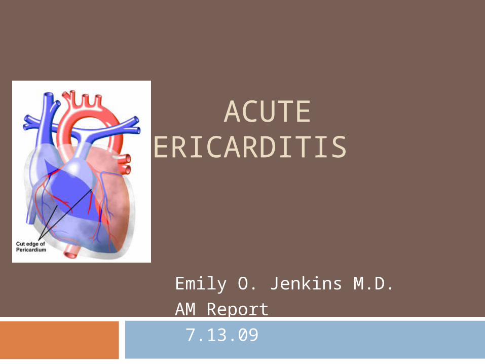
ACUTE PERICARDITIS
Emily O. Jenkins M.D.
AM Report
7.13.09
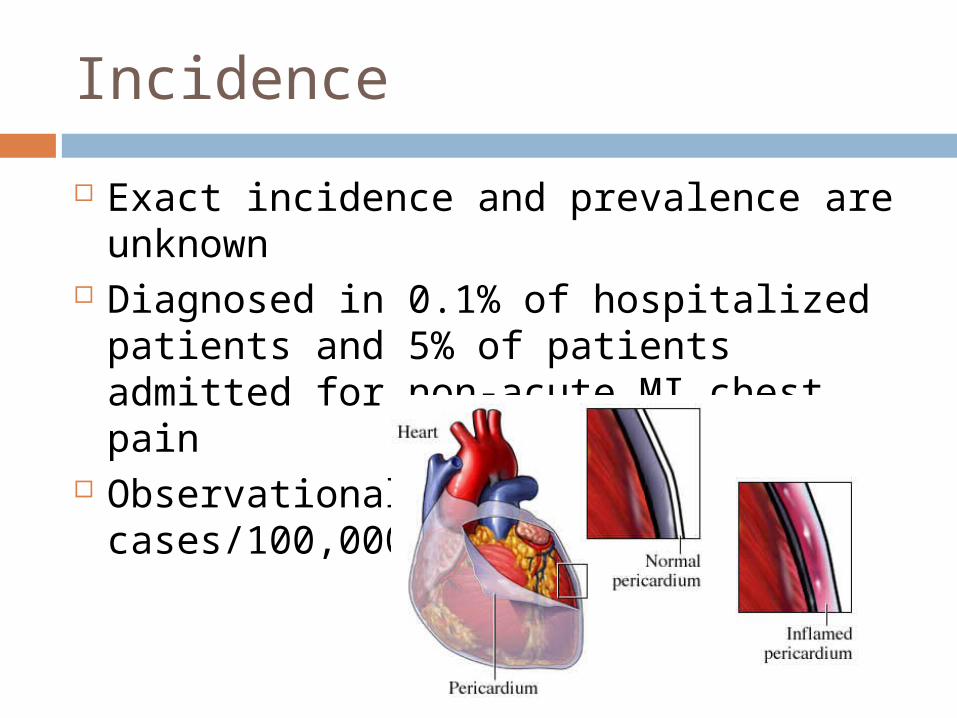
Incidence
Exact incidence and prevalence are unknown
Diagnosed in 0.1% of hospitalized patients and 5% of patients admitted for non-acute MI chest pain
Observational study: 27.7 cases/100,000 population/year
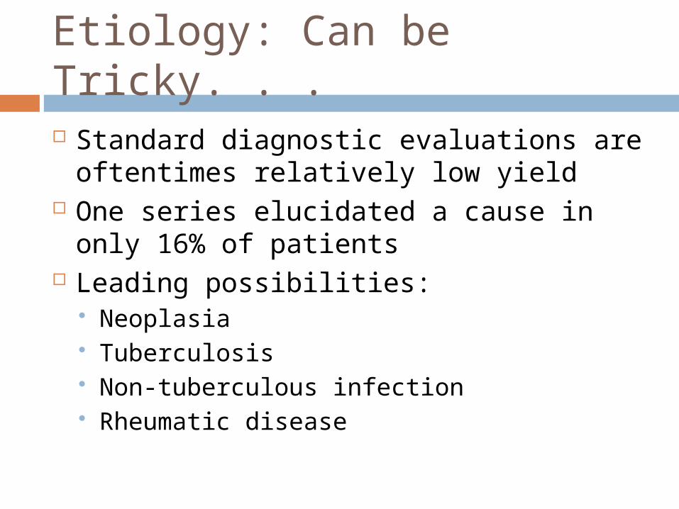
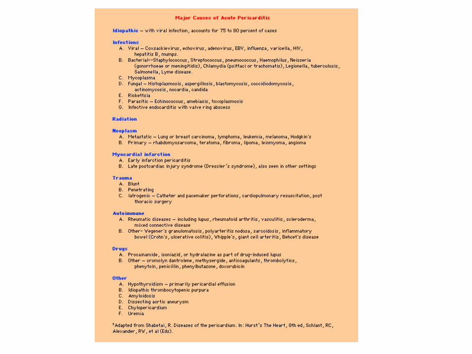
Etiology: Can be Tricky. . .
Standard diagnostic evaluations are oftentimes relatively low yield
One series elucidated a cause in only 16% of patients
Leading possibilities: Neoplasia Tuberculosis Non-tuberculous infection Rheumatic disease
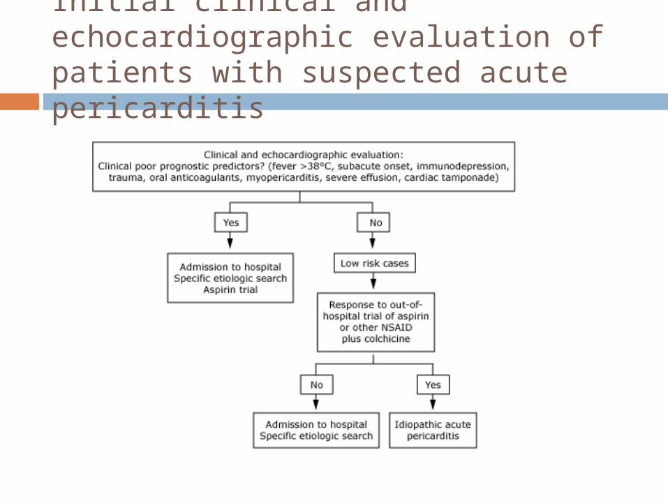
Initial clinical and echocardiographic evaluation of patients with suspected acute pericarditis
Diagnostic Criteria
Chest pain: anterior chest, sudden onset, pleuritic; may decrease in intensity when leans forward, may radiate to one or both trapezius ridges
Pericardial friction rub: most specific, heard best at LSB
EKG changes: new widespread ST elevation or PR depression
Pericardial effusion: absence of does not exclude diagnosis of pericarditis
Supporting signs/symptoms: Elevated ESR, CRP Fever leukocytosis
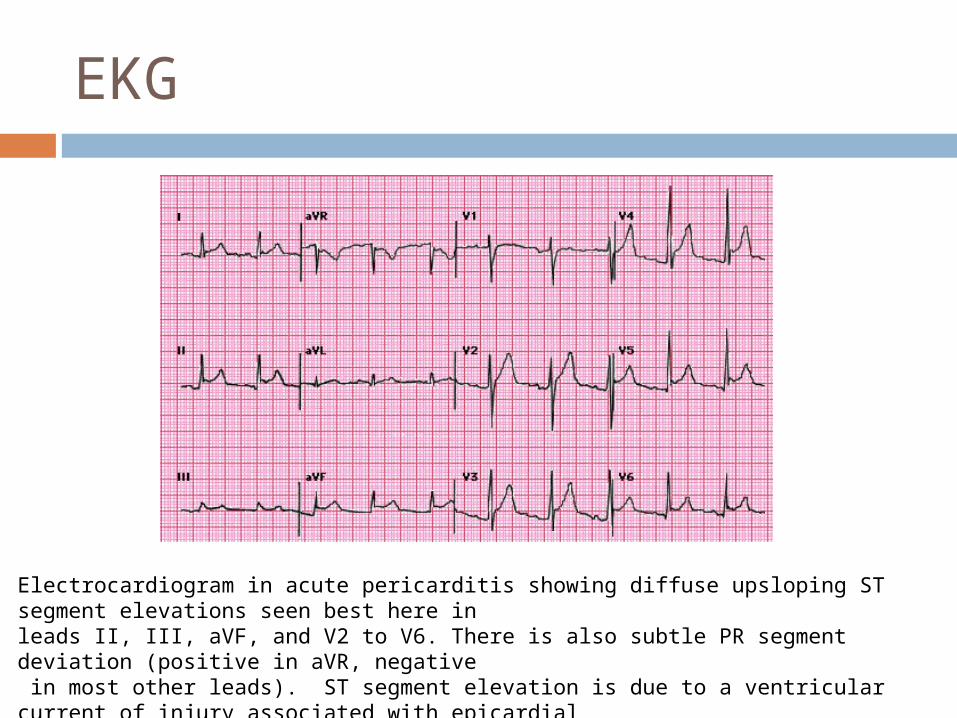
EKG
Electrocardiogram in acute pericarditis showing diffuse upsloping ST segment elevations seen best here in leads II, III, aVF, and V2 to V6. There is also subtle PR segment deviation (positive in aVR, negative in most other leads). ST segment elevation is due to a ventricular current of injury associated with epicardial inflammation; similarly, the PR segment changes are due to an atrial current of injury which, in pericarditis, typically displaces the PR segment upward in lead aVR and downward in most other leads.
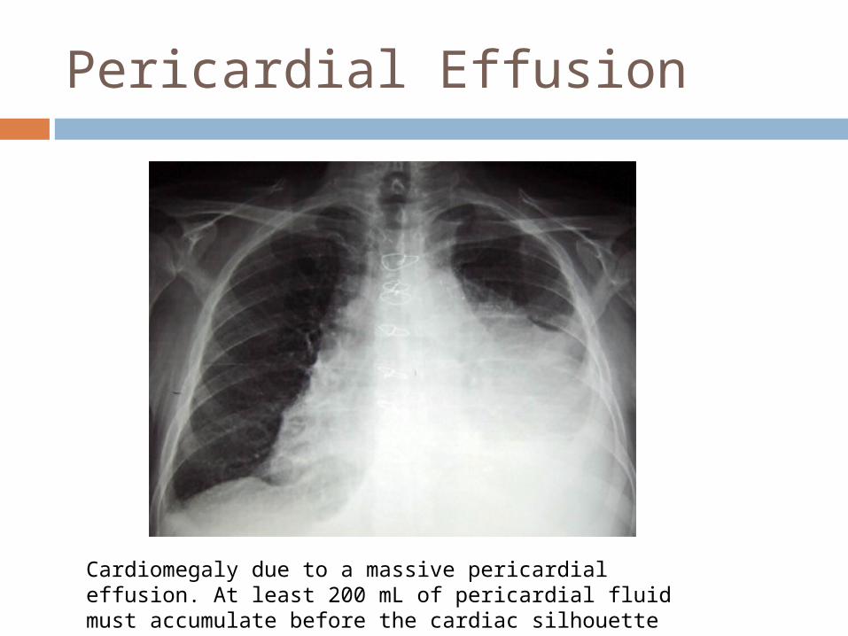
Pericardial Effusion
Cardiomegaly due to a massive pericardial effusion. At least 200 mL of pericardial fluid must accumulate before the cardiac silhouette enlarges.
Tests
EKG CXR PPD ANA HIV Blood cultures Urgent echocardiogram if evidence of
pericardial effusion Not necessary:
Viral studies b/c yield is low and management is not altered
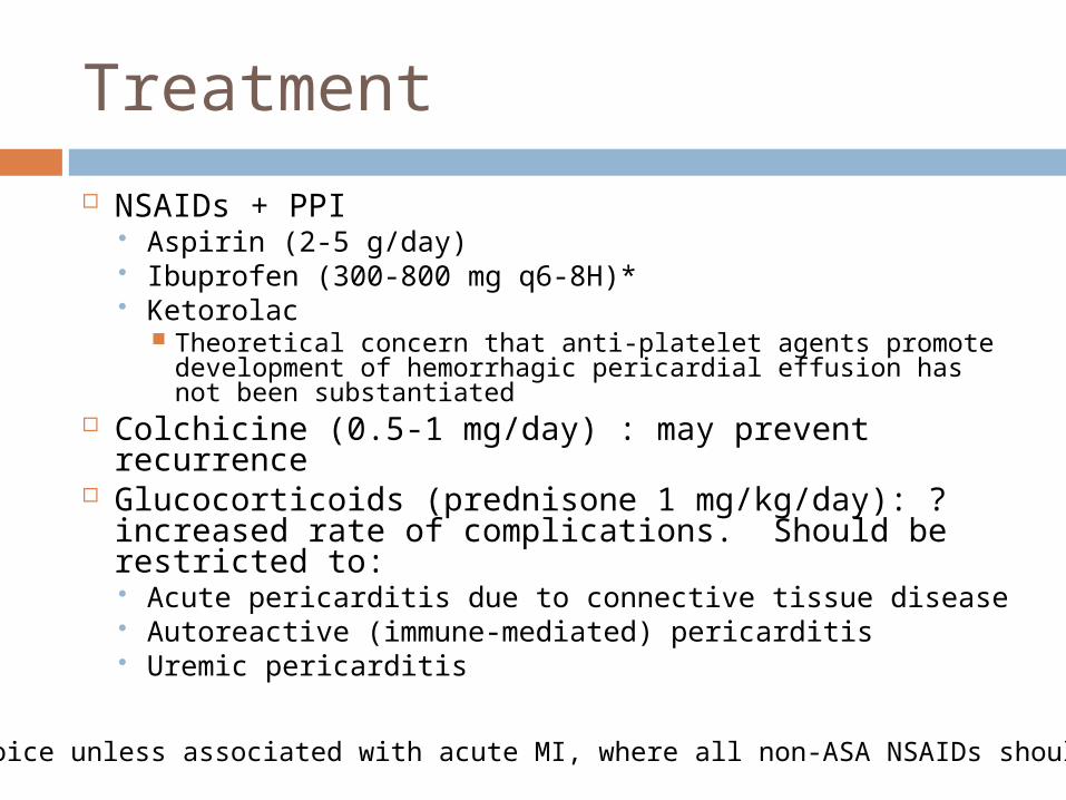
Treatment
NSAIDs + PPI Aspirin (2-5 g/day) Ibuprofen (300-800 mg q6-8H)* Ketorolac
Theoretical concern that anti-platelet agents promote development of hemorrhagic pericardial effusion has not been substantiated
Colchicine (0.5-1 mg/day) : may prevent recurrence
Glucocorticoids (prednisone 1 mg/kg/day): ? increased rate of complications. Should be restricted to: Acute pericarditis due to connective tissue disease Autoreactive (immune-mediated) pericarditis Uremic pericarditis
*NSAID of choice unless associated with acute MI, where all non-ASA NSAIDs should be avoided
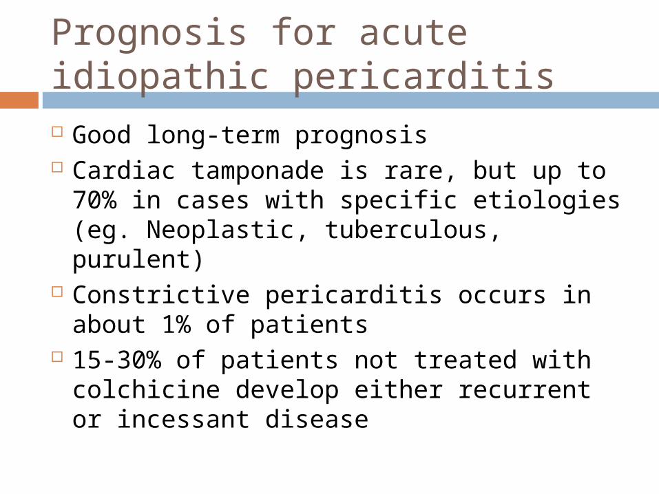
Prognosis for acute idiopathic pericarditis Good long-term prognosis Cardiac tamponade is rare, but up to
70% in cases with specific etiologies (eg. Neoplastic, tuberculous, purulent)
Constrictive pericarditis occurs in about 1% of patients
15-30% of patients not treated with colchicine develop either recurrent or incessant disease
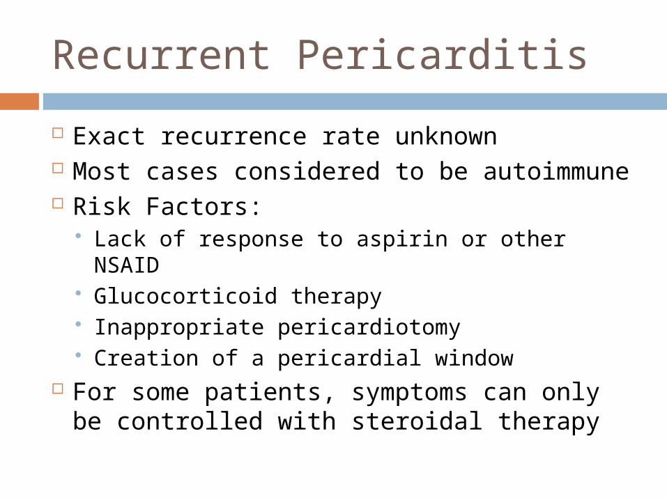
Recurrent Pericarditis
Exact recurrence rate unknown Most cases considered to be autoimmune Risk Factors:
Lack of response to aspirin or other NSAID Glucocorticoid therapy Inappropriate pericardiotomy Creation of a pericardial window
For some patients, symptoms can only be controlled with steroidal therapy
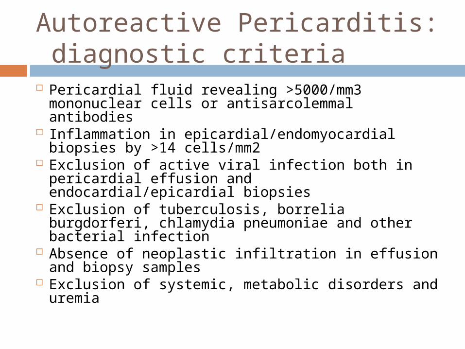
Autoreactive Pericarditis: diagnostic criteria Pericardial fluid revealing >5000/mm3
mononuclear cells or antisarcolemmal antibodies Inflammation in epicardial/endomyocardial
biopsies by >14 cells/mm2 Exclusion of active viral infection both in
pericardial effusion and endocardial/epicardial biopsies
Exclusion of tuberculosis, borrelia burgdorferi, chlamydia pneumoniae and other bacterial infection
Absence of neoplastic infiltration in effusion and biopsy samples
Exclusion of systemic, metabolic disorders and uremia
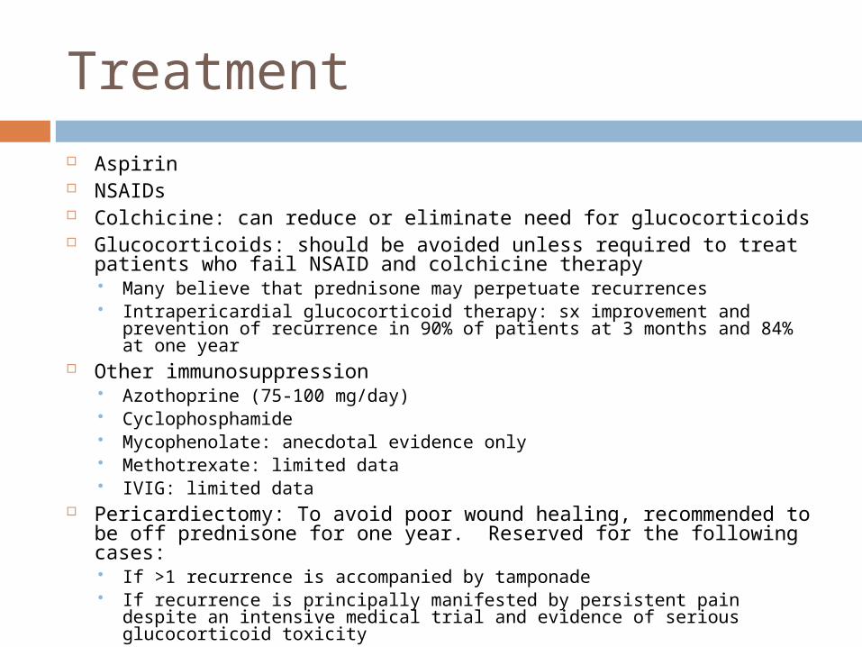
Treatment
Aspirin NSAIDs Colchicine: can reduce or eliminate need for glucocorticoids Glucocorticoids: should be avoided unless required to treat
patients who fail NSAID and colchicine therapy Many believe that prednisone may perpetuate recurrences Intrapericardial glucocorticoid therapy: sx improvement and prevention
of recurrence in 90% of patients at 3 months and 84% at one year Other immunosuppression
Azothoprine (75-100 mg/day) Cyclophosphamide Mycophenolate: anecdotal evidence only Methotrexate: limited data IVIG: limited data
Pericardiectomy: To avoid poor wound healing, recommended to be off prednisone for one year. Reserved for the following cases: If >1 recurrence is accompanied by tamponade If recurrence is principally manifested by persistent pain despite an
intensive medical trial and evidence of serious glucocorticoid toxicity